dentistry for medical students maxillofacial and...
TRANSCRIPT

DENTISTRY FOR MEDICAL STUDENTS
Maxillofacial and Oral SurgeryUniversity Medical Centre
Ljubljana
PURPOSE OF THIS TRAINING
• Familiarize non-dental medical providers with basic dental terminology
• Prepare non-dental medical providers to diagnose and treat common dental emergencies – Lost fillings/caries– Common oral pathology – Pulpal & Periodontal conditions– Infection– Trauma– Administration of local anesthesia

DENTAL CLASSIFICATIONS
Deployable:• Dental Class 1
– no disease present– no treatment needed
• Dental Class 2– Disease present– no emergency anticipated
within 12 months
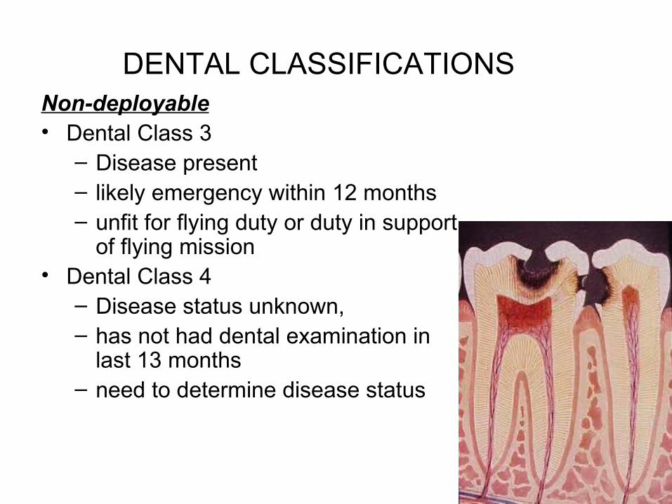
DENTAL CLASSIFICATIONSNon-deployable• Dental Class 3
– Disease present– likely emergency within 12 months– unfit for flying duty or duty in support
of flying mission• Dental Class 4
– Disease status unknown, – has not had dental examination in
last 13 months – need to determine disease status

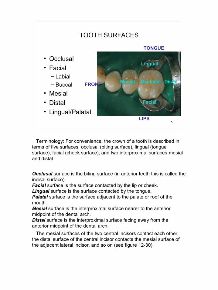
TOOTH SURFACES
• Occlusal• Facial
– Labial– Buccal
• Mesial• Distal• Lingual/Palatal
LIPS
TONGUE
FRONT Occlusal
Facial
Lingual
Mesial Distal

EU TOOTH NUMBERING SYSTEMSfor the adult dentition
• 11-18 Begins with maxillary first incisor right
• 21-28 Begins with maxillary first incisor left
• 31-38 Continues with mandibular first left incisor
• 41-48 Continues with mandibular first right incisor

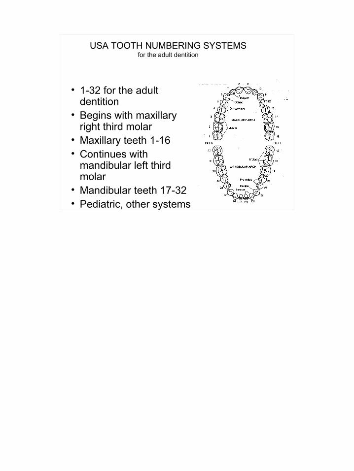
USA TOOTH NUMBERING SYSTEMSfor the adult dentition
• 1-32 for the adult dentition
• Begins with maxillary right third molar
• Maxillary teeth 1-16• Continues with
mandibular left third molar
• Mandibular teeth 17-32• Pediatric, other systems
DENTAL SICK CALL
Key for Medical Providers• Diagnosis
– Medical History and Vital Signs– Chief Complaint– History of Present Illness
- Pain frequency and duration - Pain characterization (dull/sharp, throbbing) - Pain elicited or spontaneous?
– LISTEN!– Use S.O.A.P. Format
• Early Management– Initial treatment in the absence of a dentist

OROFACIAL PAIN
• Orofacial pain from non-odontogenic sources:– Maxillary sinus– Temporomandibular joint– Referred pain/nerve distribution
• Odontogenic pain:– Arising from teeth or supporting structures– Diagnosis complicated by pain referral and other
factors– However, in most cases dental cause can be found
with exam
ODONTOGENIC PAIN
• Pulpal origin– Erroneously referred to as the “nerve”– Vascularized connective tissue with odontoblasts
and unmyelinated C fibers– Pulpalgia
• Caused by inflammatory changes to pulp – Apical periodontitis
• Inflammatory changes at root apex secondary to pulpal disease
• Periodontal origin– Gingivitis– Periodontitis– Acute periodontal abscess

DENTAL DIAGNOSTIC TESTS
• Attempt to replicate dental symptoms– Percussion
• Tap on teeth with finger tip – Palpation
• Press on intraoral structures and teeth with digital pressure
– Thermal• Place cold (ice) or squirt
warm liquids on tooth– Biting pressure
• Have patient bite on tongue blade, cotton tipped applicator
Spontaneous or lingering pain is indicative of an advanced problem
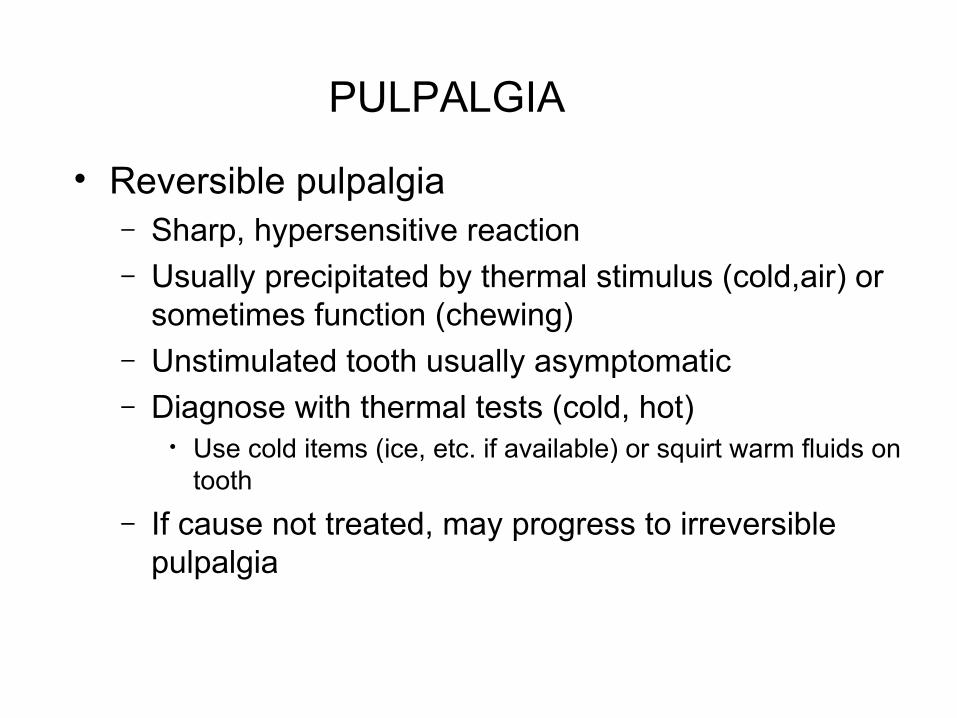
PULPALGIA
• Reversible pulpalgia– Sharp, hypersensitive reaction – Usually precipitated by thermal stimulus (cold,air) or
sometimes function (chewing)– Unstimulated tooth usually asymptomatic– Diagnose with thermal tests (cold, hot)
• Use cold items (ice, etc. if available) or squirt warm fluids on tooth
– If cause not treated, may progress to irreversible pulpalgia

PULPALGIA
• Irreversible pulpalgia– Typically hallmarked by prolonged
unstimulated pain• Spontaneous pain is common• Pain usually persists after removal of thermal
stimulus• Pain usually only partially/temporarily relieved by
analgesics– Diagnose with thermal tests– Untreated, will progress to pulpal necrosis
and apical periodontitis
PULPALGIA
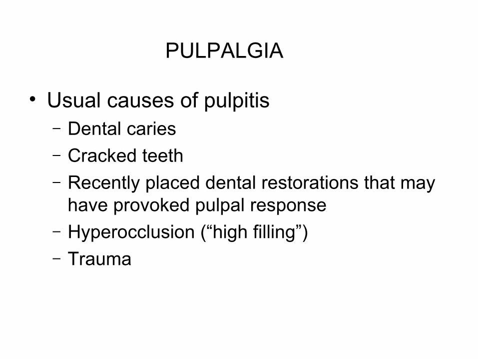
• Usual causes of pulpitis– Dental caries– Cracked teeth– Recently placed dental restorations that may
have provoked pulpal response– Hyperocclusion (“high filling”)– Trauma

DENTAL CARIES
• Extremely common • Unusual as an emergency (unless
severe)• Temporary restorations helpful• Dental treatment required

Caries Progression
CARIES PROGRESSION
Pulpal Involvement

DENTAL CARIES TREATMENT - causing symptoms
– Isolate tooth as possible with cotton (gauze, rolls); teeth need to be as dry as possible
– Mix Fuji IX glass ionomer material
– Pack into tooth & smooth with gloved finger
– Keep material below the height of the tooth
– Have patient bite down to ensure that material is not too high in occlusion
– Pain meds (NSAID’s or acetaminophen) prn
MIXING TEMPORARY FILLING MATERIAL
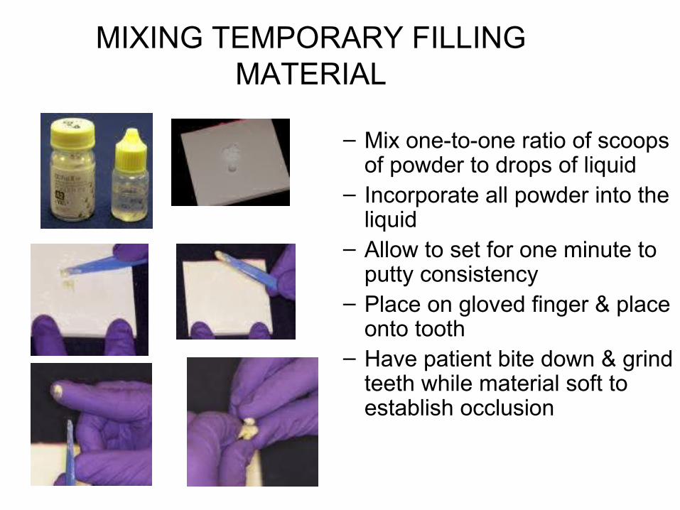
– Mix one-to-one ratio of scoops of powder to drops of liquid
– Incorporate all powder into the liquid
– Allow to set for one minute to putty consistency
– Place on gloved finger & place onto tooth
– Have patient bite down & grind teeth while material soft to establish occlusion

BROKEN FILLING
• Treatment– Treat the same as dental
caries– Try to remove fragments
with instruments– Mix and pack restorative
material into voids

CRACKED TOOTH (Incomplete Tooth Fracture)
Fracture Line
• Difficult to diagnose• Tooth appears intact, but patient
indicates exquisite pain to biting (on release)
• Other symptoms include unexplained sensitivity to cold and sweets
• May progress to complete fracture if untreated
• Confirmed by – Bite test using cotton tip applicator or
section of tongue blade. – Thermal tests, ice, warm water, air spray– Transillumination with light is also helpful to
look for cracks• Definitive diagnosis is not always
possible without specialized equipment

CRACKED TEETH (Incomplete tooth fracture)
• Treatment– Depends upon symptoms
• Reversible pulpalgia– Pain meds, avoidance of function on
offending tooth• Irreversible pulpalgia
– Med Evac out for definitive dental Tx; or– Extraction of symptomatic tooth
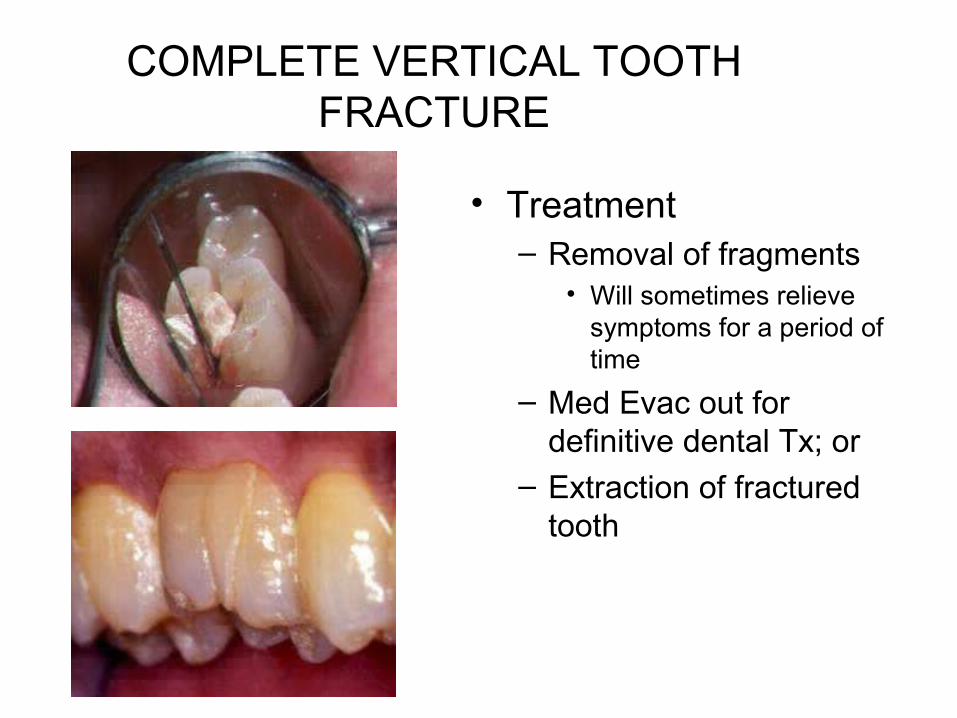
COMPLETE VERTICAL TOOTH FRACTURE
• Treatment– Removal of fragments
• Will sometimes relieve symptoms for a period of time
– Med Evac out for definitive dental Tx; or
– Extraction of fractured tooth
PAIN FROM RECENT RESTORATION
• Treatment– Reversible pulpalgia:
• Pain meds and allow time to evaluate– Irreversible pulpalgia:
• Med Evac patient out for definitive dental Tx; or• Extraction of symptomatic tooth

APICAL PERIODONTITIS
• Classic “Dental abscess”– Usually a single tooth– Secondary to complete or partial
necrosis of the dental pulp– Inflammatory response at root
apex– Tooth may be sensitive to touch-
test by percussion or tapping with finger
– Negative response to cold test will usually confirm lack of pulp vitality
– Untreated, may lead to cellulitis or space infection
Broken down tooth
Draining Abscess

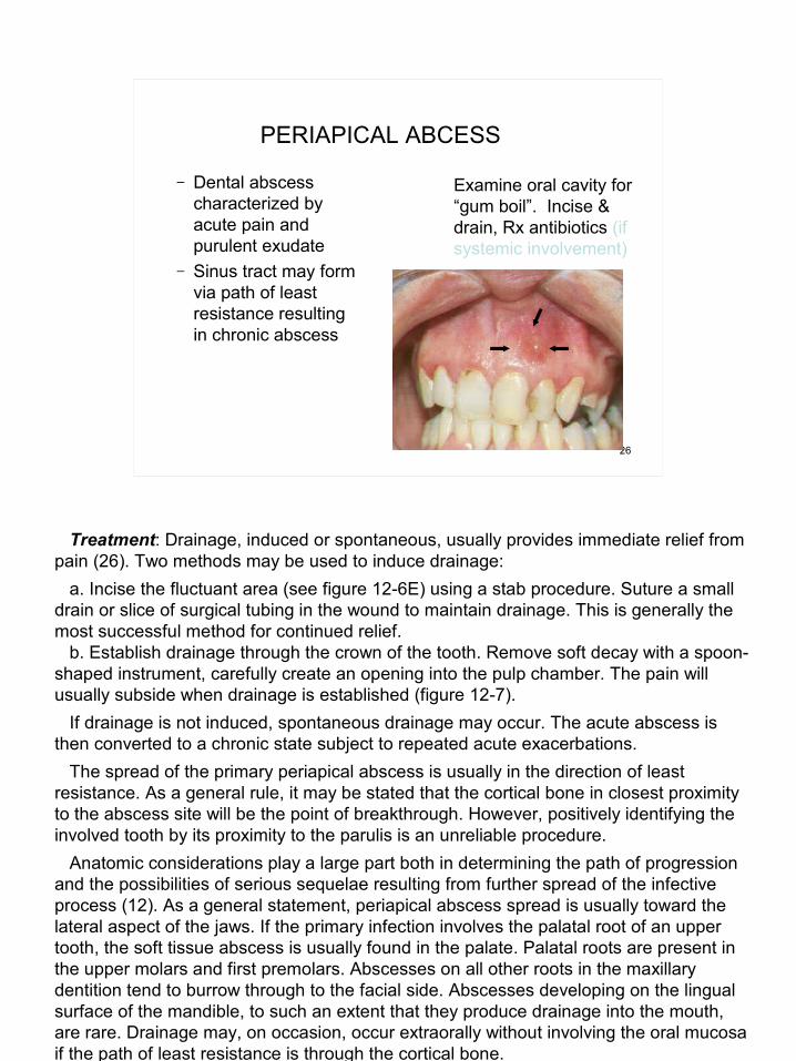
PERIAPICAL ABCESS
Examine oral cavity for“gum boil”. Incise &drain, Rx antibiotics (if systemic involvement)
– Dental abscess characterized by acute pain and purulent exudate
– Sinus tract may form via path of least resistance resulting in chronic abscess

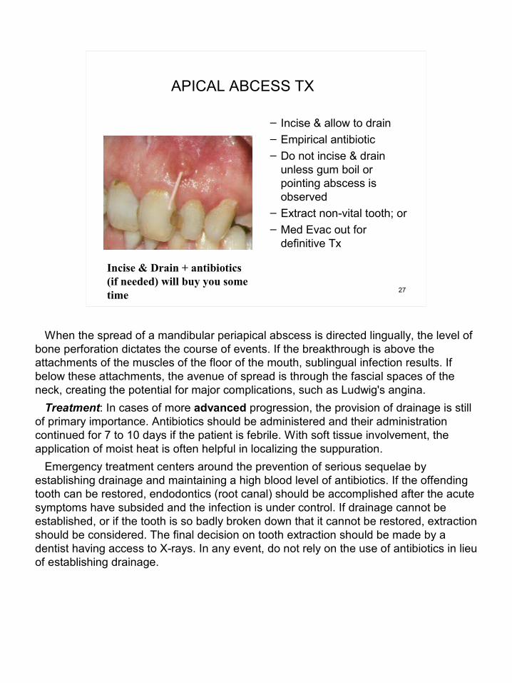
APICAL ABCESS TX
Incise & Drain + antibiotics (if needed) will buy you some time
– Incise & allow to drain– Empirical antibiotic – Do not incise & drain
unless gum boil or pointing abscess is observed
– Extract non-vital tooth; or– Med Evac out for
definitive Tx

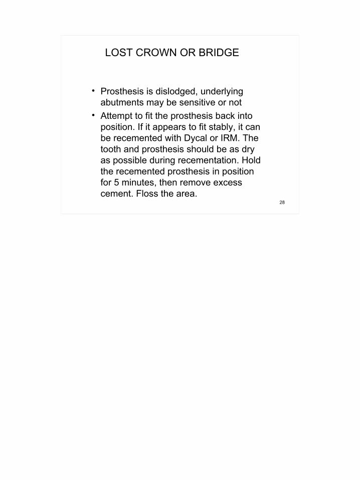
LOST CROWN OR BRIDGE
• Prosthesis is dislodged, underlying abutments may be sensitive or not
• Attempt to fit the prosthesis back into position. If it appears to fit stably, it can be recemented with Dycal or IRM. The tooth and prosthesis should be as dry as possible during recementation. Hold the recemented prosthesis in position for 5 minutes, then remove excess cement. Floss the area.

BROKEN/LOOSE DENTURE
• The patient tends to view this problem as an emergency. It is not.
• Refer to dentist as soon as possible• Prosthesis may be modified for
comfortable wear, but use caution so as not to ruin a repairable prosthesis

ODONTOGENIC SPACE INFECTIONS
• May present as cellulitis or abscess
• Can be life threatening– Invasion of parapharyngeal spaces– Airway compromise – Cavernous sinus thrombosis– Endotoxic shock
• Infections involving the muscles of mastication may result in trismus

ODONTOGENIC SPACE INFECTIONS
Buccal space infection that isspreading to submand & Sublingual spaces

ODONTOGENIC SPACE INFECTIONS
Ludwig’sAngina

ODONTOGENIC SPACE INFECTIONS
• Aggressive management indicated• Incision and drainage
– Intra-oral approach, if possible– Irrigate copiously
• Parental antibiotics + supportive care• Tooth extraction (if possible) and Med
Evac for definitive dental Tx is imperative

EXTRAORAL INCISION AND DRAINAGE

PERIODONTAL CONDITIONS
• Gingivitis • Acute Periodontal Conditions
– Necrotizing Ulcerating Gingivitis (NUG)– Periodontal Abscess– Herpetic Stomatitis– Recurrent Apthous Ulcers– Pericornitis

GINGIVITIS
• Inflammation of marginal gingiva– Swelling and easy bleeding– Usually asymptomatic– Occurs quickly after interruption of oral
hygiene practices • Untreated, may progress to NUG in
susceptible individuals
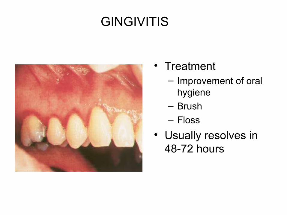
GINGIVITIS
• Treatment– Improvement of oral
hygiene– Brush– Floss
• Usually resolves in 48-72 hours

N.U.G. Necrotizing Ulcerative Gingivitis
• Previously known as Vincent’s Infection or “Trench Mouth”
• Signs and symptoms include:– Acute pain– Foul breath– Interproximal gingival
necrosis (“punched out papillae”)
Responsible flora is usuallymixed, fuso-spirochetal

NECROTIZING ULCERATIVE GINGIVITIS
• Usually associated with:– Poor oral hygiene– Situational STRESSSTRESS– Smoking
• NUG is now considered as an oral hallmark of immunosuppression– HIV/AIDS– Diabetes– Malnutrition

NECROTIZING ULCERATIVE GINGIVITIS
• Treatment– Debridement (at least
aggressive toothbrush use)
– 50/50 mix of viscous lidocaine & mouthwash (if available)
– Antibiotics ONLY if clinical signs of systemic involvement
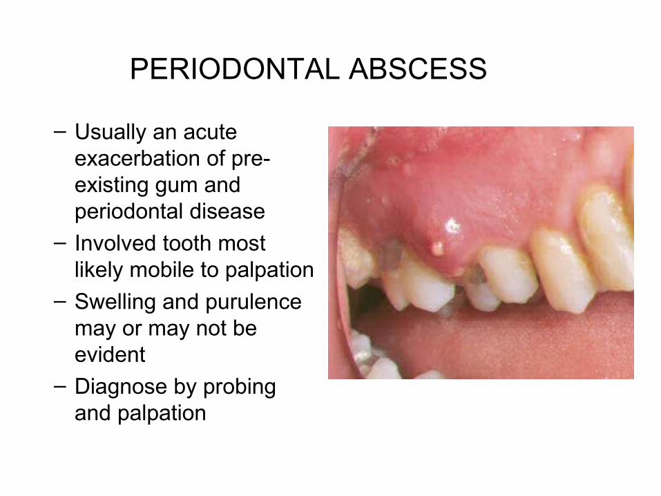
PERIODONTAL ABSCESS
– Usually an acute exacerbation of pre-existing gum and periodontal disease
– Involved tooth most likely mobile to palpation
– Swelling and purulence may or may not be evident
– Diagnose by probing and palpation

Periodontal Abscess
• Treatment– Establish drainage
• Incise & drain• Curettage along the
tooth root• or Extract
– Irrigation– Antibiotics if systemic
involvement

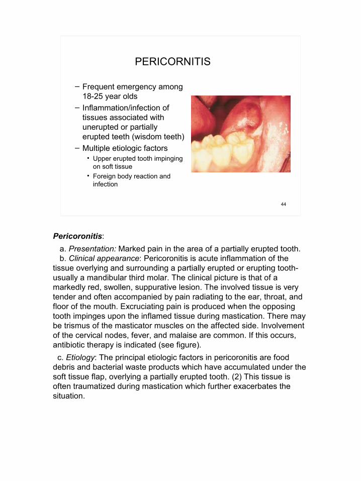
PERICORNITIS
– Frequent emergency among 18-25 year olds
– Inflammation/infection of tissues associated with unerupted or partially erupted teeth (wisdom teeth)
– Multiple etiologic factors• Upper erupted tooth impinging
on soft tissue• Foreign body reaction and
infection
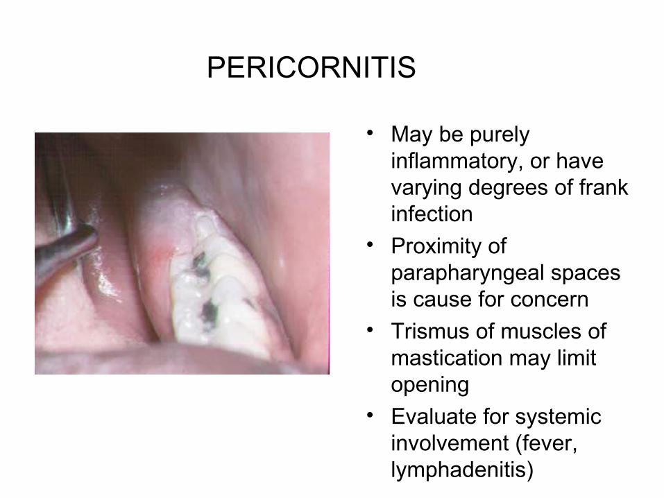
PERICORNITIS
• May be purely inflammatory, or have varying degrees of frank infection
• Proximity of parapharyngeal spaces is cause for concern
• Trismus of muscles of mastication may limit opening
• Evaluate for systemic involvement (fever, lymphadenitis)
PERICORNITIS
• Aggressive management is indicated– Irrigate infected area with saline or Peridex – Initiate antibiotic therapy if systemic
involvement or space infection is evident – Extract opposing tooth that is occluding on
inflamed tissue– If symptoms are not resolved by conservative
treatment, med-evac for removal of affected teeth

OTHER CAUSES OF ORAL-FACIAL PAIN:other things to keep in mind . . .
• Non-odontogenic causes of orofacial pain which may mimic dental conditions– Maxillary sinusitis – Sialadenitis– Temporomandibular joint pain– Angina pectoris (radiating to left posterior
mandible)– Neuralgia and cluster headaches

EMERGENT ORAL PATHOLOGY
• Apthous ulcers– Discrete, shallow ulceration– moveable, unattached intra-oral
mucosa– erythemous halo– self-limiting, – 7-10 day duration

APHTHOUS ULCERS
• Treatment– palliative– topical meds
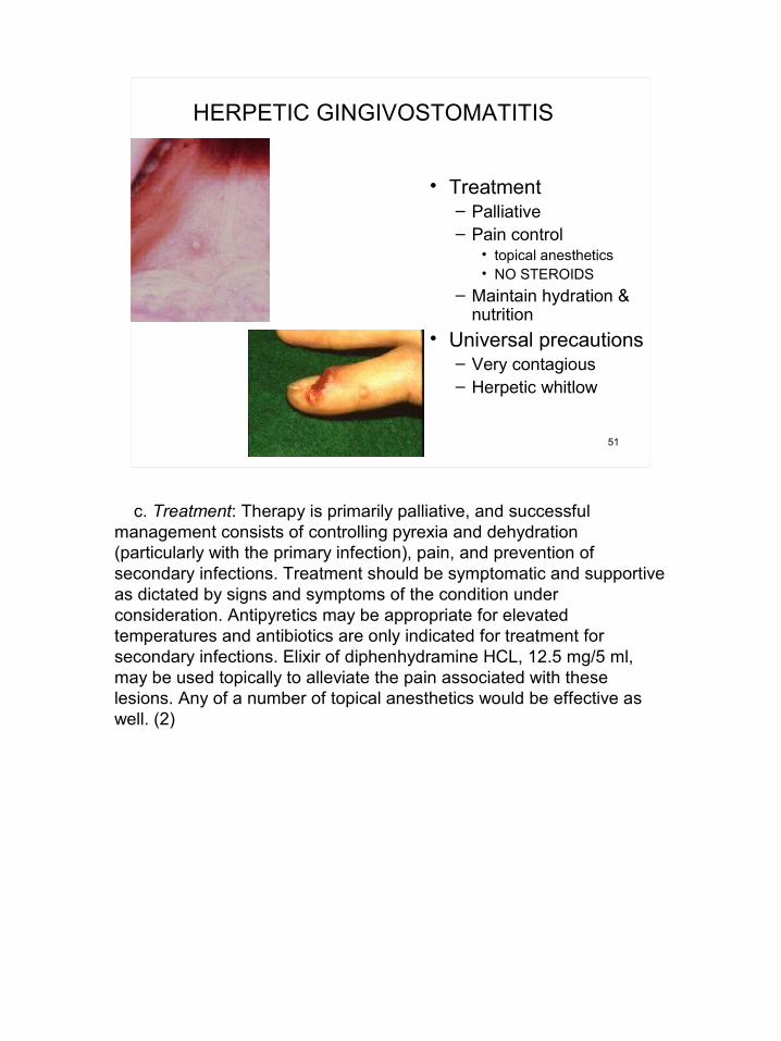
HERPETIC GINGIVOSTOMATITIS
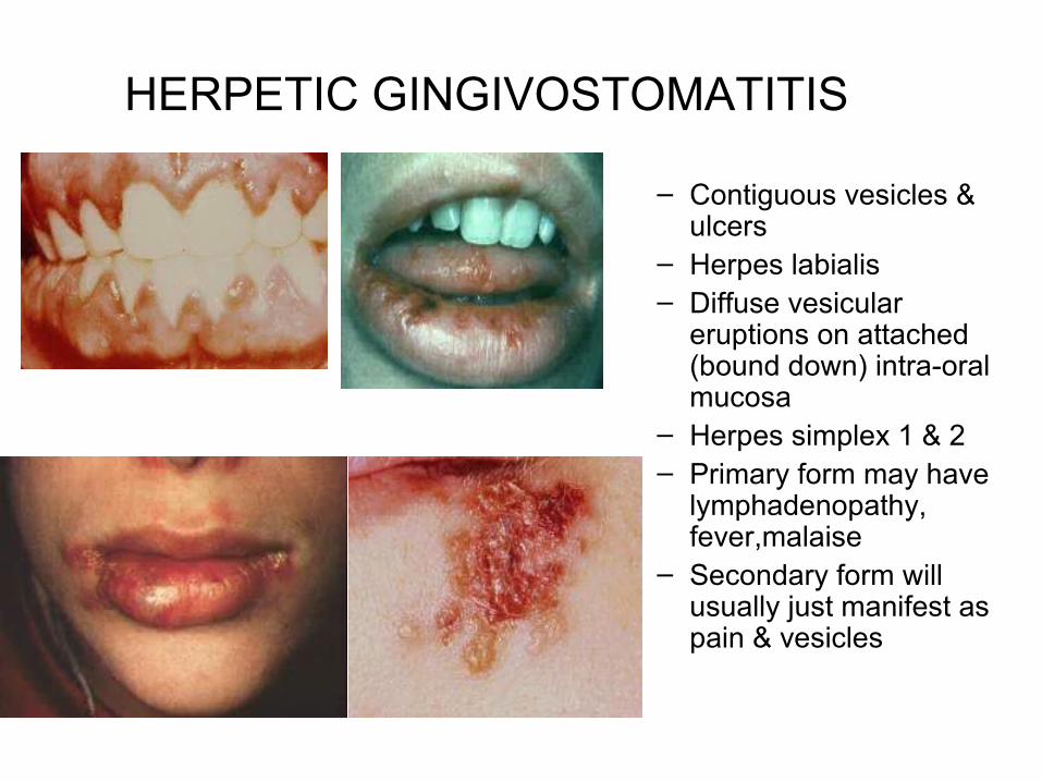
– Contiguous vesicles & ulcers
– Herpes labialis – Diffuse vesicular
eruptions on attached (bound down) intra-oral mucosa
– Herpes simplex 1 & 2– Primary form may have
lymphadenopathy, fever,malaise
– Secondary form will usually just manifest as pain & vesicles

HERPETIC GINGIVOSTOMATITIS
• Treatment– Palliative– Pain control
• topical anesthetics• NO STEROIDS
– Maintain hydration & nutrition
• Universal precautions– Very contagious– Herpetic whitlow

TEMPOROMANDIBULAR DISORDERS
• Myofascial pain dysfunction syndrome (MPD)– Involves muscles of mastication– Situational stress is a contributing factor– Clenching and bruxism are associated with MPD
• Temporomandibular joint (TMJ) disorder– May involve anterior disc displacement or other
pathologic changes in the TMJ– Asymptomatic joint noises are not a cause for concern
in field and are not always predictive of future problems

TMJ DISORDERS
• Treatment is palliative– NSAIDS, muscle relaxants– Moist heat to affected muscles – Limit opening, un-necessary function– Soft diet as much as possible– Pain is frequently cyclical and may resolve
with or without intervention

TRAUMATIC DENTAL INJURIES
• Crown fractures• Tooth Avulsion/Luxation• Intraoral soft tissue injury

CROWN FRACTURES CONSERVATIVE FIELD TX
• Exposed dentin of tooth may be sensitive
• Dry tooth with cotton• Observe if tooth is bleeding
from the pulp– if not bleeding, cover tooth
with Fuji IX – If bleeding, will need root
canal in near future, but restore with Fuji IX for the current time

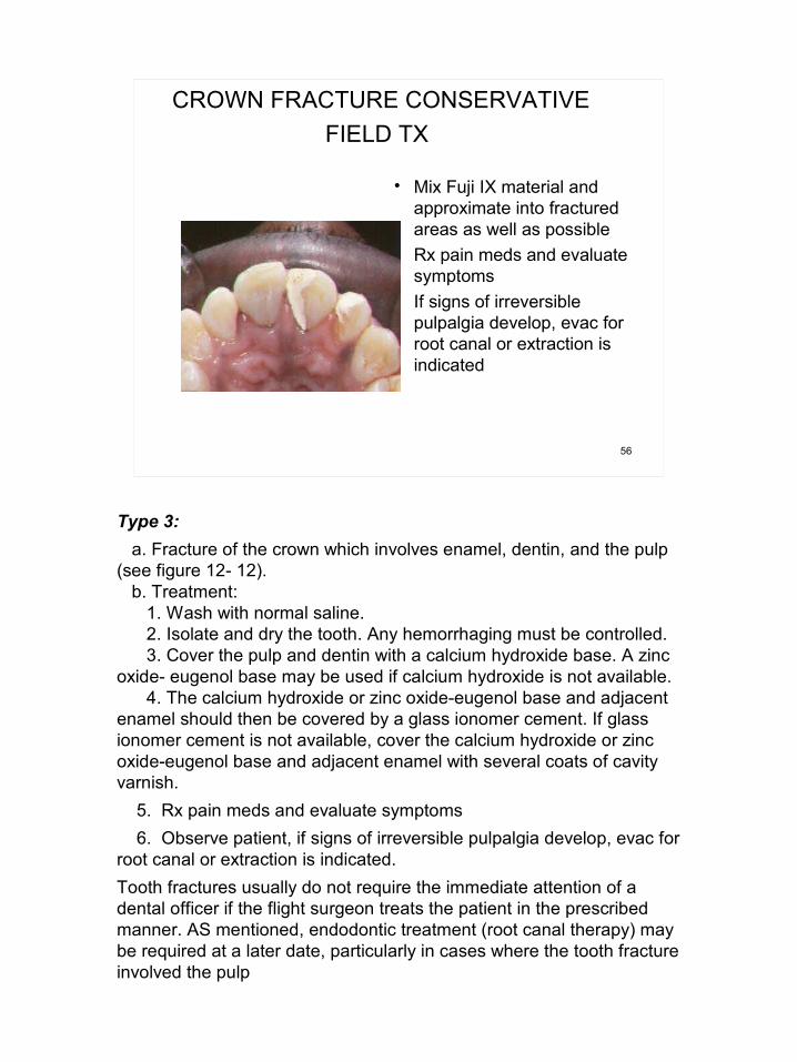
CROWN FRACTURE CONSERVATIVE FIELD TX
• Mix Fuji IX material and approximate into fractured areas as well as possible
• Rx pain meds and evaluate symptoms
• If signs of irreversible pulpalgia develop, evac for root canal or extraction is indicated

TOOTH AVULSION
• Usually anterior teeth• If tooth is not recovered
assume ingestion or aspiration (check lung sounds--future X-rays may be indicated)
• Prompt reimplantation critical for good prognosis– Rinse with saline or clean
water (Do not scrub)– Re-implant and stabilize
as well as possible– Check tetanus
immunization

INTRAORAL SOFT TISSUE INJURIES• Isolate source of intraoral
hemorrhage• Obtain anesthesia by
inflitration or block if needed • Irrigate and debride, remove
foreign debris• Close mucosa with 3-0 or 4-
0 gut or silk• Cover bone--especially over
roots• For through and through
lesions, close intraoral lacerations first

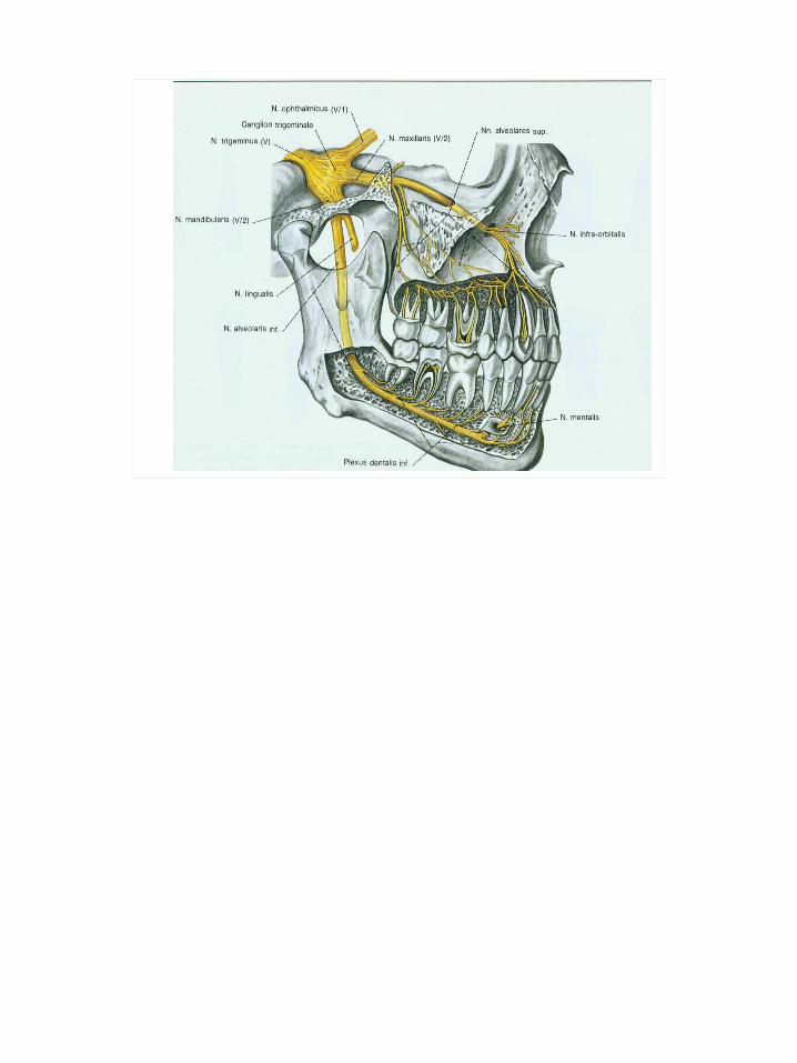
LOCAL ANESTHESIA FOR DENTISTRY
• Infiltration along 2nd division trigeminal (maxilla)
• Nerve block 3rd division trigeminal (mandible)

– Aspirating Syringe– 1.8 ml cartridge– Sterile Disposable
Needle• Long: 1 5/8 inches• Short: 1 inch
LOCAL ANESTHESIA - armamentarium

ARMAMENTARIUM
Cream (EMLA)Spray (Xylocain)Syringe Anesthetic solution
(Ultracain)SterileSafty

COMMON DENTAL NEEDLES

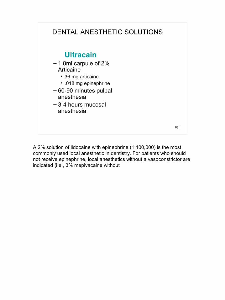
Ultracain– 1.8ml carpule of 2%
Articaine• 36 mg articaine• .018 mg epinephrine
– 60-90 minutes pulpal anesthesia
– 3-4 hours mucosal anesthesia
DENTAL ANESTHETIC SOLUTIONS

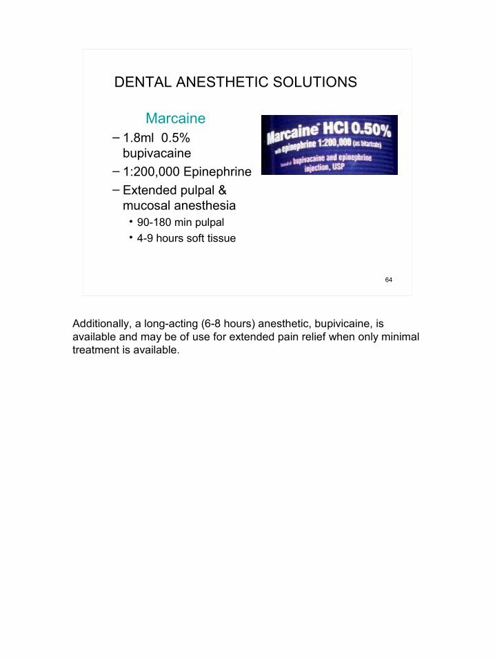
Marcaine– 1.8ml 0.5%
bupivacaine– 1:200,000 Epinephrine– Extended pulpal &
mucosal anesthesia• 90-180 min pulpal• 4-9 hours soft tissue
DENTAL ANESTHETIC SOLUTIONS

Infiltration – Injected directly in the area to be
anesthetized– Mechanism is diffusion– Indicated in porous bone (Maxilla)– Short 27 or 30 gauge needle– Onset: 2-3 minutes
Dental Anesthetic Injections


Maxillary Infiltration Injection – Short needle, 27 or 30 gauge– Penetrate soft tissue over root
apex– Direct the needle toward bone
(45 degrees)– Contact bone and aspirate– Inject one-half to three-
quarters carpule– Repeat for each tooth– Usually WILL NOT anesthetize
palatal tissues
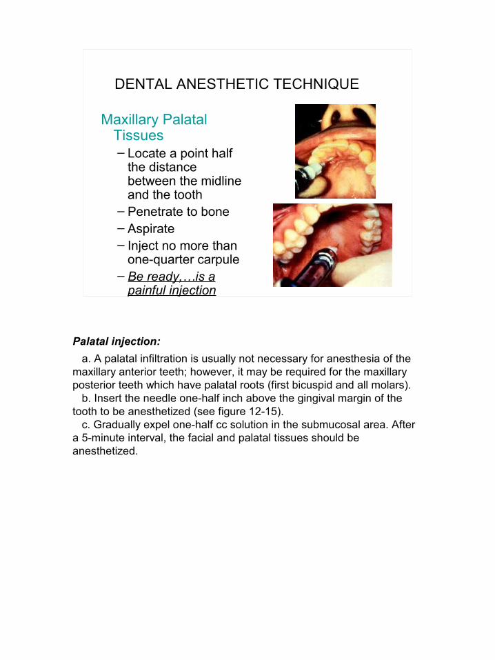
DENTAL ANESTHETIC TECHNIQUE

Maxillary Palatal Tissues– Locate a point half
the distance between the midline and the tooth
– Penetrate to bone– Aspirate– Inject no more than
one-quarter carpule– Be ready,…is a
painful injection
DENTAL ANESTHETIC TECHNIQUE

MANDIBULAR NERVE BLOCK
• Inferior Alveolar, Lingual Nerve Block– 27 gauge needle, long– Parallel to mandibular
occlusal plane– Palpate (thumb) anterior
ramus of mandible (intra-orally) at Coronoid Notch
– Grasp posterior border of ramus with index finger (extra-orally)
– Injection approach from contralateral bicuspids
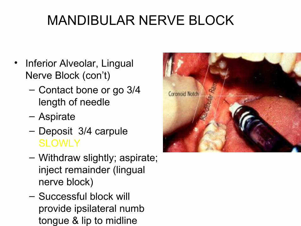
• Inferior Alveolar, Lingual Nerve Block (con’t)– Contact bone or go 3/4
length of needle– Aspirate– Deposit 3/4 carpule
SLOWLY– Withdraw slightly; aspirate;
inject remainder (lingual nerve block)
– Successful block will provide ipsilateral numb tongue & lip to midline
MANDIBULAR NERVE BLOCK
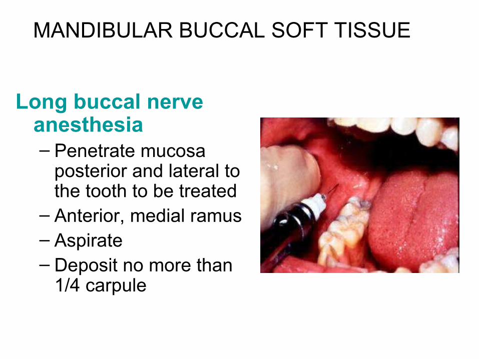
Long buccal nerve anesthesia– Penetrate mucosa
posterior and lateral to the tooth to be treated
– Anterior, medial ramus– Aspirate– Deposit no more than
1/4 carpule
MANDIBULAR BUCCAL SOFT TISSUE
BASIC EXODONTIA FOR THE FIELD
– For the resolution of problems that cannot wait or not feasible for evacuation
– Mechanical process in which the bone around the teeth is expanded to the point that tooth can be removed
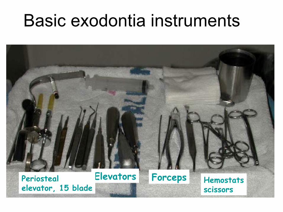
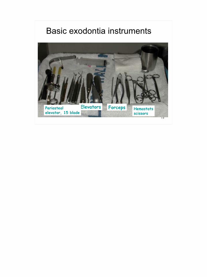
Basic exodontia instruments
Elevators ForcepsPeriostealelevator, 15 blade
Hemostatsscissors
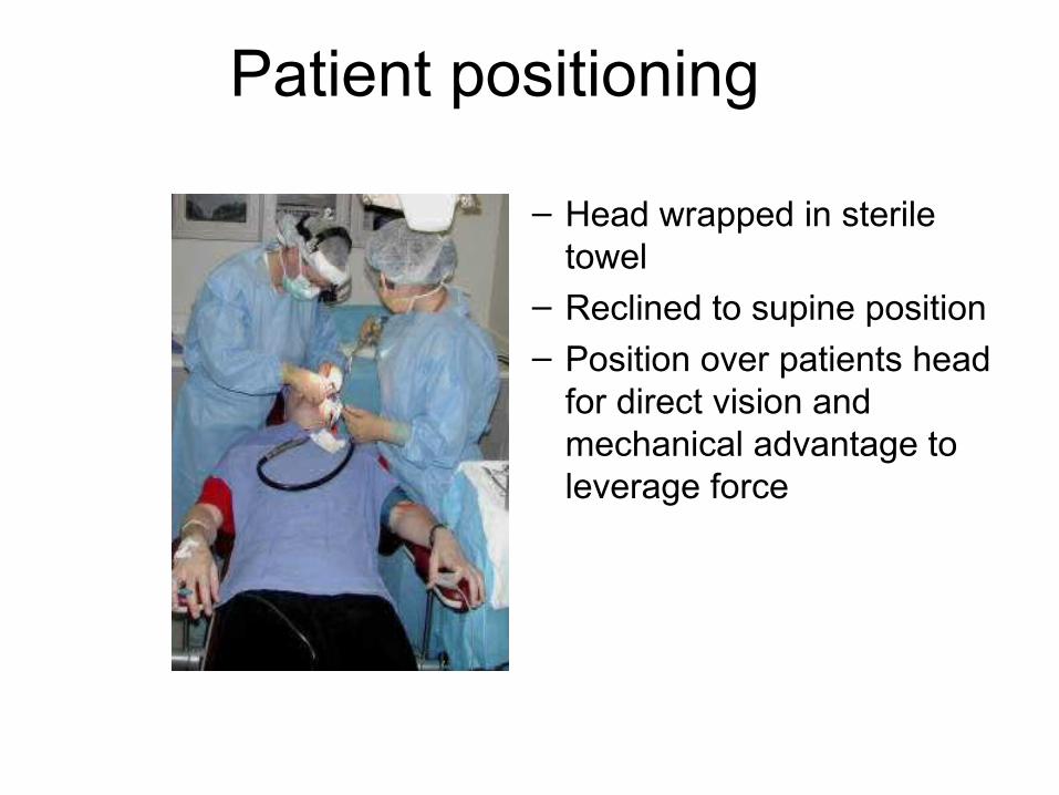
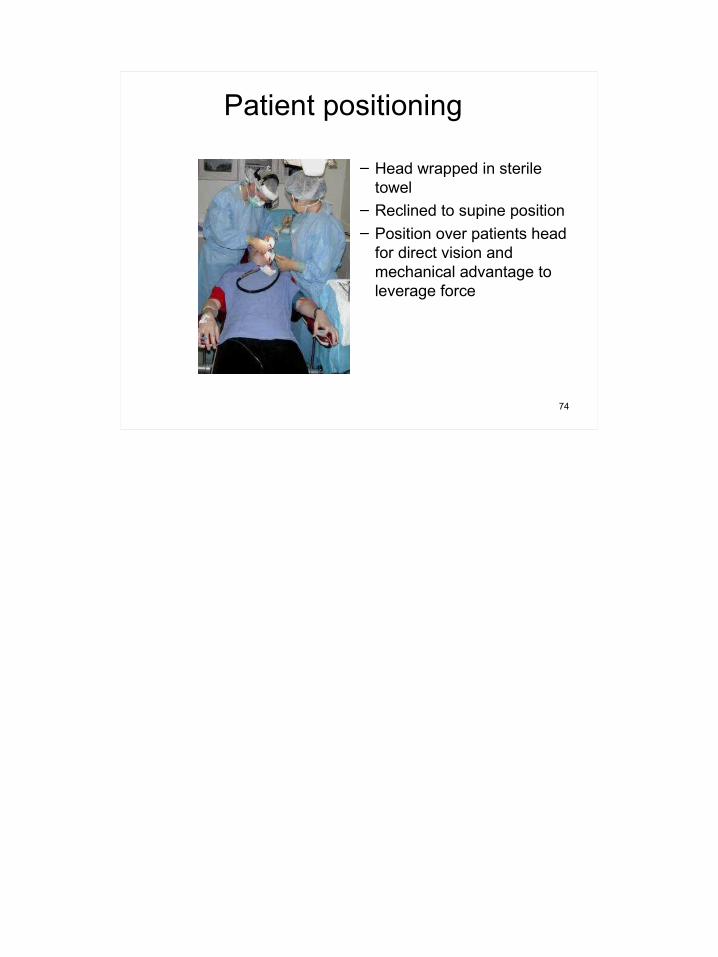
Patient positioning
– Head wrapped in sterile towel
– Reclined to supine position– Position over patients head
for direct vision and mechanical advantage to leverage force

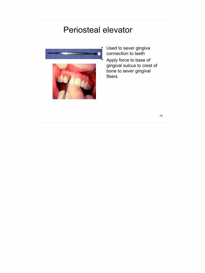
Periosteal elevator
• Used to sever gingiva connection to teeth
• Apply force to base of gingival sulcus to crest of bone to sever gingival fibers
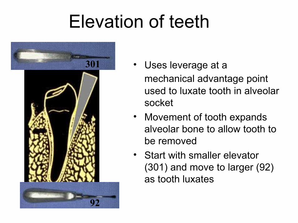
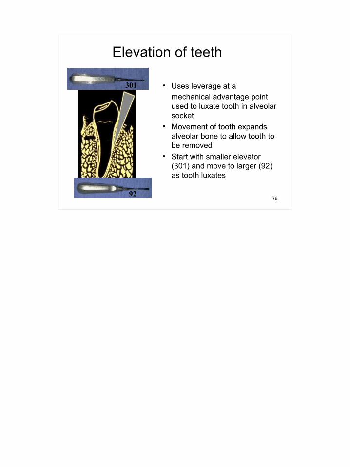
Elevation of teeth
• Uses leverage at a mechanical advantage point used to luxate tooth in alveolar socket
• Movement of tooth expands alveolar bone to allow tooth to be removed
• Start with smaller elevator (301) and move to larger (92) as tooth luxates
301
92

Elevation of teeth
– Wedge elevator between tooth and bone at neck of tooth and rotate handle with slight twisting, quarter-turn movement
– Observe for tooth movement– Do not use excessive force
• Crown fracture• Loosen adjacent teeth
– As tooth loosens, move elevator more into bone towards root end
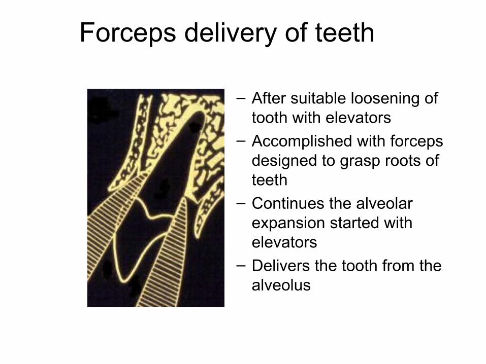
Forceps delivery of teeth
– After suitable loosening of tooth with elevators
– Accomplished with forceps designed to grasp roots of teeth
– Continues the alveolar expansion started with elevators
– Delivers the tooth from the alveolus
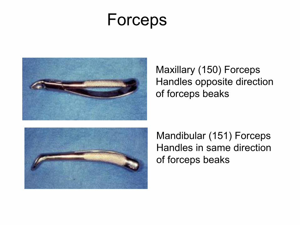
Forceps
Maxillary (150) ForcepsHandles opposite direction of forceps beaks
Mandibular (151) ForcepsHandles in same direction of forceps beaks
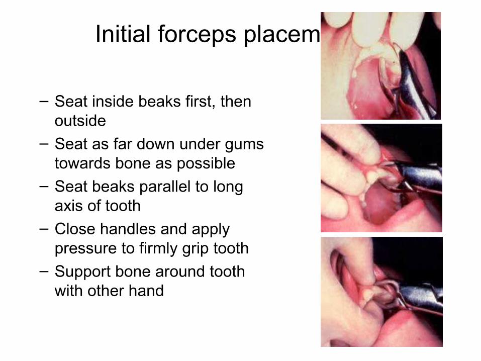
Initial forceps placement
– Seat inside beaks first, then outside
– Seat as far down under gums towards bone as possible
– Seat beaks parallel to long axis of tooth
– Close handles and apply pressure to firmly grip tooth
– Support bone around tooth with other hand

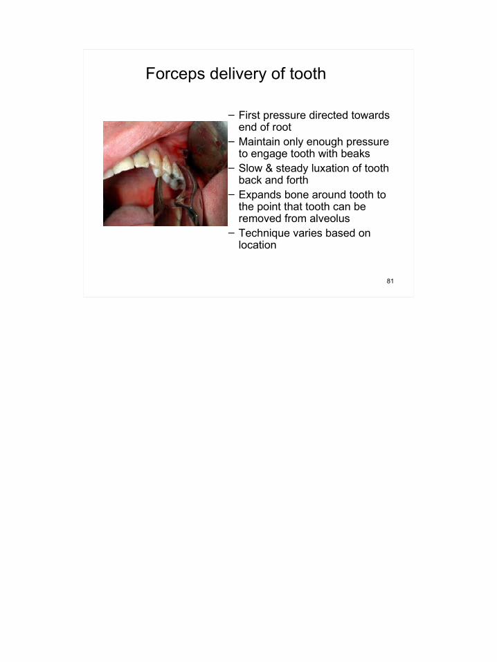
Forceps delivery of tooth
– First pressure directed towards end of root
– Maintain only enough pressure to engage tooth with beaks
– Slow & steady luxation of tooth back and forth
– Expands bone around tooth to the point that tooth can be removed from alveolus
– Technique varies based on location

Anterior tooth technique (most cases)
– Roots of teeth mostly cylindrical
– Luxate first to facial– Then luxate to lingual– As tooth loosens, rotate and
deliver

Posterior tooth technique (most cases)
– Similar technique as anterior tooth, BUT
– Teeth usually multi-rooted, rotation not possible
– Use predominately facial/lingual rotation to slowly expand alveolar plate before attempting to deliver

After extraction
• Check for root completeness– If root is fractured: can attempt remove root fragments if observed,
otherwise annotate so follow-up can be accomplished• Irrigate socket with saline• Evaluate need for suture• Use finger pressure to compress expanded
alveolar bone• Place slightly moistened gauze over socket and
apply pressure

Instructions to patient
• Instructions for pain meds• Apply pressure to gauze for one hour• No excessive spitting• No vigorous rinsing• No drinking through straw• Avoid tobacco use

Post extraction complications
• Pain• Bleeding• Swelling• Dry Socket

Post extraction complications
• Pain– Reassurance, maintain pain meds
• Swelling– Some post op swelling (edema) can
be expected due to trauma of surgery
– Usually resolves 24-36 hours post op
– Swelling after 36 hours may indicate infection

Post extraction complications
• Bleeding– Normal oozing 6-12 hours post op
common– Maintain pressure with good
packing– Look for “liver clot”
• Remove with suction and apply pressure until hemostasis obtained

Post extraction complications
• Dry Socket– due to loss of clot,
exposed bone in socket– usually mand molars– increase in acute,
throbbing pain after 48 hrs (2-5 days)
– pain radiates to ipsilateral ear
– Analgesics ineffective– fetid odor

Post extraction complications
• Dry Socket Treatment– Anesthesia– Irrigation– Pack socket with
iodoform gauze + eugenol
– Pain meds– Repeat q 24hrs until
symptom-free, and then remove packing
Bony Fill and Maturation
1
DENTISTRY FOR MEDICAL STUDENTS
Maxillofacial and Oral SurgeryUniversity Medical Centre
Ljubljana
2
PURPOSE OF THIS TRAINING
• Familiarize non-dental medical providers with basic dental terminology
• Prepare non-dental medical providers to diagnose and treat common dental emergencies – Lost fillings/caries– Common oral pathology – Pulpal & Periodontal conditions– Infection– Trauma– Administration of local anesthesia
3
DENTAL CLASSIFICATIONS
Deployable:• Dental Class 1
– no disease present– no treatment needed
• Dental Class 2– Disease present– no emergency anticipated
within 12 months
Class I No pathologic oral conditions exist and no treatment is required. This includes: a. No dental caries or defective restorations. b. Healthy periodontium; oral prophylaxis not indicated. c. Replacement of missing teeth not indicated. d. Unerupted, partially erupted, or malposed teeth that are without historical, clinical, or radiographic signs or symptoms of pathosis, and are not recommended for removal.
e. Stable occlusion; asymptomatic temporomandibular joint. Class II Oral conditions exist that will not require emergency care within 12 months. These include: a. Dental caries with minimal extension into dentin; minor defective restorations easily maintainable by the patient. b. Interim restorations or prostheses which can be maintained by the patient for a 12- month period. c. Periodontal conditions limited to: 1. Requiring oral prophylaxis 2. Generalized marginal gingivitis, 3. Early periodontitis. 4. Maintenance therapy. 5. Supragingival or subgingival calculus. d. Unerupted, partially erupted, or malposed teeth recommended for removal that are without historical, clinical, or radiographic signs or symptoms of pathosis. e. Edentulous areas not requiring immediate replacement. f. Active orthodontic treatment. g. Temporomandibular joint dysfunction or myofascial pain patients in maintenance therapy.

4
DENTAL CLASSIFICATIONSNon-deployable• Dental Class 3
– Disease present– likely emergency within 12 months– unfit for flying duty or duty in support
of flying mission• Dental Class 4
– Disease status unknown, – has not had dental examination in
last 13 months – need to determine disease status
Class III Oral conditions exist that may require emergency care within 12 months. These include: a. Dental caries with moderate or advanced extension into dentin; tooth fractures; or defective restorations not maintainable. b. Interim (temporary) restorations or prostheses which cannot be maintained by the patient for a 12-month period. c. Periodontal considerations: 1. Acute gingivitis or pericoronitis. 2. Moderate to advanced periodontitis. d. Edentulous areas or teeth requiring prosthodontic treatment for adequate mastication, communication, or acceptable esthetics. e. Unerupted, partially erupted, or malposed teeth with historical, clinical, or radiographic signs or symptoms of pathosis. f. Chronic oral infections (other than periodontal disease); pathological lesions including: 1. Pulpal or periapical pathology. 2. Lesions requiring biopsy or awaiting biopsy report. g. Emergency situations requiring therapy to relieve pain, treat trauma, treat acute oral infections, or provide timely follow-up care (e.g., drain or suture removal) until resolved. h. Temporomandibular joint dysfunction or myofascial pain requiring treatment. Class IV A dental examination is required. This includes: a. Overdue for periodic dental examination b. Determination made that a dental examination is required because of irregularities in dental record, clearance for remote assignment, etc.
5
TOOTH SURFACES
• Occlusal• Facial
– Labial– Buccal
• Mesial• Distal• Lingual/Palatal
LIPS
TONGUE
FRONT Occlusal
Facial
Lingual
Mesial Distal
Terminology: For convenience, the crown of a tooth is described in terms of five surfaces: occlusal (biting surface), lingual (tongue surface), facial (cheek surface), and two interproximal surfaces-mesial and distal
Occlusal surface is the biting surface (in anterior teeth this is called the incisal surface). Facial surface is the surface contacted by the lip or cheek. Lingual surface is the surface contacted by the tongue. Palatal surface is the surface adjacent to the palate or roof of the mouth. Mesial surface is the interproximal surface nearer to the anterior midpoint of the dental arch. Distal surface is the interproximal surface facing away from the anterior midpoint of the dental arch. The mesial surfaces of the two central incisors contact each other; the distal surface of the central incisor contacts the mesial surface of the adjacent lateral incisor, and so on (see figure 12-30).

6
EU TOOTH NUMBERING SYSTEMSfor the adult dentition
• 11-18 Begins with maxillary first incisor right
• 21-28 Begins with maxillary first incisor left
• 31-38 Continues with mandibular first left incisor
• 41-48 Continues with mandibular first right incisor
7
USA TOOTH NUMBERING SYSTEMSfor the adult dentition
• 1-32 for the adult dentition
• Begins with maxillary right third molar
• Maxillary teeth 1-16• Continues with
mandibular left third molar
• Mandibular teeth 17-32• Pediatric, other systems

8
DENTAL SICK CALL
Key for Medical Providers• Diagnosis
– Medical History and Vital Signs– Chief Complaint– History of Present Illness
- Pain frequency and duration - Pain characterization (dull/sharp, throbbing) - Pain elicited or spontaneous?
– LISTEN!– Use S.O.A.P. Format
• Early Management– Initial treatment in the absence of a dentist

9
OROFACIAL PAIN
• Orofacial pain from non-odontogenic sources:– Maxillary sinus– Temporomandibular joint– Referred pain/nerve distribution
• Odontogenic pain:– Arising from teeth or supporting structures– Diagnosis complicated by pain referral and other
factors– However, in most cases dental cause can be found
with exam

10
ODONTOGENIC PAIN
• Pulpal origin– Erroneously referred to as the “nerve”– Vascularized connective tissue with odontoblasts
and unmyelinated C fibers– Pulpalgia
• Caused by inflammatory changes to pulp – Apical periodontitis
• Inflammatory changes at root apex secondary to pulpal disease
• Periodontal origin– Gingivitis– Periodontitis– Acute periodontal abscess

11
DENTAL DIAGNOSTIC TESTS
• Attempt to replicate dental symptoms– Percussion
• Tap on teeth with finger tip – Palpation
• Press on intraoral structures and teeth with digital pressure
– Thermal• Place cold (ice) or squirt
warm liquids on tooth– Biting pressure
• Have patient bite on tongue blade, cotton tipped applicator
Spontaneous or lingering pain is indicative of an advanced problem

12
PULPALGIA
• Reversible pulpalgia– Sharp, hypersensitive reaction – Usually precipitated by thermal stimulus (cold,air) or
sometimes function (chewing)– Unstimulated tooth usually asymptomatic– Diagnose with thermal tests (cold, hot)
• Use cold items (ice, etc. if available) or squirt warm fluids on tooth
– If cause not treated, may progress to irreversible pulpalgia

13
PULPALGIA
• Irreversible pulpalgia– Typically hallmarked by prolonged
unstimulated pain• Spontaneous pain is common• Pain usually persists after removal of thermal
stimulus• Pain usually only partially/temporarily relieved by
analgesics– Diagnose with thermal tests– Untreated, will progress to pulpal necrosis
and apical periodontitis

14
PULPALGIA
• Usual causes of pulpitis– Dental caries– Cracked teeth– Recently placed dental restorations that may
have provoked pulpal response– Hyperocclusion (“high filling”)– Trauma

15
DENTAL CARIES
• Extremely common • Unusual as an emergency (unless
severe)• Temporary restorations helpful• Dental treatment required
Diagnosis: Dental caries (decay) generally does not cause pain when diagnosed and treated early. (2) Undetected or if left untreated, caries will eventually cause mild discomfort to severe pain as the carious lesion progresses (see figure 12- 5,6). The patient with tooth pain from dental caries will usually present with one or more of the following signs and symptoms: a. Intermittent or continuous pain often associated with eating. b. A visible break in the continuity of the enamel surface. c. A brownish-black discoloration of the dentin and overlying enamel.

16
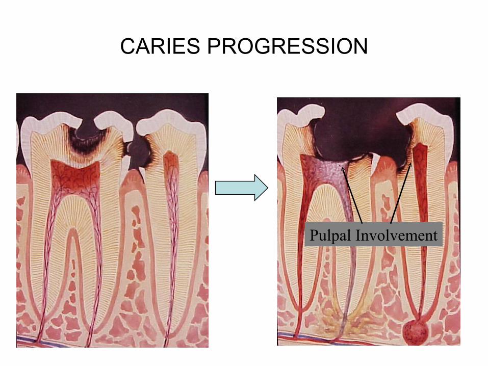
Caries Progression
A. These teeth are clinically free of caries although bacterial plague may exist on the surface.
B. Cavitation of the enamel has started. This is difficult to observe, but the lesion is detectable in an X-ray.
C. Caries has invaded the dentin and is approaching the pulp.

17
CARIES PROGRESSION
Pulpal Involvement
D. Early pulp involvement. E. The crown of the tooth is destroyed; the pulp necrotic; and a periapical abscess present.

18
DENTAL CARIES TREATMENT - causing symptoms
– Isolate tooth as possible with cotton (gauze, rolls); teeth need to be as dry as possible
– Mix Fuji IX glass ionomer material
– Pack into tooth & smooth with gloved finger
– Keep material below the height of the tooth
– Have patient bite down to ensure that material is not too high in occlusion
– Pain meds (NSAID’s or acetaminophen) prn
Treatment: Since the majority of carious dentin is necrotic debris, it is frequently possible to remove much of the carious dentin required for palliation without using a local anesthetic. A local anesthetic should be used if soft caries removal elicits pain. a. Remove most of the soft decayed material with a spoon-shaped instrument (see figure 12-7). b. Since these large carious lesions frequently approximate the pulp, considerable caution should be exerted to avoid penetrating the pulp chamber. Remove as much of the soft decayed material as practical. c. Irrigate the cavity with warm water until loose debris has been removed. d. Isolate the tooth with cotton rolls, or gauze packs, and gently dry the cavity with cotton pledgets (see figure 12-8). e. Restore with glass ionomer (Fuji IX), the preferred restorative material, … if this is not available you may substitute IRM f. Relieve possible interference with opposing teeth by asking the patient to bite several times. Surplus filling material is easily removed and surface contour of the restoration established by applying light pressure with a moist cotton pledget. The IRM will harden within a few minutes. Caution the patient not to use the treated tooth in masticatory function for the next 24 hours (i.e. don't chew on the side of the mouth with the treated tooth). g. If glass ionomer, IRM or zinc oxide is not available, a cotton pledget impregnated with a liquid anodyne can be left in the cavity. h. Instruct the patient that the restoration is temporary. Emphasize that it will keep the patient comfortable until a permanent restoration can be placed by a dental officer.

19
MIXING TEMPORARY FILLING MATERIAL
– Mix one-to-one ratio of scoops of powder to drops of liquid
– Incorporate all powder into the liquid
– Allow to set for one minute to putty consistency
– Place on gloved finger & place onto tooth
– Have patient bite down & grind teeth while material soft to establish occlusion
If glass ionomer is available, it is the restorative material of choice. Mix the powder and liquid as per the manufacturer's instruction and place in the prepared cavity. If this is not available, IRM or ZOE mixtures can be used as described below. If intermediate restorative material (IRM) is available, mix the powder and liquid as per the manufacturer's instruction and place in the prepared cavity. If IRM is not available, zinc oxide powder can be mixed with two or three drops of eugenol until a very thick mix is obtained. The cavity can then be filled with the mixed zinc oxide eugenol. If a suitable mixing surface is not available, use a finger cot or finger cut from a glove. Add the powder and liquid and kneed the mixture to a putty-like consistency.

20
BROKEN FILLING
• Treatment– Treat the same as dental
caries– Try to remove fragments
with instruments– Mix and pack restorative
material into voids

21
CRACKED TOOTH (Incomplete Tooth Fracture)
Fracture Line
• Difficult to diagnose• Tooth appears intact, but patient
indicates exquisite pain to biting (on release)
• Other symptoms include unexplained sensitivity to cold and sweets
• May progress to complete fracture if untreated
• Confirmed by – Bite test using cotton tip applicator or
section of tongue blade. – Thermal tests, ice, warm water, air spray– Transillumination with light is also helpful to
look for cracks• Definitive diagnosis is not always
possible without specialized equipment

22
CRACKED TEETH (Incomplete tooth fracture)
• Treatment– Depends upon symptoms
• Reversible pulpalgia– Pain meds, avoidance of function on
offending tooth• Irreversible pulpalgia
– Med Evac out for definitive dental Tx; or– Extraction of symptomatic tooth

23
COMPLETE VERTICAL TOOTH FRACTURE
• Treatment– Removal of fragments
• Will sometimes relieve symptoms for a period of time
– Med Evac out for definitive dental Tx; or
– Extraction of fractured tooth

24
PAIN FROM RECENT RESTORATION
• Treatment– Reversible pulpalgia:
• Pain meds and allow time to evaluate– Irreversible pulpalgia:
• Med Evac patient out for definitive dental Tx; or• Extraction of symptomatic tooth

25
APICAL PERIODONTITIS
• Classic “Dental abscess”– Usually a single tooth– Secondary to complete or partial
necrosis of the dental pulp– Inflammatory response at root
apex– Tooth may be sensitive to touch-
test by percussion or tapping with finger
– Negative response to cold test will usually confirm lack of pulp vitality
– Untreated, may lead to cellulitis or space infection
Broken down tooth
Draining Abscess
This condition can result when an infection of the pulp reaches out through the apex of the tooth and involves the periodontal tissues (20). Diagnosis: The patient may give a history of repeated episodes of pain that have gradually become continuous and intense. Increased pressure at the root apex may make the tooth feel elongated to the patient. It may seem to be the first tooth to strike its antagonist when the teeth are brought together. There may be severe pain on percussion. This is a most significant symptom. An untreated periapical abscess may burrow through alveolar bone and manifest clinically as a bright red elevation of mucous membrane (parulis or gum boil), (see figure ). A cellulitis may even develop. Also malaise, anorexia, and an elevated temperature may be noted when there is systemic involvement.
26
PERIAPICAL ABCESS
Examine oral cavity for“gum boil”. Incise &drain, Rx antibiotics (if systemic involvement)
– Dental abscess characterized by acute pain and purulent exudate
– Sinus tract may form via path of least resistance resulting in chronic abscess
Treatment: Drainage, induced or spontaneous, usually provides immediate relief from pain (26). Two methods may be used to induce drainage: a. Incise the fluctuant area (see figure 12-6E) using a stab procedure. Suture a small drain or slice of surgical tubing in the wound to maintain drainage. This is generally the most successful method for continued relief. b. Establish drainage through the crown of the tooth. Remove soft decay with a spoon-shaped instrument, carefully create an opening into the pulp chamber. The pain will usually subside when drainage is established (figure 12-7). If drainage is not induced, spontaneous drainage may occur. The acute abscess is then converted to a chronic state subject to repeated acute exacerbations. The spread of the primary periapical abscess is usually in the direction of least resistance. As a general rule, it may be stated that the cortical bone in closest proximity to the abscess site will be the point of breakthrough. However, positively identifying the involved tooth by its proximity to the parulis is an unreliable procedure. Anatomic considerations play a large part both in determining the path of progression and the possibilities of serious sequelae resulting from further spread of the infective process (12). As a general statement, periapical abscess spread is usually toward the lateral aspect of the jaws. If the primary infection involves the palatal root of an upper tooth, the soft tissue abscess is usually found in the palate. Palatal roots are present in the upper molars and first premolars. Abscesses on all other roots in the maxillary dentition tend to burrow through to the facial side. Abscesses developing on the lingual surface of the mandible, to such an extent that they produce drainage into the mouth, are rare. Drainage may, on occasion, occur extraorally without involving the oral mucosa if the path of least resistance is through the cortical bone.
27
APICAL ABCESS TX
Incise & Drain + antibiotics (if needed) will buy you some time
– Incise & allow to drain– Empirical antibiotic – Do not incise & drain
unless gum boil or pointing abscess is observed
– Extract non-vital tooth; or– Med Evac out for
definitive Tx
When the spread of a mandibular periapical abscess is directed lingually, the level of bone perforation dictates the course of events. If the breakthrough is above the attachments of the muscles of the floor of the mouth, sublingual infection results. If below these attachments, the avenue of spread is through the fascial spaces of the neck, creating the potential for major complications, such as Ludwig's angina. Treatment: In cases of more advanced progression, the provision of drainage is still of primary importance. Antibiotics should be administered and their administration continued for 7 to 10 days if the patient is febrile. With soft tissue involvement, the application of moist heat is often helpful in localizing the suppuration. Emergency treatment centers around the prevention of serious sequelae by establishing drainage and maintaining a high blood level of antibiotics. If the offending tooth can be restored, endodontics (root canal) should be accomplished after the acute symptoms have subsided and the infection is under control. If drainage cannot be established, or if the tooth is so badly broken down that it cannot be restored, extraction should be considered. The final decision on tooth extraction should be made by a dentist having access to X-rays. In any event, do not rely on the use of antibiotics in lieu of establishing drainage.
28
LOST CROWN OR BRIDGE
• Prosthesis is dislodged, underlying abutments may be sensitive or not
• Attempt to fit the prosthesis back into position. If it appears to fit stably, it can be recemented with Dycal or IRM. The tooth and prosthesis should be as dry as possible during recementation. Hold the recemented prosthesis in position for 5 minutes, then remove excess cement. Floss the area.
29
BROKEN/LOOSE DENTURE
• The patient tends to view this problem as an emergency. It is not.
• Refer to dentist as soon as possible• Prosthesis may be modified for
comfortable wear, but use caution so as not to ruin a repairable prosthesis

30
ODONTOGENIC SPACE INFECTIONS
• May present as cellulitis or abscess
• Can be life threatening– Invasion of parapharyngeal spaces– Airway compromise – Cavernous sinus thrombosis– Endotoxic shock
• Infections involving the muscles of mastication may result in trismus

31
ODONTOGENIC SPACE INFECTIONS
Buccal space infection that isspreading to submand & Sublingual spaces

32
ODONTOGENIC SPACE INFECTIONS
Ludwig’sAngina

33
ODONTOGENIC SPACE INFECTIONS
• Aggressive management indicated• Incision and drainage
– Intra-oral approach, if possible– Irrigate copiously
• Parental antibiotics + supportive care• Tooth extraction (if possible) and Med
Evac for definitive dental Tx is imperative

34
EXTRAORAL INCISION AND DRAINAGE

35

36
PERIODONTAL CONDITIONS
• Gingivitis • Acute Periodontal Conditions
– Necrotizing Ulcerating Gingivitis (NUG)– Periodontal Abscess– Herpetic Stomatitis– Recurrent Apthous Ulcers– Pericornitis

37
GINGIVITIS
• Inflammation of marginal gingiva– Swelling and easy bleeding– Usually asymptomatic– Occurs quickly after interruption of oral
hygiene practices • Untreated, may progress to NUG in
susceptible individuals

38
GINGIVITIS
• Treatment– Improvement of oral
hygiene– Brush– Floss
• Usually resolves in 48-72 hours

39
N.U.G. Necrotizing Ulcerative Gingivitis
• Previously known as Vincent’s Infection or “Trench Mouth”
• Signs and symptoms include:– Acute pain– Foul breath– Interproximal gingival
necrosis (“punched out papillae”)
Responsible flora is usuallymixed, fuso-spirochetal
Necrotizing ulcerative gingivitis (NUG, Vincent's Infection, Trench Mouth): a. Presentation: A gnawing pain and marked gingival sensitivity are usually the outstanding complaints. These subjective symptoms are characteristically accompanied by pronounced gingival hemorrhage, a foul metallic taste and fetid odor in the mouth, general malaise, and anorexia (12). b. Clinical appearance: Necrosis and ulceration are the principal characteristics of this exceedingly painful inflammatory disease of the gingival tissues. Necrotic lesions typically appear between the teeth in the interproximal spaces. These crater like ulcerations, giving the gingiva a "reverse architecture" appearance and covered by a grayish pseudomembrane, are generally pathognomonic.

40
NECROTIZING ULCERATIVE GINGIVITIS
• Usually associated with:– Poor oral hygiene– Situational STRESSSTRESS– Smoking
• NUG is now considered as an oral hallmark of immunosuppression– HIV/AIDS– Diabetes– Malnutrition
c. Etiology: Although it was felt for many years that fusospirochetal organisms were responsible, the precise etiology has not been established. It is now considered by many to be an endogenous infection arising as a result of the action of ordinarily harmless surface parasites exposed to an altered environment. It is beyond doubt that general health, diet, fatigue, stress, and oral hygiene are more important as precipitating factors than are proximity, intimacy, and contamination. Indeed, the term "trench mouth" is associated with high numbers of troops suffering from similar conditions of high anxiety, poor diet, compromised general health, etc., not from crowded conditions (20).

41
NECROTIZING ULCERATIVE GINGIVITIS
• Treatment– Debridement (at least
aggressive toothbrush use)
– 50/50 mix of viscous lidocaine & mouthwash (if available)
– Antibiotics ONLY if clinical signs of systemic involvement
d. Treatment: Therapy is the establishment of good oral hygiene so as to break the cycle of poor oral hygiene, pain, aversion to brushing, infection, anorexia, and so on. Simple emergency treatment is outlined as follows: 1. Gross debridement. Initial debridement can be achieved by having the patient brush the teeth and gums using a soft toothbrush and warm water. Clean the teeth supragingivally with a dental scaler. Thorough rinsing with 3% hydrogen peroxide diluted with water, or a flavored mouthwash, should follow the initial debridement. 2. Place the patient on an adequate diet and advise a copious fluid intake. 3. Dental home care instructions. Emphasize the importance of good home care. Provide the patient with the soft brush used for initial debridement. Recall the patient daily until acute phase subsides. Unless the patient develops systemic manifestations, antibiotic therapy should not be instituted. Institution of the above regimen will usually suffice for the management of the typical acute case. As a result of this treatment, which can be considered by no means definitive, the acute form subsides and the chronic phase ensues. Although clinical symptoms are minimal, tissue destruction continues unabated unless further corrective measures are instituted. For this reason, definitive dental treatment must be obtained.
If the patient is not improving after 24-hours, blood workups are indicated to rule out other systemic diseases.

42
PERIODONTAL ABSCESS
– Usually an acute exacerbation of pre-existing gum and periodontal disease
– Involved tooth most likely mobile to palpation
– Swelling and purulence may or may not be evident
– Diagnose by probing and palpation
a. Presentation: A deep, throbbing, well-localized pain and a tenderness of the gingiva is characteristic (see figure 12-3).
b: Clinical appearance: This acute suppurative process presents as an ovoid elevation of the gingiva on the lateral aspect of the root (19). The overlaying gingiva is usually red and edematous, with a smooth, shiny surface caused by the edema. The following symptoms may be present: 1. Sensitivity of the tooth to percussion. 2. Mobility of the tooth. 3. Lymphadenitis. 4. General malaise and elevation of temperature in severe cases. c. Etiology: This condition results from an extension of inflammation and infection from a periodontal pocket into deeper periodontal tissues.

43
Periodontal Abscess
• Treatment– Establish drainage
• Incise & drain• Curettage along the
tooth root• or Extract
– Irrigation– Antibiotics if systemic
involvement
d. Treatment consists of the following procedures: 1. Establish drainage through the gingival crevice using a periodontal probe or scalpel blade. 2. Spread the tissues and irrigate to remove pus and debris from the abscess area. 3. Instruct the patient to use warm saline mouth rinses hourly. With definitive treatment, prognosis is good, depending upon the extent of supporting bone loss around the teeth involved. The symptoms of a periodontal abscess may resemble those of a periapical abscess. Because of variations in prognosis, it is important to differentiate positively between the two. Involvement of the lateral aspect of a tooth root by a single suppurative lesion, which can be entered from the gingival crevice, is indicative of a periodontal abscess. The presence of suppurative material in the gingival crevice of the affected tooth also points to this diagnosis.
44
PERICORNITIS
– Frequent emergency among 18-25 year olds
– Inflammation/infection of tissues associated with unerupted or partially erupted teeth (wisdom teeth)
– Multiple etiologic factors• Upper erupted tooth impinging
on soft tissue• Foreign body reaction and
infection
Pericoronitis: a. Presentation: Marked pain in the area of a partially erupted tooth. b. Clinical appearance: Pericoronitis is acute inflammation of the tissue overlying and surrounding a partially erupted or erupting tooth-usually a mandibular third molar. The clinical picture is that of a markedly red, swollen, suppurative lesion. The involved tissue is very tender and often accompanied by pain radiating to the ear, throat, and floor of the mouth. Excruciating pain is produced when the opposing tooth impinges upon the inflamed tissue during mastication. There may be trismus of the masticator muscles on the affected side. Involvement of the cervical nodes, fever, and malaise are common. If this occurs, antibiotic therapy is indicated (see figure). c. Etiology: The principal etiologic factors in pericoronitis are food debris and bacterial waste products which have accumulated under the soft tissue flap, overlying a partially erupted tooth. (2) This tissue is often traumatized during mastication which further exacerbates the situation.

45
PERICORNITIS
• May be purely inflammatory, or have varying degrees of frank infection
• Proximity of parapharyngeal spaces is cause for concern
• Trismus of muscles of mastication may limit opening
• Evaluate for systemic involvement (fever, lymphadenitis)

46
PERICORNITIS
• Aggressive management is indicated– Irrigate infected area with saline or Peridex – Initiate antibiotic therapy if systemic
involvement or space infection is evident – Extract opposing tooth that is occluding on
inflamed tissue– If symptoms are not resolved by conservative
treatment, med-evac for removal of affected teeth
d. Treatment: Satisfactory emergency treatment is as follows: 1. Carefully cleanse beneath the tissue flap using a dental scaler if available. Then flush thoroughly with an irrigating syringe and warm saline. 2. Instruct the patient to rinse with warm saline hourly. 3. Prescribe a soft diet and instruct the patient to refrain from chewing on the affected side of the mouth. 4. Repeat treatment daily until the inflammatory reaction subsides. 5. Initiate antibiotic therapy if systemic involvement or space infection is evident Since extraction of the offending tooth, or its antagonist, is frequently necessary, definitive dental treatment is usually indicated. The antagonist is, in the case of the third molar, the opposing maxillary tooth which may have been the culprit in originally impacting food debris under the flap of skin overlying the partially erupted tooth. This opposing tooth then serves to macerate the swollen flap of tissue causing great pain. (20) When deciding to extract the antagonist, it is important to explain this rationale to the patient so as to minimize misunderstanding. If symptoms are not resolcved by conservative treatment, med-evac for removal of affected tooth.

47
OTHER CAUSES OF ORAL-FACIAL PAIN:other things to keep in mind . . .
• Non-odontogenic causes of orofacial pain which may mimic dental conditions– Maxillary sinusitis – Sialadenitis– Temporomandibular joint pain– Angina pectoris (radiating to left posterior
mandible)– Neuralgia and cluster headaches

48
EMERGENT ORAL PATHOLOGY
• Apthous ulcers– Discrete, shallow ulceration– moveable, unattached intra-oral
mucosa– erythemous halo– self-limiting, – 7-10 day duration
Aphthous Ulcers (Aphthous Stomatitis, Canker Sores): Recurrent aphthous stomatitis (RAS) is one of the most common oral lesions. The lesions are painful, recurrent, single or multiple necrotizing ulcerations of the oral mucosa, and their course is essentially unaltered by modern therapy (21). a. Clinical appearance: These lesions are similar in appearance to oral herpetic lesions; however, they are usually seen on oral mucosa that does not overlay bone. They may be found on the tongue, mucobuccal folds, floor of the mouth, and soft palate whereas intra-oral herpes is usually found only on mucosa overlying bone. The lesion begins as a small macule that enlarges and progresses to a shallow ulceration of 0.5 to 3.0 cm in diameter. The ulcer is usually round or ovoid, with erythematous, rolled margins. The central area is coated with a gray-white fibrinopurulent pseudomembrane. This gray membrane can be removed with rubbing or trauma and a red bleeding ulcer base is exposed. Lesions persist for 7-14 days and heal spontaneously without scarring. b. Etiology: Two distinct groups of factors are important to the cause of aphthous ulcers. These are exogenous microbes (viruses or bacteria) and endogenous precipitating factors (hormonal changes, psychological disease). The most obvious and most common precipitating factor for aphthous ulcers is trauma.

49
APHTHOUS ULCERS
• Treatment– palliative– topical meds
c. Treatment: As with oral herpetic lesions, treatment is usually palliative. Several modalities have been attempted to eliminate recurrence and reduce duration of aphthous ulcers, but the therapeutic value of many commonly used treatments remains unproved. Symptomatic therapy is useful with patients with more severe cases of recurrent aphthous stomatitis. Viscous lidocaine or diphenhydramine elixir mouthwashes prove temporary relief, and coagulating agents, such as Negatan, provide relief of pain through caustic action on sensory nerves. Several commercially available over-the- counter remedies are often used to control the pain of aphthous lesions while awaiting healing. Aphthous type ulcers have been reported as a component of other more serious systemic and local diseases including periadenitis mucosa necrotica recurrens (PMNR, Sutton's disease), Beheets disease, Reiter's syndrome, leukopenias, Crohn's disease, and ulcerative colitis. The foregoing should be considered with patients with severe recurrent aphthous stomatitis (RAS) (22).

50
HERPETIC GINGIVOSTOMATITIS
– Contiguous vesicles & ulcers
– Herpes labialis – Diffuse vesicular
eruptions on attached (bound down) intra-oral mucosa
– Herpes simplex 1 & 2– Primary form may have
lymphadenopathy, fever,malaise
– Secondary form will usually just manifest as pain & vesicles
Herpetic Infections: The herpes simplex virus (HSV) can be divided into two distinct types: type 1 (HSV-1) commonly associated with oral and facial lesions, and type 2 (HSV-2) associated with genital infection. Although less than 1% of the population has a history of clinically evident HSV-1 primary gingivostomatitis, 70 to 90% show serologic evidence of previous exposure. a. Primary gingivostomatitis generally occurs early in life (1-3 years) and presents symptoms of headache, pain, extreme sore mouth, cervical lymphadenopathy, and fever. The oral mucosa is fiery red with numerous vesicles that rupture to form painful ulcers. The gingivae are intensely inflamed and appear red on both the marginal and attached gingival. The lesions are self-limiting and heal without scarring in 12-20 days. The virus may become sequestered in a latent state in the regional sensory ganglia (trigeminal), only to reappear as recurrent intraoral herpetic lesions or as herpes labialis. Infectious virus may be spread by these recurrent lesions as well as by some patients who are clinically free of lesions; thus, transmission of the virus to others is possible (21).
b. Recurrent intra-oral herpetic lesions usually present with prodromal symptoms of tingling or slight pain, followed by the formation of vesicles which soon rupture. Characteristically these lesions occur on firmly attached mucosa, and heal in 7 to 10 days. Herpes labialis (cold sores) represent the prime site of the secondary attack of the HSV-1 virus. Precipitating factors, such as the common cold, febrile diseases, exposure to direct sunlight, emotional stress, and mechanical trauma trigger viral multiplication and manifestation of disease.
51
HERPETIC GINGIVOSTOMATITIS
• Treatment– Palliative– Pain control
• topical anesthetics• NO STEROIDS
– Maintain hydration & nutrition
• Universal precautions– Very contagious– Herpetic whitlow
c. Treatment: Therapy is primarily palliative, and successful management consists of controlling pyrexia and dehydration (particularly with the primary infection), pain, and prevention of secondary infections. Treatment should be symptomatic and supportive as dictated by signs and symptoms of the condition under consideration. Antipyretics may be appropriate for elevated temperatures and antibiotics are only indicated for treatment for secondary infections. Elixir of diphenhydramine HCL, 12.5 mg/5 ml, may be used topically to alleviate the pain associated with these lesions. Any of a number of topical anesthetics would be effective as well. (2)

52
TEMPOROMANDIBULAR DISORDERS
• Myofascial pain dysfunction syndrome (MPD)– Involves muscles of mastication– Situational stress is a contributing factor– Clenching and bruxism are associated with MPD
• Temporomandibular joint (TMJ) disorder– May involve anterior disc displacement or other
pathologic changes in the TMJ– Asymptomatic joint noises are not a cause for concern
in field and are not always predictive of future problems

53
TMJ DISORDERS
• Treatment is palliative– NSAIDS, muscle relaxants– Moist heat to affected muscles – Limit opening, un-necessary function– Soft diet as much as possible– Pain is frequently cyclical and may resolve
with or without intervention

54
TRAUMATIC DENTAL INJURIES
• Crown fractures• Tooth Avulsion/Luxation• Intraoral soft tissue injury
Tooth Fracture The anterior teeth are particularly susceptible to traumatic tooth fracture. (4) Three general types of fractures may be encountered. The classification and emergency treatment for the bulk of these injuries are summarized. Type 1: a. Fracture of the tooth which involves enamel only b. No treatment required. An emory board may smooth the sharp "snags."

55
CROWN FRACTURES CONSERVATIVE FIELD TX
• Exposed dentin of tooth may be sensitive
• Dry tooth with cotton• Observe if tooth is bleeding
from the pulp– if not bleeding, cover tooth
with Fuji IX – If bleeding, will need root
canal in near future, but restore with Fuji IX for the current time
Type 2: a. Fracture of the tooth which involves enamel and dentin, but not pulp (see figure 12-11). b. Treatment: 1. Wash tooth with normal saline or water. 2. Isolate and dry the tooth. 3. The best temporary covering for the exposed dentine is a chemically activated glass ionomer cement Fuji IX. These cements chemically bond to dentine and are the best material for covering dentinal exposures. 4. If a GI cement is not available, use a zinc oxide-eugenol or a calcium hydroxide base. 5. If a glass ionomer cement was not used, cover the base and adjacent enamel with several coats of cavity varnish (CopaliteR). Cavity varnish has low solubility in oral fluids. 6. Rx pain meds and evaluate symptoms 7. Observe patient, if signs of irreversible pulpalgia develop, evac for root canal or extraction is indicated.
56
CROWN FRACTURE CONSERVATIVE FIELD TX
• Mix Fuji IX material and approximate into fractured areas as well as possible
• Rx pain meds and evaluate symptoms
• If signs of irreversible pulpalgia develop, evac for root canal or extraction is indicated
Type 3: a. Fracture of the crown which involves enamel, dentin, and the pulp (see figure 12- 12). b. Treatment: 1. Wash with normal saline. 2. Isolate and dry the tooth. Any hemorrhaging must be controlled. 3. Cover the pulp and dentin with a calcium hydroxide base. A zinc oxide- eugenol base may be used if calcium hydroxide is not available. 4. The calcium hydroxide or zinc oxide-eugenol base and adjacent enamel should then be covered by a glass ionomer cement. If glass ionomer cement is not available, cover the calcium hydroxide or zinc oxide-eugenol base and adjacent enamel with several coats of cavity varnish. 5. Rx pain meds and evaluate symptoms 6. Observe patient, if signs of irreversible pulpalgia develop, evac for root canal or extraction is indicated. Tooth fractures usually do not require the immediate attention of a dental officer if the flight surgeon treats the patient in the prescribed manner. AS mentioned, endodontic treatment (root canal therapy) may be required at a later date, particularly in cases where the tooth fracture involved the pulp

57
TOOTH AVULSION
• Usually anterior teeth• If tooth is not recovered
assume ingestion or aspiration (check lung sounds--future X-rays may be indicated)
• Prompt reimplantation critical for good prognosis– Rinse with saline or clean
water (Do not scrub)– Re-implant and stabilize
as well as possible– Check tetanus
immunization
Tooth Avulsion Another condition to which the anterior teeth are particularly susceptible is tooth avulsion. This usually involves anterior teeth and it is important that if the tooth is not recovered that the possibility of ingestion or aspiration be explored. Checking lung sounds and potentially evacuation for radiographic confirmaiton/diagnosis may be indicated. The patient who presents with an avulsed tooth in hand can be adequately treated by the flight surgeon. The treatment should be initiated as soon as possible. The tooth should be washed by flushing with saline (never scrape the tooth or attempt to clean with hand instruments) and if possible reimplant in the socket immediately (3,20). If a dentist is nearby, and if clot formation in the socket precludes reimplantation, the tooth can be stored in saline and the patient transported to the dentist for treatment. If no dentist is available, the clot should be removed from the socket and the tooth reimplanted. The reimplanted tooth should then be stabilized. Today most dentists do this with composite resin using an acid etching technique. The flight surgeon probably would not have this material available, therefore, the tooth should be stabilized with wire. If the patient can be transported to a dentist within a reasonable period of time, wiring the implanted tooth is not necessary. In this case, the tooth can be stabilized in the socket with the patient's fingers. Considering the nature of the injury, checking the patients tetanus immunization would be indicated.

58
INTRAORAL SOFT TISSUE INJURIES• Isolate source of intraoral
hemorrhage• Obtain anesthesia by
inflitration or block if needed • Irrigate and debride, remove
foreign debris• Close mucosa with 3-0 or 4-
0 gut or silk• Cover bone--especially over
roots• For through and through
lesions, close intraoral lacerations first
59
LOCAL ANESTHESIA FOR DENTISTRY
• Infiltration along 2nd division trigeminal (maxilla)
• Nerve block 3rd division trigeminal (mandible)
The pain control required for dental procedures may be accomplished by local or general anesthetic methods. Local anesthesia is usually the method of choice (11).

60
– Aspirating Syringe– 1.8 ml cartridge– Sterile Disposable
Needle• Long: 1 5/8 inches• Short: 1 inch
LOCAL ANESTHESIA - armamentarium
Armamentarium Local anesthetic agents used for dentistry are administered intra-orally with a cartridge type aspirating syringe. vasoconstrictor) (11). These have a needle attached to them which are used to deliver the local anesthetic which comes in 1.8 ml cartridges

61
ARMAMENTARIUM
Cream (EMLA)Spray (Xylocain)Syringe Anesthetic solution
(Ultracain)SterileSafty

62
COMMON DENTAL NEEDLES
A 27 gauge, 1 3/8-inch needle is usually used for conduction (block) or infiltration; whereas, the 27 gauge, 1-inch needle is usually used for infiltration.30 gauge needles, short are also used for some maxillary infiltrations
63
Ultracain– 1.8ml carpule of 2%
Articaine• 36 mg articaine• .018 mg epinephrine
– 60-90 minutes pulpal anesthesia
– 3-4 hours mucosal anesthesia
DENTAL ANESTHETIC SOLUTIONS
A 2% solution of lidocaine with epinephrine (1:100,000) is the most commonly used local anesthetic in dentistry. For patients who should not receive epinephrine, local anesthetics without a vasoconstrictor are indicated (i.e., 3% mepivacaine without
64
Marcaine– 1.8ml 0.5%
bupivacaine– 1:200,000 Epinephrine– Extended pulpal &
mucosal anesthesia• 90-180 min pulpal• 4-9 hours soft tissue
DENTAL ANESTHETIC SOLUTIONS
Additionally, a long-acting (6-8 hours) anesthetic, bupivicaine, is available and may be of use for extended pain relief when only minimal treatment is available.

65
Infiltration – Injected directly in the area to be
anesthetized– Mechanism is diffusion– Indicated in porous bone (Maxilla)– Short 27 or 30 gauge needle– Onset: 2-3 minutes
Dental Anesthetic Injections
Maxillary Area Infiltration will provide adequate anesthesia in the maxillary teeth. The facial and palatal injections required for effective anesthesia are carried out as follows:
66

67
Maxillary Infiltration Injection – Short needle, 27 or 30 gauge– Penetrate soft tissue over root
apex– Direct the needle toward bone
(45 degrees)– Contact bone and aspirate– Inject one-half to three-
quarters carpule– Repeat for each tooth– Usually WILL NOT anesthetize
palatal tissues
DENTAL ANESTHETIC TECHNIQUE
Facial injection: a. Insert the needle into the mucobuccal fold directly above the tooth to be anesthetized. This fold is formed by the junction of the alveolar mucosa with that of the lip or cheek (see figure 12-14). b. Advance the needle upward for about three-eighths inch, approximately to the apical end of the root. Maintain the point of the needle in close proximity to the maxilla. c. Slowly deposit 1 1/2 cc of solution.
68
Maxillary Palatal Tissues– Locate a point half
the distance between the midline and the tooth
– Penetrate to bone– Aspirate– Inject no more than
one-quarter carpule– Be ready,…is a
painful injection
DENTAL ANESTHETIC TECHNIQUE
Palatal injection: a. A palatal infiltration is usually not necessary for anesthesia of the maxillary anterior teeth; however, it may be required for the maxillary posterior teeth which have palatal roots (first bicuspid and all molars). b. Insert the needle one-half inch above the gingival margin of the tooth to be anesthetized (see figure 12-15). c. Gradually expel one-half cc solution in the submucosal area. After a 5-minute interval, the facial and palatal tissues should be anesthetized.

69
MANDIBULAR NERVE BLOCK
• Inferior Alveolar, Lingual Nerve Block– 27 gauge needle, long– Parallel to mandibular
occlusal plane– Palpate (thumb) anterior
ramus of mandible (intra-orally) at Coronoid Notch
– Grasp posterior border of ramus with index finger (extra-orally)
– Injection approach from contralateral bicuspids
Mandibular Area Conduction anesthesia is the method of choice in anesthetizing the lower teeth (11). The inferior alveolar nerve is blocked as it enters the mandibular foramen on the medial aspect of the ramus. This foramen is located midway between the anterior and posterior borders of the ramus and approximately one-half inch above the biting surface of the lower molar teeth. The width of the ramus at this level can be estimated by placing the thumb on the anterior surface of the ramus intra-orally, and the index finger on the posterior surface extra-orally. The inferior alveolar and lingual nerve are anesthetized by a single injection (see figure 12-16).

70
• Inferior Alveolar, Lingual Nerve Block (con’t)– Contact bone or go 3/4
length of needle– Aspirate– Deposit 3/4 carpule
SLOWLY– Withdraw slightly; aspirate;
inject remainder (lingual nerve block)
– Successful block will provide ipsilateral numb tongue & lip to midline
MANDIBULAR NERVE BLOCK
The inferior alveolar-lingual injection is carried out as follows: a. Place the index finger on the biting surface of the lower molar teeth so that the ball of the finger will contact the junction of the medial surface and the anterior border of the ramus. The fingernail will then be parallel to and facing the sagital plane. b. Place the barrel of the syringe on the lower bicuspids of the side opposite that to be anesthetized. c. Insert the needle at a point one-half inch posterior to the tip of the finger and on a line bisecting the finger nail. The angulations established by carrying out steps b and c are maintained throughout the procedure (see figure 12-17). d. Advance the needle to contact the medial surface of the ramus. One-inch penetration will usually suffice to position the needle point in direct proximity with the mandibular foramen. e. Pull back approximately 3mm on the syringe plunger to provide slight aspirating suction. If blood is drawn in the glass anesthetic carpule, withdraw the needle and try again. f. If no blood is aspirated, slowly deposit approximately 1 1/2 cc of solution at this position. g. Withdraw the needle halfway, aspirate as before, and inject one-half cc of the agent to anesthetize the lingual nerve.

71
Long buccal nerve anesthesia– Penetrate mucosa
posterior and lateral to the tooth to be treated
– Anterior, medial ramus– Aspirate– Deposit no more than
1/4 carpule
MANDIBULAR BUCCAL SOFT TISSUE
Sometimes the innervation to the buccal soft tissues will not be adequately anesthetized with an inferior alveolar block technique and a separate long buccal nerve injection is indicated.

72
BASIC EXODONTIA FOR THE FIELD
– For the resolution of problems that cannot wait or not feasible for evacuation
– Mechanical process in which the bone around the teeth is expanded to the point that tooth can be removed
TOOTH EXTRACTION Today the vast majority of tooth pain can be successfully treated without tooth extraction. A flight surgeon treating a patient with tooth pain should consider extraction only when all other emergency treatment modalities have failed. If extraction is required, the patient should be treated palliatively with analgesics (including local anesthesia) and antibiotics until someone trained in surgical tooth removal is available (2). Even an extraction which appears relatively simple can be deceiving. It is important to remember that the potential complications of tooth extraction can be far more painful and debilitating than the original condition requiring extraction. The complications include fractures of bone (ranging from a minor alveolar fracture to a major fracture of the mandible), fractured roots, hemorrhage, paresthesia, infection, and displacement of a tooth, or tooth fragment, into a sinus or tissue space. As a result, the flight surgeon with no training in oral surgery should treat pain that requires extraction palliatively and make arrangements for definitive treatment as soon as possible. Post Extraction Complications Even though most flight surgeons will never have to extract a tooth, many may be faced with the problem of treating a patient with a post-extraction complication. The two most common post-extraction complications are pain and hemorrhage (3).
73
Basic exodontia instruments
Elevators ForcepsPeriostealelevator, 15 blade
Hemostatsscissors
74
Patient positioning
– Head wrapped in sterile towel
– Reclined to supine position– Position over patients head
for direct vision and mechanical advantage to leverage force
75
Periosteal elevator
• Used to sever gingiva connection to teeth
• Apply force to base of gingival sulcus to crest of bone to sever gingival fibers
76
Elevation of teeth
• Uses leverage at a mechanical advantage point used to luxate tooth in alveolar socket
• Movement of tooth expands alveolar bone to allow tooth to be removed
• Start with smaller elevator (301) and move to larger (92) as tooth luxates
301
92

77
Elevation of teeth
– Wedge elevator between tooth and bone at neck of tooth and rotate handle with slight twisting, quarter-turn movement
– Observe for tooth movement– Do not use excessive force
• Crown fracture• Loosen adjacent teeth
– As tooth loosens, move elevator more into bone towards root end

78
Forceps delivery of teeth
– After suitable loosening of tooth with elevators
– Accomplished with forceps designed to grasp roots of teeth
– Continues the alveolar expansion started with elevators
– Delivers the tooth from the alveolus

79
Forceps
Maxillary (150) ForcepsHandles opposite direction of forceps beaks
Mandibular (151) ForcepsHandles in same direction of forceps beaks

80
Initial forceps placement
– Seat inside beaks first, then outside
– Seat as far down under gums towards bone as possible
– Seat beaks parallel to long axis of tooth
– Close handles and apply pressure to firmly grip tooth
– Support bone around tooth with other hand
81
Forceps delivery of tooth
– First pressure directed towards end of root
– Maintain only enough pressure to engage tooth with beaks
– Slow & steady luxation of tooth back and forth
– Expands bone around tooth to the point that tooth can be removed from alveolus
– Technique varies based on location

82
Anterior tooth technique (most cases)
– Roots of teeth mostly cylindrical
– Luxate first to facial– Then luxate to lingual– As tooth loosens, rotate and
deliver

83
Posterior tooth technique (most cases)
– Similar technique as anterior tooth, BUT
– Teeth usually multi-rooted, rotation not possible
– Use predominately facial/lingual rotation to slowly expand alveolar plate before attempting to deliver

84
After extraction
• Check for root completeness– If root is fractured: can attempt remove root fragments if observed,
otherwise annotate so follow-up can be accomplished• Irrigate socket with saline• Evaluate need for suture• Use finger pressure to compress expanded
alveolar bone• Place slightly moistened gauze over socket and
apply pressure

85
Instructions to patient
• Instructions for pain meds• Apply pressure to gauze for one hour• No excessive spitting• No vigorous rinsing• No drinking through straw• Avoid tobacco use

86
Post extraction complications
• Pain• Bleeding• Swelling• Dry Socket
Post Extraction Complications Even though most flight surgeons will never have to extract a tooth, many may be faced
with the problem of treating a patient with a post-extraction complication. The most common post-extraction complications are pain swelling, hemorrhage and dry socket (3).

87
Post extraction complications
• Pain– Reassurance, maintain pain meds
• Swelling– Some post op swelling (edema) can
be expected due to trauma of surgery
– Usually resolves 24-36 hours post op
– Swelling after 36 hours may indicate infection
Pain: Mild pain or discomfort immediately following tooth extraction is not unusual. Placing a patient on the appropriate analgesics before the local anesthesia has worn off will usually prevent the immediate type of post surgical discomfort or pain. If a patient has not taken the post operative medication and does present with pain, the condition can usually be corrected with the prescription of appropriate analgesics. This pain is usually easily controlled with mild analgesics although some cases may require more powerful medication. Severe pain immediately following surgery is an indication that there may be other complications. The patient with severe pain immediately after surgery should be referred back to the dentist who performed the surgery, if possible. Sometimes reinjection of local anesthetic is the only effective pain control in such cases.
Swelling:Some p;ost op swelling (edema) is to be expected due to the trauma from the surgery. This will usually resolve in 24-36 hours from the time of surgery. If swelling continues or worsens after 36 hours, this may indicate an underlying infection and be treated accordingly.

88
Post extraction complications
• Bleeding– Normal oozing 6-12 hours post op
common– Maintain pressure with good
packing– Look for “liver clot”
• Remove with suction and apply pressure until hemostasis obtained
Hemorrhage: The hemorrhage normally associated with tooth extraction is easily controlled initially by placing gauze sponges over the extraction site and having the patient close to create pressure. The gauze is then removed 20 to 30 minutes postoperatively. In the majority of cases this will be all that is needed to control post-op hemorrhage.
Discoloration of the saliva due to the presence of the blood clot at the extraction site or intermittent capillary hemorrhaging is to be expected postoperatively. Heavy hemorrhaging is not normal and must be treated immediately. Generally this heavy bleeding is caused by the loss of the blood clot at the extraction site or the failure of a clot to form there. In either case, if repeated attempts by the patient to control the hemorrhage by applying pressure gauze dressings have failed, they will usually seek dental or medical attention. If no dentist is available the following treatment should be rendered if bleeding persists:
Remove the clot and pack the extraction site with an absorbable gelatin sponge (Gelfoam) and a hemostatic agent (surgicel). Place gauze sponges over the surgical site and have the patient close and maintain pressure on the gauze for 30 minutes. A new, and expensive treatment modality available for this procedure is microfibriller collagen (AviteneR). Local anesthesia using an anesthetic with a vasoconstrictor can reduce bleeding enough to allow clot formation. In addition, suturing across the socket and papillae with 3-0 silk or chromic suture should be considered. An x-ray of the area should be taken at this point. If dental periapical x-ray film is not available, a lateral oblique or occlusal film should be taken and studied for possible fractures, bony fragments, or retained root fragments. At this time a blood workup should also be initiated to include a platelet count, PT, PTT, and bleeding time. If a major vessel, such as the inferior alveolar artery, was lacerated during surgery, one to two inches of iodoform gauze impregnated with bacitracin ointment should be tightly packed into the wound and left in place for 2 to 3 days.
89
Post extraction complications
• Dry Socket– due to loss of clot,
exposed bone in socket– usually mand molars– increase in acute,
throbbing pain after 48 hrs (2-5 days)
– pain radiates to ipsilateral ear
– Analgesics ineffective– fetid odor
Occasionally a patient will complain of a severe, constant, radiating pain that began 3 to 5 days postoperatively. This delayed pain arises from a condition known as local osteitis or "dry socket."

90
Post extraction complications
• Dry Socket Treatment– Anesthesia– Irrigation– Pack socket with
iodoform gauze + eugenol
– Pain meds– Repeat q 24hrs until
symptom-free, and then remove packing
Bony Fill and Maturation
The treatment for this condition is simply to place a eugenol gauze dressing within the bony socket. Pain relief will be almost immediate. Change the dressing daily until the site becomes desensitized, usually 7-10 days. At the initial appointment the tooth socket should be irrigated with warm saline. The socket should then be "packed" with one to two inches of 1/4 inch iodoform gauze saturated with eugenol. This dressing should be changed daily for the first 3 to 5 days. Generally the pack changes can then be extended to every other day. The patient will inform you if more frequent pack changes are necessary. Patient comfort determines when the treatment is no longer required.