dependenceofagonist activation residue in the third intracellular ... · the at1 receptor to...
TRANSCRIPT
Proc. Natl. Acad. Sci. USAVol. 93, pp. 10040-10045, September 1996Biochemistry
Dependence of agonist activation on a conserved apolar residuein the third intracellular loop of the AT1 angiotensin receptor
(G protein-coupled receptors/inositol phosphate signaling/receptor internalization/site-directed mutagenesis)
LASZLO HUNYADY*t, MENG ZHANG*, GOWRAGANAHALLI JAGADEESHt, MARTA BOR*, TAMA&S BALLA*,AND KEVIN J. CArr*§*Endocrinology and Reproduction Research Branch, National Institute of Child Health and Human Development, National Institutes of Health, Bethesda, MD20892; and *Division of Cardio-Renal Drug Products, Center for Drug Evaluation and Research, Food and Drug Administration, Rockville, MD 20857
Communicated by J. Edward Rall, National Institutes of Health, Bethesda, MD, April 12, 1996 (received for review December 20, 1995)
ABSTRACT The coupling of agonist-activated seventransmembrane domain receptors to G proteins is known toinvolve the amino-terminal region of their third cytoplasmicloop. Analysis of the amino acids in this region of the rat typela angiotensin (ATia) receptor identified Leu-222 as anessential residue in receptor activation by the physiologicalagonist, angiotensin II (Ang II). Nonpolar replacements forLeu-222 yielded functionally intact AT1 receptors, while polaror charged residues caused progressive impairment of AngII-induced inositol phosphate generation. The decrease inagonist-induced signal generation was associated with a par-allel reduction of receptor internalization, and was mostpronounced for the Lys-222 mutant receptor. Although thismutant showed normal binding of the peptide antagonist,[Sar',Ile8]Ang II, its affinity forAng II was markedly reduced,consistent with its inability to adopt the high-affinity confor-mation. A search revealed that many Gq-coupled receptorscontain an apolar amino acid (frequently leucine) in theposition corresponding to Leu-222 of the AT1 receptor. Thesefindings suggest that such a conserved apolar residue in thethird intracellular loop is a crucial element in the agonist-induced activation of the AT1 and possibly many other Gprotein-coupled receptors.
The type 1 angiotensin (AT,) receptor is a typical G protein-coupled receptor (GPCR) and has a major physiological rolein mediating the cardiovascular and other actions of theoctapeptide pressor hormone, angiotensin II (Ang II) (1, 2).Members of this receptor family couple to one or more specificG proteins that mediate their activation of plasma-membraneeffector systems (3-6). In most Ang II target cells, coupling ofthe AT1 receptor to Gq-type proteins results in activation ofphospholipase C, generation of inositol 1,4,5-trisphosphateand diacylglycerol, and stimulation of Ca2+ signaling andprotein kinase C (7-9). In addition, the agonist-receptorcomplex usually undergoes rapid internalization (10-16). Theinositol phosphate responses (17-21) and internalization ki-netics (22-27) of cloned AT, receptors expressed in trans-fected cells are similar to those of endogenous receptors. Themolecular mechanisms by which agonist-activated GPCRscouple to signal generation, and subsequently undergo inter-nalization, have yet to be determined.
In most GPCRs the binding site for physiological agonists isformed by amino acids located in superficial regions of thetransmembrane helices and the extracellular regions of thereceptor (4). The conformational change evoked by agonistbinding is transmitted by the transmembrane helices to theintracellular loops, which are believed to couple the receptorto its cognate G protein(s) and thence to intracellular signalingsystems. Mutagenesis and biochemical studies have demon-
strated that the amino-terminal portion of the third intracel-lular loop is a major structural element involved in receptoractivation and G protein recognition (3-6). Secondary struc-ture predictions suggest that this region is an amphiphilica-helical extension of the fifth transmembrane helix (4). Inaccordance with the proposed importance of this region in Gprotein activation, synthetic peptides corresponding to theN-terminal portion of the third intracellular loop of certainGPCRs can activate the appropriate G proteins in vitro (4, 5).However, relatively few studies have identified specific aminoacids that are required for receptor activation (28-31), and thestructural determinants of the coupling specificity of GPCRsare still unclear (3).
Site-directed mutagenesis studies with muscarinic (28),f3-adrenergic (30), formyl peptide (31), and Ang 11 (20)receptors have found no evidence that charged residues in thisamphiphilic segment participate in G protein coupling. How-ever, uncharged amino acids in this portion of the thirdintracellular loop may be important determinants ofG proteinrecognition and activation (30). One such residue is Tyr-254 inthe M3 muscarinic receptor (28, 32), which is conservedbetween receptor subtypes that activate phosphoinositide sig-naling (M1, M3, and M5) but is replaced by a serine residue inthose (M2 and M4) that inhibit adenylyl cyclase (28). However,most of the GPCRs that stimulate phosphoinositide turnoverdo not possess a tyrosine residue in this position (6). Identi-fication of other amino acids that determine G protein cou-pling is important in defining the nature of the interactionbetween the receptor and its G protein(s), and the mechanismsof G protein activation and specificity.
Recently, deletion studies (18) and the use of chimericAT1/AT2 receptors (33) have identified the amino-terminalportion of the third intracellular loop as a critical region for Gprotein coupling of the rat type la angiotensin (ATia) recep-tor. In addition, substitution of phenylalanine for a conservedtyrosine residue (Tyr-215) in the fifth transmembrane domain,near the origin of the third loop, caused loss of ATia receptoractivation (34). In the present study, the N-terminal portion ofthe third intracellular loop of the AT,a receptor was analyzedto identify its determinants for receptor activation.
Abbreviations: ATla receptor, type la angiotensin receptor; Ang II,angiotensin II; GPCR, G protein-coupled receptor; InsP3, inositoltrisphosphate; InsP2, inositol bisphosphate. [Mutant rat ATia recep-tors are designated by the one-letter code of the original amino acidfollowed by the position number and the one letter code of thereplacement amino acid. Del(AL) refers to a mutant with deletions ofAla-221 and Leu-222; del(AY) has deletions of Ala-225 and Tyr-226.Del221, de1222, and de1223 are single amino acid deletions of Ala-221,Leu222, and Lys-223, respectively.]tPresent address: Department of Physiology, Semmelweis Universityof Medicine, P.O. Box 259, H-1444 Budapest, Hungary.§To whom reprint requests should be addressed at: Endocrinology andReproduction Research Branch, National Institute of Child Healthand Human Development, Building 49, Room 6A36, 49 ConventDrive, National Institutes of Health, Bethesda, MD 20892-4510.
10040
The publication costs of this article were defrayed in part by page chargepayment. This article must therefore be hereby marked "advertisement" inaccordance with 18 U.S.C. §1734 solely to indicate this fact.

Proc. Natl. Acad. Sci. USA 93 (1996) 10041
MATERIALS AND METHODSMaterials. The cDNA clone (pCal8b) of the rat smooth
muscle ATla receptor subcloned into the mammalian expres-sion vector pCDM8 (Invitrogen) was kindly provided byKenneth E. Bernstein (17). COS-7 cells were obtained fromAmerican Type Culture Collection. Restriction enzymes werepurchased from Boehringer Mannheim or New England Bio-labs. Culture media were from Biofluids (Rockville, MD).Lipofectamine and Opti-MEM I were from Life Technologies(Gaithersburg, MD). 125I-labeled Ang II (2200 Ci/mmol; 1Ci = 37 GBq) and 125I-labeled [Sarl,lle8]Ang 11 (2000 Ci/mmol) were obtained from DuPont/New England Nuclear orCorning Hazleton (Vienna, VA), and [3H]inositol was fromAmersham.
Mutagenesis and Expression of the Rat Smooth MuscleAT,a Receptor cDNA. The rat ATla receptor cDNA wassubcloned into the mammalian expression vector pcDNAI/Amp (Invitrogen) as described (18). Mutant rat ATla receptorswere created using the Mutagen kit (Bio-Rad), which is basedon the method of Kunkel et al. (35), using oligonucleotidesobtained from the Midland Certified Reagent (Midland, TX).Most mutant colonies contained a silent restriction site tofacilitate the screening of bacterial colonies, and all mutationswere verified by dideoxy sequencing by using Sequenase II(United States Biochemical). The wild-type and mutant ATIareceptor cDNAs were transiently expressed in COS-7 cellsusing Lipofectamine (36).
Inositol Phosphate Measurements. Cells were prelabeledwith 20 ,uCi/ml [3H]inositol in 0.5 ml inositol-free Dulbecco'smodified Eagle's medium for 24 h (18), then washed twice andincubated for 30 min at 37°C in inositol-free modified Medium199 containing 10 mM LiCl. After stimulation with 1 ,tM AngII for 20 min, reactions were stopped with perchloric acid andinositol phosphates were extracted and analyzed by HPLC (36,37). Inositol phosphates formed under these conditions accu-mulate in the inositol bisphosphate (InsP2) and inositoltriphosphate (InsP3) fractions, which were combined to eval-uate the signaling efficacies of the activated receptors. Sinceinositol phosphate accumulation is proportional to the numberof receptors expressed in transiently transfected cells (18),corrections for variations in expression levels of the mutantreceptors were made by normalizing inositol phosphate re-sponses to the number of cell-surface binding sites measuredin the same experiment. These were quantitated by analysis of[Sarl,lle8]Ang II binding-inhibition data derived from trans-fected COS-7 cells as described earlier (36), and inositolphosphate responses were plotted as a percentage of that of thewild-type ATla receptor.Ang II Binding to COS-7 Cell Membranes. Forty-eight
hours after transfection the cells were washed and scraped into1.5 ml ice-cold 10 mM Tris HCl, pH 7.4/1 mM EDTA andlysed by freezing and thawing. The membrane pellet preparedby centrifuging the samples at 16,000 x g was resuspended inbinding buffer (containing 100 mM NaCl/5 mM MgCl2/20mM Tris HCl, pH 7.4) and the protein content was deter-mined. Binding assays were performed in 0.2 ml binding buffersupplemented with 2 g/liter bovine serum albumin at 25°C.Each sample contained 0.05-0.1 ACi 125I-labeled Ang II andcrude membranes (15-30 jig protein), and inhibition of radio-ligand binding by Ang II was determined in the presence orabsence of 10 ,uM GTP-yS. After 90 min incubation at 25°C theunbound tracer was removed by rapid filtration and the boundradioactivity was measured by y-spectrometry. The apparentKD values were calculated using a one-site model with theRADLIG program (version 4) (Biosoft, Ferguson, MO).
Internalization of Wild-Type and Mutant AT,a Receptors.To determine the internalization kinetics of the mutant andwild-type ATla receptors, 125I-labeled Ang 11 (0.05-0.1 ,Ci)was added in Hepes-buffered Medium 199 and the cells were
incubated at 37°C for the indicated times. Incubations werestopped by placing the cells on ice and rapidly washing themtwice with 1 ml ice-cold Dulbecco's phosphate-buffered saline.The cells were incubated for 10 min in 0.5 ml acid wash solution(150 mM NaCl/50 mM acetic acid) to remove the surface-bound radioligand. The supernatant containing the acid-released radioactivity was collected and the cells were treatedwith 0.5 M NaOH and 0.05% SDS to solubilize the acid-resistant (internalized) radioactivity. The nonspecific bindingwas measured in a parallel experiment using nontransfectedcells. The radioactivities were measured by -y spectrometry andthe percent internalization for each individual point wascalculated, after deducting the respective nonspecific value,from the ratio of the acid-resistant binding to the total(acid-resistant plus acid-released) binding. The endocytic rateconstants were calculated as described by Wiley and Cunning-ham (38). For these calculations, values of 3.5%, and 10% wereused for surface to cell, and intracellular to released spillover,respectively.
Statistical Analysis. All data are shown as means ± SEMunless otherwise indicated. Statistical analyses were per-formed using analysis of variance combined with Tukey'smultiple range test performed at 1% and 5% significancelevels.
RESULTSThe properties ofwild-type ATia receptors expressed in COS-7cells were similar to those previously reported (18, 34, 36, 39),with binding affinity (Kd) for [Sar1,lle8]Ang II at 4°C of 1.5 ±0.2 nM and expression level of 1.0 ± 0.2 pmol/mg protein (n =8). The affinities of the mutant receptors for [Sar',Ile8]Ang IIwere 1.1-2.1 nM, and their expression level was 0.6-1.7pmol/mg protein. The only exception was the de1221 mutantreceptor, which was expressed at a level of 0.28 pmol/mgprotein (n = 3). The mean InsP2 plus InsP3 response showeda 6.1 ± 0.6-fold increase afterAng II stimulation in COS-7 cellsexpressing the wild-type receptor (n = 8).
In earlier studies, the amino terminal region of the thirdcytoplasmic loop (from Tyr-215 to Tyr-226) was implicated inATIa receptor activation (18). This sequence was furtheranalyzed by constructing mutant receptors with double alaninesubstitutions for Leu-217 and Ile-218 (L217A/I218A), Trp-219and Lys-220 (W219A/K220A), and Lys-223 and Lys-224(K223A/K224A) (Fig. 1). Since this segment of the receptorcontains two alanine residues (Ala-221 and Ala-225), doubleamino acid deletions were created for Ala-221 and Leu-222[del(AL)], and Ala-225 and Tyr-226 [del(AY)]. In transiently
NH2~~~~~~~HExtracellular
Membrane
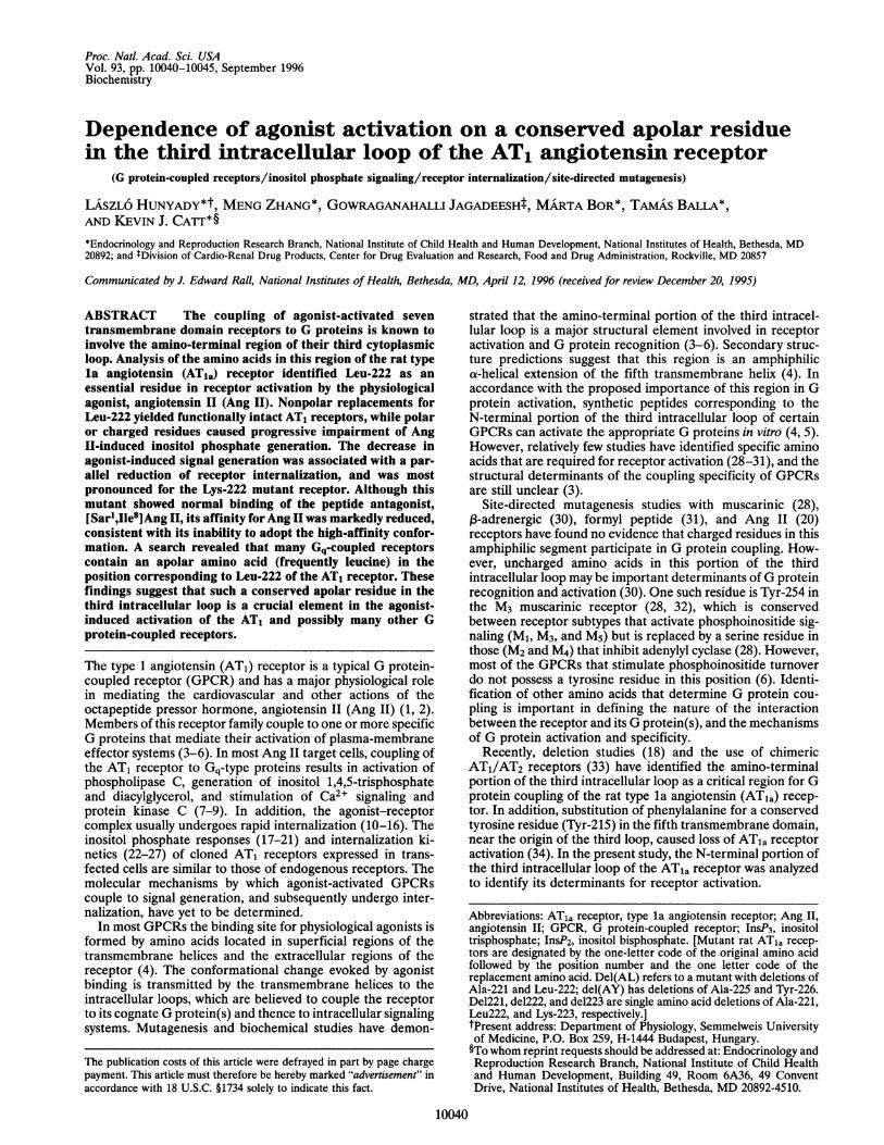
So-- g 2~~ ~ ~~~~~~222Xrr08 s~~~~~~~~~~~~~~ntracellular~~~~~~~COOHFIG. 1. The structure of the ATla receptor. The locations of
residues in the intramembrane helices are based on the model ofBaldwin (40). The most conserved residues in the GPCRs are shownas white letters on a black background. The amino acid residues fromLeu-217 to Tyr-226 analyzed in this paper, and the putative glycosy-lation sites of the receptor, are also shown.
Biochemistry: Hunyady et al.

10042 Biochemistry: Hunyady et al.
transfected COS-7 cells, Ang II-induced inositol phosphateresponses mediated by the L217A/1218A, W219A/K220A,K223A/K224A, and del(AY) receptors were unaffected,whereas deletion of Ala-221 and Leu-222 almost abolishedinositol phosphate production (n = 3, P < 0.01) (Fig. 2).
Since secondary structure algorithms predict that the ami-no-terminal region of the third intracellular loop has ana-helical conformation, the attenuation of signaling by thedel(AL) mutant receptor could result from a change in therelative positioning of the amino acids distal to the deletion.Consistent with this, deletion of either Ala-221 or Leu-222almost completely eliminated the Ang 11-induced inositolphosphate response (n = 3, P < 0.01) (Fig. 3 Upper). However,deletion of Lys-223 had no effect, suggesting that the adjacentN-terminal amino acids are involved in ATia receptor activa-tion. The replacement of Ala-221 with polar (Ser) or charged(Lys or Arg) residues had no significant effect on the inositolphosphate response (Fig. 3 Lower). Thus, it is unlikely thatAla-221 has a specific function in Ang II receptor activation,and its deletion probably affected receptor signaling indirectlyby altering the position of a distal amino acid in the helicalstructure.
Since the above data pointed to Leu-222 as the single mostimportant determinant of ATla receptor activation in thisregion, several mutants with replacements of this residue wereconstructed. As shown in Fig. 4 Upper, alanine replacementcaused only a modest reduction of the inositol phosphateresponse that was not statistically significant, probably reflect-ing the presence of another nonpolar amino acid albeit with a
{L
I-o
%14-0
0
0
cs
0~u)
4)L.
cL
C4
100
50
0
i/CY" 44 4 4
FIG. 2. Inositol phosphate responses of wild-type, double alaninemutant (LI/AA = L217A/I218A; WK/AA = W219A/K220A; KK/AA = K223A/K224A), and two amino acid deletion mutant [del(AL)and del(AY)] rat ATia receptors. Experiments were performed in[3H]inositol-labeled transiently transfected COS-7 cells as described.Maximal stimulation was achieved by incubation with 1 ,uM Ang II inthe presence of 10 mM lithium. The combined accumulation of InsP2and InsP3 following Ang II treatment is shown after normalization ofthe data to the number of expressed receptor sites (18, 36). Results are
expressed as percent of the wild-type response. Data are presented asmeans ± SEM of three independent experiments, each performed induplicate.
100
0
1 60
._40
°~ 20
-0
0
U)
00 6co
- 800-coE 420-
Deletions
T
w.t. de1221 del222 de1223
Ala mutations
T
20 F
0 L......
w.t. Ser Arg Lys
FIG. 3. Inositol phosphate responses of single amino acid deletionmutant (Upper) and Ala-221 mutant (Lower) rat ATla receptors.Combined InsP2 and InsP3 responses of mutant and wild-type ATlareceptors were measured in lithium-treated COS-7 cells after stimu-lation with a maximally active concentration ofAng II (1 ,uM), and thedata were normalized to the number of receptor sites. Results werecalculated as percent of the wild-type response (w.t.) and are shown asmeans ± SEM of three independent experiments, each performed induplicate. On the lower panel the indicated amino acid refers to thereplacement for Ala-221; Ser is A221S, Arg is A221R, and Lys isA221K.
shorter side chain. However, replacement by polar amino acids(L222S and L222N) significantly impaired inositol phosphatesignaling (P < 0.05) and substitution of charged amino acids,particularly lysine, had an even more pronounced effect (P <0.01). In contrast, replacement of Leu-222 with other aliphaticresidues such as valine or isoleucine did not impair the abilityof the mutant receptors to elicit inositol phosphate responses.Although these receptors had a tendency for augmentedinositol phosphate generation compared with the wild-typereceptor, this change was not statistically significant (Fig. 4Upper).Measurements of agonist affinity of the L222K mutant ATia
receptor, and the effect of guanyl nucleotide treatmentthereon, were performed to determine the basis of its inabilityto elicit inositol phosphate responses. Like the other Leu-222mutants, the L222K receptor had normal affinity for thepeptide antagonist, [Sar1,ile8]Ang II (Kd = 1.6 ± 0.1 nM, n =3). However, in membrane binding experiments its affinity forthe native agonist, Ang II, was markedly reduced, with anincrease in apparent Kd from 1.0 nM to 12.0 ± 0.8 nM (n = 3).Furthermore, while the agonist binding affinity of the wild-type receptorwas decreased in the presence of GTPyS to 3.1 ±0.3 nM (n = 3), the low affinity of the L222K mutant ATiareceptor was unaffected by the guanine nucleotide (14.6 ± 0.5nM, n = 3).The internalization kinetics of the several Leu-222 mutant
ATia receptors were determined using 125I-labeled Ang II. Asobserved for the inositol phosphate responses, internalization
Proc. Natl. Acad. Sci. USA 93 (1996)
80 W

Proc. Natl. Acad. Sci. USA 93 (1996) 10043
Leu mutations-, 200
1 80u0
1 60o 1 400.
" 120,,, 1 00n 80c
+ 60
(\4 400cco 20
._
0
C 70C-oK 60.0--- 50
00. 40
-o0 30N
O 20C
10
0
200
K 150
0
W
DL0CU)0
L-io0
0.-c
0-C
CL 50
0c
w.t. Ala Ser Asn Arg Lys Val lle
0 5 10 15 20 25 30
Time (min)
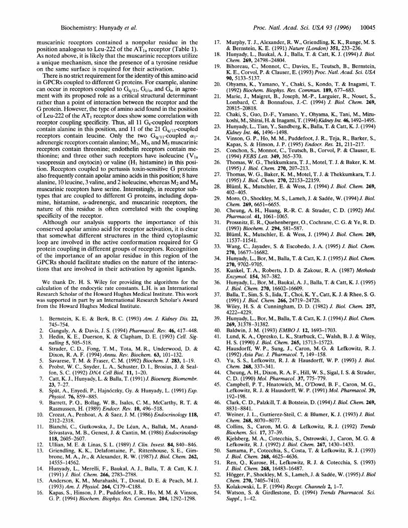
FIG. 4. Combined InsP2 and InsP3 responses (Upper) and inter-nalization kinetics (Lower) of Leu-222 mutant AT1a receptors. Pointmutant rat AT1a receptors are identified by the amino acid replace-ment for Leu-222 (e.g., Ala refers to L222A). Experimental conditionsand data presentation (Upper) are as shown in Fig. 3; results are
means + SEM of three independent experiments, each performed induplicate. Internalization kinetics (Lower) were measured after addi-tion of 1251-Ang II at zero time as described, and are shown as means
of three independent experiments, each performed in duplicate. TheSEM value (not shown) was less then 10% for each point shown.
of the mutant AT1a receptors was impaired only when Leu-222was replaced with polar or charged residues (Fig. 4 Lower). Theendocytic rate constants of these receptors (38, 41) were
closely correlated with the impairment of their inositol phos-phate responses (Fig. 5).
DISCUSSION
Activation of the AT, receptor and other GPCRs is a conse-
quence of conformational changes that result from agonistbinding to specific regions of the receptor protein. Althoughthe exact nature of these changes is not yet known, there are
several parallel events that reflect their occurrence (4). Mostactivated GPCRs exhibit high-affinity agonist binding that isbelieved to result from the formation of a ternary ligand-receptor-G protein complex and is abolished by guaninenucleotides that promote dissociation of the G protein. Inaddition to coupling to G proteins that stimulate secondmessenger systems, many activated GPCRs undergo phosphor-ylation and rapid internalization (42). Recent studies haveshown that agonists can induce internalization of mutantreceptors that are deficient in G protein coupling, indicatingthat endocytosis is a direct consequence of agonist activationrather than of signal generation by the stimulated receptor (18,42, 43).
0,
0 20 40 60 80 100
Endocytic rate constant (% of w.t.)
FIG. 5. Correlation between inositol phosphate signaling (alsoshown on Fig. 4) and endocytic rate constants of Leu-222 mutant AT,areceptors. Data are expressed as percent of the wild-type (w.t.)response or rate constant, and are shown as means + SEM of threeto six independent experiments, each performed in duplicate.
The amino terminal region of the third cytoplasmic loop ofGPCRs is generally considered to be an important determi-nant of G protein coupling and specificity (3-6). In manyGPCRs, the amphiphilic helix formed by this region hasnonpolar and positively charged surfaces. In general, muta-tional studies targeting the charged surface have not revealedmajor defects in the functional characteristics of the mutantreceptors (20, 30, 31). In fact, replacement of all positivelycharged residues in this region did not affect the ability of theATia receptor to activate G proteins (20).The present study demonstrates that replacement of Leu-
222 with polar or charged amino acids progressively interfereswith the internalization and signal generation of the ATiareceptor. In contrast, substitution of alanine with its shortapolar side-chain had only a minor effect, and replacementwith other aliphatic amino acids such as valine or isoleucine didnot impair receptor function. Analysis of the functional prop-erties of Leu-222 mutant receptors revealed that receptorinternalization and inositol phosphate signaling have a parallelamino acid requirement at this position. Since receptor inter-nalization can occur independently of G protein activation (18,25, 43-45), the correlation between impairment of receptorinternalization and signaling of the Leu-222 mutant ATiareceptor suggests that mutations of this amino acid interferewith an event that affects both processes. In the yeast phero-mone receptor (Ste2), replacement of a similarly locatedleucine residue in the third cytoplasmic loop with alanine (46),histidine, or arginine (47), interfered with a-factor-inducedresponses. Thus, the role of this residue in receptor activationmight be conserved in distantly related GPCRs.
In the absence of the agonist, GPCRs adopt an inactiveconformation that is characterized by low binding affinity andlack of G protein activation (48). In most GPCRs, the confor-mational change caused by receptor activation is associated withan increase in agonist binding attributed to the formation of aligand-receptor-G protein complex. Conversely, uncoupling ofthe receptor from the G protein (e.g., in the presence of GTP-yS)prevents the development of the high-affinity conformation.
Biochemistry: Hunyady et al.
10044 Biochemistry: Hunyady et al.
However, in constitutively active adrenergic receptors the high-affinity conformation is an intrinsic property of the receptor andoccurs in the absence of the G protein (49-51). In our study theagonist binding affinity of the most severely impaired mutantreceptor (L222K) was markedly reduced, and unlike that of thewild-type receptor, was unaffected by GTPyS. Such impairedagonist affinity, together with unchanged binding affinity for thepeptide antagonist, [Sar',Ile8]Ang II, suggests that the L222Kreceptor is unable to adopt the activated conformation. It isnoteworthy that the agonist affinity of the L222K receptor was anorder of magnitude lower than that of the uncoupled nativereceptor (measured in the presence of GTPyS). These findingssuggest that unlike G protein coupling, which regulates only theaffinity state of the receptor, replacement of Leu-222 by lysineinterferes with the basic mechanism of the conformationalchange required for receptor activation. In this regard, the L222Kreceptor behaves in an opposite manner to the constitutivelyactive mutants described for adrenergic GPCRs (49-51), since itis unable to adopt the high-affinity conformation and cannot beactivated by agonists. It is for this reason that mutations ofLeu-222 interfere with all known consequences of ATla receptoractivation. The Leu-222 residue thus appears to be an essentialelement in the agonist-induced change in ATia receptor confor-mation, and probably participates in an intramolecular interac-tion that is required for receptor activation.The conserved Tyr-215 residue of the ATIa receptor, which
we have previously identified as critical for ATla receptoractivation (34), is located on the same nonpolar surface of thehelix as the Leu-222 residue identified in this study. Althoughthe amino acid requirements at the two positions are different,point mutations at both locations produced a similar receptorphenotype with impaired G protein coupling and internaliza-tion, as well as reduced agonist binding but normal affinity forpeptide antagonists (34). These findings suggest that bothresidues, which are located within two turns of the helix and onthe same surface, are essential for the active conformation ofthe receptor. The proposed structural role of this nonpolar
surface in the formation of the active conformation of GPCRsis consistent with previous findings that mutations in thisregion of the third intracellular loop cause parallel impairmentof receptor signaling and internalization in all GPCRs studiedto date (4-6). In the rat M3 muscarinic receptor, the samenonpolar surface has been proposed to be a recognition site forG proteins, based on the finding that mutation of Tyr-254impairs inositol phosphate signaling (32). This residue islocated on the same nonpolar surface as Tyr-215 and Leu-222of the ATla receptor, in a position corresponding to Trp-219of the ATIa receptor. Furthermore, in the human M1 musca-rinic receptor the corresponding tyrosine residue (Tyr-212)and the adjacent isoleucine residue (Ile-211) were found to beimportant for G protein coupling (52). These amino acids arelocated on the same nonpolar surface as Leu-222, furtherimplicating this structure in G protein coupling. However,while a common surface appears to be involved in G proteinrecognition, the critical residues concerned in M3 and ATiareceptor signaling are not identical. Thus, double alaninereplacement of Trp-219 and Lys-220 of the ATla receptor doesnot affect ATla receptor function. These data suggest thatalthough this region probably has a conserved role in receptoractivation, the amino acids on the nonpolar-aromatic surfacethat are required for activation differ among GPCRs.GPCRs can be divided on the basis of sequence homology
into five families, of which family I includes the AT1 receptoramong more than 200 hormone and neurotransmitter recep-tors (53). The natures of the amino acid residues in theanalogous position to Leu-222 in other members of family Iwere determined by examining the sequences of a recentcompilation of human GPCRs (54). The position analogous toLeu-222 of the ATla receptor was identified based on itsdistance from the conserved proline and tyrosine residues inthe fifth transmembrane domain. If either of these two residueswas not present, or if the signal transduction pathway was notknown, the sequence was excluded from the analysis. In 57human GPCR sequences that met these criteria, all except
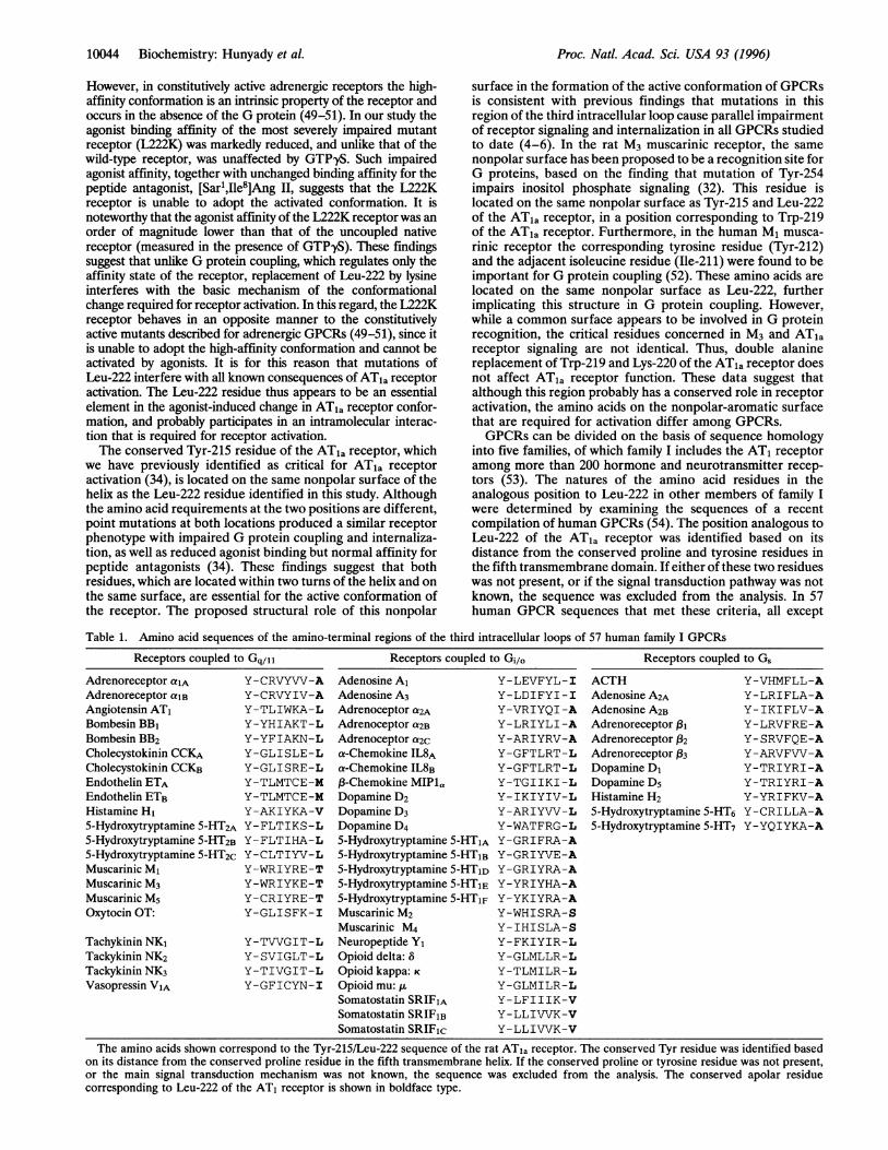
Table 1. Amino acid sequences of the amino-terminal regions of the third intracellular loops of 57 human family I GPCRs
Receptors coupled to Gq/11 Receptors coupled to Gi/o Receptors coupled to G,
Adrenoreceptor alAAdrenoreceptor alBAngiotensin AT,Bombesin BB,Bombesin BB2Cholecystokinin CCKACholecystokinin CCKBEndothelin ETAEndothelin ETBHistamine H15-Hydroxytryptamine 5-HT2A5-Hydroxytryptamine 5-HT2B5-Hydroxytryptamine 5-HT2cMuscarinic MlMuscarinic M3Muscarinic M5Oxytocin OT:
Tachykinin NK1Tackykinin NK2Tackykinin NK3Vasopressin VIA
Y-CRVYVV-AY-CRVYIV-AY-TLIWKA-LY-YHIAKT-LY-YFIAKN-LY-GLISLE-LY-GLISRE-LY-TLMTCE-MY-TLMTCE-MY-AKIYKA-VY-FLTIKS-LY-FLTIHA-LY-CLTIYV-LY-WRIYRE-TY-WRIYKE-TY-CRIYRE-TY-GLISFK-I
Y-TVVGIT-LY-SVIGLT-LY-TIVGIT-LY-GFICYN-I
Adenosine A,Adenosine A3Adrenoceptor a2AAdrenoceptor a2BAdrenoceptor a2Ca-Chemokine IL8Aa-Chemokine IL8Bf3-Chemokine MIPlaDopamine D2Dopamine D3Dopamine D45-Hydroxytryptamine 5-HTlA5-Hydroxytryptamine 5-HTlB5-Hydroxytryptamine 5-HTlD5-Hydroxytryptamine 5-HTIE5-Hydroxytryptamine 5-HT1FMuscarinic M2Muscarinic M4Neuropeptide Y,Opioid delta: SOpioid kappa: KOpioid mu: ,Somatostatin SRIFlASomatostatin SRIFlBSomatostatin SRIFic
Y-LEVFYL-IY-LDIFYI-IY-VRIYQI-AY-LRIYLI-AY-ARIYRV-AY-GFTLRT-LY-GFTLRT-LY-TGIIKI-LY-IKIYIV-LY-ARIYVV-LY-WATFRG-LY-GRIFRA-AY-GRIYVE-AY-GRIYRA-AY-YRIYHA-AY-YKIYRA-AY-WHISRA-SY-IHISLA-SY-FKIYIR-LY-GLMLLR-LY-TLMILR-LY-GLMILR-LY-LFIIIK-VY-LLIVVK-VY-LLIVVK-V
ACTHAdenosine A2AAdenosine A2BAdrenoreceptor 13iAdrenoreceptor X32Adrenoreceptor 133Dopamine D1Dopamine D5Histamine H2
Y-VHMFLL-AY-LRIFLA-AY-IKIFLV-AY-LRVFRE-AY-SRVFQE-AY-ARVFVV-AY-TRIYRI-AY-TRIYRI-AY-YRIFKV-A
5-Hydroxytryptamine 5-HT6 Y-CRILLA-A5-Hydroxytryptamine 5-HT7 Y-YQIYKA-A
The amino acids shown correspond to the Tyr-215/Leu-222 sequence of the rat ATia receptor. The conserved Tyr residue was identified basedon its distance from the conserved proline residue in the fifth transmembrane helix. If the conserved proline or tyrosine residue was not present,or the main signal transduction mechanism was not known, the sequence was excluded from the analysis. The conserved apolar residuecorresponding to Leu-222 of the AT1 receptor is shown in boldface type.
Proc. Natl. Acad. Sci. USA 93 (1996)
Proc. Natl. Acad. Sci. USA 93 (1996) 10045
muscarinic receptors contained a nonpolar residue in theposition analogous to Leu-222 of the AT,a receptor (Table 1).As noted above, it is likely that the muscarinic receptors utilizea unique mechanism, since the presence of a tyrosine residueon the same surface is required for their activation.There is no strict requirement for the identity of this amino acid
in GPCRs coupled to different G proteins. For example, alaninecan occur in receptors coupled to Gq/11, Gi/o, and Gs, in agree-ment with its proposed role as a critical structural determinantrather than a point of interaction between the receptor and theG protein. However, the type of amino acid found in the positionof Leu-222 of the AT1 receptor does show some correlation withreceptor coupling specificity. Thus, all 11 Gs-coupled receptorscontain alanine in this position, and 11 of the 21 Gq/11-coupledreceptors contain leucine. Only the two Gq/11-coupled al-adrenergic receptors contain alanine; Ml, M3, and M5 muscarinicreceptors contain threonine; endothelin receptors contain me-thionine; and three other such receptors have isoleucine (Vlavasopressin and oxytocin) or valine (H1 histamine) in this posi-tion. Receptors coupled to pertussis toxin-sensitive G proteinsalso frequently contain apolar amino acids in this position; 8 havealanine, 10 leucine, 3 valine, and 2 isoleucine, whereas M2 and M4muscarinic receptors have serine. Interestingly, in receptor sub-types that are coupled to different G proteins, including dopa-mine, histamine, a-adrenergic, and muscarinic receptors, thenature of this residue is often correlated with the couplingspecificity of the receptor.Although our analysis supports the importance of this
conserved apolar amino acid for receptor activation, it is clearthat somewhat different structures in the third cytoplasmicloop are involved in the active conformation required for Gprotein coupling in different groups of receptors. Recognitionof the importance of an apolar residue in this region of theGPCRs should facilitate studies on the nature of the interac-tions that are involved in their activation by agonist ligands.
We thank Dr. H. S. Wiley for providing the algorithms for thecalculation of the endocytic rate constants. L.H. is an InternationalResearch Scholar of the Howard Hughes Medical Institute. This workwas supported in part by an International Research Scholar's Awardfrom the Howard Hughes Medical Institute.
1. Bernstein, K. E. & Berk, B. C. (1993) Am. J. Kidney Dis. 22,745-754.
2. Ganguly, A. & Davis, J. S. (1994) Pharmacol. Rev. 46, 417-448.3. Hedin, K. E., Duerson, K. & Clapham, D. E. (1993) Cell. Sig-
nalling 5, 505-518.4. Strader, C. D., Fong, T. M., Tota, M. R., Underwood, D. &
Dixon, R. A. F. (1994) Annu. Rev. Biochem. 63, 101-132.5. Savarese, T. M. & Fraser, C. M. (1992) Biochem. J. 283, 1-19.6. Probst, W. C., Snyder, L. A., Schuster, D. I., Brosius, J. & Seal-
fon, S. C. (1992) DNA Cell Biol. 11, 1-20.7. Catt, K. J., Hunyady, L. & Balla, T. (1991) J. Bioenerg. Biomembr.
23, 7-27.8. Spat, A., Enyedi, P., Hajn6czky, Gy. & Hunyady, L. (1991) Exp.
Physiol. 76, 859-885.9. Barrett, P. Q., Bollag, W. B., Isales, C. M., McCarthy, R. T. &
Rasmussen, H. (1989) Endocr. Rev. 10, 496-518.10. Crozat, A., Penhoat, A. & Saez, J. M. (1986) Endocrinology 118,
2312-2318.11. Bianchi, C., Gutkowska, J., De Lean, A., Ballak, M., Anand-
Srivastava, M. B., Genest, J. & Cantin, M. (1986) Endocrinology118, 2605-2607.
12. Ullian, M. E. & Linas, S. L. (1989) J. Clin. Invest. 84, 840-846.13. Griendling, K. K., Delafontaine, P., Rittenhouse, S. E., Gim-
brone, M. A., Jr., & Alexander, R. W. (1987) J. Biol. Chem. 262,14555-14562.
14. Hunyady, L., Merelli, F., Baukal, A. J., Balla, T. & Catt, K. J.(1991) J. Biol. Chem. 266, 2783-2788.
15. Anderson, K. M., Murahashi, T., Dostal, D. E. & Peach, M. J.(1993) Am. J. Physiol. 264, C179-C188.
16. Kapas, S., Hinson, J. P., Puddefoot, J. R., Ho, M. M. & Vinson,G. P. (1994) Biochem. Biophys. Res. Commun. 204, 1292-1298.
17. Murphy, T. J., Alexander, R. W., Griendling, K. K., Runge, M. S.& Bernstein, K. E. (1991) Nature (London) 351, 233-236.
18. Hunyady, L., Baukal, A. J., Balla, T. & Catt, K. J. (1994) J. Biol.Chem. 269, 24798-24804.
19. Bihoreau, C., Monnot, C., Davies, E., Teutsch, B., Bernstein,K. E., Corvol, P. & Clauser, E. (1993) Proc. Natl. Acad. Sci. USA90, 5133-5137.
20. Ohyama, K., Yamano, Y., Chaki, S., Kondo, T. & Inagami, T.(1992) Biochem. Biophys. Res. Commun. 189, 677-683.
21. Marie, J., Maigret, B., Joseph, M.-P., Larguier, R., Nouet, S.,Lombard, C. & Bonnafous, J.-C. (1994) J. Biol. Chem. 269,20815-20818.
22. Chaki, S., Guo, D.-F., Yamano, Y., Ohyama, K., Tani, M., Mizu-koshi, M., Shirai, H. & Inagami, T. (1994) Kidney Int. 46,1492-1495.
23. Hunyady, L., Tian, Y., Sandberg, K., Balla, T. & Catt, K. J. (1994)Kidney Int. 46, 1496-1498.
24. Vinson, G. P., Ho, M. M., Puddefoot, J. R., Teja, R., Barker, S.,Kapas, S. & Hinson, J. P. (1995) Endocr. Res. 21, 211-217.
25. Conchon, S., Monnot, C., Teutsch, B., Corvol, P. & Clauser, E.(1994) FEBS Lett. 349, 365-370.
26. Thomas, W. G., Thekkumkara, T. J., Motel, T. J. & Baker, K. M.(1995) J. Biol. Chem. 270, 207-213.
27. Thomas, W. G., Baker, K. M., Motel, T. J. & Thekkumkara, T. J.(1995) J. Biol. Chem. 270, 22153-22159.
28. Bluml, K., Mutschler, E. & Wess, J. (1994) J. Biol. Chem. 269,402-405.
29. Moro, O., Shockley, M. S., Lameh, J. & Sadee, W. (1994) J. Biol.Chem. 269, 6651-6655.
30. Cheung, A. H., Huang, R.-R. C. & Strader, C. D. (1992) Mol.Pharmacol. 41, 1061-1065.
31. Prossnitz, E. R., Quehenberger, O., Cochrane, C. G. & Ye, R. D.(1993) Biochem. J. 294, 581-587.
32. Bluml, K., Mutschler, E. & Wess, J. (1994) J. Biol. Chem. 269,11537-11541.
33. Wang, C., Jayadev, S. & Escobedo, J. A. (1995) J. Biol. Chem.270, 16677-16682.
34. Hunyady, L., Bor, M., Balla, T. & Catt, K. J. (1995)J. Biol. Chem.270, 9702-9705.
35. Kunkel, T. A., Roberts, J. D. & Zakour, R. A. (1987) MethodsEnzymol. 154, 367-382.
36. Hunyady, L., Bor, M., Baukal, A. J., Balla, T. & Catt, K. J. (1995)J. Biol. Chem. 270, 16602-16609.
37. Balla, T., Sim, S. S., lida, T., Choi, K. Y., Catt, K. J. & Rhee, S. G.(1991) J. Biol. Chem. 266, 24719-24726.
38. Wiley, H. S. & Cunningham, D. D. (1982) J. Biol. Chem. 257,4222-4229.
39. Hunyady, L., Bor, M., Balla, T. & Catt, K. J. (1994)J. Biol. Chem.269, 31378-31382.
40. Baldwin, J. M. (1993) EMBO J. 12, 1693-1703.41. Lund, K. A., Opresko, L. K., Starbuck, C., Walsh, B. J. & Wiley,
H. S. (1990) J. Biol. Chem. 265, 15713-15723.42. Hausdorff, W. P., Sung, J., Caron, M. G. & Lefkowitz, R. J.
(1992) Asia Pac. J. Pharmacol. 7, 149-158.43. Yu, S. S., Lefkowitz, R. J. & Hausdorff, W. P. (1993) J. Biol.
Chem. 268, 337-341.44. Cheung, A. H., Dixon, R. A. F., Hill, W. S., Sigal, I. S. & Strader,
C. D. (1990) Mol. Pharmacol. 37, 775-779.45. Campbell, P. T., Hnatowich, M., O'Dowd, B. F., Caron, M. G.,
Lefkowitz, R. J. & Hausdorff, W. P. (1991) Mol. Pharmacol. 39,192-198.
46. Clark, C. D., Palzkill, T. & Botstein, D. (1994)J. Biol. Chem. 269,8831-8841.
47. Weiner, J. L., Guttierez-Steil, C. & Blumer, K. J. (1993) J. Biol.Chem. 268, 8070-8077.
48. Collins, S., Caron, M. G. & Lefkowitz, R. J. (1992) TrendsBiochem. Sci. 17, 37-39.
49. Kjelsberg, M. A., Cotecchia, S., Ostrowski, J., Caron, M. G. &Lefkowitz, R. J. (1992) J. Biol. Chem. 267, 1430-1433.
50. Samama, P., Cotecchia, S., Costa, T. & Lefkowitz, R. J. (1993)J. Biol. Chem. 268, 4625-4636.
51. Ren, Q., Kurose, H., Lefkowitz, R. J. & Cotecchia, S. (1993)J. Biol. Chem. 268, 16483-16487.
52. Hogger, P., Shockley, M. S., Lameh, J. & Sadee, W. (1995) J. BiolChem. 270, 7405-7410.
53. Kolakowski, L. F. (1994) Recept. Channels 2, 1-7.54. Watson, S. & Girdlestone, D. (1994) Trends Pharmacol. Sci.
Suppl., 1-42.
Biochemistry: Hunyady et al.