depletion of runx1&eto in t(8;21) aml cells leads to
TRANSCRIPT

Depletion of RUNX1/ETO in t(8;21)AML cells leads to genome-widechanges in chromatin structureand transcription factor binding
The Harvard community has made thisarticle openly available. Please share howthis access benefits you. Your story matters
Citation Ptasinska, A, S A Assi, D Mannari, S R James, D Williamson, JDunne, M Hoogenkamp, et al. 2012. Depletion of RUNX1/ETO int(8;21) AML cells leads to genome-wide changes in chromatinstructure and transcription factor binding. Leukemia 26(8):1829-1841.
Published Version doi:10.1038/leu.2012.49
Citable link http://nrs.harvard.edu/urn-3:HUL.InstRepos:10471527
Terms of Use This article was downloaded from Harvard University’s DASHrepository, and is made available under the terms and conditionsapplicable to Other Posted Material, as set forth at http://nrs.harvard.edu/urn-3:HUL.InstRepos:dash.current.terms-of-use#LAA
ORIGINAL ARTICLE
Depletion of RUNX1/ETO in t(8;21) AML cells leads togenome-wide changes in chromatin structure andtranscription factor bindingA Ptasinska1,2,8, SA Assi3,8, D Mannari4,9, SR James1,9, D Williamson5, J Dunne4, M Hoogenkamp1, M Wu6, M Care1, H McNeill5,P Cauchy2, M Cullen1, RM Tooze1, DG Tenen6,7, BD Young4, PN Cockerill1,2, DR Westhead3, O Heidenreich5 and C Bonifer1,2
The t(8;21) translocation fuses the DNA-binding domain of the hematopoietic master regulator RUNX1 to the ETO protein.The resultant RUNX1/ETO fusion protein is a leukemia-initiating transcription factor that interferes with RUNX1 function. Theresult of this interference is a block in differentiation and, finally, the development of acute myeloid leukemia (AML). To obtaininsights into RUNX1/ETO-dependant alterations of the epigenetic landscape, we measured genome-wide RUNX1- and RUNX1/ETO-bound regions in t(8;21) cells and assessed to what extent the effects of RUNX1/ETO on the epigenome depend on itscontinued expression in established leukemic cells. To this end, we determined dynamic alterations of histone acetylation, RNAPolymerase II binding and RUNX1 occupancy in the presence or absence of RUNX1/ETO using a knockdown approach.Combined global assessments of chromatin accessibility and kinetic gene expression data show that RUNX1/ETO controls theexpression of important regulators of hematopoietic differentiation and self-renewal. We show that selective removal ofRUNX1/ETO leads to a widespread reversal of epigenetic reprogramming and a genome-wide redistribution of RUNX1 binding,resulting in the inhibition of leukemic proliferation and self-renewal, and the induction of differentiation. This demonstrates thatRUNX1/ETO represents a pivotal therapeutic target in AML.
Leukemia (2012) 26, 1829 -- 1841; doi:10.1038/leu.2012.49
Keywords: acute myeloid leukemia; RUNX1/ETO; epigenetic regulation; chromatin; integrated analysis of high-throughput data
INTRODUCTIONChromosomal rearrangements are a hallmark of hematopoieticmalignancies. Many of these translocations generate aberranttranscriptional regulators that reproducibly lead to defined blocksin differentiation.1 A subclass of acute myeloid leukemia (AML) isassociated with a group of chromosomal translocations that eachresult in disruption of the function of the core factor-binding (CBF)complex, which consists of a heterodimer of RUNX transcriptionfactor family members and their binding partner CBFb.2 The best-characterized CBF-type translocation is t(8;21), which fuses RUNX1(AML1), a gene that is essential for normal hematopoiesis,3 withthe gene encoding transcriptional co-repressor ETO (RUNX1T1or MTG8).4,5 The ectopic expression of RUNX1/ETO redirectsthe specific gene expression program of normal precursor cells.6
The mechanism of such deregulation is based on RUNX1/ETOinterfering with normal RUNX1 function.7 -- 11 However, the moleculardetails of this interference are poorly understood on a genome-widelevel. Several studies have demonstrated that RUNX1/ETO acts as aconstitutive repressor by recruiting histone deacetylase complexes,and that it interferes directly with other transcriptional regulatorsof hematopoiesis such as C/EBPa.12 -- 16 The presence of RUNX1/ETO can alternatively lead to gene activation.17 -- 20
The t(8;21) translocation is a leukemia-initiating event, andfusion gene sequences can be found long before the onset ofleukemia in the blood from newborn children.21 However, theinduction of fully developed AML in t(8;21) patients requiressecondary genetic alterations,22 -- 25 which complicates the estab-lishment of in vitro model systems for gain-of-function studies.In order to evaluate the suitability of RUNX1/ETO as a therapeutictarget, it is therefore necessary to identify its target sites at thegenome-wide level in actual leukemic cells, to define its indi-vidual role in reprogramming gene expression networks and todetermine whether its continued presence is required formaintaining deregulation. As RUNX1/ETO and RUNX1 occupy thesame binding sites, it is also important to elucidate how chromatinand RUNX1 occupancy respond to loss of RUNX1/ETO bindingat its target genes.
To this end, we determined genome-wide patterns ofRUNX1/ETO occupancy in primary cells from patients and t(8;21)cell lines, and compared histone acetylation profiles, RNA-Polymerase II occupancy, RUNX1 binding and gene expressionbefore and after siRNA-mediated depletion of RUNX1/ETO. Globalloss of RUNX1/ETO binding results in widespread and complexchanges in chromatin structure patterns, RUNX1 occupancy and
Received 14 February 2012; accepted 15 February 2012; accepted article preview online 20 February 2012; advance online publication, 16 March 2012
1Section of Experimental Haematology, Leeds Institute of Molecular Medicine, University of Leeds, Leeds, UK; 2School of Cancer Sciences, Institute of Biomedical Research,College of Medical and Dental Sciences, University of Birmingham, Birmingham, UK; 3Faculty of Biological Sciences, University of Leeds, Leeds, UK; 4Barts Cancer Institute, QueenMary University of London, London, UK; 5Northern Institute of Cancer Research, University of Newcastle, Newcastle upon Tyne, UK; 6Cancer Science Institute, National Universityof Singapore, Singapore, Singapore and 7Harvard Stem Cell Institute, Harvard Medical School, Boston, MA, USA. Correspondence: Professor C Bonifer, School of Cancer Sciences,Institute of Biomedical Research, College of Medical and Dental Sciences, University of Birmingham, Birmingham B15 2TT, UK.E-mail: [email protected] Dr O Heidenreich, Northern Institute of Cancer Research, University of Newcastle, Paul O’Gorman Building, Framlington Place, Newcastle upon Tyne NE2 4HH, UK.E-mail: [email protected] authors contributed equally to this work.9These authors contributed equally to this work.
Leukemia (2012) 26, 1829 -- 1841& 2012 Macmillan Publishers Limited All rights reserved 0887-6924/12
www.nature.com/leu
gene expression, indicating that RUNX1/ETO-mediated transcrip-tional reprogramming is amenable to therapeutic intervention.
MATERIALS AND METHODSHuman primary cells and cell linesPatient material was obtained with the required ethical approval from theNHS Research Ethics Committees (Leeds Teaching Hospitals NHS Trust andNewcastle upon Tyne Hospitals NHS Foundation Trust) or with informedconsent from adult patients at St Bartholomew’s Hospital, London.
The Kasumi-1 cell line was obtained from the DSMZ cell line repository(http://www.dsmz.de/) and was cultured in RPMI1640 containing 10% fetalcalf serum. SKNO-1 cells were maintained in RPMI1640 supplemented with20% FCS and 7 ng/ml granulocyte -- macrophage colony-stimulating factor.RAW264.7 cells were cultured in Dulbecco’s modified Eagle’s Mediumcontaining 10% fetal calf serum.
siRNA transfectionsKasumi-1 and SKNO-1 cells and AML blasts were transfected with 200 nM ofsiRNA using a Fischer EPI 3500 electroporator (Fischer, Heidelberg,Germany) as described previously.26 For time course experiments,sequential siRNA electroporations were performed at days 0, 4 and 7.The following siRNAs were used: RUNX1/ETO siRNA (sense, 50-CCUCGAAAUCGUACUGAGAAG-30 ; antisense, 50-UCUCAGUACGAUUUCGAGGUU-30), mismatchcontrol siRNA (sense, 50-CCUCGAAUUCGUUCUGAGAAG-30 ; antisense, 50-UCUCAGAACGAAUUCGAGGUU-30).
RNA and protein isolation, real-time PCR and western blottingRNA was isolated 2, 4, 7 and 10 days after siRNA electroporation usingRNeasy columns (Qiagen, Hilden, Germany). Total protein was precipitatedby adding two volumes of acetone to the flow through of the RNeasycolumns and then resuspended in urea buffer (9 M urea, 4% (w/w)3-((3-cholamidopropyl)-dimethylammonio)-propanesulphonate, 1% (w/w)dithiothreitol). Reverse transcription, real-time PCR and western blottingwere performed as described.27
Chromatin immunoprecipitationChromatin immunoprecipitation (ChIP) was performed using thefollowing antibodies: ETO (C-terminus-specific, Santa Cruz Biotechnology,Wembley, UK, sc-9737X), RUNX1 (C-terminus-specific, Abcam, Cambridge,UK, ab23980), RNA-Polymerase II phospho S2 (ab5095) and H3K9Ac(ab4441).
DNase I hypersensitive site mappingGenome-wide hypersensitive sites were mapped as described in Leddinet al.28
Library generation and sequencingLibraries of DNA fragments from ChIP or DNase I treatment were preparedfrom B10 ng of DNA using standard procedures. DNA libraries weresubject to massively parallel DNA sequencing on an Illumina GenomeAnalyzer (Illumina, Little Chesterford, UK).
Luciferase reporter assayReporter constructs were generated in the pGL4-Basic plasmid withpromoter sequences or in the pGL4-TK plasmid (Promega, Southampton,UK) with distal sequences. Regions of interest were amplified from purifiedgenomic DNA from Kasumi-1 cells. Transient transfections of RAW264.7cells were conducted using the Lipofectamine 2000 Reagent (Invitrogen,Paisley, UK) according to the manufacturer’s protocol. Luciferase assayswere performed using a dual-luciferase reporter assay system (Promega).
Expression arraysTotal RNA was labeled and hybridized to the Illumina HT-12 v4 BeadChip(Illumina).
Identification of DNasel- and ChIP-sequencing peaksThe raw sequence data were aligned to the hg18 assembly (NCBI Build 36.1)using BWA,29 and data were displayed using the UCSC Genome Browser.30
Regions of enrichment of DNaseI and ChIP data were identified using MACSsoftware.31 De novo motif analysis was performed using HOMER.32
Microarray gene expression data analysisMicroarray gene expression data were analyzed in GenomeStudiosoftware (Illumina) with background subtraction. The raw data outputwas analyzed using the Lumi R package33 with quantile normalization. The10% threshold (P-value p0.1) was applied to all data. Genes were selectedwith at least twofold change in expression.
Gene set enrichment analysis (GSEA) was performed using thestand-alone application (Broad Institute) by ranking genes according totheir correlation (Pearson) with the F1 metagene, the metagene highlyexpressed in the cells treated with siRNA for RUNX1/ETO. David/EASE analysis was performed using the online tool at http:\\david.abcc.ncifcrf.gov.34
A more detailed description of Methods is given in SupplementaryMaterial.
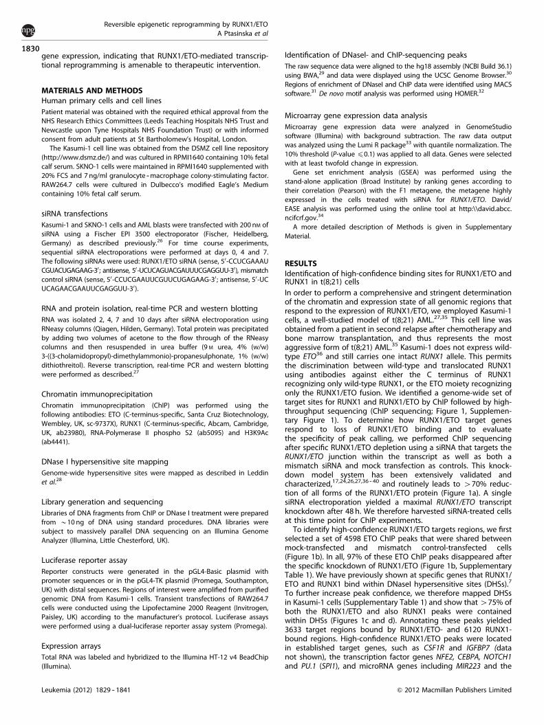
RESULTSIdentification of high-confidence binding sites for RUNX1/ETO andRUNX1 in t(8;21) cellsIn order to perform a comprehensive and stringent determinationof the chromatin and expression state of all genomic regions thatrespond to the expression of RUNX1/ETO, we employed Kasumi-1cells, a well-studied model of t(8;21) AML.27,35 This cell line wasobtained from a patient in second relapse after chemotherapy andbone marrow transplantation, and thus represents the mostaggressive form of t(8;21) AML.35 Kasumi-1 does not express wild-type ETO36 and still carries one intact RUNX1 allele. This permitsthe discrimination between wild-type and translocated RUNX1using antibodies against either the C terminus of RUNX1recognizing only wild-type RUNX1, or the ETO moiety recognizingonly the RUNX1/ETO fusion. We identified a genome-wide set oftarget sites for RUNX1 and RUNX1/ETO by ChIP followed by high-throughput sequencing (ChIP sequencing; Figure 1, Supplemen-tary Figure 1). To determine how RUNX1/ETO target genesrespond to loss of RUNX1/ETO binding and to evaluatethe specificity of peak calling, we performed ChIP sequencingafter specific RUNX1/ETO depletion using a siRNA that targets theRUNX1/ETO junction within the transcript as well as both amismatch siRNA and mock transfection as controls. This knock-down model system has been extensively validated andcharacterized,17,24,26,27,36 -- 40 and routinely leads to 470% reduc-tion of all forms of the RUNX1/ETO protein (Figure 1a). A singlesiRNA electroporation yielded a maximal RUNX1/ETO transcriptknockdown after 48 h. We therefore harvested siRNA-treated cellsat this time point for ChIP experiments.
To identify high-confidence RUNX1/ETO targets regions, we firstselected a set of 4598 ETO ChIP peaks that were shared betweenmock-transfected and mismatch control-transfected cells(Figure 1b). In all, 97% of these ETO ChIP peaks disappeared afterthe specific knockdown of RUNX1/ETO (Figure 1b, SupplementaryTable 1). We have previously shown at specific genes that RUNX1/ETO and RUNX1 bind within DNaseI hypersensitive sites (DHSs).7
To further increase peak confidence, we therefore mapped DHSsin Kasumi-1 cells (Supplementary Table 1) and show that 475% ofboth the RUNX1/ETO and also RUNX1 peaks were containedwithin DHSs (Figures 1c and d). Annotating these peaks yielded3633 target regions bound by RUNX1/ETO- and 6120 RUNX1-bound regions. High-confidence RUNX1/ETO peaks were locatedin established target genes, such as CSF1R and IGFBP7 (datanot shown), the transcription factor genes NFE2, CEBPA, NOTCH1and PU.1 (SPI1), and microRNA genes including MIR223 and the
Reversible epigenetic reprogramming by RUNX1/ETOA Ptasinska et al
1830
Leukemia (2012) 1829 -- 1841 & 2012 Macmillan Publishers Limited
MIR23A---27A---24-2 cluster41 (Figure 1e, Supplementary Figure 1A).High-confidence peaks were also found between the telomeraseprotein gene TERT and CLPTM1L, a locus associated with severaltypes of cancer42 (Supplementary Figure 1A, Supplementary Table4). Selected peaks were validated manually in Kasumi-1 cells(Supplementary Figures 1B and C) and in primary cells from at(8;21) AML patient (Supplementary Figures 1D -- F), with andwithout RUNX1/ETO knockdown.
To validate our genome-wide cell line ChIP-sequencing data, wecompared the RUNX1/ETO-binding pattern of Kasumi-1 cells to
that of primary, patient-derived t(8;21) AML cells. As with t(8;21)cell lines, the wild-type ETO protein is neither expressed in t(8;21)-negative nor in t(8;21)-positive AML cells,36,43,44 which agrees witha lack of DHSs at the ETO (RUNX1T1) locus in all hematopoietic celltypes studied here (data not shown). With primary cells from twopatients (patients A and B), we obtained a large number of smallpeaks, many of which disappeared when analyzed at higherstringency (data not shown), but 2629 peaks occurred in bothpatients. In all, 76% of joint peaks intersected with DHS from thetwo other t(8;21) patients (no. 1 and no. 2, Supplementary Figure
RUNX1/ETO siREKasumi-1
(1595)
RUNX1/ETO siMMKasumi-1
(6341)
RUNX1/ETO control Kasumi-1(7813)
305845981581
144
1420
18 13
RUNX1Kasumi-1
(8008)DHSKasumi-1(33832)
27712 6120 1888
RUNX1 highconfidence peaks
DHSKasumi-1
(33832)
RUNX1/ETO highconfidence peaks
RUNX1/ETOcontrol & siMM
Kasumi-1
(4742)
363330199 1109
mRNA
0
0.2
0.4
0.6
0.8
1
1.2
Nor
m. t
rans
crip
t lev
els
RUNX1/ETOβ-ACTIN
48h24h 96h
protein
RUNX1/ETOa b
c d
NFE2 SPI1 (PU.1) e
DHS t(8;21) # 1
DHS t(8;21) # 2
RUNX1/ETO patient A
DHS Kasumi-1
Sequence conservation
RUNX1/ETO patient B
RUNX1/ETO control
RUNX1/ETO siMM
RUNX1/ETO siRE
RUNX1
control siMM siRE siMM siRE siMM siRE
Figure 1. Identification of high-confidence binding sites for RUNX1/ETO and RUNX1. (a) Time course of RUNX1/ETO knockdown in Kasumi-1cells. Top panel: real-time PCR analysis of mRNA expression; bottom panels: immunoblot detection of RUNX1/ETO protein. b-ACTIN served asloading control. Time points are indicated at the bottom. RUNX1/ETO transcript levels are recovering 48 h after siRNA electroporation, whereasprotein levels remain low for another 24 h. siRE, RUNX1/ETO siRNA; siMM, mismatch siRNA; control, mock-transfected cells. (b) Intersectionanalysis of RUNX1/ETO peaks. The Venn diagram shows the overlap between RUNX1/ETO peaks in mock, mismatch siRNA or RUNX1/ETOsiRNA-treated Kasumi-1 cells. The vast majority of RUNX1/ETO peaks were common to mock and siMM-treated Kasumi-1 cells, and 497% ofthe common peaks disappeared after RUNX1/ETO depletion. (c, d) Identification of high-confidence peaks for RUNX1/ETO and RUNX1 inKasumi-1 cells. More than 75% of both the RUNX1/ETO and RUNX1 peaks colocalize with DHS in Kasumi-1 cells, thus constitutinghigh-confidence peaks. (e) UCSC Genome Browser image depicting the human NFE2 and SPI1 (PU.1) loci demonstrating a colocalization ofRUNX1/ETO peaks in patient cells, specific RUNX1/ETO and RUNX1 peaks in Kasumi-1 cells as well as DHS.
Reversible epigenetic reprogramming by RUNX1/ETOA Ptasinska et al
1831
Leukemia (2012) 1829 -- 1841& 2012 Macmillan Publishers Limited

1G, Supplementary Table 1) and thus represented high-confidencepeaks. More than 50% of genes bound by RUNX1/ETO in Kasumi-1cells were also specifically bound in patient cells (SupplementaryTable 5). This high degree of concordance was also confirmed bycomparing RUNX1/ETO binding to specific genes across thegenome (Figure 1e, Supplementary Figure 1A), including knowntargets of RUNX1/ETO such as LAT2.45
RUNX1/ETO- and RUNX1-binding sites overlap only partially int(8;21) cellsBoth RUNX1 and RUNX1/ETO showed a similar pattern ofdistribution of binding sites for the subsets of peaks located
within 1.5 kb of transcription start sites, and 60% of the RUNX1/ETO peaks overlapped with RUNX1 peaks (Figures 2a and b). Theunbiased de novo identification of enriched consensus bindingmotifs showed that, in contrast to RUNX1-unique peaks, bothRUNX1/ETO-associated and RUNX1/ETO-unique peaks were en-riched in motifs for E-box-binding proteins (Figure 2c), inagreement with observations that these factors form stableinteractions with the NHR1 domain of RUNX1/ETO.12 The samemotifs were found when examining the entire set of RUNX1- orRUNX1/ETO-bound sequences in Kasumi-1 cells (SupplementaryFigures 2A and B). Both RUNX1/ETO- and RUNX1-binding regionswere enriched for RUNX1 consensus sequences, but only 30% ofRUNX1-unique peaks contained such motifs, indicating that at
c Motifs associated withshared RUNX1/ETO
and RUNX1 peaks in Kasumi-1
Motifs associated with RUNX1 unique
peaks in Kasumi-1
Motifs associated with RUNX1/ETO unique peaks in Kasumi-1
motif match score
(log p-value)
ETS -1769
ETS -1638
ETS -1538
ETS -1418
RUNX -1139
RUNX -1111
RUNX -948
C/EBP -314
motif match score
(log p-value)
RUNX -1318
ETS -1302
ETS -1158
RUNX -567
ETS: E-box
-437
motif match score
(log p-value)
RUNX -835
ETS -682
ETS -602
E-box -489
E-box -488
E-box -447
ETS -224
b d
a
0
10
20
30
40
50
60
70
80
RUNX1 ETS E-box C/EBPα
% m
otifs
in p
eaks
in K
asum
i - 1
RUNX1 & RUNX1/ETO overlap RUNX1 unique
RUNX1/ETO unique
RUNX1 Kasumi-1
(6120)
RUNX1/ETO Kasumi-1
(3633)
2181 3939 1452
10 3.5
3
2.5
2
1.5
1
0.5
RUNX1in Kasumi-1
RUNX1/ETOin Kasumi-19
8
7
6
5
4
3
2
% N
um
ber
of
pea
ks
% N
um
ber
of
pea
ks
1
-1500 -1100 -700
Distance from TSS Distance from TSS
-300 0 300 700 1100 1500 -1500 -1100 -700 -300 0 300 700 1100 1500
Figure 2. Identification and characterization of RUNX1/ETO and RUNX1 target regions in t(8;21) cells. (a) Positional distribution of RUNX1- (left)or RUNX1/ETO- (right) binding sites relative to the transcription start site (TSS) of their nearest gene. (b) Intersection of RUNX1 and RUNX1/ETO peaks in Kasumi-1 cells. The Venn diagram shows the numbers of high-confidence peaks bound by RUNX1 and RUNX1/ETO. (c) De novomotif discovery performed on the set of regions bound by the RUNX1 and/or RUNX1/ETO in Kasumi-1 cells identified enriched RUNXconsensus and different types of ETS factor-binding sites. E-box motifs were significantly enriched in peaks either unique for RUNX1/ETOor common to RUNX1/ETO and RUNX1. (d) RUNX, ETS, E-box and C/EBP consensus sequence were mapped back to all regions bound byRUNX1/ETO and RUNX1. A large proportion of regions bound by RUNX1 did not contain RUNX (TGYGGT) and/or E-box (CANNTG) consensusbinding motifs, whereas most regions contain ETS (GGAW) sites.
Reversible epigenetic reprogramming by RUNX1/ETOA Ptasinska et al
1832
Leukemia (2012) 1829 -- 1841 & 2012 Macmillan Publishers Limited
sites not occupied by RUNX1/ETO, RUNX1 may bind via interactionwith other transcription factors. These are likely to be ETS familymembers, which present the top-scoring motif in this peak
population (Figure 2c). In contrast, up to 70% of shared andunique RUNX1/ETO-bound sequences contained a RUNX1motif (Figure 2d), suggesting that the fusion protein needs to
RUNX1KN-AML(14552)
22449
DHSKN-AML(36033)
13584 968
RUNX1 KN-AML highconfidence peaks
motif match score(log p-value)
PU.1/ETS -1084
ETS -1043
PU.1/ETS -964
ETS -916
RUNX1 -804
ETS -224
motif matchscore
(log p-value)
ETS -1978
RUNX1 -1809
E-box -98
0
20
40
60
80
100
RUNX1 ETS E-box C/EBPα AP1
% m
oti
fs in
pea
ksin
KN
-AM
L
motif matchscore
(log p-value)
RUNX1 -4786
ETS -4072
ETS -3158
AP1 -2320
E-box -1806
CEBP -651
AP1 -487
NF-kB -480
CEBP -435
NF-kB -319
Motifs associated with RUNX1 peaksunique to KN-AML
Motifs associated with RUNX1 peakscommon to t(8;21) and KN-AML
RUNX1KN-AML(13548)
RUNX1Kasumi-1
(6120)354410004 2576
Motifs associated with RUNX1 peaks unique to t(8;21)
7 RUNX1 non-t(8;21)
6
5
4
3
2% N
um
ber
of
pea
ks
1
-1500-1100 -700
Distance from TSS-300 0 300 700 1100 1500
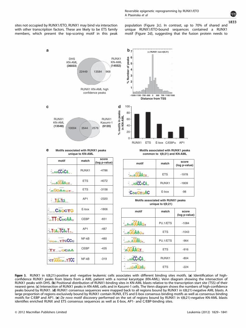
Figure 3. RUNX1 in t(8;21)-positive and -negative leukemic cells associates with different binding sites motifs. (a) Identification of high-confidence RUNX1 peaks from blasts from a AML patient with a normal karyotype (KN-AML). Venn diagram showing the intersection ofRUNX1 peaks with DHS. (b) Positional distribution of RUNX1-binding sites in KN-AML blasts relative to the transcription start site (TSS) of theirnearest gene. (c) Intersection of RUNX1 peaks in KN-AML cells and in Kasumi-1 cells. The Venn diagram shows the numbers of high-confidencepeaks bound by RUNX1. (d) RUNX1 consensus sequences were mapped back to all regions bound by RUNX1 in t(8;21)-negative AML blasts. Alarge proportion of regions exclusively bound by RUNX1 contain RUNX, ETS and E-box consensus binding motifs as well as consensus bindingmotifs for C/EBP and AP1. (e) De novo motif discovery performed on the set of regions bound by RUNX1 in t(8;21)-negative KN-AML blastsidentifies enriched RUNX and ETS consensus sequences as well as E-box, AP1- and C/EBP-binding sites.
Reversible epigenetic reprogramming by RUNX1/ETOA Ptasinska et al
1833
Leukemia (2012) 1829 -- 1841& 2012 Macmillan Publishers Limited

RUNX1 siMM
Kasumi-1
8368
RUNX1 siREKasumi-1
11827
61765651
2108 2634
RUNX1/ETOcontrol & siMM
4742
5134229 5138
Unique peaksShared peaks
FGR NFIL3
NFIL3NFIL3
NFIL3
RUNX1/ETO control_
RUNX1/ETO siMM _
RUNX1/ETO siRE_
RUNX1_
RUNX1 siMM_
RUNX1 siRE_
Sequence conservation
RUNX1 peaks uniquefor siRE
5651
2192
RUNX1/ETOcontrol & siMM
4742
RUNX1 shared peakssiRE & siMM
6176
4068
Figure 4. Knockdown of RUNX1/ETO leads to a redistribution of RUNX1 binding. Kasumi-1 cells were electroporated with either mismatchcontrol siRNA (siMM) or RUNX1/ETO siRNA (siRE). Two days after siRNA electroporation, RUNX1 binding was measured by ChIP sequencing inboth populations. (a) Top panel: Venn diagram showing the appearance of new RUNX1-binding sites after RUNX1/ETO depletion. Bottom left:Venn diagram showing the overlap between RUNX1/ETO-bound regions and de novo RUNX1 sites, demonstrating that the latter are distinctfrom sites previously bound by RUNX1/EO. Bottom right: Venn diagram showing the overlap between RUNX1/ETO-bound sequences and sitesbound by RUNX1 before and after RUNX1/ETO depletion. (b) Example of alterations in RUNX1 binding. Left panel shows UCSC browser imagesdepicting one gene (NFIL3) at a site not previously bound by RUNX1/ETO and another gene (FGR) showing a de novo RUNX1 with a RUNX1/ETO-bound site showing an increase in RUNX1 binding at this site after knockdown.
Figure 5. Analysis of RUNX1/ETO-dependent gene expression patterns. (a) Hierarchical clustering of genes responding by an at least twofoldchange in expression levels to RUNX1/ETO knockdown in Kasumi-1 cells. The heat map shows early upregulated (Group I), downregulated(Group II) and late upregulated genes (Group III) over a time course of 10 days. At the bottom of the heat map note non-clustered genes thatare either upregulated more than threefold or show a more complex response pattern (Group IV). Expression levels were compared betweenRUNX1/ETO siRNA and mismatch siRNA-treated cells. Time points are indicated on the top of the heat map. Dark red indicates highlyupregulated genes and black indicates highly downregulated genes. (b) Effect of RUNX1/ETO depletion on gene expression in primary t(8;21)AML blasts. The graph shows real-time PCR-based validation of early responding genes. The columns represent the means for three t(8;21)AML patients and the error bars the s.e.m. Inset: immunoblot showing siRNA-mediated depletion of RUNX1/ETO in blasts from a t(8;21) AMLpatient. (c) GSEA ranked according to the correlation of genes with a metagene (F1) summarizing the gene expression profile of Kasumi-1 cellsafter RUNX1/ETO knockdown. From left to right: significant enrichment of gene sets downregulated in human hematopoietic stem cells uponRUNX1/ETO overexpression6 and enrichment of genes determined in this study to have a high-confidence RUNX1/ETO-binding sitewith a corresponding DHS in the region 2 kb upstream of the start of transcription in Kasumi-1 cells. P, nominal P-value; q, false discovery rate.(d) The numbers of upregulated and downregulated genes upon RUNX1/ETO depletion in Kasumi-1 cells. The bottom row indicatesgenes with RUNX1/ETO peaks. (e) Classification of RUNX1/ETO target genes. The columns show the percentage of genes with high-confidence RUNX1/ETO peaks of all genes with changed gene expression upon RUNX1/ETO knockdown. siMM, mismatch siRNA; siRE, RUNX1/ETO siRNA.
Reversible epigenetic reprogramming by RUNX1/ETOA Ptasinska et al
1834
Leukemia (2012) 1829 -- 1841 & 2012 Macmillan Publishers Limited

be recruited by directly anchoring it to DNA. These results contrastto recently published findings by Maiques-Diaz et al.,46
who reported a substantially lower fraction of RUNX1 motifs in
RUNX1/ETO peaks. This discrepancy may be due to the fact thatin these experiments a ChIP-chip proximal promoter arraywas used.
Time (h)
p < 0.0001; q < 0.0001 p < 0.0001; q < 0.003
48 96 168 240
Regulation up down up down up down up down
Total 90 68 174 106 293 256 656 741
Direct RUNX1/ETO targets 46 13 63 27 106 57 152 112
0
2
4
6
8
10
RU
NX
1/E
TO
CS
T7
IGF
BP
7
NF
E2
PR
G2
PT
PN
22
RN
AS
E2
Fol
d ch
ange
Transcript
siMMsiREsi
RE
siM
M
RUNX1/ETO
RUNX1
a
b
c
0
10
20
30
40
50
60
% g
enes
with
R/E
pea
ks
Early directtarget genes
Late direct target genes
48 96 168
Time (h)
240
e
d
I
II
III
IV
Fold-change0.5 2.1
Time after knock-down96h48h 240h168h
RUNX1/ETO targetsdown in HSC(Tonks 2007)
DHS +/- 2 kb
Reversible epigenetic reprogramming by RUNX1/ETOA Ptasinska et al
1835
Leukemia (2012) 1829 -- 1841& 2012 Macmillan Publishers Limited

Differential binding of RUNX1 in AML cells without a CBF complexmutationThe incomplete overlap between RUNX1 and RUNX1/ETO peaksprompted us to investigate whether RUNX1 bound to differentsequences in the human genome in RUNX1/ETO-expressing cellscompared to AML cells without CBF mutations. To this end weidentified genome-wide high-confidence RUNX1-binding sites inAML cells with a normal karyotype (KN-AML) from a patient thatexpressed a full-length RUNX1 protein (data not shown) butdisplayed a block at a similar differentiation stage as the t(8;21)patients and had a surface marker expression profile highly similarto the two t(8;21) AML samples (Supplementary Table 2). Similar toKasumi-1 cells, 90% of all RUNX1 peaks in the KN-AML blast cellscolocalized with DHSs (Figure 3a). Direct RUNX1 targets includedgenes controlling hematopoietic differentiation such as CSF1R,LAT2, and LMO2 (Supplementary Figure 3). The KN-AML RUNX1peaks showed a similar pattern of distribution around thetranscription start sites as the RUNX1 peaks from Kasumi-1 cells(Figure 3b), but their genome-wide distribution and enriched motifcomposition was strikingly different. When directly compared(Figure 3c), only 26% of the KN-AML RUNX1-binding sites werefound in Kasumi-1 cells. GATA3, GCHFR and KLF13 are examples forthis notion (Supplementary Figure 3). Most importantly, in contrastto t(8;21) cells, the RUNX1 peaks unique for KN-AML showed ahighly significant enrichment for binding sites for induciblefactors such as AP1, C/EBP and NF-kB (Figures 3d and e).
RUNX1/ETO knockdown leads to a shift in the pattern of RUNX1occupancyOur RUNX1 ChIP data indicate that the genome of hematopoieticprecursor cells contains a large number of functional RUNX1-binding sites which are differentially occupied in t(8;21) AML andKN-AML cells and are associated with different binding site motifs.We therefore tested by ChIP sequencing whether depletion ofRUNX1/ETO led to an alteration of RUNX1 occupancy in RUNX1/ETO expressing cells (Figure 4). The comparison between RUNX1binding before and after knockdown showed that the depletion ofRUNX1/ETO led not only to an increase in the number of RUNX1peaks, but also to the formation of a large number of de novoRUNX1-binding sites (Figure 4a), as exemplified by the transcrip-tion factor gene NFIL3, which is upregulated by RUNX1/ETOdepletion (Figure 4b, right panel). Only 10% of de novo peaksoverlapped with RUNX1/ETO-bound sites (Figure 4a, bottom left).In contrast, a much larger proportion (34%) of peaks sharedbetween knockdown and control samples were originally boundby RUNX1/ETO (Figure 4a, bottom right). Interestingly, although atmost tested target regions depletion of RUNX1/ETO led to anupregulation of RUNX1 binding and gene expression (an exampleof this is shown with another upregulated gene, FGR, a member ofthe SRC kinase family, in Figure 4b, left panel), this was not alwaysthe case (Supplementary Figure 4). For instance, PU.1 (SPI1)represents a class of genes, where RUNX1/ETO binding did not oronly marginally affect RUNX1 occupancy. The PU.1 upstreamregulatory element contains an upstream enhancer element (PU.1(E)) that is bound by RUNX1 at multiple sites, and this binding isvital for enhancer activity,47 but it is also bound by RUNX1/ETO(this study). At this element RUNX1 binding did not respond toRUNX1/ETO knockdown, and gene expression was barely altered,which may indicate a complex binding pattern with multipleoccupancies of both RUNX1/ETO and RUNX1.
RUNX1/ETO depletion can lead to both activation and repressionof its direct target genesIn order to examine the effects of RUNX1/ETO depletion on geneexpression at the global level and to correlate these changes withidentified RUNX1/ETO target genes, we generated expressionprofiles during a time course of sustained RUNX1/ETO knockdown
for 10 days in Kasumi-1 cells (Figure 5, Supplementary Table 6,Supplementary Figure 5A). Hierarchical clustering identified fourgroups of genes differentially responding to knockdown, consist-ing of early (Group I) and late (Group III) upregulated genes,downregulated genes (Group II) as well as genes showing acomplex pattern of response (Group IV; Figure 5a). These resultswere validated by manual analysis with a selection of earlyresponding genes both in Kasumi-1 cells and in primary t(8,21)cells obtained from three different AML patients (Figure 5b,Supplementary Figure 5B).
To compare global gene expression profiles, we produced twocoordinately regulated metagenes F1 and F2 in an unsupervisedmanner that summarized the expression of RUNX1/ETO-dependent genes in a single score using non-negative matrixfactorization.48,49 The F1 and F2 metagenes were highly expressedin Kasumi-1 cells with and without RUNX1/ETO knockdown,respectively. We also tested the relevance of the RUNX1/ETO-dependent global gene expression changes for another t(8;21)-positive cell line, SKNO-1, and for primary t(8;21)-positive AML.Kasumi-1-derived RUNX1/ETO-dependent metagene expressionwas mirrored and validated in the SKNO-1 and primary AML datasets, demonstrating an excellent concordance between theRUNX1/ETO-associated global gene expression changes in thesethree different t(8;21)-positive cell types (Supplementary Figure5C). Furthermore, RUNX/ETO-associated gene expression changesinversely correlated with gene expression changes observed inprevious knockdown studies and in overexpression studies withprimary human CD34þ cells (Figure 5c, Supplementary Figures5D and E).6,27,39 These combined analyses confirmed that RUNX1/ETO-associated shifts in gene expression in Kasumi-1 faithfullyreflect gene expression features in the human t(8;21) AML.
The metagene was also used to rank genes for the purpose ofGSEA according to their correlation with the metagene score(Supplementary Table 7). Genes with high-confidence RUNX1/ETOpeaks were highly enriched in the RUNX1/ETO knockdownsignature (Figure 5c). However, not all genes responding toRUNX1/ETO knockdown were associated with RUNX1/ETO peaks.Genes responding early at day 2, such as CST7, IGFBP7 and MTSS1,consisted of 50% direct RUNX1/ETO target genes, whereas thiswas true for only 20% of the late responding genes at day 10(Figures 5d and e). Moreover, almost 80% of all early respondingtarget genes were upregulated upon RUNX1/ETO knockdown, incontrast to 60% of all late responding target genes (Figures 5dand e). This indicates that downstream effects of the removalof RUNX1/ETO contribute rapidly and progressively to thereorganization of the transcriptional network.
The depletion of RUNX1/ETO leads to complex changes in thehistone acetylation pattern and RNA Polymerase II occupancyAs RUNX1/ETO recruits histone deacetylases to its target promo-ters13,14 and alters gene transcription, we examined the immediateconsequences of RUNX1/ETO depletion on histone H3 lysine 9acetylation (H3K9Ac) and occupancy of the elongating form of RNAPolymerase II (RNA Pol II). We therefore performed ChIP experimentswith RUNX1/ETO-depleted and -control cells at day 2 afterknockdown, and integrated RNA Pol II and H3K9Ac data inKasumi-1 cells on RUNX1/ETO target loci with the gene expressiondata (Figures 6a and b, Supplementary Table 6). After 2 days ofknockdown, half of the early upregulated genes, including IGFBP7,NFE2 and PRG2, exhibited more than twofold increased RNA Pol IIoccupancy, whereas most downregulated genes such as CD34 and75% of the late upregulated genes generally showed no or littlechange in RNA Pol II binding at this stage (Supplementary Figure 6A).
A more complex pattern of changes at RUNX1/ETO peakswas seen when measuring H3K9Ac. Although after 2 days ofknockdown most upregulated genes displayed a rapid increase inH3K9Ac, we also found an increase at 50% of the downregulated
Reversible epigenetic reprogramming by RUNX1/ETOA Ptasinska et al
1836
Leukemia (2012) 1829 -- 1841 & 2012 Macmillan Publishers Limited

POLIIChIP
Expression
Time after knock-down 240h48h 48h 96h 168h 240h48h 48h 96h 168h
Downregulated
Downregulated
Late-Up-regulated
Late-Up-regulated
Early-Up-regulated
Early-Up-regulated
H3K9AcChIP
Expression
Time after knock-down 2.12.1 0.50.5a b
dc ZBTB16 WT1
RUNX1/ETO control
RUNX1/ETO siMM
RUNX1/ETO siRE
POLII siMM
POLII siRE
H3K9Ac siMM
H3K9Ac siRE
Sequence
Conservation
Figure 6. RUNX1/ETO silencing leads to changes in RNA-Polymerase II occupancy and the histone H3K9 acetylation pattern at RUNX1/ETOtarget genes. (a) Comparison of RNA Pol II occupation with transcriptional profiling reveals a substantial correlation between changes in geneexpression and changes in RNA Pol II association upon RUNX1/ETO knockdown. RNA Pol II occupation was analyzed 48 h after siRNAtreatment, ranked according to fold change in occupancy and compared with changes in gene expression during a time course of 10 dayswith siRNA treatment. Dark red indicates high occupancy or upregulation, black low occupancy or downregulation, respectively. (b) TheH3K9Ac pattern correlates partially with changes in gene expression associated with RUNX1/ETO depletion. H3K9Ac occupation was analyzedand compared analogously to (a). (c, d) UCSC Genome Browser image of the human ZBTB16 and WT1 loci depicting ChIP-Seq tags for RUNX1/ETO, RNA-Pol II and H3K9Ac in mock-treated (control), mismatch siRNA (siMM) and RUNX1/ETO siRNA (siRE)-treated Kasumi-1 cells. (e) Heatmap resulting from an unsupervised clustering of H3K9Ac- and RUNX1/ETO-binding sites with and without RUNX1/ETO knockdown (siMMand siRE, respectively) as well as without transfection (control) showing two groups of sequences and their genomic location. In each lane, theChIP enrichment score is shown 10 kb upstream and downstream from the peak center. (f ) Integration of the sequence enrichment of eachgroup into a density plot showing the location of RUNX1/ETO-binding sites with and without knockdown in relation to H3K9Ac. Dark blue:H3K9Ac (siMM); pink: H3K9Ac (siRE); green: RUNX1/ETO (control); yellow RUNX1/ETO (siMM); light blue: RUNX1/ETO (siRE).
Reversible epigenetic reprogramming by RUNX1/ETOA Ptasinska et al
1837
Leukemia (2012) 1829 -- 1841& 2012 Macmillan Publishers Limited
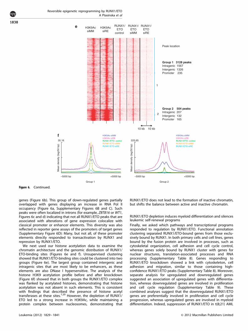
genes (Figure 6b). This group of down-regulated genes partiallyoverlapped with genes displaying an increase in RNA Pol IIoccupancy (Figure 6a, Supplementary Figures 6B and C). Suchpeaks were often localized in introns (for example, ZBTB16 or WT1,Figures 6c and d) indicating that not all RUNX1/ETO peaks that areassociated with alterations of gene expression colocalize withclassical promoter or enhancer elements. This diversity was alsoreflected in reporter gene assays of the promoters of target genes(Supplementary Figure 6D): Many, but not all, of these promoterelements directly responded to transactivation by RUNX1 andrepression by RUNX1/ETO.
We next used our histone acetylation data to examine thechromatin architecture and the genomic distribution of RUNX1/ETO-binding sites (Figures 6e and f). Unsupervised clusteringshowed that RUNX1/ETO-binding sites could be clustered into twogroups (Figure 6e). The largest group contained intergenic andintragenic sites that are most likely to be enhancers, as theseelements are also DNase I hypersensitive. The analysis of thehistone H3K9 acetylation profile before and after knockdown(Figure 6f) showed that in both groups the RUNX1/ETO complexwas flanked by acetylated histones, demonstrating that histoneacetylation was not absent in such elements. This is consistentwith findings that described the presence of histone acetyltransferases at these sites.7,20 However, the depletion of RUNX1/ETO led to a strong increase in H3K9Ac, while maintaining aprotein complex between nucleosomes, demonstrating that
RUNX1/ETO does not lead to the formation of inactive chromatin,but shifts the balance between active and inactive chromatin.
RUNX1/ETO depletion induces myeloid differentiation and silencesleukemic self-renewal programsFinally, we asked which pathways and transcriptional programsresponded to regulation by RUNX1/ETO. Functional annotationclustering separated RUNX1/ETO-bound genes from those exclu-sively bound by RUNX1. In both primary cells and cell lines, genesbound by the fusion protein are involved in processes, such ascytoskeletal organization, cell adhesion and cell cycle control,whereas genes solely bound by RUNX1 cluster with genes fornuclear structures, translation-associated processes and RNAprocessing (Supplementary Table 8). Genes responding toRUNX1/ETO knockdown showed a link with cytoskeleton, celladhesion and migration, similar to those containing high-confidence RUNX1/ETO peaks (Supplementary Table 8). Moreover,separate analysis for upregulated and downregulated genessuggested an association of upregulated genes with differentia-tion, whereas downregulated genes are involved in proliferationand cell cycle regulation (Supplementary Table 9). Thesecombined analyses suggest that the downregulated RUNX1/ETOgenes are preferentially involved in proliferation and cell cycleprogression, whereas upregulated genes are involved in myeloiddifferentiation. Indeed, suppression of RUNX1/ETO in t(8;21) AML
Group 1 3128 peaks Intragenic 1567Intergenic 1326Promoter 235
Group 2 504 peaks Intragenic 207Intergenic 132Promoter 165
Peak location
1
2
H3K9AcsiMM
H3K9AcsiRE
RUNX1/ETO
control
RUNX1/ETOsiMM
RUNX1/ETOsiRE
e
10 kb 10 kb
225.6535.16
Mea
n de
nsity
(ta
g/50
bp)
Mea
n de
nsity
(ta
g/50
bp)
Group 1
–5000 bp +5000 bp –5000 bp +5000 bp
Group 2
f H3K9Ac-siMMH3K9Ac-siRERUNX1/ETO- controlRUNX1/ETO-siMMRUNX1/ETO-siRE
H3K9Ac-siMMH3K9Ac-siRERUNX1/ETO- controlRUNX1/ETO-siMMRUNX1/ETO-siRE
Figure 6. Continued.
Reversible epigenetic reprogramming by RUNX1/ETOA Ptasinska et al
1838
Leukemia (2012) 1829 -- 1841 & 2012 Macmillan Publishers Limited
cell lines impairs clonogenicity and proliferation, and leads to cellcycle arrest in G1 phase.26
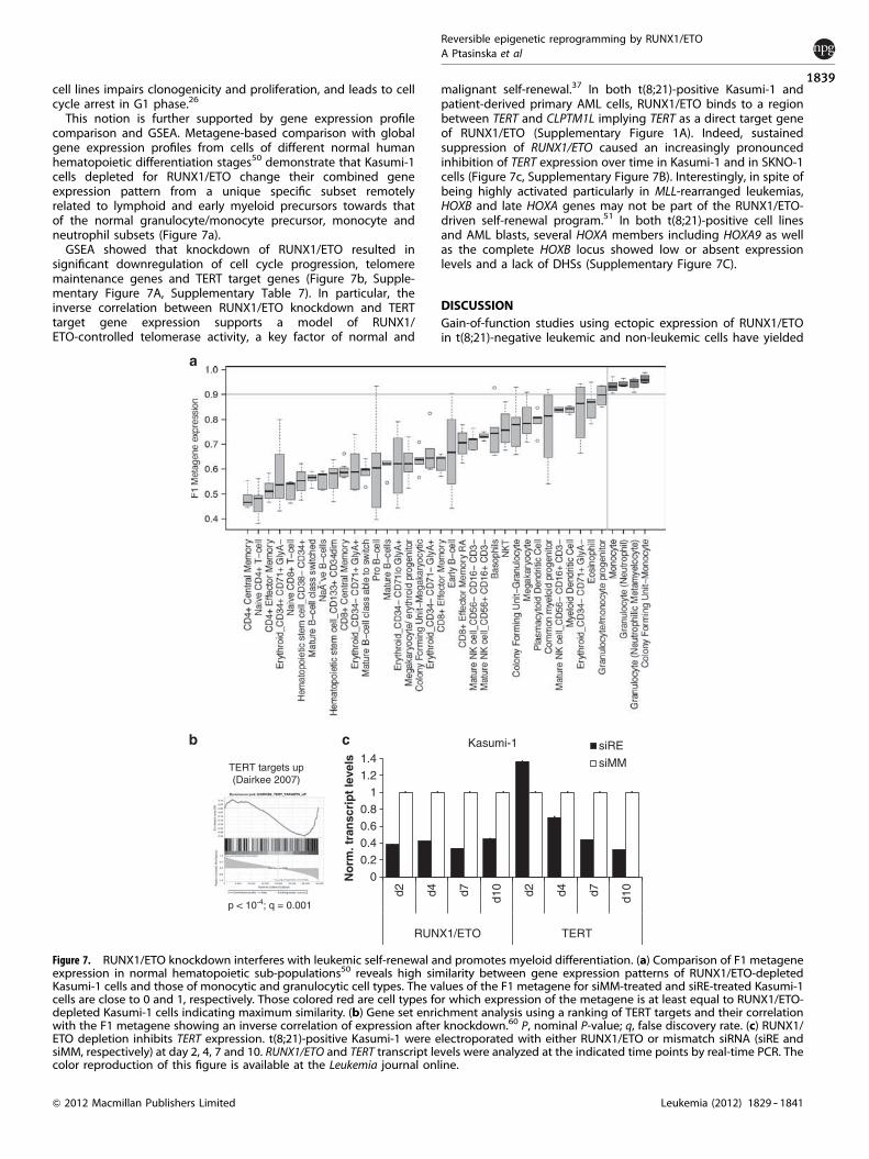
This notion is further supported by gene expression profilecomparison and GSEA. Metagene-based comparison with globalgene expression profiles from cells of different normal humanhematopoietic differentiation stages50 demonstrate that Kasumi-1cells depleted for RUNX1/ETO change their combined geneexpression pattern from a unique specific subset remotelyrelated to lymphoid and early myeloid precursors towards thatof the normal granulocyte/monocyte precursor, monocyte andneutrophil subsets (Figure 7a).
GSEA showed that knockdown of RUNX1/ETO resulted insignificant downregulation of cell cycle progression, telomeremaintenance genes and TERT target genes (Figure 7b, Supple-mentary Figure 7A, Supplementary Table 7). In particular, theinverse correlation between RUNX1/ETO knockdown and TERTtarget gene expression supports a model of RUNX1/ETO-controlled telomerase activity, a key factor of normal and
malignant self-renewal.37 In both t(8;21)-positive Kasumi-1 andpatient-derived primary AML cells, RUNX1/ETO binds to a regionbetween TERT and CLPTM1L implying TERT as a direct target geneof RUNX1/ETO (Supplementary Figure 1A). Indeed, sustainedsuppression of RUNX1/ETO caused an increasingly pronouncedinhibition of TERT expression over time in Kasumi-1 and in SKNO-1cells (Figure 7c, Supplementary Figure 7B). Interestingly, in spite ofbeing highly activated particularly in MLL-rearranged leukemias,HOXB and late HOXA genes may not be part of the RUNX1/ETO-driven self-renewal program.51 In both t(8;21)-positive cell linesand AML blasts, several HOXA members including HOXA9 as wellas the complete HOXB locus showed low or absent expressionlevels and a lack of DHSs (Supplementary Figure 7C).
DISCUSSIONGain-of-function studies using ectopic expression of RUNX1/ETOin t(8;21)-negative leukemic and non-leukemic cells have yielded
p < 10-4; q = 0.001
TERT targets up(Dairkee 2007)
0
0.2
0.4
0.6
0.8
1
1.2
1.4
d2 d4 d7 d10 d2 d4 d7 d10
RUNX1/ETO
No
rm. t
ran
scri
pt
leve
ls
Kasumi-1 siRE
siMM
TERT
Figure 7. RUNX1/ETO knockdown interferes with leukemic self-renewal and promotes myeloid differentiation. (a) Comparison of F1 metageneexpression in normal hematopoietic sub-populations50 reveals high similarity between gene expression patterns of RUNX1/ETO-depletedKasumi-1 cells and those of monocytic and granulocytic cell types. The values of the F1 metagene for siMM-treated and siRE-treated Kasumi-1cells are close to 0 and 1, respectively. Those colored red are cell types for which expression of the metagene is at least equal to RUNX1/ETO-depleted Kasumi-1 cells indicating maximum similarity. (b) Gene set enrichment analysis using a ranking of TERT targets and their correlationwith the F1 metagene showing an inverse correlation of expression after knockdown.60 P, nominal P-value; q, false discovery rate. (c) RUNX1/ETO depletion inhibits TERT expression. t(8;21)-positive Kasumi-1 were electroporated with either RUNX1/ETO or mismatch siRNA (siRE andsiMM, respectively) at day 2, 4, 7 and 10. RUNX1/ETO and TERT transcript levels were analyzed at the indicated time points by real-time PCR. Thecolor reproduction of this figure is available at the Leukemia journal online.
Reversible epigenetic reprogramming by RUNX1/ETOA Ptasinska et al
1839
Leukemia (2012) 1829 -- 1841& 2012 Macmillan Publishers Limited
insights into the molecular mechanisms of initiating leukemogen-esis,6,46,52 -- 54 whereas loss of function studies in actual leukemict(8;21) cells (including the Kasumi-1 cells described here) informabout the role of RUNX1/ETO in maintaining leukemia.24,25,36,39
Our experiments and novel integrative data analyses demonstratea substantial concordance between these two approaches,indicating that integral parts of the t(8;21)-specific leukemia-initiating program are also required for maintaining the leukemicphenotype. In both primary cells and cell lines, RUNX1/ETObinds genes associated with the control of the cell cycleand cell structure. Concordantly, siRNA-mediated depletion ofRUNX1/ETO affects transcriptional programs associatedwith myeloid differentiation, proliferation and self-renewal,in addition to those promoting cell cycle progression and DNAsynthesis.
Our siRNA knockdown shows that RUNX1/ETO binding leads tolarge-scale, but reversible, alterations throughout the epigenome.H3K9Ac at RUNX1/ETO-binding sites was mostly increased afterknockdown, which is consistent with a repressive role of the fusionprotein at these sites. However, RUNX1/ETO depletion was alsoassociated with upregulation of gene expression. In a recentstudy, RUNX1/ETO was shown to also regulate gene activation in ap300-dependent manner.20 Concurrent with this observation,when combined with gene expression data, RUNX1/ETO depletionconcurred with strikingly complex regulatory patterns, with anincrease of H3K9Ac and RNA Pol II binding alterations beingassociated with both upregulated and downregulated genes. Anillustrative example for this notion is the CD34 locus (Supplemen-tary Figure 6C, lower panel), which contains an intragenic RUNX1/ETO site and is downregulated by RUNX1/ETO depletion, butshows increased histone acetylation at the main promoter. At theWT1 locus, RUNX1/ETO binds to an element downstream of themain start site that contains a bidirectional promoter driving analternative WT1 transcript and an inhibitory antisense intronictranscript.55 RUNX1/ETO may therefore maintain WT1 expressionby repressing the expression of non-coding RNAs.55 This is ofsignificant clinical interest because the level of WT1 transcription isa prognostic factor in leukemia diagnosis.56 Moreover, we alsofound widespread siRNA-mediated changes in H3K9Ac at RUNX1/ETO peaks that were not associated with alterations of steady-state mRNA levels. These elements may represent sites thatrespond to the upregulation of myeloid regulators such as C/EBPaand prime genes for the onset of myeloid differentiation. Follow-up of these observations is outside of the scope of this study, butour datasets provide a wealth of resources for experimentsunravelling the mechanistic details of such changes.
Another important result from this study is our finding that thedepletion of RUNX1/ETO and subsequent cell differentiation isassociated with a redistribution of RUNX1-binding activitythroughout the genome. We observed a large number of newbinding sites distinct from those previously bound by RUNX1/ETO.The induction of myeloid differentiation after RUNX1/ETO deple-tion therefore involves not only loss of repression, but alsoincreased recruitment of transcriptional activators to additionalsites. Currently, we do not know how RUNX1 is directed to newsites, but it is likely that this involves the interaction of RUNX1 withother transcription factors whose activity is altered by RUNX1/ETO,such as C/EBPa and PU.1.15,57 Our observation of a differentialbinding of RUNX1 is also important in the context of leukemogen-esis in general because it implies that mutations of RUNX1 that arewidespread in leukemogenesis and cause specific diseasephenotypes may differentially affect alternate subsets of genesdepending on how the interaction with cooperating transcriptionfactors is altered at each gene.58,59 Therefore, one of the futurechallenges in leukemia research will be to unravel the differentialactivity of transcription factors in a system-wide manner and tomodel the regulatory consequences of such differences for eachspecific type of leukemia.
In conclusion, our study demonstrates that epigenetic altera-tions mediated by RUNX1/ETO are reversible at a global scale bysolely interfering with its function, emphasizing the feasibilityof targeted therapeutic approaches either using siRNA or smallmolecules.
CONFLICT OF INTERESTThe authors declare no conflict of interest.
ACKNOWLEDGEMENTSThis work was funded by a specialist program grant from Leukaemia and LymphomaResearch (CB, PC, RT), grants from Yorkshire Cancer Research (PC and CB), CancerResearch UK (BDY), the NIH (NIH R01 CA118316, DGT) and Kay Kendall LeukaemiaFund (OH). We thank Berthold Gottgens (Cambridge) for helpful comments on themanuscript.
REFERENCES1 Bonifer C, Bowen DT. Epigenetic mechanisms regulating normal and malignant
haematopoiesis: new therapeutic targets for clinical medicine. Expert Rev Mol Med2010; 12: e6.
2 Wang Q, Stacy T, Miller JD, Lewis AF, Gu TL, Huang X et al. The CBFbeta subunit isessential for CBFalpha2 (AML1) function in vivo. Cell 1996; 87: 697 -- 708.
3 Okuda T, van Deursen J, Hiebert SW, Grosveld G, Downing JR. AML1, the target ofmultiple chromosomal translocations in human leukemia, is essential for normalfetal liver hematopoiesis. Cell 1996; 84: 321 -- 330.
4 Erickson P, Gao J, Chang KS, Look T, Whisenant E, Raimondi S et al. Identificationof breakpoints in t(8;21) acute myelogenous leukemia and isolation of a fusiontranscript, AML1/ETO, with similarity to Drosophila segmentation gene, runt.Blood 1992; 80: 1825 -- 1831.
5 Miyoshi H, Kozu T, Shimizu K, Enomoto K, Maseki N, Kaneko Y et al. The t(8;21)translocation in acute myeloid leukemia results in production of an AML1-MTG8fusion transcript. EMBO J 1993; 12: 2715 -- 2721.
6 Tonks A, Pearn L, Musson M, Gilkes A, Mills KI, Burnett AK et al. Transcriptionaldysregulation mediated by RUNX1-RUNX1T1 in normal human progenitor cellsand in acute myeloid leukaemia. Leukemia 2007; 21: 2495 -- 2505.
7 Follows GA, Tagoh H, Lefevre P, Hodge D, Morgan GJ, Bonifer C. Epigeneticconsequences of AML1-ETO action at the human c-FMS locus. EMBO J 2003; 22:2798 -- 2809.
8 Frank R, Zhang J, Uchida H, Meyers S, Hiebert SW, Nimer SD. The AML1/ETO fusionprotein blocks transactivation of the GM-CSF promoter by AML1B. Oncogene1995; 11: 2667 -- 2674.
9 Okuda T, Cai Z, Yang S, Lenny N, Lyu CJ, van Deursen JM et al. Expression of aknocked-in AML1-ETO leukemia gene inhibits the establishment of normaldefinitive hematopoiesis and directly generates dysplastic hematopoieticprogenitors. Blood 1998; 91: 3134 -- 3143.
10 Yergeau DA, Hetherington CJ, Wang Q, Zhang P, Sharpe AH, Binder M et al.Embryonic lethality and impairment of haematopoiesis in mice heterozygous foran AML1-ETO fusion gene. Nat Genet 1997; 15: 303 -- 306.
11 Meyers S, Downing JR, Hiebert SW. Identification of AML-1 and the (8;21)translocation protein (AML-1/ETO) as sequence-specific DNA-binding proteins:the runt homology domain is required for DNA binding and protein-proteininteractions. Mol Cell Biol 1993; 13: 6336 -- 6345.
12 Zhang J, Kalkum M, Yamamura S, Chait BT, Roeder RG. E protein silencing by theleukemogenic AML1-ETO fusion protein. Science 2004; 305: 1286 -- 1289.
13 Gelmetti V, Zhang J, Fanelli M, Minucci S, Pelicci PG, Lazar MA. Aberrantrecruitment of the nuclear receptor corepressor-histone deacetylase complex bythe acute myeloid leukemia fusion partner ETO. Mol Cell Biol 1998; 18: 7185 -- 7191.
14 Lutterbach B, Westendorf JJ, Linggi B, Patten A, Moniwa M, Davie JR et al. ETO, atarget of t(8;21) in acute leukemia, interacts with the N-CoR and mSin3corepressors. Mol Cell Biol 1998; 18: 7176 -- 7184.
15 Pabst T, Mueller BU, Harakawa N, Schoch C, Haferlach T, Behre G et al. AML1-ETOdownregulates the granulocytic differentiation factor C/EBPalpha in t(8;21)myeloid leukemia. Nat Med 2001; 7: 444 -- 451.
16 Wang J, Xie LY, Allan S, Beach D, Hannon GJ. Myc activates telomerase. Genes Dev1998; 12: 1769 -- 1774.
17 Berg T, Fliegauf M, Burger J, Staege MS, Liu S, Martinez N et al. Transcriptionalupregulation of p21/WAF/Cip1 in myeloid leukemic blasts expressing AML1-ETO.Haematologica 2008; 93: 1728 -- 1733.
18 Peterson LF, Yan M, Zhang DE. The p21Waf1 pathway is involved inblocking leukemogenesis by the t(8;21) fusion protein AML1-ETO. Blood 2007;109: 4392 -- 4398.
Reversible epigenetic reprogramming by RUNX1/ETOA Ptasinska et al
1840
Leukemia (2012) 1829 -- 1841 & 2012 Macmillan Publishers Limited
19 Viale A, De Franco F, Orleth A, Cambiaghi V, Giuliani V, Bossi D et al. Cell-cyclerestriction limits DNA damage and maintains self-renewal of leukaemia stem cells.Nature 2009; 457: 51 -- 56.
20 Wang L, Gural A, Sun XJ, Zhao X, Perna F, Huang G et al. The leukemogenicity ofAML1-ETO is dependent on site-specific lysine acetylation. Science 2011; 333:765 -- 769.
21 Wiemels JL, Xiao Z, Buffler PA, Maia AT, Ma X, Dicks BM et al. In utero originof t(8;21) AML1-ETO translocations in childhood acute myeloid leukemia.Blood 2002; 99: 3801 -- 3805.
22 Yuan Y, Zhou L, Miyamoto T, Iwasaki H, Harakawa N, Hetherington CJ et al.AML1-ETO expression is directly involved in the development of acute myeloidleukemia in the presence of additional mutations. Proc Natl Acad Sci USA 2001; 98:10398 -- 10403.
23 Wang YY, Zhou GB, Yin T, Chen B, Shi JY, Liang WX et al. AML1-ETO and C-KITmutation/overexpression in t(8;21) leukemia: implication in stepwise leukemo-genesis and response to Gleevec. Proc Natl Acad Sci USA 2005; 102: 1104 -- 1109.
24 Fazi F, Zardo G, Gelmetti V, Travaglini L, Ciolfi A, Di Croce L et al. Heterochromaticgene repression of the retinoic acid pathway in acute myeloid leukemia.Blood 2007; 109: 4432 -- 4440.
25 Wichmann C, Chen L, Heinrich M, Baus D, Pfitzner E, Zornig M et al. Targeting theoligomerization domain of ETO interferes with RUNX1/ETO oncogenic activity int(8;21)-positive leukemic cells. Cancer Res 2007; 67: 2280 -- 2289.
26 Martinez N, Drescher B, Riehle H, Cullmann C, Vornlocher HP, Ganser A et al.The oncogenic fusion protein RUNX1-CBFA2T1 supports proliferation andinhibits senescence in t(8;21)-positive leukaemic cells. BMC Cancer 2004; 4: 44.
27 Dunne J, Cullmann C, Ritter M, Soria NM, Drescher B, Debernardi S et al.siRNA-mediated AML1/MTG8 depletion affects differentiation and proliferation-associated gene expression in t(8;21)-positive cell lines and primary AML blasts.Oncogene 2006; 25: 6067 -- 6078.
28 Leddin M, Perrod C, Hoogenkamp M, Ghani S, Assi S, Heinz S et al. Two distinctauto-regulatory loops operate at the PU.1 locus in B cells and myeloid cells. Blood2011; 117: 2827 -- 2838.
29 Li H, Durbin R. Fast and accurate long-read alignment with Burrows-Wheelertransform. Bioinformatics 2010; 26: 589 -- 595.
30 Kent WJ, Sugnet CW, Furey TS, Roskin KM, Pringle TH, Zahler AM et al. The humangenome browser at UCSC. Genome Res 2002; 12: 996 -- 1006.
31 Zhang Y, Liu T, Meyer CA, Eeckhoute J, Johnson DS, Bernstein BE et al.Model-based analysis of ChIP-Seq (MACS). Genome Biol 2008; 9: R137.
32 Heinz S, Benner C, Spann N, Bertolino E, Lin YC, Laslo P et al. Simple combinationsof lineage-determining transcription factors prime cis-regulatory elementsrequired for macrophage and B cell identities. Mol Cell 2010; 38: 576 -- 589.
33 Du P, Kibbe WA, Lin SM. Lumi: a pipeline for processing Illumina microarray.Bioinformatics 2008; 24: 1547 -- 1548.
34 Huang da W, Sherman BT, Lempicki RA. Systematic and integrative analysis oflarge gene lists using DAVID bioinformatics resources. Nat Protoc 2009; 4: 44 -- 57.
35 Asou H, Tashiro S, Hamamoto K, Otsuji A, Kita K, Kamada N. Establishment of ahuman acute myeloid leukemia cell line (Kasumi-1) with 8;21 chromosometranslocation. Blood 1991; 77: 2031 -- 2036.
36 Heidenreich O, Krauter J, Riehle H, Hadwiger P, John M, Heil G et al. AML1/MTG8oncogene suppression by small interfering RNAs supports myeloid differentiationof t(8;21)-positive leukemic cells. Blood 2003; 101: 3157 -- 3163.
37 Gessner A, Thomas M, Castro PG, Buchler L, Scholz A, Brummendorf TH et al.Leukemic fusion genes MLL/AF4 and AML1/MTG8 support leukemic self-renewalby controlling expression of the telomerase subunit TERT. Leukemia 2010; 24:1751 -- 1759.
38 Thomas M, Gessner A, Vornlocher HP, Hadwiger P, Greil J, Heidenreich O.Targeting MLL-AF4 with short interfering RNAs inhibits clonogenicity andengraftment of t(4;11)-positive human leukemic cells. Blood 2005; 106: 3559 --3566.
39 Corsello SM, Roti G, Ross KN, Chow KT, Galinsky I, DeAngelo DJ et al. Identificationof AML1-ETO modulators by chemical genomics. Blood 2009; 113: 6193 -- 6205.
40 Martinez Soria N, Tussiwand R, Ziegler P, Manz MG, Heidenreich O. Transientdepletion of RUNX1/RUNX1T1 by RNA interference delays tumour formationin vivo. Leukemia 2009; 23: 188 -- 190.
41 Zaidi SK, Dowdy CR, van Wijnen AJ, Lian JB, Raza A, Stein JL et al. AlteredRunx1 subnuclear targeting enhances myeloid cell proliferation and blocks
differentiation by activating a miR-24/MKP-7/MAPK network. Cancer Res 2009; 69:8249 -- 8255.
42 Rafnar T, Sulem P, Stacey SN, Geller F, Gudmundsson J, Sigurdsson A et al.Sequence variants at the TERT-CLPTM1L locus associate with many cancer types.Nat Genet 2009; 41: 221 -- 227.
43 Ross ME, Mahfouz R, Onciu M, Liu HC, Zhou X, Song G et al. Gene expressionprofiling of pediatric acute myelogenous leukemia. Blood 2004; 104: 3679 -- 3687.
44 Debernardi S, Lillington DM, Chaplin T, Tomlinson S, Amess J, Rohatiner A et al.Genome-wide analysis of acute myeloid leukemia with normal karyotype revealsa unique pattern of homeobox gene expression distinct from thosewith translocation-mediated fusion events. Genes Chromosomes Cancer 2003;37: 149 -- 158.
45 Duque-Afonso J, Yalcin A, Berg T, Abdelkarim M, Heidenreich O, Lubbert M.The HDAC class I-specific inhibitor entinostat (MS-275) effectively relievesepigenetic silencing of the LAT2 gene mediated by AML1/ETO. Oncogene 2011;30: 3062 -- 3072.
46 Maiques-Diaz A, Chou FS, Wunderlich M, Gomez-Lopez G, Jacinto FV, Rodriguez-Perales S et al. Chromatin modifications induced by the AML1-ETO fusion proteinreversibly silence its genomic targets through AML1 and Sp1 binding motifs.Leukemia 2012; e-pub ahead of print 13 January 2012, doi:10.1038/leu.2011.376.
47 Rosenbauer F, Wagner K, Kutok JL, Iwasaki H, Le Beau MM, Okuno Y et al. Acutemyeloid leukemia induced by graded reduction of a lineage-specific transcriptionfactor, PU.1. Nat Genet 2004; 36: 624 -- 630.
48 Brunet JP, Tamayo P, Golub TR, Mesirov JP. Metagenes and molecularpattern discovery using matrix factorization. Proc Natl Acad Sci USA 2004; 101:4164 -- 4169.
49 Tamayo P, Scanfeld D, Ebert BL, Gillette MA, Roberts CW, Mesirov JP.Metagene projection for cross-platform, cross-species characterization of globaltranscriptional states. Proc Natl Acad Sci USA 2007; 104: 5959 -- 5964.
50 Novershtern N, Subramanian A, Lawton LN, Mak RH, Haining WN, McConkey MEet al. Densely interconnected transcriptional circuits control cell states in humanhematopoiesis. Cell 2011; 144: 296 -- 309.
51 Smith LL, Yeung J, Zeisig BB, Popov N, Huijbers I, Barnes J et al. Functionalcrosstalk between Bmi1 and MLL/Hoxa9 axis in establishment of normalhematopoietic and leukemic stem cells. Cell Stem Cell 2011; 8: 649 -- 662.
52 Alcalay M, Meani N, Gelmetti V, Fantozzi A, Fagioli M, Orleth A et al. Acute myeloidleukemia fusion proteins deregulate genes involved in stem cell maintenance andDNA repair. J Clin Invest 2003; 112: 1751 -- 1761.
53 Gardini A, Cesaroni M, Luzi L, Okumura AJ, Biggs JR, Minardi SP et al. AML1/ETOoncoprotein is directed to AML1 binding regions and co-localizes with AML1 andHEB on its targets. PLoS Genet 2008; 4: e1000275.
54 Mulloy JC, Cammenga J, Berguido FJ, Wu K, Zhou P, Comenzo RL et al.Maintaining the self-renewal and differentiation potential of human CD34+hematopoietic cells using a single genetic element. Blood 2003; 102: 4369 -- 4376.
55 Hancock AL, Brown KW, Moorwood K, Moon H, Holmgren C, Mardikar SH et al. ACTCF-binding silencer regulates the imprinted genes AWT1 and WT1-AS andexhibits sequential epigenetic defects during Wilms’ tumourigenesis. Hum MolGenet 2007; 16: 343 -- 354.
56 Bergmann L, Miething C, Maurer U, Brieger J, Karakas T, Weidmann E et al.High levels of Wilms’ tumor gene (wt1) mRNA in acute myeloid leukemiasare associated with a worse long-term outcome. Blood 1997; 90: 1217 -- 1225.
57 Vangala RK, Heiss-Neumann MS, Rangatia JS, Singh SM, Schoch C, Tenen DG et al.The myeloid master regulator transcription factor PU.1 is inactivated byAML1-ETO in t(8;21) myeloid leukemia. Blood 2003; 101: 270 -- 277.
58 Matheny CJ, Speck ME, Cushing PR, Zhou Y, Corpora T, Regan M et al. Diseasemutations in RUNX1 and RUNX2 create nonfunctional, dominant-negative, orhypomorphic alleles. EMBO J 2007; 26: 1163 -- 1175.
59 Osato M. Point mutations in the RUNX1/AML1 gene: another actor in RUNXleukemia. Oncogene 2004; 23: 4284 -- 4296.
60 Dairkee SH, Nicolau M, Sayeed A, Champion S, Ji Y, Moore DH et al. Oxidativestress pathways highlighted in tumor cell immortalization: association with breastcancer outcome. Oncogene 2007; 26: 6269 -- 6279.
This work is licensed under the Creative Commons Attribution-NonCommercial-No Derivative Works 3.0 Unported License. To view a
copy of this license, visit http://creativecommons.org/licenses/by-nc-nd/3.0/
Supplementary Information accompanies the paper on the Leukemia website (http://www.nature.com/leu)
Reversible epigenetic reprogramming by RUNX1/ETOA Ptasinska et al
1841
Leukemia (2012) 1829 -- 1841& 2012 Macmillan Publishers Limited