description of the disease - home:...
TRANSCRIPT
Description of the disease: Lumpy skin disease (LSD, knopvelsiekte) is a poxvirus disease of
cattle characterised by fever, nodules on the skin, mucous membranes and internal organs,
emaciation, enlarged lymph nodes, oedema of the skin, and sometimes death. The disease is of
economic importance as it can cause a temporary reduction in milk production, temporary or
permanent sterility in bulls, damage to hides and death due to secondary bacterial infections.
Various strains of capripoxvirus are responsible for the disease. These are antigenically
indistinguishable from strains causing sheep pox and goat pox yet distinct at the genetic level. LSD
has a partially different geographical distribution from sheep and goat pox, suggesting that cattle
strains of capripoxvirus do not infect and transmit between sheep and goats. Transmission of LSD
virus (LSDV) is thought to be predominantly by arthropods, natural contact transmission in the
absence of vectors being inefficient. Lumpy skin disease is endemic in most African and Middle
Eastern countries. In 2015 and 2016 the disease spread to south-east Europe, the Balkans and the
Caucasus.
Pathology: the nodules are firm, and may extend to the underlying subcutis and muscle. Acute
histological key lesions consist of epidermal vacuolar changes with intracytoplasmic inclusion
bodies and dermal vasculitis. Chronic key histological lesions consist of fibrosis and necrotic
sequestrum
Identification of the agent: Laboratory confirmation of LSD is most rapid using a real-time or
conventional polymerase chain reaction (PCR) method specific for capripoxviruses in combination
with a clinical history of a generalised nodular skin disease and enlarged superficial lymph nodes in
cattle. Ultrastructurally, capripoxvirus virions are distinct from parapoxvirus virions, which causes
bovine papular stomatitis and pseudocowpox, but cannot be distinguished morphologically from
orthopoxvirus virions, including cowpox and vaccinia viruses, both of which can cause disease in
cattle. Neither of these viruses, however, causes generalised infection and both are uncommon in
cattle. LSDV will grow in tissue culture of bovine, ovine or caprine origin, although maximum yield is
obtained using lamb testis or bovine dermis cells. In cell culture, LSDV causes a characteristic
cytopathic effect and intracytoplasmic inclusion bodies that is distinct from infection with Bovine
herpesvirus 2, which causes pseudo-lumpy skin disease and produces syncytia and intranuclear
inclusion bodies in cell culture. Capripoxvirus antigens can be demonstrated in tissue culture using
immunoperoxidase or immunofluorescent staining and the virus can be neutralised using specific
antisera.
A variety of conventional and real-time PCR tests as well as isothermal amplification tests using
capripoxvirus-specific primers have been published for use on a variety of samples.
Serological tests: The virus neutralisation test (VNT) is the only validated serological test
available. The agar gel immunodiffusion test and indirect immunofluorescent antibody test are less
specific than the VNT due to cross-reactions with antibody to other poxviruses. Western blotting
using the reaction between the P32 antigen of LSDV with test sera is both sensitive and specific,
but is difficult and expensive to carry out. Some antibody-detecting enzyme-linked immunosorbent
assays (ELISAs) have been described but none is sufficiently validated to be recommended for
use.
Requirements for vaccines: All strains of capripoxvirus examined so far, whether derived from
cattle, sheep or goats, are antigenically similar. Attenuated cattle strains, and strains derived from
sheep and goats have been used as live vaccines against LSDV.
Lumpy skin disease (LSD) was first seen in Zambia in 1929, spreading into Botswana by 1943 (Haig, 1957), and then into South Africa, where it affected over eight million cattle causing major economic loss. In 1957 it entered Kenya, associated with an outbreak of sheep pox (Weiss, 1968). In 1970 LSD spread north into the Sudan, by 1974 it had spread west as far as Nigeria, and in 1977 was reported from Mauritania, Mali, Ghana and Liberia. Another epizootic of LSD between 1981 and 1986 affected Tanzania, Kenya, Zimbabwe, Somalia and the Cameroon, with reported mortality rates in affected cattle of 20%. The occurrence of LSD north of the Sahara desert and outside the African continent was confirmed for the first time in Egypt and Israel between 1988 and 1989, and was reported again in 2006 (Brenner et al., 2006). In the past decade, LSD occurrences have been reported in the Middle Eastern, European and west Asian regions (OIE, 2016). Lumpy skin disease outbreaks tend to be sporadic, depending upon animal movements, immune status, and wind and rainfall patterns affecting vector populations. The principal method of transmission is thought to be mechanical by arthropod vector (Tuppurainen et al., 2015).
The severity of the clinical signs of LSD depends on the strain of capripoxvirus and the age, immunological status and breed of host. Bos taurus is more susceptible to clinical disease than Bos indicus; the Asian buffalo has also been reported to be susceptible. Within Bos taurus, the fine-skinned Channel Island breeds develop more severe disease, with lactating cows appearing to be the most at risk. However, even among groups of cattle of the same breed kept together under the same conditions, there is a large variation in the clinical signs presented, ranging from subclinical infection to death (Carn & Kitching, 1995). There may be failure of the virus to infect the whole group, probably depending on the virulence of the virus isolate, immunological status of the host, host genotype, and vector prevalence.
The incubation period under field conditions has not been reported, but following inoculation is 6–9 days until the onset of fever. In the acutely infected animal, there is an initial pyrexia, which may exceed 41°C and persist for 1 week. All the superficial lymph nodes become enlarged. In lactating cattle there is a marked reduction in milk yield. Lesions develop over the body, particularly on the head, neck, udder, scrotum, vulva and perineum between 7 and 19 days after virus inoculation (Coetzer, 2004). The characteristic integumentary lesions are multiple, well circumscribed to coalescing, 0.5–5 cm in diameter, firm, flat-topped papules and nodules. The nodules involve the dermis and epidermis, and may extend to the underlying subcutis and occasionally to the adjacent striated muscle. These nodules have a creamy grey to white colour on cut section, which may initially exude serum, but over the ensuing 2 weeks a cone-shaped central core or sequestrum of necrotic material/necrotic plug (“sit-fast”) may appear within the nodule. The acute histological lesions consist of epidermal vacuolar changes with intracytoplasmic inclusion bodies and dermal vasculitis. The inclusion bodies are numerous, intracytoplasmic, eosinophilic, homogenous to occasionally granular and they may occur in endothelial cells, fibroblasts, macrophages, pericytes, and keratinocytes. The dermal lesions include vasculitis with fibrinoid necrosis, oedema, thrombosis, lymphangitis, dermal-epidermal separation, and mixed inflammatory infiltrate. The chronic lesions are characterised by an infarcted tissue with a sequestered necrotic core, often rimmed by granulation tissue gradually replaced by mature fibrosis. At the appearance of the nodules, the discharge from the eyes and nose becomes mucopurulent, and keratitis may develop. Nodules may also develop in the mucous membranes of the mouth and alimentary tract, particularly the abomasum and in the trachea and the lungs, resulting in primary and secondary pneumonia. The nodules on the mucous membranes of the eyes, nose, mouth, rectum, udder and genitalia quickly ulcerate, and by then all secretions, ocular and nasal discharge and saliva contain LSD virus (LSDV). The limbs may be oedematous and the animal is reluctant to move. Pregnant cattle may abort, and there is a report of intrauterine transmission (Rouby & Aboulsoudb, 2016). Bulls may become permanently or temporarily infertile and the virus can be excreted in the semen for prolonged periods (Irons et al., 2005). Recovery from severe infection is slow; the animal is emaciated, may have pneumonia and mastitis, and the necrotic plugs of skin, which may have been subject to fly strike, are shed leaving deep holes in the hide (Prozesky & Barnard, 1982).
The main differential diagnosis is pseudo-LSD caused by bovine herpesvirus 2 (BoHV-2). This is usually a milder clinical condition, characterised by superficial nodules, resembling only the early stage of LSD. Intra-nuclear inclusion bodies and viral syncytia are histopathological characteristics of BoHV-2 infection not seen in LSD. Other differential diagnoses (for integumentary lesions) include: dermatophilosis, dermatophytosis, bovine farcy, photosensitisation, actinomycosis, actinobacilosis, urticaria, insect bites, besnoitiosis, nocardiasis, demodicosis, onchocerciasis, pseudo-cowpox, and cowpox. Differential diagnoses for mucosal lesions include: foot and mouth disease, bluetongue, bovine viral diarrhoea, malignant catarrhal fever, infectious bovine rhinotracheitis, and bovine popular stomatitis.
LSDV is not transmissible to humans. However, all laboratory manipulations must be performed at an appropriate containment level determined by biorisk analysis (see Chapter 1.1.4 Biosafety and biosecurity: Standard for managing biological risk in the veterinary laboratory and animal facilities).
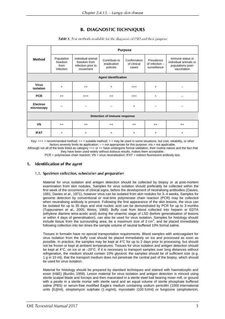
Method
Purpose
Population freedom
from infection
Individual animal freedom from
infection prior to movement
Contribute to eradication
policies
Confirmation of clinical
cases
Prevalence of infection – surveillance
Immune status in individual animals or
populations post-vaccination
Agent identification
Virus isolation
+ ++ + +++ + –
PCR ++ +++ ++ +++ + –
Electron microscopy
– – – + – –
Detection of immune response
VN ++ ++ ++ ++ ++ ++
IFAT + + + + + +
Key: +++ = recommended method; ++ = suitable method; + = may be used in some situations, but cost, reliability, or other factors severely limits its application; – = not appropriate for this purpose; n/a = not applicable.
Although not all of the tests listed as category +++ or ++ have undergone formal validation, their routine nature and the fact that they have been used widely without dubious results, makes them acceptable.
PCR = polymerase chain reaction; VN = virus neutralisation; IFAT = indirect fluorescent antibody test.
Material for virus isolation and antigen detection should be collected by biopsy or at post-mortem examination from skin nodules. Samples for virus isolation should preferably be collected within the first week of the occurrence of clinical signs, before the development of neutralising antibodies (Davies, 1991; Davies et al., 1971), however virus can be isolated from skin nodules for 3–4 weeks. Samples for genome detection by conventional or real-time polymerase chain reaction (PCR) may be collected when neutralising antibody is present. Following the first appearance of the skin lesions, the virus can be isolated for up to 35 days and viral nucleic acid can be demonstrated by PCR for up to 3 months (Tuppurainen et al., 2005; Weiss, 1968). Buffy coat from blood collected into heparin or EDTA (ethylene diamine tetra-acetic acid) during the viraemic stage of LSD (before generalisation of lesions or within 4 days of generalisation), can also be used for virus isolation. Samples for histology should include tissue from the surrounding area, be a maximum size of 2 cm
3, and be placed immediately
following collection into ten times the sample volume of neutral buffered 10% formal saline.
Tissues in formalin have no special transportation requirements. Blood samples with anticoagulant for virus isolation from the buffy coat should be placed immediately on ice and processed as soon as possible. In practice, the samples may be kept at 4°C for up to 2 days prior to processing, but should not be frozen or kept at ambient temperatures. Tissues for virus isolation and antigen detection should be kept at 4°C, on ice or at –20°C. If it is necessary to transport samples over long distances without refrigeration, the medium should contain 10% glycerol; the samples should be of sufficient size (e.g. 1 g in 10 ml), that the transport medium does not penetrate the central part of the biopsy, which should be used for virus isolation.
Material for histology should be prepared by standard techniques and stained with haematoxylin and eosin (H&E) (Burdin, 1959). Lesion material for virus isolation and antigen detection is minced using sterile scalpel blade and forceps and then macerated in a sterile steel ball bearing mixer mill, or ground with a pestle in a sterile mortar with sterile sand and an equal volume of sterile phosphate buffered saline (PBS) or serum-free modified Eagle‟s medium containing sodium penicillin (1000 international units [IU]/ml), streptomycin sulphate (1 mg/ml), mycostatin (100 IU/ml) or fungizone (amphotericin,
2.5 µg/ml) and neomycin (200 IU/ml). The suspension is freeze–thawed three times and then partially clarified by centrifugation using a bench centrifuge at 600 g for 10 minutes. In cases where bacterial contamination of the sample is expected (such as when virus is isolated from skin samples), the supernatant can be filtered through a 0.45 µm pore size filter after the centrifugation step. Buffy coats may be prepared from unclotted blood by centrifugation at 600 g for 15 minutes, and the buffy coat carefully removed into 5 ml of cold double-distilled water using a sterile Pasteur pipette. After 30 seconds, 5 ml of cold double-strength growth medium is added and mixed. The mixture is centrifuged at 600 g for 15 minutes, the supernatant is discarded and the cell pellet is suspended in 5 ml growth medium, such as Glasgow‟s modified Eagle‟s medium (GMEM). After centrifugation at 600 g for a further 15 minutes, the resulting pellet is suspended in 5 ml of fresh GMEM. Alternatively, the buffy coat may be separated from a heparinised sample by using a Ficoll gradient.
LSDV will grow in tissue culture of bovine, ovine or caprine origin, although primary or secondary culture of bovine dermis cells or lamb testis (LT) cells are considered to be the most susceptible.
One ml of clarified supernatant or buffy coat, is inoculated on to a confluent monolayer in a 25 cm2 culture flask at 37°C and allowed to adsorb for 1 hour. The culture is then washed with warm PBS and covered with 10 ml of a suitable medium, such as GMEM, containing antibiotics and 2% fetal calf serum. If available, tissue culture tubes containing LT cells and a flying cover-slip, or tissue culture microscope slides, are also infected.
The flasks are examined daily for 7–14 days for evidence of cytopathic effects (CPE). Infected cells develop a characteristic CPE consisting of retraction of the cell membrane from surrounding cells, and eventually rounding of cells and margination of the nuclear chromatin. At first only small areas of CPE can be seen, sometimes as soon as 2 days after infection; over the following 4–6 days these expand to involve the whole cell sheet. If no CPE is apparent by day 14, the culture should be freeze–thawed three times, and clarified supernatant inoculated on to fresh LT culture. At the first sign of CPE in the flasks, or earlier if a number of infected cover-slips are being used, a cover-slip should be removed, fixed in acetone and stained using H&E. Eosinophilic intracytoplasmic inclusion bodies, which are variable in size but up to half the size of the nucleus and surrounded by a clear halo, are diagnostic for poxvirus infection. PCR may be used as an alternative to H&E for confirmation of the diagnosis. The CPE can be prevented or delayed by adding specific anti-LSDV serum to the medium. In contrast, the herpesvirus that causes pseudo-LSD produces a Cowdry type A intranuclear inclusion body. It also forms syncytia, which are not usually a feature of capripoxvirus infection (although they may be seen in Madin–Darby bovine kidney [MDBK] cells).
Strains of capripoxvirus that cause LSD have been adapted to grow on the chorioallantoic membrane of embryonated chicken eggs and African green monkey kidney (Vero) cells. This is not recommended for primary isolation. Ovine testis secondary cell line (OA3.Ts) has been evaluated for the propagation of capripoxvirus isolates (Babiuk et al., 2007).
The characteristic poxvirus virion can be visualised using a negative staining preparation technique followed by examination with an electron microscope. There are many different negative staining protocols, an example is given below.
Before centrifugation, material from the original biopsy suspension is prepared for examination under the transmission electron microscope by floating a 400-mesh hexagonal electron microscope grid, with pioloform-carbon substrate activated by glow discharge in pentylamine vapour, on to a drop of the suspension placed on parafilm or a wax plate. After 1 minute, the grid is transferred to a drop of Tris/EDTA buffer, pH 7.8, for 20 seconds and then to a drop of 1% phosphotungstic acid, pH 7.2, for 10 seconds. The grid is drained using filter paper, air-dried and placed in the electron microscope. The capripox virion is brick shaped, covered in short tubular elements and measures approximately 290 × 270 nm. A host-cell-derived membrane may surround some of the virions, and as many as possible should be examined to confirm their appearance (Kitching & Smale, 1986).
The virions of capripoxvirus are indistinguishable from those of orthopoxvirus, but, apart from vaccinia virus and cowpox virus, which are both uncommon in cattle and do not cause generalised infection, no other orthopoxvirus causes lesions in cattle. However, vaccinia virus may cause generalised infection in young immunocompromised calves. In contrast, orthopoxviruses are a common cause of skin disease in domestic buffalo causing buffalo pox, a disease that usually manifests as pock lesions on the teats, but may cause skin lesions at other sites, such as the perineum, the medial aspects of the thighs and the head. Orthopoxviruses that cause buffalo pox cannot be readily distinguished from

capripoxvirus by electron microscopy. The virions of parapoxvirus that cause bovine papular stomatitis and pseudocowpox are smaller, oval in shape and each is covered in a single continuous tubular element that appears as striations over the virion. The capripoxvirus is also distinct from the herpesvirus that causes pseudo-LSD (Allerton – herpes mammillitis).
Capripoxvirus antigen can be identified on the infected cover-slips or tissue culture slides using fluorescent antibody tests. Cover-slips or slides should be washed and air-dried and fixed in cold acetone for 10 minutes. The indirect test using immune cattle sera is subject to high background colour and nonspecific reactions. However, a direct conjugate can be prepared from sera from convalescent cattle (or from sheep or goats convalescing from capripox) or from rabbits hyperimmunised with purified capripoxvirus. Uninfected tissue culture should be included as a negative control as cross-reactions can cause problems due to antibodies to cellular components.
Immunohistochemistry using F80G5 monoclonal antibody specific for capripoxvirus ORF 057 has been described for detection of LSDV antigen in the skin of experimentally infected cattle (Babiuk et al., 2008).
The conventional gel-based PCR method described below is a simple, fast and sensitive method for the detection of capripoxvirus genome in EDTA blood, semen or tissue culture samples. More recently, quantitative real-time PCR methods have been described that are reported to be faster and to have higher sensitivity (Balinsky et al., 2008; Bowden et al., 2008). A real-time PCR method which differentiates between LSDV, sheep pox virus and goat pox virus has been published (Lamien et al.,
2011).
An example of a published conventional gel-based PCR method is described below (Tuppurainen et al., 2005).
The extraction method described below can be replaced using commercially available DNA extraction kits.
i) Freeze and thaw 200 µl of blood in EDTA, semen or tissue culture supernatant and suspend in 100 µl of lysis buffer containing 5 M guanidine thiocyanate, 50 mM potassium chloride, 10 mM Tris/HCl (pH 8); and 0.5 ml Tween 20.
ii) Cut skin and other tissue samples into fine pieces using sterile scalpel blade and forceps. Grind with a pestle in a mortar. Suspend the tissue samples in 800 µl of the above mentioned lysis buffer.
iii) Add 2 µl of proteinase K (20 mg/ml) to blood samples and 10 µl of proteinase K to tissue samples. Incubate at 56°C for 2 hours or overnight, followed by heating at 100°C for 10 minutes. Add phenol:chloroform:isoamylalcohol (25:24:1 [v/v]) to the samples in 1:1 ratio. Vortex and incubate at room temperature for 10 minutes. Centrifuge the samples at 16,060 g for 15 minutes at 4°C. Carefully collect the upper, aqueous phase (up to 200 µl)
and transfer into a clean 2.0 ml tube. Add two volumes of ice cold ethanol (100%) and 1/10 of 3 M sodium acetate (pH 5.3). Place the samples at –20°C for 1 hour. Centrifuge again at 16,060 g for 15 minutes at 4°C and discard the supernatant. Wash the pellets with ice cold 70% ethanol (100 µl) and centrifuge at 16,060 g for 1 minute at 4°C. Discard the supernatant and dry the pellets thoroughly. Suspend the pellets in 30 µl of nuclease-free water and store immediately at –20°C (Tuppurainen et al., 2005).
iv) The primers were developed from the gene encoding the viral attachment protein. The size of the expected amplicon is 192 bp (Ireland & Binepal, 1998). The primers have the following gene sequences:
Forward primer 5‟-TCC-GAG-CTC-TTT-CCT-GAT-TTT-TCT-TAC-TAT-3‟
Reverse primer 5‟-TAT-GGT-ACC-TAA-ATT-ATA-TAC-GTA-AAT-AAC-3‟.
v) DNA amplification is carried out in a final volume of 50 µl containing: 5 µl of 10 × PCR buffer, 1.5 µl of MgCl2 (50 mM), 1 µl of dNTP (10 mM), 1 µl of forward primer, 1 µl of
reverse primer, 1 µl of DNA template (~10 ng), 0.5 µl of Taq DNA polymerase and 39 µl of nuclease-free water. The volume of DNA template required may vary and the volume of nuclease-free water must be adjusted to the final volume of 50 µl.
vi) Run the samples in a thermal cycler as follows: 2 minutes at 95°C; then 45 seconds at 95°C, 50 seconds at 50°C and 1 minute at 72°C (34 cycles); 2 minutes at 72°C and hold at 4°C until analysis.
vii) Mix 10 µl of each sample with loading dye and load onto a 1.5% agarose gel in TAE buffer (Tris/acetate buffer containing EDTA). Load a parallel lane with a 100 bp DNA-marker ladder. Electrophoretically separate the products approximately 8–10 V/cm for 40–60 minutes and visualise with a suitable DNA stain.
Molecular tests using loop-mediated isothermal amplification to detect capripoxvirus genomes are reported to provide sensitivity and specificity similar to real-time PCR with a simpler method and lower cost (Das et al., 2012; Murray et al., 2013). Field validation of the Das et al. method was reported by Omoga et al. (2016).
All the viruses in the genus Capripoxvirus share a common major antigen for neutralising antibodies and it is thus not possible to distinguish strains of capripoxvirus from cattle, sheep or goats using serological techniques.
A test serum can either be titrated against a constant titre of capripoxvirus (100 TCID50 [50% tissue
culture infective dose]) or a standard virus strain can be titrated against a constant dilution of test serum in order to calculate a neutralisation index. Because of the variable sensitivity of tissue culture to capripoxvirus, and the consequent difficulty of ensuring the use of 100 TCID50, the neutralisation index
is the preferred method although it does require a larger volume of test sera. The test is described using 96-well flat-bottomed tissue-culture grade microtitre plates, but it can be performed equally well in tissue culture tubes with the appropriate changes to the volumes used, although it is more difficult to read an end-point in tubes. The use of Vero cells in the virus neutralisation test has been reported to give more consistent results.
i) Test sera, including a negative and a positive control, are diluted 1/5 in Eagle‟s/HEPES (N-2-hydroxyethylpiperazine, N-2-ethanesulphonic acid) and inactivated at 56°C for 30 minutes.
ii) Next, 50 µl of the first inactivated serum is added to columns 1 and 2, rows A to H of the microtitre plate. The second serum is placed in columns 3 and 4, the third in columns 5 and 6, the positive control serum is placed in columns 7 and 8, the negative control serum is placed in columns 9 and 10, and 50 µl of Eagle‟s/HEPES without serum is placed in columns 11 and 12 and to all wells in row H.
iii) A reference strain of capripoxvirus, usually a vaccine strain known to grow well in tissue culture, with a titre of over log10 6 TCID50 per ml is diluted in Eagle‟s/HEPES in bijoux
bottles to give a log dilution series of log10 5.0, 4.0, 3.5, 3.0, 2.5, 2.0, 1.5 TCID50 per ml
(equivalent to log10 3.7, 2.7, 2.2, 1.7, 1.2, 0.7, 0.2 TCID50 per 50 µl).
iv) Starting with row G and the most diluted virus preparation, 50 µl of virus is added to each well in that row. This is repeated with each virus dilution, the highest titre virus dilution being placed in row A.
v) The plates are covered and incubated for 1 hour at 37°C.
vi) LT cells are prepared from pregrown monolayers as a suspension of 105 cells/ml in Eagle‟s medium containing antibiotics and 2% fetal calf serum. Following incubation of the microtitre plates, 100 µl of cell suspension is added to all the wells, except wells H11 and
H12, which serve as control wells for the medium. The remaining wells of row H are cell and serum controls.
vii) The microtitre plates are covered and incubated at 37°C for 9 days.
viii) Using an inverted microscope, the monolayers are examined daily from day 4 for evidence of CPE. There should be no CPE in the cells of row H. Using the 0240 KSGP vaccine strain of capripoxvirus, the final reading is taken on day 9, and the titre of virus in each duplicate titration is calculated by the Kärber method. If left longer, there is invariably a „breakthrough‟ of virus in which virus that was at first neutralised appears to disassociate from the antibody.
ix) Interpretation of the results: The neutralisation index is the log titre difference between the titre of the virus in the negative serum and in the test serum. An index of ≥1.5 is positive. The test can be made more sensitive if serum from the same animal is examined before and after infection. Because the immunity to capripoxviruses is predominantly cell mediated, a negative result, particularly following vaccination, after which the antibody response may be low, does not imply that the animal from which the serum was taken is not protected.
A constant-virus/varying-serum method has been described using serum dilutions in the range 1/5 to 1/500 and fetal calf muscle cells. Because these cells have a lower sensitivity to capripoxvirus than LT cells, the problem of virus „breakthrough‟ is overcome.
Antibodies to capripoxvirus can be detected from day 2 after the onset of clinical signs. These remain detectable for about 7 months, but a significant rise in titre is usually seen between days 21 and 42.
The AGID test cannot be recommended as a serological test for the diagnosis of LSD because of the cross-reaction with antibodies to bovine papular stomatitis and pseudocowpox virus. A consequence of this cross-reaction is false-positive results. Lack of sensitivity of the test can also lead to false-negative results.
Capripoxvirus-infected tissue culture grown on cover-slips or tissue culture microscope slides can be used for the indirect fluorescent antibody test. Uninfected tissue culture control, and positive and negative control sera, should be included in the test. The infected and control cultures are fixed in acetone at –20°C for 10 minutes and stored at 4°C. Dilutions of test sera are made in PBS, starting at 1/20 or 1/40, and positive samples are identified using an anti-bovine gamma-globulin conjugated with fluorescein isothiocyanate. Antibody titres may exceed 1/1000 after infection. Sera may be screened at 1/50 and 1/500. Cross-reactions can occur with orf (contagious pustular dermatitis virus of sheep), bovine papular stomatitis and perhaps other poxviruses.
Western blotting of test sera against capripoxvirus-infected cell lysate provides a sensitive and specific system for the detection of antibody to capripoxvirus structural proteins, although the test is expensive and difficult to carry out.
Capripoxvirus-infected LT cells should be harvested when 90% CPE is seen, freeze–thawed three times, and the cellular debris pelleted by centrifugation. The supernatant should be decanted, and the proteins should be separated by SDS/PAGE (sodium dodecyl sulphate/polyacrylamide gel electrophoresis). A vertical discontinuous gel system, using a stacking gel made up of acrylamide 5% in Tris (125 mM), pH 6.8, and SDS (0.1%), and a resolving gel made up of acrylamide (10–12.5%) in Tris (560 mM), pH 8.7, and SDS (0.1%), is recommended for use with a glycine running buffer containing Tris (250 mM), glycine (2 M), and SDS (0.1%). Samples of supernatant should be prepared by boiling for 5 minutes with an appropriate lysis buffer prior to loading. Alternatively, purified virus or recombinant antigens may replace tissue-culture-derived antigen.
Molecular weight markers should be run concurrently with the protein samples. The separated proteins in the SDS/PAGE gel should be transferred electrophoretically to a nitrocellulose membrane (NCM). After transfer, the NCM is rinsed thoroughly in PBS and blocked in 3% bovine serum albumin (BSA) in
PBS, or 5% skimmed milk powder in PBS, on a rotating shaker at 4C overnight. The NCM can then be separated into strips by employing a commercial apparatus to allow the concurrent testing of multiple
serum samples, or may be cut into strips and each strip incubated separately thereafter. The NCM is washed thoroughly with five changes of PBS for 5 minutes on a rotating shaker, and then incubated at room temperature on the shaker for 1.5 hours, with the appropriate serum at a dilution of 1/50 in blocking buffer (3% BSA and 0.05% Tween 20 in PBS; or 5% milk powder and 0.05% Tween 20 in PBS). The membrane is again thoroughly washed and incubated (in blocking buffer) with anti-species immunoglobulin horseradish-peroxidase-conjugated immunoglobulins at a dilution determined by titration. After further incubation at room temperature for 1.5 hours, the membrane is washed and a solution of diaminobenzidine tetrahydrochloride (10 mg in 50 ml of 50 mmTris/HCl, pH 7.5, and 20 µl of 30% [v/v] hydrogen peroxide) is added. This is then incubated for approximately 3–7 minutes at room temperature on a shaker with constant observation, and the reaction is stopped by washing in PBS before excessive background colour is seen. A positive and negative control serum should be used on each occasion.
Positive test samples and the positive control will produce a pattern consistent with reaction to proteins of molecular weights 67, 32, 26, 19 and 17 kDa – the major structural proteins of capripoxvirus – whereas negative serum samples will not react with this pattern. Hyperimmune serum prepared against parapoxvirus (bovine papular stomatitis or pseudocowpox virus) will react with some of the capripoxvirus proteins, but not the 32 kDa protein that is specific for capripoxvirus.
Attempts to develop and validate an enzyme-linked immunosorbent assay (ELISA) for the detection of capripoxviral antibodies are in progress but the technique is not currently recommended for use.
Live attenuated strains of capripoxvirus have been used as vaccines specifically for the control of LSD (Brenner et al., 2006; Capstick & Coakley, 1961; Carn, 1993). Capripoxviruses are cross-reactive within the genus. Consequently, it is possible to protect cattle against LSD using strains of capripoxvirus derived from sheep or goats (Coakley & Capstick, 1961). However, it is recommended to carry out controlled trials, using the most susceptible breeds, prior to introducing a vaccine strain not usually used in cattle. The duration of protection provided by LSD vaccination is unknown. Capripoxvirus vaccine strains can produce a large local reaction at the site of inoculation in Bos taurus breeds (Davies, 1991), which some stock owners find unacceptable. This has discouraged the use of vaccine, even though the consequences of an outbreak of LSD are invariably more severe. Risk–benefit of vaccination should be assessed following stakeholder discussion.
General requirements set for the facilities used for the production of vaccines and for the documentation and record keeping throughout the whole manufacturing process are described in Chapter 1.1.8 Principles of veterinary vaccine production. The documentation should include standard operating procedures (SOP) for the method of manufacture and each step for the testing of cells and reagents used in the process, each batch and the final product.
Each seed strain of capripoxvirus used for vaccine production must be accompanied by records clearly and accurately describing its origin, isolation and tissue culture or animal passage history.
A quantity of master seed vaccine virus should be prepared, frozen or desiccated and stored at low temperatures such as –40°C or –80°C in order to provide a consistent working seed for regular vaccine production. The virus should be cultured in primary or secondary LT cells of wool sheep origin for maximum yield. Vero cells may also be used.
Each seed strain must be safe to use in all breeds of cattle for which it is intended, including young and pregnant animals. It must also be non-transmissible, remain attenuated after further tissue culture passage, and provide complete protection against challenge with virulent field strains for a minimum of 1 year. It must produce a minimum clinical reaction in all breeds of cattle when given by the recommended route.
The necessary safety and potency tests are described in Section C.2.2.4 Final product batch tests.
Each master seed must be tested to ensure its identity and shown to be free from adventitious viruses, in particular pestiviruses, such as border disease and bovine viral diarrhoea virus, and free from contamination with bacteria, fungi or mycoplasmas.
The general procedures for sterility or purity tests are described in Chapter 1.1.9 Tests for sterility and freedom from contamination of biological materials intended for veterinary use.
The method of manufacture should be documented as the Outline of Production.
Vaccine batches are produced on fresh monolayers of secondary LT cells. A vial of seed virus is reconstituted with GMEM or other appropriate medium and inoculated onto an LT monolayer that has been previously washed with warm PBS, and allowed to absorb for 15 minutes at 37°C before being overlaid with additional GMEM. Cells should be harvested after 4–6 days when they exhibit 50–70% CPE for maximum viral infectivity, or earlier if CPE is extensive and they appear ready to detach. The culture is freeze–thawed three times, and the suspension is recovered and centrifuged at 600 g for 20 minutes. Before harvest, the culture should be
examined for any evidence of nonspecific CPE, medium cloudiness or change in medium pH. A second passage may be required to produce sufficient virus for a production batch (to produce
enough for 106 doses, the yield from five 175 cm2 flasks is required).
The procedure is repeated and the harvests (consisting of clarified supernatants) from individually numbered flasks are each mixed separately with an equal volume of sterile, chilled 5% lactalbumin hydrolysate and 10% sucrose (dissolved in double-distilled water or appropriate balanced salt solution), and transferred to individually numbered bottles for storage at –20°C. Prior to storage, 0.2 ml is removed from each bottle for sterility control. An additional 0.2 ml is removed; 2 ml pools composed of 0.2 ml samples taken from ten bottles are used for virus titration. A written record of all the procedures must be kept for all vaccine batches.
The specification and source of all ingredients used in the manufacturing procedure should be documented and the freedom of extraneous agents: bacteria, fungi, mycoplasma and viruses should be tested. The detailed testing procedure is described in chapter 1.1.9. The use of antibiotics must meet the requirements of the licensing authority.
i) Cells
Records of the source of the master cell stocks should be maintained. The highest and lowest passage numbers of the cells that can be used for vaccine production must be indicated in the outline of the production. In case primary lamb testis cells are used, cells should be obtained from the testis of a healthy young lamb from a scrapie-free flock of a wool sheep breed. During cultivation, cells must be observed for any evidence of CPE, and for normal morphology (predominantly fibroblastic). They can usually be passaged successfully up to ten times. When used for vaccine production, uninfected control cultures should be grown in parallel and maintained for at least one additional passage for further observation. They should be checked for the presence of noncytopathic strains of pestiviruses such as bovine viral diarrhoea or border disease viruses by immunofluorescence or immunoperoxidase or by other appropriate techniques. If possible, master cell stocks should be prepared and screened prior to vaccine production, and a
stock stored in sterile DMSO (dimethyl sulphoxide) in liquid nitrogen (1–2 ml aliquots
containing 20×106 cells/ml).
ii) Serum
Bovine serum used in the growth or maintenance medium must be tested free from antibody to capripoxvirus and contamination with pestivirus or any other viruses, extraneous bacteria, mycoplasma or fungi.
iii) Medium
must be tested free from antibody to capripoxvirus and contamination with pestivirus or any other extraneous viruses, bacteria, mycoplasma or fungi.
iv) Virus
Seed virus and final vaccine must be titrated in tissue culture tubes or microtitre plates. The minimum recommended field dose of the South African Neethling strain vaccines (Mathijs et al., 2016). is log10 3.5 TCID50, although the minimum protective dose is log10
2.0 TCID50. Capripoxvirus is highly susceptible to inactivation by sunlight, and allowance
should be made for loss of activity in the field. The recommended field dose of the Romanian sheep pox vaccine for cattle is log10 2.5 sheep infective doses (SID50), and the
recommended dose for cattle of the RM65-adapted strain of Romanian sheep pox vaccine is log10 3 TCID50 (Coakley & Capstick, 1961). Vaccine samples must be examined for the
presence of adventitious viruses including cytopathic and noncytopathic strains of pestivirus, and should be mixed with a high-titre capripoxvirus-immune serum that has previously tested negative for antibodies to pestiviruses, to prevent the vaccine virus itself from interfering with the test. The vaccine can be held at –20°C until all sterility tests and titrations have been completed, at which time it should be freeze-dried. A further titration is carried out on five randomly chosen vials of the freeze-dried preparation to confirm the titre.
i) Sterility/purity
Tests for sterility and freedom from contamination of biological materials intended for veterinary use may be found in chapter 1.1.9.
ii) Safety and efficacy
The efficacy and safety studies should be demonstrated by statistically valid vaccination–challenge studies using seronegative young LSDV susceptible dairy cattle breeds. The group numbers recommended here can be varied if statistically justified. Fifteen cattle are placed in a high containment level large animal unit and serum samples are collected. Five randomly chosen vials of the freeze-dried vaccine are reconstituted in sterile PBS and pooled. Two cattle are inoculated with 10 times the field dose of the vaccine, and eight cattle are inoculated with the recommended field dose. The remaining five cattle are unvaccinated control animals. The animals are clinically examined daily and rectal temperatures are recorded. On day 21 after vaccination, the six animals are again serum sampled and challenged with a known virulent capripoxvirus strain by intravenous and intradermal inoculation (the challenge virus solution should also be tested free from extraneous viruses). The clinical response is recorded during the following 14 days. Animals in the unvaccinated control group should develop the typical clinical signs of LSD, whereas there should be no local or systemic reaction in the vaccinates other than a raised area in the skin at the site of vaccination, which should disappear after 4 days. Serum samples are again collected on day 30 after vaccination. The day 21 serum samples are examined for seroconversion to selected viral diseases that could have contaminated the vaccine, and the days 0 and 30 samples are compared to confirm the absence of antibody to pestivirus. Because of the variable response in cattle to LSD challenge, generalised disease may not be seen in all of the unvaccinated control animals, although there should be a large local reaction.
iii) Batch potency
Potency tests in cattle must be undertaken for vaccine strains of capripoxvirus if the minimum immunising dose is not known. This is usually carried out by comparing the titre of a virulent challenge virus on the flanks of vaccinated and control animals. Following

vaccination, the flanks of at least three animals and three controls are shaved of hair. Log10 dilutions of the challenge virus are prepared in sterile PBS and six dilutions are
inoculated intradermally (0.1 ml per inoculum) along the length of the flank; four replicates of each dilution are inoculated down the flank. An oedematous swelling will develop at possibly all 24 inoculation sites on the control animals, although preferably there will be little or no reaction at the four sites of the most dilute inocula. The vaccinated animals should develop an initial hypersensitivity reaction at sites of inoculation within 24 hours, which should quickly subside. Small areas of necrosis may develop at the inoculation site of the most concentrated challenge virus. The titre of the challenge virus is calculated for the vaccinated and control animals; a difference in titre >log10 2.5 is taken as evidence of
protection.
i) Target and non-target animal safety
The vaccine must be safe to use in all breeds of cattle for which it is intended, including young and pregnant animals. It must also be non-transmissible and remain attenuated after further tissue culture passage.
Safety tests should be carried out on the final product of each batch as described in Section C.2.2.4.
ii) Reversion-to-virulence for attenuated/live vaccines
The selected final vaccine should not revert to virulence during further passages in target animals.
iii) Environmental consideration
Attenuated vaccine should not be able to perpetuate autonomously in a cattle population. Strains of LSDV are not a hazard to human health.
i) For animal production
The efficacy of the vaccine must be demonstrated in statistically valid vaccination challenge experiments under laboratory conditions. The group numbers recommended here can be varied if statistically justified. Fifteen cattle are placed in a high containment level large animal unit and serum samples are collected. Five randomly chosen vials of the freeze-dried vaccine are reconstituted in sterile PBS and pooled. Two cattle are inoculated with 10 times the field dose of the vaccine, eight cattle are inoculated with the recommended field dose. The remaining five cattle are unvaccinated control animals. The animals are clinically examined daily and rectal temperatures are recorded. On day 21 after vaccination, the six animals are again serum sampled and challenged with a known virulent capripoxvirus strain by intravenous and intradermal inoculation (the challenge virus solution should also be tested and shown to be free from extraneous viruses). The clinical response is recorded during the following 14 days. Animals in the unvaccinated control group should develop the typical clinical signs of LSD, whereas there should be no local or systemic reaction in the vaccinates other than a raised area in the skin at the site of vaccination which should disappear after 4 days. Serum samples are again collected on day 30 after vaccination. The day 21 serum samples are examined for seroconversion to selected viral diseases that could have contaminated the vaccine, and the days 0 and 30 samples are compared to confirm the absence of antibody to pestivirus. Because of the variable response in cattle to challenge with LSDV, generalised disease may not be seen in all of the unvaccinated control animals, although there should be a large local reaction.
Once the potency of the particular strain being used for vaccine production has been determined in terms of minimum dose required to provide immunity, it is not necessary to repeat this on the final product of each batch, provided the titre of virus present has been ascertained.
ii) For control and eradication
Vaccination is the only effective way to control LSD outbreaks in endemic countries. Unfortunately, currently no marker vaccines allowing the differentiation of infected from vaccinated animals are available.
Immunity to virulent field virus following vaccination lasts 2 years with the Kenyan strain and 3 years with the South African vaccine, and protection against generalised infection following intradermal challenge is effectively lifelong. The duration of immunity produced by other vaccine strains should be ascertained in cattle by undertaking controlled trials in an environment in which there is no possibility of field strains of capripoxvirus interfering with the results.
All vaccines are initially given a shelf life of 24 months before expiry. Real-time stability studies are then conducted to confirm the appropriateness of the expiry date. Multiple batches of the vaccine should be re-titrated periodically throughout the shelf-life to determine the vaccine variability.
Properly freeze-dried preparations of LSDV vaccine, particularly those that include a protectant, such as sucrose and lactalbumin hydrolysate, are stable for over 25 years when stored at –20°C and for 2–4 years when stored at 4°C. There is evidence that they are stable at higher temperatures, but no long-term controlled experiments have been reported. No preservatives other than a protectant, such as sucrose and lactalbumin hydrolysate, are required for the freeze-dried preparation.
A new generation of capripox vaccines is being developed that uses the LSDV as a vector for the expression and delivery of immuno-protective proteins of other ruminant pathogens with the potential for providing dual protection (Boshra et al., 2013; Wallace & Viljoen, 2005).
Currently, no recombinant capripox vaccines are commercially available.
Not applicable at present.
BABIUK S., BOWDEN T.R., PARKYN G., DALMAN B., MANNING L., NEUFELD J., EMBURY-HYATT C., COPPS J. & BOYLE D.B. (2008). Quantification of lumpy skin disease virus following experimental infection in cattle. Transbound. Emerg. Dis., 55, 299–307.
BABIUK S., PARKYN G., COPPS J., LARENCE J.E., SABARA M.I., BOWDEN T.R., BOYLE D.B. & KITCHING R.P. (2007). Evaluation of an ovine testis cell line (OA3.Ts) for propagation of capripoxvirus isolates and development of an immunostaining technique for viral plaque visualization. J. Vet. Diagn.Invest., 19, 486–491.
BALINSKY C.A, DELHON G, SMOLIGA G, PRARAT M, FRENCH R.A, GEARY S.J, ROCK D.L & RODRIGUEZ L.L. (2008). Rapid preclinical detection of sheep pox virus by a real-time PCR assay. J. Clin. Microbiol., 46, 438–442.
BOSHRA H., TRUONG T., NFON C., GERDTS V., TIKOO S., BABIUK L.A., KARA P., MATHER A., WALLACE D. & BABIUK S. (2013). Capripoxvirus-vectored vaccines against livestock diseases in Africa. Antiviral Res., 98, 217–227.
BOWDEN, T.R, BABIUK S.L, PARKYN G.R., COPPS J.S. & BOYLE D.B. (2008). Capripox virus tissue tropism and shedding: A quantitative study in experimentally infected sheep and goats. Virology, 371, 380–393.
BRENNER J., HAIMOVITZ M., ORON E., STRAM Y., FRIDGUT O., BUMBAROV V., KUZNETZOVA L., OVED Z., WASERMAN A., GARAZZI S., PERL S., LAHAV D., EDERY N. & YADIN H. (2006). Lumpy skin sease (LSD) in a large dairy herd in Israel. Isr. J. Vet. Med., 61, 73–77.
BURDIN M.L. (1959). The use of histopathological examination of skin material for the diagnosis of lumpy skin disease in Kenya. Bull. Epizoot. Dis. Afr., 7, 27–36.
CAPSTICK P.B. & COAKLEY W. (1961). Protection of cattle against lumpy skin disease. Trials with a vaccine against Neethling type infection. Res. Vet. Sci., 2, 362–368
CARN V.M. (1993). Control of capripoxvirus infections. Vaccine, 11, 1275–1279.
CARN V.M. & KITCHING, R.P. (1995). The clinical response of cattle following infection with lumpy skin disease (Neethling) virus. Arch. Virol., 140, 503–513.
COAKLEY W. & CAPSTICK P.B. (1961). Protection of cattle against lumpy skin disease. Factors affecting small scale production of tissue culture propagated virus vaccine. Res. Vet. Sci., 2, 369–371.
COETZER J.A.W. (2004). Lumpy skin disease. In: Infectious Diseases of Livestock, Second Edition Coetzer J.A.W. & Justin R.C., eds. Oxford University Press, Cape Town, South Africa, 1268–1276.
DAS A., BABIUK S. & MCINTOSH M.T. (2012). Development of a loop-mediated isothermal amplification assay for rapid detection of capripoxviruses. J. Clin. Microbiol., 50, 1613–1620.
DAVIES F.G. (1991). Lumpy Skin Disease, a Capripox Virus Infection of Cattle in Africa. FAO, Rome, Italy.
DAVIES F.G., KRAUSS H., LUND L.J. & TAYLOR M. (1971). The laboratory diagnosis of lumpy skin disease. Res. Vet. Sci., 12, 123–127.
HAIG D. (1957). Lumpy skin disease. Bull. Epizoot. Dis. Afr., 5, 421–430.
IRELAND D.C. & BINEPAL Y.S. (1998). Improved detection of capripoxvirus in biopsy samples by PCR. J. Virol. Methods, 74, 1–7.
IRONS P.C., TUPPURAINEN E.S.M. & VENTER E.H. (2005). Excretion of lumpy skin disease virus in bull semen. Theriogenology, 63, 1290–1297.
KITCHING R.P. & SMALE C. (1986). Comparison of the external dimensions of capripoxvirus isolates. Res. Vet. Sci., 41, 425–427.
LAMIEN C.E., LELENTA M., GOGER W., SILBER R., TUPPURAINEN E., MATIJEVIC M., LUCKINS A.G. & DIALLO A. (2011). Real time PCR method for simultaneous detection, quantitation and differentiation of capripoxviruses J. Virol. Methods, 171, 134–140.
MATHIJS E., VANDENBUSSCHE F., HAEGEMAN A., KING A., NTHANGENI B., POTGIETER C., MAARTENS L., VAN BORM S. &DE CLERCQ K. (2016). Complete Genome Sequences of the Neethling-Like Lumpy Skin Disease Virus Strains Obtained Directly from Three Commercial Live Attenuated Vaccines. Genome Announc., 4 (6). pii: e01255-16.
doi: 10.1128/genomeA.01255-16.
MURRAY L., EDWARDS L., TUPPURAINEN E.S., BACHANEK-BANKOWSKA K., OURA C.A., MIOULET V. & KING D.P. (2013). Detection of capripoxvirus DNA using a novel loop-mediated isothermal amplification assay. BMC Vet. Res., 9,
90.
OIE (World Organisation for Animal Health) (2015). Lumpy Skin Disease. World Animal Health Information Database: http://www.oie.int/wahis_2/public/wahid.php/Wahidhome/Home.
OMOGA D.C.A., MACHARIA M., MAGIRI E., KINYUA J., KASIITI J. & HOLTON T. (2016). Molecular based detection, validation of a LAMP assay and phylogenetic analysis of capripoxvirus in Kenya. J. Adv. Biol. Biotech., 7, 1–12.
PROZESKY L. & BARNARD B.J.H. (1982). A study of the pathology of lumpy skin disease in cattle. Onderstepoort J. Vet. Res., 49, 167–175.
Rouby S. & Aboulsoudb E. (2016). Evidence of intrauterine transmission of lumpy skin disease virus. Vet. J., 209,
193–195.
TUPPURAINEN E.S.M., VENTER E.H. & COETZER J.A.W. (2005). The detection of lumpy skin disease virus in samples of experimentally infected cattle using different diagnostic techniques. Onderstepoort J. Vet. Res., 72, 153–164.
TUPPURAINEN E.S., VENTER E.H., COETZER J.A. & BELL-SAKYI L. (2015). Lumpy skin disease: attempted propagation in tick cell lines and presence of viral DNA in field ticks collected from naturally-infected cattle. Ticks Tick Borne Dis., 6, 134–140.
WALLACE D.B. & VILJOEN G.J. (2005). Immune responses to recombinants of the South African vaccine strain of lumpy skin disease virus generated by using thymidine kinase gene insertion. Vaccine, 23, 3061–3067.
WEISS K.E. (1968). Lumpy skin disease. Virol. Monogr., 3, 111–131.
*
* *
NB: There are OIE Reference Laboratories for Lumpy skin disease
(see Table in Part 4 of this Terrestrial Manual or consult the OIE Web site for the most up-to-date list: http://www.oie.int/en/our-scientific-expertise/reference-laboratories/list-of-laboratories/).
Please contact the OIE Reference Laboratories for any further information on diagnostic tests, reagents and vaccines for lumpy skin disease
NB: FIRST ADOPTED IN 1989. MOST RECENT UPDATES ADOPTED IN 2017