design of primers and amplification of their respective ... · design of primers and amplification...
TRANSCRIPT

UPTEC X 01 046 ISSN 1401-2138OCT 2001
Tomas Risberg
Design of primers andamplification of theirrespective genes throughPCR from a Mus musculuscDNA-library
Master’s degree project

Molecular Biotechnology ProgrammeUppsala University School of Engineering
UPTEC X 01 046 Date of issue 2001-10Author
Tomas Risberg
Title (English)Design of primers and amplification of their respective genes
through PCR from a Mus musculus cDNA-library
Title (Swedish)
AbstractBirth defects are some of the more common deformation seen in humans. Spina bifida relatesto the first trimester of the pregnancy. It is desired to map the genes that are involved in theprocess of raising and closing the neural tube. This project aims to do that. By using themicroarray technique, it is possible to monitor the differences in expression of multiple genessimultaneously. I have located the genetic sequences to a carefully selected number of genesand then designed appropriate primers to these. Each gene was amplified in a PCR, usingthese primers, from a cDNA-library, extracted from day 9 p.c. M. musculus embryos. Theamplified genes are to be used as probes on the microarray that is developing.
Keywords
Spina bifida, exencephaly, embryonic development, microarray, PCR, retinoic acid
SupervisorsProf. Lennart Dencker & Anne-Lee Gustafsson
Division of Toxicology, Uppsala University
ExaminerMatti Nikkola
Program co-ordinator ”Molekylär bioteknik”, Uppsala University
Project name Sponsors
LanguageEnglish
Security
ISSN 1401-2138Classification
Supplementary bibliographical information Pages41
Biology Education Centre Biomedical Center Husargatan 3 UppsalaBox 592 S-75124 Uppsala Tel +46 (0)18 4710000 Fax +46 (0)18 555217

Design of primers and amplification oftheir respective genes through PCRfrom a Mus musculus cDNA-library
Tomas Risberg
Sammanfattning
En av de vanligaste medfödda defekterna är ryggmärgsbråck. Ungefär en på tusen födslardrabbas. Andra exempel på defekter är gomspalt & hjärt- lungdefekter. Det är välkänt atttoxiska substanser såsom alkohol, mediciner, rökning, tungmetaller m.m. kan påverkautvecklingen i livmodern. Man vet dock inte ännu varför. Nästan ingenting är känt om vilkadelar av utvecklingen som påverkas av toxiska ämnen.
Min undersökning ingår som en tidig del i en större studie, vars syfte är att finna just dessadelar som påverkas. Metoden som kommer att användas går ut på att mäta hur mängderna avolika delar varierar när man utsätter embryon (i det här fallet mus-embryon) för en substanssom förhöjer risken för just ryggmärgsbråck.
Det finns i nuläget, en preventiv åtgärd man kan som moder vidtaga för att minska risken förryggmärgsbråck hos barnet, nämligen att mödrarna äter tillskott av folsyra med början föregraviditeten och gör så under åtminstone hela först tredjedelen av denna. Varför folsyra har enskyddande effekt är inte heller det känt för forskare.
Studien syftar till att kunna besvara vissa av dessa frågor. Min del av projektet har gått ut påatt bestämma de delar som ska undersökas och se till att dessa finns tillgängliga så att senaremätningar kan ske.
Examensarbete 20 p i Molekylär bioteknikprogrammetUppsala universitet Oktober 2001
1
1 INTRODUCTION............................................................................................................ 3
2 BACKGROUND .............................................................................................................. 4
2.1 DISEASES AND ENVIRONMENTAL PATHOGENS............................................................. 52.1.1 Diabetes ............................................................................................................. 52.1.2 Epileptic seizures ............................................................................................... 62.1.3 Retinoic acid ...................................................................................................... 72.1.4 Alcohol ............................................................................................................. 10
EMBRYONIC DEVELOPMENT AND MALFORMATIONS .............................................................. 112.1.5 Development of backbone and brain................................................................ 112.1.6 Spina bifida ...................................................................................................... 122.1.7 Exencephaly ..................................................................................................... 132.1.8 Cleft lip and palate........................................................................................... 14
2.2 TECHNIQUES ............................................................................................................. 152.2.1 Normal PCR..................................................................................................... 152.2.2 Reverse transcription PCR (rtPCR)................................................................. 162.2.3 Primer design................................................................................................... 172.2.4 Microarrays ..................................................................................................... 17
3 MATERIALS AND METHODS .................................................................................. 19
3.1 MRNA SEQUENCE SEARCH ....................................................................................... 193.2 PRIMER DESIGN ......................................................................................................... 19
3.2.1 Software ........................................................................................................... 193.2.2 Primer design criteria...................................................................................... 193.2.3 Primer production – oligo synthesis ................................................................ 193.2.4 Primer concentration ....................................................................................... 19
3.3 REVERSE TRANSCRIPTION PCR................................................................................. 193.3.1 DNase I digestion............................................................................................. 193.3.2 Reverse transcription of small amounts of RNA .............................................. 20
3.4 PCR .......................................................................................................................... 203.4.1 Specific primers................................................................................................ 203.4.2 T3 and T7 primers............................................................................................ 203.4.3 PCR conditions ................................................................................................ 203.4.4 Gel .................................................................................................................... 203.4.5 Purification of a DNA-fragment from an agarose gel ..................................... 20
4 RESULTS ....................................................................................................................... 21
4.1 PRIMERS.................................................................................................................... 214.2 PCR .......................................................................................................................... 21
4.2.1 General............................................................................................................. 214.2.2 Photos............................................................................................................... 214.2.3 Listing of the successful probes ....................................................................... 25
5 DISCUSSION ................................................................................................................. 27
ACKNOWLEDGEMENT..................................................................................................... 29
REFERENCES....................................................................................................................... 30
SELECTED READING ........................................................................................................ 33

2
APPENDIX A ......................................................................................................................... 34
APPENDIX B ......................................................................................................................... 39
APPENDIX C ......................................................................................................................... 41
APPENDIX D ......................................................................................................................... 42
3
1 IntroductionBirth defects are a common, global problem. Failure to close the neural tube and palatecleft/lip are among the more usual. The main goal with this project is to map the genes thatare involved in cranial and spinal development and especially those that are responsible forthe defects and malformations. The methods that will be used include PCR, 2d-gels, hybridi-sation to microarrays and computational bioinformatics. To study gene expression involved inthese embryonic events, retinoic acid (RA) will be used to induce neural tube defects (NTD)in the embryos of pregnant mice females. RA is administered prior to the development of thebrain and the spinal chord and the embryos will be collected during these events.The department has been working with issues of embryonic defects for several years. It isthough only recently they started with this microarray project and hence, any real result lies inthe future. The initial part of the project revolves around the selection of genes, the amplifica-tion of these and the attachment of the formed probes to a microarray.My part of the project has been to design primers to the selected genes and to amplify themusing a PCR-machine. The genetic data of the genes were extracted from web-based data-bases. And software was used to calculate the primer sequences.Differences in gene expression between treated and untreated embryos will be measured usingthe technique of microarrays. This technique allows labelled gene fragment to compete for thebinding to a probe on the microarray. In theory, the microarray can host indefinitely manyprobes, but for practical matters the numbers are restricted. There are however commercialmicroarrays that can be purchased, which e.g. contains probes for all the genes of S. cervisiae.Here, though, the design of the microarray is made in the department. This means that it ispossible to customize the microarray with the genes that are wanted.The department of toxicology deals with the initial selection of genes and later on with thereading and interpretation of the scanned microarrays When it comes to the more technicalaspects of the microarray, such as spotting of the probes, hybridisation and scanning of he mi-croarray, Akademiska Sjukhuset in Uppsala will help out.At present, the only advice to women is to take nutritional additives prior to and during apregnancy, as a way to decrease the chances of a malformation in the embryo. The most im-portant additive is folic acid (vitamin B9), but other vitamins are also recommended.A 70% decrease of NTD incidents is reported after folic acid treatment. The rest of thewomen seem to be resistant to this treatment.Since most birth defects arise from the first few weeks of pregnancy, an unplanned child maynot have been noticed yet. The foetus might therefore be exposed in a larger degree to certainchemicals, e.g. alcohol and tobacco.Planning for a child means that the woman starts to take folic acid additives prior to the preg-nancy, and that she completely cease drinking alcohol or smoking cigarettes during the preg-nancy.Projects like this will reveal the genetic pathways involved in the early embryogenesis. Thismight lead to better and more secure pre-pregnancy precautions.

4
2 BackgroundDuring the early pregnancy, an embryo will be exposed to many things that are known to in-terfere with its development. Chemicals, nutritional side-products or even enzymatic deficien-cies of the mother can lead to hap-hazardous birth defects. Serious damages early in the earlyembryogenesis most often result in a spontaneous abortion of the foetus. This is because theteratogenic effect disturbs and influences a greater number of undifferentiated cells, whereasteratogenic exposure restricted to the later part of the first trimester may affect more restrictedareas of development and differentiation parts of the body and not necessarily the whole em-bryo. Damages in restricted areas give rise to birth defects, which can express themselves asappearance challenges, physical or mental handicaps, cardio respiratory problems or enzy-matic disturbances of e.g. nutritional digestion. The primary streak and the neural plate of amouse embryo are formed on day 9 and 9.5 respectively (Bergman et al., 1998). The brainforms earlier, day 8.5 (van Straaten & Copp, 2001) Since these areas are primary targets forthis investigation, treatment with a teratogen (retinoic acid) is made days or hours before theseevents and the embryos are collected on day 9 and day 11.Birth defects occur with an incidence of 5% out of which 2% are detected upon birth1 and therest are discovered later, if ever. About 50% of all foetuses are actually aborted spontane-ously. Out of these about one half of them show embryonic defects. Adding it all up, about30%2 of all pregnancies are affected by some form of defect (Bergman et al., 1998).Rough estimates say that out of these, 20% are inherited effects, 5% are accounted for bychromosomal discrepancies, another 2-3% are caused by infections, bacteria- or virus medi-ated, 1-2% originate from diabetic pregnancies (hyperglycaemia) or cretinism and finallydrugs or other chemical substances (alcohol, tobacco, vitamin deficiencies, medications)cause 2-3%. This leaves two thirds of all incidences without a direct, known cause. A reason-able assumption is that they are due to a combined effect of several of the previously men-tioned causes (Bergman et al., 1998).Timing is important and many genes need to be activated within a short frame of time and invery strictly regulated amounts. Teratogenes can act upon a growing foetus’ well-orchestratedmachinery and disturb the expression of one or more genes. Failure of expression especiallyin growth stimulants and transcriptional factors may cause severe damage.One of the more common birth defect is some form of neural tube defect (NTD). The pheno-type of NTD is an open spine, head and/or neck. Spina bifida3 refers to an opening in the cau-dal zone of the spinal chord. In humans this is the most dominant birth defect. Anencephaly(head and neck defects) embryos most often commit spontaneous abortion.The superciliously most comprehensively studied system, regarding NTDs, is that of mouseSpina bifida. This makes the search for similar research and neighbouring areas easy. Labo-ratory mouse is also easy to breed, easy to feed and has a very short generation time withmultiple offspring in each cycle. It has been estimated that less than 1% off the genes found inmouse lacks a counterpart in the human genome(http://www.ornl.gov/hgmis/faq/compgen.html 2001-07-25). Furthermore, the genetic andmolecular pathways of mice and human resemble each other I most cases, although terato-genes in mouse do not always display the same effect in humans and vice versa (Bergman etal., 1998).
1 These statistics do not include more diffuse defects such as intellectual or motorical retardations. Nor do theyinclude functional disturbances, abnormal sexual development or inborn error of metabolism (Bergman et al.,1998).2 Half of 50% means 25% of total. This plus the 5% of the remaining 50% that are born gives 27.5%.3 Ryggmärgsbråck.
5
Diseases such as diabetes poses a potential risk for the foetus, just as well as teratogenes suchas alcohol does. I will discuss some of the more common diseases and also mention a littleabout some teratogenes. The discussion will be based upon selected information concerningmostly the genes that are involved in my master thesis and also information, which has rele-vance to birth, defects. I will try to give some basic knowledge about the embryonic develop-ment, with the focus on neural tube defects. Finally brief information is provided to cover thetechniques that have been used in this master thesis.
2.1 Diseases and environmental pathogens
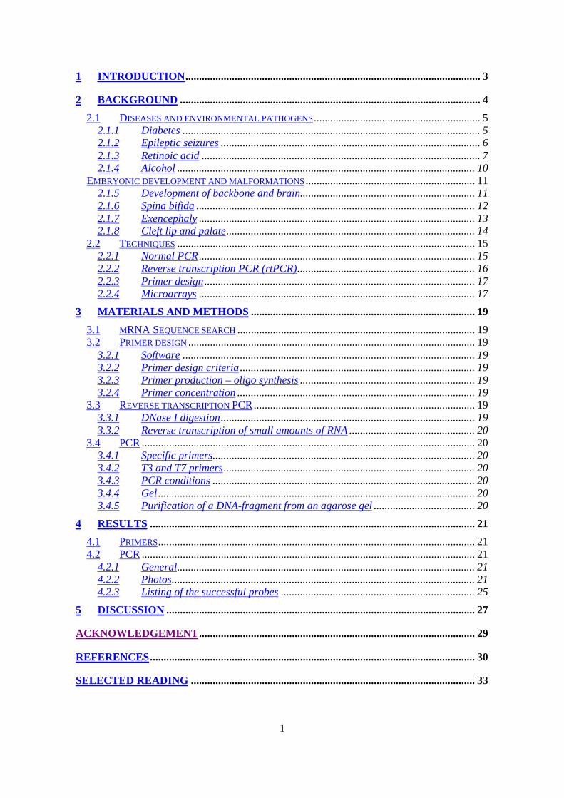
2.1.1 DiabetesCongenital malformations are increased two- to five fold in foetus when the pregnant femaleis exposed to pregestational diabetes (Fine et al., 1999)4.Neural tube defects (NTD) are the most commonly observed defects. The transcriptionalregulator Pax-3 seems to be associated with this embryopathy. Loss of function of Pax-3 leadsto a 100% incidence of NTD in mice. Reduced expression of the gene in neuroepithelial cellsalong the migrating ridge of the dorsal surface of the neural tube commits these cells to un-dergo apoptosis (Fine et al., 1999).
Figure 2.1.1-1 A challenged expression of Pax3causes the migrating ridge cells (yellow circles) toundergo apoptosis. As a consequence they will notmeet and form the necessary bridge to form thehollow tube in which the spinal chord is supposedto grow.(http://www.med.unc.edu/embryo_images/unit-bdyfm/bdyfm_htms/bdyfm024a.htm 2001-07-26).
Experiments have shown that glucose administered to produce a state of hyperglycaemia 24 hbefore the onset of pax-3 expression can inhibit the expression with decreased amounts ofPax-3 as a consequence. The rate of NTD is correlated to the severity of hyperglycaemia(Fine et al., 1999).Hyperglycaemia can cause proteins to become glycosylated. Such an effect could account forthe malfunctions in the chain of gene expressions leading to Pax-3.Acetylsalicylic acid prevents glycosylation of proteins and has been shown to have a positiveeffect on the development of hyperglycaemia-induced embryos5 (Kubow et al., 1993).
4 Suhonen et al., 2000 report a three times increase of foetal malformation rate in mice with type I diabetes mel-litus.

6
Another hypothesis is that glucose mediated malformations is due to the fact that a largenumber of glucose molecules produces a high stress on the membrane transport proteins andon the membrane incorporation of fatty acids and proteins (Fine et al., 1999). This idea issupported by the fact that subcutaneous injections of arachidonic acid (the membrane buildingfatty acid) into pregnant diabetic rats both reduced the NTD defects and also largely de-creased the number of cleft palate incidents (Goldman et al., 1985).An increase in the number of oxygen free radicals is followed in the wake of hyperglycaemia.Free radicals activate transcription of hypoxia-induced factors (HIF), which may cause altera-tions in gene expression elsewhere and thus produce the observed malformations. The factthat treatment with antioxidants such as vitamin E reduces the diabetic pregnancy induced rateof NTDs supports this third hypothesis (Fine et al., 1999).
2.1.2 Epileptic seizuresSeizures are serious problems and they need to be controlled. Several potent medicines exist.Some of the most used include Absensor®, Ergenyl® and Orfiril®. The pharmacologically ac-tive compound in these is valproic acid6 (VPA).The teratogenecity of valproic acid has been well established in NTDs, the effect being con-genital malformations of the heart as well as cleft lip and/or palate(http://www.mentalhealth.com/drug/p30-d02.html 2001-08-05; PMID: 10689198).The mechanism of VPA is not yet known, but there are some hypothesis and some interestinggenes that are currently being investigated.One study with VPA on gene expression, involving some ten genes expressed in the spinalchord-development, that was conducted on two murine strains revealed highly increased lev-els of Tgf-_ and Tgf-_1, -_2 and _3 in the susceptible strain compared to the resistant strain7
(Bennet et al., 2000).Lampen et al., 2001, have been investigating the effect of different peroxisome proliferatoractivating receptors (Ppar) and found that Ppar-_ may be affected by VPA. Pennati et al.,2001 have shown that VPA, at least in Xenopus laevis, disturbs the expression of Pax genes ingeneral and Pax-6 in particular.Two unrelated studies also tests the role of methionine in VPA induced NTDs. In the firststudy by Ehlers et al., 1996, methionine, as well as VPA, is administered to mice embryos anda significant decrease in the rate of NTDs. The other study made by Beck, 2001 investigatesthe role of genomic imprinting and administration of VPA. And it is shown that in females,the genetic imprinting in the treated embryos differs from the expected rate. Since genetic im-printing refers to the methylation of DNA, genes and even whole chromosomes (gene silenc-ing of one of the X-chromosomes in females), the tie with Ehlers et al. 1996 gets clearer andby adding one more hypothesis, the connections will get even more clearer. Folic acid isknown to reduce the rate of NTDs. And it is strongly recommended that pregnant women takefolic acid supplements prior to and during the pregnancy.The initial suggestions, regarding the role of folate, were that a deficiency of folate itself wasthe causative of the NTDs. Further research, though, have pointed more in the direction of fo-lic acid aiding in overcoming a metabolic block.
5 Acetylsalicylic acid is not to be viewed as a drug to be used prophylactically against NTDs, as sialic acids be-long to the group of drugs, which can cause NTD, see Table 4.2.3-1. Acetylsalicylic acid is merely to be seen asa tool in the understanding of the mechanisms behind diabetic related NTDs.6 Champel et al., (1999) concludes that another anticonvulsant substance, carbamazepine (Hermolepsin®, Te-gretol® and Trimonil®) equally well as valproic acid increases the risk of NTDs in exposed foetuses.7 The susceptible and resistant strain refers to different murine strains response to folic acid. In the resistantstrain, folic acid supplements do not decrease the rate of NTDs.
7
Examination of folic acid metabolism reveals several possible candidates and several path-ways. Folic acid is e.g. strongly involved in the synthesis of methionine by donating a methylgroup to homocystein.Methylation is a very important feature of e.g. the CNS metabolism. There is a cycle of meth-ylated and unmethylated molecules that are involved and methionine and homocystein areparts of this cycle (McElhone et al., 1998)8. There is more on this matter described in 2.1.7.
2.1.3 Retinoic acid
C H 2 O H
C H 3 C H 3
C H 3
C H 3CH 3
C O O H
C H 3 C H 3
C H 3
C H 3CH 3
C H 3 C H 3C H 3
C H 3
C H 3
C H O
C H 3 C H 3
C H 3
C H 3CH 3
C H 3 C H 3C H 3
C H 3
C H 3
O H
O H
C H 3
C H 3CH 3
C O O H
C H 3CH 3
Ret
inyl
est
er
(retin
ol +
fatty
aci
d)
Sto
rage
Not
sho
wn
All-
trans
retin
oic
acid
Diff
eren
tiatio
n
Ret
inal
dehy
de
Vis
ion
Ary
lhyd
rore
tinol
Gro
wth
inhi
bitio
n
14-h
ydro
xi re
tro re
tinol
Gro
wth
sup
port
Ret
inol
(Vita
min
A)
9-ci
s-re
tinoi
c ac
id
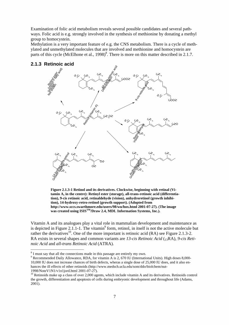
Figure 2.1.3-1 Retinol and its derivatives. Clockwise, beginning with retinal (Vi-tamin A, in the centre): Retinyl ester (storage), all-trans-retinoic acid (differentia-tion), 9-cis retinoic acid, retinaldehyde (vision), anhydroretinol (growth inhibi-tion), 14-hydroxy-retro-retinol (growth support). (Adopted fromhttp://www.sccs.swarthmore.edu/users/98/wu/hox.html 2001-07-27). (The imagewas created using ISISTM/Draw 2.4, MDL Information Systems, Inc.).
Vitamin A and its analogues play a vital role in mammalian development and maintenance asis depicted in Figure 2.1.1-1. The vitamin9 form, retinol, in itself is not the active molecule butrather the derivatives10. One of the more important is retinoic acid (RA) see Figure 2.1.3-2.RA exists in several shapes and common variants are 13-cis Retinoic Acid (13RA), 9-cis Reti-noic Acid and all-trans Retinoic Acid (ATRA). 8 I must say that all the connections made in this passage are entirely my own.9 Recommended Daily Allowance, RDA, for vitamin A is 2, 670 IU (International Units). High doses 8,000-10,000 IU does not increase chances of birth defects, wheras a single dose of 25,000 IU does, and it also en-hances the ill effects of other retinoids (http://www.medsch.ucla.edu/som/ddo/biolchem/nut-1998/NoteV1N1/v1n1jord.html 2001-07-27).10 Retinoids make up a class of over 2,000 agents, which include vitamin A and its derivatives. Retinoids controlthe growth, differentiation and apoptosis of cells during embryonic development and throughout life (Adams,2001).

8
Figure 2.1.3-2 Vitamin A is transformed to RA by specific enzymes. RA isconverted by another enzyme into 4-oxo Retinoic Acid.(http://www.shriver.org/Research/Biomedical/Projects/RetinoicAcid.htm2001-07-25).
RA acts through binding to its receptors - retinoic response elements (RAR) and retinoid “X”receptors (RXR). Most teratogenic studies involving retinoic acid have been performed withATRA, which only binds RAR receptors(http://www.sccs.swarthmore.edu/users/98/wu/hox.html 2001-07-25).2.1.3.1 Disturbances of RA levelsFrom Figure 2.1.3-2 it is clear that proper concentrations of RA requires that in vivo forma-tion and metabolization are well balanced by the respective enzymes. Elevated or loweredconcentrations of RA can be due to factors that disturb the vitamin A-converting enzyme orthe RA converting enzyme as depicted in Figure 2.1.3-3 and Figure 2.1.3-4.Sakai et al., 2001 report that RA degradation by the P450 enzyme CYP26 is required to pro-duce an uneven distribution of RA along the anterior-posterior axis. This would be importantin the patterning of the hindbrain, vertebrae and tailbud.
Figure 2.1.3-3 A block of the Vitamin A con-verting enzyme by e.g. Disulfiram (antabuse)would lead to a decrease in the amounts ofavailable RA.(http://www.shriver.org/Research/Biomedical/Projects/RetinoicAcid.htm 2001-07-25).
Figure 2.1.3-4 A block caused by e.g. ketocona-zole (antifungid) of the RA converting enzymeon the other hand would result in an increase ofRA.(http://www.shriver.org/Research/Biomedical/Projects/RetinoicAcid.htm 2001-07-25).
Another way to disturb the levels of RA would be the administration of the substance itself.RA can be introduced in teratogenic amounts by a number of sources. 13RA is used to treatcystic acne, ATRA to treat leukaemia and etretinat, another form of RA, to treat psoriasis.(http://www.shriver.org/Research/Biomedical/Projects/RetinoicAcid.htm 2001-07-25).

9
Figure 2.1.3-5 depicts some malformation seen in the human head after foetal exposure toteratogenic concentrations of RA (Bergman et al., 1998).
Figure 2.1.3-5 1 Brain damages - especially in thecerebellum. 2. Flattened midface. 3. Small chin. 4. &5. Underdeveloped thymus. Failure to split thecommon vesicular origin to the aorta and the Arteriapulmonalis. 6. Ears placed in a low position and un-derdeveloped. (Bergman et al., 1998).
The teratogenecity of RA became very obvious in the beginning of the 1980´s when a coupleof hundred children in the USA were born with malformations in the skull region(http://www.medsch.ucla.edu/som/ddo/biolchem/nut-1998/NoteV1N1/v1n1jord.html 2001-07-25) due to the fact that their mothers had been treated for diseases requiring RA of someform11.2.1.3.2 RA receptor functions and actionsThe RARs activate or repress the expression of other genes through the RA response elementsand the AP-1 binding site respectively. These events occur in the nucleus and it is thereforelikely that proper levels of available RA in the nucleus is mediated through the cellular reti-noic acid binding proteins (Crabp) I and II, which bind to RA, thus preventing RA to enter thenucleus (http://www.sccs.swarthmore.edu/users/98/wu/hox.html 2001-07-25). By studyingradio labelled cellular retinol binding protein (Crbp) 1 and Crabp-1, both vitamin A and reti-noic acid has been shown to accumulate in spatially close but non-overlapping areas sur-rounding limbs and craniofacial structures (Gustafsson et al., 1993).Disturbances in CRBP, CRABP, RAR or RXR levels may account for the malformations thathave been observed in some foetuses.Especially important and interesting is the activation of hox genes that is associated with RAand the binding of RA to its receptor.Rar-_ has been shown to act upon the hox genes in the early development of the brain, _i.e._is prior to any visual segmentation can be seen in the hindbrain (Gustafsson et al., 1993).The hox genes are a clustered together and they are activated sequentially in an orderly fash-ion, thus dictating the fate of the cells in which they are expressed. The order of the hox genes
11 Just as well as an excess of RA causes malformation, so does a deficiency of vitamin A (Sakai et al., 2001).Vitamin A deficiency syndrome (VAD) is a major public health problem in parts of Africa, Asia, Latin America,and the Western Pacific (PMID: 11432674) and causes a spectrum of malformations.

10
is correlated to the anterior posterior axis. Sonic hedgehog (Shh) is, together with the polar-izing region (ZPA), a key player in the activation of hox genes, but only through close coordi-nation with RA and bone morphogenic protein (Bmp) 2(http://www.sccs.swarthmore.edu/users/98/wu/hox.html 2001-07-25).
2.1.4 Alcohol
Figure 2.1.4-1 Typical characteristics observedin FAS-children. 1. microcephali 2. short eye slit3. flat midface 4. diffuse groove (filtrum) on up-per lip 5. thin upper lip 6. epicanthic fold 7. lowbase of nose 8. minor ear changes 9. short nose10. weakly developed chin (micrognathi). Thefive first are the more characteristic ones. Thelatter five are less common characteristics.(Bergman et al., 1998).
Ethanol has been linked to abnormalities in virtually every system of the body (Lynch et al.,2001). The alterations include abnormalities of the limbs, eyes, brain, head, heart, kidneys,abdominal organs, skeletal and urogenital system. The physiological, mental, morphologicaland behavioural impairments seen are collectively called foetal alcohol syndrome (FAS) fig-ure 2.5.1 a.Ethanol may cause alterations in the expression of the insulin-like growth factor (Igf-1) and/orthe insulin-like growth factor binding proteins (IGFBP), which are necessary for IGF bindingavailability to the IGF type-1 receptor.Virtually every biological action of IGFs is believed to be mediated through the IGF type-1receptor. Ethanol has been shown to increase Igfbp-1, which impairs the physiological activ-ity of Igf-1 and inhibit the autophosphorylation of IGF type-1 receptor. This has a negative ef-fect on IGF action and prevents downstream signalling (Lynch et al., 2001).Zachman and Grummer investigates the possibility that ethanol interacts with vitamin A or itsmetabolites and thus FAS, which display similar phenotypes as RA exposed embryos, wouldbe caused only indirectly by ethanol. The synthesis of RA from retinol is catalysed by alcoholdehydrogenase. An excess of ethanol may inhibit the synthesis of RA by competitive inhibi-tion of the enzyme.

11
Embryonic development and malformations
2.1.5 Development of backbone and brainThe spinal chord can be divided into four elevation zones, A-D Figure 2.1.5-1. Beginningwhere indicated by arrows the neural cells elevates and starts to fuse like a zipper in both di-rections until they reach the endpoint of another elevation zone (Juriloff & Harris, 2000).
Figure 2.1.5-1 Elevation zones A-D and their respec-tive sites of initiation for neural fold and fusion12.(Juriloff & Harris, 2000).
The elevation itself poses a difficult task, since it means bringing the cells from a convex po-sition into an elevated concave position and then fold inwards to meet and fuse with the op-posing side Figure 2.1.5-2.
Figure 2.1.5-2 (Juriloff & Harris, 2000).
12 Rachischisis is the medical term of a failure to close the spinal chord, whereas Spina bifida refers to the caudalzone of the spinal chord.

12
Figure 2.1.5-3 Mouse embryo, 8 daysold. The neural folds fuse in a zipperlike fashion. The hollow tube that sub-sequently forms will host the brain andthe spinal chord. The image has beencomputationally manipulated from itsoriginal appearance.(http://www.med.unc.edu/embryo_images/unit-nervous/nerv_htms/nerv001.htm.2001-07-28).
In mice, the critical time for cranial neuralation is day 8.5 while the low neural folds pass itscritical stages on day 9.5 (van Straaten & Copp, 2001)13.A more complete list of the major events of the organogenesis can be found in Appendix C.
2.1.6 Spina bifidaSpina bifida14 aperta (‘open spine’) is the most common birth defect in the United States. Aneven greater number of people have Spina bifida occulta, which is a dorsal gap in the verte-bral arches over an intact neural tube and is believed to be genetically and developmentallyunrelated to the more severe defects exencephaly and Spina bifida aperta, (Harris & Juriloff,1999). Spina bifida occulta is difficult to detect and can be unnoticed for several years.People suffering with Spina bifida aperta may lack the ability to control various parts of thebody since the open spine means that the nervous system fails to develop from the openingand caudally towards the base of the spine(http://www.dreamwater.org/sbcommunity/main.html 2001-08-06) and hence the brain mightnot be able to communicate with e.g. legs, kidneys or bladder.Almost all exencephalies and rachischisis of genetic origin are caused by failure of neural foldelevation (Harris & Juriloff, 1999).The mutant mouse strain curly tail (ct) is heavily investigated in matters concerning NTDs.The incidence rate is considerably higher in this mutant compared to wildtype (van Straaten &
13 The human equivalence of this would be that the neural cells fold and fuse on day 19(http://www.medsch.ucla.edu/som/ddo/biolchem/nut-1998/NoteV1N1/v1n1jord.html 2001-07-26) and a com-plete closure of the spinal chord is observed on day 28 p.c. (http://www.wcox.com.au/glossary.htm#ntd 2001-07-26).14 Ryggmärgsbråck.
Brainregion
Spinalchordregion
13
Copp, 2001). The high number of Spina bifida in ct appears to originate from a retardation inthe growth of the neural cells compared to that of the hindgut, forming a bent tail (Figure2.1.1-1) and also making the fusion of the caudal elevation zone impossible, rather than fail-ure to elevate (van Straaten & Copp, 2001). Interestingly RA, an otherwise known teratogenand inducer of NTDs, has been observed to decrease the incidence of Spina bifida aperta butnot of exencephaly in ct. The cause for this decrease is probably that RA stimulates the pro-duction of Rar-_, which in ct is kept on a lower level compared to wildtype. Rar- _ might aidin pacing up the growth of the neural cells (van Straaten & Copp, 2001).Nutritional supplements have, in numerous experiments, been shown to eliminate the risk forNTDs almost completely. The most potent supplement seems to be folic acid. Folic acid (vi-tamin B9) reduces the NTD incidence in both humans and mice by more than 70%.NTDs in ct mice differ from the ordinary incidences of NTDs observed in wildtype mice inthe respect that they are resistant to folic acid treatment (van Straaten and Copp, 2001)15.Myo-inositol has recently been shown to reduce the risk for NTDs16 both in ct and wildtype.Hopefully, adding inositol to the folic acid supplement will completely reduce the risk ofSpina bifida in humans (Greene & Copp, 1997).
2.1.7 ExencephalyExencephaly17 is a more severe form of neural tube defect. The neural cells in the elevationzone B or C fail to fuse leaving an opening in the skull, and thus the brain forms on the out-side
15 This would of course be consistent with the two different models that have been proposed. Spina bifida in ctoriginates in an un-natural bending of the tail that causes an opening regardless of any metabolic or other geneticdefect or disturbance. In wildtype strains of mouse on the other hand, the NTD originates from some malfunctionin gene expression or some metabolic or nutritional error.16 16 A 70% reduction of Spina bifida was observed after a single injection of myo-inositol on day 9.5 in ct(Greene & Copp, 1997).17 The human counterpart of exencephaly is anencephaly.

14
Figure 2.1.7-1 A mouse embryodisplaying exencephaly, a caudalopening in the spine and curly tail.(van Straaten and Copp, 2001).
Many mutants have been isolated that show exencephalic phenotypes. Some of these relate tothe genes in our study and they include arnt, cart1, cbp, dlx5, rar-_/_ and twist (Juriloff &Harris, 2000).An interesting feature of exencephaly is the observation that female embryos are more proneto the defect18 (Juriloff & Harris, 2000). In trying to connect this phenomenon with the folateand methionine pathways - Juriloff and Harris say that the fact that in females, one of the Xchromosomes of every cell is blocked from expression by heavy methylation and they thinkthat it is more than coincidental. They continue to state that, in rapid growing cells the needfor methylation of the chromosome may cause deprivation in other methylation chains. Thisshortage of methyl groups would cause the observed NTDs and thus both connecting to folicacid as an NTD preventing agent and to the higher incidence rate observed in females.
2.1.8 Cleft lip and palateCleft lip and/or cleft palate19 are the most common20 congenital deformity of head and neck(Kirschner & LaRossa, 2000). The malformation may have its origin in genetic differences,although a more frequent cause is drugs. Alcohol, Dilantin (a seizure medication) and excessof vitamin A are known to induce palate cleft. Palate cleft originates so early in the embryo-genesis that the woman has not even noticed the pregnancy yet. The best way to avoid thesebirth defects seems to be to avoid unplanned pregnancies(http://www.widesmiles.org/cleftlinks/WS-368.html 2001-08-03).
18 Compare with 2.1.2.19 Harmynthet resp gomspalt.20 The asian population has the highest incident rate, 1 in 500, and the african population has the lowest, 1 in1000 (http://www.widesmiles.org/cleftlinks/WS-104.html 2001-08-03)
15
2.2 Techniques
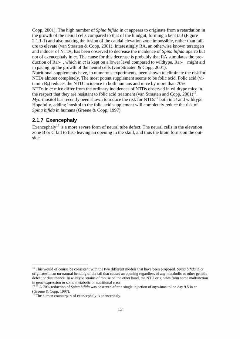
2.2.1 Normal PCRThe PCR (polymerase chain reaction)-method was developed in the 1980´s by Nobel laureateKary Mullis. It has ever since been one of the major tools in genetic analysis and has contin-ued to generate new areas of usage. Kary Mullis invention has since undergone several im-provements in efficiency. And various adaptations have been made to his general idea(Reverse transcription PCR (rtPCR)). The basic principle of PCR can be summarized as inFigure 2.2.1-1.
Figure 2.2.1-1 The 3 basic steps of PCR. The first step is called thedenaturation step, in which separation of the double stranded DNAoccurs. In the second step, the annealing step, primers anchor them-selves to the recently formed single stranded DNA-molecules. Thetemperature in this step is the most variable one of all steps. It is de-sired to have a temperature as close as possible to the melting tem-perature of the amplimers in order to get the most highly specificprimer hybridisation. If the melting point is close to the primer ex-tension temperature, this step may even be omitted. Finally theprimer extension step, in which the PCR machine adjusts the tem-perature working temperature of the polymerase. Most often thismeans 72°C, which corresponds to the optimal performance tem-perature of the DNA- polymerase from Thermus aquaticus (Walker& Rapley, 1997). (Walker & Rapley, 1997).
A normal PCR is run between 30-40 cycles and in each cycle the amount of DNA is doubledso that the final outcome is a billion fold amplification (Figure 2.2.1-2) of the original DNA.With the improvements made, a single DNA-molecule can be detected and amplified (Albertset al., 1994).

16
Figure 2.2.1-2 With each-cycle in the PCR, a doubling of thenumber of copies is achieved, yielding some 109 copies after30 cycles in the PCR. There are however limitations to thistheoretical analysis, e.g. polymerase- accessibility and per-formance, availability of building blocks. The size of thefragment also poses a limit to the performance, due to kinet-ics of the polymerase and it should not exceed a few thou-sand base pairs (Walker & Rapley, 1997).
2.2.2 Reverse transcription PCR (rtPCR)Reverse transcript PCR is a modification of the original PCR. Prior the PCR step, an extractof mRNA is mixed with a reverse transcriptase (a polymerase found in retroviruses) togetherwith other necessary components (dNTPs, a proper buffer and primers). The primers are de-signed differently and given different features depending on the purpose of the rtPCR (Figure2.2.2-1) (Walker & Rapley, 1997).
Figure 2.2.2-1 If it is desired to get an as complete cDNA library aspossible; the two leftmost techniques are to be used. The middletechnique has the advantage of capturing all mRNA, the leftmost thatit only captures polyadenylated mRNAs. The third technique is ap-plied when one and only one mRNA should be amplified (Walker &Rapley, 1997). (Adopted from Walker & Rapley, 1997).
rtPCR performed this way on an mRNA preparation creates a cDNA library. Following thecreation of this library it will be possible to use specific primers in a normal PCR to amplify aparticular gene of interest (Normal PCR). The advantage of rtPCR compared to amplifying the

17
whole genome is very obvious; the only products that can be amplified are the genes that havebeen expressed within the system21.
2.2.3 Primer designDesigning primers means finding a pair of templates for a known DNA-sequence that can actas the starting point for Taq-polymerase in the PCR-machine. Completely complementaryprimers offer a perfect match with target DNA, at least when the annealing temperature isfairly optimal (Walker & Rapley, 1997).
2.2.4 MicroarraysMicroarrays offer a first great hope for a “global view” of biological processes i.e. a simulta-neous readout of all components (Lander, 1999). The basic principle of the microarray tech-nique can be summarized as in Figure 2.2.4-1.The probes are generated by collecting all genomic DNA in the cells and then amplifyingeach gene that are supposed to be spotted on the microarray through standard PCR usinghighly specific primer pairs and thus generating a pool of each gene (Duggan et al., 1999).The probes are, as depicted in the lower left of Figure 2.2.4-1, within precisely defined bor-ders, robotically spotted onto the microarray to keep the different probes apart from eachother and to make later analysis possible (Duggan et al., 1999).Extracting mRNA from untreated/wildtype mouse foetuses produces a reference sample. Thesample is reverse transcribed and labelled with a dye. A similar procedure is performed ontreated/mutant foetuses, only with another colour of the dye. The two labelled cDNA samplesare then simultaneously allowed to hybridise with the probes on the microarray (Duggan etal., 1999).A laser is used to bring the dyes to fluoresce so that a scanner can read the intensities of theemitted light from each of the spots on the microarray. The result can be read as: no differ-ence in expression or as an over- or under-expression of each of the monitored genes (Dugganet al., 1999).
21 Using the whole genome as a target for gene amplification, although using highly specific primers, would notbe possible in a microarray experiment. The very idea of the microarray design is that it is supposed to measurevariations in the genetic expression between a reference group and the test group.

18
Figure 2.2.4-1 Lower left: A robot makes a spot of each andevery probe. Top left and middle: Reference and test sam-ples are prepared, reverse transcribed and labelled. Lowermiddle: Hybridisation between samples and probes. Lowerright and up: A scanner reads the emitted intensities; a com-puter calculates the values and presents the result as an im-age (top right). The image has been computationally modi-fied from its original appearance.(http://www.biologie.ens.fr/microarrays.html 2001-07-23).
A computer is used to calculate and compare the intensities of the two colours of the dyes.The intensity of a dye is proportional to the amount of mRNA that was originally present inthe foetuses (Cheung et al., 1999).The collected information does not directly reveal information about the underlying geneticpathways, but do provide basic information about gene expression and variations that can befurther used together with previously known interprotein interactions to determine the cellularprocesses.Microarray data is not absolute calibrated, which means that the values in the experiment areonly relative to each other within the experiment and does not indicate exactly how much thatwas actually transcribed within the system (Brazma et al., 2000).The possibility remains, even if the expression of a gene is the same in both control and ex-periment group, that the mRNA is not properly translated into a functional protein in the sameextension when comparing the two groups.

19
3 Materials and methods
3.1 mRNA Sequence searchFor finding mRNA sequences, databases at the national centre for biotechnology information(NCBI) (http://www.ncbi.nlm.nih.gov 2000-05-29) were used. My supervisors, Estibaliz Lo-pez, Simon Haile and Anne-Lee Gustafsson, had selected the genes for which to find se-quences.
3.2 Primer design
3.2.1 SoftwarePrimer design was carried out with Primer3, a web-based software found at(http://eve.mscs.mu.edu/RESEARCH/PPICK/pp_info.html 2000-05-2922).
3.2.2 Primer design criteriaThe following settings were used together with Primer3: Maximum Tm Difference 2°C, Mini-mum GC Content 40% and Maximum GC Content 55%. Maximum Complementarity 2 bp andMaximum 3’ Complementarity 2 bp.We wanted the product to be in the range of 300-500 bp, except of course when the actualgene was smaller in size. Optimum Primer Size 20 bp and Maximum Primer Size 23 bp. Opti-mum Tm 65°C.In all the other entries default values were used. In cases where no primer was found the con-dition Maximum Complementarity was increased by increments of one bp until a pair of prim-ers could be calculated.
3.2.3 Primer production – oligo synthesisPrimers were provided by Interactiva, Germany (http://www.interactiva.de 2000-05-29).
3.2.4 Primer concentration23
The primers were diluted using ddH2O to a stock solution with a concentration of 100 pmol/µl.The stock solution was then further diluted to reach a working concentration of 5 µM.
3.3 Reverse transcription PCR
3.3.1 DNase I digestion1.5 µg total RNA, 1 µl DNase I buffer 200 MM TRIS-HCL PH 8.4; 500 MM KCL; 20 MM MGCL2, 1 µlDNase I 1 U/µL LIFE TECHNOLOGIES INC 18068-015 and ddH20 was mixed to a final volume of 40 µl.The tubes were incubated for 15 min at room temperature followed by an inactivation of theDNase by the addition of 1 µl EDTA 25 MM PH 8.0 for 10 min at 60°C.
22 Primer3 has moved to a new adress: http://binfo.ym.edu.tw/shen/meeting/Primer3.htm 2001-08-2223 In some cases the particular gene of interest was expressed in plasmids, carrying specific T3 and T7 primersites. I had nothing to do with the preparation or making of these, except occasional runs in the PCR.
20
3.3.2 Reverse transcription of small amounts of RNAThe RNA tubes were preincubated with 1 µl oligo pd(T) 12-16 primer 1 µM, PHARMACIA, UPPSALA,
SWEDEN for 3 min at 75°C and then cooled down on ice for 2 min. The tubes were briefly spunin a micro centrifuge.A master mix was prepared of 8 µl 5*RT buffer PROMEGA 250 MM TRIS-HCL PH 8.4; 375 MM KCL; 15 MM
MGCL2; 50 MM DTT, 4 µl dNTP mix 10 MM, 2 µl MMLV-RT 200 U/µL PROMEGA, MADISON, WI M5301,0.4 µl RNAguard PHARMACIA and 14.6 µl ddH2O.Master mix was added to the tubes until a total volume of 40 µl was reached. The tubes werebriefly spun and then incubated at 37°C for 60 min. The reactions were terminated at 75°C for10 min and then stored at -20°C.
3.4 PCR
3.4.1 Specific primers10 µl 10_PCR Buffer, 2 µl dNTP 25 MM, 2 µl TaqPol, 10µl of primers 5µM and 2 µl cDNA wasmixed with ddH20 to reach a total volume of 100 µl.
3.4.2 T3 and T7 primersThe same conditions as with specific primers applied, except that the amount of primers waschanged to 2,5 µl each of T3 5 µM and T7 5 µM and also that only 1 µl of the plasmid wasadded to the mixture.
3.4.3 PCR conditionsA standard Perkin-Elmer PCR machine was used. The conditions were: 2 min 92°C, followedby 30-40 cycles of: 30 seconds 92°C, 1 min 60°-65°C, 1 min 68°C. Individual variations aremarked in the gel photos in the result chapter.
3.4.4 GelThe PCR reactions were checked for purity on a standard 1,5 % agarose gel, using ethidiumbromide for staining of the DNA.
3.4.5 Purification of a DNA-fragment from an agarose gelA small piece of the original gel in front of the band was cut out and the empty space was re-placed with a low melt agarose. The voltage was turned on and when the band had diffusedinto the low melt area, the electrophoresis was stopped. The DNA-piece was excised (narrowpiece), crushed and transferred to an Eppendorf tube. ddH2O was added to a total volume of400 µl. The Eppendorf was placed in a 70°C water bath for 10 min. The tube was then shakenand an addition of 400 µl of phenol was made. The mixture was vortexed and spun for 2 min.The upper phase was transferred to a new tube. The phenol procedure was repeated once.400 µl of chloroform was added to the to the isolated upper phase and then vortexed and spunfor 2 min. The upper phase was transferred to a new tube and 1/10 of the volume of DNA ofsterile 3 M NaAc pH 5.2 was added together with 2-3 times the volume of EtOH 99%. Themixture was precipitated for 30 min at -20°C, spun for 20 min 14 000 rpm, removed of its su-pernatant and finally washed with EtOH 70%, dried and diluted in ddH2O.
21
4 Results
4.1 PrimersPrimers could be designed for all genes except ece-1, gli-1 and prx-1, to which no satisfyinggenetic data could be located in the databases.In every case we chose the primer pair, which Primer3 ranked as the best choice. In only onecase did we interfere with the design. We removed the two finishing g’s of the 3’-primer ofmsx-1.Appendix B lists primer pair sequences.
4.2 PCR
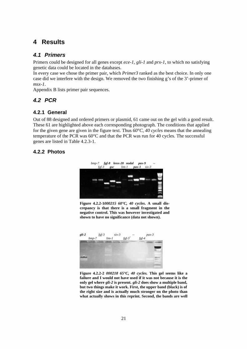
4.2.1 GeneralOut of 88 designed and ordered primers or plasmid, 61 came out on the gel with a good result.These 61 are highlighted above each corresponding photograph. The conditions that appliedfor the given gene are given in the figure text. Thus 60°C, 40 cycles means that the annealingtemperature of the PCR was 60°C and that the PCR was run for 40 cycles. The successfulgenes are listed in Table 4.2.3-1.
4.2.2 Photos
bmp-7 fgf-8 krox-20 nodal pax-9 -- fgf-3 gsc lim-1 pax-1 six-3
Figure 4.2.2-1000215 60°C, 40 cycles. A small dis-crepancy is that there is a small fragment in thenegative control. This was however investigated andshown to have no significance (data not shown).
gli-2 fgf-3 six-3 -- pax-3 bmp-7 lim-1 fgf-3* fgf-4
Figure 4.2.2-2 000218 65°C, 40 cycles. This gel seems like afailure and I would not have used if it was not because it is theonly gel where gli-2 is present. gli-2 does show a multiple band,but two things make it work. First, the upper band (black) is ofthe right size and is actually much stronger on the photo thanwhat actually shows in this reprint. Second, the bands are well
22
separated and thus easily separated by cutting out the piece ofgel containing the correct fragment.
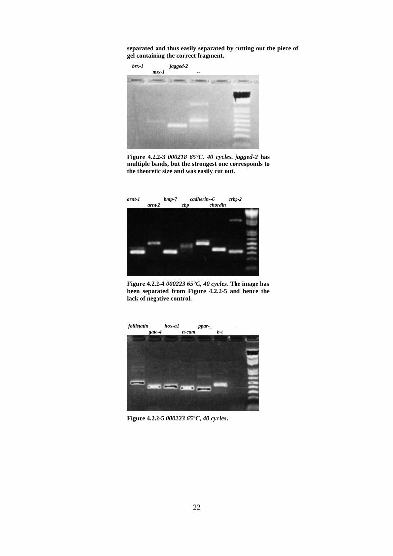
brx-1 jagged-2 msx-1 --
Figure 4.2.2-3 000218 65°C, 40 cycles. jagged-2 hasmultiple bands, but the strongest one corresponds tothe theoretic size and was easily cut out.
arnt-1 bmp-7 cadherin--6 crbp-2 arnt-2 cbp chordin
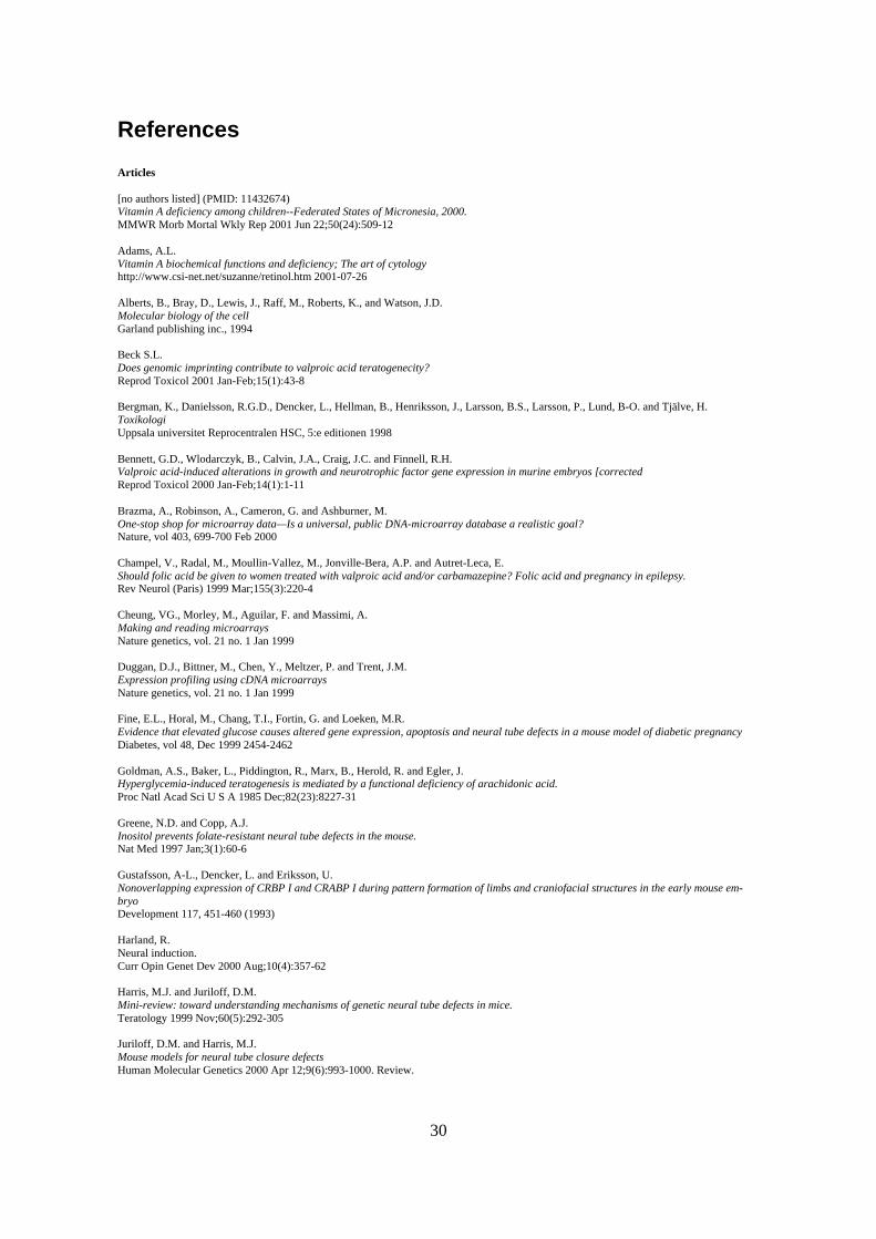
Figure 4.2.2-4 000223 65°C, 40 cycles. The image hasbeen separated from Figure 4.2.2-5 and hence thelack of negative control.
follistatin hox-a1 ppar-_ _ gata-4 n-cam b-t
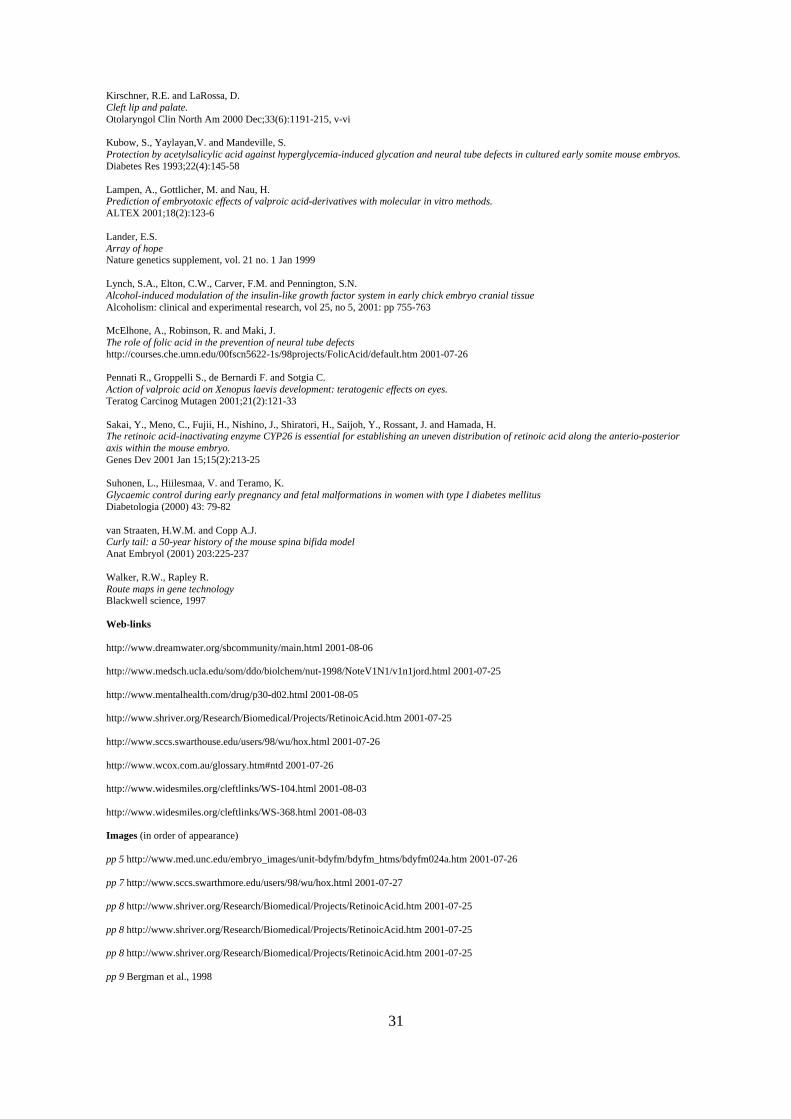
Figure 4.2.2-5 000223 65°C, 40 cycles.

23
en-2 pdgfr-_ pkC-_ erk-2 oct-6 hif-1_ erk-1 igf-1
Figure 4.2.2-6 000225 upper 65°C, 40 cycles. The im-age has been separated from Figure 4.2.2-7 andhence the lack of negative control. igf-1 has a strongmultiple band, which was disturbing. The upperband was however cut out and amplified separately,producing a single band (data not shown).
igf-2 e214-k wee-1 fgfr-1 tcof-1 --
Figure 4.2.2-7 000225 lower 65°C, 40 cycles . There isa strong indication of some form of contamination inthe negative control. The size 350 bp and the verystrong single band might however point to an erro-neous filling of the lane in question of e.g. e214-kwhich coincides in size. A test gel (data not shown)verified that it was an erroneous filling and not areal contamination. No pun intended, but wee-1shows a too small fragment. The fragment should be360 bp according to DNA sequence, but the signal isonly approximately 250 bp.
bmp-2 cart-1 dlx-1 dlx-5 emx-2 bmp-4 c-jun dlx-2 dlx-6 et-1
Figure 4.2.2-8 000302 upper 62°C, 40 cycles. The im-age has been separated from Figure 4.2.2-9 andhence the lack of negative control.

24
gli-3 pax-6 rar-_ tgf-_2 twist jagged-1 pax-7 tgf-_1 tgf-_3 --
Figure 4.2.2-9 000302 lower 62°C, 40 cycles.
et-A c-fos bfgf fgf-3 six-3 wnt-1 collagenIII pdgf-_ lim-1 --
Figure 4.2.2-10 000314 62°C, 40 cycles. c-fos is toweak in this gel to show in this reprint, but it is therein the original photo.
mhox bcl-2 lim-1 noggin wee-1 bf-1 collagen IV bfgf rar-_ --
Figure 4.2.2-11 000419 62°C, 34 cycles, day 11.bcl-2 is much to large compared with the theoreti-cal value, but my supervisors thought it was okay.The possibility remains that the size of bcl-2 hasbeen misinterpreted since it comes from anotherdepartment.

25
4.2.3 Listing of the successful probesGene Theoretical size Actual size on the gel Conditions
Tmcycles
arnt-1 307 300 6540
arnt-2 472 480 6540
bcl-2 536 650 6234
bmp-2 353 350 6540
bmp-4 342 325 6540
bmp-7 308 325 6540
brx-1 451 450 6540
b-T 439 450 6540
cadherin-6 484 525 6540
cart-1 401 425 6540
cbp 470 450 6540
c-fos 449 450 6050
chordin 359 375 6540
c-jun 407 400 6240
collagen III 401 400 6240
crbp-2 320 325 6540
dlx-1 426 400 6540
dlx-2 467 475 6540
dlx-5 370 375 6540
dlx-6 391 375 6540
e214K 363 350 6540
emx-2 173 175 6235
en-2 486 475 6540
erk-1 395 400 6540
erk-2 451 450 6540
et-1 365 350 6240
et-A 380 375 6240
fgf-8* 6040
fgfr-1 472 475 6540
follistatin 482 475 6540
gata-4 392 400 6540
gli-2 475 475 6540
gli-3 448 400 6540
gsc* 6040
hif-1_ 345 350 5840
hox-A1 401 425 6540
igf-1 350 352 6540
igf-2 452 450 6540
jagged-1 350 350 6540
jagged-2 489 475 6540
krox-20* 6040
msx-1 376 375 6540
n-cam 375 375 6540
nodal* 6040
oct-6 424 450 6540
pax-1* 6040
pax-6 318 300 6540
pax-7 358 350 6540
pax-9* 6040
pdgf-_ 386 400 6240
pdgfr-_ 344 350 6540

26
pkC-_ 416 400 6540
ppar-_ 354 375 6540
rar-_ 446 450 6235
tcof-1 437 450 6540
tgf-_1 342 350 6240
tgf-_2 403 400 6240
tgf-_3 456 450 6240
twist 157 125 6240
wee-1 360 375 6240
wnt-1 433 425 6240
Table 4.2.3-1. * = plasmid. 61 genes in total.

27
5 DiscussionThe procedures involved in this master thesis were mostly very straightforward. The main dif-ficulty in the beginning was of course learning to browse the web in an effective way. Since Iwas not involved in the project in the same way as my colleagues, most of the names of thegenes were cryptic. My colleagues knew the individual processes each gene was involved inand they also knew the complete name as well as eventual nickname of all the genes. All Iknew was their abbreviations, which in most instances was sufficient, but in some casesturned out to be insufficient.E.g. cbp is used to describe both “creb binding protein” and “cytohesin binding protein”. Theformer being the correct gene in this thesis, but the latter was the one I found.Even though the primary aim at this early stage of the project was to get a test microarray upand running as soon as possible, I think that it would have been interesting to investigate the25 % of the genes that we did not manage to amplify. Time, though, did not allow any suchresearch at the moment, since that would have meant delays in the test microarray develop-ment.It does not, however, stop me from speculating in reasons for a gene to fail to amplify.There are two ways for a gene to fail in the amplification. The first is that there is no amplifi-cation at all and the other is that there is multiple amplification, which in turn show up asmultiple bands or worse, as a smear, on the gel. If there is a case of multiple, but distinctbands, there is still a possibility to excise the band with a size that correlates with the theoreti-cal value of the size. I f there is a smear this method is useless.There are some things that might disturb a proper amplification of a gene. Obviously; theprimer sequences can be wrong. Even though we triple-checked most sequences, type errorsmight occur in the final ordering of the primers. And since we often used the same raw-sequence and the same software, the algorithm for calculating the primers might be erroneous.It is also a known fact that the databases on the Internet containing genetic data are filled witherrors. The amount of data that is being deposited annually far exceeds any chance of proof-reading.A first step would therefore include the verification of the sequences, starting with the initialselection of the gene and then followed by the whole chain of procedures. If it turns out thatwe have made no error, maybe we should consider choosing some other primer pair. primer3often lists more then one pair of primers. Alternatively, it could be worthwhile to examine theexact sequence. Some base sequences are known to interfere with polymerase performance.Another cause of failure is that the primers are not be specific, which would explain theproblems that we observed in some cases, were the gel showed multiple bands24.Multiple bands could of course also depend on alternative splicing of the gene in question,which would need to be considered. The primers might also function as an amplimer for somecompletely unrelated gene. We tried to eliminate that risk; by crosschecking each pair ofprimers with the databases to check whether it was specific for one gene only. Maybe it couldbe interesting to check the genes with multiple bands once more and investigate how specificthey really were. It could also be so that the a gene which mimics our target is, due to that ithas yet to be found and sequenced, not present in the database.The mRNA batch I was using came from 9-day old embryos. There is a possibility that somegenes were not expressed at this time of the embryogenesis, although I think that my col-
24 In some of the cases with multiple bands it was possible to cut the band with the correct size out of the gel.Still it would have been interesting to examine why there was a multiple band in the first place. E.g. igf-1 whichhad two equally bright bands whereas the others often displayed one strong band and one or two weaker.

28
leagues would have that covered. mRNA-extraction from both younger and older embryosmight therefore be worthwhile.A low copy number or a peculiar fold of a gene could make it less accessible to the polym-erase in the rtPCR, which in turn would render the gene impossible to amplify in the PCR.Generally I found the plasmid genes a bit more troublesome and not as easy as the ordinarygenes to get to work in the PCR. I had one fine gel but it was nearly impossible to repeat theresults of that experiment and the plasmid genes were a menace throughout the project, con-tinuously avoiding to be amplified.Much research is needed before the completion of a project this size can be reached. In theproximal future it is more likely that some smaller parts of the big puzzle will be revealed.Even closer to that, another round and perhaps even yet another round of genes must be se-lected, amplified and spotted to the microarray. From there, these must be tested and run overand over until the procedure becomes reliable. Only then will it be able to start doing actualexperiments, which means exposing embryos to teratogenic concentrations of RA. Doing thisand reading the array is the easy part. The difficult part lies ahead and involves connecting allstrings and forming a pattern of gene expression, gene effectants and gene affectants.

29
AcknowledgementThis master thesis was carried out at the department of toxicology at Uppsala University withProfessor Lennart Dencker and Lab assistant Anne-Lee Gustafsson as supervisors. Apart fromthem, my closest co-workers were the two post-graduates Simon Haile and Estibaliz Lopez.
I wish to thank the following people in no particular order.My parents for always supporting me.My grandparents for always believing in me.My girlfriend Hanna.Fredrik Johansson and Pär Matsson for reading my work.Matti Nikkola.
30
References
Articles
[no authors listed] (PMID: 11432674)Vitamin A deficiency among children--Federated States of Micronesia, 2000.MMWR Morb Mortal Wkly Rep 2001 Jun 22;50(24):509-12
Adams, A.L.Vitamin A biochemical functions and deficiency; The art of cytologyhttp://www.csi-net.net/suzanne/retinol.htm 2001-07-26
Alberts, B., Bray, D., Lewis, J., Raff, M., Roberts, K., and Watson, J.D.Molecular biology of the cellGarland publishing inc., 1994
Beck S.L.Does genomic imprinting contribute to valproic acid teratogenecity?Reprod Toxicol 2001 Jan-Feb;15(1):43-8
Bergman, K., Danielsson, R.G.D., Dencker, L., Hellman, B., Henriksson, J., Larsson, B.S., Larsson, P., Lund, B-O. and Tjälve, H.ToxikologiUppsala universitet Reprocentralen HSC, 5:e editionen 1998
Bennett, G.D., Wlodarczyk, B., Calvin, J.A., Craig, J.C. and Finnell, R.H.Valproic acid-induced alterations in growth and neurotrophic factor gene expression in murine embryos [correctedReprod Toxicol 2000 Jan-Feb;14(1):1-11
Brazma, A., Robinson, A., Cameron, G. and Ashburner, M.One-stop shop for microarray data—Is a universal, public DNA-microarray database a realistic goal?Nature, vol 403, 699-700 Feb 2000
Champel, V., Radal, M., Moullin-Vallez, M., Jonville-Bera, A.P. and Autret-Leca, E.Should folic acid be given to women treated with valproic acid and/or carbamazepine? Folic acid and pregnancy in epilepsy.Rev Neurol (Paris) 1999 Mar;155(3):220-4
Cheung, VG., Morley, M., Aguilar, F. and Massimi, A.Making and reading microarraysNature genetics, vol. 21 no. 1 Jan 1999
Duggan, D.J., Bittner, M., Chen, Y., Meltzer, P. and Trent, J.M.Expression profiling using cDNA microarraysNature genetics, vol. 21 no. 1 Jan 1999
Fine, E.L., Horal, M., Chang, T.I., Fortin, G. and Loeken, M.R.Evidence that elevated glucose causes altered gene expression, apoptosis and neural tube defects in a mouse model of diabetic pregnancyDiabetes, vol 48, Dec 1999 2454-2462
Goldman, A.S., Baker, L., Piddington, R., Marx, B., Herold, R. and Egler, J.Hyperglycemia-induced teratogenesis is mediated by a functional deficiency of arachidonic acid.Proc Natl Acad Sci U S A 1985 Dec;82(23):8227-31
Greene, N.D. and Copp, A.J.Inositol prevents folate-resistant neural tube defects in the mouse.Nat Med 1997 Jan;3(1):60-6
Gustafsson, A-L., Dencker, L. and Eriksson, U.Nonoverlapping expression of CRBP I and CRABP I during pattern formation of limbs and craniofacial structures in the early mouse em-bryoDevelopment 117, 451-460 (1993)
Harland, R.Neural induction.Curr Opin Genet Dev 2000 Aug;10(4):357-62
Harris, M.J. and Juriloff, D.M.Mini-review: toward understanding mechanisms of genetic neural tube defects in mice.Teratology 1999 Nov;60(5):292-305
Juriloff, D.M. and Harris, M.J.Mouse models for neural tube closure defectsHuman Molecular Genetics 2000 Apr 12;9(6):993-1000. Review.
31
Kirschner, R.E. and LaRossa, D.Cleft lip and palate.Otolaryngol Clin North Am 2000 Dec;33(6):1191-215, v-vi
Kubow, S., Yaylayan,V. and Mandeville, S.Protection by acetylsalicylic acid against hyperglycemia-induced glycation and neural tube defects in cultured early somite mouse embryos.Diabetes Res 1993;22(4):145-58
Lampen, A., Gottlicher, M. and Nau, H.Prediction of embryotoxic effects of valproic acid-derivatives with molecular in vitro methods.ALTEX 2001;18(2):123-6
Lander, E.S.Array of hopeNature genetics supplement, vol. 21 no. 1 Jan 1999
Lynch, S.A., Elton, C.W., Carver, F.M. and Pennington, S.N.Alcohol-induced modulation of the insulin-like growth factor system in early chick embryo cranial tissueAlcoholism: clinical and experimental research, vol 25, no 5, 2001: pp 755-763
McElhone, A., Robinson, R. and Maki, J.The role of folic acid in the prevention of neural tube defectshttp://courses.che.umn.edu/00fscn5622-1s/98projects/FolicAcid/default.htm 2001-07-26
Pennati R., Groppelli S., de Bernardi F. and Sotgia C.Action of valproic acid on Xenopus laevis development: teratogenic effects on eyes.Teratog Carcinog Mutagen 2001;21(2):121-33
Sakai, Y., Meno, C., Fujii, H., Nishino, J., Shiratori, H., Saijoh, Y., Rossant, J. and Hamada, H.The retinoic acid-inactivating enzyme CYP26 is essential for establishing an uneven distribution of retinoic acid along the anterio-posterioraxis within the mouse embryo.Genes Dev 2001 Jan 15;15(2):213-25
Suhonen, L., Hiilesmaa, V. and Teramo, K.Glycaemic control during early pregnancy and fetal malformations in women with type I diabetes mellitusDiabetologia (2000) 43: 79-82
van Straaten, H.W.M. and Copp A.J.Curly tail: a 50-year history of the mouse spina bifida modelAnat Embryol (2001) 203:225-237
Walker, R.W., Rapley R.Route maps in gene technologyBlackwell science, 1997
Web-links
http://www.dreamwater.org/sbcommunity/main.html 2001-08-06
http://www.medsch.ucla.edu/som/ddo/biolchem/nut-1998/NoteV1N1/v1n1jord.html 2001-07-25
http://www.mentalhealth.com/drug/p30-d02.html 2001-08-05
http://www.shriver.org/Research/Biomedical/Projects/RetinoicAcid.htm 2001-07-25
http://www.sccs.swarthouse.edu/users/98/wu/hox.html 2001-07-26
http://www.wcox.com.au/glossary.htm#ntd 2001-07-26
http://www.widesmiles.org/cleftlinks/WS-104.html 2001-08-03
http://www.widesmiles.org/cleftlinks/WS-368.html 2001-08-03
Images (in order of appearance)
pp 5 http://www.med.unc.edu/embryo_images/unit-bdyfm/bdyfm_htms/bdyfm024a.htm 2001-07-26
pp 7 http://www.sccs.swarthmore.edu/users/98/wu/hox.html 2001-07-27
pp 8 http://www.shriver.org/Research/Biomedical/Projects/RetinoicAcid.htm 2001-07-25
pp 8 http://www.shriver.org/Research/Biomedical/Projects/RetinoicAcid.htm 2001-07-25
pp 8 http://www.shriver.org/Research/Biomedical/Projects/RetinoicAcid.htm 2001-07-25
pp 9 Bergman et al., 1998

32
pp 10 Bergman et al., 1998
pp 11 Juriloff and Harris, 2000
pp 11 Juriloff and Harris, 2000
pp 12 http://www.med.unc.edu/embryo_images/unit-nervous/nerv_htms/nerv001.htm. 2001-07-28
pp 14 van Straaten and Copp, 2001
pp 15 Walker and Rapley, 1997
pp 16 Tomas Risberg, 2001
pp 16 Walker and Rapley, 1997
pp 18 http://www.biologie.ens.fr/microarrays.html 2001-07-23
33
Selected readingEmbyo images online:http://www.med.unc.edu/embryo_images/unit-welcome/welcome_htms/contents.htm
Phelan, S.A., Ito, M. and Loeken, M.R.Neural tube defects in embryos of diabetic mice: role of the Pax-3 gene and apoptosis.Diabetes 1997 Jul;46(7):1189-97
Graviditet och läkemedel—“Pregnancy and drugs”FASS, 2000
Southern, E., Mir, K. and Shchepinov, M.Molecular interactions on microarraysNature genetics, vol. 21 no. 1 Jan 1999
Lipshutz, R.J., Fodor, S.P.A., Gingeras, T.R. and Lockhart, D.J.High density synthetic oligonucleotide arraysNature genetics, vol. 21 no. 1 Jan 1999
Bowtell, D.D.L.Options available—from start to finish—for obtaining expression data by microarraysNature genetics, vol. 21 no. 1 Jan 1999
Brown, P.O. and Botstein, D.Exploring the new world of the genome with DNA microarraysNature genetics, vol. 21 no. 1 Jan 1999
Debouck, C. and Goodfellow, P.N.DNA microarrays in drug discovery and developmentNature genetics, vol. 21 no. 1 Jan 1999
Bassett, D.E. Jr., Eisen, M.B. and Boguski, M.S.Gene expression informatics—it´s all in your mineNature genetics, vol. 21 no. 1 Jan 1999
Chakravati, A.Population genetics—making sense out of sequenceNature genetics, vol. 21 no. 1 Jan 1999
Arbor, A.Copper is crucial for embryonic development say UM scientistshttp://www.lifesciences.umich.edu/news/featurestory.html 2001-07-26
Romert, A., Tuvendal, P., Simon, A., Dencker, L. and Eriksson, U.The identification of a 9-cis retinol dehydrogenase in the mouseembryo reveals a pathway for synthesis of 9-cis retinoic acidDevelopmental biology, vol. 95, pp. 4404-4409, April 1998

34
Appendix AThis is a list of occurring genes. The abbreviation of the gene is found to the left and to theright, whenever applicable, the full name of the it is given. For most of the genes there arealso some facts of whereabouts, "whenabouts" and "howabouts" of the them. Many of thegenes are involved in very complex patterns scattered in all kinds of tissue and throughout thedevelopment. In such cases I have selected some facts that mainly regard the brain and thecraniofacial area or genes relevant to the spinal chord or the peripheral system. Finally, if theyare involved in some way with the pharyngeal arches and the cardiac, pulmonary or the ven-eric system, I have tried to fit this in.I have also tried to select information, which includes the drugs, chemicals or teratogenes dis-cussed in the background chapter.Genes who are members of larger families have been lumped together within the samebracket, but I have tried to point out which information concerns which of the family member.This appendix is not to be used as a standalone spreadsheet of information, but as a guide.Good starting points for further information of the genes and their effects would be the Pub-med indexes included with each statement. All indexes in this appendix correspond to Internetdatums between 2001-07-30 and 2001-08-03 and they were found using the Pubmed searchengine provided by NCBI (http://www.ncbi.nlm.nih.gov/).Gene Codes forarnt Aryl hydrocarbon receptor nuclear translocator (Arnt) is a ubiquitously ex-
pressed helix-loop-helix (HLH) protein capable of forming hetero dimers withother HLH-proteins such as Hif-1_ and arylhydrocarbon receptor. The formedcomplexes in turn, regulate gene expression in response to hypoxia and xeno-biotics respectively. Arnt-2 functions in much the same way although it alsohas some unique features that are essential in embryonic development (PMID:11381139).
bcl-2bf-1 Brain factor 1, a regulator of forebrain development. It is a winged helix tran-
scription factor expressed in the telencephalic neuroepithelium (PMID:7815060). B-T is essential for the development of the cerebral hemispheres(PMID: 8738140). It also controls the morphogenesis by regulating the rate ofneuroepithelial cell proliferation and the timing of neuronal differentiation(PMID: 7605629).
bfgf Basic fibroblast growth factor. bFGF modulates capillary growth and facili-tates arteriolar growth. It also interacts with vascular endothelial growth factor(VEGF) to establish the normal hierarchy of the arteriolar tree (PMID:11397779).
bmp Bone morphogenic proteins are important factors in vertebrate development.They signal through Smad molecules (PMID: 11121043). Neural induction isfacilitated in cells were the BMP proteins are antagonized. Proteins thought tobe involved in silencing BMPs are Noggin, Follistatin and Chordin (Harland,2000).
b-t Brachyury T. Embryos lacking normal gene activity fail to form the noto-chord, the entire posterior region and the allantoise. They also die rather soon(PMID: 2154694). B-T has a direct role in the early events of mesoderm for-mation (PMID: 1689462).
brx-1 A homeobox gene, comprising two almost identical genes brx-1a and brx-1b.Is expressed heavily in the mammillary area as well as in the zona limitans in-
35
trathalamica of the mouse embryonic brain (PMID: 9347917).cadherin-6 Cadherins are cellular adhesion molecules (PMID: 9615235). Cadherin-6 me-
diates cell-cell binding in a homophilic manner, contributing to the sorting ofheterogeneous cell types and the maintenance of orderly structures, such as theepithelium (NM_004932).
cart-1 Cartilage paired-class homeoprotein 1. It is necessary for the survival of theforebrain mesenchyme. Mutations lead to birth defects such as acrania andmeroanencephaly (NM_006982).
cbp Creb binding protein. Functions as a coactivator of transcription factors suchas Creb, C-fos and C-jun. In studies Cbp-mutant mouse embryos died betweenday 9.5 and 10.5 post coitus and exhibited defects in neural tube closure. Lackof vascular network could also be observed (PMID: 10216070).
chordin Important developmental protein, that dorzalizes early vertebrate tissue, bybinding to ventralizing Tgf-_-like Bmps and sequestering them in latent com-plexes (PMID: 9782094). Chordin is supposed to be involved in the chain thatneuralizes cells (Harland, 2000).
c-fos A, during the embryonic stages, abundantly expressed proto-oncogene (PMID:10410909). Overexpression leads to early postnatal heterotopic chondrogene-sis and osteogenesis (PMID: 9577414). C-fos expression in striatum is ef-fected by psychostimulants such as amphetamine (PMID: 8996801).
c-jun Protooncogene, encoding the transcription factor AP-1. It is rapidly activatedby serum, Pdgf or Fgf (PMID: 3186736).
collagen Collagen type III is a major structural component of the developing palate(PMID: 11040401). Perturbations of collagen metabolism can results in theproduction of cleft palate. Tgf-_ 1 stimulates accumulation of collagen type IIIby acting on the level of synthesis and degradation (PMID: 7964554).
crbp-II Cellular retinol-binding protein type II.. May mediate actions derived fromretinol, just as its ‘cousin’ cellular retinoic acid-binding protein may mediatethe actions of RA (PMID: 2554331).
creb Cyclic AMP responsive element binding protein (PMID: 10350641). A nu-clear transcription factor, which mediates transcription of genes containingCRE recognition sequences in their promotors (PMID: 9056415).
dlx Distal-less homeobox, which are primarily expressed in the developing fore-brain, derivatives of the cranial neural crest and restricted epidermal craniofa-cial and limb (PMID: 8812481). Dlx-2 and Dlx-3 are differentially expressedin the branchial region, suggesting an important role in the craniofacial pat-terning and morphogenesis (PMID: 7893603).
e214k Ubiquitin-conjugating enzyme (accession: U57690). Has been proposed to bea key regulator of the ubiquitin proteolytic pathway (PMID: 9950926).Acutely diabetic rats show higher expression of e214k (PMID: 10329962).E214K may be involved in sepsis, it is though very uncertain (PMID:9950926).
ece Endothelin converting enzyme. Convert big endothelin-1 into endothelin-1(ET-1). Null mutations of the gene in mouse embryo exhibited abnormalitiesvirtually identical to the defects seen in ET-1 and endothelin A receptor defi-cient embryos, i.e. craniofacial and cardiac abnormalities. Mutations in ECE-1may cause developmental defects in humans, such as Hirschsprungs disease,velocardiofacial syndrome and related neurocristopathies (PMID: 9449665).
emx Homeobox genes, expressed early (day 8.5- 9.5 and onwards) in the cerebralcortex. The genes are related to empty spiracles in D. melanogaster (PMID:

36
1352754).en-2 Engrailed-2 controls the survival of the midbrain dopaminergic neurons
(PMID: 11312297). Null mutants show impaired motor learning abilities(PMID: 8652061).
erk Extracellular signal regulated kinase. Erk-1 is a serine/threonine kinase thathas the potential to phosphorylate tyrosine (PMID: 1717989). ERK is requiredfor full N-cadherin and laminin induced neurite outgrowth (PMID: 10356298).
et Endothelin. Et-1 is implicated in a wide variety of functions. 9.5 day old em-bryos seem to have a high expression in the branchial epithelium, optic vesicleand endothelial cells of large blood vessels including the dorsal aorta and aor-tic arches (PMID: 8575440).
et-A Endothelin receptor subtype A. Appears to coordinate specific aspects of pha-ryngeal arch development by inducing expression of transcription factors suchas e.g. gsc, Dlx-2 and Dlx-3 in the post migratory ectomesenchyme (PMID:10625532).
fgf Fibroblast growth factor-2 is important for vessel formation and/or mainte-nance of vascular integrity in the embryo (PMID: 10930413).
fgfr Fgf receptor. Fgfr-1 is the only high affinity receptor to Fgf-2 (PMID:10533610). Fgf signals through this receptor to act in the development andmaintenance of a mature vascular network in the embryo (PMID: 10930413).It is essential for normal growth and development of the heart (PMID:10533610).
follistatin Follistatin is an activin antagonist. Activin is important for the mesoderm for-mation. It may also have other functions, presumably in somites and hindbrain(PMID: 7600958) Follistatin RNA is located in the Spemann organizer andnotochord. Both the RNA and the protein of follistatin are capable of directlyneuralizing ectodermal explants (PMID: 8168135).
gata GATA-binding protein. A zinc finger transcription factor expressed in the de-veloping heart. Activates a variety of cardiac-specific genes (PMID:10625555). GATA-4 can drive A1AR promotor activity, which is abundant inthe brain and atria (PMID: 10318839). GATA-4 is a tissue-specific, retinoicacid-inducible, and developmentally regulated transcription factor (PMID:8455608).
gli Zinc finger transcription factor and oncogene. A role for the gli family couldbe in the craniofacial development and also in the digital development (PMID:8364225).
gsc Goosecoid is an evolutionarily conserved homeobox gene, expressed brieflyduring gastrulation with a DNA-binding specificity similar to that of Droso-phila bicoid (PMID: 1352187). Gastrula formed mesoderm generates varioustissues such as notochord, muscle, kidney and blood (PMID: 7905664). Cellsexpressing Gsc is destined to become pharyngeal endoderm, head mesodermor notochord (PMID: 1363720). Gsc s not essential for organizer activity but itis important in the embryogenesis for craniofacial and rib cage development(PMID: 7555726).
hif Hypoxia inducible factor mediates cellular responses to low oxygen tension,iron chelators and low intracellular glucose concentration. Hif-1_ forms di-mers with Arnt and the complex binds to DNA, which promotes transcriptionof targeted genes(PMID: 9368100).
hox Homeobox genes, regulates gene expression for different body parts.igf Insulin-like growth factor. Igf-1 plays a major role in growth and regenerative
37
process as a mediator of many of the biological effects of growth hormone(PMID: 3034909). Igf-2 may be involved in myogenic differentiation (PMID:2537977).
jagged Jagged1 interacts with Notch-2 and other Notch receptors (PMID: 10551863).krox-20 It is a regulatory protein containing zinc fingers. A transcription factor, which
binds to CpG methylated sites and regulates hox genes during hindbrain seg-mentation (PMID: 8929813).
mhox A homeodomain protein completely restricted to mesodermally derived celltypes during embryogenesis. Probably a transcriptional regulator of the estab-lishment of diverse mesodermal cell types (PMID: 1360403).
msx-1 Drosophila msh-like homeobox. The conserved consensus sequence makes itlikely to be a regulatory gene (PMID: 9655249).
n-cam Neural cell adhesion molecule, which is an important mediator of calcium in-dependent cell-cell interactions (PMID: 1716358).
noggin Inducer of neural tissue (PMID: 7666191). A Bmp antagonist. Expressed incondensing cartilage (PMID: 9603738). Complete knockout lead to a loss ofcaudal vertebrae and malformation of the limbs (PMID: 10087923).
oct-6 Octamer binding protein. It is present in embryonic stem cells. It is downregulated upon embryonic stem cell differentiation, increasing again duringbrain development, where it is present in telencephalon, mesencephalon andbrain stem (PMID: 1976514).
p27pax A family of transcription factors characterized by a common DNA motif; the
paired box, which encodes a DNA binding motif. Pax genes are also potentialproto-oncogenes (PMID: 9021154). From day 8, Pax-6 is expressed in discreteregions of the forebrain and hindbrain. It is also expressed in the neural tube,the developing eye and the nasal epithelium (PMID: 1687460). Pax-3 acts,with beginning on day 8.5, in the hindbrain, midbrain and diencephalon. Laterit also acts in the neural crest cells, the craniofacial mesectoderm and the limbmesenchyme (PMID: 2022185). Pax-9 is expressed in the vertebral column, inthe pharyngeal pouches and some parts of face and limbs (PMID: 9021154).
pdgf _ Platelet-derived growth factor _ plays a role in cell proliferation during vas-cular growth (PMID: 10375497). The monomer stimulates migration in limbmyoblasts (PMID: 9303348). A possible role as a neorotrophic and neurorel-gulatory factor in developing and mature brain (PMID: 1521324).
pdgfr _ Pdgf receptor _, vital for the development of vertebrate embryos. Expressed incranial neural crest cells, lung smooth muscle progenitors and oligodendrocyteprogenitors. Knockouts suffer from profound craniofacial abnormalities andspina bifida (PMID: 11023856). Pdgf _ receptor is involved in growth and de-velopment of blood vessels (PMID: 8018985).
pkc-_ Protein kinase C - _ is a phospholipid-dependent kinase. It is markedly en-hanced by non-nuclear acting retinoids such as 14-hydroxy-retro-retinol.These retinoids function as regulatory cofactors for cell survival/cell death de-cisions (PMID: 11099483). Kinases task in general is to phosphorylate aminoacids of other proteins (Genes VI). However, in order for Pkc- _ to becomeactive, it needs to be phosphorylated. The phosphorylation is carried outthrough autophosphorylation (PMID: 11073945). Indications point to PKC- _ ,being important for a cell to transform into a differentiated cell, which meansthat the cell will fail to respond to normal signals of regulation and growth(PMID: 2161238).

38
ppar-_ Peroxisome proliferator-activated receptor-_ is activated by linoleic acid(PMID: 8041794) Ppar’s act to increase the number of peroxisomes and thelevels of several enzymes (PMID: 8168153). In rat expression is most abun-dant in brain, heart, skeletal muscle, kidney and lung (PMID: 8554552). Ppar-_ dimers with RXR to bind DNA response elements (PMID: 11237216). It hasbeen supposed that it affects white adipose tissues adaptive responses to nutri-tional changes (PMID: 10991946).
prx A homeobox protein, which is expressed in lateral plate and bud mesoderm.prx-1 cooperates with prx-2 to regulate palm and hind limb morphogenesisthrough Bmp-mediated signalling pathways (PMID: 9882503). prx-1 is alsoexpressed in postmigratory cranial mesenchyme of all facial prominences andis required for the formation of proximal first arch derivatives (PMID:9876178). From day 9.5 prx-1 exhibit distinct expression in the ventral part ofthe hypothalamus, as well as in a broader region of the telencephalon (PMID:7577675).
rar Retinoic acid receptor. Most all of the actions of retinoids are mediatedthrough these.
rho-_ Small Ras-like GTPase. It participates in the regulation of the actin cytoskele-ton and cell adhesion through specific targets (PMID: 10872457).
ski An oncoprotein, which functions as an antagonist to Bmps, by interacting withBmp-specific Smad complexes (PMID: 11121043).
tcof1 Treacher Collins Franceschetti syndrome 1. The syndrome is autosomal andaffects the craniofacial region. Mutations in tcof1 lead to the phenotype of theTreacher Collins syndrome (PMID: 9299440). Evidence point to the protein isinvolved in nucleolar-cytoplasmic transport (PMID: 9158147).
tgf Transforming growth factor. Proteins involved in a variety of cellular trans-forming processes. They regulate, as do retinoic acid, the expression of CrabpI and II (PMID: 7556459).
twist Mutations cause craniosynostosis by haploinsufficiency (PMID: 10751173).versican Versican is a proteoglycan, implicated in hair follicle induction and influences
axon outgrowth (PMID: 11231081). Appears from day 16 in newly formedcondylar cartilage (PMID: 11273038). It is developmentally regulated in thebrain (PMID: 8149594).
wee A cell cycle checkpoint gene.wnt Wingless- and int-related protein (PMID: 11336784).
Wnt-1 is important for the development of the mouse central nervous system.Wnt-1 transfected cells can cause cells to differentiate into neurons without theinduction of RA (PMID: 11324569). In chick Wnt signals from the neural tubeblock cardiogenesis in the adjacent anterior paraxial mesoderm (PMID:11159906).
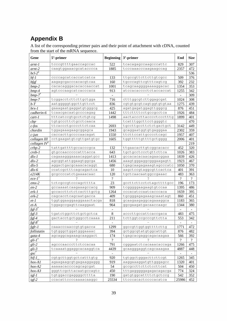
39
Appendix BA list of the corresponding primer pairs and their point of attachment with cDNA, countedfrom the start of the mRNA sequence.
Gene 5’-primer Beginning 3’-primer End Size
arnt-1 tcccgttttgaaccagccac 522 tcacagagccaagcccattc 829 307arnt-2 caagtggaaacgcatacccca 1885 tcccaaacccaagaagccag 2357 472bcl-2‡ 536bf-1 ccccagcatcaccatcatca 133 ttgccgttcttcttgtcgcc 509 376bfgf aagagcgacccacacgtcaa 160 tgcccagttcgtttcagtcg 392 232bmp-2 cacacagggacacaccaaccat 1001 tcagcaaggggaaaaggacac 1354 353bmp-4 agtcccaagcatcacccaca 913 atccacacccctctaccaccat 1255 342bmp-7* - - - - 309bmp-7 tcggacctcttcttgctgga 716 ctttggcgtcttggagcgat 1024 308b-T aatgggggtggcttgttcct 836 cgtgtgcgtcagtggtgtgtaa 1275 439brx-1 gaaagaatgaggatgtgggcg 425 agatgagatggagttgggcg 876 451cadherin-6 tcaacgacaatgccccagag 1442 tttcttttcctgccgcctca 1926 484cart-1 tttcatccgtgcctctgtcg 1498 aactacccttacccctccctttg 1899 401cbp_ tgtgcccttctgcctcaaca tcatttggcttcctggggtc 470c-fos ttcaccctgccccttctcaa 2693 tgccttgccttctctgactgct 3142 449chordin tggagaaagaagcggagca 1943 gcaggaatggtgtgagggaa 2302 359c-jun caccacttgccccaacagat 1550 ttcttccattgcccctcagc 1957 407collagen III cctgaagatgtcgttgatgtgc 1605 tggtttttgttttgctgggg 2006 401collagen IV‡ 219crbp-2 tattgattttgccacccgca 132 ttgaacacttgtcggcacacc 452 320creb-1 gtgccaacccccatttacca 643 tgctgcctccctgttcttca 1026 383dlx-1 cagaaagggaaaacaggatgcc 1413 gccacacacaacagaacggaa 1839 426dlx-2 agcggtattggaagtggcga 1456 aaagtgggaggcgggaagagtt 1923 467dlx-5 aggactgacgcaaacacaggt 680 tgagcaagagaaagtagcccatc 1050 370dlx-6 ccatcgctttcagcagactca 10 aagctccgtaggcgttcactca 401 391e214K gcgccccatctgaaaacaac 120 tgctcaacaatggccgaaac 483 363ece-1† ? ? ? ? ?emx-2 tctcgccgtcccagctttta 23 gccttcttcctctagcttttgcc 196 173en-2 gccaaaatcaagaaagccacg 909 tcgggggaagaagtgtccaa 1395 486erk-1 gccaccttctctcactttgctg 1264 cccacatccaatcacccaca 1659 395erk-2 cagcacctcagcaatgacca 409 tgcgggagagaaagcaaatagt 860 451et-1 tggtggaaggaaggaaactacga 818 gcaagaagaggcagaaaggca 1183 365et-A tggagccgagttcaaggaat 964 ggcgaagatgacaaccaagc 1344 380fgf-3* - - - - -fgf-3 tgatctggcttctgctgctca 8 acccttgccattcaccgaca 483 475fgf-4 gactacctgctgggcctcaaaa 211 tcttggtccgcccgttctta 553 342fgf-8* - - - - -fgfr-1 caaacccaaccgtgtgacca 1299 ggccgttggtggttttcttg 1771 472follistatin tgtgggctggatgggaaaac 394 gctggcgtatgtggcattgt 876 482gata-4 agcaggcagaaagcaaggact 174 tgagcacgaggcagacaagaa 566 392gli-1† ? ? ? ? ?gli-2 agcccaacccttctccacaa 791 cgggaatctcacaaacaccaga 1266 475gli-3 tcaaaatggaggcacaaggtca 4439 gcaagggaggtcagcaaagaa 4887 448gsc* - - - - -hif-1_ cgtgcttggtgctcatttgtg 920 tgtggctgggacttcttcgt 1265 345hox-A1 agaagaagcgtgagaaggaggg 919 aaggaaaggatgttgggagcc 1320 401hox-A2 aaaaacaccccagcagcgat 54 gccgccttcttctccttcat 504 450hox-A3 gggttcgcttacaatgccagtc 450 tttgagggggagagacagacga 774 324igf-1 tgtggaccgaggggctttta 190 gatgtggcattttctgctccg 542 352igf-2 ccacattccccaaaacaaggc 25534 ttcccacactccccacatca 25986 452

40
jagged-1 tggcttgggtctgttgcttg 1584 agttcgggtgttttgtcggc 1934 350jagged-2 caccatcgccaagaacagca 2252 aggcttccatccgcaccata 2741 489krox-20* - - - - -lim-1* - - - - -lim-1 gggatggaaaaagaaaggaagga 704 atggcgggttgggtaggata 1177 473mhox tcccaccctctcgtttctctct 12 tgttgtcctgtttctccgctg 480 468msx-1 ccgaaagccccgagaaacta 524 accgccaagaggaaaagaga_ 900 376n-cam tggacaaaggatggggaacc 900 tcaaggaggacacacgagca 1275 375nodal* - - - - -noggin atctacacatccgcccagca 335 caggcgctgtttcttgcctt 657 322oct-6 gcagttcgccaagcagttca 781 tcatgcgcttctccttctgc 1205 424p27‡ 515pax-1* - - - - -pax-3 tcgctttttcgtctcgcctt 241 tcacctgcttgggtttgctg 625 384pax-6 gcccttccatctttgcttgg 485 gcttcatccgagtcttctccgt 803 318pax-7 ccgaaccacatccgtcacaa 49 tcctcgtcgtcctctttcttcc 407 358pax-9* - - - - -pax-9 tgtgcctttggttgttttga 1821 gcctttttgttgtcctgtgc 2191 370pax-9 agcactcaagcctctttcagcc 2 acaaggtcggatgcccagtt 314 312pdgf-_ accactccatccgctccttt 104 gggcttctttcgcacaatctc 490 386pdgfr-_ ccttgggggctttccttgtt 4229 tctcgtggtctcttctgtgtcct 4573 344pkC-_ aaccatccgctccaacctga 894 ttcccaaaactccccttccc 1310 416ppar-_ cgccaagaacatccccaact 786 tagagcccgcagaatggtgt 1140 354prx-1† ? ? ? ? ?rar-_ aagaagaaagaggcacccaagc 493 ggcgaaggcaaagaccaagt 939 446rar-_‡ ~500six-3* - - - - -six-3 tggcgagcagaagacccatt 799 ttacgaagaggaggggaaggtg 1241 442ski tggtggttttgctctgtggg 39 ccgcttgatgttggggtgat 382 343tcof-1 atcccaacacgcagcagaca 3326 ttgccaccagcactcccttt 3763 437tgf-_1 gagacggaatacagggctttcg 984 ggtcccagacagaagttggcat 1326 342tgf-_2 tgtgaaaaccagagcggagg 1784 tgtaaagagggcgaaggcag 2187 403tgf-_3 tcaccacaacccacacctga 640 tggcagttttccctcctctg 1096 456twist tcacaagaatcagggcgtgg 946 tcgtcaaaaagtggggtggg 1103 157versican agcgacaccttccaactatccg 7004 agccaccccaaatgaccaga 7336 332wee-1 tcttgggaggaggagggttt 629 cttggggagtttgccgtgta 989 360wnt-1 tcctgatgtttgcccaccct 4549 ttccgtgccctttcaactcg 4982 433* = plasmid insertion.† = no sequence could be found and hence no primers.‡ = came from another branch of the institution and no data are available._ = the only sequence which was shortened. The two ending g’s were excluded from the primer3 result._ = I did not design this sequence. There is no significant counterpart that matches cbp when compared withknown sequences. I ordered this sequence based upon my supervisors’ advice, when my own search had endedon the wrong protein Cytohesin binding protein also shortened Cbp. Instead of the intended Creb binding pro-tein.- = no primer data available in the cases of plasmid inserted sequences.? = no primers could be designed since the DNA sequences could not be located.

41
Appendix C
Event Rat HumanFormation of blastocysts 3-5 4-6Implantation 5-6 6-7Organogenesis 6-17 21-56Primitive streak 9 16-18Neural plate 9.5 18-20First somite 10 20-21First pharyngeal arch 10 20Ten somites 10-11 25-26Anterior extremites 10.5 29-30Posterior extremites 11.2 31-32Finger 13.4 35Intestinal differentiation 14.5 43Cardiac barriers 15.5 46-47Closure of the palate 16-17 56-58Length of pregnancy 22 267
Comparison between the rat and the human
scheme of organogenesis. (Source Bergman, K et al. 1998.)
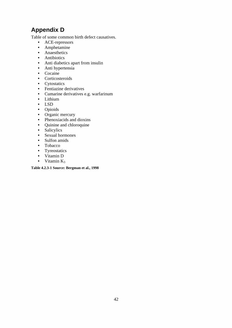
42
Appendix DTable of some common birth defect causatives.
• ACE-repressors• Amphetamine• Anaesthetics• Antibiotics• Anti diabetics apart from insulin• Anti hypertensia• Cocaine• Corticosteroids• Cytostatics• Fentiazine derivatives• Cumarine derivatives e.g. warfarinum• Lithium• LSD• Opioids• Organic mercury• Phenoxiacids and dioxins• Quinine and chloroquine• Salicylics• Sexual hormones• Sulfon amids• Tobacco• Tyreostatics• Vitamin D• Vitamin K3
Table 4.2.3-1 Source: Bergman et al., 1998