detailed experimental methods as well as additional experimental
TRANSCRIPT
Supplementary Material (ESI) for Chemical Communications This journal is © The Royal Society of Chemistry 2009
1
Electronic Supplementary Information for:
A General Electrochemical Method for the Label-Free Screening of Protein-Small Molecule Interactions Kevin J. Cash,a Francesco Riccib,d and Kevin W. Plaxcob,c*
Reagents and DNA Sequences 5
Tris(2-carboxyethyl) phosphine hydrochloride (TCEP; Invitrogen, Eugene, OR), 6-Mercapto-1-hexanol (MCH; Fluka), and sulfuric acid (Fisher Scientific) were all used as received. 1000 μg/mL solutions of 2,6-dinitrotoluene and 2,4,6-trinitrotoluene in acetonitrile (Supelco, Bellefonte, PA) 10
were aliquoted and stored at 4°C until use. 2,4-dinitrophenol (DNPOL, Sigma-Aldrich), and 4-(Dimethylamino)-phenylacetic acid (4DMAPA, Sigma-Aldrich) were dissolved in deionized water (DI water; 18 MΩ cm Milli-Q Ultrapure Water Purification, Millipore, Billerica, MA), aliquoted and 15
stored at 4°C for immediate use or -20°C for long term storage. Anti-TNT antibody (Strategic Biosolutions, Newark, DE) was aliquoted and stored at 4°C. Seawater samples were taken from the Pacific Ocean near the University of California, Santa Barbara campus and stored at 4°C. Soil 20
samples were taken from the University of California, Santa Barbara campus and extracted similar to EPA Method 8330B3. A 10 g soil sample was ground with morter and pestle, and mixed with 20 mL acetonitrile. The mixture was vortexed for 1 minute, and shaken overnight. After settling for 30 minutes, 25
8 mL of acetonitrile was removed, and filtered with a 0.45 micron PTFE syringe filter (discarding the first mL). This mixture was then dried, and resuspeded with 4 mL of deionized water and sonicated, then stored at 4°C until use. A similar soil sample was also directly suspended in DI water at 30
a concentration of 10% w/v for storage, and diluted to 5% w/v with buffer before use. Cells used were a kind gift of Patrick Daugherty and Abeer Jabaiah, and were FreeStyle 293-F cells (Invitrogen) grown to 106 cells/mL in FreeStyle Expression Medium (Invitrogen), and before use lysed by sonication and 35
diluted to 50% with buffer. Saline-sodium citrate buffer (SSC) was diluted from a 20X stock (20XSSC, Sigma Aldrich) with DI water to either 6XSSC (for sensor fabrication and buffer experiments) or 12XSSC (for dilution of seawater, soil and cell lysate samples to 50%). 40
The thiolated, methylene blue-tagged anchoring strand as well as the DNP-labeled recognition strands (HPLC purified, Biosearch Technologies Inc., Novato, CA) were used as received without further purification. The sequences of the various strands are shown in Table S1. 45
Table S1: DNA sequences used in this research
Anchor DNA Sequence Anchor 5’-HS-(CH2)6-GCAGTAACAAGAATAAAACGC
CACTGC-(CH2)7-MB DNP recognition strands (triethylene glycol linkage between DNA and
DNP) DNP25 5’-DNP-TEG-CAGTGGCGTTTTATTCTTGTTACTG DNP23 5’-DNP-TEG-AGTGGCGTTTTATTCTTGTTACT DNP21 5’-DNP-TEG-GTGGCGTTTTATTCTTGTTAC DNP19 5’-DNP-TEG-TGGCGTTTTATTCTTGTTA DNP17 5’-DNP-TEG-GGCGTTTTATTCTTGTT DNP15 5’-DNP-TEG-GCGTTTTATTCTTGT
Single-stranded DNP Probe Sequences TL1 5’-HS-(CH2)6-AAGGTGGAATGGTTDGTC-(CH2)7-MB TL2 5’-HS-(CH2)6-AAGGTGGAATGGTTTGTCD
D Location of DNP-TEG, T Thymine labeled with -(CH2)7-MB
Electrode cleaning and Sensor Preparation A detailed sensor fabrication procedure can be found in the literature20. Briefly, polycrystalline gold disk electrodes 50
(2mm diameter; BAS, West Lafayette, IN) were prepared by polishing with diamond and alumina (BAS) with sonication in ethanol or water after each step. Following polishing, electrochemical cleaning (a series of oxidation and reduction cycling in 0.5M H2SO4, 0.01M KCl/0.1M H2SO4, and 0.05M 55
H2SO4) and area determination (based on the area of the gold oxide reduction peak in the final cleaning step) were performed. Anchoring strand DNA (0.1mM) was incubated with TCEP (1 μM) for 1 hour to allow reduction of disulfide bonds. This 60
solution was diluted to 25 nM with 6XSSC. Electrodes (thoroughly rinsed with DI water) were incubated in 250 μL of anchoring DNA for 1 hour. Electrodes were rinsed with DI water, and incubated in 3mM MCH in 6XSSC for 1 hour to displace nonspecifically adsorbed DNA and passivate the 65
remaining electrode area21. After thoroughly rinsing with DI water, electrodes were stored in 6XSSC for 30 minutes before use. Probe packing density of DNA was determined using a previously described method22. The probe density used in this research was in the range of 2.2 to 3.0 x 10¹¹ molecules/cm2 70
unless otherwise noted. The modified electrodes were then incubated in 200 nM recognition strand solutions for 1 hour to allow hybridization prior to use. For optimization of antibody concentration, antibody was titrated into buffer containing the electrodes, and allowed to equilibrate for one hour before 75
measurements. For detection of TNT (and other compounds), 4 nM of antibody was added to the solution (buffer or complex sample), and allowed to equilibrate for 3 hours prior to measurements. TNT (and other compounds) in stock solutions were then added to that solution to the desired 80
concentration for measurement, allowing 1 hour incubation to
Supplementary Material (ESI) for Chemical Communications This journal is © The Royal Society of Chemistry 2009
2
allow near complete signal saturation for even the lowest concentrations used (with the exception of kinetic measurements).
Electrochemical Measurements All electrochemical measurements were performed using a 5
CHI630C potentiostat with a CHI684 Multiplexer (CH Instruments, Austin, TX) and a standard 3-electrode cell containing a platinum counter electrode (BAS) and a Ag/AgCl (3M NaCl) reference electrode (BAS). Alternating current voltammograms were obtained in 6XSSC using a 25 mV 10
amplitude signal at 10 Hz from -0.05 to -0.45 V vs. Ag/AgCl for the purpose of determining probe packing density of DNA. Experimental data were collected using square wave voltammetry from -0.05 to -0.45V in increments of 0.001V vs. Ag/AgCl, with an amplitude of 50 mV and a frequency of 60 15
Hz. Peak currents were fit using the manual fit mode in the CH Instruments software. Results are presented as signal change (difference in peak currents obtained before and after target binding divided by initial peak current) to allow for better comparison of electrodes differing in surface area. 20
Voltammograms are presented with the current at -0.05V set equal to 0A.
Surface Probe Density Experiments Surface probe density was controlled through the use of different concentrations of anchoring strand DNA during the 25
immobilization procedure. Before addition of the recognition strand, probe packing density of DNA was determined. Breifly, this method uses electrochemical interrogation with alternating current voltammetry, and relates the peak current to the number of methylene blue redox tags by assuming 30
perfect electron transfer. This method is described more fully in previous research22.
Sensor Response in Soil Suspension Of note, detection of TNT was not possible upon addition to soil suspensions (data not shown), presumably from the high 35
adsorption of TNT by particulate organic matter23. As a result, selectivity was tested using soil extracted in a method similar to EPA8330B3. 40
45
50
Supplementary Figures and Experimental Data
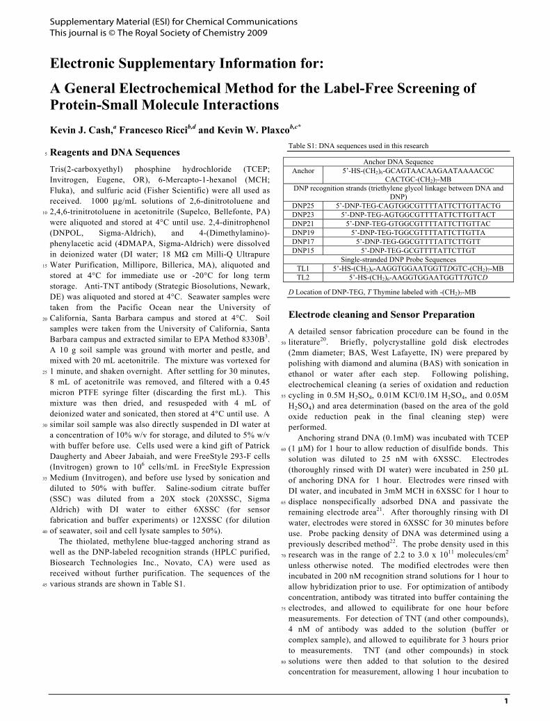
Figure S1: The apparent electron transfer rates are significantly slower in
the presence of anti-TNT antibody, supporting the hypothesis that the scaffold dynamics are reduced upon antibody binding. Data represents 55
averaged normalized peak current, for three electrodes in the absence or presence of anti-TNT antibody (70 nM). The frequency with maximal
normalized peak current is related to the electron transfer rate constant24, and can be used to compare the antibody bound and unbound states. Error
bars represent standard deviations of the three electrodes. 60
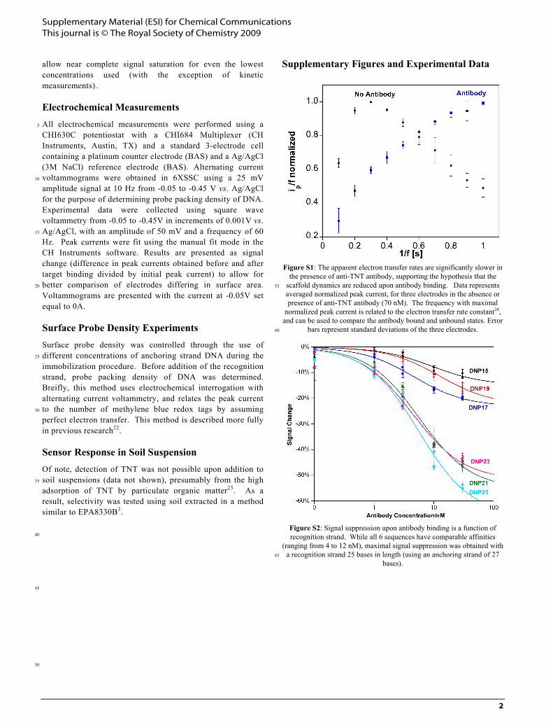
Figure S2: Signal suppression upon antibody binding is a function of recognition strand. While all 6 sequences have comparable affinities
(ranging from 4 to 12 nM), maximal signal suppression was obtained with a recognition strand 25 bases in length (using an anchoring strand of 27 65
bases).
Supplementary Material (ESI) for Chemical Communications This journal is © The Royal Society of Chemistry 2009
3
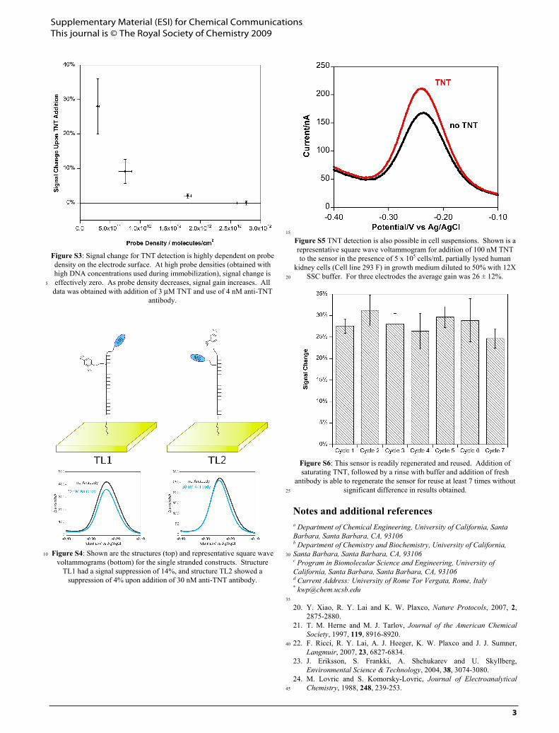
Figure S3: Signal change for TNT detection is highly dependent on probe density on the electrode surface. At high probe densities (obtained with high DNA concentrations used during immobilization), signal change is effectively zero. As probe density decreases, signal gain increases. All 5
data was obtained with addition of 3 μM TNT and use of 4 nM anti-TNT antibody.
Figure S4: Shown are the structures (top) and representative square wave 10
voltammograms (bottom) for the single stranded constructs. Structure TL1 had a signal suppression of 14%, and structure TL2 showed a
suppression of 4% upon addition of 30 nM anti-TNT antibody.
15
Figure S5 TNT detection is also possible in cell suspensions. Shown is a representative square wave voltammogram for addition of 100 nM TNT to the sensor in the presence of 5 x 10⁵ cells/mL partially lysed human
kidney cells (Cell line 293 F) in growth medium diluted to 50% with 12X SSC buffer. For three electrodes the average gain was 26 ± 12%. 20
Figure S6: This sensor is readily regenerated and reused. Addition of saturating TNT, followed by a rinse with buffer and addition of fresh
antibody is able to regenerate the sensor for reuse at least 7 times without significant difference in results obtained. 25
Notes and additional references a Department of Chemical Engineering, University of California, Santa Barbara, Santa Barbara, CA, 93106 b Department of Chemistry and Biochemistry, University of California, Santa Barbara, Santa Barbara, CA, 93106 30 c Program in Biomolecular Science and Engineering, University of California, Santa Barbara, Santa Barbara, CA, 93106 d Current Address: University of Rome Tor Vergata, Rome, Italy * [email protected] 35
20. Y. Xiao, R. Y. Lai and K. W. Plaxco, Nature Protocols, 2007, 2, 2875-2880.
21. T. M. Herne and M. J. Tarlov, Journal of the American Chemical Society, 1997, 119, 8916-8920.
22. F. Ricci, R. Y. Lai, A. J. Heeger, K. W. Plaxco and J. J. Sumner, 40
Langmuir, 2007, 23, 6827-6834. 23. J. Eriksson, S. Frankki, A. Shchukarev and U. Skyllberg,
Environmental Science & Technology, 2004, 38, 3074-3080. 24. M. Lovric and S. Komorsky-Lovric, Journal of Electroanalytical
Chemistry, 1988, 248, 239-253. 45