detection and characterization of bat sarbecovirus
TRANSCRIPT
Page 1 of 9
Article DOI: https://doi.org/eid2612/203386
Detection and Characterization of Bat Sarbecovirus Phylogenetically Related to
SARS-CoV-2, Japan
Appendix
Additional Methods
Sample Collection
We captured 4 Rhinolophus cornutus bats in a cave in Iwate prefecture, Japan, in
2013, with permission from the prefectural local government. Each bat was kept in a separate
plastic bag. Fresh feces samples were collected, transferred into tubes containing sterilized
saline, and frozen in dry ice. We released the bats after feces collection.
Reverse Transcription-PCR
RNA was extracted from the feces samples using an RNeasy PowerMicrobiome Kit
(QIAGEN, https://www.qiagen.com). Next, we detected the partial RNA-dependent RNA
polymerase (RdRp) gene of sarbecovirus in 2 samples by real-time reverse transcription-PCR
by using RNA-direct SYBR Green Realtime PCR Master Mix (TOYOBO,
https://www.toyobo-global.com) and a pair of primers (5′-CATATGCAGTAGTGGCATCA-
3′ and 5′-GCTGTAACTTGTCACATCGT-3′) that were designed based on a previous report
(1).
Next-Generation Sequencing
A cDNA library was prepared from RNA extracted from the feces sample by using
the SMARTer Stranded RNA-Seq Kit (Takara-Bio, https://www.takarabio.com). The library

Page 2 of 9
was sequenced by using a NovaSeq 6000 (Illumina, https://www.illumina.com) sequencer.
The read sequences were mapped on RaTG13 (GenBank accession no. MN996532), and the
Rc-o319 sequence was determined by using CLC genomic workbench version 8.0.1
(QIAGEN, https://www.qiagen.com) software. The sequence was deposited in GenBank
(accession no. LC556375).
Phylogenetic Analysis
The nucleotide and amino acid (aa) sequences of sarbecoviruses were aligned by
using ClustalW version 2.1(Clustal, https://www.clustal.org). Phylogenetic trees were
constructed by performing maximum-likelihood analysis with MEGA version X (2), in
combination with 500 bootstrap replicates.
Plasmids
We cloned the spike protein (S) genes of Rc-o319 and severe acute respiratory
syndrome coronavirus 2 (SARS-CoV-2; 2019-nCoV/Japan/AI/I-004/2020, GenBank
accession no. LC521925), with a 19–aa deletion at the C-terminus (pCAGGS-o319-S-del19),
into protein expression pCAGGS vectors (pCAGGS-SARS-CoV-2-del19). Earlier reports
have suggested that this deletion leads to the efficient production of vesicular stomatitis virus
(VSV) pseudotyped with the S gene of SARS-CoV (3). We used the SARS-CoV S gene
expression plasmid pKS-SARS-St19 (3) and the vesicular stomatitis Indiana virus
glycoprotein (G)-expression plasmid pCAGGS-VSV-G (4). We also constructed angiotensin-
converting enzyme 2 (ACE2)-expression plasmids derived from humans (hACE2; GenBank
accession no. NM_001371415), greater horseshoe bats (R. ferrumequinum) (Rf-ACE2;
GenBank accession no. AB297479), and Chinese rufous horseshoe bats (R. sinicus) (Rs-
ACE2; GenBank accession no. KC881004). The hACE2 cDNA was amplified from RNA
isolated from Caco-2 cells by RT-PCR, and the Rf-ACE2 and Rs-ACE2 genes were
Page 3 of 9
artificially synthesized and cloned into pCAGGS plasmids (pCAGGS-hACE2, pCAGGS-Rs-
ACE2, or pCAGGS-Rf-ACE2, respectively).
Because fresh RNA samples from R. cornutus bats were not available, the ACE2 gene
of R. cornutus (Rc-ACE2) was amplified from the exon regions of genomic DNA extracted
from the kidney of a bat carcass (GenBank accession no. LC564973) and cloned into the
pCAGGS vector that was designated as pCAGGS-Rc-ACE2. We also prepared a chimeric
bat ACE2 (referred to as Rc/Rf chimera), which consisted of the S interaction domain of Rc-
ACE2 (aa positions 15–116) and the remaining region from Rf-ACE2, to form the expression
plasmid pCAGGS-Rc/Rf-ACE2.
Western Blot Analysis
HEK293T cells were transfected with pCAGGS-RcACE2, pCAGGS-hACE2,
pCAGGS-RfACE2, pCAGGS-RsACE2, pCAGGS-Rc/RfACE2, or empty pCAGGS vectors.
One day after transfection, the cells were lysed with SDS sample buffer and subjected to
western blotting by using rabbit anti-ACE2 antibody (Abcam, https://www.abcam.com).
Production of VSV-Pseudotyped Virus
VSVΔG*-GFP, which expresses a GFP reporter gene instead of the viral G gene, was
used to produce VSV pseudotyped with S genes from sarbecoviruses, as described in earlier
reports (3,5). HEK293T cells seeded on 6-well plates were transfected with 2 μg of either
pCAGGS-o319-S-del19, pCAGGS-SARS-CoV-2-del19, pKS-SARS-St19, or control
pCAGGS-VSV-G. At 24 h post-transfection, the cells were infected with VSVΔG*-GFP and
incubated for 24 h. The culture fluid was collected, centrifuged, filtered through a 0.45-μm
filter to remove cells and cell debris, and stored at −80°C until use. The pseudotyped viruses
with Rc-o319 S were designated as VSV-Rc-o319, for SARS-CoV S were designated VSV-
SARS, for SARS-CoV-2 S were designated VSV-SARS-2, and for VSV-G were designated
VSV-VSV-G. The viruses pseudotyped with sarbecovirus S proteins were incubated with the
Page 4 of 9
anti-VSV-G neutralizing antibody I1 (6) for 30 min at 21°C to eliminate the remaining
VSVΔG*-GFP.
Cell Entry Assay
HEK293T cells seeded on 24-well plates were transfected with 0.5 μg of either
pCAGGS-Rc-ACE2, pCAGGS-Rf-ACE2, pCAGGS-Rs-ACE2, pCAGGS-Rc/Rf-ACE2,
pCAGGS-hACE2, or control empty pCAGGS plasmids, and incubated for 24 h. Each
pseudotyped virus (200 μL) was used to inoculate each ACE2-expression cell culture. After
incubation at 37°C for 1 h, the cells were washed once with Opti-MEM (Thermo Fisher
Scientific, https://www.thermofisher.com) and incubated with Opti-MEM at 37°C for 20 h.
The number of GFP-positive cells within 1 microscopic field (3.1 mm2) was counted under
an Axio Vert.A1 fluorescent microscope (Carl Zeiss, https://www.zeiss.com). The virus titers
were determined as the number of GFP-positive cells per well in a 24-well plate (1.9 cm2),
calculated for 5 microscopic fields. The virus titers are expressed in terms of the average
values with standard deviations from 3 independent experiments.
Cell Fusion Assay
HEK293T cells were cotransfected with 1 of the S-expression plasmids (pCAGGS-
o319-S-del19, pCAGGS-SARS-CoV-2-del19, or pKS-SARS-St19) and 1 of the ACE2-
expression plasmids (pCAGGS-Rc-ACE2, pCAGGS-Rf-ACE2, pCAGGS-Rs-ACE2, or
pCAGGS-hACE2), with or without the TMPRSS2-expression plasmid, and a fluorescent
reporter Venus-expression plasmid (7) for the convenient visualization of fused cells under a
fluorescence microscope. After transfection, the cells were incubated at 37°C for 24 h. The
fused cells were observed under an Axio Vert.A1 fluorescence microscope (Carl Zeiss). As a
control, we confirmed that no appreciable syncytium was observed in HEK293T cells
transfected with each S-expression plasmid, which indicates that endogeneous hACE2 in
these cells did not affect the results of the cell fusion assay.
Page 5 of 9
References
1. Suzuki J, Sato R, Kobayashi T, Aoi T, Harasawa R. Group B betacoronavirus in rhinolophid bats,
Japan. J Vet Med Sci. 2014;76:1267–9. PubMed https://doi.org/10.1292/jvms.14-0012
2. Kumar S, Stecher G, Li M, Knyaz C, Tamura K. MEGA X: Molecular evolutionary genetics
analysis across computing platforms. Mol Biol Evol. 2018;35:1547–9. PubMed
https://doi.org/10.1093/molbev/msy096
3. Fukushi S, Mizutani T, Saijo M, Matsuyama S, Miyajima N, Taguchi F, et al. Vesicular stomatitis
virus pseudotyped with severe acute respiratory syndrome coronavirus spike protein. J Gen
Virol. 2005;86:2269–74. PubMed https://doi.org/10.1099/vir.0.80955-0
4. Murakami S, Horimoto T, Mai Q, Nidom CA, Chen H, Muramoto Y, et al. Growth determinants
for H5N1 influenza vaccine seed viruses in MDCK cells. J Virol. 2008;82:10502–9. PubMed
https://doi.org/10.1128/JVI.00970-08
5. Takada A, Robison C, Goto H, Sanchez A, Murti KG, Whitt MA, et al. A system for functional
analysis of Ebola virus glycoprotein. Proc Natl Acad Sci U S A. 1997;94:14764–9. PubMed
https://doi.org/10.1073/pnas.94.26.14764
6. Iwasa A, Shimojima M, Kawaoka Y. sGP serves as a structural protein in Ebola virus infection. J
Infect Dis. 2011;204:S897–903. PubMed https://doi.org/10.1093/infdis/jir313
7. Nagai T, Ibata K, Park ES, Kubota M, Mikoshiba K, Miyawaki A. A variant of yellow fluorescent
protein with fast and efficient maturation for cell-biological applications. Nat Biotechnol.
2002;20:87–90. PubMed https://doi.org/10.1038/nbt0102-87
Page 6 of 9
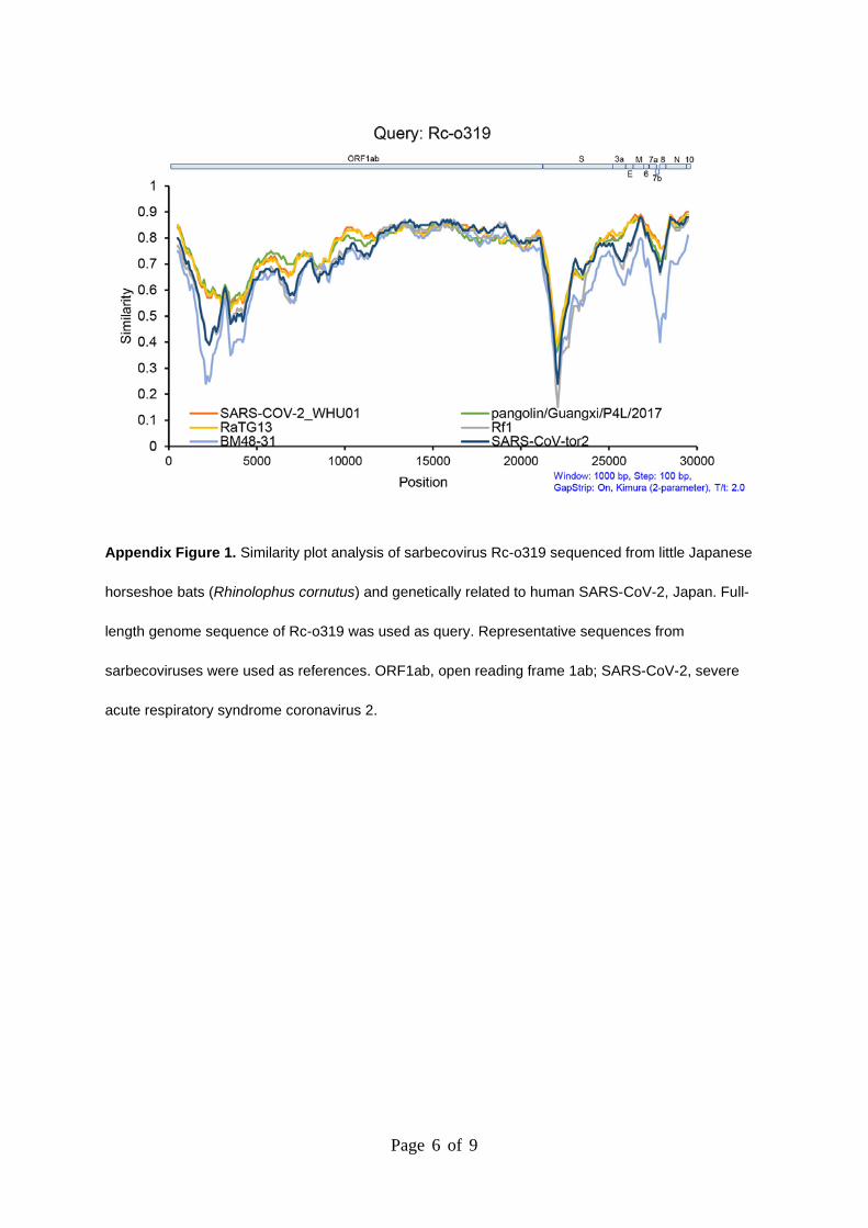
Appendix Figure 1. Similarity plot analysis of sarbecovirus Rc-o319 sequenced from little Japanese
horseshoe bats (Rhinolophus cornutus) and genetically related to human SARS-CoV-2, Japan. Full-
length genome sequence of Rc-o319 was used as query. Representative sequences from
sarbecoviruses were used as references. ORF1ab, open reading frame 1ab; SARS-CoV-2, severe
acute respiratory syndrome coronavirus 2.
Page 7 of 9
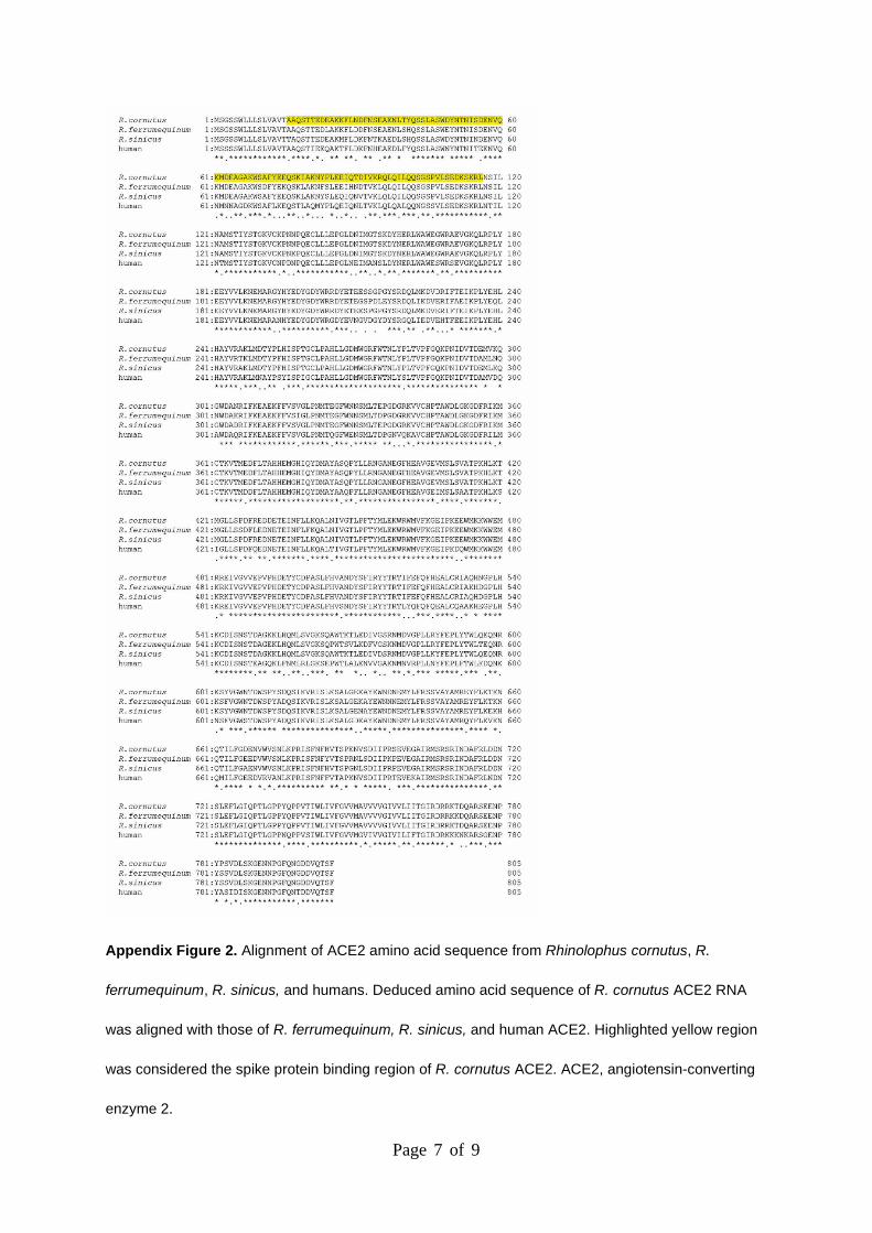
Appendix Figure 2. Alignment of ACE2 amino acid sequence from Rhinolophus cornutus, R.
ferrumequinum, R. sinicus, and humans. Deduced amino acid sequence of R. cornutus ACE2 RNA
was aligned with those of R. ferrumequinum, R. sinicus, and human ACE2. Highlighted yellow region
was considered the spike protein binding region of R. cornutus ACE2. ACE2, angiotensin-converting
enzyme 2.
Page 8 of 9
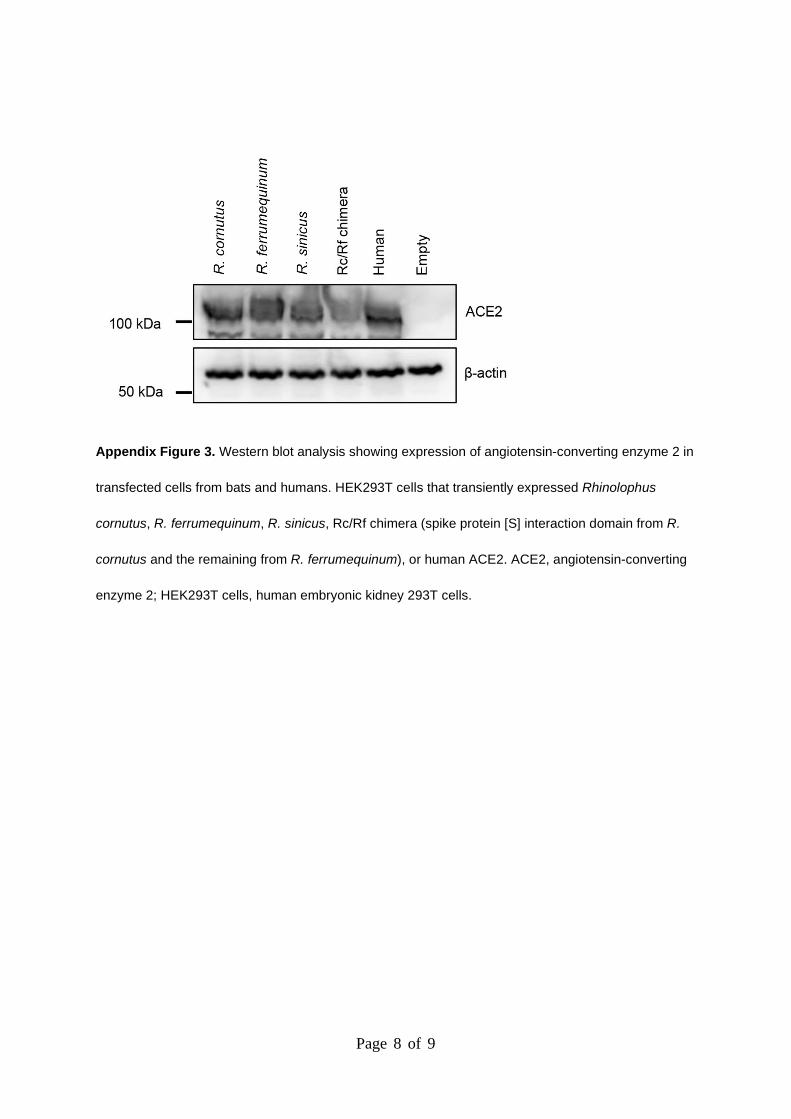
Appendix Figure 3. Western blot analysis showing expression of angiotensin-converting enzyme 2 in
transfected cells from bats and humans. HEK293T cells that transiently expressed Rhinolophus
cornutus, R. ferrumequinum, R. sinicus, Rc/Rf chimera (spike protein [S] interaction domain from R.
cornutus and the remaining from R. ferrumequinum), or human ACE2. ACE2, angiotensin-converting
enzyme 2; HEK293T cells, human embryonic kidney 293T cells.
Page 9 of 9
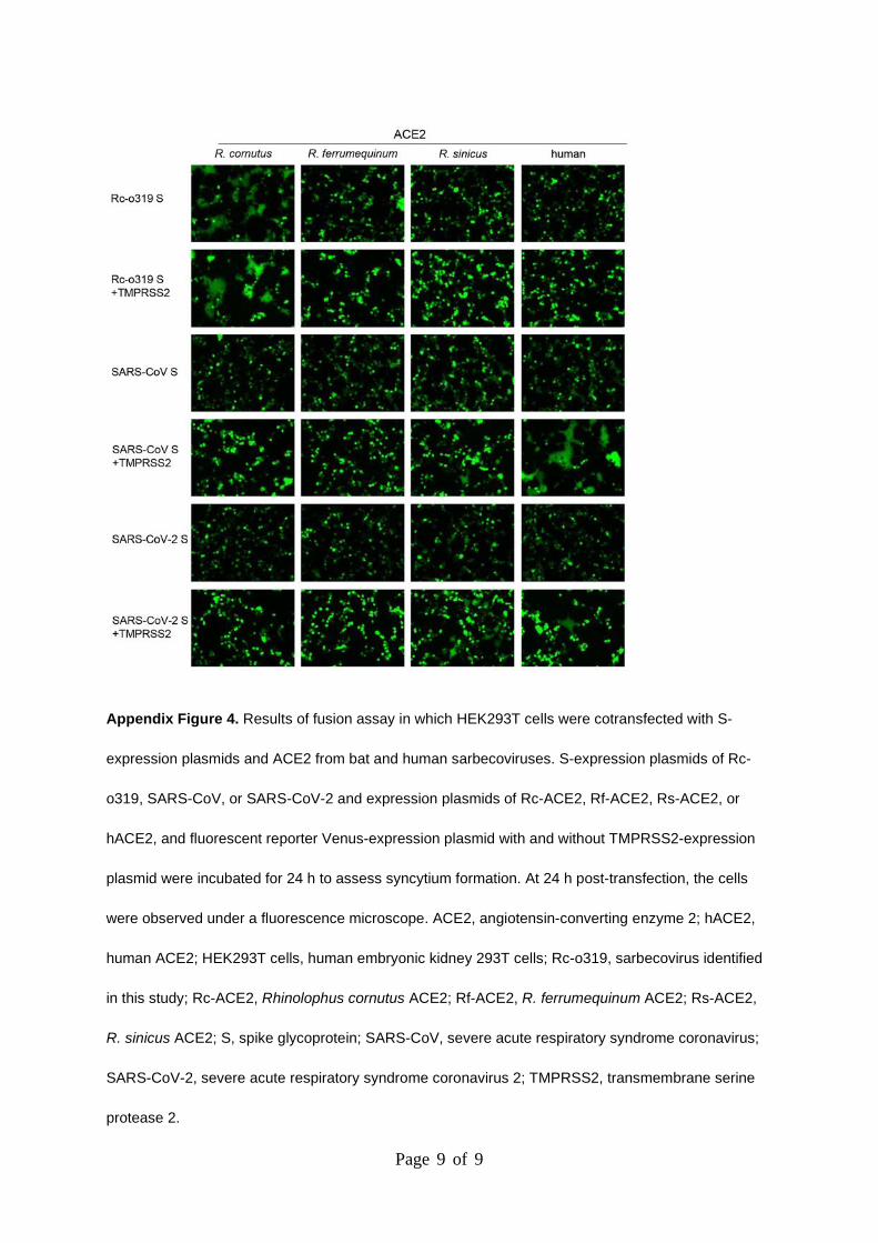
Appendix Figure 4. Results of fusion assay in which HEK293T cells were cotransfected with S-
expression plasmids and ACE2 from bat and human sarbecoviruses. S-expression plasmids of Rc-
o319, SARS-CoV, or SARS-CoV-2 and expression plasmids of Rc-ACE2, Rf-ACE2, Rs-ACE2, or
hACE2, and fluorescent reporter Venus-expression plasmid with and without TMPRSS2-expression
plasmid were incubated for 24 h to assess syncytium formation. At 24 h post-transfection, the cells
were observed under a fluorescence microscope. ACE2, angiotensin-converting enzyme 2; hACE2,
human ACE2; HEK293T cells, human embryonic kidney 293T cells; Rc-o319, sarbecovirus identified
in this study; Rc-ACE2, Rhinolophus cornutus ACE2; Rf-ACE2, R. ferrumequinum ACE2; Rs-ACE2,
R. sinicus ACE2; S, spike glycoprotein; SARS-CoV, severe acute respiratory syndrome coronavirus;
SARS-CoV-2, severe acute respiratory syndrome coronavirus 2; TMPRSS2, transmembrane serine
protease 2.