development and validation of a novel in situ - usask.cas new/tompsett... · development and...
TRANSCRIPT

Department of Veterinary Biomedical Sciences and Toxicology Centre, University of Saskatchewan
Development and validation of a novel in situ hybridization system to detect gene
expression and histological structure as biomarkers of chemical exposure in Japanese
medaka
Amber TompsettAquatic Toxicity Workshop
Halifax, Nova ScotiaOctober 3, 2007

Department of Veterinary Biomedical Sciences and Toxicology Centre, University of Saskatchewan
Background• EDC’s hypothesized to elicit a wide variety of adverse effects:
- Promotion of hormone-dependent cancers - Reproductive tract disorders - Reduction in reproductive fitness
• Most screening for the effects of EDCs was limited to measuring direct effects- Receptor binding of steroid hormone receptors
• However, chemicals can cause both direct (receptor-mediated) and indirect effects through changes in signal transduction pathways.

Department of Veterinary Biomedical Sciences and Toxicology Centre, University of Saskatchewan
Research Needs
• Methods are needed that:– Permit the analysis of multiple effects. – Can look at these effects simultaneously in a
number of tissues, including during critical windows of development
– Effects analyzed spatially
Department of Veterinary Biomedical Sciences and Toxicology Centre, University of Saskatchewan
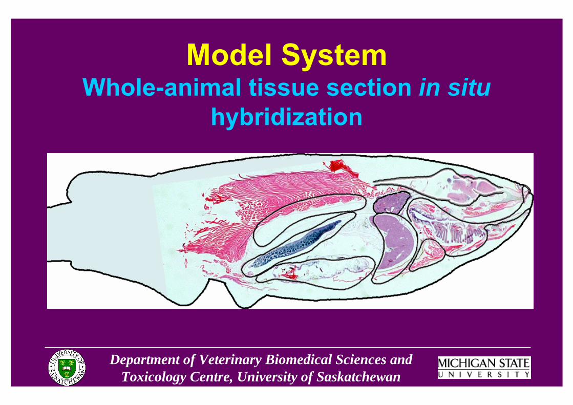
Model SystemWhole-animal tissue section in situ
hybridization

Department of Veterinary Biomedical Sciences and Toxicology Centre, University of Saskatchewan
Fixed RNA in tissue
Probe-RNA hybrid in tissue
In situ hybridization
Antisense RNA probe
In situ hybridization

Department of Veterinary Biomedical Sciences and Toxicology Centre, University of Saskatchewan
In situ hybridization
Fixed RNA in tissue
Fixed RNA in tissue
In situ hybridization
Sense RNA probe

Department of Veterinary Biomedical Sciences and Toxicology Centre, University of Saskatchewan
Whole Animal In situ hybridization
• Compatible with histopathology and IHC methods
• Allows spatial analysis of gene expression
• Small samples are preferable
• Some drawbacks: time and labor intensive

Department of Veterinary Biomedical Sciences and Toxicology Centre, University of Saskatchewan
CYP11A & B; CYP17; CYP19A; 3β-HSD; 11β-HSD; 17β-HSD; StAR; ERα & β; AR; GtHreceptors; Activin/Inhibin
Gonads
CYP11A & B; CYP17; 3β-HSD; 17β-HSD; StAR; AR; GtH receptors; Activin/InhibinAdrenals
GnRH receptors; GtH I & II; ERα & β; AR; Activin/InhibinPituitary
CYP19B; GnRHs; ERα & β; ARBrain
Primary Gene TargetsTissue
Gonads
LiverBrain
Pituitary
GnRH GRIF
Gonadotropins
Feedback
Sex SteroidsVitellogenin
Adrenals
Target Genes Along the HPG-Axis
Department of Veterinary Biomedical Sciences and Toxicology Centre, University of Saskatchewan
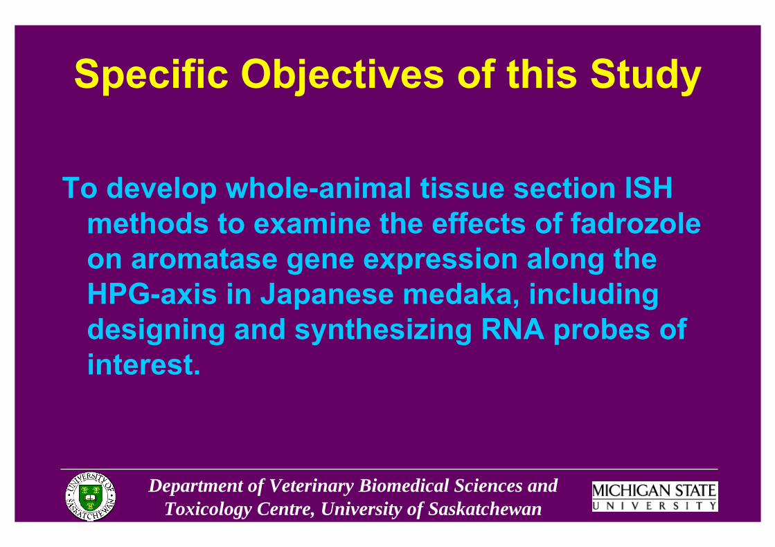
Specific Objectives of this Study
To develop whole-animal tissue section ISH methods to examine the effects of fadrozole on aromatase gene expression along the HPG-axis in Japanese medaka, including designing and synthesizing RNA probes of interest.

Department of Veterinary Biomedical Sciences and Toxicology Centre, University of Saskatchewan
Methods• Fixed and paraffin embedded samples
• 7μm tissue sections on slides-ISH with 35S-labeled CYP19a probe-H & E staining
• Detection of ISH signal-Radiography-Categorical classification system

Department of Veterinary Biomedical Sciences and Toxicology Centre, University of Saskatchewan
Methods – What didn’t work
• Cryosectioning • DIG-labeled probes

Department of Veterinary Biomedical Sciences and Toxicology Centre, University of Saskatchewan
Fadrozole Validation Exposure • 4 month old medaka
• Control and 1, 10, and 100 ug/L fadrozole treatments
• 2 replicate tanks of each treatment-10 fish per tank-5 male, 5 female
• 7 day static renewal
Department of Veterinary Biomedical Sciences and Toxicology Centre, University of Saskatchewan
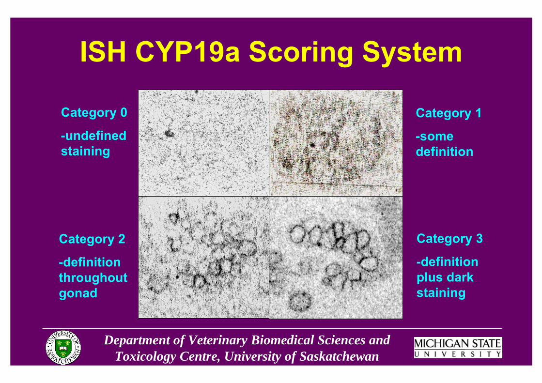
ISH CYP19a Scoring System
Category 0
-undefined staining
Category 3
-definition plus dark staining
Category 2
-definition throughout gonad
Category 1
-some definition
Department of Veterinary Biomedical Sciences and Toxicology Centre, University of Saskatchewan
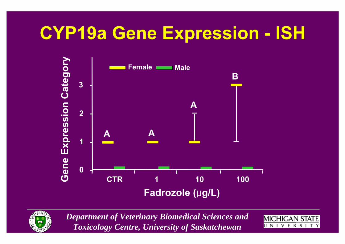
CYP19a Gene Expression - ISH
Fadrozole (μg/L)
Gen
e Ex
pres
sion
Cat
egor
y
CTR 1 10 1000
1
2
3
A A
A
BFemale Male
Department of Veterinary Biomedical Sciences and Toxicology Centre, University of Saskatchewan
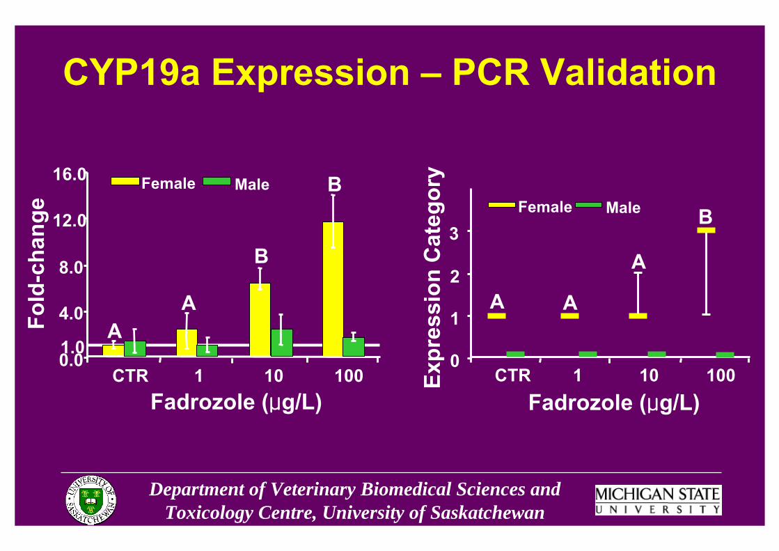
CYP19a Expression – PCR Validation
Fadrozole (μg/L)
Fold
-cha
nge
CTR 1 10 1000.0
4.0
8.0
12.0
16.0
1.0A
A
B
BFemale Male
Expr
essi
on C
ateg
ory
CTR 1 10 1000
1
2
3
A A
A
BFemale Male
Fadrozole (μg/L)

Department of Veterinary Biomedical Sciences and Toxicology Centre, University of Saskatchewan
Histological Evaluation - Male
SC
SC
SZ SZ
CTR 100μg/L

Department of Veterinary Biomedical Sciences and Toxicology Centre, University of Saskatchewan
Histological Evaluation - FemaleCTR 1μg/L
100μg/L 100μg/L
VOMO
MO
VO
VO
VO

Department of Veterinary Biomedical Sciences and Toxicology Centre, University of Saskatchewan
Exposure Conclusions• Fadrozole increased expression of CYP19a in female
medaka gonads
• Induced morphological changes in female gonads
• No measurable effects on male gene expression or histology
• ISH useful for determining spatial aspects of gene expression

Department of Veterinary Biomedical Sciences and Toxicology Centre, University of Saskatchewan
Overall Conclusions
• Successfully developed an ISH method that permitted the identification of changes in CYP19a gene expression along the HPG-axis in medaka
• Applied histological techniques to analyze gonadal morphology in the same fish
Department of Veterinary Biomedical Sciences and Toxicology Centre, University of Saskatchewan
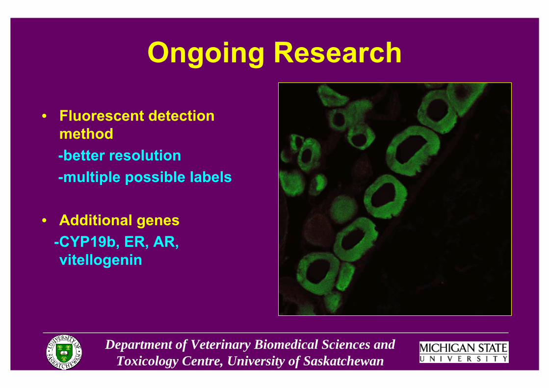
Ongoing Research
• Fluorescent detection method-better resolution-multiple possible labels
• Additional genes-CYP19b, ER, AR, vitellogenin
Department of Veterinary Biomedical Sciences and Toxicology Centre, University of Saskatchewan
Funding for this project was provided through an EPA STAR Grant (EPA Project #RD831849601-0).

Department of Veterinary Biomedical Sciences and Toxicology Centre, University of Saskatchewan
Acknowledgements
• Dr. John Giesy• June Woo Park• Dr. Markus Hecker• Dr. Paul Jones• Dr. John Newsted• Howard Zhang
• MSU students and staff• City U students and staff• ENTRIX staff• Committee Members:
-Dr. Norbert Kaminski-Dr. Steve Bursian

Department of Veterinary Biomedical Sciences and Toxicology Centre, University of Saskatchewan
Contact Information
• Amber TompsettEnvironmental Toxicology LaboratoryUniversity of SaskatchewanSaskatoon, SK S7N [email protected]
Department of Veterinary Biomedical Sciences and Toxicology Centre, University of Saskatchewan
Endocrine Disruption
“...an exogenous agent that interferes with the synthesis, secretion, transport, binding, action, or elimination of natural hormones in the body that are responsible for the maintenance of homeostasis, reproduction, development, and/or behavior.”
Kavlock et al., 1996Research needs for the assessment of environmental effects of endocrine disruptors: a report of the USEPA-sponsored workshop

Department of Veterinary Biomedical Sciences and Toxicology Centre, University of Saskatchewan
CYP19a Radiographs – Proof of Concept
CO EO
Radiographs of female fish from control (A) and 100 ug/L fadrozole (B) treatments. The control ovary (CO) shows no CYP19a expression; the exposed ovary (EO) shows CYP19a expression.
A B

Department of Veterinary Biomedical Sciences and Toxicology Centre, University of Saskatchewan
CYP19a Radiographs
CT ET
Radiographs of male fish from control (A) and 100 ug/L fadrozole (B) treatments. Both the control testis (CT) and exposed testis (ET) show similar levels of CYP19a expression.
A B
Department of Veterinary Biomedical Sciences and Toxicology Centre, University of Saskatchewan
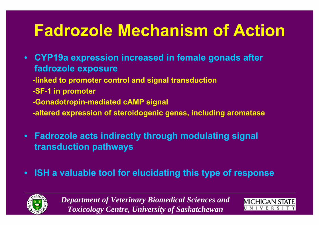
Fadrozole Mechanism of Action• CYP19a expression increased in female gonads after
fadrozole exposure-linked to promoter control and signal transduction-SF-1 in promoter-Gonadotropin-mediated cAMP signal-altered expression of steroidogenic genes, including aromatase
• Fadrozole acts indirectly through modulating signal transduction pathways
• ISH a valuable tool for elucidating this type of response