development of analytical tools to enrich and identify
TRANSCRIPT
Development of analytical tools to enrich and identify protein dopaminylation
Emily J Myers
A dissertation
submitted in partial fulfillment of the
requirements for the degree of
Doctor of Philosophy
University of Washington
2020
Reading Committee:
Shao-En Ong, Chair
Sandra Bajjalieh
Larry Zweifel
Program Authorized to Offer Degree:
Department of Pharmacology

©Copyright 2020
Emily J Myers
iii
University of Washington
Abstract
Development of analytical tools to enrich and identify protein dopaminylation
Emily J Myers
Chair of Supervisory Committee:
Shao-En Ong
Department of Pharmacology
Dopamine homeostasis and oxidative stress are thought to be key players in the etiology of
Parkinson’s disease (PD). PD is a disorder characterized by the death of dopaminergic neurons in the
substantia nigra and the formation of protein aggregates containing α-synuclein. Aberrant cytosolic
dopamine levels have been linked to mitochondrial and lysosomal dysfunction, oxidative stress, and
increased expression of α-synuclein in dopaminergic neurons. Outside of vesicles, dopamine is rapidly
oxidized and accumulates in cells as various polymerized forms including neuromelanin. Oxidized
dopamine can also covalently modify nearby proteins leading to structural and functional consequences,
including aggregation. Currently, little is known about dopaminylation of proteins as dopamine is not
widely recognized as a post translational modification. While several studies have identified dopamine-
protein adducts, these studies are limited in scope or rely on a priori knowledge of likely candidate
proteins due to lack of relevant discovery tools. Here, we describe several non-targeted approaches to
investigate dopaminylation, including a method for enrichment, identification, and site-localization of
dopaminylated peptides. Our method uses m-aminophenylboronic acid resin for covalent capture of the
cis-diol moiety of dopamine to enrich dopaminylated peptides followed by identification using tandem
mass spectrometry. Using this strategy, we identify over 1800 dopaminylated peptides from cultured cells
and 20 dopaminylated peptides from post-mortem human brain tissue. In both cultured cells and brain

iv
tissue, we identified dopaminylation at Cys147 in SOD1. SOD1 is a key protein in oxidative stress and
neurodegeneration, and modifications of Cys147 promote protein dysfunction and aggregation. We
believe these approaches to be powerful tools to characterize site-specific dopaminylation of proteins,
enabling mechanistic studies into the role of dopaminylation in the pathogenesis of Parkinson’s disease.

v
List of Figures ............................................................................................................................................... vii
List of Tables ............................................................................................................................................... viii
Chapter 1. Introduction .................................................................................................................................. 1
1.1 Dopamine and its Role in Physiology .......................................................................................... 1
1.2 Neural Circuitry of Dopamine Pathways ...................................................................................... 3
1.3 Parkinson’s Disease .................................................................................................................... 4
1.4 Dopamine Reactivity and Chemistry ........................................................................................... 6
1.5 Mass Spectrometry to Study Post-Translational Modificationss ............................................... 10
Chapter 2. Developing unbiased proteomic approaches to identify dopaminylated peptides .................... 12
2.1 Introduction ................................................................................................................................ 12
2.2 Results ....................................................................................................................................... 13
2.2.1 Dopamine modifies proteins in vitro ...................................................................................... 13
2.2.2 Enrichment of dopaminylated peptides and site localization ................................................ 15
2.2.3 Enrichment and identification of in vivo dopaminylated peptides .......................................... 18
2.2.4 Enrichment and indentification of dopaminylated peptides from human brain ..................... 23
2.3 Materials and Methods .............................................................................................................. 25
2.4 Discussion.................................................................................................................................. 29
2.5 Supplemental Figures ................................................................................................................ 31
2.6 Acknowledgements .................................................................................................................... 31
Chapter 3. Alternative methods to study dopaminylation ........................................................................... 32
3.1 Introduction ................................................................................................................................ 32
3.2 Results ....................................................................................................................................... 33
3.2.1 Biotin-dopamine..................................................................................................................... 33
3.2.2 nIRF confocal imaging ........................................................................................................... 39
3.3 Materials and Methods .............................................................................................................. 41

vi
3.4 Discussion.................................................................................................................................. 44
3.5 Suppplemental Figures .............................................................................................................. 46
References .................................................................................................................................................. 47
Vita .............................................................................................................................................................. 53

vii
List of Figures
Scheme 1.1. Biosynthesis of dopamine ........................................................................................... 1
Figure 1.1. Dopamine metabolism in the neuron ............................................................................. 2
Scheme 1.2. Steps of DA oxidation to form neuromelanin .............................................................. 7
Figure 2.1. Dopamine oxidation and protein modification .............................................................. 15
Figure 2.2. Dopaminylated peptides and enrichment .................................................................... 17
Figure 2.3. Proteins from biological samples are dopaminylated .................................................. 22
Figure 2.4. Dopaminylated peptides from human brain ................................................................. 24
Figure S2.1. Additional dopaminylated peptides............................................................................ 29
Figure 3.1. Biotin-dopamine modifies peptides and proteins ......................................................... 35
Figure 3.2. Biotin-dopamine differentially modifies proteins in N2A cells ...................................... 37
Figure 3.3. Biotin-dopamine differentially modifies protiens in differentiated SH-SY5Y cells ........ 38
Figure 3.4. nIRF detection of dopamine quinones in HEK-VMAT cells ......................................... 40
Figure S3.1. Optimizing BD treatment ........................................................................................... 46

viii
List of Tables
Table 2.1. Synthetic peptides ......................................................................................................... 16
Table 2.2. Comparison of enriched and unenriched samples ....................................................... 19

ix
Glossary
Abbreviation Full Name
AADC Amino acid decarboxylase
ADH Aldehyde dehydrogenase
ALS Amyotrophic lateral sclerosis
APLP2 Amyloid-like precursor protein 2
APP Amyloid precursor protein
ATP Adenosine triphosphate
ATP13A1 ATPase 13A1
Aβ42 Amyloid-β 42
BAG3 BCL2 associated athanogene 3
BD Biotin-dopamine
BK Bromomethyl ketone
BSA Bovine serum albumin
CAM Chloroacetamide
CAMK2G Calcium/Calmodulin dependent protein kinase II gamma
CCB Colloidal coomassie blue
CDC34 Cell division cycle 34
CISD2 CDGSH iron sulfur domain 2
CNS Central nervous system
COMT Catechol-O-methyltransferase
CSF Cerebral spinal fluid
Cys Cysteine
DA Dopamine
DAQ Dopamine quinone
DASQ Semiquinone dopamine radical
DAT Dopamine active transporter
DJ-1 Protein deglycase
DOPAC 3,4-Dihydroxyphenylacetic acid
DOPAL 3,4-Dihydroxyphenylacetaldehyde
DSS Disuccinimidylsuberate
DTT Dithiothreitol
DβH Dopamine-β-hydroxylase
FDR False discovery rate
GABA ɣ-Aminobutyric acid
GAPDH Glyceraldehyde 3-phosphate dehydrogenase
GBA β-glucocerebrosidase
GPHN Gephyrin
HCD Higher-energy collisional dissociation
HEK Human embryonic kidney

x
Glossary, cont.
Abbreviation
Full Name
His Histidine
HMOX2 Heme oxygenase 2
HMW High molecular weight
HRP Horseradish peroxidase
HSP Heat shock protein
HVA Homovanillic acid
IA Iodoacetamide
iBAQ Intensity-based absolute quantification
IMAC Immobilized metal affinity chromatography
LB Lewy body
LC Liquid chromatography
LDAC Leucodopaminechrome
L-DOPA Levodopa, l-3,4-dihdroxyphenyalanine
LRRK2 Leucine-rich repeat kinase 2
Lys Lysine
m/z Ratio of mass to charge
MAO Monoamine oxidase
MAPT Microtubule-associated protein tau
MDMA Methylenedioxymethamphetamine
MeCN Acetonitrile
MPTP 1-methyl-4-phenyl-1,2,3,6-tetrahydropyridine
MS Mass spectrometry
N2A Neuro-2a, mouse neuroblastoma cell line
NE Norepinephrine
nIRF Near-infrared fluorescence
NM Neuromelanin
PBA m-Aminophenylboronate
PBS Phosphate buffered saline
PD Parkinson's disease
PINK1 PTEN induced kinase 1
PPP5C Protein phosphatase 5 catalytic subunit
PRDX Peroxiredoxin
PRKN Parkin RBR E3 ubiquitin protein ligase
PSM Peptide-spectrum matches
PTM Post-translational modification
RA Retinoic acid
ROS Reactive oxygen species
RTN3 Reticulon 3
SDS-PAGE Sodium dodecyl sulfate-polyacrylamide gel electrophoresis
xi
Glossary, cont.
Abbreviation Full Name
SH-SY5Y Human neuroblastoma cell line
SNCA α-Synuclein
SNpc Substantia nigra pars compacta
SOD1 Superoxide dismutase 1
SOD2 Superoxide dismutase 2
TECP Tris(2-carboxyethyl)phosphine
TFA Trifluoroacetic acid
TH Tyrosine hydroxylase
TPA 12-O-tetradecanoyl-phorbol-13 acetate
Tyr Tyrosine
UbE2G1 Ubiquitin conjugating enzyme E2 G1
VMAT2 Vesicular monoamine transporter 2
VTA Ventral tegmental area
XIC Extracted ion chromatograms

xii
Acknowledgements
I would like to acknowledge my mentor, Shao-En, for his guidance and support throughout
graduate school. The freedom to explore my interests both scientifically and professionally instilled a
strong sense of purpose and direction in me. And I am grateful to have learned how to take the failures
with the success, and roll with the punches.
I also want to thank my thesis committee members, Sandra Bajjalieh, Larry Zweifel, Ning Zheng,
and Jim Bruce who each provided advice and support throughout graduate school. I also want to thank
my collaborators who invested in my research by sharing reagents and tissues that were critical to my
thesis work: Gary Miller and Dirk Keene. I also want to thank the department of Pharmacology, especially
Debbie, Jenny, and Diane.
I acknowledge and thank my lab mates for their guidance, discussion, support, and engagement.
Ho-Tak and Martin were instrumental in my training as a scientist, a mass spectrometrist, and a lab
community member. I will miss sharing beers on Fridays and cookies any day they were needed.
Next, I would like to thank my cohort: Eedann, Kristine, Rigney, Sabrina, Samara, and Stacey.
We started this journey together and I am so grateful for your friendship and for each of your unique
perspectives. Eedann, I knew that first of day of recruitment we would be friends and I am so grateful to
have your friendship. Rigney, I never imagined a person who is so kind and loving and also an amazing
scientist and dancer. I am so proud of you, my sister, and am humbled by your friendship.
I thank my big brother, Samuel Myers, for your support and guidance both throughout graduate
school and life. I am unbelievably lucky to have you as a role model, colleague, and friend. Sam, you
may not have been on my thesis advisory committee, but you will always be on my life advisory
committee.
I also thank my mother Jill for always encouraging me to follow my dreams and create a life filled
with love. To my grandfather Don, who nurtured my curiosity about the word and answered my unending
questions, thank you. I know you will always walk beside me. And to my dearest friend Kim, thank you for
your love and for always being my home base.

xiii
Finally, I thank my women scientist friends: Sarah Myhre, Jeanna Wheeler, Heather Currey,
Laura Osburn, Jodie Katon, Katie Reichard, and Sharona Gordon. With each of you, I have found
community and nourishment throughout my time in graduate school and am thrilled to support you on
your journeys ahead.
1
Chapter One: Introduction
1.1. Dopamine and its Role in Physiology
Dopamine (DA) is one of the most important and abundant catecholamines in the human body.
DA is a common precursor to other neurotransmitters including epinephrine and norepinephrine and is
itself a critical neurotransmitter. In the periphery, DA acts as an important hormone for electrolyte balance
and is a primary neurotransmitter in important functions such as reward, cognition, movement and motor
control, and lactation. Therefore, disruption of DA homeostasis and signaling can lead to disease and
psychiatric disorders such as Parkinson’s Disease, schizophrenia, and substance abuse disorders [1, 2].
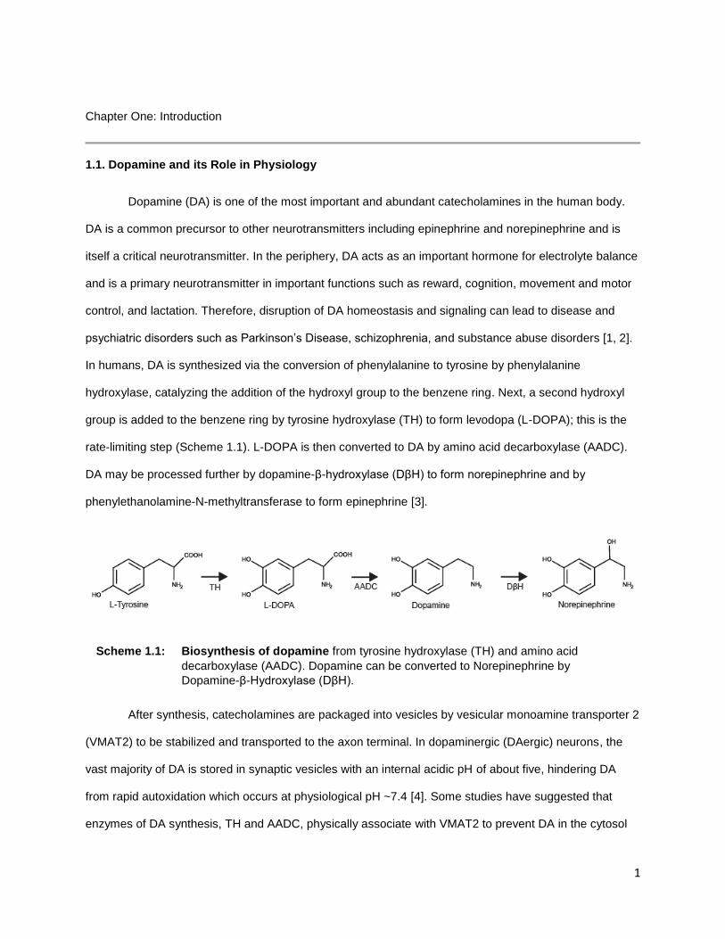
In humans, DA is synthesized via the conversion of phenylalanine to tyrosine by phenylalanine
hydroxylase, catalyzing the addition of the hydroxyl group to the benzene ring. Next, a second hydroxyl
group is added to the benzene ring by tyrosine hydroxylase (TH) to form levodopa (L-DOPA); this is the
rate-limiting step (Scheme 1.1). L-DOPA is then converted to DA by amino acid decarboxylase (AADC).
DA may be processed further by dopamine-β-hydroxylase (DβH) to form norepinephrine and by
phenylethanolamine-N-methyltransferase to form epinephrine [3].
After synthesis, catecholamines are packaged into vesicles by vesicular monoamine transporter 2
(VMAT2) to be stabilized and transported to the axon terminal. In dopaminergic (DAergic) neurons, the
vast majority of DA is stored in synaptic vesicles with an internal acidic pH of about five, hindering DA
from rapid autoxidation which occurs at physiological pH ~7.4 [4]. Some studies have suggested that
enzymes of DA synthesis, TH and AADC, physically associate with VMAT2 to prevent DA in the cytosol
Scheme 1.1: Biosynthesis of dopamine from tyrosine hydroxylase (TH) and amino acid
decarboxylase (AADC). Dopamine can be converted to Norepinephrine by
Dopamine-β-Hydroxylase (DβH).

2
[5]. In DAergic neurons, like most neurons, chemical neurotransmission is initiated by a depolarization of
the presynaptic terminal often due to an action potential. Upon depolarization, synaptic vesicles fuse with
the plasma membrane, releasing DA into the synaptic cleft where it can interact with DA receptors.
DA receptors are G-protein coupled receptors prominent in the central nervous system, cardio-
pulmonary system, and renal system. DA receptors can be divided into two general subtypes: Gs coupled
(D1 and D5) and Gi coupled (D2, D3, and D4) to affect downstream signaling [6]. Neurotransmission is
halted when DA is cleared from the synapse, through both diffusion and uptake by the dopamine
transporter (DAT) and recycled or degraded (Figure 1.1). The major metabolic pathway for DA
degradation starts with monoamine oxidase (MAO) acting on DA through oxidative deamidation to
produce 3,4-dihydroxyphenylacetaldehyde (DOPAL), hydrogen peroxide, and ammonia. DOPAL is
converted to its acid (DOPAC) by aldehyde dehydrogenase (ADH). DOPAL and DOPAC metabolites are
also easily oxidized [7]. Finally catechol-O-methyltransferase (COMT) converts DOPAC to the
nonreactive metabolite homovanillic acid (HVA) [8].
Figure 1.1. Dopamine metabolism in the neuron. As L-DOPA is converted to DA by AADC, DA is
transported into synaptic vesicles by VMAT2. Unpackaged DA in the cytosol undergoes oxidative
deamidation by MAO to produce DOPAL, which undergoes additional reactions to produce unreactive
HVA. When stimulated, the neuron releases DA into the synaptic cleft to transduce signal to post-
synaptic DA receptors. Excess DA is transported back into the presynaptic neuron via DAT and either
recycled or degraded.
3
The DA molecule appears simple: a primary amine connected by an ethyl chain to a benzene ring
with two hydroxyl groups (a catechol), but DA’s chemistry is complex. DA, and to a lesser extent other
catecholamines, are spontaneously oxidized to form dopamine quinone (DAQ) in the presence of ambient
oxygen, reactive oxygen species, redox-capable metals, peroxidases, and at neutral pH [9]. DAQ is
capable of many downstream reactions including protein modification (dopaminylation) and forming DA
polymers. Some plants and animals use these reactions to their advantage. For example, zebra mussels
incorporate DA into their foot proteins where DA forms protein aggregates and covalent attachments with
rocks upon exposure to the basic ocean water at pH 8.1 [10, 11]. Grasshoppers use polyDA to crosslink
the structural proteins in the mandible to strengthen the mandible and prevent breakdown [12]. Simply
stated, dopamine can be used as a glue to crosslink proteins together and form insoluble aggregates. In
the brain, insoluble protein aggregates are toxic and the bases for several neurodegenerative diseases.
Therefore, to protect against potential “protein glue”, DA must be tightly regulated and contained. If DA
leaks into the cytosol, DA will oxidize at cytosolic pH and may modify nearby proteins. Small amounts of
dopaminylation is unlikely to induce cell death, but cells may accumulate modified proteins as they age,
eventually leading to protein aggregates. While protein aggregation is a common feature in many
neurodegenerative diseases, the role of dopamine modifications in protein aggregation remains unknown.
Tools to study DA as a protein post-translational modification (PTM) are limited, and currently no
unbiased method exists to identify specific protein sites of dopaminylation.
1.2. Neural Circuitry of Dopamine Pathways
Identification of DA as a neurotransmitter in the brain was a key finding by Arvid Carlsson and led
Carlsson, Eric Kandel, and Paul Greengard to win the Nobel Prize in Physiology or Medicine in 2000 for
their work on catecholamine neurotransmission [13]. Since their discovery four major DA circuits have
been described in the brain: mesocortical, mesolimbic, nigrostriatal, and tuberoinfundibular pathways.
The tuberoinfundibular pathway DAergic neurons project from the arcuate nucleus of the hypothalamus to
the pituitary gland to affect prolactin release. In the mesocortical pathway, DAergic neurons project from
the ventral tegmental area (VTA) to the prefrontal cortex and are implicated in cognitive and emotional
behavior. The mesolimbic pathway also originates with DAergic neurons in the VTA and projects to
4
multiple areas of the limbic system, including the olfactory bulb, amygdala, piriform cortex, lateral septal
nuclei, and the nucleus accumbens. In this neural circuit, DA functions in emotion and reward systems.
[14] Together, the mesolimbic and mesocortical dopamine circuitry are engaged in reward learning and
concentration, whereas dysfunction in this circuitry contributes to neuropsychiatric disorders including
substance abuse disorder, attention deficit disorders, and schizophrenia [15].
The DAergic neurons of the nigrostriatal pathway have cell bodies in the substantia nigra pars
compacta (SNpc) and project to the caudate nucleus and the putamen in the striatum. The release of DA
in the striatum plays a significant role in motor function, regulating the direct and indirect pathways
important for fine motor control. In collaboration with other neurotransmitters like glutamate and ɣ-
aminobutyric acid (GABA), DA activation of D1-like receptors of the putamen, the “direct” pathway,
activates the motor cortices, while DA activation of D2-like receptors reduces activation of motor cortices,
stymieing voluntary movement [16]. Disruption of this system leads to loss of fine motor control and
difficulties initiating movement, and the loss of DAergic neurons in the SNpc produce the classical
symptoms of Parkinson’s disease: akinesia, rigidity, and tremor at rest.
Altered DA homeostasis in the central nervous system (CNS) is implicated in many other
psychiatric and neurodegenerative disorders beyond Parkinson’s disease. Schizophrenia is associated
with increased amphetamine-stimulated striatal DA release. DA pathway dysfunction has been implicated
in other mood disorders like depression and mania [17]. Many addictive drugs act on the dopamine
system, and pharmacological blockade of DA signaling can attenuate drug-associated reward [18]. DA
neurotransmission has also been implicated in Alzheimer’s disease, Huntington’s disease, and multiple
sclerosis, among others [19].
1.3. Parkinson’s Disease
Parkinson’s disease affects an estimated 10 million people worldwide and is the second most
common neurodegenerative disorder. This debilitating movement disorder is a result of degeneration of
several neuronal populations, however, SNpc DAergic neurons are impacted early, leading to loss of DA
in the nigrostriatal system. Death of these neurons is progressive and early intervention is difficult as
5
onset of clinical motor symptoms are associated with loss of 70-80% of striatal DA. PD can be classified
into four sub-types: 1. Idiopathic Parkinson’s (or Primary Parkinson’s) disease where the underlying
mechanisms are unclear; 2. Acquired Parkinson’s (or Secondary) disease which stems from exposure to
toxins; 3. Hereditary Parkinson’s disease which has a direct, genetic basis, and 4. Parkinson’s-like or
Parkinson’s-plus syndromes which represents a larger variety of diseases with similar symptoms and
pathology, including Dementia with Lewy Body disease and related synucleinopathies [20]. The canonical
symptoms of PD include tremor, dyskinesia and slow movement, rigidity, and progressive motor
dysfunction. Motor dysfunction is the main symptom at time of diagnosis, non-motor symptoms develop
much earlier but are often go undiagnosed. These include altered circadian rhythm, hyposmia,
depression, speech impairments, and constipation [21]. As disease progresses, additional non-motor
symptoms manifest including pain, fatigue, cognitive impairment, orthostatic hypotension, and dementia
[22].
The etiology of Parkinson’s disease is still largely unknown, although age, sex, genetics, and
environmental exposures have all been implicated. The majority of cases of PD (up to 90%) are
considered sporadic and have no direct genetic cause [23]. However, improvements in genome
sequencing technology have allowed researchers to identify a number of genes that are implicated in
disease, including SNCA, PRKN, PINK1, DJ-1, LRRK2, GBA, and MAPT [24]. Several of the proteins
from these genes are known to be prion-like and prone to aggregation. Specifically, α-synuclein (SNCA)
aggregates to form insoluble fibrils and are the major component in Lewy bodies [25]. Tau, when
hyperphosphorylated, self-assembles in “tau tangles” to form insoluble plaques. In addition to prion-like
proteins, transition metals with redox activity have also been implicated in the development of protein
aggregates in PD [26]. Patients with PD have higher levels of iron in the substantia nigra compared to
controls and copper levels are known to be elevated in the cerebral spinal fluid (CSF) of PD patients [27,
28]. Exposure to these metals promote oxidative stress and catalyze oligomerization of α-synuclein in the
brain [28, 29]. Other toxins including pesticides and herbicides are thought to play a role in disease
through increasing oxidative stress [30]. Rotenone, paraquat, dichlorodiphenyltrichloroethane (DDT),
dichlorophenoxyacetic acid, diazinon, chlorpyrifos, propargite, dieldrin, benomyl, maneb, and arsenic
6
exposure have been directly correlated to increased risk of PD [31]. Due to the multifactorial nature of this
disease, it is likely an interaction of genetics and environmental factors that lead to most cases of PD.
The role of dopamine in PD
The selective loss of DAergic cells in PD begs the question of both if and how DA contributes to
disease progression. Without tight regulation, DA is toxic in vivo and autoxidizes to form to reactive DA
metabolites, producing reactive oxygen species (ROS) including O2- and H2O2 [32]. Due to the clear toxic
effects of DA, DA metabolism and storage is an active area of research for neurobiologists. Studies in
post-mortem brain tissue suggest Parkinson’s patients have lower expression of mRNA for both DAT and
VMAT2 per cell and significant loss of VMAT2 function [33, 34]. Decreased expression and
pharmacological inhibition of VMAT2 result in Parkinson’s-like symptoms and neurodegeneration in
animal models [35]. Recently, mutations in VMAT2 that reduce function were linked to an infantile
parkinsonism-like condition [36]. Conversely, increased expression of VMAT2 has been associated with
decreased risk of PD and improved outcomes for DA toxicity [35]. Decades of research demonstrate the
importance of strict control and modulation of DA, however, the full consequence of mismanaged DA and
its toxicity remains to be seen.
1.4. Dopamine Reactivity and Chemistry
DA is widely acknowledged as toxic, both in vivo and in vitro. The relatively low free energy
barrier to oxidation of DA means it is a highly reactive molecule and can undergo spontaneous
autoxidation. Reactivity of DA increases under certain conditions, such as higher pH and the presence of
redox-active metals or other oxidants like oxygen or H2O2 [37]. DA reactions are complex and can
proceed through one or two electron processes to generate semiquinone dopamine radical (DASQ) or
dopamine quinone (DAQ), respectively. These reactive intermediates can undergo further oxidation or
spontaneous rearrangement to generate other reactive species which can modify proteins, oligomerize to
form polydopamine chains and neuromelanin (NM), react with antioxidants like ascorbate and glutathione,
or some combination of these depending on reaction conditions [32]. (Scheme 1.2)

7
In vivo, DA oxidation can be induced via H2O2 activation by heme or peroxidases, prostaglandin
H synthase, and activated microglia [38-40]. The presence of redox-active metal ions, such as copper
and iron, have also been implicated in DA induced neurodegeneration. For example, iron accumulates in
Lewy bodies of PD patients, and plaque deposits in Alzheimer’s patients contain iron bound by amyloid-
Beta [41, 42]. Researchers have also shown both copper and iron are necessary for the aggregation of α-
synuclein and elevation of DA levels induces aggregation [43]. DA oxidation promoted by metal ions can
generate several toxic species including DAQ, DASQ, O2-, H2O2, and -OH, creating a cascade of ROS
and oxidative stress [9].
Neuromelanin
Neuromelanin is a dark pigment found primarily in DAergic neurons of the SNpc and
noradrenergic cells of the locus coeruleus. NM is formed from the polymerization of DA and other
catecholamines (Scheme 1.2) and is structurally similar to other melanins, such as melanin found in skin.
The precursors to NM and melanin overlap: tyrosine, L-Dopa, DA, norepinephrine, and thiols [44].
However, unlike melanin synthesis which is tightly controlled through enzymatic reactions, generation of
Scheme 1.2: Steps of DA oxidation to form neuromelanin from DAQ by DA oxidative
polymerization (bottom). DAQ also modifies cysteine side chains (middle) and can
form poly-dopamine cysteine conjugates (top).
8
NM appears to be non-enzymatic and driven by autoxidation of catecholamines to quinones,
polymerization of quinone species, and recent studies suggest a role for cytosolic seeds of aggregated
protein or peptides in NM formation [45-47]. pH is also critical to DA oxidation as at physiological pH or
higher, the protons of the hydroxyl groups dissociate to form an electrophilic quinone, whereas DA at the
vesicular pH (pH ~5) is stable [4]. Redox-active metals likely play a role in NM catalysis; NM isolated from
the SNpc also contains significant amounts of metal ions, especially iron [48]. The resulting chemically
complex NM precursors, a mixture of DA and its derivatives, proteins, and metal ions, are engulfed in
autophagic vacuoles and fuse with lysosomes for degradation. However, lysosomal proteases are unable
to degrade NM precursors [47]. As the autolysosome fuses with other lysosomes and autophagic
vacuoles, NM precursors continue to accrue modifications including different proteins and lipids from
other lysosomes [47, 49]. Eventually, the NM complex is sequestered into specialized double membrane
autolysosome where it accumulates.
Human and primate brains have higher levels of NM compared to other vertebrates, and small
vertebrates like mice have almost undetectable levels making NM difficult to study in animal models [50,
51]. In humans, dark, pigmented cells are absent at birth, but NM can be seen by age 5 and continues to
accumulate throughout a lifetime [52]. This complex molecule has been used as a hallmark for
Parkinson’s disease and implicated in other age-related neurodegenerative diseases. The loss of
pigmentation in the SNpc, where NM-rich cells undergo cell death unlike non-pigmented DAergic cells in
the VTA, is a classic characteristic of Parkinson’s disease. However, the formation of NM may provide a
protective mechanism against accumulation of cytosolic DA by binding quinones and buffering against
subsequent ROS [53]. NM may act to chelate and trap metals, bind proteins including ferritin, and
sequester neurotoxins like paraquat and 1-methyl-4-phenyl-1,2,3,6-tetrahydropyridine (MPTP) [54-56]. It
is proposed that as NM accumulates these toxins, metals, and modified proteins, its capacity diminishes
and these are released back into the neuron, resulting in cell death [55, 57, 58] Once freed from the
granule, NM itself can activate microglia, initiate an inflammatory response, and exacerbate damage to
surviving cells [59]. While NM is thought to play a protective role in DAergic neurons, the accumulation of
toxins may contribute to age-related neuron loss. A complete picture of the function and structure of NM
and its role in PD has not been fully elucidated at this time.

9
Protein modification by DA and its derivatives
The modifications of proteins by DA and its oxidation products have long been thought to be a
major component of DA toxicity. The autoxidation of DA under physiological pH leads to DAQ formation, a
strong electrophile, and generates ROS. Studies from the 1980s demonstrated DA’s toxic reactivity
towards nucleophilic protein side-chains, specifically Cys, His, and Lys residues [60, 61]. In vitro studies
with free amino acids or small peptides found that the thiol groups of Cys residues were highly
susceptible to DA modification; Cys modification is preferred over ring closure to form
leucodopaminechrome (LDAC) (shown in Scheme 1.2) [62]. Research in this field is ongoing and relies
heavily on candidate-driven approaches and a priori knowledge of cellular stress. For example, in isolated
mitochondria exposed to DA, proteins of complex I, III, and IV of the mitochondrial electron transport
chain were found to be dopaminylated and showed reduced ATP production [63]. Actin, tubulin, GAPDH,
parkin, GBA, SOD2, and other cysteine containing proteins have also been described as dopaminylated
[63-65]. Furthermore, DA metabolites like DOPAL, have also been shown to modify both thiol and amine
groups in proteins [66, 67]. DA and DOPAL mediate the aggregation of α-synuclein, the primary
component of Lewy bodies, and DOPAL modified α-synuclein can activate microglia [68]. It is clear that
accumulation of DA and reactive metabolites depletes cellular stores of antioxidants and increases ROS,
promotes protein aggregation and modifications that lead to apoptosis, and increase microglia activation
and inflammation in the brain. Unfortunately, technologies previously available to study dopaminylation
have been very limited and currently there are no unbiased methods to identify dopaminylated sites on
proteins from biological samples. Much of the previous work used purified proteins and DA oxidation,
which is unlikely to accurately predict dopaminylation in cells. One previous study relied on treating
mitochondria with radiolabeled DA and 2-D gels coupled to LC-MS/MS to nominate potentially
dopaminylated proteins. However, without identification of specific sites of DA modification (site IDs),
functional analysis of dopaminylation is remained elusive. A more comprehensive methodology is needed
to identify modification sites on individual proteins from in vivo DA breakdown, to understand how DA
modifications affect protein structure and function, and to identify localization of toxic DA reactions in
cells. Furthermore, an unbiased approach to identify protein dopaminylation from biological samples
10
would be clinically relevant as none of the current methods can be applied to identify dopaminylation in
human post-mortem brain tissue.
1.5. Mass Spectrometry to Study Post-Translational Modifications
Post-translational modifications (PTMs) multiply the complexity of the proteome beyond the
genetically encoded protein sequence. PTMs covalently modify amino acid side chains and increase the
variety of functional groups beyond the standard 20 amino acids. These modifications include
phosphorylation, glycosylation, acetylation, ubiquitination, etc. and can affect a protein’s function,
localization, stability, or interaction partners. Proteins have a variety of possible PTMs and the
addition/removal of PTMs are commonly used in cellular signaling cascades. Many PTMs are added
enzymatically, but non-enzymatic PTMs also exist. For example, advanced glycation end products are a
collection of modifications on lysines and arginines that are associated with metabolic stress from glucose
metabolism. In high glucose conditions, the degradation of reducing sugars produces reactive species
that modify proteins [69]. These modifications have been associated with serious health conditions,
including the development of atherosclerotic plaques as well as uncontrolled blood sugar levels in
diabetes mellitus.
PTMs can be detected and identified by a variety of methods. Antibody-based methods, in which
an antibody is raised against a PTM side chain, are used to detect PTMs via Western blot and
immunofluorescence. Such antibodies that recognize specific PTMs have also been important in studying
the functional importance of these PTMs in certain biological contexts, examples include
phosphotyrosine-specific antibodies (4G10) or antibodies against epigenetic marks like Histone H3 lysine
9 tri-methylation (H3K9me3) [70, 71].
Alternatively, proteomic approaches to study PTMs utilize mass spectrometry (MS). MS is a
powerful and unbiased analytical technique that identifies proteins or peptides, including their modified
forms, by measuring their mass to charge (m/z) ratio of precursor ions and subsequent product ions to
determine peptide sequence. To do this, proteins are extracted from biological samples and digested to
peptides. Peptides are then separated via liquid chromatography (LC) over time, introduced into the gas
11
phase by electrospray ionization, and injected into a MS. The MS measures the m/z values for ions,
fragments them and analyzes mass of these fragments to produce a tandem mass spectrum (MS/MS).
On state-of-the-art MS instrumentation, a single LC-MS/MS run may produce tens of thousands of mass
spectra for complex peptide samples. Mass spectra are analyzed using automated peptide identification
software like MaxQuant/Andromeda, which searches the MS spectra against the user specified library of
possible peptides [72]. To identify PTMs, we expand our searches to include mass shifts of the
modification of interest.
For PTM analysis, it is often necessary to enrich pools of modified proteins or peptides from the
bulk protein sample as PTMs are substoichiometric, typically representing a small fraction of total protein
present in the cell. MS is typically used to analyze modified proteins or peptides isolated with affinity or
covalent capture enrichment from complex mixtures. One of the most widely used affinity-MS methods is
immobilized metal affinity chromatography (IMAC) enrichment of phosphorylated peptides coupled with
LC-MS/MS. This method has expanded our understanding of the phosphoproteome exponentially by
allowing researchers to study how phosphorylation patterns differ in cell types and change in cell
signaling and disease states. Unlike phosphorylation, however, enrichment strategies for DA-modified
peptides and proteins have not been explored for unbiased proteomics in such depth. This work
addresses strategies to enrich and identify dopaminylation with LC-MS/MS.
12
Chapter 2: Developing unbiased proteomic approaches to identify dopaminylated peptides
2.1. Introduction
PD affects an estimated 10 million people worldwide and is the second most common
neurodegenerative disorder. This movement disorder is a result of degeneration of several neuronal
populations, however, substantia nigra pars compacta dopaminergic neurons are impacted early, leading
to loss of DA in the nigrostriatal system [20]. Death of these neurons is progressive and takes years as
onset of clinical motor symptoms are associated with loss of 70-80% of striatal DA. In addition to
neurodegeneration, DA metabolism and resulting oxidants have been implicated in synaptic nerve
terminals damage caused by psychoactive drug use, including cocaine, methamphetamine, and
methylenedioxymethamphetamine (MDMA) [73-75]. Furthermore, known environmental toxins such as 1-
methyl-4-phenyl-1,2,3,6-tetrahydropyridine (MPTP) and rotenone cause oxidative damage to
dopaminergic neurons, which may be exacerbated by excess DA metabolism and altered DA
homeostasis [76, 77].
The toxic effects of DA are thought to be attributed, in part, to increases in cytosolic DA, which is
rapidly oxidized at physiological pH and modifies proteins and peptides (7). Protein dopaminylation can
alter function and structure of proteins and is an important outcome of increases in cytosolic DA.
However, dopaminylation is relatively unrecognized as a PTM despite its implications in PD. Currently,
studies of dopaminylation are limited to a few proteins due to lack of unbiased tools to identify
dopaminylated proteins. For example, proteins which have been identified as dopaminylated were treated
to high doses of DA on purified proteins [64, 65]. These studies fail to provide direct evidence of
dopaminylation in physiologically relevant conditions. Other groups have used 14C-DA to treat isolated
mitochondria or cell lines to indirectly identify several dopaminylated proteins, but this assay does not
specify dopaminylation at specific sites [63, 78]. Furthermore, neither of these approaches can be
translated to a wide range of biological and/or clinical samples. Most importantly, these techniques cannot
be used in clinical investigations to validate targets of dopaminylation or further elucidate functional
consequences as they rely on radioactive labeling or purified candidate proteins. Common approaches to
13
study PTMs, including antibodies to dopaminylation or affinity enrichment coupled to LC-MS/MS, have yet
to be developed. Therefore, compared to other PTMs, dopaminylation remains relatively under-examined.
This work will describe a novel chemical biological method for enrichment and identification of
native dopaminylated peptides extracted from cells or tissues via mass spectrometric-based proteomics.
We a use boronic acid-based covalent capture strategy, commonly used for enrichment of peptides
bearing other PTMs such as glycosylation and PARylation, for DA-modified peptide enrichment [79, 80].
This matrix, m-aminophenylboronate acid agarose (PBA), covalently but reversibly binds molecules
bearing cis-diols groups by forming boronic esters. We take advantage of the cis-diol moiety of dopamine
to capture dopaminylated peptides on PBA beads, release these peptides by hydrolysis in acidic
conditions, and analyze peptides via LC-MS/MS. We demonstrate enrichment of thousands of
dopaminylated peptides from 2 mg of protein with ~90% of total peptide signal from dopaminylated
peptides. We also identify dopaminylation sites on several key proteins implicated in oxidative stress and
DA metabolism. This method may facilitate future studies to identify pathological events leading
to dopaminergic neuron death in neurodegenerative conditions such as PD.
2.2. Results
2.2.1. Dopamine modifies proteins in vitro
Since DA reactivity is highly variable and can result in polyDA chains, melanin, and single or
multiple protein modifications including crosslinks, we initially sought to establish experimental conditions
to quickly induce single protein modifications and avoid extensive crosslinks and complicated polyDA
species. We tested various concentrations of DA on several different purified proteins: Aβ42, CDC34 and
UbE2G1. Aβ is of interest due to its established role in neurodegeneration and propensity to aggregate.
Alternatively, CDC34 and UbE2G1 were chosen due to their availability. Unfortunately, both Aβ42 and
CDC34 are prone to spontaneously form higher molecular weight species even in the absence of DA
(data not shown). Further experiments used UbE2G1 as it does not self-aggregate and contains residues
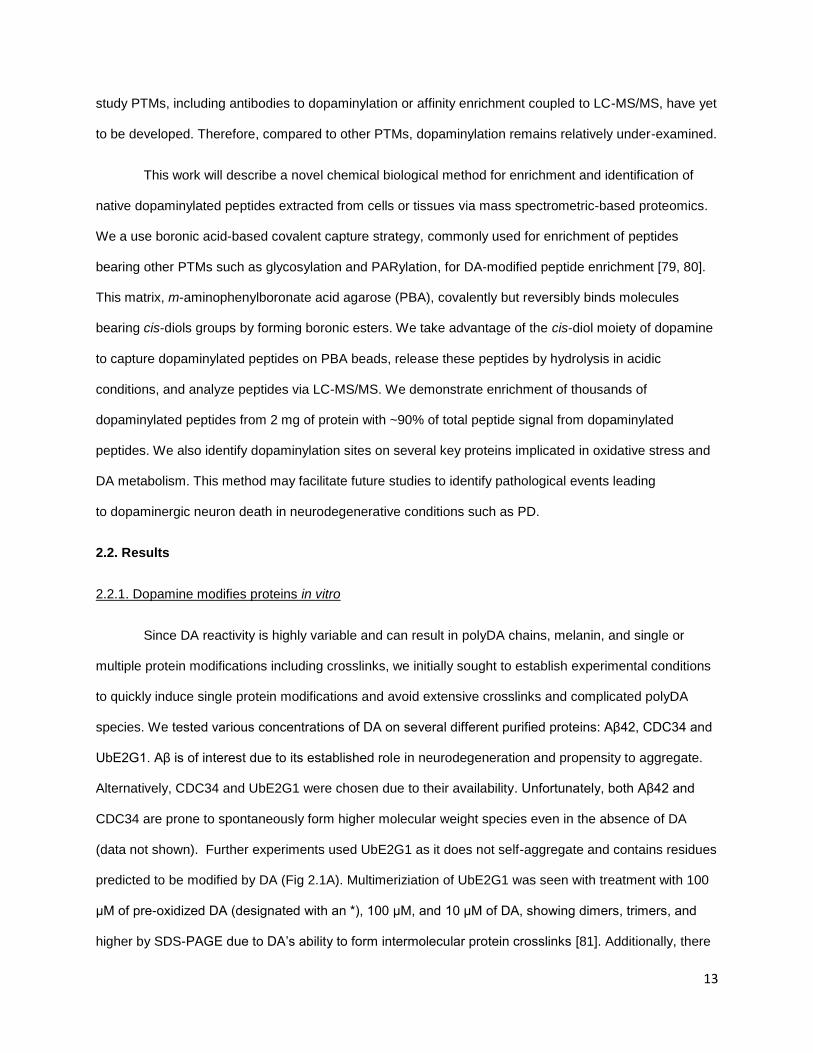
predicted to be modified by DA (Fig 2.1A). Multimeriziation of UbE2G1 was seen with treatment with 100
μM of pre-oxidized DA (designated with an *), 100 μM, and 10 μM of DA, showing dimers, trimers, and
higher by SDS-PAGE due to DA’s ability to form intermolecular protein crosslinks [81]. Additionally, there
14
appears to be a lower molecular weight band which is indicative of intramolecular crosslinks, as shown by
our disuccinimidylsuberate (DSS) control (Figure 2.1B)
After confirming DA’s ability to modify and crosslink UbE2G1, we looked to shorten our 96 hour
incubation time to decrease degradation products and increase experimental efficiency. To decrease
reaction time, we hypothesized that a peroxidase could be used to increase the reaction kinetics.
Peroxidases, like horseradish peroxidase (HRP), are known to catalyze DA oxidation in the presence of
H2O2 [39]. Therefore, we used a combination of IgG-HRP, DA, and H2O2 to treat UbE2G1 for 25 min. This
reaction yields high molecular weight (HMW) products, some which appear too large to enter the gel
(Figure 2.1C). HRP controls were included to distinguish UbE2G1 from IgG-HRP and any unintended
cross-linked species. UbE2G1 crosslinks are seen in 25 minutes in the presence of DA without HRP
(Figure 2.1C, lanes 5 & 6). However, in the presence of HRP, there was an increase in higher molecular
weight species (lanes 2 & 4). Activation of HRP with H2O2 generates increased high molecular weight
(HMW) species and depletion of monomers of UbE2G1 at 20 kDa (lane 1 & 3). In addition to these HMW
species, HRP catalyzed oxidation of DA generated black pigments in the test tube (not shown), consistent
with oxidative products polymerizing, including polyDA and melanin (see Scheme 1.2 for details).
Peroxidase activity increased kinetics of the reaction, but the high concentration of DA and rapid oxidation
induced aggregation that are challenging for downstream mass spectrometry analysis.
To address these challenges, we tested our peroxidase-catalyzed reaction with variable
concentrations of DA. In the presence of IgG-HRP, H2O2, and 100 μM, 10 μM, or 1 μM DA, similar
amounts of intermolecular cross-linked proteins are seen across DA concentrations. To determine which
concentration of DA generated protein modifications without crosslinks, we used near-infrared (nIRF)
imaging to detect the quinone group of the dopaminylated protein [82]. Interestingly, 10 μM DA shows a
5-fold increase in quinone fluorescence compared to the 100 μM DA (Figure 2.1.D-E). Despite our
visualization of dopaminylated proteins, when we excised the fluorescent gel band, digested, and ran LC-
MS/MS, we were unable to detect dopaminylated peptides.

15
2.2.2. Enrichment of dopaminylated peptides and site localization
Excess cytosolic DAis a known source of oxidative stress in the brain through formation of DAQ
and other toxic metabolites [9]. Formation of DA oxidation products drive DA oxidative polymerization
(neuromelanin) and aberrant protein modification through Michael addition with nucleophilic residues Cys,
His, and Lys [60, 83]. Tyrosine should also be considered for dopaminylation as Tyr are the primary
residue of modification in other peroxidase-catalyzed protein modification [84].
UbE2G1 (19.5kDa) Amino Acid Sequence:
SMTELQSALLLRRQLAELNK NPVEGFSAGLIDDNDLYRWE VLIIGPPDTLYEGGVFKAHL TFPKDYPLRPPKMKFITEIW HPNVDKNGDVCISILHEPGE DKYGYEKPEERWLPIHTVET IMISVISMLADPNGDSPANV DAAKEWREDRNGEFKRKVAR CVRKSQETAFE
0
5
10
15
20
25
Pix
el In
tensity
a b c
d e CCB nIRF Detection:
Figure 2.1. Dopamine oxidation and protein modification a) UbE2G1 amino acid sequence which contains potential residues of interest: C, H, K, Y. b) UbE2G1 (5 μg) was incubated with 10 mM, 100 μM pre-oxidized*, 100 μM, 10 μM, or 1 μM DA for 96 hours and analyzed by SDS-PAGE stained with Colloidal Coomassie Blue (CCB) to visualize protein. c) UbE2G1 (5 μg) was incubated with 100 μM DA, 2 mM H2O2, and IgG-HRP for 25 minutes and analyzed by SDS-
PAGE stained with Colloidal Coomassie to visualize protein. Lanes with IgG-HRP are run as controls. d) UbE2G1 (5 μg) was incubated with HRP, H2O2, and 100 μM, 10 μM, or 1 μM DA for
25 minutes and analyzed by SDS-PAGE. Gels were scanned using a near-infrared imager (right panel), then stained with Colloidal Coomassie Blue (CCB) to visualize total protein (left panel). e) The fluorescent signal was quantified by measuring pixel intensity at 20kDa and dividing by background pixel intensity. The molecular weight marker on the left side of the gel indicates protein size in kDa. DA=Dopamine, DSS= disuccinimidyl suberate
16
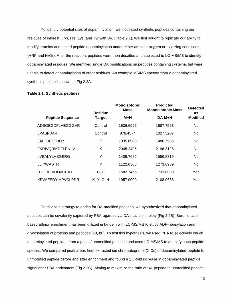
To identify potential sites of dopaminylation, we incubated synthetic peptides containing our
residues of interest: Cys, His, Lys, and Tyr with DA (Table 2.1). We first sought to replicate our ability to
modify proteins and tested peptide dopaminylation under either ambient oxygen or oxidizing conditions
(HRP and H2O2). After the reaction, peptides were then desalted and subjected to LC-MS/MS to identify
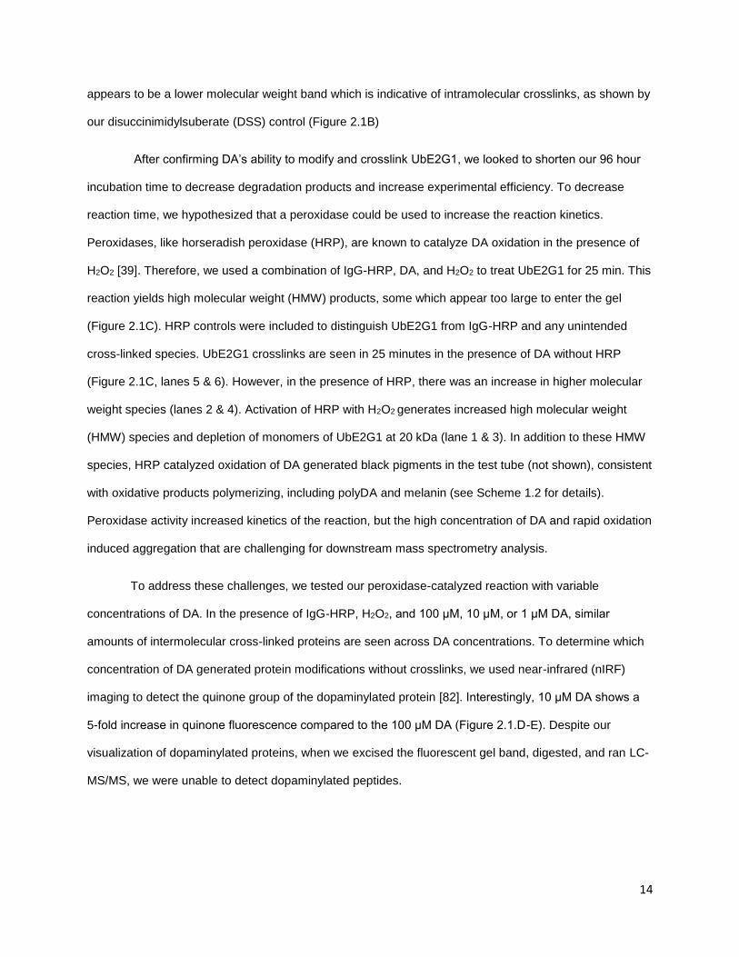
dopaminylated residues. We identified single DA modifications on peptides containing cysteine, but were
unable to detect dopaminylation of other residues. An example MS/MS spectra from a dopaminylated
synthetic peptide is shown in Fig 2.2A.
Table 2.1: Synthetic peptides
Peptide Sequence
Residue
Target
Monoisotopic
Mass
M+H
Predicted
Monoisotopic Mass
DA-M+H
Detected
as
Modified
ADSGEGDFLAEGGGVR Control 1536.6925 1687.7558 No
LPASFDAR Control 876.4574 1027.5207 No
EIAQDFKTDLR K 1335.6903 1486.7536 No
FKRIVQRIKDFLRNLV K 2045.2495 2196.3128 No
LVEALYLVSGERG Y 1405.7686 1556.8319 No
LLIYWASTR Y 1122.6306 1273.6939 No
HTGREIVDLMCHAT C, H 1582.7465 1733.8098 Yes
KPVAFSDYIHPVCLPDR K, Y, C, H 1957.0000 2108.0633 Yes
To devise a strategy to enrich for DA-modified peptides, we hypothesized that dopaminylated
peptides can be covalently captured by PBA-agarose via DA’s cis-diol moiety (Fig 2.2B). Boronic-acid
based affinity enrichment has been utilized in tandem with LC-MS/MS to study ADP-ribosylation and
glycosylation of proteins and peptides [79, 80]. To test this hypothesis, we used PBA to selectively enrich
dopaminylated peptides from a pool of unmodified peptides and used LC-MS/MS to quantify each peptide
species. We compared peak areas from extracted ion chromatograms (XICs) of dopaminylated-peptide to
unmodified peptide before and after enrichment and found a 2.5-fold increase in dopaminylated peptide
signal after PBA enrichment (Fig 2.2C). Aiming to maximize the ratio of DA-peptide to unmodified peptide,
17
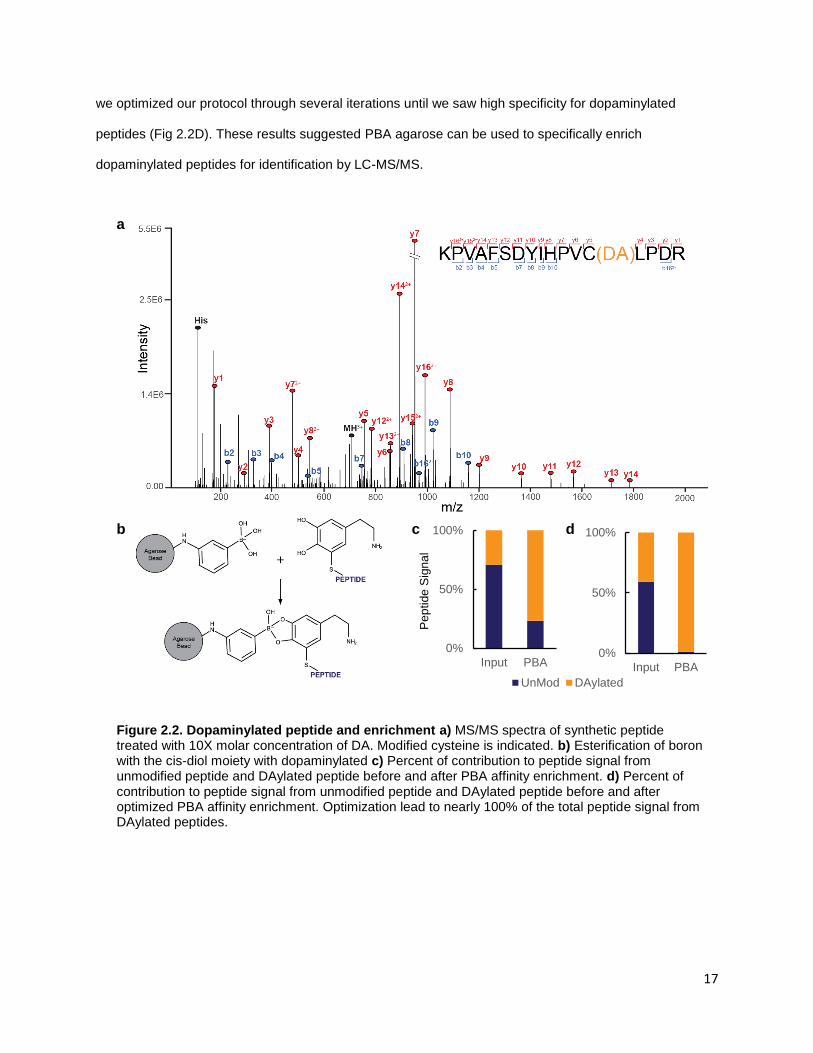
we optimized our protocol through several iterations until we saw high specificity for dopaminylated
peptides (Fig 2.2D). These results suggested PBA agarose can be used to specifically enrich
dopaminylated peptides for identification by LC-MS/MS.
0%
50%
100%
Input PBA
0%
50%
100%
Input PBA
UnMod DAylated
Pe
ptide
Sig
na
l
a
b c d
Figure 2.2. Dopaminylated peptide and enrichment a) MS/MS spectra of synthetic peptide treated with 10X molar concentration of DA. Modified cysteine is indicated. b) Esterification of boron with the cis-diol moiety with dopaminylated c) Percent of contribution to peptide signal from unmodified peptide and DAylated peptide before and after PBA affinity enrichment. d) Percent of contribution to peptide signal from unmodified peptide and DAylated peptide before and after optimized PBA affinity enrichment. Optimization lead to nearly 100% of the total peptide signal from DAylated peptides.

18
2.2.3. Enrichment and identification of in vivo dopaminylated peptides
To test our ability to enrich and identify substoichiometric dopaminylated peptides from complex
mixtures including cell or tissue lysates, we looked for a cell model that expressed the DA transporters
VMAT2 and DAT, were sensitive to DA, and grew quickly. Initially, we used the neuronal cell line SH-
SY5Y differentiated with retinoic acid (RA) and 12-O-tetradecanoyl-phorbol-13 acetate (TPA) for six days.
This differentiation protocol increases expression of dopaminergic markers like tyrosine hydroxylase [85].
However, these cells grow slowly with a doubling time of several days, were difficult to passage, were
extremely sensitive to perturbations, and died easily. As an alternative, we utilized a cell model of HEK-
293 cells which stably express both DAT and VMAT2, termed HEK-VMAT, from Gary Miller’s lab at
Columbia University [86]. These cells uptake exogenous DA and have similar doubling times to standard
HEK cells, making them a useful cellular model to study protein dopaminylation. Furthermore, these cells
can be treated with DA concentrations up to 50 μM without forming melanin pigments or inducing massive
cell death.
Our early experiments continued optimizing our DA-modified peptide enrichment method.
Optimization was done over 30 biological sample sets, with each set containing dopamine treated and
control cells with DA with up to four process replicates. Together, optimization of PBA affinity enrichment
from our HEK-VMAT cells model used over 220 LC-MS/MS runs. A summary of these various
optimization conditions is included under Methods and an in-depth protocol is included. Briefly, proteins
from snap frozen biological samples are extracted in 8 M Urea (100 mM ammonia bicarbonate, 75 mM
NaCl, and 5 mM DTT) and sonicated to shear DNA. Proteins were reduced and alkylated, digested, and
desalted at 4°C on Waters Oasis Cartridges. Peptides were resuspended in PBA Buffer (30% Acetonitrile
(MeCN), 50 mM Phosphate pH 8.5), mixed with PBA beads and incubated for two hours. The bead
volume was transferred to StageTip and washed five times with PBA Buffer and eluted with StageTip
buffer A (1% MeCN, 0.1% trifluoroacetic acid (TFA)) for 1 hour, followed by an additional wash with 0.1%
TFA. We found that performing our washes in a StageTip format increased our reproducibility between
process replicates by reducing bead loss compared to batch washes. The eluted fractions were mixed,
desalted on StageTips, and analyzed on either a Thermo Orbitrap Elite or Lumos Tribrid mass
spectrometer (Figure 2.3A). Raw files were analyzed with variable modifications DA and

19
Carbamidomethyl (CAM) on cysteine residues. Peptide IDs from the most recent 10 sample sets are
summarized in Figure 2.3B, where sets 8, 9, and 10 represent the optimized protocol.
Utilizing our optimized PBA affinity enrichment of dopaminylated peptides, we identified between
800 and 4500 dopaminylated peptides from HEK-VMAT cells treated with 50 μM DA for 24 hours (Figure
2.3B). From each biological sample, we identified between 600-3200 dopaminylated cysteine sites with
localization scores greater than 0.75. Our PBA enrichment has low background with over 94% of peptide
intensity coming from dopaminylated peptides (Table 2.3). To demonstrate the specificity of our protocol,
we also analyzed input whole cell lysates without enrichment. From these unenriched samples, we see
fewer than 24 dopaminylated peptides and 15 dopaminylated cysteine sites, which is below our peptide
and site ID false discovery rate of 0.01, suggesting that these modified peptides identified from a dataset
of 25,450 or 28,222 (unenriched HEK-VMAT 9 or 10, respectively) peptide-spectrum matches (PSM) are
likely false-positive PSMs.
Table 2.2: Comparison of enriched and unenriched samples
Biological
Sample
Total # of
Peptides
# of
DAylated
Peptides
# DA
Sites
% DAylated
Peptides of
Peptide Signal
PBA
Enriched
HEK-VMAT 8 946 870 658 95.95
HEK-VMAT 9 1403 1198 901 89.45
HEK-VMAT 10 4884 4591 3483 98.40
No
Enrichment
HEK-VMAT 9 25450 12 15 0.02
HEK-VMAT 10 28222 24 15 0.06
Next, we questioned if dopaminylation of proteins occurs randomly as this modification is non-
enzymatic. A random modification would have equal probability to modify proteins with a solvent exposed
cysteine, and would be directly correlated to protein abundance. To interrogate the relationship between
abundance and dopaminylation, we overlaid our data from a representative biological replicate (HEK-
VMAT 9) onto the proteome of HEK cells. Utilizing the MaxQB database, we extracted the background
proteome of HEK cells and ranked proteins by abundance [87]. By overlaying our 481 dopaminylated
20
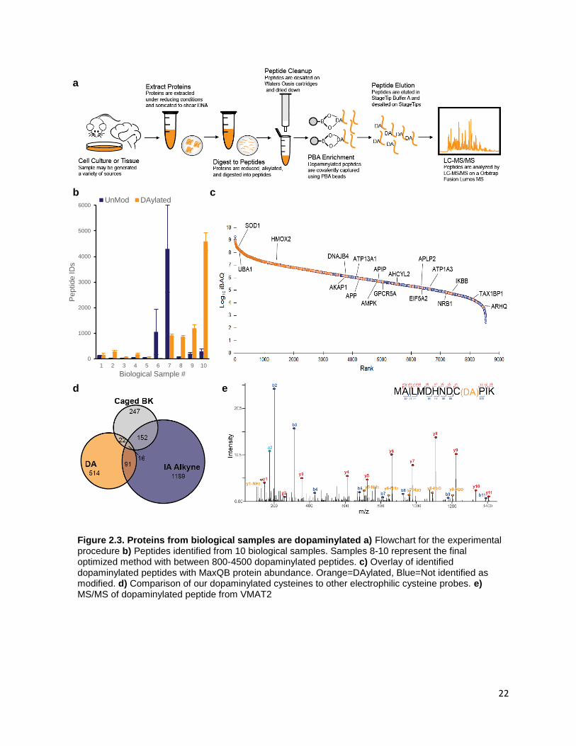
proteins detected in this study on the ranked proteins from the MaxQB HEK database, we saw a large
portion of our dopaminylated peptides come from highly abundant proteins (Fig 2.3C). However, in the
lower two quartiles of proteins ranked by abundance, we identified dopaminylation of 18 proteins. Of
these 18, we detected two copper binding amyloid proteins: amyloid precursor protein (APP) and
amyloid-like precursor protein 2 (APLP2). APP is processed in the cell to form amyloid-β, the major
component of amyloid plaques in Alzheimer’s disease. Previous work has shown potential dopaminylation
of tyrosine on amyloid-β, but cysteine modification on APP is unknown [88]. Dopaminylation of lower
abundance proteins such as these suggests a mechanism that is not restricted to spontaneous DA
oxidation resulting in nearby cysteine modification. Unlike other protein modifications like phosphorylation,
we were unable to identify a common motif surrounding our dopaminylated cysteine residues
(Supplemental Figure 2.1A). It is possible some cysteine residues have microenvironments that make
them more likely to be modified, such as proteins which bind redox-active metal ions.
To address the potential of differential susceptibility of cysteine to DA modification, we compared
our dopaminylated Cys sites to those identified by LC-MS/MS with two established reactive cysteine
probes. These cysteine probes are electrophilic chemical probes used to identify and quantify reactive
cysteines; one probe is an iodoacetamide-based chemical probe (IA-alkyne) and applied to cell lysates,
while the other is a caged bromomethyl ketone electrophile (caged-BK) which can be used in living cells
[89]. We reasoned the caged-BK probe applied to living cells may have a similar cysteine reactivity profile
to DA in living cells if dopaminylation is indeed directed by abundance and cysteine availability. We
compared our dataset of dopaminylated cysteine sites to reactive cysteines identified using caged-BK
probe, and IA-Alkyne (Figure 2.3D). Modified cysteine residues in both DA and caged-BK have only 4.8%
overlap; the overlap between DA and IA-Alkyne is 5.9%. These small overlaps suggest dopaminylation
may not be a generic modification, but rather some cysteine residues are more prone to specific
dopaminylation. The selective dopaminylation of particular cysteines may be important in understanding
what role dopaminylation plays under physiological conditions.
Of the 481 dopaminylated proteins we identified in the representative biological replicate (HEK-
VMAT 9), 32 are not represented in the MaxQB database for HEK cell line. This may be due to low
21
expression of these proteins in HEK cells. For example, we identified dopaminylated peptides from our
transgenes SLC18A2 (VMAT2) and SLC6A3 (DAT), which normally have low expression in HEK cells
[90]. Identification of dopaminylated sites on the DA transporters is especially interesting. In neurons, high
expression and activity of VMAT2 has been shown to be neuroprotective against DA oxidation [35]. We
found in our HEK-VMAT cells, Cys488 of VMAT2 is dopaminylated (Figure 2.3E). This residue is on the
C-terminal tail of VMAT2 and is susceptible to S-glutationylation in the presence of ROS which leads to
decreased VMAT activity and impaired DA packaging [91]. Implications of dopaminylation at this site will
require further investigation. From the other DA transporter, DAT, we identified two dopaminylations at
Cys6 and Cys342 (Supplemental Figure 2.1B,C). Oxidative modifications on Cys342 have been shown to
inactivate the transporter and contribute to oxidative stress in DAergic neurons [92]. Cys6 can be S-
palmitoylated and is thought to be involved in the function and localization of DAT in neurons [93]. It is
possible, given the functional significance of these sites, that dopaminylation of these transporters may
alter the homeostasis of DA packaging and increase cytosolic DA, and potentially representing a toxic
feed forward mechanism of DA oxidation and oxidative stress.
22
Figure 2.3. Proteins from biological samples are dopaminylated a) Flowchart for the experimental procedure b) Peptides identified from 10 biological samples. Samples 8-10 represent the final optimized method with between 800-4500 dopaminylated peptides. c) Overlay of identified dopaminylated peptides with MaxQB protein abundance. Orange=DAylated, Blue=Not identified as modified. d) Comparison of our dopaminylated cysteines to other electrophilic cysteine probes. e) MS/MS of dopaminylated peptide from VMAT2
a
d e
b c
0
1000
2000
3000
4000
5000
6000
1 2 3 4 5 6 7 8 9 10
Peptid
e ID
s
Biological Sample #
UnMod DAylated

23
2.2.4. Enrichment and identification of dopaminylated peptides from human brain
Having established an approach to reliably enrich dopaminylated peptides from a complex cell
lysate, we next tested our method with more physiological relevant tissue: human brain. We obtained two
control cases and two Lewy body (LB) positive post-mortem, flash-frozen brain tissue samples from the
University of Washington Neuropathology Core and subjected them to our boronic acid-based enrichment
and LC-MS/MS. We identified over 20 dopaminylated peptides directly from brain (Figure 2.4A). The
concentration of cytosolic DA in human neurons is much lower than those used in our HEK-VMAT model
system, potentially explaining the reduced number of dopaminylated peptide identifications. Furthermore,
our cell line model was optimized to avoid generation of neuromelanin and extensive modifications which
are not under our control in human tissue. Further optimization of our protocol may be taken to improve
dopaminylated peptide enrichment from complex tissue samples.
Amongst the DA sites identified from human brain tissue, the cysteine residue, Cys147, of
superoxide dismutase (SOD1), is consistently found to be dopaminylated in HEK-VMAT cells and in two
of four human brain samples, one control and one LB positive (Figure 2.4 B and C, respectively).
Interestingly, SOD1 binds both copper and zinc, and copper is linked with DA oxidation and oxidative
stress [94]. Furthermore, SOD1 is a key protein involved in detoxification of ROS and oxidative stress and
has been studied extensively in neurodegeneration. For example, one previous study demonstrated
dopaminylated of SOD1 in differentiated SH-SY5Y cells exposed to DA but was unable to identify the site
of dopaminylation [63]. With our method, we identified dopaminylation of Cys147, which forms a critical
disulfide bridge at C58-C147. Mutations at Cys147 and subsequent loss of this disulfide bridge promote
SOD1 aggregation [94]. Furthermore, SOD1 mutations are key drivers in amyotrophic lateral sclerosis
(ALS), a neurodegenerative disorder affecting motor neurons in the brain and lower motor neurons in the
brain stem and spinal cord. Whether dopaminylation of SOD1 drives disease or is a consequence of
already impaired dopamine homeostasis remains to be determined, but our method will help to elucidate
the role of dopaminylation from various biological samples.

24
0
200
400
600
800
1000
1200
1468 1273 1833 1355
UnMod DAylated
Pe
ptide
ID
s
Brain ID #
c
Figure 2.4 Dopaminylated peptides from human brain a) Peptide IDs from brain tissue across 4 patients. b) MS/MS spectra of Cys147 of SOD1 from HEK-VMAT cells treated with DA c) MS/MS spectra of Cys147 of SOD1
a b

25
2.3. Materials and Methods
Peptides and Reagents
Peptide standards were purchased from AnaSpec and New England Peptides. Dopamine was
purchased from Acros Organics and m-aminophenylboronic acid-agarose was purchased from Sigma.
Protein Labeling and in-Gel Detection
UbE2G1 was gifted by Haibin Mao for early experiments. 5 μg of purified protein was incubated
with DA in phosphate buffered saline (PBS) for 96 hours at room temperature. For peroxidase-induced
reactions, 5 μg of purified protein was incubated with DA, IgG-HRP (Sigma), and 2 mM H2O2 for 25
minutes. Experiments were also carried out in 50mM Tris buffer, pH 7.4, which may increase DA
oxidation. Reactions were quenched by adding 4X Bolt LDS Sample Buffer (Thermo) supplemented with
5 mM DTT. Proteins were analyzed by sodium dodecyl sulfate-polyacrylamide gel electrophoresis (SDS-
PAGE). Gels were scanned on an Odyssey infrared imager (Li-Cor Biosciences, ex = 685 nm) and
quantified in Adobe Photoshop. Total protein was visualized by staining with colloidal blue coommassie
and imaged on a FluorChem E imager.
Peptide Labeling and Enrichment
Peptides were dissolved to 100-500 μM in PBS and incubated with equimolar fresh DA for 96
hours at room temperature. The reaction was stopped by addition of 10% TFA to acidify peptide mixtures,
and desalted using StageTips [95] and analyzed by LC-MS/MS. The stoichiometry of peptide
dopaminylation was calculated by integrating the area under the curve for the monoisotopic mass of the
modified peptide rationed to the unmodified peptide. A mass window of +/-0.3 amu was allowed, and the
ICIS peak detection algorithm feature of Xcalibur was used for automated detection of peaks.
Identification of Dopaminylated Peptides from cell culture
HEK293 cells stably expressing VMAT2 and DAT were gifted by Dr. Gary Miller (Columbia
University). Cells were grown in DMEM media supplemented with 10% FBS (Hyclone, GE) and Penicillin-
Streptomycin-Glutamine (100X, Thermo Scientific), 100 μg/ml Zeocin (Invitrogen) and 250 μg/ml G-418
(GoldBio) in an incubator at 37°C and 5% CO2.

26
Cells were plated in six 15 cm dishes and grown to 60% confluence. Fresh media (20 mLs) was
added 16-18 hours prior to DA treatment (the day before). Three of six plates were treated with either 10
or 50 μM DA for 24 hours. DA was made at 1000x stock in PBS and aliquots were stored at -80°C. It is
important to avoid freeze/thaw cycles as DA will oxidize quickly. After treatment, cells were washed 3X
with 20 mls of ice-cold PBS. An additional 1 mL of cold PBS was added to dishes and cells are scraped
with a rubber spatula and collected in a 15 mL conical vial. Cells were kept on ice while untreated dishes
were collected. Cells were pelleted via centrifugation for 2 min at 2,325 x g. The supernatant was
aspirated and cell pellets snap frozen in liquid nitrogen. Cell pellets were stored at -80°C until ready to
process.
Frozen cell pellets were lysed in 1.5 mL of 8 M Urea (100 mM ammonia bicarbonate, 75 mM
NaCl, 5 mM DTT). Pellets were broken up by pipetting up and down with P-1000 until clumps dissolved.
The lysate was transferred to a low-retention 2 mL microcentrifuge tube and sonicated in a Cup Horn
sonicator (QSonica) for 20 minutes (30s on/off). The sonication cup must be filled with ice and
replenished after 10 minutes to keep samples cold. Lysates are clarified by centrifugation at 21,000 x g
and 4°C for 10 minutes. Soluble protein fractions were transferred to a fresh microcentrifuge tube.
The protein content was measured using Pierce 660 nm Assay Reagent (Thermo Scientific) using
a BSA standard curve. 8 mg (4 process replicates of 2 mg each) of protein was transferred into a 15 mL
conical vial. Proteins were reduced with 1 mM TCEP for 10 minutes at room temperature and alkylated
with 10 mM chloroacetamide for 45 minutes at room temperature in the dark. Proteins were diluted 2-fold
with 50 mM Tris pH 8 and digested with LysC (Wako Chemicals) at 1:100 for 2 hours at room
temperature. Proteins are diluted further until Urea < 1.5 M, and digested with Trypsin (Thermo Scientific)
at 1:100 at room temperature while shaking overnight. In morning, another 5 μg trypsin was added and
incubated for 1 hour. Samples were acidified to pH 2 with 10% TFA. Any insoluble proteins were pelleted
by centrifuging samples at 5000 x g for 5 minutes and then placed on ice. Peptides were then extracted
with Oasis HLB 3cc 60mg cartridges at 4°C in a cold room. Cartridges were conditioned with 3 mL of
100% MeCN, followed by 3 mL StageTip Buffer B (50% MeCN, 0.1% TFA), and washed two times with 3
mL StageTip Buffer A (1% MeCN, 0.1% TFA). Peptides are loaded on the cartridges and washed two
times with cold StageTip Buffer A. Cartridges were moved back to room temperature, and peptides are
27
eluted two times with StageTip Buffer B into two 2 mL microcentrifuge tubes. Peptides were dried to
completion using a Thermo Scientific Speedvac (about 4 hours). Dried peptides were stored at -80°C.
Peptides were resuspended in 800 μL PBA Buffer (30% MeCN, 50 mM Phosphate pH 8.5) and
bath sonicated for 5 minutes to help dissolve peptides. Peptides were clarified via centrifugation at 14,000
x g for 5 minutes and transferred to a new tube. While peptides were cleared by centrifugation, 2x 40 μL
of m-aminophenlyboronic acid-agarose (PBA) (10 μL for each 2mgs of peptide) were aliquoted into low
retention 1.5 mL microcentrifuge tubes. Beads were mixed prior to pipetting. PBA beads were washed
with 1 mL 30% MeCN, 0.1% TFA for 5 minutes, centrifuged for 30s on a tabletop centrifuge, and allowed
beads to settle for 5 minutes. The supernatant was removed and beads were primed with 1 mL 100mM
Phosphate Buffer pH 10 for 5 minutes, centrifuged for 30s on a tabletop centrifuge, and beads allowed to
settle for 5 minutes. The supernatant was discarded. 400 μL PBA buffer was mixed with beads and
aliquoted into four 1.5 mL microcentrifuge tube using a cut, widebore P-200 pipette tip. 200 μL of
peptides was added to beads and incubated for 2 hours with end-over-end rotation.
While peptides and PBA beads incubated, StageTip were made by adding two plugs of C18 to P-
200 pipette tips. These were used as fritted columns to wash beads.
Peptide/PBA bead mixtures were collected via tabletop centrifuge and ~80% of the supernatant
was removed. The remaining mixture was transferred StageTips and centrifuged using StageTip adaptors
to remove remaining liquid. Caution must be used to avoid the beads drying. PBA beads were washed
with 400 μL PBA buffer five times and centrifuged to remove supernatant. Extended pipette tips were
used when adding washes to mix beads and PBA Buffer each time to ensure adequate washing.
Peptides were released from PBA beads by adding 300 μL StageTip Buffer A and incubating for one
hour. The elution was collected by centrifugation into a fresh microcentrifuge tube. An additional 300 μL of
0.1% TFA was added to PBA beads and incubated for 20 minutes and centrifuged to collect elution.
Finally, 30 μL of 35% MeCN 0.1% TFA was added (to elute peptides bound to C18 plug). These three
elutions were combined and desalted on StageTips at 4°C. Briefly, each StageTip contains two plugs of
Empore SDB-XC. StageTips were conditioned with 50 μL MeOH, 50 μL StageTip Buffer B (50% MeCN,
0.1% TFA), and two 50 μL washes of StageTip Buffer A (1% MeCN, 0.1% TFA). Peptides were added
28
and bound. StageTips were washed with 100 μL of cold StageTip Buffer A followed by elution by 50 μL
35% MeCN 0.1% TFA. Peptides were then dried via speedvac and resuspended in StageTip Buffer A.
LC-MS/MS
Peptides were separated on a Thermo EASY-nLC 1200 UHPLC instrument (Sunnyvale, CA) with
10 cm long fused silica capillary columns made in-house with a laser puller (Sutter, Novato, CA) and
packed with 3 μm 120 Å reversed phase C18 beads (Dr. Maisch, Ammerbuch, DE). The LC gradient was
133 min long with 4–38% B at 200 nL/min. LC solvent A was 0.1% acetic acid and LC solvent B was
0.1% acetic acid, 80% acetonitrile. MS data was collected with a Thermo Orbitrap Fusion Lumos Tribrid
mass spectrometer. Data-dependent analysis was applied using Top15 selection with HCD fragmentation
with 31% HCD collision energy. Raw files were analyzed by MaxQuant/Andromeda [72] version 1.5.2.8
using protein, peptide and site FDRs of 0.01 and a minimum score of 40 for modified peptides, 0 for
unmodified peptides; minimum delta score of 17 for modified peptides, 0 for unmodified peptides. MS/MS
spectra were searched against the UniProt human database (updated July 22nd, 2016). MaxQuant
search parameters: Variable modifications included Oxidation (M), Carbamidomethyl (C), and
Dopaminylation (C). For dopaminylation, the chemical formula was C(8) O(2) N H(9) and a mass of
151.0633 g/mol. Max. labeled amino acids was 3, max. missed cleavages was 2, enzyme was Trypsin/P,
max. charge was 7. The initial search tolerance for FTMS scans was 20 ppm and 0.5 Da for ITMS MS/MS
scans. Data and raw spectra were analyzed in Excel, R, and Perseus. Modified peptides were only
considered identified if MaxQuant is able to compute a peptide intensity in three process replicates (of
four).
29
2.4. Discussion
Protein and peptide dopaminylation is an important but understudied PTM. Currently, the
functional significance of increased cytosolic DA is unknown although studies suggest cytosolic DA is
important for neurodegenerative disease. The role that DA plays in neurodegeneration is linked to
oxidative stress as unregulated DA quickly forms reactive oxidation products like DAQ, which may modify
proteins. Modification of proteins occurs at physiological pH; therefore it is possible to dopaminylate
proteins both in the cytosol and extracellular matrix when DA is released into the synapse. While in most
cases cytosolic DA concentrations remain low, the presence of NM suggests there is adequate cytosolic
DA that eludes standard antioxidants like glutathione to allow the formation of complex, DA-protein
aggregates. Unbiased tools to characterize dopaminylation are critical to advance this field and elucidate
the functional significance of this PTM.
We describe a new method for direct detection of protein dopaminylation utilizing boronic-acid
based enrichment and LC-MS/MS. Our data indicates that our enrichment protocol yields 450-fold
increase of peptide signal from dopaminylated peptides which are undetectable without enrichment. For
dopaminylated peptides which are detected pre and post-enrichment, we see a 208-fold increase in
peptide signal after PBA enrichment. We employed our protocol to identify 1860 dopaminylated proteins
with roughly 3400 unique dopaminylated cysteine residues in HEK-VMAT cells treated with DA. Several
of these proteins have been previously described as DA-modified, though our method now provides
specific site identification of DA modifications. Identification of sites is critical to elucidate the functional
consequence of DA modifications. For example, we identify Cys 488 of VMAT2 as dopaminylated.
Previous work has shown that under oxidative stress, this site can be modified by glutathione and slows
VMAT2 activity [91]. To our knowledge, this is the first report to find DA modification of VMAT2.
Unlike other methods which employ radioactive DA or purified proteins, the method described in
this study translated to study human tissue or fluids. Here, we show that from four clinical patient
samples, we can identify 20 dopaminylated sites, including Cys146 of SOD1. Cys146 of SOD1 forms a
critical disulfide bridge and loss of this bridge promotes protein aggregation [94]. Currently, the role of

30
dopaminylation in SOD1 aggregation is unknown, but our findings suggest a potential mechanism and
further study is needed.
The method described here should also facilitate the study the effect of dopaminylation and the
consequences in DA neurons. We believe our method can be used to study range of neurotoxic effects
due to DA, from neurodegeneration to pesticide-induced toxicity.
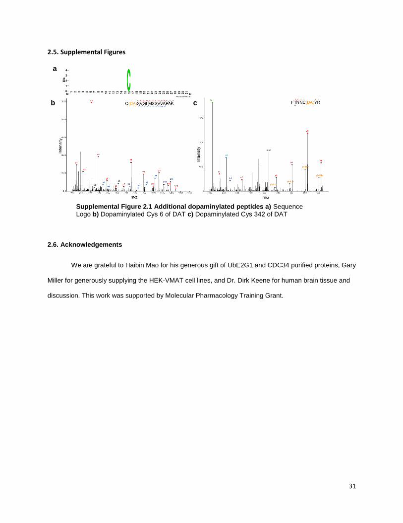
31
2.5. Supplemental Figures
2.6. Acknowledgements
We are grateful to Haibin Mao for his generous gift of UbE2G1 and CDC34 purified proteins, Gary
Miller for generously supplying the HEK-VMAT cell lines, and Dr. Dirk Keene for human brain tissue and
discussion. This work was supported by Molecular Pharmacology Training Grant.
Supplemental Figure 2.1 Additional dopaminylated peptides a) Sequence Logo b) Dopaminylated Cys 6 of DAT c) Dopaminylated Cys 342 of DAT
a
b c

32
Chapter Three: Alternative methods to study dopaminylation
3.1. Introduction
The repertoire of tools is limited to study the dopaminylation of proteins and peptides as a post-
translational modification (PTM). Other PTMs like phosphorylation or ubiquitylation can be studied using
antibodies which have been developed against the PTM itself. For example, antibodies for phospho-
tyrosine detect global phosphorylation of tyrosine independent of protein sequence and may be used for
unbiased study of tyrosine phosphorylation [96]. However, reliable antibodies have not been developed
for dopaminylation and, therefore, even common antibody-based techniques like Western blotting and
immunofluorescence (IF) are unavailable. To address this, we proposed an alternative strategy to
investigate dopaminylation: a modified dopamine containing a biotin group. If biotinylated dopamine (BD)
underwent oxidation and modified proteins as DA does, the suite of avidin-based enrichment tools would
be available to study dopaminylation. The interaction between biotin and avidin is strong and can be
utilized to enrich biotinylated proteins for MS analysis, and used for Western blots or IF. Other groups
have taken advantage of a similar mechanism using a genetically modified peroxidase (APEX) to catalyze
the covalent addition of proteins with biotin-tyramide [97]. Biotin-tyramide is similar in structure to BD
except for the additional hydroxyl group on BD, however biotin-tyramide requires a peroxidase to
biotinylate proteins. The APEX strategy has been used to identify the mitochondrial proteome, the cilia
proteome, and proteins at a specific genomic loci using various avidin-based tools [97-99]. Therefore, we
believe avidin-based tools can be adopted for use with BD to study dopaminylation in cell models.
Another alternative method we explored was the use of near-infrared (nIRF) imaging of
dopamine-protein modifications. Dopaminylated proteins exposed to oxidative conditions can undergo
further oxidation from to form protein-dopamine o-quinone (DAQ-protein). These o-quinones have
specific excitation-emission properties in the nIRF range and can be detected using a Li-Cor Odyssey
infrared imager [82]. We hypothesized that this approach can be adapted using a confocal microscope
with nIRF capabilities and will discuss our findings in this chapter.

33
3.2. Results
3.2.1. Biotin-dopamine
To determine if a biotinylated dopamine strategy was feasible, we used biotin-dopamine (BD) to
compare to with our work on dopaminylation. The structure of BD maintains the reactive catechol group
but substitutes a biotin group on the primary amine (Figure 3.1A). The addition of a biotin group to
dopaminylation was attractive because it allows us to utilize the suite of avidin-based technology to study
dopaminylation.
To determine BD reactivity, we tested three concentrations of BD on our test protein, UbE2G1, for
96 hours. After Ponceau staining to visualize total protein, a faint band at 40 kDa after incubation with 125
μM BD was visible, but this effect was lost with decreasing concentration of BD (Figure 3.1B left). This
suggests that at higher concentrations, BD is able to crosslink UbE2G1 to form dimers in a concentration
dependent manner similar to DA but less efficiently (in comparison with Figure 2.1B). Next, we probed our
membrane with avidin-HRP to detect BD modifications of UbE2G1 (Figure 3.1B right). We saw strong
evidence of BD modification of UbE2G1 in both 125 and 12.5 μM BD. Additionally, we detected BD signal
at 40 kDa and 60 kDa, suggesting multimerization of UbE2G1 in the presence of BD. Next, we treated our
standard peptides (Table 2.1) with BD and detected modification on Cys residues (Figure 3.1C). From
these results we reasoned that BD may have slower rates of polymerization needed for crosslinking,
however BD has similar reactivity to DA to modify cysteine residues of proteins and peptides.
Next, we reasoned that BD may cross cell membranes because BD is not charged, unlike DA
which carries a plus charge on the primary amine at physiological pH and requires specialized
transporters. We treated a mouse neuroblastoma (N2A) cell line with five concentrations of BD for 96
hours and saw increasing biotin-dopaminylation of proteins with increasing BD concentrations (Fig 3.1D).
We also saw a similar, concentration dependent increase in biotin-dopaminylation in human
neuroblastoma cells (SH-SY5Y) (Supplemental Figure S3.1A). This suggests BD is oxidized in cells and
modifies proteins. Interestingly, we found that biotin-tyramide, which lacks a second hydroxyl group, does
not spontaneously modify proteins at the same concentrations (Supplemental Figure S3.1B). Finally, to
optimize conditions, we treated N2A cells with 50 μM BD and detected biotin-dopaminylated proteins after

34
30 minutes (Supplemental Figure S3.1C). We see that the banding pattern between six and 24 hours is
similar, with signal loss at 48 hours. Therefore, we chose 24 hours for BD treatment in further
experiments.
To determine if biotin-dopaminylation was localized in the cell, we carried out
immunofluorescence on N2A cells treated with BD for 24 hours. Biotin-dopaminylation appears to be
absent in the nuclei and localized throughout the cells. We note that BD localizes near but is not
completely constrained to the mitochondria (Fig 3.1E).
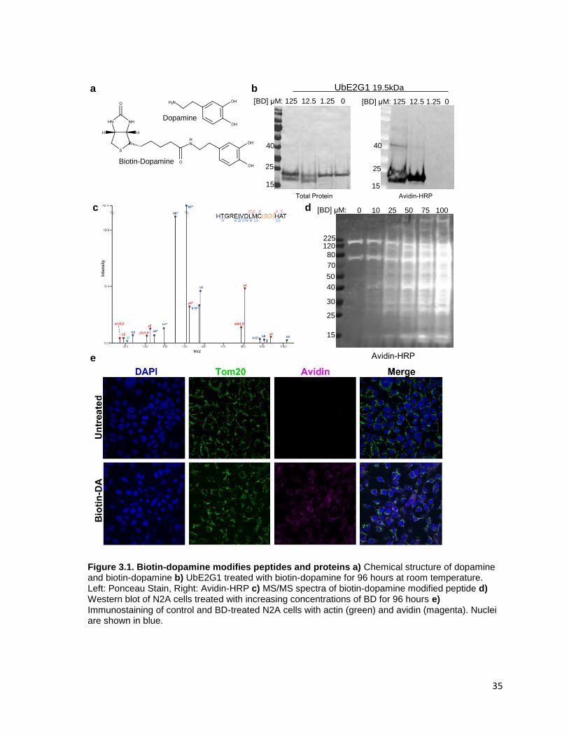
35
Figure 3.1. Biotin-dopamine modifies peptides and proteins a) Chemical structure of dopamine and biotin-dopamine b) UbE2G1 treated with biotin-dopamine for 96 hours at room temperature. Left: Ponceau Stain, Right: Avidin-HRP c) MS/MS spectra of biotin-dopamine modified peptide d) Western blot of N2A cells treated with increasing concentrations of BD for 96 hours e) Immunostaining of control and BD-treated N2A cells with actin (green) and avidin (magenta). Nuclei are shown in blue.
Total Protein Avidin-HRP
25
15
40
UbE2G1 19.5kDa
[BD] μM: 125 12.5 1.25 0
25
15
40
[BD] μM: 0 10 25 50 75 100
25
15
40
30
50 70 80
120 225
Avidin-HRP
Dopamine
Biotin-Dopamine
[BD] μM: 125 12.5 1.25 0 a b
c d
e

36
Finally, to identify biotin-dopaminylated proteins, we utilized streptavidin-based enrichment
coupled with label-free quantitative proteomics. We identified 105 unique proteins by MS, 97 of these are
significantly enriched across two biological replicates of N2A cells treated with 50 μM compared to
untreated controls (Figure 3.2A). We find several proteins previously implicated in neurodegenerative
disease enriched in BD treatment such as proteins found in amyloid plaques or Lewy Bodies including
MAPT, GPHN, and RTN3, as well as other proteins associated with neurodegeneration including:
CAMK2G, CISD2, PPP5C, and HMOX2 [100-106]. Proteins involved in protein homeostasis were also
found to be significantly enriched with biotin-dopamine treatment. For example, BAG3 functions as a co-
chaperone with HSP70 and HSPB8 under acute stress and in cellular aging to degrade proteins via
macroautophagy [107]. Furthermore, BAG3-mediated protein degradation is critical for aggregation-prone
proteins implicated in various neurodegenerative disorders.
Curiously, we identified several proteins that were not significantly enriched after BD treatment
despite their known sensitivity to oxidative modifications. For example, the peroxiredoxins (PRDX) family
of proteins are not significantly enriched in BD (shown in purple). Peroxiredoxins are abundant enzymes
in mammalian cells that use their redox-active cysteine to neutralize oxidative species and modulate
oxidative stress [108, 109]. Another protein family we identified but was not enriched was the heat-shock
(HSPs) chaperone family (shown in green). These molecular chaperones protect against oxidative stress,
regulate redox homeostasis, and have been shown to be oxidative modified in neurodegeneration [110,
111]. As both PRDXs and HSPs are abundant, sensitive to changes in oxidation, and are known to
undergo oxidative modifications, it is curious that these enzymes are not susceptible to biotin-
dopaminylation.
To determine if biotin-dopaminylation is related to abundance, we compared our biotin-
dopaminylated proteins with the proteome of N2A cells. We ranked the proteome using the extracted
iBAQ values from MaxQB and plotted the rank and iBAQ values (shown in blue). We then colored our
identified biotin-dopaminylated proteins in orange (Fig 2.2B). Of the 97 proteins identified to be
biotinylated by BD, eight are in the lower half of the abundance rank.

37
Last, we compared our biotin-dopaminylated proteins to our dopaminylated proteins from our
PBA enrichment experiments. We see that very few proteins appear in both conditions (Fig 3.2C). Of the
18 which appear in both conditions, HMOX2, ATP13A1, and BAG3 are associated with
neurodegeneration.
Figure 3.2. Biotin-dopamine differentially modified proteins in N2A cells. a) Distribution of biotinylated proteins from no treatment (WT) and 50 μM BD treatment (BD). Volcano plot shows proteins significantly enriched via streptavidin pull-down. T-test performed in Perseus. Proteins are distributed along x-axis by Log2 Ratio of BD/WT and along the y-axis by p-value. Orange: Proteins implicated in neurodegeneration. Blue: endogenously biotinylated proteins. b) Overlay of Biotin-dopamine modified proteins with protein abundance from N2A cells. Orange= Biotin-dopaminylated, Blue=Not identified as modified. c) Venn diagram of protein names identified as biotin-dopaminylated in N2A or dopaminylated in HEK-VMAT (Chapter 2, Replicate 9)
a
b c
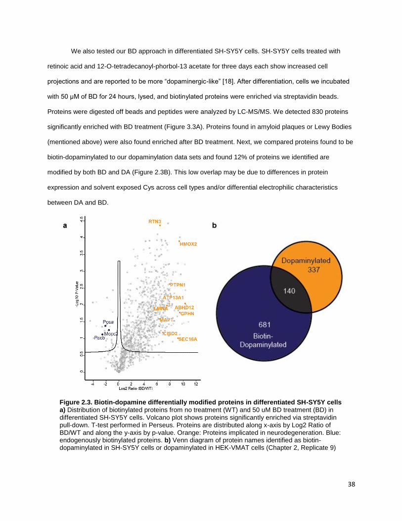
38
We also tested our BD approach in differentiated SH-SY5Y cells. SH-SY5Y cells treated with
retinoic acid and 12-O-tetradecanoyl-phorbol-13 acetate for three days each show increased cell
projections and are reported to be more “dopaminergic-like” [18]. After differentiation, cells we incubated
with 50 μM of BD for 24 hours, lysed, and biotinylated proteins were enriched via streptavidin beads.
Proteins were digested off beads and peptides were analyzed by LC-MS/MS. We detected 830 proteins
significantly enriched with BD treatment (Figure 3.3A). Proteins found in amyloid plaques or Lewy Bodies
(mentioned above) were also found enriched after BD treatment. Next, we compared proteins found to be
biotin-dopaminylated to our dopaminylation data sets and found 12% of proteins we identified are
modified by both BD and DA (Figure 2.3B). This low overlap may be due to differences in protein
expression and solvent exposed Cys across cell types and/or differential electrophilic characteristics
between DA and BD.
Figure 2.3. Biotin-dopamine differentially modified proteins in differentiated SH-SY5Y cells a) Distribution of biotinylated proteins from no treatment (WT) and 50 uM BD treatment (BD) in differentiated SH-SY5Y cells. Volcano plot shows proteins significantly enriched via streptavidin pull-down. T-test performed in Perseus. Proteins are distributed along x-axis by Log2 Ratio of BD/WT and along the y-axis by p-value. Orange: Proteins implicated in neurodegeneration. Blue: endogenously biotinylated proteins. b) Venn diagram of protein names identified as biotin-dopaminylated in SH-SY5Y cells or dopaminylated in HEK-VMAT cells (Chapter 2, Replicate 9)
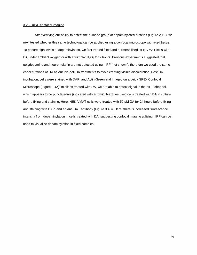
39
3.2.2. nIRF confocal imaging
After verifying our ability to detect the quinone group of dopaminylated proteins (Figure 2.1E), we
next tested whether this same technology can be applied using a confocal microscope with fixed tissue.
To ensure high levels of dopaminylation, we first treated fixed and permeabilized HEK-VMAT cells with
DA under ambient oxygen or with equimolar H2O2 for 2 hours. Previous experiments suggested that
polydopamine and neuromelanin are not detected using nIRF (not shown), therefore we used the same
concentrations of DA as our live-cell DA treatments to avoid creating visible discoloration. Post DA
incubation, cells were stained with DAPI and Actin-Green and imaged on a Leica SP8X Confocal
Microscope (Figure 3.4A). In slides treated with DA, we are able to detect signal in the nIRF channel,
which appears to be punctate-like (indicated with arrows). Next, we used cells treated with DA in culture
before fixing and staining. Here, HEK-VMAT cells were treated with 50 μM DA for 24 hours before fixing
and staining with DAPI and an anti-DAT antibody (Figure 3.4B). Here, there is increased fluorescence
intensity from dopaminylation in cells treated with DA, suggesting confocal imaging utilizing nIRF can be
used to visualize dopaminylation in fixed samples.
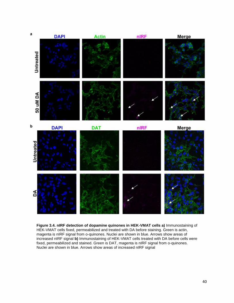
40
Figure 3.4. nIRF detection of dopamine quinones in HEK-VMAT cells a) Immunostaining of HEK-VMAT cells fixed, permeabilized and treated with DA before staining. Green is actin, magenta is nIRF signal from o-quinones. Nuclei are shown in blue. Arrows show areas of increased nIRF signal b) Immunostaining of HEK-VMAT cells treated with DA before cells were fixed, permeabilized and stained. Green is DAT, magenta is nIRF signal from o-quinones. Nuclei are shown in blue. Arrows show areas of increased nIRF signal
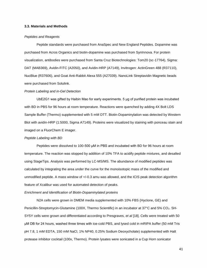
41
3.3. Materials and Methods
Peptides and Reagents
Peptide standards were purchased from AnaSpec and New England Peptides. Dopamine was
purchased from Acros Organics and biotin-dopamine was purchased from SynInnova. For protein
visualization, antibodies were purchased from Santa Cruz Biotechnologies: Tom20 (sc-17764), Sigma:
DAT (MAB369), Avidin-FITC (A2050), and Avidin-HRP (A7149), Invitrogen: ActinGreen 488 (R37110),
NucBlue (R37606), and Goat Anti-Rabbit Alexa 555 (A27039). NanoLink Streptavidin Magnetic beads
were purchased from Solulink.
Protein Labeling and in-Gel Detection
UbE2G1 was gifted by Haibin Mao for early experiments. 5 μg of purified protein was incubated
with BD in PBS for 96 hours at room temperature. Reactions were quenched by adding 4X Bolt LDS
Sample Buffer (Thermo) supplemented with 5 mM DTT. Biotin-Dopaminylation was detected by Western
Blot with avidin-HRP (1:5000, Sigma A7149). Proteins were visualized by staining with ponceau stain and
imaged on a FluorChem E imager.
Peptide Labeling with BD
Peptides were dissolved to 100-500 μM in PBS and incubated with BD for 96 hours at room
temperature. The reaction was stopped by addition of 10% TFA to acidify peptide mixtures, and desalted
using StageTips. Analysis was performed by LC-MS/MS. The abundance of modified peptides was
calculated by integrating the area under the curve for the monoisotopic mass of the modified and
unmodified peptide. A mass window of +/-0.3 amu was allowed, and the ICIS peak detection algorithm
feature of Xcalibur was used for automated detection of peaks.
Enrichment and Identification of Biotin-Dopaminylated proteins
N2A cells were grown in DMEM media supplemented with 10% FBS (Hyclone, GE) and
Penicillin-Streptomycin-Glutamine (100X, Thermo Scientific) in an incubator at 37°C and 5% CO2. SH-
SY5Y cells were grown and differentiated according to Presgraves, et al [18]. Cells were treated with 50
μM DB for 24 hours, washed three times with ice-cold PBS, and lysed cold in mRIPA buffer (50 mM Tris
pH 7.8, 1 mM EDTA, 150 mM NaCl, 1% NP40, 0.25% Sodium Deoxycholate) supplemented with Halt
protease inhibitor cocktail (100x, Thermo). Protein lysates were sonicated in a Cup Horn sonicator
42
(QSonica) for 20 minutes (30s on/off). The sonication cup must be filled with ice and replenished after 10
minutes to keep samples cold. Lysates are clarified by centrifugation at 21,000 x g and 4°C for 10
minutes. The supernatant containing the soluble protein fraction was transferred to a fresh
microcentrifuge tube. The protein content was measured using Pierce 660 nm Assay Reagent (Thermo
Scientific) using a BSA standard curve.
For each enrichment, 10 μL of NanoLink beads were washed two times with PBS, mixed with 500
μg of protein, and incubated with end-over-end rotation for one hour at room temperature. Beads were
washed two times with mRIPA buffer, once with 1 M KCl, once with 0.1 M Na2CO3, and twice with 2 M
Urea (50 mM Tris pH 8). Beads were resuspended in 100 μL 8 M Urea (50 mM Tris pH 8, 150 mM NaCl),
reduced with 1 mM TCEP and alkylated with 2 mM CAM. Urea was diluted to under 1.5 M with 100 mM
triethylammonium bicarbonate buffer. Proteins were digested off beads with 0.25 μg Lys-C for 2 hours,
followed by 0.25 μg Trypsin overnight. Peptides were acidified to pH 2 with t 10% TFA and desalted on
StageTips [95]. Peptides were then dried via speedvac and resuspended in StageTip Buffer A.
LC-MS/MS and data analysis
Peptides were separated on a Thermo-Dionex RSLCNano UHPLC instrument with 10 cm long
fused silica capillary columns made in-house with a laser puller (Sutter) and packed with 3 μm 120 Å
reversed phase C18 beads (Dr. Maisch). The LC gradient was 90 min long with 10–35% B at 200 nL/min.
LC solvent A was 0.1% acetic acid and LC solvent B was 0.1% acetic acid, 99.9% acetonitrile. MS data
was collected with a Thermo Orbitrap Elite spectrometer. Data-dependent analysis was applied using
Top15 selection with CID fragmentation. Raw files were analyzed by MaxQuant/Andromeda [72] version
1.5.7.4 using protein, peptide and site FDRs of 0.01 and a score minimum of 40 for modified peptides, 0
for unmodified peptides; delta score minimum of 6 for modified peptides, 0 for unmodified peptides.
MS/MS spectra were searched against the UniProt human database (updated July, 2016). MaxQuant
search parameters: Variable modifications included Oxidation (M) and Acetyl (Protein N-term).
Carbamidomethyl (C) was a fixed modification. Max. missed cleavages was 2, enzyme was Trypsin/P,
max. charge was 7. The MaxQuant match between runs feature was enabled. The initial search tolerance
for FTMS scans was 20 ppm and 0.5 Da for ITMS MS/MS scans. Data was further processed using the
Perseus software package (version 1.5.8.5), the R environment, and Microsoft Excel. Significantly
43
enriched proteins in the 50 μM BD experiments were determined by applying a moderated two-tailed,
two-sample t test in Perseus with an FDR of 0.05 and a small constant s0 = 0.1.
Immunofluorescence (IF)
HEK293 cells stably expressing VMAT2 and DAT were gifted by Dr. Gary Miller (Columbia). Cells
were grown in DMEM media supplemented with 10% FBS (Hyclone, GE) and Penicillin-Streptomycin-
Glutamine (100X, Thermo Scientific), 100 μg/ml Zeocin (Invitrogen) and 250 μg/ml G-418 (GoldBio) in an
incubator at 37°C and 5% CO2. For BD experiments, the same protocol was followed.
Cells were plated on acid washed cover slips and grown to 60% confluence. Fresh media was
added 16-18 hours prior to DA treatment (the day before). Cells were treated with 50 μM DA for 24 hours.
DA was made at 1000x stock in PBS and aliquots were frozen at -80°C. It is important to avoid
freeze/thaw cycles as DA will oxidize quickly. After treatment, cells were washed 3X with ice-cold PBS
and fixed with 4% paraformaldehyde for 20 minutes. Coverslips were washed three times with PBS,
quenched with 50 mM NH4Cl, washed again, and permeabilized with 0.1% TritonX-100 for four minutes at
room temperature. Slips were washed again, and blocked in 2% goat serum, 1% bovine serum albumin
(BSA) in PBS for one hour. After blocking, slips were washed once with PBS before incubation with
primary antibody (DAT 1:200, Tom20 1:500) in 0.2% goat serum, 0.1% Tween-20, 0.5% BSA in PBS at
4°C overnight. Slips were washed three times with PBS, and secondary antibody (1:500, diluted into
same buffer at primary antibody) with NucBlue and AvidinGreen (were specified) for one hour at room
temperature. Slips were washed three times with PBS, once with water, and mounted with ProLong
Antifade Diamond (Invitrogen) and cured overnight.
Confocal Microscopy
IF experiments were imaged on a Leica SP8X Confocal Microscope. For nIRF experiments, the
following settings were used: The scan direction was unidirectional at 200 Hz. Images were taken at 63X
in oil immersion. The aperture was set to 1.4, and the pinhole was calculated for 700 nM and set to 115.3
μm. Images were collective with a line average of 3 and a frame average 1. UV laser was set to 3% and
detected in PMT, Alexa 488 or 555 was excited with laser at 5% power and detected in PMT. The nIRF
44
was excited at 670 nm with the laser power at 50% and detected in HyD. Images were processed in
ImageJ.
3.4. Discussion
We describe two alternative strategies to study dopaminylation: biotin-dopamine and nIRF. A
biotinylated dopamine molecule is attractive as many tools already exist to enrich or visualize
biotinylation. However, we first needed to determine if BD is comparable to DA. Our data indicates that
BD has similar properties to dopamine in that BD modifies cysteine residues in standard peptides and
modifies proteins in cultured cells. Furthermore, the BD maintains its reactive catechol group; the two
hydroxyl groups on the benzene ring are critical for BD modification as biotin-tyramide is unable to modify
proteins in cultured cells. Another potential benefit of using BD is that it is applied exogenously, it crosses
cell membranes and therefore BD can be used in multiple cell types. Here, we show the BD modifies
proteins in both mouse neuroblastoma cells (N2A) and differentiated human neuroblatoma cells (dSH-
SY5Y) without requiring expression of dopamine transporters or specialized peroxidases.
We utilized several biotin-avidin tools to study biotin-dopaminylation of proteins including affinity
pull-downs coupled with LC-MS/MS. From these studies, we identified several proteins of interest in
neurodegeneration including tau, gephyrin, and reticulin-3, which are found in protein plaques in human
brain. Comparing our data set from BD experiments to proteins we found dopaminylated in our PBA
experiments, we see a 3% overlap in our dopaminylated proteins from HEK-VMAT cells and biotin-
dopaminylated proteins from N2A cells. This may be partially due to differential susceptibility to
dopamine oxidation between species. Furthermore, we noticed our human cells were more vulnerable to
high doses of BD or DA than mouse cells, which may be expected as mouse cells are known to be more
resistant to the toxic effects of DA [64]. Comparing biotin-dopaminylated proteins from differentiated SH-
SY5Y and dopaminylated proteins from HEK-VMAT cells, we found 12% of modified proteins are
common between BD and DA. While some differences may be due to differential gene expression, we
wanted to look for direct evidence of biotin-dopaminylation of proteins. Unfortunately, the biotin moieties
are difficult to elute from solid phase streptavidin beads and attempts to identify sites of biotinylation yield
45
few results. We iterated through several attempts to use biotin antibodies for biotin-dopaminylation site ID
[84], but were unable to successfully enrich biotin-dopaminylated peptides.
Throughout our study of BD, some differences between dopamine and biotin-dopamine were
observed. For example, BD appears to be more stable in solution than DA and BD solutions color much
more slowly. Also, BD does not form black pigments in a comparable time to DA. Whereas DA turns
pinkish within hours at room temperature and form black pigments in a few days, BD colors only after
several days and we never observed pigmentation. This may be due to the modified amine group
blocking cyclization making polymerization occur through alternative pathways. A modified amine group
may also slow the oxidation rate for BD. Together, these differences may have implications for BD
modification in cells.
Finally, while biotin-dopamine may be an interesting tool to probe proteins of interest like tau, our
biotin-dopamine approach cannot be translated to study tissue. Our method for covalent capture of
dopaminylated peptides described in Chapter 2 can be used to study dopaminylation from endogenous
DA modification and does not require derivatization of dopamine analogs. Biotin-dopamine may benefit
from future study to identify modified residues or be used as a chemical probe in living cells, but this is
beyond the scope of this work.
To get a more visual based detection approach, we tested nIRF confocal imaging for the
detection of dopaminylation in cultured cells. These experiments aimed to complement our identification
of dopaminylated peptides (Chapter 2) and were an extension of the work done by Mazzulli et al., to
detect and quantify oxidized catechols by nIRF. Confocal microscopy using nIRF to detect oxidized DA
represents a potential method to track dopaminylation in fixed tissue. Our experiments with confocal
microscopy and nIRF suggest fluorescence specific to DA treated cells. However, we found the signal to
noise ratio to be fairly low and difficult to interpret. Unlike fluorophore-based IF, nIRF of dopaminylation
relies on direct detection of the o-quinone and no amplification of signal is possible. Unable to increase
the fluorescent signal, we are unable to determine if the nIRF signal is co-localizing with organelles or
proteins of interest. Lastly, we saw background signal in our control experiments (no DA treatments)
increased over time, making re-imaging of microscope slides difficult. Overall, we believe this technique
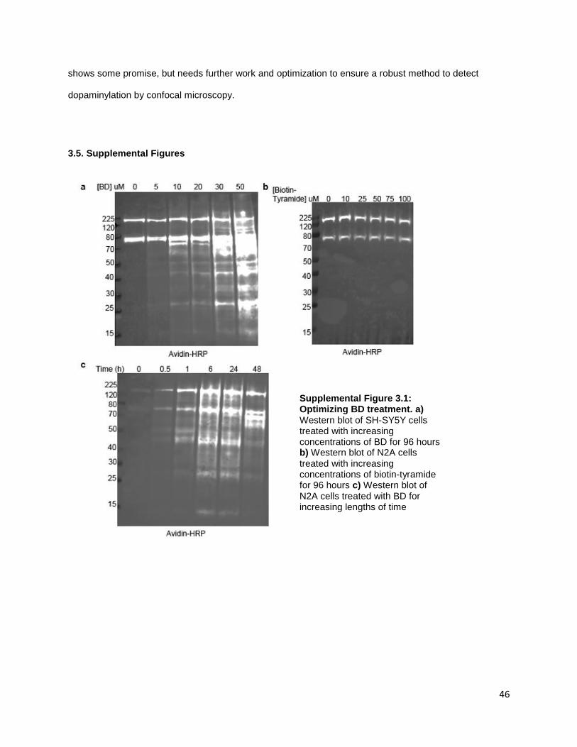
46
shows some promise, but needs further work and optimization to ensure a robust method to detect
dopaminylation by confocal microscopy.
3.5. Supplemental Figures
Supplemental Figure 3.1: Optimizing BD treatment. a) Western blot of SH-SY5Y cells treated with increasing concentrations of BD for 96 hours b) Western blot of N2A cells treated with increasing concentrations of biotin-tyramide for 96 hours c) Western blot of N2A cells treated with BD for increasing lengths of time
47
References
1. Schultz, W., Behavioral dopamine signals. Trends Neurosci, 2007. 30(5): p. 203-10. 2. Palmiter, R.D., Dopamine Signaling as a Neural Correlate of Consciousness. Neuroscience, 2011.
198: p. 213-220. 3. Medina, M.A., et al., Biogenic amines and polyamines: Similar biochemistry for different
physiological missions and biomedical applications. Critical Reviews in Biochemistry and Molecular Biology, 2003. 38(1): p. 23-59.
4. Canovas, F.G., et al., The role of pH in the melanin biosynthesis pathway. J Biol Chem, 1982. 257(15): p. 8738-44.
5. Lohr, K.M., et al., Membrane transporters as mediators of synaptic dopamine dynamics: implications for disease. Eur J Neurosci, 2017. 45(1): p. 20-33.
6. Neve, K.A., J.K. Seamans, and H. Trantham-Davidson, Dopamine receptor signaling. J Recept Signal Transduct Res, 2004. 24(3): p. 165-205.
7. Marchitti, S.A., R.A. Deitrich, and V. Vasiliou, Neurotoxicity and metabolism of the catecholamine-derived 3,4-dihydroxyphenylacetaldehyde and 3,4-dihydroxyphenylglycolaldehyde: the role of aldehyde dehydrogenase. Pharmacol Rev, 2007. 59(2): p. 125-50.
8. Sharman, D.F., The catabolism of catecholamines. Recent studies. Br Med Bull, 1973. 29(2): p. 110-5.
9. Cobley, J.N., M.L. Fiorello, and D.M. Bailey, 13 reasons why the brain is susceptible to oxidative stress. Redox Biology, 2018. 15: p. 490-503.
10. Lee, H., et al., Mussel-inspired surface chemistry for multifunctional coatings. Science, 2007. 318(5849): p. 426-30.
11. Jiang, L.Q., et al., Surface ocean pH and buffer capacity: past, present and future. Sci Rep, 2019. 9(1): p. 18624.
12. Lee, K., et al., Role of Dopamine Chemistry in the Formation of Mechanically Strong Mandibles of Grasshoppers. Chemistry of Materials, 2015. 27(19): p. 6478-6481.
13. Carlsson, A., et al., On the presence of 3-hydroxytyramine in brain. Science, 1958. 127(3296): p. 471.
14. Bjorklund, A. and S.B. Dunnett, Dopamine neuron systems in the brain: an update. Trends Neurosci, 2007. 30(5): p. 194-202.
15. Dichter, G.S., C.A. Damiano, and J.A. Allen, Reward circuitry dysfunction in psychiatric and neurodevelopmental disorders and genetic syndromes: animal models and clinical findings. J Neurodev Disord, 2012. 4(1): p. 19.
16. Gerfen, C.R. and D.J. Surmeier, Modulation of striatal projection systems by dopamine. Annu Rev Neurosci, 2011. 34: p. 441-66.
17. Grace, A.A., Dysregulation of the dopamine system in the pathophysiology of schizophrenia and depression. Nat Rev Neurosci, 2016. 17(8): p. 524-32.
18. Willuhn, I., et al., Excessive cocaine use results from decreased phasic dopamine signaling in the striatum. Nat Neurosci, 2014. 17(5): p. 704-9.
19. Rangel-Barajas, C., I. Coronel, and B. Floran, Dopamine Receptors and Neurodegeneration. Aging Dis, 2015. 6(5): p. 349-68.
20. Jankovic, J., Parkinson's disease: clinical features and diagnosis. J Neurol Neurosurg Psychiatry, 2008. 79(4): p. 368-76.
21. Postuma, R.B., et al., Identifying prodromal Parkinson's disease: pre-motor disorders in Parkinson's disease. Mov Disord, 2012. 27(5): p. 617-26.

48
22. Kalia, L.V. and A.E. Lang, Parkinson disease in 2015: Evolving basic, pathological and clinical concepts in PD. Nat Rev Neurol, 2016. 12(2): p. 65-6.
23. Verstraeten, A., J. Theuns, and C. Van Broeckhoven, Progress in unraveling the genetic etiology of Parkinson disease in a genomic era. Trends Genet, 2015. 31(3): p. 140-9.
24. Spatola, M. and C. Wider, Genetics of Parkinson's disease: the yield. Parkinsonism Relat Disord, 2014. 20 Suppl 1: p. S35-8.
25. Bellomo, G., et al., The vicious cycle between alpha-synuclein aggregation and autophagic-lysosomal dysfunction. Mov Disord, 2019.
26. Vogels, T., et al., Propagation of Tau Pathology: Integrating Insights From Postmortem and In Vivo Studies. Biol Psychiatry, 2019.
27. Telling, N.D., et al., Iron Biochemistry is Correlated with Amyloid Plaque Morphology in an Established Mouse Model of Alzheimer's Disease. Cell Chem Biol, 2017. 24(10): p. 1205-1215 e3.
28. Wright, J.A., X. Wang, and D.R. Brown, Unique copper-induced oligomers mediate alpha-synuclein toxicity. FASEB J, 2009. 23(8): p. 2384-93.
29. Wan, W., et al., Iron Deposition Leads to Neuronal alpha-Synuclein Pathology by Inducing Autophagy Dysfunction. Front Neurol, 2017. 8: p. 1.
30. Bellou, V., et al., Environmental risk factors and Parkinson's disease: An umbrella review of meta-analyses. Parkinsonism Relat Disord, 2016. 23: p. 1-9.
31. Goldman, S.M., et al., Genetic modification of the association of paraquat and Parkinson's disease. Mov Disord, 2012. 27(13): p. 1652-8.
32. Monzani, E., et al., Dopamine, Oxidative Stress and Protein-Quinone Modifications in Parkinson's and Other Neurodegenerative Diseases. Angewandte Chemie-International Edition, 2019. 58(20): p. 6512-6527.
33. Harrington, K.A., et al., Dopamine transporter (Dat) and synaptic vesicle amine transporter (VMAT2) gene expression in the substantia nigra of control and Parkinson's disease. Brain Res Mol Brain Res, 1996. 36(1): p. 157-62.
34. Pifl, C., et al., Is Parkinson's disease a vesicular dopamine storage disorder? Evidence from a study in isolated synaptic vesicles of human and nonhuman primate striatum. J Neurosci, 2014. 34(24): p. 8210-8.
35. Lohr, K.M., et al., Increased vesicular monoamine transporter enhances dopamine release and opposes Parkinson disease-related neurodegeneration in vivo. Proc Natl Acad Sci U S A, 2014. 111(27): p. 9977-82.
36. Rilstone, J.J., R.A. Alkhater, and B.A. Minassian, Brain dopamine-serotonin vesicular transport disease and its treatment. N Engl J Med, 2013. 368(6): p. 543-50.
37. Jellinger, K., et al., Chemical evidence for 6-hydroxydopamine to be an endogenous toxic factor in the pathogenesis of Parkinson's disease. J Neural Transm Suppl, 1995. 46: p. 297-314.
38. Galzigna, L., A. De Iuliis, and L. Zanatta, Enzymatic dopamine peroxidation in substantia nigra of human brain. Clin Chim Acta, 2000. 300(1-2): p. 131-8.
39. Hastings, T.G., Enzymatic oxidation of dopamine: the role of prostaglandin H synthase. J Neurochem, 1995. 64(2): p. 919-24.
40. Block, M.L., L. Zecca, and J.S. Hong, Microglia-mediated neurotoxicity: uncovering the molecular mechanisms. Nat Rev Neurosci, 2007. 8(1): p. 57-69.
41. Bulk, M., et al., Quantitative comparison of different iron forms in the temporal cortex of Alzheimer patients and control subjects. Scientific Reports, 2018. 8.
42. Castellani, R.J., et al., Sequestration of iron by Lewy bodies in Parkinson's disease. Acta Neuropathologica, 2000. 100(2): p. 111-114.
43. Mor, D.E., et al., Dopamine induces soluble alpha-synuclein oligomers and nigrostriatal degeneration. Nat Neurosci, 2017. 20(11): p. 1560-+.
49
44. Fedorow, H., et al., Neuromelanin in human dopamine neurons: Comparison with peripheral melanins and relevance to Parkinson's disease. Progress in Neurobiology, 2005. 75(2): p. 109-124.
45. Solomon, E.I., et al., Copper Active Sites in Biology. Chemical Reviews, 2014. 114(7): p. 3659-3853.
46. Zucca, F.A., et al., Interactions of iron, dopamine and neuromelanin pathways in brain aging and Parkinson's disease. Progress in Neurobiology, 2017. 155: p. 96-119.
47. Zucca, F.A., et al., Neuromelanin organelles are specialized autolysosomes that accumulate undegraded proteins and lipids in aging human brain and are likely involved in Parkinson's disease. Npj Parkinsons Disease, 2018. 4.
48. Shima, T., et al., Binding of iron to neuromelanin of human substantia nigra and synthetic melanin: An electron paramagnetic resonance spectroscopy study. Free Radical Biology and Medicine, 1997. 23(1): p. 110-119.
49. Sulzer, D., et al., Neuronal pigmented autophagic vacuoles: lipofuscin, neuromelanin, and ceroid as macroautophagic responses during aging and disease. J Neurochem, 2008. 106(1): p. 24-36.
50. Marsden, C.D., Pigmentation in the nucleus substantiae nigrae of mammals. J Anat, 1961. 95: p. 256-61.
51. Marsden, C.D., The Development of Pigmentation and Enzyme Activity in the Nucleus Substantiae Nigrae of the Cat. J Anat, 1965. 99: p. 175-80.
52. Zecca, L., et al., The absolute concentration of nigral neuromelanin, assayed by a new sensitive method, increases throughout the life and is dramatically decreased in Parkinson's disease. Febs Letters, 2002. 510(3): p. 216-220.
53. Venkateshappa, C., et al., Increased oxidative damage and decreased antioxidant function in aging human substantia nigra compared to striatum: Implications for Parkinson's disease. Movement Disorders, 2014. 29: p. S35-S35.
54. Tribl, F., et al., Identification of L-ferritin in neuromelanin granules of the human substantia nigra: a targeted proteomics approach. Mol Cell Proteomics, 2009. 8(8): p. 1832-8.
55. D'Amato, R.J., Z.P. Lipman, and S.H. Snyder, Selectivity of the parkinsonian neurotoxin MPTP: toxic metabolite MPP+ binds to neuromelanin. Science, 1986. 231(4741): p. 987-9.
56. Lindquist, N.G., B.S. Larsson, and A. Lyden-Sokolowski, Autoradiography of [14C]paraquat or [14C]diquat in frogs and mice: accumulation in neuromelanin. Neurosci Lett, 1988. 93(1): p. 1-6.
57. Haining, R.L. and C. Achat-Mendes, Neuromelanin, one of the most overlooked molecules in modern medicine, is not a spectator. Neural Regen Res, 2017. 12(3): p. 372-375.
58. Zucca, F.A., et al., Neuromelanin of the human substantia nigra: an update. Neurotox Res, 2014. 25(1): p. 13-23.
59. Viceconte, N., et al., Neuromelanin activates proinflammatory microglia through a caspase-8-dependent mechanism. J Neuroinflammation, 2015. 12: p. 5.
60. Li, H. and G. Dryhurst, Irreversible inhibition of mitochondrial complex I by 7-(2-aminoethyl)-3,4-dihydro-5-hydroxy-2H-1,4-benzothiazine-3-carboxyli c acid (DHBT-1): a putative nigral endotoxin of relevance to Parkinson's disease. J Neurochem, 1997. 69(4): p. 1530-41.
61. Ito, S., A. Palumbo, and G. Prota, Tyrosinase-catalyzed conjugation of dopa with glutathione. Experientia, 1985. 41(7): p. 960-1.
62. Xu, R., et al., Characterization of products from the reactions of N-acetyldopamine quinone with N-acetylhistidine. Arch Biochem Biophys, 1996. 329(1): p. 56-64.
63. Van Laar, V.S., et al., Proteomic identification of dopamine-conjugated proteins from isolated rat brain mitochondria and SH-SY5Y cells. Neurobiol Dis, 2009. 34(3): p. 487-500.
64. Burbulla, L.F., et al., Dopamine oxidation mediates mitochondrial and lysosomal dysfunction in Parkinson's disease. Science, 2017. 357(6357): p. 1255-1261.

50
65. Belluzzi, E., et al., Human SOD2 modification by dopamine quinones affects enzymatic activity by promoting its aggregation: possible implications for Parkinson's disease. PLoS One, 2012. 7(6): p. e38026.
66. Mexas, L.M., V.R. Florang, and J.A. Doorn, Inhibition and covalent modification of tyrosine hydroxylase by 3,4-dihydroxyphenylacetaldehyde, a toxic dopamine metabolite. Neurotoxicology, 2011. 32(4): p. 471-7.
67. Werner-Allen, J.W., et al., Toxic Dopamine Metabolite DOPAL Forms an Unexpected Dicatechol Pyrrole Adduct with Lysines of alpha-Synuclein. Angew Chem Int Ed Engl, 2016. 55(26): p. 7374-8.
68. Longhena, F., et al., The Contribution of alpha-Synuclein Spreading to Parkinson's Disease Synaptopathy. Neural Plast, 2017. 2017: p. 5012129.
69. Kalousova, M., J. Skrha, and T. Zima, Advanced glycation end-products and advanced oxidation protein products in patients with diabetes mellitus. Physiol Res, 2002. 51(6): p. 597-604.
70. van der Mijn, J.C., et al., Evaluation of different phospho-tyrosine antibodies for label-free phosphoproteomics. J Proteomics, 2015. 127(Pt B): p. 259-63.
71. Herberg, S., et al., Histone H3 lysine 9 trimethylation is required for suppressing the expression of an embryonically activated retrotransposon in Xenopus laevis. Sci Rep, 2015. 5: p. 14236.
72. Cox, J., et al., Andromeda: a peptide search engine integrated into the MaxQuant environment. J Proteome Res, 2011. 10(4): p. 1794-805.
73. Dietrich, J.B., et al., Acute or repeated cocaine administration generates reactive oxygen species and induces antioxidant enzyme activity in dopaminergic rat brain structures. Neuropharmacology, 2005. 48(7): p. 965-74.
74. LaVoie, M.J. and T.G. Hastings, Dopamine quinone formation and protein modification associated with the striatal neurotoxicity of methamphetamine: evidence against a role for extracellular dopamine. J Neurosci, 1999. 19(4): p. 1484-91.
75. Granado, N., et al., Persistent MDMA-induced dopaminergic neurotoxicity in the striatum and substantia nigra of mice. J Neurochem, 2008. 107(4): p. 1102-12.
76. Lotharius, J. and K.L. O'Malley, The parkinsonism-inducing drug 1-methyl-4-phenylpyridinium triggers intracellular dopamine oxidation. A novel mechanism of toxicity. J Biol Chem, 2000. 275(49): p. 38581-8.
77. Betarbet, R., et al., Chronic systemic pesticide exposure reproduces features of Parkinson's disease. Nat Neurosci, 2000. 3(12): p. 1301-6.
78. LaVoie, M.J., et al., Dopamine covalently modifies and functionally inactivates parkin. Nat Med, 2005. 11(11): p. 1214-21.
79. Malaker, S.A., et al., Identification of Glycopeptides as Posttranslationally Modified Neoantigens in Leukemia. Cancer Immunol Res, 2017. 5(5): p. 376-384.
80. Zhang, Y., et al., Site-specific characterization of the Asp- and Glu-ADP-ribosylated proteome. Nat Methods, 2013. 10(10): p. 981-4.
81. Burzio, L.A. and J.H. Waite, Cross-linking in adhesive quinoproteins: studies with model decapeptides. Biochemistry, 2000. 39(36): p. 11147-53.
82. Mazzulli, J.R., et al., Detection of Free and Protein-Bound ortho-Quinones by Near-Infrared Fluorescence. Anal Chem, 2016. 88(4): p. 2399-405.
83. Napolitano, A., P. Manini, and M. d'Ischia, Oxidation chemistry of catecholamines and neuronal degeneration: an update. Curr Med Chem, 2011. 18(12): p. 1832-45.
84. Udeshi, N.D., et al., Antibodies to biotin enable large-scale detection of biotinylation sites on proteins. Nat Methods, 2017. 14(12): p. 1167-1170.
85. Presgraves, S.P., et al., Terminally differentiated SH-SY5Y cells provide a model system for studying neuroprotective effects of dopamine agonists. Neurotox Res, 2004. 5(8): p. 579-98.

51
86. Bernstein, A.I., K.A. Stout, and G.W. Miller, A fluorescent-based assay for live cell, spatially resolved assessment of vesicular monoamine transporter 2-mediated neurotransmitter transport. J Neurosci Methods, 2012. 209(2): p. 357-66.
87. Geiger, T., et al., Comparative proteomic analysis of eleven common cell lines reveals ubiquitous but varying expression of most proteins. Mol Cell Proteomics, 2012. 11(3): p. M111 014050.
88. Smith, D.G., R. Cappai, and K.J. Barnham, The redox chemistry of the Alzheimer's disease amyloid beta peptide. Biochim Biophys Acta, 2007. 1768(8): p. 1976-90.
89. Abo, M. and E. Weerapana, A Caged Electrophilic Probe for Global Analysis of Cysteine Reactivity in Living Cells. J Am Chem Soc, 2015. 137(22): p. 7087-90.
90. Barretina, J., et al., The Cancer Cell Line Encyclopedia enables predictive modelling of anticancer drug sensitivity. Nature, 2012. 483(7391): p. 603-7.
91. Hedges, D.M., et al., Methamphetamine Induces Dopamine Release in the Nucleus Accumbens Through a Sigma Receptor-Mediated Pathway. Neuropsychopharmacology, 2018. 43(6): p. 1405-1414.
92. Park, S.U., et al., Peroxynitrite inactivates the human dopamine transporter by modification of cysteine 342: potential mechanism of neurotoxicity in dopamine neurons. J Neurosci, 2002. 22(11): p. 4399-405.
93. Rastedt, D.E., R.A. Vaughan, and J.D. Foster, Palmitoylation mechanisms in dopamine transporter regulation. J Chem Neuroanat, 2017. 83-84: p. 3-9.
94. Alvarez-Zaldiernas, C., et al., Cellular Redox Systems Impact the Aggregation of Cu,Zn Superoxide Dismutase Linked to Familial Amyotrophic Lateral Sclerosis. J Biol Chem, 2016. 291(33): p. 17197-208.
95. Rappsilber, J., M. Mann, and Y. Ishihama, Protocol for micro-purification, enrichment, pre-fractionation and storage of peptides for proteomics using StageTips. Nat Protoc, 2007. 2(8): p. 1896-906.
96. Rikova, K., et al., Global survey of phosphotyrosine signaling identifies oncogenic kinases in lung cancer. Cell, 2007. 131(6): p. 1190-203.
97. Rhee, H.W., et al., Proteomic mapping of mitochondria in living cells via spatially restricted enzymatic tagging. Science, 2013. 339(6125): p. 1328-1331.
98. Mick, D.U., et al., Proteomics of Primary Cilia by Proximity Labeling. Dev Cell, 2015. 35(4): p. 497-512.
99. Myers, S.A., et al., Discovery of proteins associated with a predefined genomic locus via dCas9-APEX-mediated proximity labeling. Nat Methods, 2018. 15(6): p. 437-439.
100. Goedert, M., D.S. Eisenberg, and R.A. Crowther, Propagation of Tau Aggregates and Neurodegeneration. Annu Rev Neurosci, 2017. 40: p. 189-210.
101. Hales, C.M., et al., Abnormal gephyrin immunoreactivity associated with Alzheimer disease pathologic changes. J Neuropathol Exp Neurol, 2013. 72(11): p. 1009-15.
102. Heath, J.E., et al., Widespread distribution of reticulon-3 in various neurodegenerative diseases. Neuropathology, 2010. 30(6): p. 574-9.
103. Attems, J. and K. Jellinger, Proteomics for synaptic markers of cognitive decline in neurodegenerative diseases. Brain, 2018. 141(2): p. 329-331.
104. Chen, Y.F., et al., Cisd2 deficiency drives premature aging and causes mitochondria-mediated defects in mice. Genes Dev, 2009. 23(10): p. 1183-94.
105. Sanchez-Ortiz, E., et al., Protein phosphatase 5 protects neurons against amyloid-beta toxicity. J Neurochem, 2009. 111(2): p. 391-402.
106. Ayuso, P., et al., A polymorphism located at an ATG transcription start site of the heme oxygenase-2 gene is associated with classical Parkinson's disease. Pharmacogenet Genomics, 2011. 21(9): p. 565-71.
52
107. Sturner, E. and C. Behl, The Role of the Multifunctional BAG3 Protein in Cellular Protein Quality Control and in Disease. Front Mol Neurosci, 2017. 10: p. 177.
108. Riquier, S., et al., Peroxiredoxin post-translational modifications by redox messengers. Redox Biology, 2014. 2: p. 777-85.
109. Wu, C., et al., Sulfonation of the resolving cysteine in human peroxiredoxin 1: A comprehensive analysis by mass spectrometry. Free Radic Biol Med, 2017. 108: p. 785-792.
110. Ikwegbue, P.C., et al., Roles of Heat Shock Proteins in Apoptosis, Oxidative Stress, Human Inflammatory Diseases, and Cancer. Pharmaceuticals (Basel), 2017. 11(1).
111. Castegna, A., et al., Proteomic identification of oxidatively modified proteins in Alzheimer's disease brain. Part II: dihydropyrimidinase-related protein 2, alpha-enolase and heat shock cognate 71. J Neurochem, 2002. 82(6): p. 1524-32.

53
Vita
Emily Jane Myers was born in Fullerton, California. After graduating high school, she attended Fullerton
Community College prior to transferring to the University of California, Irvine. Emily earned a Bachelor of
Science from the University of California Irvine in Biochemistry and Molecular Biology in 2011. While
working on her undergraduate degree, Emily worked in the lab of Dr. Jefferson Y Chan where she
investigated Nrf1 trafficking via retrotranslocation of deglycosylated substrates and validation of novel
genes under Nrf1 transcriptional control in disease state and cell lines. After graduation, Emily worked as
a project coordinator and researcher for the Women’s Interagency HIV Study at the University of
Southern California investigating accelerating aging mechanisms due to latent HIV infection. In 2013, she
joined the University of Washington’s Department of Pharmacology as a graduate research associate,
and earned a Doctor of Philosophy in 2019.