development of western blots for actin without the use of radioactivity geoff theobald step summer...
TRANSCRIPT

Development of Western Blots for Actin without the use of
radioactivity
Geoff Theobald
STEP Summer Internship Program
June 2003
Objective
To create a Western Blot without the use of radioactivity.
Significance
• Safety & disposal
• Use in college lab: students learn about gene expression and molecular biology

Background Information
Actin:• Is the protein we detected
using a Western Blot.• Has a molecular weight of
42,000-43,000 daltons. • Works with myosin
(another protein) in muscle contraction.
• Is also involved in the structure in most cells.
Methods
• Polyacrylamide gel electrophoresis
• Western Blot
• Probing with an antibody to actin
• Detection by fluorescence
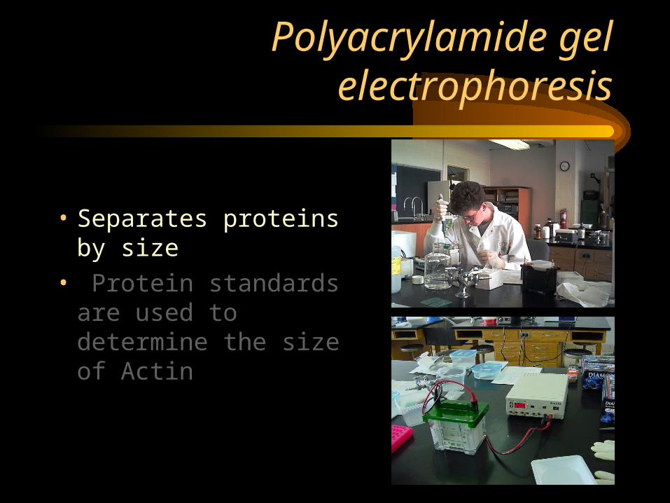
Polyacrylamide gel electrophoresis
• Separates proteins by size
• Protein standards are used to determine the size of Actin

Western Blot
• A western is when you transfer protein out of polyacrylamide gel and into a membrane.
• A Western Blot is the result of the transfer.
Gel
Pic. Of membrane
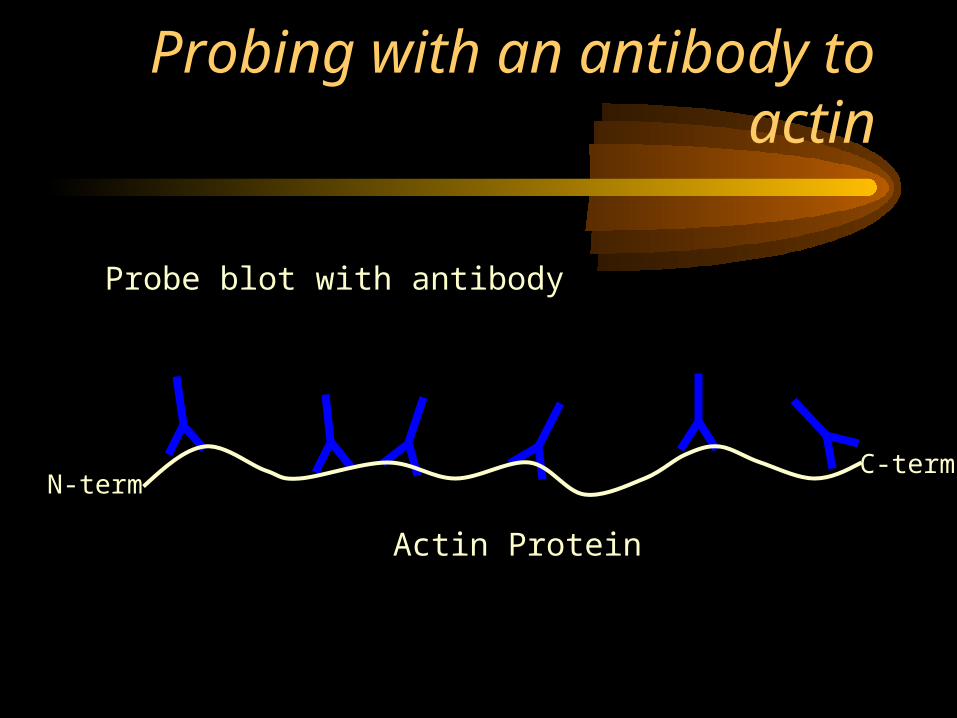
Probing with an antibody to actin
Probe blot with antibody
N-termC-term
Actin Protein
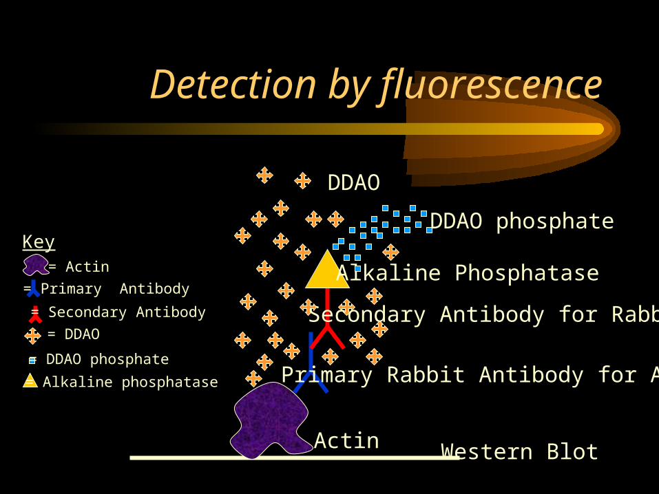
Detection by fluorescence
Key= Actin
= Primary Antibody
= Secondary Antibody
= DDAO
= DDAO phosphate
= Alkaline phosphatase
Actin
Primary Rabbit Antibody for Actin
Secondary Antibody for Rabbit
Alkaline Phosphatase
DDAO phosphate
DDAO
Western Blot

Results: detection with UV light
• There was no clear signal.
Pic. of Blot exposed to UV light

Fluorescent dye can be visualized with UV or white light
Advantages
• UV: we can distinguish red light
• White light: stronger excitation
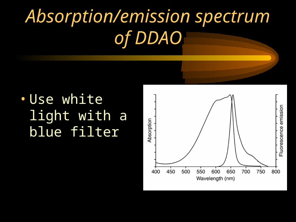
Absorption/emission spectrum of DDAO
• Use white light with a blue filter
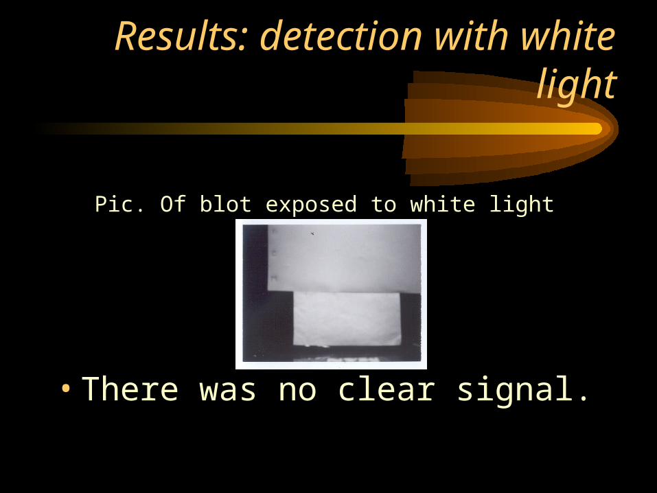
Results: detection with white light
• There was no clear signal.
Pic. Of blot exposed to white light

Chemiluminescence assay
• Worked well. Continued experiments with this assay.
Pic of dot blot with all actin antibody
Pic of membrane with all actin antibody

Conclusions
1. Detection of fluorescence with UV light did not work.
2. Detection of fluorescence with white light and a blue filter did not work.
3. Since fluorescence assay did not work well, but chemiluminescence worked, we will concentrate on that assay.
Acknowledgements
• Dr. Guzman, Biology Dept.
• Dr. Metz, Biology Dept.
• Mrs. Bloom, STEP program
I would like to thank each and everyone of you for all of your advice, support, and fun I had these past two weeks.