di(2-ethylhexyl) phthalate 1. exposure datamonographs.iarc.fr/eng/monographs/vol77/mono77-6.pdf ·...
TRANSCRIPT
DI(2-ETHYLHEXYL) PHTHALATE
This substance was considered by previous Working Groups, in October 1981(IARC, 1982) and March 1987 (IARC, 1987). Since that time, new data have becomeavailable, and these have been incorporated into the monograph and taken into consi-deration in the present evaluation.
1. Exposure Data
1.1 Chemical and physical data
1.1.1 Nomenclature
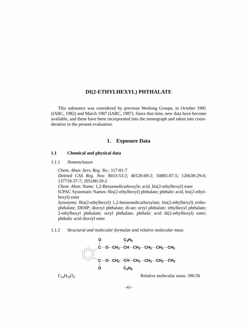
Chem. Abstr. Serv. Reg. No.: 117-81-7Deleted CAS Reg. Nos: 8033-53-2; 40120-69-2; 50885-87-5; 126639-29-0;137718-37-7; 205180-59-2Chem. Abstr. Name: 1,2-Benzenedicarboxylic acid, bis(2-ethylhexyl) esterIUPAC Systematic Names: Bis(2-ethylhexyl) phthalate; phthalic acid, bis(2-ethyl-hexyl) esterSynonyms: Bis(2-ethylhexyl) 1,2-benzenedicarboxylate; bis(2-ethylhexyl) ortho-phthalate; DEHP; dioctyl phthalate; di-sec octyl phthalate; ethylhexyl phthalate;2-ethylhexyl phthalate; octyl phthalate; phthalic acid di(2-ethylhexyl) ester;phthalic acid dioctyl ester
1.1.2 Structural and molecular formulae and relative molecular mass
C24H38O4 Relative molecular mass: 390.56
–41–
C
C
O CH2 CH
C2H5
CH2 CH2 CH2 CH3
O
O CH2 CH CH2 CH2 CH2 CH3
O
C2H5

1.1.3 Chemical and physical properties of the pure substance
(a) Description: Light-coloured liquid with a slight odour (Agency for ToxicSubstances and Disease Registry, 1993; Verschueren, 1996)
(b) Boiling-point: 384 °C (Lide, 1999)(c) Melting-point: –55 °C (Lide, 1999)(d) Density: 0.981 g/cm3 at 25 °C (Lide, 1999)(e) Spectroscopy data: Infrared (prism [28]; grating [18401]), ultraviolet [22080],
nuclear magnetic resonance (proton [9392]; C-13 [5201]) and mass [NIST,43511] spectral data have been reported (Sadtler Research Laboratories, 1980;Lide & Milne, 1996)
(f) Solubility: Slightly soluble in water (0.285 mg/L at 24 °C)1; slightly solublein carbon tetrachloride (Environmental Protection Agency, 1998; Lide, 1999)
(g) Volatility: Vapour pressure, 8.6 × 10⁻⁴ Pa at 25 °C, 160 Pa at 200 °C; relativevapour density (air = 1), 13.4 (Howard et al., 1985; Verschueren, 1996)
(h) Octanol/water partition coefficient (P): log P, 7.45 (Hansch et al., 1995)(i) Conversion factor2: mg/m3 = 16.0 × ppm
1.1.4 Technical products and impuritiesDi(2-ethylhexyl) phthalate is available in a variety of technical grades (including a
special grade for capacitor applications and a low residuals grade). Typical specificationsare: minimal ester content, 99.0–99.6%; maximal moisture content, 0.1%; and acidity(as acetic acid or phthalic acid), 0.007–0.01% (Aristech Chemical Corp., 1992; WHO,1992).
Trade names for di(2-ethylhexyl) phthalate include: Bisoflex; Compound 889;Diacizer DOP; DOP; Eastman DOP Plasticizer; Ergoplast; Etalon; Eviplast; Fleximel;Flexol DOP; Good-rite GP 264; Hatco-DOP; Kodaflex DOP; Monocizer DOP; Octoil;Palatinol AH; Pittsburgh PX 138; Plasthall DOP; Platinol AH; Reomol D 79P; Sicol150; Staflex DOP; Truflex DOP; Vestinol AH; Vinycizer; Witcizer 312 (National Toxi-cology Program, 1991; WHO, 1992; American Conference of Governmental IndustrialHygienists, 1999).
1.1.5 AnalysisDetection and quantification of very low levels of di(2-ethylhexyl) phthalate are
seriously limited by the presence of this compound as a contaminant in almost all labo-ratory equipment and reagents. Plastics, glassware, aluminium foil, cork, rubber, glasswool, solvents and Teflon® sheets have all been found to be contaminated (Agency forToxic Substances and Disease Registry, 1993).
IARC MONOGRAPHS VOLUME 7742
1 Lower values have been proposed, based on models (Staples et al., 1997).2 Calculated from: mg/m3 = (relative molecular mass/24.45) × ppm, assuming a temperature of 25 °C anda pressure of 101 kPa

Determination of di(2-ethylhexyl) phthalate in air, water, soil/sediments and foodis usually accomplished by gas chromatographic analysis; a high-performance liquidchromatography method for food has also been developed. Di(2-ethylhexyl) phthalateis usually extracted from environmental samples with an organic solvent such ashexane, chloroform, dichloromethane or acetonitrile. Air samples are drawn througha solid sorbent material and desorbed with carbon disulfide. A purge-and-trap methodhas been developed for separation of di(2-ethylhexyl) phthalate from the fat in foods(Agency for Toxic Substances and Disease Registry, 1993).
Selected methods for the analysis of di(2-ethylhexyl) phthalate in various matricesare presented in Table 1.
1.2 Production
The worldwide production of di(2-ethylhexyl) phthalate has been increasing duringrecent decades and in the late 1980s amounted to approximately 1 million tonnes peryear (WHO, 1992). Production of di(2-ethylhexyl) phthalate in the United Statesincreased during the 1980s, from approximately 114 000 tonnes in 1982 to over 130 000tonnes in 1986 (Environmental Protection Agency, 1998). In 1994, production of di(2-ethylhexyl) phthalate in the United States was 117 500 tonnes; production in Japan in1995 was 298 000 tonnes; production in Taiwan in 1995 was 207 000 tonnes, downfrom 241 000 tonnes in 1994 (Anon., 1996).
Di(2-ethylhexyl) phthalate is produced commercially by the reaction of excess2-ethylhexanol with phthalic anhydride in the presence of an acid catalyst such assulfuric acid or para-toluenesulfonic acid. It was first produced in commercial quan-tities in Japan around 1933 and in the United States in 1939 (IARC, 1982).
Information available in 1999 indicated that di(2-ethylhexyl) phthalate was pro-duced by 30 companies in China, 15 companies in India, 12 companies in Japan, eightcompanies in Mexico, seven companies in Taiwan, five companies each in Germany andthe Russian Federation, four companies each in Argentina, Brazil, the Philippines andthe United States, three companies each in Canada, Chile, Spain, Thailand, Turkey andVenezuela, two companies each in Belgium, Colombia, Ecuador, France, Indonesia,Iran, Italy, Korea (Republic of), Malaysia and Poland, and one company each in Albania,the Czech Republic, Finland, Kazakhstan, Pakistan, Peru, Romania, South Africa,Sweden, Ukraine, the United Kingdom and Viet Nam (Chemical Information Services,1999).
1.3 Use
Di(2-ethylhexyl) phthalate is widely used as a plasticizer in flexible vinyl products.Plastics may contain from 1 to 40% di(2-ethylhexyl) phthalate by weight and are usedin consumer products such as imitation leather, rainwear, footwear, upholstery, flooring,wire and cable, tablecloths, shower curtains, food packaging materials and children’s
DI(2-ETHYLHEXYL) PHTHALATE 43

toys. Poly(vinyl chloride) (PVC) containing di(2-ethylhexyl) phthalate is also used fortubing and containers for blood products and transfusions. Di(2-ethylhexyl) phthalate isalso used as a hydraulic fluid and as a dielectric fluid (a non-conductor of electriccurrent) in electrical capacitors (Agency for Toxic Substances and Disease Registry,1989). Other uses are in rubbing alcohol, liquid detergents, decorative inks, munitions,industrial and lubricating oils and defoaming agents during paper and paperboardmanufacture (Environmental Protection Agency, 1998). It is also used as an acaricide in
IARC MONOGRAPHS VOLUME 7744
Table 1. Selected methods for the analysis of di(2-ethylhexyl) phthalate
Sample matrix Sample preparation Assayprocedurea
Limit ofdetection
Reference
Air Collect on cellulose estermembrane filter; desorb withcarbon disulfide
GC/FID 10 μg/sample Eller (1994)[Method 5020]
Drinking-water,and sourcewater
Extract in liquid–solid extractor;elute with dichloromethane;concentrate by evaporation
GC/MS 0.5 μg/L EnvironmentalProtectionAgency (1995a)[Method 525.2]
Drinking-water Extract in liquid–liquid extractor;isolate; dry; concentrate
GC/PID 2.25 μg/L EnvironmentalProtectionAgency (1995b)[Method 506]
Wastewater,municipal andindustrial
Extract with dichloromethane;dry; exchange to hexane andconcentrate
GC/ECD 2.0 μg/L EnvironmentalProtectionAgency (1999a)[Method 606]
Extract with dichloromethane;dry; concentrate
GC/MS 2.5 μg/L EnvironmentalProtectionAgency (1999b)[Method 625]
Add isotope-labelled analogue;extract with dichloromethane;dry over sodium sulfate;concentrate
GC/MS 10 μg/L EnvironmentalProtectionAgency (1999c)[Method 1625]
Groundwater,leachate, soil,sludge,sediment
Aqueous sample: extract withdichloromethane; elute withacetonitrile; exchange to hexane;Solid sample: extract withdichloromethane/acetone (1:1) orhexane/acetone (1:1); clean-up
GC/ECD 0.27 μg/L(aqueous)
EnvironmentalProtectionAgency (1996)[Method 8061A]
a Abbreviations: GC, gas chromatography; ECD, electron capture detection; FID, flame ionization detec-tion; MS, mass spectrometry; PID, photoionization detection

orchards, an inert ingredient in pesticides, a detector for leaks in respirators, in thetesting of air filtration systems and as a component of cosmetic products (National Toxi-cology Program, 1991).
World consumption of phthalates in the early 1990s was estimated to be 3.25 milliontonnes, of which di(2-ethylhexyl) phthalate accounted for approximately 2.1 milliontonnes. The estimated total consumption of di(2-ethylhexyl) phthalate by geographicalregion was (thousand tonnes): western Europe, 465; North America, 155; eastern Asia,490; Japan, 245; and others, 765 (Towae et al., 1992).
1.4 Occurrence
Concern regarding exposure to di(2-ethylhexyl) phthalate rose to prominence whenJaeger and Rubin (1970) reported its presence in human blood that had been stored inPVC bags. The same authors later reported the presence of di(2-ethylhexyl) phthalatein tissue samples of the lung, liver and spleen from patients who had received bloodtransfusions before death (Jaeger & Rubin, 1972). While occupational inhalation is asignificant potential route of exposure, medical procedures such as haemodialysis,extracorporeal membrane oxygenation, blood transfusion, umbilical catheterizationand short-term cardiopulmonary by-pass can also result in high exposures (Huber et al.,1996; Karle et al., 1997). Patients undergoing haemodialysis are considered to have thehighest exposure, due to the chronic nature of the treatment. Further, because of thewidespread use of di(2-ethylhexyl) phthalate in plastic containers and its ability toleach out of PVC, humans are exposed to this substance on a regular basis. Theextensive manufacture of di(2-ethylhexyl) phthalate-containing plastics has resulted inits becoming a ubiquitous environmental contaminant (Huber et al., 1996).
1.4.1 Natural occurrence
Di(2-ethylhexyl) phthalate is not known to occur naturally.
1.4.2 Occupational exposure
According to the 1981–83 US National Occupational Exposure Survey, approxi-mately 341 000 workers in the United States were potentially exposed to di(2-ethylhexyl) phthalate. Occupational exposure to di(2-ethylhexyl) phthalate may occurduring its manufacture and its use mostly as a plasticizer of PVC (compounding,calendering and coating operations). Printing and painting occupations account alsofor a large number of workers potentially exposed (NOES, 1999). Occupationalexposure to di(2-ethylhexyl) phthalate occurs by inhalation, essentially in the form ofan aerosol (mist) because of the very low vapour pressure of the substance (Fishbein,1992). Indeed, di(2-ethylhexyl) phthalate aerosols are used to test the efficiency ofhigh-efficiency particulate air filters during their manufacture (Roberts, 1983).
DI(2-ETHYLHEXYL) PHTHALATE 45

Few data are available on levels of occupational exposure to di(2-ethylhexyl)phthalate (Table 2). Huber et al. (1996) observed that concentrations in air reported inolder studies were well above (up to 60 mg/m3) those determined later; these olderstudies, however, reported concentrations of total phthalates.
Urinary levels of di(2-ethylhexyl) phthalate, its metabolites and total phthalates havebeen shown in a few studies to be higher in di(2-ethylhexyl) phthalate-exposed workersthan in non-exposed workers and in post-shift samples than in pre-shift samples. Nostandard method has been proposed for biological monitoring of exposure to di(2-ethylhexyl) phthalate (Liss et al., 1985; Nielsen et al., 1985; Dirven et al., 1993a).
Other exposures may occur concurrently with exposure to di(2-ethylhexyl)phthalate, e.g., phthalic anhydride, other phthalates and hydrogen chloride, depending onthe type of industry (Liss et al., 1985; Nielsen et al., 1985; Vainiotalo & Pfäffli, 1990).
1.4.3 Environmental occurrence
The environmental fate of phthalate esters has been reviewed (Staples et al., 1997). Di(2-ethylhexyl) phthalate is considered a priority and/or hazardous pollutant in
the United States (Environmental Protection Agency, 1984; Kelly et al., 1994),Canada (Meek & Chan, 1994; Meek et al., 1994; Environment Canada, 1997) and theNetherlands (Wams, 1987) because of the very large quantities that have been emittedduring its production, use and disposal and its ubiquitous occurrence and stability inthe environment. It is known to be widely distributed, generally at very low levels, inair, precipitation, water, sediment soil and biota (with the highest levels found inindustrial areas), in food samples and in human and animal tissues (Peterson &Freeman, 1982; Giam et al., 1984; Wams, 1987; WHO, 1992; Agency for ToxicSubstances and Disease Registry, 1993; Kelly et al., 1994; Sharman et al., 1994;Huber et al., 1996). The principal route by which it enters the environment is viatransport in air or via leaching from plastics and plasticizer plants or other sourcessuch as sewage treatment plants, paper and textile mills and refuse incinerators.Patients receiving blood products or undergoing treatments requiring extracorporealblood circulation may be exposed by leaching of di(2-ethylhexyl) phthalate from PVCbags and tubing (Wams, 1987; WHO, 1992; Agency for Toxic Substances and DiseaseRegistry, 1993). Human daily intakes of di(2-ethylhexyl) phthalate from variousexposure pathways have been estimated (Table 3) (Meek & Chan, 1994).
(a) AirAccording to the Toxics Release Inventory (Environmental Protection Agency,
1999d), air emissions of di(2-ethylhexyl) phthalate from 298 industrial facilities in theUnited States amounted to 107 tonnes in 1997.
In Canada, 27 tonnes of di(2-ethylhexyl) phthalate were released to air in 1995,according to the Canadian National Pollutant Release Inventory (Environment Canada,1997).
IARC MONOGRAPHS VOLUME 7746

DI(2-ETH
YLH
EXY
L) PHTH
ALATE
47
Table 2. Workplace air levels of di(2-ethylhexyl) phthalate
Air concentration(mg/m3)
Production Country
Mean Range
Sampling No. ofsamples
Reference
Di(2-ethylhexyl) phthalate manufacturingplant Chemical operators, technicians and maintenance workers
USA
ND–4.11a
Personal—whole-shift
50b
Liss & Hartle(1983)
Poly(vinyl chloride) processing industry Thick film department: calender operators/machine attendants
Sweden0.4c 0.1–0.8
Personal—2-h16
Nielsen et al.(1985)
Poly(vinyl chloride) processing plants Extrusion Extrusion Calendering Hot embossing Welding Injection moulding Compounding Thermoforming High-frequency welding
Finland0.050.30.50.050.30.020.020.02< 0.02
± 0.03± 0.2± 0.5± 0.02± 0.05± 0.01± 0.01± 0.02
Area—1.5–3 h 4 5 7 5 4 2 5 2
Vainiotalo &Pfäffli (1990)
Poly(vinyl chloride) processing plants Boot factory Mixing process Extruder process Cable factory Mixing process Extruder process
Netherlands
0.260.12
0.180.24
0.1–1.20.05–0.28
0.01–0.810.01–1.27
Personal—2-h
1611
813
Dirven et al.(1993a)

IARC M
ON
OG
RAPH
S VO
LUM
E 7748
Table 2 (contd)
Air concentration(mg/m3)
Production Country
Mean Range
Sampling No. ofsamples
Reference
Various plants Di(2-ethylhexyl) phthalate manufacture Two aerosol filter testing facilities Poly(vinyl chloride) sheet processing plant
USAND
0.01–0.140.06–0.29
Personal—4–5 h843
Roberts (1983)
a ND, not detectedb Only six measurements were above the detection limitc Presented as total phthalates, but where di(2-ethylhexyl) phthalate was the main plasticizer

Di(2-ethylhexyl) phthalate concentrations of up to 790 ng/m3 have been found inurban and polluted air, but usually the levels in ambient air are well below 100 ng/m3
(WHO, 1992: Agency for Toxic Substances and Disease Registry, 1993).Di(2-ethylhexyl) phthalate released into air can be carried for long distances in the
troposphere and it has been detected over the Atlantic and Pacific Oceans; wash-outby rain appears to be a significant removal process (Atlas & Giam, 1981; Giam et al.,1984; WHO, 1992).
Di(2-ethylhexyl) phthalate in air has been monitored in the North Atlantic, theGulf of Mexico and on Enewetak Atoll in the North Pacific and levels ranged from notdetectable to 4.1 ng/m3 (Giam et al., 1978, 1980; Atlas & Giam, 1981). Concentrationsof di(2-ethylhexyl) phthalate in the atmosphere of the northwestern Gulf of Mexicoaveraged 1.16 ng/m3 for ten samples, with 57% of the compound measured in thevapour phase only. The concentration was one to two orders of magnitude lower inmaritime air than in continental atmospheres (Giam et al., 1978, 1980).
Somewhat similar levels in air, between 0.5 and 5 ng/m3 (mean, 2 ng/m3 ) of di(2-ethylhexyl) phthalate have been found in the Great Lakes ecosystem (Canada andUnited States). The concentration of di(2-ethylhexyl) phthalate in precipitation rangedfrom 4 to 10 ng/L (mean, 6 ng/L). Atmospheric fluxes to the Great Lakes are a com-bination of dry and wet removal processes. The total deposition of di(2-ethylhexyl)phthalate into Lakes Superior, Michigan, Huron, Erie and Ontario was estimated toamount to 16, 11, 12, 5.0 and 3.7 tonnes per year, respectively (Eisenreich et al.,1981).
DI(2-ETHYLHEXYL) PHTHALATE 49
Table 3. Estimated daily intake of di(2-ethylhexyl) phthalate by the populationof Canada
Estimated intake for various age ranges (ng/kg bw per day)Substrate/medium
0–0.5 yearsa 0.5–4 yearsb 5–11 yearsc 12–19 yearsd 20–70 yearse
Ambient air: Great Lakes region
0.03–0.3 0.03–0. 3 0.04–0.4 0.03–0.3 0.03–0.3
Indoor air 860 990 1200 950 850Drinking-water 130–380 60–180 30–100 20–70 20–60Food 7900 18 000 13 000 7200 4900Soil 0.064 0.042 0.014 0.04 0.03Total estimated intake 8900–9100 19 000 14 000 8200 5800
From Meek & Chan (1994)a Assumed to weigh 6 kg, breathe 2 m3 air, drink 0.75 L water and ingest 35 mg soilb Assumed to weigh 13 kg, breathe 5 m3 air, drink 0.8 L water and ingest 50 mg soilc Assumed to weigh 27 kg, breathe 12 m3 air, drink 0.9 L water and ingest 35 mg soild Assumed to weigh 57 kg, breathe 21 m3 air, drink 1.3 L water and ingest 20 mg soile Assumed to weigh 70 kg, breathe 23 m3 air, drink 1.5 L water and ingest 20 mg soil

In Sweden, air concentrations of di(2-ethylhexyl) phthalate were measured at 14monitoring stations (53 samples). The median air concentration was 2.0 ng/m3 (range,0.3–77 ng/m3) and the average fallout was 23.8 μg/m2 per month (range, 6.0–195.5 μg/m2 per month). The total annual fallout of di(2-ethylhexyl) phthalate inSweden was estimated to be 130 tonnes (Thurén & Larsson, 1990).
During 1995, four sets of samples from two monitoring stations of the breathablefraction of atmospheric particulates including phthalates were measured in the air ofRieti urban area in Italy. The concentrations of di(2-ethylhexyl) phthalate ranged from20.5 to 31 ng/m3 (normalized) and from 34.8 to 503.5 ng/m3 (normalized), respec-tively, at the two monitoring stations (Guidotti et al., 1998).
The concentration of di(2-ethylhexyl) phthalate in air at Lyngby, Denmark wascalculated to be 22 ng/m3 based on the analysis of snow samples (Løkke & Rasmussen,1983). Levels of 26–132 ng/m3 were measured in four samples from a residential areain Antwerp, Belgium (Cautreels et al., 1977). The yearly average concentrations atthree air sampling stations in New York City in 1978 ranged from 10 to 17 ng/m3,respectively (Bove et al., 1978).
There is a paucity of data concerning concentrations of di(2-ethylhexyl) phthalateconcentrations in indoor air, although its volatilization from plastic products has beennoted (Wams, 1987; Agency for Toxic Substances and Disease Registry, 1993).
Di(2-ethylhexyl) phthalate has been shown to account for 69 and 52% of the totalamount of phthalates adsorbed to sedimented dust and particulate matter, respectively,in a number of Oslo dwellings. It was found at levels of 11–210 μg/100 mg [110–2100 mg/kg] sedimented dust in 38 dwellings and at levels of 24–94 μg/100 mg[240–940 mg/kg] suspended particulate matter (mean ± SD, 60 ± 30) in six dwellings.It was suggested that suspended particulate exposure to di(2-ethylhexyl) phthalate isone- to three-fold higher than the estimated vapour phase exposure (Øie et al., 1997).
(b) Water and sedimentsIn general, concentrations of di(2-ethylhexyl) phthalate in fresh waters are in the
range of < 0.1–10 μg/L, although occasionally much higher values have been observed(~ 100 μg/L) when water basins are surrounded by heavy concentrations of industry(WHO, 1992; Agency for Toxic Substances and Disease Registry, 1993).
Surface water discharges of di(2-ethylhexyl) phthalate from 298 industrial faci-lities in 1994 in the United States amounted to 264 kg, as reported in the Environ-mental Protection Agency Toxics Release Inventory (Environmental ProtectionAgency, 1999d).
Di(2-ethylhexyl) phthalate has been detected in 24% of 901 surface water suppliesat a median concentration of 10 μg/L and in 40% of 367 sediment samples at a medianconcentration of 1000 μg/kg in samples recorded in the STORET database in the UnitedStates (Staples et al., 1985). Di(2-ethylhexyl) phthalate concentrations in water fromGalveston Bay, Texas, ranged from < 2 to 12 000 ng/L (average, 600 ng/L) (Murrayet al., 1981), somewhat higher than those found earlier for the Mississippi Delta
IARC MONOGRAPHS VOLUME 7750

(23–225 ng/L; average, 70 ng/L) and the Gulf of Mexico coast (6–316 ng/L; average,130 ng/L) (Giam et al., 1978). Levels of di(2-ethylhexyl) phthalate up to 720 ng/L werefound in two sampling stations of the Mississippi River in the summer of 1984 (DeLeonet al., 1986).
Levels of dissolved di(2-ethylhexyl) phthalate in samples from the River Merseyestuary, Liverpool, United Kingdom, ranged from 0.125 to 0.693 μg/L (Preston &Al-Omran, 1989).
Levels of up to 1.9 μg/L di(2-ethylhexyl) phthalate were found in rivers of thegreater Manchester area, United Kingdom (Fatoki & Vernon, 1990) and at unspecifiedlevels as contaminants of the Elbe River and its tributaries in Germany during theperiod 1992–94 (Franke et al., 1995).
Levels of di(2-ethylhexyl) phthalate in two rivers in southern Sweden were0.32–3.10 μg/L and 0.39–1.98 μg/L. The highest value was in samples taken near anindustrial effluent discharge (Thurén, 1986).
In a 12-day survey, di(2-ethylhexyl) phthalate at levels ranging from 0.2 to0.6 μg/L was found in the River Rhine near Lobith and levels ranging from < 0.1 to0.3 ng/L were found in the IJsselmeer, The Netherlands (Ritsema et al., 1989).
Levels of di(2-ethylhexyl) phthalate in water samples from 12 stations in the KlangRiver Basin in central Malaysia ranged from 3.1 to 64.3 μg/L between January 1992 andFebruary 1993. The highest levels of phthalates in the water and sediment samples werecollected near industrial areas where direct discharge points were found (Tan, 1995).
Di(2-ethylhexyl) phthalate has been reported in the leachate from municipal andindustrial landfills at levels ranging from < 0.01 to 150 μg/mL (Ghassemi et al., 1984).It has also been detected in 13% of 86 samples of urban storm water runoff evaluatedfor the National Urban Runoff Program at concentrations ranging from 7 to 39 μg/L(Cole et al., 1984).
Since di(2-ethylhexyl) phthalate is lipophilic, it tends to be absorbed onto sediment,which serves as a sink (WHO, 1992). Di(2-ethylhexyl) phthalate has been measured inrivers and lake sediments in Europe (Schwartz et al., 1979; Giam & Atlas, 1980; Thurén,1986; Preston & Al-Omeron, 1989; Ritsema et al., 1989) and in river and bay sedimentsin the United States (Peterson & Freeman, 1982; Ray et al., 1983; Hollyfield & Sharma,1995). Concentrations ranged from 0.029 to 70 mg/kg. Near direct discharge points fromindustry in Sweden and Malaysia, concentrations of di(2-ethylhexyl) phthalate insediments were above 1000 mg/kg (Thurén, 1986; Tan, 1995), and ranged from 190 to700 μg/kg near industrial discharges in marine sediments around coastal Taiwan (Jeng,1986).
In experimental studies of a marine environment of Narragansett Bay, Rhode Island,United States, it was shown that biodegradation by the surface microlayer biotaaccounted for at least 30% of the removal of di(2-ethylhexyl) phthalate (Davey et al.,1990).
Water solubility is a major factor limiting the degradation of phthalate esters undermethanogenic conditions. In a study of the degradation of di(2-ethylhexyl) phthalate
DI(2-ETHYLHEXYL) PHTHALATE 51

and its intermediate hydrolysis products, 2-ethylhexanol and mono(2-ethylhexyl)phthalate in a methanogenic phthalic acid ester-degrading enrichment culture at 37 °C,the culture readily degraded 2-ethylhexanol via 2-ethylhexanoic acid to methane,mono(2-ethylhexyl) phthalate was degraded to stoichiometric amounts of methane withphthalic acid as a transient intermediate, while di(2-ethylhexyl) phthalate remainedunaffected throughout the 330-day experimental period (Ejlertsson & Svensson, 1996;Ejlertsson et al., 1997).
(c) SoilThe principal source of di(2-ethylhexyl) phthalate release to land is disposal of
industrial and municipal waste to landfills (Agency for Toxic Substances and DiseaseRegistry, 1993; Bauer & Herrmann, 1997). Additionally, di(2-ethylhexyl) phthalatefrom various sources such as food wraps is released to municipal waste. Waste disposalof PVC products containing varying amounts of di(2-ethylhexyl) phthalate to landfillsis another source (Swedish Environmental Protection Agency, 1996). Releases of di(2-ethylhexyl) phthalate to land from 298 industrial facilities in the United States in 1997amounted to 32 137 kg (Environmental Protection Agency, 1999d). According to theCanadian National Pollutant Release Inventory, 33 tonnes of di(2-ethylhexyl) phthalatewere released from Canadian facilities to land (Environment Canada, 1997).
Five soils and leachate-sprayed soils from the Susquehanna River basin inPennsylvania and New York had levels of di(2-ethylhexyl) phthalate ranging from0.001 to 1.2 mg/kg (Russell & McDuffie, 1983). Contaminated soil in the Netherlandswas found to contain up to 1.5 mg/kg dry matter (Wams, 1987). Residues of di(2-ethylhexyl) phthalate in soil collected in the vicinity of a di(2-ethylhexyl) phthalatemanufacturing plant amounted to up to 0.5 mg/kg (Persson et al., 1978).
(d) FoodsThe most common route of human exposure to di(2-ethylhexyl) phthalate is
through food contamination. The average daily exposure from food in the United Stateshas been estimated to be about 0.3 mg/day per individual, with a maximum exposureof 2 mg/day (WHO, 1992; Agency for Toxic Substances and Disease Registry, 1993).
Di(2-ethylhexyl) phthalate has been found at generally low levels in a broadvariety of foods, including milk, cheese, margarine, butter, meat, cereals, fish andother seafood (Cocchieri, 1986; Giam & Wong, 1987; Castle et al., 1990; Petersen,1991; WHO, 1992; Agency for Toxic Substances and Disease Registry, 1993; Gilbert,1994). It can originate from PVC wrapping materials, manufacturing processes orfrom the animals which had produced the milk or meat (Giam & Wong, 1987; Gilbert,1994; Sharman et al., 1994). The highest levels of di(2-ethylhexyl) phthalate havebeen measured in milk products, meat and fish as well as in other products which havea high fat content. The use of di(2-ethylhexyl) phthalate in food contact applicationsis reported to be decreasing (Page & Lacroix, 1995).
IARC MONOGRAPHS VOLUME 7752

Di(2-ethylhexyl) phthalate was determined in milk, cream, butter and cheesesamples from a variety of sources from Norway, Spain and the United Kingdom.Samples of Norwegian milk obtained at various stages during collection, transportationand packaging operations showed no apparent trends in phthalate contamination, withtotal phthalate levels (expressed as di(2-ethylhexyl) phthalate equivalents) in the rawmilk of between 0.12 and 0.28 μg/kg. On processing the di(2-ethylhexyl) phthalate wasconcentrated in the cream at levels up to 1.93 μg/g, whereas low-fat milk contained from< 0.01 to 0.07 μg/g [mg/L]. In the United Kingdom, pooled milk samples from doorstepdelivery in different regions of the country contained < 0.01–0.09 μg/g [mg/L] di(2-ethylhexyl) phthalate. Concentrations of di(2-ethylhexyl) phthalate in 10 samples ofretail cream and 10 samples of butter obtained in the United Kingdom ranged from 0.2to 2.7 μg/g and 2.5 to 7.4 μg/g, respectively. Thirteen retail milk and cream productsfrom Spain had levels of di(2-ethylhexyl) phthalate ranging from < 0.01 to 0.55 μg/g(Sharman et al., 1994).
Milk samples were collected from a dairy in Norway at various stages of themilking process to determine the extent of migration of di(2-ethylhexyl) phthalate fromplasticized tubing used in commercial milking equipment. Concentrations for eachindividual cow averaged 0.03 μg/g [mg/L] and rose to 0.05 μg/g [mg/L] in the centralcollecting tank. In control milk samples obtained by hand-milking, the concentration ofdi(2-ethylhexyl) phthalate was below 0.005 μg/g [mg/L]. In Norway and the UnitedKingdom, di(2-ethylhexyl) phthalate in milk tubing has been replaced by other typesof plasticizers, such as di(2-ethylhexyl) adipate (see monograph in this volume) anddiisodecyl phthalate (Castle et al., 1990).
An investigation of residues of di(2-ethylhexyl) phthalate in retail whole milksamples from 14 Danish dairies about six months after the use of di(2-ethylhexyl)phthalate-plasticized milk tubing was banned in Denmark in August 1989 revealed amean concentration lower than 50 μg/L (Petersen, 1991).
Retail samples of Canadian butter and margarine wrapped in aluminium foil–paperlaminate were found to contain di(2-ethylhexyl) phthalate at levels up to 11.9 mg/kg.Ten samples of butter (454 g each) had levels of di(2-ethylhexyl) phthalate rangingfrom 2.9 to 11.9 mg/kg and six samples of margarine (454 g each) had levels rangingfrom 0.8 to 11.3 mg/kg. Analysis of the wrappers showed little correlation between thelevels of di(2-ethylhexyl) phthalate in the total wrapper and the corresponding food.When di(2-ethylhexyl) phthalate was not present in the wrapper, a background level ofdi(2-ethylhexyl) phthalate from about 3 to 7 mg/kg was found in butter while, withdi(2-ethylhexyl) phthalate present in the wrapper, an average level in the butter of9.4 mg/kg of the phthalate was found (Page & Lacroix, 1992).
Di(2-ethylhexyl) phthalate was found in both the packaging and in a number ofcontacted foods sampled in a 1985–89 survey as part of the Canadian Health ProtectionBranch Total Diet Program. Low levels (65 μg/kg [L] average in beverages and 29μg/kg average in foods) associated with the use of di(2-ethylhexyl) phthalate-plasticized cap or lid seals were found in a variety of glass-packaged foods. It was
DI(2-ETHYLHEXYL) PHTHALATE 53

found in 14 types of cheese at levels up to 5.5 mg/kg [average, 2.2 mg/kg] and, on abutter–fat basis, these levels averaged about 8 mg/kg di(2-ethylhexyl) phthalate. Levelsin factory-packaged fish were 0.2 mg/kg in halibut and 2.1 mg/kg in pollack and in twosmoked salmon samples were 0.3 and 3.9 mg/kg. Di(2-ethylhexyl) phthalate was foundin nine varieties of factory-packaged non-frozen meats at levels that ranged from 0.1 to3.7 mg/kg (Page & Lacroix, 1995).
Analysis of dairy food composite samples showed the presence of di(2-ethylhexyl)phthalate in all samples at 0.1–3.4 mg/kg. The levels in total diet samples of meat,poultry and fish ranged from 0.1 to 2.6 mg/kg and, in total diet cereal products, rangedfrom 0.02 to 1.5 mg/kg. Low incidence and low levels of di(2-ethylhexyl) phthalate werefound in total diet samples of fruits and vegetables (mostly not detected to 0.07 mg/kg)(Page & Lacroix, 1995).
Di(2-ethylhexyl) phthalate was detected in 80, 71, 84 and 52% of Italian plasticpackaged salted meat, jam, baby food and milk samples, respectively, and in all thecheese and vegetable soups samples. The mean di(2-ethylhexyl) phthalate levels rangedbetween 0.21 and 2.38 mg/kg (Cocchieri, 1986).
A German study in which 22 samples of baby milk, baby food, mothers’ milk andcows’ milk were analysed for their content of phthalates found a relatively narrow rangeof 50–210 mg/kg di(2-ethylhexyl) phthalate, with hardly any differences between thefood items (Gruber et al., 1998).
Di(2-ethylhexyl) phthalate was found (mg/kg wet weight) in the followingcommercial fish (pooled samples of 10 individuals each): herring (fillets), 4.71;mackerel (fillets), 6.50; cod (liver), 5.19; plaice (fillets), < 0.010; and redfish (fillets),< 0.010 (Musial et al., 1981).
In an investigation of 2-ethyl-1-hexanol as a contaminant in some samples ofbottled Italian drinking-water, di(2-ethylhexyl) phthalate was also found in 12 glassbottled drinking-water samples (sealed with caps with plastic internal gasket) at levelsranging from 2.4 to 17.7 μg/L (mean, 6.0 μg/L). It was also found in 13 poly(ethyleneterephthalate) bottled drinking-water samples (sealed with caps with plastic internalgasket) at levels ranging from 2.7 to 31.8 μg/L (mean, 10.5 μg/L) (Vitali et al., 1993).
(e) Exposure from medical devicesDi(2-ethylhexyl) phthalate at concentrations up to 40% by weight is generally
used as a plasticizer in PVC materials which have been widely used for a variety ofmedical purposes (e.g., infusion–transfusion, dialysis systems or feeding tubes andcatheters in disposable medical devices). It is known to leach from PVC blood packsinto whole blood, platelet concentrates and plasma during storage; the concentrationof di(2-ethylhexyl) phthalate increases with storage time and it is converted by aplasma enzyme to a more toxic metabolite, mono(2-ethylhexyl) phthalate (Rock et al.,1978). Di(2-ethylhexyl) phthalate has been detected in the blood and tissues ofpatients receiving blood transfusions and haemodialysis treatments (Jaeger & Rubin,1972; Rock et al., 1978; Sasakawa & Mitomi, 1978; Cole et al., 1981; Rock et al.,
IARC MONOGRAPHS VOLUME 7754

1986; Christensson et al., 1991; Dine et al., 1991; Huber et al., 1996; Mettang et al.,1996a).
Di(2-ethylhexyl) phthalate was detected in whole blood at levels ranging from 16.8to 46.1 μg/mL [mg/L] and in packed cells at levels ranging from 32.6 to 55.5 μg/mL[mg/L] in PVC blood bags stored at 5 °C. These levels increased with storage. Theaverage content was 6.7 ± 4.6 μg/mL in cryoprecipitate and 7.4 ± 2.8 μg/mL in freshfrozen plasma. Both values were independent of the storage period (Sasakawa &Mitomi, 1978).
The accumulation of di(2-ethylhexyl) phthalate in platelet-poor plasma stored forseven and 14 days in PVC bags sterilized by steam, ethylene oxide or irradiationrevealed seven-day storage levels of di(2-ethylhexyl) phthalate of 378 ± 19, 362 ± 10and 275 ± 15 mg/L, respectively, and 14-day storage levels of 432 ± 24, 428 ± 22 and356 ± 23 mg/L, respectively (Dine et al., 1991).
In one study of newborn infants who received exchange transfusion, the plasmalevels of di(2-ethylhexyl) phthalate in six patients varied between 3.4 and 11.1 mg/L,while mono(2-ethylhexyl) phthalate levels in the corresponding samples ranged from2.4 to 15.1 mg/L. In newborn infants subjected to a single exchange transfusion,concentrations of di(2-ethylhexyl) phthalate in plasma from the blood taken from thetransfusion set varied between 36.8 and 84.9 mg/L, while mono(2-ethylhexyl)phthalate in these samples ranged between 3.0 and 15.6 mg/mL (Sjöberg et al., 1985a).The concentrations in blood of both di- and mono(2-ethylhexyl) phthalates weresimilar in PVC bags stored for four days or less (Rock et al., 1986).
In an additional study to investigate further the disposition of di(2-ethylhexyl)phthalate and mono(2-ethylhexyl) phthalate during a single exchange transfusion in fournewborn infants, the amounts of di(2-ethylhexyl) phthalate and mono(2-ethylhexyl)phthalate infused ranged from 0.8 to 3.3 and 0.05 to 0.20 mg/kg bw, respectively. Therewere indications that about 30% of the infused di(2-ethylhexyl) phthalate originatedfrom parts of the transfusion set other than the blood bag. Approximately 30% of theinfused amount of di(2-ethylhexyl) phthalate was withdrawn during the course of eachtransfusion. Immediately after the transfusions, the plasma levels of di(2-ethylhexyl)phthalate levels ranged between 5.8 and 19.6 mg/L and subsequently declined rapidly(reflecting its distribution within the body), followed by a slower elimination phase. Thehalf-life of this phase was approximately 10 h (Sjöberg et al., 1985b).
Serum levels of di(2-ethylhexyl) phthalate in 16 newborn infants undergoingexchange transfusion indicated an undetectable level (< 1 mg/L) before exchange butlevels ranging from 6.1 to 21.6 mg/L serum (average, 12.5 ± 6.2 mg/L) after a singleexchange transfusion. In 13 newborn infants receiving a second blood unit, the serumlevels of di(2-ethylhexyl) phthalate ranged from 12.3 to 87.8 mg/L and, in six patientsreceiving a third blood unit, the serum levels ranged from 24.9 to 93.1 mg/L (Plonaitet al., 1993).
The degree of exposure to di(2-ethylhexyl) phthalate was assessed in 11 patientsundergoing haemodialysis for treatment of renal failure and showed that on average,
DI(2-ETHYLHEXYL) PHTHALATE 55

an estimated 105 mg di(2-ethylhexyl) phthalate was extracted from the dialyser duringa single 4-h dialysis session, with a range of 23.8–360 mg. Time-averaged circulatingconcentrations of mono(2-ethylhexyl) phthalate during the session (1.33 ± 0.58 mg/L)were similar to those of di(2-ethylhexyl) phthalate (1.91 ± 2.11 mg/L). Assuming aschedule of treatment three times per week, the average patient in the study wouldhave received approximately 16 g di(2-ethylhexyl) phthalate over the course of a year,with a range of 3.7–56 g (Pollack et al., 1985).
Di(2-ethylhexyl) phthalate was found at concentrations ranging from 0.8 to4.2 mg/L serum in 17 haemodialysis patients after dialysis and 0.1–0.9 mg/L in fourof seven continuous ambulatory peritoneal dialysis (CAPD) patients. In three of theCAPD patients and in all of the predialysis patients, di(2-ethylhexyl) phthalate was notdetected (< 0.1 mg/L); in no case could the hydrolysis product mono(2-ethylhexyl)phthalate be detected (< 0.4 mg/L) (Nässberger et al., 1987).
A comparative evaluation of haemodialysis tubing plasticized with di(2-ethylhexyl) phthalate and with tri-2-ethylhexyl trimellitate was made in 11 patients (10men, one woman) with chronic renal failure on haemodialysis for a period of sixmonths. During treatment with tubing containing di(2-ethylhexyl) phthalate, theplasma level of di(2-ethylhexyl) phthalate rose from 0.10 mg/L (range, < 0.05–0.17)to 0.70 mg/L (range, 0.30–1.6) (detection limit, 0.05 mg/L) (Christensson et al., 1991).
The degree of exposure to and the fate of di(2-ethylhexyl) phthalate and derivedmono(2-ethylhexyl) phthalate, 2-ethylhexanol and phthalic acid in seven elderlypatients undergoing regular CAPD were compared with those in six aged-matchedhealthy controls during a 4-h dwell period. Serum concentrations of di(2-ethylhexyl)phthalate and phthalic acid were significantly higher (p = 0.027 and p = 0.026, respec-tively) in patients (median, 0.079 mg/L; range, 0.032–0.210 mg/L; and 0.167 mg/L;range, 0.097–0.231 mg/L, respectively) than in controls (median, 0.0195 mg/L; range,0.016–0.025 mg/L; and 0.012 mg/L; range, 0.006–0.034 mg/L, respectively). Theconcentration of mono(2-ethylhexyl) phthalate in the fluid of CAPD bags before usewas four times higher than that of di(2-ethylhexyl) phthalate. During the first 4 h ofdwell time, the concentrations of mono(2-ethylhexyl) phthalate and 2-ethylhexanol indialysate consistently decreased from 0.177 mg/L (range, 0.137–0.239 mg/L) to0.022 mg/L (range, 0.005–0.058 mg/L) (p = 0.017), and from 0.087 mg/L (range,0.075–0.097 mg/L) to 0.05 mg/mL (range, 0.023–0.064 mg/L) (p = 0.017),respectively, while the concentration of di(2-ethylhexyl) phthalate remained stable.Remarkably high concentrations of phthalic acid (0.129 mg/L; range, 0.038–0.466 mg/L) were found in the CAPD bags before use, and these concentrationstended to increase during dwell time but without statistical significance (0.135 mg/L;range, 0.073–0.659 mg/L; p = 0.062) (Mettang et al., 1996a).
Levels of di(2-ethylhexyl) phthalate ranging from < 1 to 4100 μg/mL [mg/L] in thecondensate from water traps of six respirators have been reported. Estimation of theinhalatory di(2-ethylhexyl) phthalate exposure to five artificially ventilated preterminfants over a 24-h period yielded values ranging between 1 μg/h and 4200 μg/h. Di(2-
IARC MONOGRAPHS VOLUME 7756

ethylhexyl) phthalate (0.23 mg/kg wet weight) was found in the lung tissue of oneinfant who died of pneumothorax soon after birth following artificial ventilation (Rothet al., 1988)
Serum samples and autopsy specimens were examined from two infants withcongenital diaphragmatic hernia who had received life support with extracorporealmembrane oxygenation (ECMO). The serum levels of di(2-ethylhexyl) phthalate after14 and 24 days of ECMO support were 26.8 and 33.5 mg/L respectively, and levels of3.5, 1.0 and 0.4 mg/kg di(2-ethylhexyl) phthalate were found in liver, heart andtesticular tissues, respectively, and trace quantities were found in the brain. The rateof di(2-ethylhexyl) phthalate extraction from the model PVC circuits was linear withtime (rate, 3.5 and 4.1 mg/L per hour). The exposure to di(2-ethylhexyl) phthalate fora 4-kg infant on ECMO support for 3–10 days was estimated to be 42–140 mg/kg(Shneider et al., 1989).
A more recent study of 18 infants on ECMO life support also reported leaching ofdi(2-ethylhexyl) phthalate from the PVC circuits at linear rates that were dependenton the surface area of the circuit. For standard 3–10-day treatment courses, the meanpeak plasma concentration of di(2-ethylhexyl) phthalate was 8.3 ± 5.7 mg/L, and theestimated exposure over 3–10 days was 10–35 mg/kg bw. No leaching of di(2-ethylhexyl) phthalate from heparin-coated PVC circuits was detected (Karle et al.,1997).
Exposure of children to di(2-ethylhexyl) phthalate by migration from PVC toysand other articles into saliva has been reported. Until the early 1980s, di(2-ethylhexyl)phthalate was the predominant plasticizer used in soft PVC children’s products. Sincethen, it has been replaced in most countries by other plasticizers, in particular di(iso-nonyl) phthalate (Steiner et al., 1998; Wilkinson & Lamb, 1999).
1.5 Regulations and guidelines
Occupational exposure limits for di(2-ethylhexyl) phthalate are given in Table 4.The World Health Organization has established an international drinking-water
guideline for di(2-ethylhexyl) phthalate of 8 μg/L (WHO, 1993). The EnvironmentalProtection Agency (1998) has set the maximum contaminant level (MCL) for di(2-ethylhexyl) phthalate in drinking-water at 6 μg/L in the United States.
The Czech Republic has set a maximum limit for plastic materials for di(2-ethyl-hexyl) phthalate of 50 mg/g as a component of plastic products permitted for contactwith food (UNEP, 1999).
The Food and Drug Administration (1999) permits the use of di(2-ethylhexyl)phthalate in the United States as a component of adhesives used in food packaging, asa plasticizer in resinous and polymeric coatings used in food packaging, as acomponent of defoaming agents used in the manufacture of paper and paperboardused in food packaging, as a flow promoter in food contact surfaces not to exceed3 wt% based on monomers, as a component of cellophane where total phthalates do
DI(2-ETHYLHEXYL) PHTHALATE 57

not exceed 5%, as a component of surface lubricants used in the manufacture ofmetallic articles that contact food and as a food-packaging plasticizer for foods of highwater content.
The European Pharmacopoeia identifies di(2-ethylhexyl) phthalate as a substancethat may be used in the manufacture of PVC plasticized containers and tubing forhuman blood and blood components, at a level of not more than 40% in the plastic(Council of Europe, 1997).
IARC MONOGRAPHS VOLUME 7758
Table 4. Occupational exposure limits for di(2-ethylhexyl)phthalatea
Countryb Year Concentration(mg/m3)b
Interpretationb
Argentina 1991 510
TWASTEL
Australia 1993 510
TWASTEL
Belgium 1993 510
TWASTEL
Canada 1994 510
TWASTEL
Czech Republic 1993 510
TWASTEL
Denmark 1993 5 (Ca) TWAFinland 1998 5 (sk)
10TWASTEL
France 1993 5 TWAGermany 1999 10 TWAHungary 1993 5 (sk)
10TWASTEL
Ireland 1997 510
TWASTEL
Japan 1998 5 TWANetherlands 1993 5 TWAPhilippines 1993 5 TWAPoland 1998 1
5TWASTEL
Russian Federation 1993 1 STELSlovakia 1993 5
10TWASTEL
Sweden 1993 3 5
TWASTEL
Switzerland 1993 5 TWAUnited Kingdom 1993 5
10TWASTEL
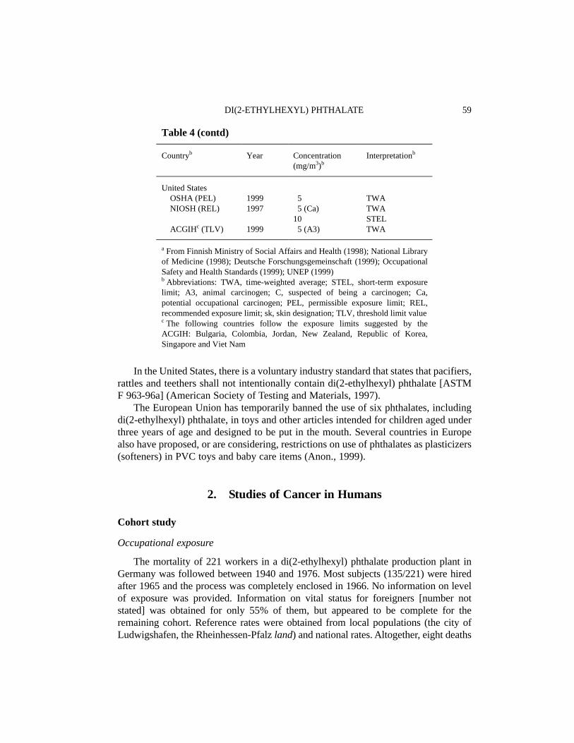
In the United States, there is a voluntary industry standard that states that pacifiers,rattles and teethers shall not intentionally contain di(2-ethylhexyl) phthalate [ASTMF 963-96a] (American Society of Testing and Materials, 1997).
The European Union has temporarily banned the use of six phthalates, includingdi(2-ethylhexyl) phthalate, in toys and other articles intended for children aged underthree years of age and designed to be put in the mouth. Several countries in Europealso have proposed, or are considering, restrictions on use of phthalates as plasticizers(softeners) in PVC toys and baby care items (Anon., 1999).
2. Studies of Cancer in Humans
Cohort study
Occupational exposure
The mortality of 221 workers in a di(2-ethylhexyl) phthalate production plant inGermany was followed between 1940 and 1976. Most subjects (135/221) were hiredafter 1965 and the process was completely enclosed in 1966. No information on levelof exposure was provided. Information on vital status for foreigners [number notstated] was obtained for only 55% of them, but appeared to be complete for theremaining cohort. Reference rates were obtained from local populations (the city ofLudwigshafen, the Rheinhessen-Pfalz land) and national rates. Altogether, eight deaths
DI(2-ETHYLHEXYL) PHTHALATE 59
Table 4 (contd)
Countryb Year Concentration(mg/m3)b
Interpretationb
United States OSHA (PEL) NIOSH (REL)
ACGIHc (TLV)
19991997
1999
5 5 (Ca)10 5 (A3)
TWATWASTELTWA
a From Finnish Ministry of Social Affairs and Health (1998); National Libraryof Medicine (1998); Deutsche Forschungsgemeinschaft (1999); OccupationalSafety and Health Standards (1999); UNEP (1999)b Abbreviations: TWA, time-weighted average; STEL, short-term exposurelimit; A3, animal carcinogen; C, suspected of being a carcinogen; Ca,potential occupational carcinogen; PEL, permissible exposure limit; REL,recommended exposure limit; sk, skin designation; TLV, threshold limit valuec The following countries follow the exposure limits suggested by theACGIH: Bulgaria, Colombia, Jordan, New Zealand, Republic of Korea,Singapore and Viet Nam

occurred during the follow-up period versus 15.9 expected using local rates[standardized mortality ratio, 0.50; 95% confidence interval, 0.22–0.99] and 17.0expected using national rates. One death from pancreatic cancer (0.13 expected) andone from bladder papilloma (0.01 expected) occurred among workers with a longexposure time (≥ 20 years). No further report on a longer follow-up for this cohort wasavailable to the Working Group (Thiess et al., 1978). [The Working Group noted thatthe majority of the cohort members were employed after exposure levels had beenconsiderably reduced, and that the methods for this study were poorly described.]
Dialysis patients
Long-term dialysis patients are likely to experience elevated exposures to di(2-ethylhexyl) phthalate, through frequent and protracted exposure to substances leachedfrom surgical tubing during dialysis (see Section 1.4.3(e)).
Cancer risk among dialysis patients has been specifically studied because ofconcern about medical conditions (for example, immunodeficiency) or incidental expo-sures from treatment (viruses, drugs) (Inamoto et al., 1991). However, exposure to di(2-ethylhexyl) phthalate resulting from treatment has not been studied as such in relationto cancer risk. Due to the medical condition of this population, follow-up is usuallyvery short and incompatible with induction of chemical carcinogenesis. In conclusion,the Working Group was not aware of any study of dialysis patients for which studymethods were suitable for the evaluation of carcinogenic risk associated with di(2-ethylhexyl) phthalate.
3. Studies of Cancer in Experimental Animals
3.1 Oral administration
3.1.1 Mouse
Groups of 50 male and 50 female B6C3F1 mice, six weeks of age, were fed dietscontaining 3000 or 6000 mg/kg diet (ppm) di(2-ethylhexyl) phthalate (> 99% pure) for103 weeks. All surviving mice were killed at 104–105 weeks. There was a clear dose-related decrease in body weight gain in females. Survival at the end of the study wasmore than 60% in males and more than 50% in females. High-dose males had a slightlydecreased body weight gain. In male mice, significant increases in the incidence ofhepatocellular carcinomas were observed (control, 9/50; low-dose, 14/48; high-dose,19/50; p = 0.022, Fisher’s exact test). The Cochran–Armitage test also indicated a signi-ficant trend (p = 0.018). The incidence of hepatocellular adenomas and carcinomascombined was also increased in males (control, 14/50; low-dose, 25/48, p = 0.013; high-dose, 29/50, p = 0.002, Fisher’s exact test). In females, significant increases in the inci-dence of hepatocellular carcinomas were seen (control, 0/50; low-dose, 7/50, p = 0.006;
IARC MONOGRAPHS VOLUME 7760
high-dose, 17/50, p < 0.001, Fisher’s exact test) and of hepatocellular adenoma andcarcinoma combined (control, 1/50; low-dose, 12/50; high-dose, 18/50, p < 0.001, trendand Fisher’s exact tests) (Kluwe et al.,1982; National Toxicology Program, 1982; Kluweet al., 1983, 1985).
Groups of 65 male and 65 female B6C3F1 mice, six weeks of age, were fed dietscontaining 0, 100, 500, 1500 or 6000 ppm di(2-ethylhexyl) phthalate (purity, 99.7%)for 104 weeks. Another group of 65 male and 65 female mice received the highestconcentration for 78 weeks and then control diet for a further 26 weeks (recoverygroup). Ten of these animals per group and sex were killed at week 105 for bio-chemical analyses of peroxisome proliferation. Two subgroups of 15 additional micein the 0 and 6000 ppm groups were designated for measurement of cell proliferation,biochemical analysis for peroxisome proliferation and histopathological evaluationand were killed at week 79. These additional animals were included in the finalanalysis. All surviving mice were killed at 105 weeks for histopathological exami-nation. There was a clear dose-related decrease in body weight gain in females. Thesurvival of male mice treated with 6000 ppm was significantly lower than that of thecontrols; about 80% of the untreated controls and treated animals in the other groupssurvived. In females, more than 60% of the animals survived until the end of the study.Hepatocellular tumour incidences in males were: 8/70 (control), 14/60 (100 ppm),21/65 (500 ppm), 27/65 (1500 ppm), 37/70 (6000 ppm) and 15/55 (recovery group).All but the 100 ppm group differed significantly (p < 0.05, Fisher’s exact test) fromthe control group. The respective incidences in females were 3/70, 4/60, 7/65, 19/65,44/70 and 30/55, with the 1500-ppm, 6000-ppm and recovery groups differing signifi-cantly from the concurrent control group (p < 0.05, Fisher’s exact test) (David et al.,1999)
3.1.2 Rat
Groups of 50 male and 50 female Fischer 344 rats, five to six weeks of age, werefed diets containing 6000 or 12 000 mg/kg diet (ppm) di(2-ethylhexyl) phthalate (> 99%pure) for 103 weeks. All surviving rats were killed at 104–105 weeks. Other groups of50 males and 50 females served as controls. There was a dose-related decrease in bodyweight gain in both sexes but no effect on survival. More than 60% of the animalssurvived to the end of the study. High-dose male rats had significant increases (p = 0.01,Fisher’s exact test) in the combined incidence of hepatocellular carcinomas andneoplastic nodules (control, 3/50; low-dose, 6/49; high-dose, 12/49). TheCochran–Armitage test also indicated a significant trend (p = 0.007). [The WorkingGroup noted that the term neoplastic nodule is now generally assumed to representhepatocellular adenomas.] The incidence of hepatocellular carcinomas alone orneoplastic nodules alone was not significantly increased. In female rats, the incidence ofhepatocellular carcinomas was increased in high-dose rats (8/50; p = 0.003, Fisher’sexact test) compared with controls (0/50) and that of neoplastic nodules was also
DI(2-ETHYLHEXYL) PHTHALATE 61

increased in high-dose females (5/50; p < 0.028) compared with controls (0/50). Theincidence of hepatocellular carcinomas and neoplastic nodules combined was alsoincreased in low-dose (6/49; p = 0.012) and high-dose (13/50; p < 0.001) femalescompared with controls (0/50) (Kluwe et al., 1982; National Toxicology Program, 1982;Kluwe et al., 1983, 1985).
Groups of 65 male and 65 female Fischer 344 rats, six weeks of age, were fed dietscontaining 0, 100, 500, 2500 or 12 500 ppm di(2-ethylhexyl) phthalate (purity, 99.7%)for up to 104 weeks. Another group of 65 male and 65 female rats received the highestconcentration for 78 weeks and then control diet for a further 26 weeks (recoverygroup). Ten of these animals per group and sex were killed at week 105 for bio-chemical analyses of peroxisome proliferation. Fifteen additional rats in the 0-, 2500-and 12 500-ppm groups were designated for measurement of cell proliferation, bio-chemical analyses for peroxisome proliferation and histopathological evaluation andwere killed at 79 weeks, and these animals were included in the final tumour analyses.About 65% of males and females survived until the end of the study. All surviving ratswere killed at 105 weeks for histopathological examination. For group comparison,the Fisher’s exact test was used. Hepatocellular tumour incidences in males were: 5/80(control), 5/50 (100 ppm), 4/55 (500 ppm), 11/65 (2500 ppm, p < 0.05), 34/80 (12500 ppm, p < 0.05) and 18/55 (recovery group, p < 0.05). The respective incidences infemales were 0/80, 4/50 (p < 0.05), 1/55, 3/65, 22/80 (p < 0.05) and 10/55 (p < 0.05)(David et al., 1999).
Several studies using smaller numbers of animals have also been reported, some ofwhich were not designed for carcinogenicity testing. These studies are reviewed below.
Groups of 20 female Fischer 344 rats, eight weeks of age, were fed a diet containing0 (control), 0.03, 0.10 or 1.2% di(2-ethylhexyl) phthalate [purity not specified] for twoyears. Neoplastic nodules or hepatocellular carcinomas were seen in 0/18 control, 1/18low-dose, 1/19 mid-dose and 6/20 high-dose rats (p < 0.01). Di(2-ethylhexyl) phthalatedid not induce foci of altered hepatocytes as judged by basophilia, ATPase-deficiencyor glucose-6-phosphatase-deficiency (Cattley et al., 1987).
Groups of 10 male Fischer 344 rats, six weeks of age, were fed a diet containing2% di(2-ethylhexyl) phthalate (purity, 98%) for 95 weeks. Neoplastic nodules and/orhepatocellular carcinomas were found in 0/18 controls and 6/10 rats fed di(2-ethylhexyl) phthalate (p < 0.005). Both the neoplastic nodules and hepatocellularcarcinomas were negative for γ-glutamyltranspeptidase (γ-GT) (Rao et al., 1987).
Groups of 10–14 male Fischer 344 rats were fed a diet containing 0 (control) or2% di(2-ethylhexyl) phthalate (98% pure) for 108 weeks. All liver lobes were slicedat 1–2-mm intervals and the number and size of grossly visible lesions recorded.Neoplastic nodules and/or hepatocellular carcinomas were observed in 1/10 controlsand 11/14 treated rats [no statistical analysis given] (Rao et al., 1990). [The authorssuggest that their finding of a high incidence of liver tumours was due to their grossslicing technique, although they do not report a comparison of their findings withconventional liver trimming and sectioning techniques].
IARC MONOGRAPHS VOLUME 7762

A total of 520 male Sprague-Dawley rats (180 g) were fed diets containing 0, 0.02,0.2 or 2% (200, 2000 or 20 000 ppm) di(2-ethylhexyl) phthalate (purity, > 99%) for102 weeks [group sizes not specified]. There was a significant, dose-dependentdecrease in body weights in the mid- and high-dose groups, but not in the low-doseanimals. Data on survival were not reported. For biochemical assays and histopatho-logical examinations, 7–18 rats were killed at weeks 24, 48, 72 and 96. The numberof animals at terminal sacrifice was not given. No hyperplastic nodules or hepato-cellular carcinomas were reported in either the control or treated animals (Ganninget al., 1991) [The Working Group noted the inadequate reporting.]
As part of a larger experiment for studying the characteristics of hepatocarcino-genesis, 17 male Fischer 344 rats were fed a diet containing 2% (20 000 ppm) di(2-ethylhexyl) phthalate [purity unspecified] for up to 78 weeks. A group of 18 untreatedanimals served as controls. At 52 weeks, no liver tumours had developed in 10 di(2-ethylhexyl) phthalate-treated rats or in 10 controls, while at 78 weeks, hepatocellularcarcinomas or neoplastic nodules were found in 3/7 di(2-ethylhexyl) phthalate-treatedrats and 0/8 controls (Hayashi et al., 1994).
3.2 Inhalation exposure
Hamster: Groups of 65–80 male or female Syrian golden hamsters, 12 weeks ofage, were exposed by whole-body inhalation to 15 μg/m3 di(2-ethylhexyl) phthalatevapour (> 99% pure) for 24 h per day on five days per week until natural death. Totalexposure over the lifetime was 7–10 mg/kg bw. No difference in survival was seenbetween controls and the di(2-ethylhexyl) phthalate-treated groups. Median survivalin controls was 709 days in males and 507 days in females and that in treated animalswas 703 days in males and 522 days in females. Tumour incidence was not increasedin di(2-ethylhexyl) phthalate-treated hamsters (Schmezer et al., 1988). [The WorkingGroup noted the low concentration and that it was selected to simulate occupationalexposure.]
3.3 Intraperitoneal administration
Hamster: Groups of 50 male and 50 female Syrian golden hamsters, six weeks ofage, were administered 0 (control) or 3 g/kg bw di(2-ethylhexyl) phthalate (> 99%pure) by intraperitoneal injection once per week, once per two weeks or once permonth (total doses, 24–54 g/kg bw). Hamsters were maintained for their naturallifespan. There were no differences in survival between groups (range of mediansurvival being 629–686 days for males and 465–495 days for females). The incidenceof tumours was not increased by treatment (Schmezer et al., 1988). [The WorkingGroup noted limitations in the dosing schedule.]
DI(2-ETHYLHEXYL) PHTHALATE 63

3.4 Administration with known carcinogens and modifying agents
There are numerous reports of studies of the initiating or promoting activities ofdi(2-ethylhexyl) phthalate given in combination with known carcinogens or promotingagents. Selected studies (complex protocols involving multiple promoting agents andspecial procedures were excluded) are summarized below and in Tables 5 and 6.
3.4.1 Mouse
Liver: Male B6C3F1 mice, four weeks of age, received a single intraperitonealinjection of 80 mg/kg bw N-nitrosodiethylamine (NDEA) in tricaprylin. Two weekslater, the mice were fed diets containing 0, 3000, 6000 or 12 000 ppm di(2-ethylhexyl)phthalate for up to six months. Groups of 10 mice were killed at two, four and sixmonths after NDEA treatment. Few hepatocellular foci were seen at two, four or sixmonths in mice treated with NDEA alone or di(2-ethylhexyl) phthalate alone, whilenumerous foci and neoplasms were seen in mice given di(2-ethylhexyl) phthalate afterNDEA. No tumours were found at six months in mice receiving NDEA alone. By theend of the study, the number of foci per unit volume of liver was similar in mice at alldoses of di(2-ethylhexyl) phthalate, but there was an increase in the volume of the foci(0, 1.4, 0.6, 9.4 mm3 for the control, 3000-, 6000- and 12 000-ppm groups, respec-tively) (Ward et al., 1983).
The differential effects of short- or long-term exposure to di(2-ethylhexyl)phthalate were studied in male B6C3F1 mice. Mice were given an intraperitonealinjection of 80 mg/kg bw NDEA at four weeks of age. At five weeks of age, the micewere fed diets containing 3000 ppm di(2-ethylhexyl) phthalate for periods of one,seven, 28, 84 or 168 days and were killed at 168 days. When di(2-ethylhexyl) phthalatewas fed after NDEA treatment for 28 or more days, there was an increase in incidencesof hepatocellular foci (45, 50, 67%) and adenomas (20.6, 17.8 and 46.6%) comparedwith those in mice receiving NDEA alone (foci, 20%; adenomas, 6.6%). There was alsoan increase in lesion number and size (Ward et al., 1984).
Male B6C3F1 mice, four weeks of age, received a single intraperitoneal injectionof 80 mg/kg bw NDEA in tricaprylin and, two weeks later, were fed diets containing0, 3000, 6000 or 12 000 ppm di(2-ethylhexyl) phthalate (purity, 99%) for up to 18months. Groups of 10–20 mice were killed at two, four, six or 18 months. The numbersof mice with hepatocellular foci, adenomas and carcinomas were determined. Alldoses of di(2-ethylhexyl) phthalate increased the numbers of all lesions at the timeperiods studied compared with mice receiving NDEA alone. At 18 months, carci-nomas were found in 3/10 mice treated with NDEA alone and in 10/10 and 18/20 micetreated with NDEA + di(2-ethylhexyl) phthalate at the low and mid doses, respec-tively. All mice given 12 000 ppm di(2-ethylhexyl) phthalate with NDEA initiationdied by nine months and 11/20 had liver carcinomas. In mice treated with di(2-ethylhexyl) phthalate alone, 2/30 had liver carcinomas (Ward et al., 1986).
IARC MONOGRAPHS VOLUME 7764

DI(2-ETH
YLH
EXY
L) PHTH
ALATE
65
Table 5. Selected promotion studies on di(2-ethylhexyl) phthalate (DEHP) with known carcinogens and modifying factors
Tumour typeSpecies/strain (sex)
Known carcinogen(initiator)
Route ofadminis-tration
Intervalbetweeninitiator andpromoter
Dose and duration of DEHP Route ofadminis-tration
Promotingactivityfor DEHP
Reference
LiverB6C3F1 mice (M) 80 mg/kg bw NDEA i.p. 2 weeks 3000 mg/kg diet/6 months Oral + Ward et al. (1983)B6C3F1 mice (M) 80 mg/kg bw NDEA i.p. 2 weeks 6000 mg/kg diet/6 months Oral + Ward et al. (1983)B6C3F1 mice (M) 80 mg/kg bw NDEA i.p. 2 weeks 12 000 mg/kg diet/6 months Oral + Ward et al. (1983)B6C3F1 mice (M) 80 mg/kg bw NDEA i.p. 1 week 3000 mg/kg diet/28 days Oral + Ward et al. (1984)B6C3F1 mice (M) 80 mg/kg bw NDEA i.p. 1 week 3000 mg/kg diet/84 days Oral + Ward et al. (1984)B6C3F1 mice (M) 80 mg/kg bw NDEA i.p. 1 week 3000 mg/kg diet/168 days Oral + Ward et al. (1984)B6C3F1 mice (M) 80 mg/kg bw NDEA i.p. 2 weeks 3000 mg/kg diet/18 months Oral + Ward et al. (1986)B6C3F1 mice (M) 80 mg/kg bw NDEA i.p. 2 weeks 6000 mg/kg diet/18 months Oral + Ward et al. (1986)B6C3F1 mice (M) 80 mg/kg bw NDEA i.p. 2 weeks 12 000 mg/kg diet/18 months Oral + Ward et al. (1986)C3H/HeNCr mice (M,F) 5 mg/kg bw NDEA i.p. 2 weeks 12 000 mg/kg diet/24 weeks Oral + Weghorst et al.
(1993/94)Fischer 344 rats (M) 150 mg/kg bw NDEA i.p. 3 weeks 12 000 mg/kg diet/6 months Oral – Popp et al. (1985)Fischer 344 rats (M) 282 mg/kg bw NDEA i.p. 2 weeks 12 000 mg/kg diet/14 weeks Oral – Ward et al. (1986)Fischer 344 rats (M) 200 mg/kg diet AAF
7 weeksOral 4 weeks 12 000 mg/kg diet/31 weeks Oral – Williams et al.
(1987)Fischer 344 rats (M) PH/200 mg/kg NDEA i.p. 2 weeks 3000 mg/kg diet/6 weeks Oral – Ito et al. (1988)Sprague-Dawley rats (F) 8 mg/kg NDEA i.g. 1 week 10 mg/kg 3 × weekly/
11 weeksi.g. – Oesterle & Deml.
(1988)Sprague-Dawley rats (F) 8 mg/kg NDEA i.g. 1 week 100 mg/kg 3 × weekly/
11 weeksi.g. – Oesterle & Deml
(1988)Sprague-Dawley rats (F) 8 mg/kg NDEA i.g. 1 week 200 mg/kg 3 × weekly/
11 weeksi.g. + Oesterle & Deml
(1988)Sprague-Dawley rats (F) 8 mg/kg NDEA i.g. 1 week 500 mg/kg 3 × weekly/
11 weeksi.g. + Oesterle & Deml
(1988)Fischer 344 rats (M) 200 ppm AAF
7 weeksOral 4 weeks 12 000 ppm /24 weeks Oral – Maruyama et al.
(1990)

IARC M
ON
OG
RAPH
S VO
LUM
E 7766
Table 5 (contd)
Tumour typeSpecies/strain (sex)
Known carcinogen(initiator)
Route ofadminis-tration
Intervalbetweeninitiator andpromoter
Dose and duration of DEHP Route ofadminis-tration
Promotingactivityfor DEHP
Reference
KidneyFischer 344 rats (M) 500 mg/kg diet
EHEN/2 weeksOral 0 weeks 12 000 ppm/24 weeks Oral + Kurokawa et al.
(1988)
Urinary bladderFischer 344 rats (M) 5000 ppm BBN weeks
1–4; 30 000 ppm uracilweeks 8–11
Oral 0 weeks 3000 ppm/16 weeks Oral – Hagiwara et al.(1990)
AAF, 2-acetylaminofluorene; BBN, N-butyl-N-(4-hydroxybutyl)nitrosamine; EHEN, N-ethyl-N-hydroxyethylnitrosamine; NDEA, N-nitrosodiethylamine; F, female;M, male; i.p., intraperitoneal injection; i.g., intragastric; PH, partial hepatectomy

DI(2-ETH
YLH
EXY
L) PHTH
ALATE
67
Table 6. Initiation studies on di(2-ethylhexyl) phthalate (DEHP) with promoting agents
Species/strain DEHP initiation Route ofadminis-tration
Intervalbetweeninitiation andpromotion
Dose and duration ofpromoter
Route ofadminis-tration
Initiatingactivity ofDEHP
Reference
Mouse B6C3F1(M)
Fischer 344 rats(F)
25 or 50 g/kg bw
10 g/kg bw DEHP 6,12, 24 h after PH
Gavage
Oral
2 weeks
2 weeks
500 mg/L phenobarbital6 or 18 months
200 ppm AAF 2 weeks1.5 mL/kg CCl4 once
Drinking-water
DietGavage
––
Ward et al.(1986)
Garvey et al.(1987)
Fischer 344 rats(F)
12 000 mg/kg dietDEHP 12 weeks
Oral 2 weeks 500 mg/kg dietphenobarbital, 39 weeks
Diet – Garvey et al.(1987)
PH, partial hepatectomy; AAF, 2-acetylaminofluorene; CCl4, carbon tetrachloride

In a study to test di(2-ethylhexyl) phthalate for initiating activity, groups of 7–20male B6C3F1 mice, four weeks of age, were given a single intragastric dose of 25 or50 g/kg bw di(2-ethylhexyl) phthalate (99% pure). Groups of 10–17 controls wereused. Two weeks later, phenobarbital was given as a promoting agent at a concen-tration of 500 mg/L in the drinking-water for six or 18 months. At 18 months, hepato-cellular carcinomas were found in 0/7 mice given 50 g/kg di(2-ethylhexyl) phthalate,2/15 mice given 50 g/kg bw di(2-ethylhexyl) phthalate + phenobarbital, 1/10 micegiven 25 g/kg di(2-ethylhexyl) phthalate, 2/20 mice given 25 g/kg di(2-ethylhexyl)phthalate + phenobarbital, 3/17 mice given phenobarbital alone and 0/10 untreatedmice [statistical analysis not given]. Thus the study showed no evidence of initiatingactivity of di(2-ethylhexyl) phthalate (Ward et al., 1986).
Groups of 10 male and five female C3H/HeNCr mice, 15 days of age, receivedeither a single intraperitoneal injection of 5 mg/kg bw NDEA or saline. At weaning (fourweeks of age), mice were divided into two groups and fed diets containing either 0 or12 000 ppm di(2-ethylhexyl) phthalate [purity unspecified] for 24 weeks. All mice werekilled at 28 weeks of age and the number and size of hepatic foci were measured. Di(2-ethylhexyl) phthalate in combination with NDEA increased the average numbers of fociper liver (NDEA-treated males, 176; NDEA + di(2-ethylhexyl) phthalate-treated males,366; NDEA-treated females, 47; NDEA + di(2-ethylhexyl) phthalate-treated females,169). The numbers of adenomas per liver were also increased (NDEA-treated males, 7;NDEA + di(2-ethylhexyl) phthalate-treated males, 15.8; NDEA-treated females, 0;NDEA + di(2-ethylhexyl) phthalate-treated females, 2). In male mice, treatment withNDEA + di(2-ethylhexyl) phthalate yielded larger adenomas than those seen in micetreated with NDEA alone (2.4 mm3 versus 1.3 mm3) (Weghorst et al., 1993/94).
Skin: Groups of 25 female SENCAR mice, seven weeks of age, received a singleapplication of 20 μg 7,12-dimethylbenz[a]anthracene (DMBA) in 0.2 mL acetone on theskin of the back. One week later, mice received applications of 100 mg per animal di(2-ethylhexyl) phthalate (99% pure) twice weekly for 28 weeks. 12-O-Tetradecanoyl-phorbol 13-acetate (TPA) control groups received 2 μg TPA. To test di(2-ethylhexyl)phthalate as a second-stage promoter, mice received TPA for two weeks followed bydi(2-ethylhexyl) phthalate for 26 weeks. Appropriate acetone, TPA and di(2-ethylhexyl)phthalate controls were included. Di(2-ethylhexyl) phthalate, when tested as a completepromoter (28 weeks of exposure), enhanced only slightly the numbers of papillomas(0.88 per mouse versus DMBA alone 0 per mouse) but significantly (p < 0.01) enhancedpapillomas when given for 26 weeks after two weeks of TPA first-stage promotion (6.44versus 2.2). The authors concluded that di(2-ethylhexyl) phthalate was a second-stagepromoter (Diwan et al., 1985; Ward et al., 1986).
3.4.2 Rat
Liver: Groups of 10 female Fischer 344 rats, six to eight weeks of age, received asingle intraperitoneal injection of 150 mg/kg bw NDEA followed three weeks later by
IARC MONOGRAPHS VOLUME 7768

a diet containing 1.2% di(2-ethylhexyl) phthalate (99.5% pure) for three or sixmonths. No neoplasms or nodules were identified. Di(2-ethylhexyl) phthalate did notincrease the number of foci or the mean volume of the foci, as identified by fivedifferent histological markers (Popp et al.,1985).
The initiating activity of di(2-ethylhexyl) phthalate was examined after single andsub-chronic dosing. Di(2-ethylhexyl) phthalate (99.5% pure) was administered as asingle oral dose (10 g/kg bw) or by 12 weeks of feeding at a concentration of 1.2% inthe diet followed by various known promotion regimens, such as phenobarbitaltreatment or partial hepatectomy. There was no increase in number or mean volumeof foci in liver sections examined using multiple histological markers and no tumourswere identified, indicating that di(2-ethylhexyl) phthalate had no initiating activity(Garvey et al., 1987).
Groups of 18–20 male Fischer rats (weighing 160 g) were given a single intra-peritoneal injection of 200 mg/kg bw NDEA. Two weeks later, they were fed a dietcontaining 3000 ppm di(2-ethylhexyl) phthalate [purity unspecified] for six weeks. Atweek 3, they were subjected to a partial hepatectomy. All rats were killed at week 8.Di(2-ethylhexyl) phthalate-treated rats had no increase in foci staining positively forglutathione S-transferase placental form (8.5 per cm2 versus 11.6 for NDEA alone) (Itoet al., 1988).
Male Fischer 344 rats were fed 200 ppm 2-acetylaminofluorene (AAF) for sevenweeks to induce hepatocellular altered foci, and were subsequently fed 0 or 12 000 ppmdi(2-ethylhexyl) phthalate (98% pure) in the diet. No evidence of induction of hepato-cellular altered foci or hepatic neoplasms was found when di(2-ethylhexyl) phthalatewas given alone for 24 weeks. Di(2-ethylhexyl) phthalate fed for 24 weeks increasedbasophilic foci, but showed no promoting effect on iron-excluding altered hepatic fociinduced by AAF, and produced no significant enhancement of the occurrence of AAF-induced liver neoplasms (3/6 compared with 3/12) (Williams et al., 1987).
Di(2-ethylhexyl) phthalate exerted weak promoting activity in weanling femaleSprague-Dawley rats after doses of 200 or 500 mg/kg bw, given three times per weekby gavage for 11 consecutive weeks after initiation with a single oral dose of 8 mg/kgbw NDEA. Lower doses were ineffective. The incidence of ATPase-deficient foci wasenhanced about two-fold compared with rats treated with NDEA alone. The incidenceof foci with expression of γ-GT was not affected by di(2-ethylhexyl) phthalatetreatment (Oesterle & Deml, 1988).
Male Fischer 344 rats were fed diets containing 200 ppm AAF for seven weeks toinduce hepatocellular altered foci, and were then fed diets containing either 0 or 12 000ppm di(2-ethylhexyl) phthalate (purity, 98%) for 24 weeks. In foci that were induced byAAF, di(2-ethylhexyl) phthalate reduced the activity of γ-GT, as detected histo-chemically, but did not increase the number, mean volume or volume percentage of focidetected by deficiencies in iron storage, glucose-6-phosphatase, adenosine triphospha-tase or fibronectin. Although the numbers of haematoxylin/eosin-stained foci wereincreased in di(2-ethylhexyl) phthalate-treated rats, the volume percentage was not
DI(2-ETHYLHEXYL) PHTHALATE 69

increased and no difference in the numbers of iron storage foci was seen (Maruyamaet al., 1990).
Urinary system: Groups of 20 male Fischer 344 rats were given 0.05% N-ethyl-N-hydroxyethylnitrosamine (EHEN) for two weeks in the diet followed by di(2-ethyl-hexyl) phthalate [purity unspecified] at a concentration of 0 or 1.2% in the diet for 24weeks. Rats were killed at 27 weeks. Di(2-ethylhexyl) phthalate increased the numbersof rats with renal (tubular) cell tumours (EHEN + di(2-ethylhexyl) phthalate 65%versus 20% for EHEN alone; p < 0.01) and the mean number of tumours per kidney(EHEN + di(2-ethylhexyl) phthalate 1.1 versus EHEN alone 0.2, p < 0.01) (Kurokawaet al., 1988).
The modifying potential of di(2-ethylhexyl) phthalate on second-stage N-butyl-N-(4-hydroxybutyl)nitrosamine (BBN)-initiated urinary bladder carcinogenesis wasinvestigated in male Fischer 344 rats, using a uracil-accelerated transitional-cell proli-feration model. Six-week-old animals received 0.05% BBN in their drinking-water forfour weeks followed by di(2-ethylhexyl) phthalate [purity unspecified] (0, 0.3, 0.6 or1.2% in the diet) for experimental weeks 5–8 and weeks 12–20. Uracil was adminis-tered during weeks 9–11 at a dietary level of 3.0%. Surviving animals were killed atthe end of week 20 of the experiment. Di(2-ethylhexyl) phthalate did not promotehyperplastic lesions (papillary or nodular) of the urinary bladder or papillomasinduced by BBN (Hagiwara et al., 1990).
3.4.3 Hamster
Groups of 50 male and 50 female Syrian golden hamsters, six weeks old, were givenintraperitoneal injections of 3 g/kg bw di(2-ethylhexyl) phthalate (> 99% pure) eitheronce, or once per week for two or four weeks. N-Nitrosodimethylamine (NDMA) wasgiven orally at 1.67 mg/kg bw once [exact week of dosing for both chemicals not given].Hamsters were maintained for their natural lifespan. Survival was reduced amonghamsters receiving NDMA. Di(2-ethylhexyl) phthalate did not affect tumour yield (livertumours: 16/50 and 9/50 in di(2-ethylhexyl) phthalate + NDMA and NDMA males; and6/50 and 6/50 in di(2-ethylhexyl) phthalate + NDMA and NDMA females) [statisticalanalysis not given] (Schmezer et al., 1988).
3.5 Carcinogenicity of the metabolite 2-ethylhexanol
3.5.1 Mouse
Groups of 50 male and 50 female B6C3F1 mice, seven weeks of age, were given2-ethylhexanol by gavage five times weekly at doses of 0, 50, 200 and 250 mg/kg bwfor 18 months. Body weight gain was reduced by 24–26% in the high-dose group andmortality was dose-related. In females, liver carcinomas occurred in 0/50 control, 1/50low-dose, 3/50 mid-dose and 5/50 (p < 0.05 Fisher’s exact test) high-dose mice (Astillet al., 1996).
IARC MONOGRAPHS VOLUME 7770

3.5.2 Rat
Groups of 50 male and 50 female Fischer 344 rats, six weeks of age, were given2-ethylhexanol by gavage, five times weekly, at doses of 0, 50, 150 or 500 mg/kg bwfor 104 weeks. A dose-related depression of body weight gain in male and female ratsand increased mortality in high-dose female rats were observed. There was noincrease in the incidence of tumours in any treated group (Astill et al., 1996).
4. Other Data Relevant to an Evaluation of Carcinogenicityand its Mechanisms
4.1 Absorption, distribution, metabolism and excretion
4.1.1 Humans
Human exposure to di(2-ethylhexyl) phthalate can occur via the dermal, inhalation,oral and intravenous routes. The high level of exposure has prompted many studies onthe absorption, distribution, metabolism and excretion of di(2-ethylhexyl) phthalate inhumans (Lawrence & Tuell, 1979; Thomas & Thomas, 1984; Burg, 1988; Albro &Lavenhar, 1989; Kamrin & Mayor, 1991; Huber et al., 1996; Doull et al., 1999).
In a very early study, Shaffer et al. (1945) administered single oral doses of 5 and10 g di(2-ethylhexyl) phthalate to two human subjects and reported that approxi-mately 4.5% of the dose was excreted in the urine within 24 h. Schmid and Schlatter(1985) also administered di(2-ethylhexyl) phthalate orally to two human subjects, butat the much lower dose of 30 mg per person. These authors reported that 11–15% ofthe dose was excreted in the urine and a urinary elimination half-life of about 12 h canbe estimated from the data. In the same study, the two volunteers also received 10 mgdi(2-ethylhexyl) phthalate daily for four days, there being no evidence of accumu-lation, with 11 and 33 % of the dose recovered each day in the urine. In contrast,Rubin and Schiffer (1976) reported data from two patients receiving platelet trans-fusions from bags containing di(2-ethylhexyl) phthalate, who excreted between 60and 90% of the infused dose in the urine collected for 24 h after transfusion.
Figure 1 summarizes the metabolic pathways responsible for the metabolism of di-(2-ethylhexyl) phthalate in humans and in animals, using the widely accepted metabolitenomenclature of Albro and Lavenhar (1989). The quantitative urinary metabolic profileof di(2-ethylhexyl) phthalate reported by Schmid and Schlatter (1985) is quite similar tothat found by Albro et al. (1982), who determined the urinary metabolites of di(2-ethyl-hexyl) phthalate in leukaemia patients who received platelet transfusions from bagscontaining di(2-ethylhexyl) phthalate. These data are also in good agreement with theresults of Dirven et al. (1993a), who studied the excretion of di(2-ethylhexyl) phthalatemetabolites in five workers occupationally exposed to di(2-ethylhexyl) phthalate. Themajor phase I metabolites of di(2-ethylhexyl) phthalate in human urine are mono(2-
DI(2-ETHYLHEXYL) PHTHALATE 71

IARC M
ON
OG
RAPH
S VO
LUM
E 7772
C
C
O
O
OH
O CH2 CH CH2 CH2 CH2 CH3
CH2
CH3
Di(2-ethylhexyl) phthalate
Mono(2-ethylhexyl) phthalate
R CH CH2 CH2 CH CH3
OHCH2
CH3
ω Oxidation of ethyl chain
R CH CH2 CH2 CH2 CH3
CH2
CH2OH
R CH CH2 CH2 CH2 CH2OH
CH2
CH3
ω Oxidation of hexyl chain
R CH CH2 CH2 C CH3
CH2
CH3
O
R CH CH2 CH2 CH2 CH3
CH2
COOH
R CH CH2 CH2 CH2 COOH
CH2
CH3
α-Oxidation
VI VIV
β-Oxidation
R CH CH2 CH2 CH2 CH3
COOH
R CH CH2 CH2 COOH
CH2
CH3
III
HO CH2 CH CH2 CH2 CH2
CH2
CH3
CH3
2-Ethylhexanol
HOOC CH2 CH CH2 CH2 CH2
CH2
CH3
CH3
2-Ethylhexanoic acid
VIIIX X
R = C
C
OH
O CH2
O
O
ω-Oxidation ω-Oxidation(ω 1)-Oxidation
ω 1-Oxidation of hexyl chain
The numbering of the metabolites is based on the nomenclature of Albro and Lavenhar (1989). This chart is based on the work of Huber et al. (1996) and Astill (1989). I, phthalic acid, mono(4-carboxy-2-ethylhexyl) ester; II, phthalic acid, mono(2-carboxyhexyl) ester; IV, phthalic acid, mono(2-(carboxymethyl)hexyl) ester; V, phthalic acid, mono(5-carboxy-2-ethylpentyl) ester; VI, phthalic acid,mono(2-ethyl-5-oxohexyl) ester; VII, phthalic acid, mono(2-(2-hydroxyethyl)hexyl) ester; IX, phthalic acid, mono(2-ethyl-5-hydroxyhexyl) ester; X, phthalic acid, mono(2-ethyl-6-hydroxyhexyl) ester
Figure 1. Major pathways involved in the metabolism of di(2-ethylhexyl) phthalate

ethylhexyl) phthalate, and compounds V, VI and IX (Figure 1; Albro et al., 1982;Schmid & Schlatter, 1985, Dirven et al., 1993b). Dirven et al. (1993b) found that theurinary concentration of metabolites VI and IX was 1.7 times higher than the concen-tration of metabolite V, and concluded that metabolism via (ω–1)-hydroxylation isfavoured over ω-hydroxylation in humans. Metabolites VI and IX were reported byDirven et al. (1993b) to be almost completely conjugated, while only 32–45% ofmetabolite V was present in the conjugated form. The same authors found a markedinterindividual variation in the conjugation of mono(2-ethylhexyl) phthalate. In two ofthe five subjects studied, 77–100% of mono(2-ethylhexyl) phthalate was in the freeform, whereas in urine samples from the other three subjects, only 20–38% of mono(2-ethylhexyl) phthalate was not conjugated. Both Albro et al. (1982) and Schmidt andSchlatter (1985) reported that up to 80% of the urinary metabolites of di(2-ethylhexyl)phthalate in humans were excreted as glucuronide conjugates. Conjugates of di(2-ethylhexyl) phthalate metabolites with sulfate, taurine and glycine have not been foundin any species studied to date.
Rubin and Schiffer (1976) reported peak blood plasma levels of di(2-ethylhexyl)phthalate in adult patients transfused with platelets ranging from 3 to 8 mg/L anddescribed the kinetics of plasma disappearance as mono-exponential, with a half-lifeof 28 min. The plasma levels ranged from 0.3 to 1.2 mg/kg bw di(2-ethylhexyl)phthalate. This is a low level compared with di(2-ethylhexyl) phthalate levels in wholeblood stored in blood bags for up to 21 days (Jaeger & Rubin, 1972; Rock et al., 1978;Peck et al., 1979), from which a transfusion of 2.5 L of blood to a 70-kg person wouldgive an exposure of 1.3–2.6 mg/kg bw di(2-ethylhexyl) phthalate. Similar levels ofexposure (1.7 to 4.2 mg/kg bw) have been reported in newborn infants during singleexchange transfusions (Sjöberg et al., 1985a).
Lewis et al. (1978) determined the disappearance of di(2-ethylhexyl) phthalate inseven adult patients following dialysis. The mean serum levels were 606 μg/L imme-diately after dialysis, 323 μg/L at 30 min, 167 μg/L at 1 h and 145 μg/L at 3 h aftercompletion of dialysis. They concluded that most of the di(2-ethylhexyl) phthalatepresent in serum at the completion of dialysis is likely to disappear within 5–7 h. Sjöberget al. (1985b) and Plonait et al. (1993) also described a rapid decline in serum di(2-ethyl-hexyl) phthalate levels in term newborns similar to that seen in adults. Sjöberg et al.(1985b) reported that plasma levels of di(2-ethylhexyl) phthalate declined biexponen-tially after transfusions in newborn infants, with the half-life of the terminal phase beingapproximately 10 h. The terminal half-life of the active metabolite of di(2-ethylhexyl)phthalate, mono(2-ethylhexyl) phthalate, in newborn infants was similar (Sjöberg et al.,1985b). However, in preterm infants, di(2-ethylhexyl) phthalate can persist in serum fora much longer time and it is postulated that either there are different kinetics resultingfrom immature metabolism or there is a biphasic disappearance pattern, as Stern et al.(1977) and Sjöberg et al. (1985c,d) observed in rats (Plonait et al., 1993). In adulthaemodialysis patients, circulating levels of mono(2-ethylhexyl) phthalate are similar to
DI(2-ETHYLHEXYL) PHTHALATE 73

those of di(2-ethylhexyl) phthalate, showing that this metabolite is formed after systemicintake in humans (Pollack et al., 1985). Further, di(2-ethylhexyl) phthalate can be hydro-lysed by plasma proteins in blood products to mono(2-ethylhexyl) phthalate. Hence,patients undergoing transfusion may be exposed to mono(2-ethylhexyl) phthalate atdoses of up to one tenth those of di(2-ethylhexyl) phthalate (Rock et al., 1978).
Absorption of di(2-ethylhexyl) phthalate via the lungs is considered to be themajor route of occupational exposure, as dermal absorption through preparations ofhuman skin in vitro is low (Scott et al., 1987; Dirven et al., 1993a; Huber et al., 1996).Occupational exposure to di(2-ethylhexyl) phthalate based on personal air samplingdata in the range 9–1266 μg/m3 has been reported (Dirven et al., 1993a). Furthermore,Dirven et al. (1993a) evaluated the inhalation exposure to di(2-ethylhexyl) phthalatein a group of nine volunteers in a boot factory in an attempt to evaluate the absorptionand disposition of the plasticizer. They reported that the median values of three majordi(2-ethylhexyl) phthalate metabolites studied (VI, IX and V; see Figure 1) were signi-ficantly increased (1.2–2.3-fold) in urine samples collected at the end of the workdaycompared with urine samples collected at the start of the workday. However, a similarcomparison in six cable factory workers detected no statistically significant increasein post-shift urinary di(2-ethylhexyl) phthalate metabolite concentrations comparedwith pre-shift values, although the airborne exposure levels were within a similarrange. As pointed out by the authors, a meaningful interpretation of these data isimpeded by the lack of understanding of the toxicokinetics of di(2-ethylhexyl)phthalate metabolites in humans.
4.1.2 Experimental systems
The absorption and disposition of di(2-ethylhexyl) phthalate has been extensivelyinvestigated in laboratory animals (Thomas & Thomas, 1984; Albro & Lavenhar, 1989;Astill, 1989; Huber et al., 1996; Doull et al., 1999). The first step in the metabolism ofdi(2-ethylhexyl) phthalate in all species is the hydrolysis of one of the two ethylhexylside-chains to yield mono(2-ethylhexyl) phthalate and 2-ethylhexanol. After oraladministration of di(2-ethylhexyl) phthalate, this pathway is primarily catalysed bypancreatic lipase. The level of pancreatic lipase in the intestine of laboratory animalshas been shown to exhibit marked species and strain differences (Albro & Thomas,1973). Lower levels of di(2-ethylhexyl) phthalate-hydrolysing enzyme activity areexpressed in the liver, blood and other tissues (Albro, 1986; Huber et al., 1996). It isgenerally accepted that in all mammalian species the majority of di(2-ethylhexyl)phthalate reaching the intestine is absorbed as hydrolysis products rather than as theintact diester (Albro & Lavenhar, 1989). For example, Teirlynck and Belpaire (1985)studied the disposition of both di(2-ethylhexyl) phthalate and mono(2-ethylhexyl)phthalate in immature male Wistar rats and reported that, after a single oral dose ofdi(2-ethylhexyl) phthalate (2.8 g/kg), plasma concentrations of 8.8 ± 1.7 μg/mL di(2-ethylhexyl) phthalate and 63.2 ± 8.7 μg/mL mono(2-ethylhexyl) phthalate were reached
IARC MONOGRAPHS VOLUME 7774

after 3 h. The plasma concentration of mono(2-ethylhexyl) phthalate declined with ahalf-life of 5.2 h, whereas the corresponding value for di(2-ethylhexyl) phthalate couldnot be estimated due to very low levels of the parent diester.
The efficiency of di(2-ethylhexyl) phthalate elimination from the gastrointestinaltract differs between species. Ikeda et al. (1980) fed 50 mg/kg bw di(2-ethylhexyl)phthalate per day to male Sprague-Dawley rats, male pure-bred beagles and male imma-ture swine of the Hormel strain for 21–28 days and then administered a single dose of14C-labelled di(2-ethylhexyl) phthalate at 50 mg/kg bw on the last day. In rats, 37.5 and53.2% of the administered radioactivity were excreted in the urine and faeces, respec-tively, within four days. In dogs, the figures showed that the faecal route of excretiondominated (75%), with the urinary route accounting for 20.7% of the radioactivity. Theopposite was the case in pigs, with the faecal and urinary routes accounting for 25.7%and 79.4%, respectively. In male Wistar rats given a single 2.9 mg/kg bw oral dose of[14C]di(2-ethylhexyl) phthalate, Daniel and Bratt (1974) found 42% and 57% of the dosein the urine and faeces within seven days. In the same study, in rats fed 2.6 mg/kg bw[14C]di(2-ethylhexyl) phthalate, 14% of the dose was excreted in the bile.
Rhodes et al. (1986) found that the absorption of di(2-ethylhexyl) phthalate wasconsiderably lower in marmosets than in rats. These authors reported that 45% and 7%of di(2-ethylhexyl) phthalate were absorbed by marmosets at doses of 100 mg/kg bwand 2000 mg/kg bw, respectively. However, Astill (1989) observed that, after a single100 mg/kg bw gavage dose of di(2-ethylhexyl) [carbonyl-14C]phthalate, faecal elimi-nation accounted for approximately 50% of the dose in cynomolgus monkeys, maleFischer 344 rats and male B6C3F1 mice. Urinary excretion levels were also similaracross the three species, ranging from 28% in monkeys, 33% in rats to 37% in miceover approximately the first 96 h. According to this study, the excretion of di(2-ethylhexyl) phthalate in rats varied depending on whether it was administered bygavage or in the diet. In African green monkeys, di(2-ethylhexyl) phthalate wasrapidly and extensively metabolized, with urinary excretion being 80% in the form ofglucuronide conjugates (Albro et al., 1981, 1982). Peck and Albro (1982) also showedthat di(2-ethylhexyl) phthalate was rapidly and extensively metabolized in bothAfrican green monkeys and humans. Di(2-ethylhexyl) phthalate is excreted largely inthe urine by both African green monkeys and humans (> 60% in 24 h) as glucuronideconjugates (~80%) of the oxidation products of mono(2-ethylhexyl) phthalate. Thepredominant hydrolysable urinary metabolites were metabolite IX (26.1%) and mono-(2-ethylhexyl) phthalate (19.6%) and faecal excretion in monkeys accounted forapproximately 8% of the administered di(2-ethylhexyl) phthalate 48 h after infusion.In contrast to the extent of conjugation of urinary metabolites in African greenmonkeys, only 16% of the urinary metabolites of di(2-ethylhexyl) phthalate in cyno-molgus monkeys were in the form of hydrolysable products (Astill, 1989). Di(2-ethyl-hexyl) phthalate excretion in urine and faeces and the subsequent metabolic patternsof di(2-ethylhexyl) phthalate were stated to be quantitatively similar in cynomolgusmonkeys, rats and mice.
DI(2-ETHYLHEXYL) PHTHALATE 75

In making species comparisons, careful interpretation of data is required, as thephysical state of the di(2-ethylhexyl) phthalate used can affect the outcome (Thomas &Thomas, 1984; Albro & Lavenhar, 1989; Astill, 1989). For example, the disposition half-life in the rat of [14C]di(2-ethylhexyl) phthalate solubilized in aqueous polysorbateemulsion, in plasma containing polysorbate emulsion, in plasma containing ethanol or inthe plasticizer leached from the plastic into the plasma was 263, 83, 181 and 31 min,respectively (Miripol et al., 1975, cited in Thomas & Thomas, 1984). Albro andLavenhar (1989) reported that intact di(2-ethylhexyl) phthalate was not absorbed untilthe dose reached 500 mg/kg bw in CD-1 mice and 450 mg/kg bw in Fischer 344 rats. Incontrast, no threshold was seen in B6C3F1 mice: the amount of di(2-ethylhexyl)phthalate absorbed was directly proportional to the dose down to a dose of 20 mg/kg bw.Further, Astill (1989) reported that prolonged feeding or the use of high doses of up to1000 mg/kg bw did not influence the absorption of di(2-ethylhexyl) phthalate from thegastrointestinal tract of male Fischer 344 rats. While most of a dose of di(2-ethylhexyl)phthalate is absorbed as mono(2-ethylhexyl) phthalate, more of the parent diesterreaches the circulation at high doses (Albro et al., 1982).
Because distribution studies have monitored total radioactivity, our understandingof the distribution of intact di(2-ethylhexyl) phthalate is limited. Chu et al. (1978)studied the metabolism and distribution of mono(2-ethylhexyl) phthalate in rats afteroral dosing and found that the intestine contained the highest tissues levels after 24 h.The liver, heart, lung and muscle each contained approximately half the level in theintestine. They also reported that 80% of the 14C-dose of mono(2-ethylhexyl) phthalatewas eliminated 24 h after oral administration, 72% in the urine and 8% in the faeces.Twenty minutes after the administration of an intravenous dose of [14C]mono(2-ethyl-hexyl) phthalate, Chu et al. (1978) found comparable levels in the liver, kidney andbladder, with other organs containing approximately 10–25% of the level of the liver.
Species differences in the metabolism of di(2-ethylhexyl) phthalate have beenreported and attempts have been made to explain the susceptibility of animals to di(2-ethylhexyl) phthalate-induced hepatic peroxisome proliferation based on their meta-bolic profiles (Doull et al., 1999). As mentioned above, the bulk of a di(2-ethylhexyl)phthalate dose is absorbed as the mono-ester, mono(2-ethylhexyl) phthalate, andfollowing absorption this metabolite is subjected to extensive oxidative metabolismmediated by cytochrome P450 enzymes (Albro & Lavenhar, 1989; Astill, 1989; Huberet al., 1996; Doull et al., 1999). The metabolism of mono(2-ethylhexyl) phthalate hasbeen summarized by Doull et al. (1999) as follows (see Figure 1):
1. hydroxylation of the terminal carbon atoms (ω-oxidation) of both the hexyland ethyl side-chains and the penultimate carbon (ω–1-oxidation) of thehexyl chain;
2. conversion of these hydroxyl groups to either a carboxylic acid (ω-oxi-dation) or a ketone (ω–1-oxidation); and
3. further metabolism (shortening of the carbon chains) of the dicarboxylic acidproducts of ω-oxidation by α- or β-oxidation.
IARC MONOGRAPHS VOLUME 7776

Mono(2-ethylhexyl) phthalate and metabolites VI and IX are considered to beprimarily responsible for the peroxisome-proliferating activity of di(2-ethylhexyl)phthalate (Mitchell et al., 1985; Elcombe & Mitchell, 1986; Cornu et al., 1992; Keithet al., 1992; Elcombe et al., 1996; Doull et al., 1999). These are the keto and hydroxylmetabolites of mono(2-ethylhexyl) phthalate that are produced via ω–1-oxidation.Mono(2-ethylhexyl) phthalate undergoes extensive metabolism in rats, with approxi-mately 75% of all hydrolysable urinary metabolites consisting of dicarboxylic acids,mainly metabolited V, resulting from two oxidative steps, which accounts for 51.3% ofthe urinary metabolites of di(2-ethylhexyl) phthalate in rats (Albro et al., 1981).African green monkeys, guinea-pigs and humans excrete approximately 5% of a doseof di(2-ethylhexyl) phthalate in the urine as metabolite V, while mice and hamstersexcrete 1.1% and 14%, respectively (Albro et al., 1981, 1982). Interestingly, two otherstudies have reported that this metabolite comprises approximately 20–30% of urinarymetabolites in humans (Schmid & Schlatter, 1985; Dirven et al., 1993b). In anothercomparative study, Astill (1989) reported that 5.7%, 8.4% and 0.3% of a 100-mg/kg bwsingle gavage dose of di(2-ethylhexyl) phthalate was excreted in the urine in the first24 h as metabolite V by cynomolgus monkeys, rats and mice, respectively. In mice,18.6% of hydrolysable metabolites of di(2-ethylhexyl) phthalate was excreted asmono(2-ethylhexyl) phthalate and this metabolite ranged from 71.2% in guinea-pigs,28.9% in African green monkeys, 18.3% in humans to 4.5% in hamsters (Albro et al.,1981, 1982). In contrast, mono(2-ethylhexyl) phthalate was present only in traceamounts in rat urine. In rats and mice, metabolite IX accounted for 13.3% and 12.3%of hydrolysable urinary metabolites, whereas in African green monkeys, hamsters andhumans, this metabolite could account for as much as 30% of urinary metabolites(Albro et al., 1982). In contrast, Astill (1989) reported that similar amounts of meta-bolite IX were excreted in the urine of cynomolgus monkeys, rats and mice. It shouldbe emphasized that the above data for African green monkeys and humans in thestudies by Albro and coworkers (Albro et al., 1981, 1982) were collected followingintravenous administration of di(2-ethylhexyl) phthalate. The study by Dirven et al.(1993b) involved occupational exposure of humans via inhalation, whereas all theremaining studies reported findings following a single oral dose. Therefore, the possi-bility that the route of administration affected the metabolic profile cannot be ruled out.
Dermal absorption of di(2-ethylhexyl) phthalate is slow in rats (Melnick et al.,1987; Scott et al., 1987; Albro & Lavenhar, 1989; Chu et al., 1996; Deisinger et al.,1998). Fetal exposure to [14C]di(2-ethylhexyl) phthalate or its metabolites occursfollowing intraperitoneal administration to pregnant rats (Singh et al., 1975). Livermicrosomes from fetuses (21 days of gestation) and neonates (one- and five-day-old)in vitro are capable of metabolizing mono(2-ethylhexyl) phthalate. Further, livermicrosomes from mature animals metabolize mono(2-ethylhexyl) phthalate at a ratesimilar to those from five-day-old rats, demonstrating rapid postnatal development ofmono(2-ethylhexyl) phthalate (ω–1)-hydroxylase activity in rats (Sjöberg et al.,1988). In rats given high oral doses of di(2-ethylhexyl) phthalate (2000 mg/kg bw)
DI(2-ETHYLHEXYL) PHTHALATE 77

during lactation, large amounts of di(2-ethylhexyl) phthalate (e.g., 216 μg/mL 6 h afterdosing) and smaller, yet significant amounts of mono(2-ethylhexyl) phthalate (e.g.,25 μg/mL 6 h after dosing) were transported into the milk (Dostal et al., 1987a). Whileoccupational exposure via inhalation is considered to be a major route of human expo-sure to di(2-ethylhexyl) phthalate, there is no information available on the toxico-kinetics or metabolic disposition of this compound in animals exposed via this route.
Placental transfer of di(2-ethylhexyl) phthalate has been observed following intra-peritoneal administration of di(2-ethylhexyl) [carbonyl-14C]phthalate on gestationalday 5 or 10 in rats (Singh et al., 1975). The dams were killed at 24-h intervals startingon days 8 and 11 until day 20 of gestation. Radioactivity was detected in fetal tissues,amniotic fluid and placenta at all time points. The radioactivity peaked at 48 h anddeclined rapidly thereafter. The concentration was less than that in maternal blood andless than 1% of the administered dose.
Radiolabelled di(2-ethylhexyl) phthalate (10 mL/kg) was administered by oralgavage to ddY-SLC mice on gestation day 8. Three and 12 h after exposure, levels ofdi(2-ethylhexyl) phthalate in the fetuses were 522 μg/g and 426 μg/g, respectively.The level of mono(2-ethylhexyl) phthalate in the fetus was approximately 1% that ofdi(2-ethylhexyl) phthalate (Tomita et al., 1986).
Sjöberg et al. (1985d) also studied the kinetics of orally administered di(2-ethyl-hexyl) phthalate and its excretion in groups of 10 rats 25, 40 or 60 days old. The AUC(area under the plasma curve) for 0–30 h was significantly higher (about two-fold) inthe 25-day-old rats than in the older rats; the half-life was, however, not significantlyhigher in the 25-day-old rats (3.9 h compared with 3.1 h and 2.8 h at 40 and 60 days,respectively). The amount of di(2-ethylhexyl) phthalate-derived products excreted inthe urine was twice as high in 25- as in 60-day-old rats. It was concluded that thetoxicokinetic differences may in part explain the age-dependent effect of di(2-ethyl-hexyl) phthalate on the testes (see Section 4.3.2).
4.2 Toxic effects
4.2.1 Humans
Dermally applied di(2-ethylhexyl) phthalate was considered to be moderatelyirritating, but only slightly or non-sensitizing to human skin (Shaffer et al., 1945;Mallette & von Haam, 1952). Two adults given single oral doses of either 5 or 10 gdi(2-ethylhexyl) phthalate exhibited no untoward effects apart from mild gastricdisturbances and moderate diarrhoea at the higher dose (Shaffer et al., 1945). Thereare few data on effects of occupational exposure specifically to di(2-ethylhexyl)phthalate (WHO, 1992). In a study involving workers at a Swedish PVC-processingfactory, peripheral nervous system symptoms and signs were investigated in 54workers exposed to di(2-ethylhexyl) phthalate and other phthalate diesters. Someworkers showed various peripheral nervous system symptoms and signs, but these
IARC MONOGRAPHS VOLUME 7778

were not related to the level of exposure to phthalate diesters. None of the workersreported symptoms indicating work-related obstructive lung disease and conventionallung function tests showed no association with exposure to phthalate diesters (Nielsenet al., 1985). One case of occupational exposure to di(2-ethylhexyl) phthalate wasassociated with asthma in a worker at a PVC-processing plant (WHO, 1992).
Di(2-ethylhexyl) phthalate is leached in significant amounts from the PVC tubingused in transfusion and dialysis. Dialysis patients were studied for evidence of liverperoxisome proliferation in biopsy samples (Ganning et al., 1984, 1987). Based onsubjective ultrastructural evaluation of one subject, no effect was seen after one monthof dialysis. However, in a liver biopsy from another subject after 12 months ofdialysis, an increased number of peroxisomes was reported to be present. Others havesuggested that more cautious evaluation, including objective measurements, increasednumbers of biopsy intervals, and appropriate controls, would be needed to determineconclusively whether peroxisome proliferation due to di(2-ethylhexyl) phthalateoccurs in dialysis patients (Huber et al., 1996). [The Working Group noted that biopsystudy of Ganning et al. (1984, 1987) is not adequate for evaluation.]
The toxicity of di(2-ethylhexyl) phthalate was evaluated in 28 term infants withrespiratory failure, 18 of whom received extracorporeal membrane oxygenation(ECMO) and were compared with 10 untreated infants. Various clinical parameters ofliver, pulmonary and cardiac dysfunction were found to be unaffected in treatedinfants, even though the rate of administration ranged up to 2 mg/kg bw di(2-ethylhexyl) phthalate over 3–10 days (mean peak plasma concentration, 8 μg/mL).ECMO is considered to be the clinical intervention that results in the highest intra-venous dose of di(2-ethylhexyl) phthalate (Karle et al., 1997).
Mettang et al. (1996b) investigated the relationship between di(2-ethylhexyl)phthalate exposure and uraemic pruritus in dialysis patients. There was no relationshipbetween severity of pruritus and post-dialysis serum concentrations of di(2-ethylhexyl)phthalate, mono(2-ethylhexyl) phthalate, phthalic acid or 2-ethylhexanol. Furthermore,serum concentrations of di(2-ethylhexyl) phthalate and these related compounds werenot significantly different between patients with or without uraemic pruritus.
4.2.2 Experimental systems
In-vivo studies of general toxic effects
Acute oral LD50 values for di(2-ethylhexyl) phthalate ranging from 26.3 to33.9 g/kg bw in rats, mice, guinea-pigs and rabbits have been reported (Shaffer et al.,1945; Lawrence et al., 1975; IARC, 1982). LD50 values after intraperitoneal adminis-tration were 30.7 g/kg bw in rats and 14.2 and 37.8 g/kg bw in mice (Shaffer et al.,1945; Calley et al., 1966; Lawrence et al., 1975).
Nair et al. (1998) evaluated the systemic toxicity of di(2-ethylhexyl) phthalate(0–7.5 mg/kg bw given up to six times by intraperitoneal injection on alternate days)in male Wistar rats (approximately 150 g bw). Animals were evaluated by organ
DI(2-ETHYLHEXYL) PHTHALATE 79

weight (testis and liver), light microscopy (liver, heart, brain and testis) and plasmaclinical chemistry (γ-GT, lactic dehydrogenase, alanine aminotransferase, alkalinephosphatase). No evidence of toxicity was observed.
In rats, age may be an important factor in sensitivity to the lethal effects of oraldi(2-ethylhexyl) phthalate. Dostal et al. (1987b) evaluated the effects of di(2-ethyl-hexyl) phthalate (five daily oral doses of 0, 10, 100, 1000 or 2000 mg/kg bw) in maleSprague-Dawley rats, beginning at 6, 14, 16, 21, 42 or 86 days of age. Suckling rats(starting at 6–21 days of age) suffered severe growth retardation at doses of1000 mg/kg bw and death at 2000 mg/kg bw, while older rats showed only decreasedweight gain at 2000 mg/kg bw. Lethality was observed at doses of 1000 mg/kg bwstarting at 14 days of age but not at six days or ≥ 16 days.
A number of other studies have examined the toxic effects of di(2-ethylhexyl)phthalate in rodents and other species following oral administration. Young, post-weanling male and female Sprague-Dawley (CD) rats were fed diets containing 0.2,1.0 and 2.0% di(2-ethylhexyl) phthalate for 17 weeks, resulting in mean di(2-ethylhexyl) phthalate intakes of 143, 737 or 1440 mg/kg bw per day in males and 154,797 or 1414 mg/kg bw per day in females, respectively (Gray et al., 1977). Significantdose-dependent increases in relative liver weight to 116–204% of control values wereobserved in both males and females at all levels of di(2-ethylhexyl) phthalatetreatment. Di(2-ethylhexyl) phthalate levels of 1.0 and 2.0% resulted in reduced testisweights in male rats, associated with marked seminiferous tubule atrophy. Apart fromthe liver, no relative organ weights were affected in male and female rats given 0.2%di(2-ethylhexyl) phthalate in the diet.
In another study, young male and female Sprague-Dawley rats (10 per sex pergroup) were fed diets containing 5, 50, 500 or 5000 ppm di(2-ethylhexyl) phthalate for13 weeks (Poon et al., 1997). Mean di(2-ethylhexyl) phthalate intakes were 0.4, 3.7,38 and 375 mg/kg bw per day in males and 0.4, 4.2, 42 and 419 mg/kg bw per day infemales, respectively. No clinical signs of toxicity were observed, and body weightgain and food consumption were not affected. Significant increases in relative liverweight, to 146 and 124% of control values in male and female rats, respectively, wereobserved only in animals given 5000 ppm di(2-ethylhexyl) phthalate. Relative testisweight was reduced in male rats fed 5000 ppm di(2-ethylhexyl) phthalate.Morphological examination revealed minimal to mild centrilobular hypertrophy in theliver and mild to moderate seminiferous tubule atrophy in the testis in male rats fed5000 ppm di(2-ethylhexyl) phthalate and Sertoli cell vacuolation of minimal nature inmale rats fed 500 ppm di(2-ethylhexyl) phthalate.
In-vivo studies of hepatic peroxisome proliferation in rats and mice
Many studies have examined the hepatic biochemical and morphological effects ofdi(2-ethylhexyl) phthalate (reviewed in Huber et al., 1996). Such effects in rodentsinclude peroxisome proliferation, which is characterized by increases in the number of
IARC MONOGRAPHS VOLUME 7780

hepatocellular peroxisomes, the induction of peroxisomal and microsomal fatty acid-oxidizing enzymes and hepatocellular hyperplasia.
Young male Wistar rats were given 2000 mg/kg bw di(2-ethylhexyl) phthalate perday by gavage for periods of 3–21 days (Lake et al., 1975). Treatment causedincreases in relative liver weight and in microsomal cytochrome P450 content. Ultra-structural examination revealed marked peroxisome proliferation and a dilation of thesmooth and rough endoplasmic reticulum. Rats were also treated with mono(2-ethyl-hexyl) phthalate, 2-ethylhexanol and phthalic acid at doses equimolar to 2000 mg/kgbw per day di(2-ethylhexyl) phthalate for seven days. While phthalic acid had noeffect, both mono(2-ethylhexyl) phthalate and 2-ethylhexanol increased relative liverweight and produced hepatic peroxisome proliferation.
Male Fischer 344 rats (body weight, 100–150 g) were fed 0.5–4% di(2-ethyl-hexyl) phthalate in the diet for one or four weeks and male Swiss Webster mice(20–30 g) were fed 2 or 4% di(2-ethylhexyl) phthalate in the diet for one or fourweeks (Reddy et al., 1976). Di(2-ethylhexyl) phthalate increased relative liver weightsand markedly induced hepatic carnitine acetyltransferase activity in both species (upto 25-fold in rats and 10-fold in mice). Some increase in hepatic catalase activity(approximately two-fold) was observed and subjective (non-morphometric) ultra-structural examination revealed marked peroxisome proliferation. This study alsodemonstrated that di(2-ethylhexyl) phthalate was a hypolipidaemic agent, as serumtriglyceride levels were reduced to one seventh of control values in rats and one thirdof control values in mice.
Male and female Fischer 344 rats and B6C3F1 mice were fed di(2-ethylhexyl)phthalate for up to 13 weeks (David et al., 1999). In rats fed 12 500 ppm di(2-ethylhexyl) phthalate, there was an increase in hepatocyte replicative DNA synthesis(measured after continuous bromodeoxyuridine administration (osmotic pump) forthree days before sampling) after one week (but not after two or 13 weeks) and anincrease in hepatic peroxisomal β-oxidation activity after one, two and 13 weeks’administration. In mice fed 10 000 and 17 500 ppm di(2-ethylhexyl) phthalate, therewas no increase in hepatocyte replicative DNA synthesis (measured after continuousbromodeoxyuridine three days before sampling) after one, two or 13 weeks of admi-nistration, but there was an increase in hepatic peroxisomal β-oxidation activity afterone, two and 13 weeks’ administration. In mice fed 1000 ppm di(2-ethylhexyl)phthalate, there was no statistically significant increase in hepatic peroxisomal β-oxidation activity after one, two or 13 weeks’ administration (bromodeoxyuridinelabelling was not evaluated at this lower dietary concentration of di(2-ethylhexyl)phthalate).
Treatment of five-week-old male Sprague-Dawley rats with 1000 mg/kg bw di(2-ethylhexyl) phthalate per day by gavage for 14 days caused increased relative liverweight and hepatic peroxisome proliferation (Lake et al., 1984). In addition, di(2-ethylhexyl) phthalate treatment induced cyanide-insensitive palmitoyl-coenzyme A(CoA) oxidation activity and microsomal lauric acid 12-hydroxylase activity in the
DI(2-ETHYLHEXYL) PHTHALATE 81
liver. While the former is considered to be a specific marker of the peroxisomal fattyacid β-oxidation cycle, the latter reflects induction of cytochrome P450 isoforms in theCYP4A subfamily. CYP4A induction is generally associated with peroxisome proli-feration (Huber et al., 1996). Induction of these two enzymatic markers has also beenobserved in other studies. Di(2-ethylhexyl) phthalate at dietary levels of 0.01–2.5% toyoung male and female Fischer 344 rats for 21 days produced dose-related increasesin relative liver weight and in cyanide-insensitive palmitoyl-CoA oxidation and lauricacid 12-hydroxylase activities (Barber et al., 1987). Reddy et al. (1986) fed dietscontaining 0.25–2.0% di(2-ethylhexyl) phthalate to young male Fischer 344 rats for30 days. Dose-related increases in relative liver weight, cyanide-insensitive palmitoyl-CoA oxidation activity and peroxisome volume density were observed. In this study,an excellent correlation between the enzymatic marker of the peroxisomal fatty acidβ-oxidation cycle and changes in peroxisome morphometry was observed, demons-trating that peroxisomal cyanide-insensitive palmitoyl-CoA oxidation is a goodmarker for peroxisome proliferation in rodent liver.
Di(2-ethylhexyl) phthalate-induced hepatic peroxisome proliferation in rodents isnot due to the parent diester per se, but rather to its metabolites. With mono(2-ethyl-hexyl) phthalate, the two ‘proximate’ peroxisome proliferators were found to be the(ω–1)-hydroxy (IX) and keto (VI) metabolites, while for 2-ethylhexanol the‘proximate’ peroxisome proliferator was 2-ethylhexanoic acid (Mitchell et al., 1985;Elcombe & Mitchell, 1986; Cornu et al., 1992; Lewis & Lake, 1993).
Studies of peroxisome proliferator-activated receptor α (PPARα)
It has been established that the peroxisome proliferation and hepatocellularproliferation effects of di(2-ethylhexyl) phthalate and other peroxisome proliferatorsin rodent liver are mediated through activation of the peroxisome proliferator-activated receptor α (PPARα). This nuclear receptor is a member of the steroidhormone receptor superfamily. Its role in mediating the hepatic effects of peroxisomeproliferators was demonstrated with knock-out mice that do not express the PPARαreceptor (Lee et al., 1995; Peters et al., 1997). Knock-out mice fed di(2-ethylhexyl)phthalate at 1.2% in the diet for 24 weeks showed none of the hepatic effects (liverweight increase, induction of mRNA expression for peroxisomal acyl CoA oxidaseand microsomal CYP4A) that were observed in SV129 strain control mice (Wardet al., 1998). This relatively high intake of di(2-ethylhexyl) phthalate induced histo-logical evidence of testicular and renal toxicity in both strains of mice, although earlieronset and greater severity were seen in the SV129 strain control mice than in theknock-out mice. These results establish an absolute requirement for PPARα in themanifestation of hepatic effects of di(2-ethylhexyl) phthalate, and indicate a contri-butory role of the receptor in mediating extrahepatic toxicity.
In non-hepatocytic systems in vitro, addition of mono(2-ethylhexyl) phthalateresults in activation of PPARα (Issemann & Green, 1990; Isseman et al., 1993). Such
IARC MONOGRAPHS VOLUME 7782

systems were also used to study the ability of di(2-ethylhexyl) phthalate and its meta-bolites to activate nuclear receptors PPARα from mouse and human (Maloney &Waxman, 1999). In COS-1 cells transfected to express high levels of mouse or humanPPARα, mono(2-ethylhexyl) phthalate and 2-ethylhexanoic acid, but not di(2-ethylhexyl) phthalate, caused transactivation of a responsive reporter gene construct.
Other hepatic studies in vivo
Other hepatic effects of di(2-ethylhexyl) phthalate in rodent liver include effects onreplicative DNA synthesis and oxidative stress. Male Fischer 344 rats (seven to nineweeks old) were fed di(2-ethylhexyl) phthalate in the diet for various periods up to 365days (Marsman et al., 1988; Conway et al., 1989). Replicative DNA synthesis wasstudied employing subcutaneously implanted osmotic pumps to continuously infuse[3H]thymidine over seven-day periods (Marsman et al., 1988). Liver sections wereprocessed for autoradiography and subsequent determination of the hepatocytelabelling index. Di(2-ethylhexyl) phthalate produced a burst of replicative DNAsynthesis during treatment days 1–8, whereas at later time points, rates of replicativeDNA synthesis were similar to those in control animals. Over the 365-day treatmentperiod, di(2-ethylhexyl) phthalate produced a sustained stimulation of peroxisomeproliferation, as demonstrated by cyanide-insensitive palmitoyl-CoA oxidation activityand peroxisome morphometry. Di(2-ethylhexyl) phthalate was found to increase levelsof lipofuscin, as a marker of oxidative stress, three-fold after 39 days of treatment, thislevel being maintained throughout the rest of the treatment period (Conway et al.,1989). In another study, male Sprague-Dawley rats were fed a diet containing 2% di(2-ethylhexyl) phthalate for two years (Lake et al., 1987). Levels of conjugated dieneswere increased in liver homogenates and morphological examination of liver sectionsrevealed increased lipofuscin deposition in non-nodular/tumorous areas of the liver.Takagi et al. (1990a,b) investigated the relationship between hepatic peroxisomeproliferation and levels of 8-hydroxydeoxyguanosine in hepatic DNA. Male Fischer344 rats (six weeks old) were fed 1.2% di(2-ethylhexyl) phthalate in the diet for periodsof 1–12 months. Treatment with di(2-ethylhexyl) phthalate resulted in sustained stimu-lation of cyanide-insensitive palmitoyl-CoA activity and produced up to a twofoldincrease in levels of 8-hydroxydeoxyguanosine in hepatic DNA.
Two studies have demonstrated that oral administration of di(2-ethylhexyl)phthalate to mice results in increased replicative DNA synthesis in hepatocytes. In thefirst study, administration of oral di(2-ethylhexyl) phthalate (1150 mg/kg bw per day)to male B6C3F1 mice for two days resulted in a 2.4-fold increase in replicative DNAsynthesis (James et al., 1998). In the second study, feeding a diet containing 6000 ppmdi(2-ethylhexyl) phthalate to male B6C3F1 mice for seven days resulted in a seven-fold increase in replicative DNA synthesis in hepatocytes, while no increase wasobserved when the animals were fed for 14 or 28 days (Smith-Oliver & Butterworth,1987).
DI(2-ETHYLHEXYL) PHTHALATE 83

Other hepatic effects have also been attributed to di(2-ethylhexyl) phthalate.Increases of about 30% in hepatic phosphatidylcholine and phosphatidylethanolaminewere observed in male Wistar rats fed 2% di(2-ethylhexyl) phthalate in the diet forseven days (Mizuguchi et al., 1999).
Interspecies comparisons in vivo
Many studies have demonstrated marked species differences in hepatic peroxisomeproliferation. Male Wistar rats (250–350 g), male and female Sprague-Dawley rats(140–170 g), male mice (20–30 g) and male guinea-pigs (320–340 g) were fed 2% di(2-ethylhexyl) phthalate in the diet for two weeks (Osumi & Hashimoto, 1978). Hepaticcyanide-insensitive palmitoyl-CoA oxidation activity was significantly increased inboth rat strains, with a more marked effect in male than in female Sprague-Dawley rats.Marked induction of enzyme activity was also observed in the mice, but none in guinea-pigs. In another study, male Fischer 344 rats and male Dunkin-Hartley guinea-pigswere given 950 mg/kg bw per day by gavage for four days (Hasmall et al., 2000).Significant increases in liver weight, hepatic β-oxidation activity and hepatocyte DNAreplication, and decreases in hepatocyte apoptosis were observed in the rats but not theguinea-pigs. In another study, male Sprague-Dawley rats (five weeks old) and maleSyrian hamsters (five weeks old) were given di(2-ethylhexyl) phthalate at doses of25–1000 mg/kg bw per day by gavage for 14 days (Lake et al., 1984). In rats, treatmentwith di(2-ethylhexyl) phthalate produced dose-related increases in relative liver weightand hepatic cyanide-insensitive palmitoyl-CoA oxidation and carnitine acetyltrans-ferase activities. While some effect was also observed in Syrian hamsters, this specieswas clearly less responsive to di(2-ethylhexyl) phthalate-induced hepatic peroxisomeproliferation than rats (liver weight: up to 176% increase in rats versus 122% increasein hamsters; cyanide-insensitive palmitoyl-CoA oxidation: up to 1400% increase in ratsversus 200% increase in hamsters; carnitine acetyltransferase: up to 1000% increase inrats versus 180% increase in hamsters). In the same study, rats and Syrian hamsterswere also treated with 500 mg/kg bw mono(2-ethylhexyl) phthalate per day. As withdi(2-ethylhexyl) phthalate, mono(2-ethylhexyl) phthalate produced a greater increase inrelative liver weight and a greater stimulation of enzyme activities in rats than in Syrianhamsters (liver weight: 65% increase in rats versus 10% increase in hamsters; cyanide-insensitive palmitoyl-CoA oxidation: 1380% increase in rats versus 171% increase inhamsters; carnitine acetyltransferase: 2100% increase in rats versus 162% increase inhamsters).
A number of studies have compared the ability of di(2-ethylhexyl) phthalate toinduce hepatic peroxisome proliferation in rats and primates. Male and female Wistar-derived rats (six to eight weeks old) and male and female marmosets (12–18 monthsold) were given 2000 mg/kg bw per day di(2-ethylhexyl) phthalate by gavage for 14days (Rhodes et al., 1986). Male and female marmosets (24 months old) were alsogiven 1000 mg/kg bw per day di(2-ethylhexyl) phthalate by daily intraperitoneal
IARC MONOGRAPHS VOLUME 7784

injection for 14 days. In rats, treatment with di(2-ethylhexyl) phthalate increasedrelative liver weight and produced hepatic peroxisome proliferation, as demonstratedby ultrastructural examination and increased cyanide-insensitive palmitoyl-CoAoxidation and lauric acid hydroxylation. No such effects were observed in marmosetsgiven di(2-ethylhexyl) phthalate by either oral or intraperitoneal administration.Groups of four male and four female marmosets (final body weight, around 360 g)were given 100, 500 or 2500 mg/kg bw di(2-ethylhexyl) phthalate per day by gavagefor 13 weeks (Kurata et al., 1998). A preliminary dose-setting study indicated that2500 mg/kg bw di(2-ethylhexyl) phthalate per day was close to the maximum tole-rated dose in this species. Significant suppression of body weight gain was observedin male marmosets given 2500 mg/kg di(2-ethylhexyl) phthalate per day. Treatmentwith di(2-ethylhexyl) phthalate did not affect relative liver weight or hepatic cyanide-insensitive palmitoyl-CoA oxidation and carnitine acetyltransferase activities. Ultra-structural examination did not reveal significant changes in peroxisome numbers perhepatocyte or in peroxisome volume density, although small increases in mean peroxi-somal volume were noted in male marmosets given 500 or 2500 mg/kg bw di(2-ethyl-hexyl) phthalate per day. As peroxisomal volume density is considered the mostaccurate morphometric measurement of peroxisome proliferation by ultrastructuralevaluation (IARC, 1995), the results are considered not to have demonstrated peroxi-some proliferation. Furthermore, di(2-ethylhexyl) phthalate did not produce morpho-logical changes in the livers of male and female marmosets or in the testes of themales. Thus, in addition to demonstrating a lack of hepatic peroxisome proliferation,these results indicate that di(2-ethylhexyl) phthalate treatment did not produce testi-cular damage in adult marmosets.
Male Fischer 344 rats were fed diets containing 100–25 000 ppm di(2-ethylhexyl)phthalate for 21 days, while male cynomolgus monkeys were given 100 or 500 mg/kgbw di(2-ethylhexyl) phthalate per day by gavage for 25 days (Short et al., 1987). In rats,di(2-ethylhexyl) phthalate treatment produced a dose-related increase in relative liverweight and in enzymatic markers and ultrastructural evidence (subjective evaluation) ofhepatic peroxisome proliferation. Treatment with di(2-ethylhexyl) phthalate did notaffect relative liver weight or activities of cyanide-insensitive palmitoyl-CoA oxidation,carnitine acetyltransferase and lauric acid 12-hydroxylase in male cynomolgusmonkeys. In addition, no treatment-related changes were observed by light or electronmicroscopic examination of liver sections.
Interspecies comparisons with hepatocytes in vitro
Peroxisome proliferation may also be demonstrated in vitro in cultured rat andmouse hepatocytes. Many of the known characteristics of peroxisome proliferation invivo, including increased number and size of peroxisomes, differential induction ofperoxisomal enzyme activities and stimulation of replicative DNA synthesis, havebeen demonstrated in cultured rat and mouse hepatocytes (IARC, 1995).
DI(2-ETHYLHEXYL) PHTHALATE 85

Hepatocytes isolated from male Wistar rats (180–250 g) were treated with 0.2 mMmono(2-ethylhexyl) phthalate or 1 mM 2-ethylhexanol for 48 h (Gray et al., 1982).Both di(2-ethylhexyl) phthalate metabolites increased carnitine acetyltransferaseactivity about nine-fold. In studies with hepatocytes from male Sprague-Dawley rats(180–220 g), treatment with 0.2 mM mono(2-ethylhexyl) phthalate and 1.0 mM 2-ethylhexanol for 48 h resulted in induction of carnitine acetyltransferase activity about15-fold and six-fold, respectively (Gray et al., 1983). Mono(2-ethylhexyl) phthalatewas also shown to induce cyanide-insensitive palmitoyl-CoA oxidation and, by ultra-structural examination, to increase numbers of peroxisomes. Hepatocytes were isolatedfrom Wistar-derived rats (180–220 g) and treated for 72 h with 0–0.5 mM mono(2-ethylhexyl) phthalate and some mono(2-ethylhexyl) phthalate metabolites (Mitchellet al., 1985). Treatment with mono(2-ethylhexyl) phthalate and metabolites VI and IX(see Figure 1) resulted in a concentration-dependent induction of cyanide-insensitivepalmitoyl-CoA oxidation. In addition, 0–0.5 mM mono(2-ethylhexyl) phthalate and0–1.0 mM metabolite VI produced concentration-dependent increases in lauric acidhydroxylation. Treatment with metabolites I and V resulted in only small effects on theenzymatic markers of peroxisome proliferation. In another study with hepatocytes fromWistar-derived rats (180–220 g), metabolite VI was shown by subjective ultrastructuralexamination to cause proliferation of peroxisomes (Elcombe & Mitchell, 1986).
Primary hepatocyte cultures may also be employed to study species differences inhepatic peroxisome proliferation (IARC, 1995; Doull et al., 1999). Hepatocytes wereisolated from male Sprague–Dawley rats (180–220 g), male Syrian hamsters (70–80 g)and male Dunkin-Hartley guinea-pigs (400–450 g). Treatment with 20–200 μMmono(2-ethylhexyl) phthalate for 70 h caused strong induction of cyanide-insensitivepalmitoyl-CoA oxidation activity in rat hepatocytes (up to 600% of control levels),while no marked effect was observed in Syrian hamster (up to 120% of control) orguinea-pig (down to 80% of control) hepatocytes (Lake et al., 1986).
Hepatocytes were isolated from male Wistar-derived rats (180–220 g) and maleAlderley Park guinea-pigs (400–500 g) and treated with 0–0.5 mM mono(2-ethyl-hexyl) phthalate or metabolite IX for 72 h (Mitchell et al., 1985).While both causedconcentration-dependent induction of cyanide-insensitive palmitoyl-CoA oxidation inrat hepatocytes, no such effect was observed in guinea-pig hepatocytes.
Species comparisons of hepatic peroxisomal proliferation have also includedstudies of human and non-human primate primary hepatocyte cultures. Hepatocytesisolated from Wistar-derived rats (180–220 g), male Alderley Park guinea-pigs(400–500 g), male marmosets (350–500 g) and three human liver samples (renaltransplant donors) were treated with 0–0.5 mM mono(2-ethylhexyl) phthalate for 72 h(Elcombe & Mitchell, 1986). While there was a concentration-dependent induction ofcyanide-insensitive palmitoyl-CoA oxidation in rat hepatocytes, no induction wasobserved in guinea-pig or human hepatocytes and only small non-concentration-dependent effects were observed in marmoset hepatocytes. Metabolite VI inducedcyanide-insensitive palmitoyl-CoA oxidation and lauric acid hydroxylation in cultured
IARC MONOGRAPHS VOLUME 7786

rat hepatocytes. In contrast, treatment of marmoset hepatocytes with 0–1.0 mMmetabolite VI and guinea-pig and human hepatocytes with 0–2.0 mM metabolite VIresulted in no induction of cyanide-insensitive palmitoyl-CoA oxidation activity.Similarly, lauric acid hydroxylation activity was not induced in marmoset or humanhepatocytes treated with 0–2.0 mM metabolite VI.
Hepatocytes were isolated from male Wistar rats (200 g), male Dunkin-Hartleyguinea-pigs (350 g), male New Zealand rabbits (2500 g) and cynomolgus monkeys(two to three years old) and treated with mono(2-ethylhexyl) phthalate or withmetabolites VI or V for 72 h (Dirven et al., 1993c). The treatment of rat hepatocyteswith 0–300 μM of mono(2-ethylhexyl) phthalate or metabolite VI resulted in aconcentration-dependent induction of cyanide-insensitive palmitoyl-CoA oxidase andlauric acid 12-hydroxylase activities; metabolite V had very little effect. In guinea-pig,rabbit and cynomolgus monkey hepatocytes, neither mono(2-ethylhexyl) phthalatenor metabolite VI had any effect on cyanide-insensitive palmitoyl-CoA oxidaseactivity at concentrations of up to 300 μM. Small increases in cyanide-insensitivepalmitoyl-CoA oxidase activity were observed at mono(2-ethylhexyl) phthalate andmetabolite VI concentrations of 600 μM, whereas treament with 0–600 μM of eithercompound had no effect on lauric acid 12-hydroxylase activity in guinea-pig, rabbitor cynomolgus monkey hepatocyte cultures.
Hepatocytes were isolated from male Wistar-derived rats, male Alderley Parkguinea-pigs and three human liver samples (liver transplant donors). Treatment withup to 1100 μM metabolite VI for 72 h caused a concentration-dependent induction ofcyanide-insensitive palmitoyl-CoA oxidation in rat hepatocytes, but had no significanteffect in either guinea-pig or human hepatocytes. Ultrastructural examination revealedan increase in the numbers of peroxisomes in rat hepatocytes, but no such effect wasobserved in cultured human hepatocytes (Elcombe et al., 1996).
Hepatocytes were isolated from male Fischer 344 rats and from two human liversamples (liver surgery patients). Treatment with 200 μM mono(2-ethylhexyl)phthalate for either 48 or 72 h induced carnitine acetyltransferase activity in culturedrat but not human hepatocytes (Butterworth et al., 1989).
Hepatocytes were isolated from male Fischer 344 rats and from three human liver(liver transplantation donors). Treatment with mono(2-ethylhexyl) phthalate inducedβ-oxidation activity, replicative DNA synthesis and inhibited apoptosis induced bytransforming growth factor β (TGFβ) in cultured rat but not human hepatocytes(Hasmall et al., 1999).
Hepatocytes were isolated from male Wistar rats, two dogs (age, breed and sex notstated) and two human subjects (69–71 years of age, sex not stated) (Hildebrand et al.,1999). In collagen sandwich cultures, the rat hepatocytes responded to di(2-ethylhexyl) phthalate in the culture medium with slightly increased carnitineacetyltransferase activity, while dog and human hepatocytes did not respond.
Hepatocytes were isolated from male Alderley Park (Wistar-derived) rats, maleAlderley Park (Swiss) mice, male Alderley Park (Dunkin-Hartley) guinea-pigs and
DI(2-ETHYLHEXYL) PHTHALATE 87

male captive bred (ICI, Alderley Park) marmosets. Treatment with either 2-ethyl-hexanol or 2-ethylhexanoic acid for 72 h produced a concentration-dependent induc-tion of cyanide-insensitive palmitoyl-CoA oxidation in rat and mouse but not guinea-pig or marmoset hepatocytes (Cornu et al., 1992).
Peroxisome proliferators have also been shown to induce replicative DNAsynthesis in cultured rodent hepatocytes (IARC, 1995). In contrast, several peroxisomeproliferators have failed to induce replicative DNA synthesis in human hepatocytecultures (Doull et al., 1999). Hepatocytes were isolated from male Wistar-derived ratsand from three human liver samples (liver transplantation donors) and treated with 2-ethylhexanoic acid and some other peroxisome proliferators for 72 h (Elcombe et al.,1996). While 2-ethylhexanoic acid induced replicative DNA synthesis in cultured rathepatocytes, no effect was observed in human hepatocytes. Hepatocytes were isolatedfrom male Fischer 344 rats and three humans and treated in culture with 250–2000 μMmono(2-ethylhexyl) phthalate (Hasmall et al., 1999). Increased peroxisomal ω-oxi-dation (at 250–750 μM), replicative DNA synthesis (at 500–1000 μM), and inhibitionof apoptosis (at 250–1000 μM) were observed in rat hepatocytes. None of these para-meters was affected by mono(2-ethylhexyl) phthalate in human hepatocytes.
Other effects
The effect of di(2-ethylhexyl) phthalate in diet (2% for 21 days) on lipoproteinmetabolism in male Wistar rats was evaluated (Mocchiutti & Bernal, 1997). Theobserved reduction in plasma triglyceride levels was associated with (and attributedto) increased activity of extrahepatic lipoprotein lipase.
An effect of di(2-ethylhexyl) phthalate on estrogen metabolism has been reported(Eagon et al., 1994). Male Fischer 344 rats fed diets containing 1.2% di(2-ethylhexyl)phthalate for four, eight or 16 weeks had significantly increased serum estradiol levels.This was explained by the observation that these rats showed significant loss ofhepatic activity of a major male estrogen-metabolizing enzyme, estrogen 2-hydroxy-lase, and a male-specific estrogen-sequestering protein.
The testicular toxicity of di(2-ethylhexyl) phthalate is described in Section 4.3.Female Fischer 344 rats treated with a single oral dose (up to 5000 mg/kg bw) or
with repeated doses (up to 1500 mg/kg bw per day for 14 days) of di(2-ethylhexyl)phthalate showed no neurobehavioural effects, as evaluated by functional observa-tional battery and motor activity testing (Moser et al., 1995).
4.3 Reproductive and developmental effects
4.3.1 Humans
No data were available to the Working Group.
IARC MONOGRAPHS VOLUME 7788

4.3.2 Experimental systems
The teratogenicity and reproductive toxicity of di(2-ethylhexyl) phthalate havebeen reviewed (Huber et al., 1996). It was noted that di(2-ethylhexyl) phthalateimpairs the fertility in both sexes of adult rats at doses above 100 mg/kg bw per day,and that several studies indicate that di(2-ethylhexyl) phthalate is embryotoxic andteratogenic in rodents. Di(2-ethylhexyl) phthalate was associated with a reduction inrelative testis weight, decreases in sperm production and a depletion of testicular zinc.The testicular response appeared to be species-specific, but was not consistent withthe species sensitivity to peroxisome proliferation (e.g., guinea-pigs were more sensi-tive to the testicular effects than were Syrian hamsters). The metabolite mono(2-ethyl-hexyl) phthalate was judged to be more potent in causing both teratogenicity andreproductive toxicity. Both the embryotoxic and testicular effects in adults were consi-dered to be observed at doses above those at which peroxisome proliferation wasrecorded, but no mechanism for either response was identified. Effects on testiculardevelopment in rats following exposure to di(2-ethylhexyl) phthalate prenatally andduring suckling or during adolescence at dose levels below those associated withperoxisome proliferation have been reported (Poon et al., 1997; Arcadi et al., 1998).
(a) Developmental toxicity studies(i) Rats
Sprague-Dawley rats were administered intraperitoneal injections of 5 or10 mL/kg bw [4930 or 9860 mg/kg] di(2-ethylhexyl) phthalate on gestation days 5, 10and 15. Fetal body weight was reduced in both treated groups, resorptions increasedfrom about 8% in controls and the low-dose group to 27% in the high-dose group andan increase in the incidence of abnormalities (e.g., haemangiomas of the legs) but notskeletal abnormalities was also evident in the high-dose group (Singh et al., 1972).
Groups of 25 Wistar rats were exposed by head–nose inhalation to di(2-ethyl-hexyl) phthalate (> 99% pure) at aerosol concentrations (mass median aerodynamicdiameter (MADD) 50% of < 1.2 μm) of 0, 0.1, 0.05 and 0.3 mg/L [a range-findingstudy noted a dose-related trend for hepatic peroxisome proliferation at levels of 0.2,0.5 and 1.0 mg/L (the maximum concentration that could be achieved)] for 6 h per dayon gestation days 6–15. Twenty females were killed on gestation day 20 and theirfetuses examined for viability, growth and malformations. The only dose-relatedeffect noted was an increase in the extent of retarded development (renal pelvic dila-tations) in the highest-exposure group. The remaining five females per group wereallowed to litter and their offspring were examined for postnatal growth and viability.No dose-related effects were noted. The only maternal effect seen was a reduction inbody weight on post-partum day 21 in the high-dose group (Merkle et al., 1988).
Groups of 22–25 Fischer 344 rats were fed diets containing 0, 0.5, 1.0, 1.5 or 2.0%di(2-ethylhexyl) phthalate (> 99% pure) on days 0–20 of gestation (Tyl et al., 1988).Fetuses were examined on gestation day 20 for growth, viability, external, visceral and
DI(2-ETHYLHEXYL) PHTHALATE 89

skeletal malformations and variations. Maternal food intake was decreased and waterconsumption and liver weights were increased at all dose levels. Maternal body weightgain effects and reduced fetal weights were evident at levels of 1.0% and above. Fetalviability was reduced at the highest exposure level. No significant increase in theincidence of variations or malformations was observed.
The effects on testicular development in the offspring of rats exposed to di(2-ethylhexyl) phthalate in utero were studied by Tandon et al. (1991). Groups of sixWistar rats were exposed to 0 or 1000 mg/kg di(2-ethylhexyl) phthalate by gavage forthe entire period of gestation and spermatogenesis was assessed in the offspring at 31,61 and 91 days of age. Testicular weight was significantly reduced in the offspringonly at 31 days of age. Alterations in the activity of testicular γ-GT, sorbitol dehydro-genase, lactate dehydrogenase and β-glucuronidase were noted, especially at theyoungest age, suggesting alterations in Sertoli cell function and germ cell maturation.Epididymal sperm counts were reduced by about 22% at 91 days of age.
In another study, Fischer 344 rats were exposed to 0, 0.25, 0.5 or 1% di(2-ethyl-hexyl) phthalate in the diet during gestation and offspring were evaluated for repro-ductive function and fertility. In the summary of the study, it was reported that thehigh-dose level reduced the body weight of the parental generation by 25%, whilereductions in food consumption were seen at the mid- and high-dose level. The onlysign of reproductive toxicity was an 8% decrease in pup birth weight (NationalToxicology Program, 1997a).
In a screening assay for developmental toxicity, groups of 16–21 Fischer 344 ratswere exposed to di(2-ethylhexyl) phthalate (98% pure) by oral gavage at doses of 0,1125 or 1500 mg/kg bw per day on gestation days 6–19 (Narotsky & Kavlock, 1995).The litters were followed postnatally for growth, viability and malformations.Exposure to either level resulted in decreased maternal weight gain on gestation days6–8 and a significant increase in females with fully resorbed litters. No treated pupssurvived to postnatal day 6 and malformations (e.g., microphthalmia, missing inno-minate artery and cleft palate) were present in a few pups that were found dead. In afollow-up study, groups of 10–12 Fischer 344 rats were exposed to 0, 333, 500, 750or 1125 mg/kg bw di(2-ethylhexyl) phthalate per day by gavage on gestation days6–15. There were no effects upon maternal body weight or clinical findings and theonly developmental effects were a delay in parturition and micro/anophthalmia at 750and 1125 mg/kg bw. These effects were in contrast to the earlier study, so a compa-rison study was made using dose levels of 1125 and 1500 mg/kg bw administered ongestational days 6–15 or 6–19. Detailed results were not presented, but the authorsstated that in the 6–15-day groups, there were significant maternal weight loss,increased prenatal mortality, delay in parturition and eye malformations (Narotskyet al., 1995).
Di(2-ethylhexyl) phthalate was one of several phthalate esters evaluated forprenatal toxicity in Wistar rats (Hellwig et al., 1997). Groups of 10 females receiveddi(2-ethylhexyl) phthalate at doses of 0, 40, 200 and 1000 mg/kg bw per day by oral
IARC MONOGRAPHS VOLUME 7790

gavage on gestation days 6–15. The highest dose level decreased food consumptionand body weight gains, and increased relative liver and kidney weights at term; twodams in this group showed vaginal haemorrhage during treatment. Also in the high-dose group, there were pronounced postimplantation loss, reduced numbers of livefetuses and an increase in malformations, predominantly of the tail, brain, urinarytract, gonads, vertebral column and sternum.
There are reports indicating major organ toxicity in the offspring includingirreversible testicular effects following exposure to di(2-ethylhexyl) phthalate pre-natally and during suckling (Arcadi et al., 1998). Groups of 12 female Long-Evansrats were exposed daily to drinking water containing 32.5 or 325 μL/L di(2-ethyl-hexyl) phthalate from day 1 of pregnancy to day 21 after the delivery. Based on esti-mated water intake, the exposure was roughly calculated to correspond to 3.0–3.5 and30–35 mg/kg di(2-ethylhexyl) phthalate per day during pregnancy; during sucklingthis value increased by at least 30%, which was assumed to be due to increased waterintake. At different times after delivery (21, 28, 35, 42, 56 days), eight pups per groupwere killed. Pup body weight gain and kidney, liver and testis weights were measured.Histopathology of the kidneys, liver and testes was also studied. Female pups wereused for behavioural assessment 30 days after birth in the ‘beam walking’ test,designed to assess the locomotor activity by employing a learned avoidance test.Pregnancy rate, body weight gain and gross appearance in the dams were not affectedby the treatment. Perinatal exposure produced no significant changes in body weightgain in the pups. A reduction in kidney weight (absolute and relative) was observed atboth dose levels, accompanied by histopathological findings (shrinkage of renalglomeruli with signs of glomerulonephritis, dilation of renal tubuli and light fibrosis)between weeks 0 and 4 of age. The alterations were less pronounced at week 8. Theincrease in liver weight was not dose-related. A dose-dependent reduction in testicularweight (absolute and relative) was observed and did not appear to return to normalwith growth. The perinatal exposure caused severe histological damage to the testes.At 21 and 28 days of age, there was a gross disorganization of the seminiferous tubularstructure, detachment of the spermatogonial cells from basal membrane and absenceof spermatocytes in both exposure groups. At the end of the observation period, at 56days, there were still severe histopathological changes in the testes of pups. Low-doserats exhibited only a few elongated spermatids in tubules showing a pervious lumen.In high-dose animals, the histological findings included a generalized disorganizationof the tubular epithelium, with spermatogonia detached from the basal membrane,absence of elongated spermatids and spermatozoa and tubular lumen filled withcellular deposits. Four adult male rats exposed to the same levels via the drinking-water for 42 days did not show any significant change in either kidney, liver or testisrelative weights, and only minor histological damage of the testes. Female pupsexposed perinatally to 325 μL/L di(2-ethylhexyl) phthalate showed a significantlyincreased time necessary to perform the beam walking test, indicating a behaviouraleffect expressed as reduced locomotor activity.
DI(2-ETHYLHEXYL) PHTHALATE 91
(ii) Postnatal studies in ratsParmar et al. (1985) exposed groups of five dams (albino rats) to 0 or 2000 mg/kg
bw di(2-ethylhexyl) phthalate. Litters of seven pups were dosed with di(2-ethylhexyl)phthalate through mothers’ milk throughout the lactation period (from parturition today 21). Pup body weights were recorded at five-day intervals and on day 21 when thepups were killed. Pooled liver homogenates were prepared for an assay of the acti-vities of arylhydrocarbon hydroxylase, aniline hydroxylase and ethylmorphine N-demethylase, and concentration of cytochrome P450. The body weight of the di(2-ethylhexyl) phthalate-treated pups was lower than that of the control group throughoutthe whole period. Absolute liver weight was significantly decreased in the di(2-ethylhexyl) phthalate-treated pups; relative liver weight was similar in the two groups.All four biochemical parameters showed significant decreases in the di(2-ethylhexyl)phthalate-treated pups relative to control. In the livers of the pups (5), a concentrationof 25.7 ± 0.3 μg/g di(2-ethylhexyl) phthalate was found.
Female Sprague-Dawley rats were given five oral doses of 2000 mg/kg bw per daydi(2-ethylhexyl) phthalate (> 99% pure) in corn oil by gavage on days 2–6, 6–10 or14–18 of lactation (Dostal et al., 1987a). The rats were killed 24 h after the last dose.The body weights of lactating rats and of their suckling pups were significantlyreduced at all treatment intervals. Food consumption was reduced in the dams dosedon days 14–18. Relative liver weights were increased in the lactating dams at all threestages of lactation but not in the suckling pups. The hepatic peroxisomal enzymeactivities (cyanide-insensitive palmitoyl-CoA oxidase and carnitine acetyltransferase)were increased five- to eight-fold in treated dams at all three stages of lactation.Twofold increases in these enzyme activities were also observed in pups suckling thetreated dams. Hypolipidaemia was observed in treated lactating rats at all three stagesof lactation. Plasma cholesterol and triglyceride concentrations were decreased by30–50%.
The transfer of di(2-ethylhexyl) phthalate into the milk of lactating rats was shownin groups of female Sprague-Dawley rats given three oral doses of 2000 mg/kg bw perday di(2-ethylhexyl) phthalate in corn oil by gavage on days 15–17 of lactation. Twohours after dosing on day 17, 10 pups per litter were removed from the dams to allowmilk to accumulate. Six hours after the last dose, the dams were killed and milk andmammary glands were collected. Two pups from each litter were killed 3–4 h after thethird dose. Increased activities of cyanide-insensitive palmitoyl-CoA oxidase andcarnitine acetyltransferase in dams and pups were observed. In treated rats, mammarygland weights, both absolute and relative, were significantly reduced, and total milksolids, lipid, and protein were increased relative to control rats, whereas milk lactosewas significantly decreased. Milk collected 6 h after the third dose contained216 μg/mL di(2-ethylhexyl) phthalate and 25 μg/mL mono(2-ethylhexyl) phthalate. Incontrast, plasma contained virtually no di(2-ethylhexyl) phthalate (< 0.5 μg/mL) butsubstantial amounts of mono(2-ethylhexyl) phthalate (76 μg/mL), resulting in a highmilk:plasma ratio for di(2-ethylhexyl) phthalate and a low milk:plasma ratio for
IARC MONOGRAPHS VOLUME 7792
mono(2-ethylhexyl) phthalate. Di(2-ethylhexyl) phthalate and mono(2-ethylhexyl)phthalate were not detected in the plasma of the pups (Dostal et al., 1987a).
Groups of 10 male Sprague-Dawley rats were given 0, 10, 100, 1000 or 2000mg/kg bw per day di(2-ethylhexyl) phthalate (> 99% pure) in corn oil by gavage forfive days, beginning at the age of six (one week old), 14–16 (two weeks old), 21 (threeweeks old), 42 (six weeks old) or 86 (12 weeks old) days (Dostal et al., 1987b). Thecontrol group was given the vehicle. After two doses of 2000 mg/kg bw per day,virtually all pups in the three youngest age groups died, whereas six- and 12-week-oldrats showed significantly decreased body weight but no fatalities. Five daily doses of1000 mg/kg bw per day caused significant decreases in body weight gain in one-, two-and three-week-old rats. Absolute and relative liver weights were significantlyincreased at 100 mg/kg bw per day in all age groups (except for one-week-old rats)and in all age groups at higher dose levels. Absolute kidney weight was reduced insome cases, whereas relative kidney weight was increased at doses of 1000 mg/kg bwper day or more in rats three or more weeks old. Morphological examination revealedincreased peroxisome proliferation in neonatal as well as in adult rats. The activitiesof cyanide-insensitive palmitoyl-CoA oxidase and carnitine acetyltransferase wereincreased in a dose-dependent manner in all age groups. The activities of theseenzymes were similar in control rats of all ages. Plasma cholesterol concentrationswere higher in suckling control rats (one- and two-week-old) than in weanling (three-week-old) and adult controls. In di(2-ethylhexyl) phthalate-treated rats, plasmacholesterol concentrations were significantly reduced in weanling and adult rats givendoses of 1000 mg/kg bw per day or more. In suckling rats, plasma cholesterol levelswere increased at 1000 mg/kg bw per day. Plasma triglyceride levels in the controlgroup were similar at all ages, whereas significant decreases in plasma triglycerideswere observed in weanling and adult rats; in suckling rats only small decreases (notsignificant) occurred.
Tandon et al. (1990) performed a study on testis development in male rats afterexposure during the suckling period. Groups of four female rats were given vehicle(groundnut oil) or 2000 mg/kg bw per day di(2-ethylhexyl) phthalate orally for 21days from parturition. The pups were killed at the age of 31, 61 or 91 days when testes,epididymis, prostate and seminal vesicles were examined. The offspring of di(2-ethylhexyl) phthalate-treated mothers showed a significant increase in the activity oftesticular γ-GT, lactate dehydrogenase and β-glucuronidase, and a significant decreasein the activity of acid phosphatase and sorbitol dehydrogenase at 31 and 61 days ofage compared with controls. No effect on these testicular enzymes was seen in 91-day-old rats. The authors concluded that exposure to di(2-ethylhexyl) phthalate duringearly life through mother’s milk causes biochemical alterations which may affect thefunctional development of the testis.
In a neonatal rat model used to assess di(2-ethylhexyl) phthalate toxicityfollowing intravenous administration, groups of 12 neonates, two to four days of age,were injected intravenously with 30.8, 91.7 or 164.8 mg/kg bw di(2-ethylhexyl)
DI(2-ETHYLHEXYL) PHTHALATE 93

phthalate in 4% bovine serum albumin (BSA) solution for 18 consecutive days(Greener et al., 1987). Control neonates were injected with a solution of 4% BSA orsaline, or were untreated. Neonates were examined for signs of toxicity immediatelyafter treatment and again 1–3 h later. Animals were killed 24 h after the last treatment(postnatal days 20–23) and a complete necropsy was performed and selected tissues(brain, heart, lungs, liver, spleen, kidneys, injection site, eyes, stomach, duodenum andcaecum) were prepared for histopathological evaluation. Body weight gains andaverage weight gain per day were significantly and dose-dependently decreased fromdays 4 to 21 of the treatment period. Absolute and relative liver weights were signi-ficantly increased in a dose-related manner. No conclusive histopathological alte-rations were detected in the tissues.
(iii) MiceIn a study designed to identify the gestational days when mice are particularly
sensitive to di(2-ethylhexyl) phthalate, groups of three to eight female ddY-Slc(SPF)mice, seven to eight weeks of age, were given 0, 1.0, 2.5, 5, 7.5, 10 or 30 mL/kg bwdi(2-ethylhexyl) phthalate (> 99% pure; corresponding to 1/30, 1/12, 1/6, 1/4, 1/3 or1/1 of the acute oral LD50) by gavage on day 6, 7, 8, 9 or 10 of gestation (Yagi et al.,1980). The average body weights of the fetuses were decreased at all dose levels,regardless of the day of maternal exposure. The number of resorptions was increasedlargely depending on the dose and particularly on the day of dosing. A high and dose-related increase was observed in animals dosed on days 7 and 8 of gestation at all doselevels tested. Doses of 5 or 10 mL/kg bw given on day 7 led to 100% fatality of thefetuses. The incidences of fetal deaths were 8% and 5%, respectively, when 10 mL/kgbw di(2-ethylhexyl) phthalate was administered on day 9 or 10 of gestation. Doselevels of 2.5 or 7.5 mL/kg di(2-ethylhexyl) phthalate given to mice on day 7 or 8 ofgestation induced a high incidence of gross and skeletal abnormalities includingencephaly, open eyelid and club foot. There was no information on maternal toxicity.
Groups of 6–19 ddY-Slc × CBA mice were administered 0.05–30 mL/kg di(2-ethylhexyl) phthalate on gestational day 6, 7, 8, 9 or 10 (Tomita et al., 1982a). Dose-and stage-dependent effects on fetal development were observed. The numbers of livefetuses were greatly reduced by treatment on gestational days 7–9, but particularly by2.5 mL/kg bw on day 7. High rates of external (e.g., exencephaly, open eyelid, clubfoot and bent tail) and skeletal anomalies (e.g., abnormal vertebrae and ribs) werenoted in groups receiving 2.5 or 7.5 mL/kg bw on day 7 or 8 of gestation.
Di(2-ethylhexyl) phthalate was administered in the diet to groups of 7–24 ICRmice at levels of 0, 0.05, 0.1, 0.2, 0.4 or 1.0% by weight throughout gestation andeffects on the developing fetus were assessed (Shiota & Nishimura, 1982). Maternalbody weight at the end of pregnancy was reduced at dose levels of 0.2% and higher.Dose levels of 0.1% and above significantly reduced embryonic viability. In the 0.2%di(2-ethylhexyl) phthalate group, malformations such as exencephaly, myeloschisisand tail anomalies were observed.
IARC MONOGRAPHS VOLUME 7794

Groups of 9–11 Slc-ICR mice were exposed to di(2-ethylhexyl) phthalate by oralgavage at concentrations of 0, 250, 500, 1000 or 2000 mg/kg bw or by intraperitonealinjection (3–9 per group) at doses of 0, 500, 1000, 2000, 4000 or 8000 mg/kg bw ondays 7–9 of gestation (Shiota & Mima, 1985). Controls received the olive oil vehicle.When given by the oral route, di(2-ethylhexyl) phthalate exposure caused resorptions,reduced fetal weight and an increased incidence of malformations (exencephaly,anencephaly, open eyelids, tail anomalies) at doses of 1000 mg/kg and above. One of11 females died in the highest-dose group. No effects on the fetuses were observedfollowing intraperitoneal exposure, although two of three females exposed to the highdose died. Using a similar experimental design, these authors reported that mono(2-ethylhexyl) phthalate was not toxic to development in the mouse.
Groups of 24–30 CD-1 mice were fed diets containing 0, 0.025, 0.05, 0.10 or0.15% di(2-ethylhexyl) phthalate (> 99% pure) on days 0–17 of gestation (Tyl et al.,1988). Fetuses were examined on gestational day 17 for growth, viability, external,visceral and skeletal malformations and variations. Maternal body weight gain wasreduced and liver weights were increased at 0.10% and above. Fetal body weight wasreduced in females at 0.10%, whereas this effect was not observed until the 0.15%level in male offspring. Concentrations of 0.10% and above reduced fetal viabilityand, beginning at the 0.05% level, increased the incidence of malformations (affectingthe eye, central nervous system, heart, axial skeleton and tail).
In another study, CD-1 mice were exposed via the diet to 0. 0.01, 0.025 or 0.05%di(2-ethylhexyl) phthalate in the diet on days 0–17 of gestation, and the offspring wereevaluated for acquisition of developmental landmarks, spontaneous locomotor activityand fertility. The summary report of the study indicates that no effects were observedon reproductive toxicity in either generation (National Toxicology Program, 1997b).
(b) Mechanistically oriented developmental toxicity studiesGroups of 7–10 Wistar rats were dosed by gavage with undiluted di(2-ethylhexyl)
phthalate (12.5 or 25 mmol/kg), 2-ethylhexanol (6.25 or 12.5 mmol/kg) or 2-ethyl-hexanoic acid (6.25 or 12.5 mmol/kg) on day 12 of gestation and fetuses wereexamined for viability, growth and morphology on day 20 of gestation (Ritter et al.,1987). Control females were untreated [no maternal effects were reported]. All threechemicals induced malformations (hydronephrosis, laevocardia, septal defects, shortand kinky tails, ectrodactyly, misplaced digits and bowed radius), with the order ofpotency suggesting that 2-ethylhexanoic acid may be the proximate teratogen [malfor-mation data not shown].
Fetal body weights were reduced by about 23% and relative liver weightsincreased by about 22% in rats exposed by oral gavage to 1000 mg/kg bw di(2-ethyl-hexyl) phthalate on gestational days 6–15. This dose also reduced maternal weightgain during pregnancy and decreased the activity of several enzymes (dehydrogenase,malate dehydrogenase, adenosine triphosphatase and cytochrome c oxidase) in fetallivers, although no malformations were observed (Srivastava et al., 1989).
DI(2-ETHYLHEXYL) PHTHALATE 95
Di(2-ethylhexyl) phthalate (0, 0.5, 1, 2.5 or 5 g/kg per day) was administered toFischer 344 rats by oral gavage from birth through lactation day 21 and the activity ofseveral peroxisomal enzymes was determined in the livers, kidneys and brains of thefemales and their offspring (Cimini et al., 1994). No pups survived exposure to dosesof 2.5 g/kg per day. Pup growth was impaired at the two lowest doses. In the liver,cyanide-insensitive palmitoyl-CoA oxidase activity showed similar increases in pupsand adult females.
Groups of 10–13 pregnant female mice, either homozygous wild type (+/+) orPPARα null (–/–), were administered di(2-ethylhexyl) phthalate by gavage at 0 or1000 mg/kg on days 8–9 of gestation. Offspring were evaluated on gestational days 10and 18. Similar developmental toxicicity (resorptions, growth retardation, incidence ofmalformations) was seen in mice of both genotypes, suggesting that thedevelopmental effects are not PPARα-mediated. Additional analysis showed that di(2-ethylhexyl) phthalate induced maternal hepatic CYP4A1 mRNA in the wild-typefemales only, and both genotypes showed induced MT-1 and zinc levels in the liversand reduced serum and fetal concentrations after exposure to di(2-ethylhexyl)phthalate (Peters et al., 1997a,b).
The reproductive effects of 10 known or suspected anti-androgens on sexualdifferentiation were investigated in male rats. Groups of 10 Long-Evans hooded ratswere administered 0 or 750 mg/kg di(2-ethylhexyl) phthalate by gavage from day 14 ofpregnancy to day 3 of lactation. In the offspring, di(2-ethylhexyl) phthalate inducedlower body weight, decreased anogenital distance, retained nipples and high levels oftesticular and epididymal abnormalities, including atrophy and agenesis. In eight-day-old pups, several males from different litters displayed haemorrhagic testes that werevisible by gross examination of the inguinal region. Testicular lesions of this naturehave not been observed with known anti-androgens, which indicates that di(2-ethylhexyl) phthalate affects the developing male reproductive system by a mechanismthat is distinct from that of previously described reproductive toxicants. The profile ofthe effects induced by di(2-ethylhexyl) phthalate was different from that induced bysome known androgen-receptor antagonists (i.e., vinclozolin, procymidone, p,p′-DDE(1,1-dichloro-2,2-bis(para-chlorophenyl)ethylene)) (Gray et al., 1999).
(c) Reproductive toxicity studies(i) Rats
Sprague-Dawley rats received daily intraperitoneal injections of 50 or 100 mg/kgbw di(2-ethylhexyl) phthalate on alternate days for 20 days. Prostatic and testicularzinc levels were reduced by this treatment (Curto & Thomas, 1982).
Groups of five or 15 male and female Sprague-Dawley rats were exposed to di(2-ethylhexyl) phthalate in the diet at concentrations of 0, 0.2, 1.0, or 2.0% (0, 143, 737or 1440 mg/kg bw per day in males) for two, six or 17 weeks. The absolute andrelative testicular weights of rats in the mid- and high-dose groups were lower than
IARC MONOGRAPHS VOLUME 7796
those of control rats. Histological examination revealed severe seminiferous tubularatrophy and cessation of spermatogenesis related to the dietary level of di(2-ethyl-hexyl) phthalate. These changes were observed after exposure for two weeks (Grayet al., 1977).
Testicular effects were also investigated after oral administration of 2000 mg/kgbw di(2-ethylhexyl) phthalate for seven consecutive days to 13-week-old male Wistarrats (Saxena et al., 1985). Degeneration was observed in about 40% of the semi-niferous tubules. Loss of succinic dehydrogenase, NADH-diaphorase and acid phos-phatase activity and increases in adenosine triphosphatase, glucose-6-phosphatedehydrogenase and alkaline phosphatase activity were observed in treated rats.
The reversibility of testicular effects was studied after oral administration of2000 mg/kg bw per day di(2-ethylhexyl) phthalate for 14 days (Oishi, 1985). One dayafter the last administration, 10 treated animals were killed and compared with 10 ratsfed control diet for an additional 45 days without further administration of di(2-ethylhexyl) phthalate. Testicular morphology was characterized by a marked shrinkageof the seminiferous tubules, the germinal epithelium consisting of Sertoli cells, veryfew spermatogonia and several multinucleated cells. The interstitial tissue and Leydigcells appeared normal. After a recovery period of 45 days after termination of di(2-ethylhexyl) phthalate administration, the majority of tubules showed a lack of sperma-togenesis, but some tubules had intact epithelium. The percentage of spermatogenictubules in a representative cross-section was 0 (total atrophy) or 12.8%.
Oral administration of di(2-ethylhexyl) phthalate at levels of 0, 250, 500 or1000 mg/kg bw to adult albino rats for 15 days resulted in a significant decrease insperm count in the epididymus and increased activity of β-glucuronidase, γ-GT andlactate dehydrogenase and a decrease in the activity of acid phosphatase in the testes.The authors interpreted these findings as indicating germ-cell depletion and deterio-ration of the germinal epithelium in the testes (Parmar et al., 1986).
Dietary exposure of groups of 24 adult male Fischer 344 rats to 0, 320, 1250, 5000or 20 000 ppm di(2-ethylhexyl) phthalate (> 99% pure) for 60 days resulted in dose-dependent reductions in body weight and testis, epididymis and prostate weightsbeginning at 5000 ppm. Histopathologically, the testes showed severe atrophy of theseminiferous epithelium and loss of spermatogonia at the highest dose. At this doselevel, there were reductions in the sperm density in the epididymus and percentage ofmotile sperm, while abnormal sperm shapes, testicular zinc and serum follicle-stimu-lating hormone (FSH) and luteinizing hormone (LH) were elevated. The size of thelitters at birth was significantly reduced at the highest dose (Agarwal et al., 1986).
In a 102-week study, adult male Sprague-Dawley rats were exposed to di(2-ethyl-hexyl) phthalate in the diet at dose levels of 0, 0.02, 0.2 or 2% (0, 7, 70, or 700 mg/kgbw per day) (Ganning et al., 1991). In all dose groups, di(2-ethylhexyl) phthalateinhibited spermatogenesis and general seminiferous tubular atrophy at the end of thestudy was reported. [The Working Group noted that the study was designed to study theeffects of phthalates on the liver and there was little information on testicular effects].
DI(2-ETHYLHEXYL) PHTHALATE 97

Male Sprague-Dawley rats, aged one, two, three, six or 12 weeks of age, receivedfive daily doses ranging from 10 to 2000 mg/kg bw per day di(2-ethylhexyl) phthalateby oral gavage and testicular effects were examined 24 h after the last dose (Dostalet al., 1988). Relative testicular weight was reduced at doses of 1000 mg/kg bw perday in one-, two-, three- and six-week-old rats, while 2000 mg/kg was required for thiseffect when the animals were 12 weeks of age. Sertoli cell number was transientlyreduced in rats exposed to 1000 mg/kg bw per day at one week of age. Offspring fromthis treatment showed normal fertility at six weeks of age, although there washistological evidence of decreased maturation of spermatids at doses as low as500 mg/kg bw per day. Losses of spermatocytes were evident in rats exposed to 1000or 2000 mg/kg bw per day at ages of two weeks or older. Decreased testicular zinclevels did not appear to be related to the effect on spermatogenesis.
Groups of 10 male Wistar rats, nine weeks of age, were exposed by inhalation to0, 0.01, 0.05 or 1.0 mg/L di(2-ethylhexyl) phthalate for 6 h per day on five days perweek for 28 days and had normal fertility when mated with untreated females two andsix weeks later (Klimisch et al., 1992). At the end of the exposure, relative liverweights were increased in the high-dose males, but there was no evidence of testicularhistopathological effects.
Adult male albino rats received 0, 500 or 1000 mg/kg di(2-ethylhexyl) phthalate bygavage for 15 days and were killed 24 h later (Siddiqui & Srivastava, 1992). Relativetestis weight and epididymal sperm counts were reduced at the highest dose, as was theactivity of several enzymes relevant to spermatogenesis (aldose reductase and sorbitoldehydrogenase). The activity of testicular lactate dehydrogenase was increased at thisdose.
Effects on female reproductive function were evaluated in groups of six to nineSprague-Dawley rats exposed by gavage to 2 g/kg di(2-ethylhexyl) phthalate (purity,> 99%) by gavage daily for 1–12 days (Davis et al., 1994). Di(2-ethylhexyl) phthalate-exposed females had prolonged estrous cycles, suppressed or delayed ovulations andsmaller preovulatory follicles. Endocrinologically, the treated females had reducedpreovulatory granulosa cell estrogen production, with secondary increases in FSH andinsufficient LH surge for ovulation.
In a 90-day study, groups of 10 young male Sprague-Dawley rats (105–130 g at ini-tiation of dosing) were given 0, 5, 50, 500 or 5000 ppm (0, 0.4, 3.7, 37.6 or 375.2 mg/kgbw) di(2-ethylhexyl) phthalate per day in the diet for 13 weeks. No clinical signs oftoxicity were observed. Feed consumption and body weight gain were not affected. At5000 ppm, rats had significantly increased absolute and relative liver weights and rela-tive kidney weight. In the 500-ppm dose group, a high incidence of minimal to mildSertoli cell vacuolation was observed in 7/10 rats. No other effects were noted at this doselevel. At 5000 ppm, the absolute and relative testis weights were significantly reduced.Microscopic examination revealed mild to moderate, bilateral, multifocal or completeatrophy of the seminiferous tubules with complete loss of spermatogenesis and cyto-plasmic vacuolation of the Sertoli cells lining the tubules in 9/10 rats (Poon et al., 1997).
IARC MONOGRAPHS VOLUME 7798

(ii) MiceAdult Swiss Webster mice received daily intraperitoneal injections of 50 or 100
mg/kg bw di(2-ethylhexyl) phthalate for five days or alternate daily intraperitonealinjections of 50 or 100 mg/kg bw di(2-ethylhexyl) phthalate for 20 days. Prostatic andtesticular zinc levels were not affected by treatment with di(2-ethylhexyl) phthalate(Curto & Thomas, 1982).
In a fertility assessment by continuous breeding, groups of 20 male and 20 femaleCD-1 mice were fed di(2-ethylhexyl) phthalate (> 99% pure) at dietary levels of 0.01,0.1 or 0.3% (equivalent to 20, 200 or 600 mg/kg bw per day, respectively) and acontrol group of 40 male and 40 female mice received basal diet (Lamb et al., 1987).Both male and female mice were exposed during a seven-day premating period andwere then randomly grouped as mating pairs. Treatment continued for the 98-daycohabitation period and for 21 days thereafter. Reproductive function was evaluatedby measuring the number of litters per breeding pair, number of pups per litter,proportion of pups born alive and mean pup weight. Dietary levels of 0.1 and 0.3%di(2-ethylhexyl) phthalate produced dose-dependent and significant decreases in ferti-lity and in the number and proportion of pups born alive. In males, 0.3% di(2-ethyl-hexyl) phthalate caused significantly reduced weights of the testes, epididymis,prostate and seminal vesicles. All high-dose males but one showed some degree ofbilateral atrophy of the seminiferous tubules. Sperm analysis showed a significantdecrease in the percentage of motile sperm and a significantly decreased spermconcentration in cauda epididymis. Exposure to 0.3% di(2-ethylhexyl) phthalate alsocaused an increased incidence of abnormal sperm forms. Di(2-ethylhexyl) phthalatedid not significantly decrease body-weight gain in the high-dose group. A cross-overmating trial conducted with F0 mice showed a decrease in fertility for both treatedmales and treated females. Only four litters out of 20 were born to treated males matedwith control females and the proportion of pups born alive was decreased. No pupswere born when treated females were mated with control males.
A two-generation study in CD-1 mice was performed by feeding 0.01, 0.025 or0.05% di(2-ethylhexyl) phthalate (> 99% pure) in the diet (NTIS, 1988). The doses wereequivalent to about 19, 48 or 95 mg/kg bw per day. The study was carried out to examinethe effect of prenatally administered di(2-ethylhexyl) phthalate on the growth, develop-ment and reproductive performance of the F1 generation. The F1 generation was matedwithin dose groups at sexual maturity and F2 offspring were evaluated for viability andgrowth at postnatal day 4. For F1 litters, the percentage of prenatal mortality wasincreased at the high dose from 9% to 26.4%. During the neonatal period, the percentageof viable pups was significantly decreased at the dose of 0.05% di(2-ethylhexyl)phthalate. No other effects of di(2-ethylhexyl) phthalate upon growth, viability, age ofacquisition for developmental landmarks (incisor eruption, wire grasping, eye opening,testis descent, or vaginal opening or spontaneous locomotor activity) were observed onpostnatal days 14, 21 or 50.
DI(2-ETHYLHEXYL) PHTHALATE 99

Groups of male and female ICR mice received doses of di(2-ethylhexyl) phthalateranging from 1 to 100 mL/kg bw by subcutaneous injections on experimental days 1,5 and 10 and were evaluated on day 21 (Agarwal et al., 1989). Doses of 10 mL/kg orgreater reduced the fertility of both males and females. At 15 mL/kg, relative testisweights were reduced, while changes in ovarian and testicular ATPase and lysosomalenzymes occurred at 10 mL/kg. Histopathological changes were observed in the testesfrom 10 mL/kg and in the ovaries from 1 mL/kg.
Thirty mice of an inbred colony were used to study the effect of di(2-ethylhexyl)phthalate on reproductive function (Jain & Joshi, 1991). Fifteen mice were dosedorally with 1000 mg/kg bw di(2-ethylhexyl) phthalate [purity not specified] in 0.1 mLolive oil for one week. The fertility (evaluated by the ability of the motile spermatozoato fertilize normal cycling females) was reduced from 90 to 75%. Sperm density andsperm motility were also significantly reduced.
(iii) MarmosetsIn a 13-week study, groups of four mature male marmosets were given daily doses
of 0, 100, 500 or 2500 mg/kg bw di(2-ethylhexyl) phthalate. Body-weight gain wassignificantly depressed at 2500 mg/kg bw. No significant changes were observed intestis weights or histopathology of the testis, epididymis, seminal vesicles or prostate(Kurata et al., 1998).
(d) Mechanistic-based reproductive toxicity studiesThe Sertoli cell appears to be the primary site of phthalate toxicity in the testes,
and theories have been proposed related to (1) reduced testicular zinc levels, (2)altered hormonal status, (3) altered metabolic function and (4) altered FSH reactivity.None of these alone appears to account for the observed testicular effects (reviewed inBoekelheide, 1993). Younger rats tend to be more sensitive to the testicular effects ofphthalates, although this difference appears to be related to changes in absorption,metabolism and distribution rather than to changes in tissue sensitivity (Heindel &Powell, 1992). Administration of 2 g/kg mono(2-ethylhexyl) phthalate by oral gavageto 20-day-old Fischer 344 rats resulted in a collapse of Sertoli cell vimentin filamentswithin 3 h of exposure. This response was accompanied at first by a decrease, and thenseveral hours later by an increase, in germ-cell apoptosis. The authors suggested thatthese events may be linked via signal transduction events (Richburg & Boekelheide,1996). Subsequently, it has been demonstrated that expression of Fas, a trans-membrane receptor protein, and of the Fas ligand (FasL), which occur in the germ andSertoli cells, respectively, is up-regulated in the Fischer 344 rat testes followingexposure to 2 g/kg mono(2-ethylhexyl) phthalate. Expression of Fas in the germ cellis related to apoptosis, and terminal deoxynucleotide transferase (TdT)-mediateddUTP-biotin nick-end labelling (TUNEL)-positive cells (i.e., those showing thestaining reaction indicative of apoptosis) were present in same regions as theheightened Fas expression. This suggests that damage to Sertoli cells increases the
IARC MONOGRAPHS VOLUME 77100

output of FasL, leading to increased germ-cell loss in an effort to maintain testicularhomeostasis (Lee et al., 1997).
The estrogenic activities of phthalates were investigated in competitive ligand-binding assays, yeast and mammalian gene expression assays and in a uterotrophicassay. Di(2-ethylhexyl) phthalate did not compete for estrogen receptors, induce luci-ferase activity in transfected MCF-7 cells or stably transfected HeLa cells, supportestrogen-inducible growth in yeast cells or demonstrate any estrogenic activity inmammalian assays in vivo (Zacharewski et al., 1998).
The involvement of testosterone in the testicular atrophy caused by di(2-ethyl-hexyl) phthalate was examined by co-administration of testosterone (1 mg/kg bw)subcutaneously with 2000 mg/kg bw di(2-ethylhexyl) phthalate [purity not specified]in groundnut oil to adult male Wistar rats for 15 days (Parmar et al., 1987). Adminis-tration of di(2-ethylhexyl) phthalate reduced the sperm count and also significantlyincreased the activity of γ-GT, lactate dehydrogenase and β-glucuronidase anddecreased the activity of sorbitol dehydrogenase and acid phosphatase. Co-adminis-tration of testosterone seemed to normalize the sperm count and the activity of testi-cular enzymes. The role of testosterone in the testicular toxicity of di(2-ethylhexyl)phthalate has not been fully elucidated. Several reports refer to increased or decreasedtestosterone levels in plasma and testicular tissue.
A study of the influence of a low-protein diet on the testicular toxicity of di(2-ethylhexyl) phthalate was performed in adult male Wistar rats (12 weeks old) (Tandonet al., 1992). One group received a synthetic diet containing 20% casein and the othera diet containing 8% starch. After 15 days of consumption, half of the rats in eachgroup received 1000 mg/kg bw di(2-ethylhexyl) phthalate [purity not stated] in0.2 mL groundnut oil orally for 15 days. The other half served as a control group. Thegroup on the low-protein diet had a more severe response to di(2-ethylhexyl) phthalatein terms of sperm count, and of increased activity of β-glucuronidase and γ-GT.
Gray and Butterworth (1980) found an age-dependent induction of testicularatrophy in rats, with younger rats being more sensitive than older ones. Four-, 10- and15-week-old Wistar rats were administered 2800 mg/kg bw di(2-ethylhexyl) phthalate[purity not specified] dissolved in corn oil by gavage for 10 days. In some experi-ments, testosterone propionate (200 μg/kg per day in corn oil) or FSH was given sub-cutaneously. In four-week-old rats, di(2-ethylhexyl) phthalate produced uniform semi-niferous tubular atrophy, comprising loss of spermatids and spermatocytes. In 10-week old rats, 5–50% of the tubules were atrophic; the testicular weight was notaffected. A marked decrease in testicular weight (% of control) was seen in four-week-old rats. The weights of the seminal vesicles and ventral prostate were reduced in thefour- and 10-week-old males. In four-week-old rats fed a diet containing 2.0% di(2-ethylhexyl) phthalate, the testicular effects were reversible within 12 weeks whentreatment was stopped before puberty, but recovery was slower when treatmentcontinued throughout puberty. Simultaneous administration of testosterone or FSHwith di(2-ethylhexyl) phthalate did not affect the development of testicular atrophy,
DI(2-ETHYLHEXYL) PHTHALATE 101

but prevented the reduction of accessory gland weights. The interstitial tissue appearedto be unaffected in all dosed animals. No effects were observed in 15-week-old rats.
Groups of eight male Sprague-Dawley rats (25, 40 or 60 days old) were dosed with0 or 1000 mg/kg bw di(2-ethylhexyl) phthalate in corn oil by gavage for 14 days(Sjöberg et al., 1985d). The liver weight was increased in all three age groups. In the25-day-old rats, the absolute testicular weight was decreased and histopathologicalexamination showed severe testicular damage, whereas the older animals wereunaffected. In the youngest age group, there were a marked reduction in the numberof germ cells, a high occurrence of degenerating cells and a reduction in the tubulardiameter. There was also a marked reduction in the number of spermatogonia. Theauthors suggested that this indicated that the damage was not completely reversible.In some animals that were allowed to survive for 100 days, the majority of tubuleswere totally devoid of germ cells.
To determine which compound or compounds were responsible for the testiculardamage after oral administration of di(2-ethylhexyl) phthalate, Sjöberg et al. (1986a)administered di(2-ethylhexyl) phthalate and five of its major metabolites (mono(2-ethylhexyl) phthalate, 2-ethylhexanol and three identified metabolites (V, VI, or IX) ofmono(2-ethylhexyl) phthalate) for five days. Groups of six male Sprague-Dawley rats(35 days old at the start of the experiment) were given 2.7 mmol/kg bw di(2-ethyl-hexyl) phthalate (1055 mg/kg bw) or one of the metabolites. Counting of degeneratedcells per tubular cross-section was carried out. No testicular damage was observedfollowing oral doses of di(2-ethylhexyl) phthalate or 2-ethylhexanol. The number ofdegenerated spermatocytes and spermatids was increased in animals receiving mono-(2-ethylhexyl) phthalate; no such effects were seen in animals given the mono(2-ethyl-hexyl) phthalate-derived metabolites.
In another study, Sjöberg et al. (1986b) examined the age-dependent testicular toxi-city of di(2-ethylhexyl) phthalate (1000 and 1700 mg/kg bw in the diet for 14 days) inrats at 25, 40 and 60 days of age. Body weight gain was retarded in all treated groups.Testicular weight was markedly reduced and severe testicular damage occurred in the25- and 40-day-old rats given doses of 1700 mg/kg bw. No changes were found in the60-day-old rats. Only a few seminiferous tubules (1–10%) were affected in the1000 mg/kg bw group at any age of exposure. The authors concluded that the differencein response to di(2-ethylhexyl) phthalate of male rats of different ages may be due tohigher absorption of the metabolite mono(2-ethylhexyl) phthalate in younger animals.
Groups of eight male Sprague-Dawley rats (four, 10 or 15 weeks of age) were usedto study the age-dependence of effects on male reproductive organs (Gray & Gangolli,1986). The rats were given 2800 mg/kg bw di(2-ethylhexyl) phthalate orally for 10days. Administration to four-week-old rats produced a marked reduction in absoluteweights of the testes, seminal vesicles and prostate. There was only a slight reductionin testis weight in 10-week-old rats, but the seminal vesicle and prostate weights weresignificantly reduced. Di(2-ethylhexyl) phthalate had no effect in 15-week-old rats.Histologically, the testes of the four-week-old rats showed severe atrophy affecting
IARC MONOGRAPHS VOLUME 77102

virtually all the tubules, which were populated only by Sertoli cells, spermatogoniaand occasional primary spermatocytes. In the 10-week-old rats, these histologicalchanges were evident in 5–50% of the tubules, the remainder appearing to be essen-tially normal. No histological abnormalities were seen in testes from the 15-week-oldrats. In the same study, the effects of mono(2-ethylhexyl) phthalate on Sertoli cellfunction in immature rats were studied by measuring the secretion of seminiferoustubule fluid and androgen-binding protein. A single dose of 1000 mg/kg bw mono(2-ethylhexyl) phthalate reduced fluid and protein production to around 50% of theconcurrent control group and to 25% after three repeated doses.
To elucidate further the mechanisms responsible for the enhanced sensitivity ofthe testes of developing animals to di(2-ethylhexyl) phthalate, the activities of thetesticular enzymes associated with spermatogenesis including lactate dehydrogenase,γ-GT, sorbitol dehydrogenase, β-glucuronidase and acid phosphatase were studied in25-day-old male Wistar rats (Parmar et al., 1995). Doses of 0, 50, 100, 250 or 500mg/kg bw di(2-ethylhexyl) phthalate in groundnut oil were given for 30 consecutivedays to groups of six male rats. There was an exposure-related and significantdecrease of absolute and relative testicular weight at all dose levels. From 50 mg/kgbw, a dose-dependent and significant increase in the activities of lactate dehydro-genase and γ-GT was noted, while activity of sorbitol dehydrogenase decreased. β-Glucuronidase activity was elevated at 250 or 500 mg/kg bw, while acid phosphatasedecreased at the same dose levels. The administration also resulted in marked destruc-tive changes in the advanced germ cell layers and marked degrees of vacuolar degene-ration in the testes at 250 and 500 mg/kg bw. The significant alterations in the acti-vities of sorbitol dehydrogenase, lactate dehydrogenase and γ-GT occurred thus atmuch lower levels of di(2-ethylhexyl) phthalate and before the histopathologicalchanges. The Leydig cells and the fibroblasts appeared to be normal.
4.4 Genetic and related effects
Di(2-ethylhexyl) phthalate was one of the compounds used in the IPCS evaluationof short-term tests for carcinogenicity (IPCS, 1985). The data from this and otherevaluations of the genetic and related effects of di(2-ethylhexyl) phthalate have beenreviewed (Huber et al., 1996).
4.4.1 Humans
No data were available to the Working Group.
4.4.2 Experimental systems (see Table 7 for references)
Di(2-ethylhexyl) phthalate was not mutagenic to Salmonella typhimurium strainsTA100, TA102, TA1535, TA1537, TA1538, TA98, TA97 or the TA7000 series in the
DI(2-ETHYLHEXYL) PHTHALATE 103

IARC M
ON
OG
RAPH
S VO
LUM
E 77104Table 7. Genetic and related effects of di(2-ethylhexyl) phthalate
Result aTest system
Withoutexogenousmetabolicsystem
Withexogenousmetabolicsystem
Doseb
(LED or HID)Reference
Bacillus subtilis rec, differential toxicity – NT 500 μg/disc Tomita et al. (1982b)Salmonella typhimurium, forward mutation – – 500 Liber (1985)Salmonella typhimurium TA100, TA1535, TA1537, TA1538, TA98, reverse mutation
– – 9860 Kirby et al. (1983)
Salmonella typhimurium TA100, TA98, reverse mutation – – 4000 Robertson et al. (1983)Salmonella typhimurium TA100, TA98, reverse mutation – – 2000 Yoshikawa et al. (1983)Salmonella typhimurium TA100, TA102, TA98, TA97, reverse mutation
– – 10 000 Baker & Bonin (1985)
Salmonella typhimurium TA100, TA102, TA98, TA97, reverse mutation
– – 5000 Matsushima et al. (1985)
Salmonella typhimurium TA100, TA102, TA98, TA97, reverse mutation
– – 10 000 Nohmi et al. (1985)
Salmonella typhimurium TA100, TA1535, TA1537, TA1538, TA98, reverse mutation
– – 5000 Rexroat & Probst (1985)
Salmonella typhimurium TA100, TA1535, TA98, TA97, reverse mutation
– – 10 000 Zeiger & Haworth (1985)
Salmonella typhimurium TA100, TA1535, TA1537, TA98, reverse mutation
– – 10 000 Zeiger et al. (1985)
Salmonella typhimurium TA1537, TA98, TA7001, TA7002, TA7003, TA7004, TA7005, TA7006, reverse mutation
– – 1000 Gee et al. (1998)
Escherichia coli WP2 uvrA, reverse mutation – – 2000 Yoshikawa et al. (1983)Saccharomyces cerevisiae, gene conversion (+) (+) 5000 Arni (1985)Saccharomyces cerevisiae, gene conversion – – 2000 Brooks et al. (1985)Saccharomyces cerevisiae, gene conversion – – 1000 Inge-Vechtomov et al.
(1985)

DI(2-ETH
YLH
EXY
L) PHTH
ALATE
105Table 7 (contd)
Result aTest system
Withoutexogenousmetabolicsystem
Withexogenousmetabolicsystem
Doseb
(LED or HID)Reference
Saccharomyces cerevisiae, gene conversion + + 1500 Mehta & von Borstel(1985)
Saccharomyces cerevisiae D6, aneuploidy + + 5000 Parry & Eckardt (1985)Saccharomyces cerevisiae, gene conversion – – 5000 Parry & Eckardt (1985)Saccharomyces cerevisiae, homozygosis – – 5000 Arni (1985)Saccharomyces cerevisiae, homozygosis – – 1000 Inge-Vechtomov et al.
(1985)Aspergillus nidulans haploid, mutation – NT 9900 Carere et al. (1985)Aspergillus nidulans non-disjunction – NT 9900 Carere et al. (1985)Aspergillus nidulans, mitotic crossing-over – NT 9900 Carere et al. (1985)Saccharomyces cerevisiae, forward mutation – – 1000 Inge-Vechtomov et al.
(1985)Saccharomyces cerevisiae, reverse mutation – – 5000 Arni (1985)Saccharomyces cerevisiae, reverse mutation – – 1000 Inge-Vechtomov et al.
(1985)Saccharomyces cerevisiae, reverse mutation + + 1500 Mehta & von Borstel
(1985)Saccharomyces cerevisiae, reverse mutation – – 5000 Parry & Eckardt (1985)Saccharomyces pombe, forward mutation ? – 5900 Loprieno et al. (1985)Saccharomyces cerevisiae DEL assay, and ICR recombination – – 200 000 Carls & Schiestl (1994)Drosophila melanogaster, crossing-over/recombination – 39 000 μg/g food Würgler et al. (1985)Drosophila melanogaster, somatic mutation (+) 6930 μg/cm2 food
surfaceFujikawa et al. (1985)
Drosophila melanogaster, somatic mutation (+) 780 μg/g food Vogel (1985)Drosophila melanogaster, somatic mutation – 39 000 μg/g food Würgler et al. (1985)

IARC M
ON
OG
RAPH
S VO
LUM
E 77106Table 7 (contd)
Result aTest system
Withoutexogenousmetabolicsystem
Withexogenousmetabolicsystem
Doseb
(LED or HID)Reference
Drosophila melanogaster, sex-linked recessive lethal mutation – 20 inj. Yoon et al. (1985)Drosophila melanogaster, sex-linked recessive lethal mutation – 18 600 μg/g food Zimmering et al. (1989)Drosophila melanogaster, DNA double strand breakage in vivo – 7540 μg/g food Kawai (1998)Drosophila melanogaster, DNA repair test in vivo – 7540 μg/g food Kawai (1998)Drosophila melanogaster, wing spot test, mutation in vivo – 7540 μg/g food Kawai (1998)DNA single-strand breaks, rat hepatocytes in vitro – NT 3900 Bradley (1985)DNA strand breaks, Chinese hamster ovary cells in vitro – – 39 000 Douglas et al. (1985)DNA single-strand breaks, rat or Syrian hamster hepatocytes in vitro
– NT 9750 Schmezer et al. (1988)
Unscheduled DNA synthesis, rat primary hepatocytes – NT 3900 Butterworth et al. (1984)Unscheduled DNA synthesis, rat primary hepatocytes – NT 3900 Kornbrust et al. (1984)Unscheduled DNA synthesis, rat primary hepatocytes – NT 3900 Probst & Hill (1985)URP, Unscheduled DNA synthesis, rat primary hepatocytes – NT 10 000 Williams et al. (1985)URP, Unscheduled DNA synthesis, rat primary hepatocytes – NT 1000 Astill et al. (1986)UIA, Unscheduled DNA synthesis, B6C3F1 mouse primary hepatocytes in vitro
– NT 390 Smith-Oliver &Butterworth (1987)
Gene mutation, mouse lymphoma L5178Y cells, Tk locus in vitro
(+) – 980 Kirby et al. (1983)
Gene mutation, mouse lymphoma L5178Y cells, Tk locus in vitro
? – 2500 Amacher & Turner (1985)
Gene mutation, mouse lymphoma L5178Y cells, Tk locus in vitro
– – 4900 Myhr et al. (1985)
Gene mutation, mouse lymphoma L5178Y cells, Tk locus in vitro
(+) (+) 7.5 Oberly et al. (1985)

DI(2-ETH
YLH
EXY
L) PHTH
ALATE
107Table 7 (contd)
Result aTest system
Withoutexogenousmetabolicsystem
Withexogenousmetabolicsystem
Doseb
(LED or HID)Reference
Gene mutation, mouse lymphoma L5178Y cells, Tk locus in vitro
– – 9800 Styles et al. (1985)
Gene mutation, mouse lymphoma L5178Y cells, Tk locus in vitro
– – 250 Astill et al. (1986)
Gene mutation, mouse lymphoma L5178Y cells, ouabain resistance in vitro
– – 200 Garner & Campbell (1985)
Gene mutation, mouse lymphoma L5178Y cells, ouabain resistance in vitro
– – 9800 Styles et al. (1985)
Gene mutation, BALB/c-3T3 mouse cells, ouabain resistance in vitro
NT – 1960 Matthews et al. (1985)
Sister chromatid exchange, Chinese hamster Don cells in vitro – NT 3900 Abe & Sasaki (1977)Sister chromatid exchange, Chinese hamster ovary cells in vitro – – 3900 Douglas et al. (1985)Sister chromatid exchange, Chinese hamster ovary cells in vitro (+) – 5000 Gulati et al. (1985)Sister chromatid exchange, rat liver RL4 cells in vitro – NT 1000 Priston & Dean (1985)Micronucleus formation, Chinese hamster ovary cells in vitro – – 3900 Douglas et al. (1985)Micronucleus formation, rat hepatocytes in vitro – NT 3900 Müller-Tegethoff et al.
(1995)Micronucleus formation, Syrian hamster embryo cells, in vitro + NT NG Fritzenschaf et al. (1993)Chromosomal aberrations, Chinese hamster Don cells in vitro – NT 3900 Abe & Sasaki (1977)Chromosomal aberrations, Chinese hamster lung cells in vitro – NT 160 Ishidate & Odashima
(1977)Chromosomal aberrations, Chinese hamster ovary cells in vitro – NT 781 Phillips et al. (1982)Chromosomal aberrations, Chinese hamster liver cells in vitro – NT 50 Danford (1985)Chromosomal aberrations, Chinese hamster ovary cells in vitro – – 5000 Gulati et al. (1985)Chromosomal aberrations, Chinese hamster lung cells in vitro – – 4130 Ishidate & Sofuni (1985)

IARC M
ON
OG
RAPH
S VO
LUM
E 77108Table 7 (contd)
Result aTest system
Withoutexogenousmetabolicsystem
Withexogenousmetabolicsystem
Doseb
(LED or HID)Reference
Chromosomal aberrations, rat liver RL4 cells in vitro – NT 1000 Priston & Dean (1985)Chromosomal aberrations, Syrian hamster embryo cells in vitro – + 39 Tsutsui et al. (1993)Aneuploidy, Chinese hamster liver cells in vitro (+) NT 50 Danford (1985)Mitotic aberrations, Chinese hamster primary liver cells in vitro (+) NT 50 Parry (1985)Aneuploidy, rat liver RL4 cells in vitro – NT 1000 Priston & Dean (1985)Cell transformation, BALB/3T3 mouse cells – – 25 000 Matthews et al. (1985)Cell transformation, BALB/3T3 mouse cells – – 20 Astill et al. (1986)Cell transformation, C3H10T½ mouse cells (+) (+) 40 Lawrence & McGregor
(1985)Cell transformation, C3H10T½ mouse cells – NT 3.9 Sanchez et al. (1987)Cell transformation, Syrian hamster embryo cells, clonal assay + NT 1 Barrett & Lamb (1985)Cell transformation, Syrian hamster embryo cells, clonal assay + NT 4 Sanner & Rivedal (1985)Cell transformation, Syrian hamster embryo cells, clonal assay + NT 10 Mikalsen et al. (1990)Cell transformation, Syrian hamster embryo cells, clonal assay + NT 30 Mikalsen & Sanner (1993)Cell transformation, Syrian hamster embryo cells, clonal assay (+) + 1.2 Tsutsui et al. (1993)Ornithine decarboxylase superinduction, Syrian hamster embryo cellsd
– NT 39 Dhalluin et al. (1998)
Cell transformation, RLV/Fischer rat + NT 1000 Suk & Humphreys (1985)Cell transformation, SA7/Syrian hamster embryo cells + NT 500 Hatch & Anderson (1985)Cell transformation, Syrian hamster embryo cells + NT 39 Dhalluin et al. (1998)Unscheduled DNA synthesis, human hepatocytes in vitro – NT 3900 Butterworth et al. (1984)Gene mutation, human lymphocytes, TK and HPRT loci in vitro – – 1000 Crespi et al. (1985)Sister chromatid exchange, human lymphocytes in vitro – – 1000 Obe et al. (1985)Sister chromatid exchange, human lymphocytes (metabolic activation by co-cultured with rat liver cells) in vitro
– (+) 39 Lindahl-Kiessling et al.(1989)
Chromosomal aberrations, human lymphocytes in vitro – NT 75 Turner et al. (1974)

DI(2-ETH
YLH
EXY
L) PHTH
ALATE
109
Table 7 (contd)
Result aTest system
Withoutexogenousmetabolicsystem
Withexogenousmetabolicsystem
Doseb
(LED or HID)Reference
Chromosomal aberrations, human lymphocytes in vitro – NT 60 Stenchever et al. (1976)Chromosomal aberrations, human lymphocytes in vitro – NT 160 Tsuchiya & Hattori (1976)Aneuploidy, human fetal lung cells in vitro – NT 6 Stenchever et al. (1976)Comet assay on human blood in vitro + – 156 Anderson et al. (1999)Body fluids, Sprague Dawley rat urine, microbial mutagenicity – – 2000 × 15 po DiVincenzo et al. (1985)DNA strand breaks, Wistar rat liver in vivo – 2000 × 28 po Elliott & Elcombe (1987)DNA oxidative damage, Fischer 344 rat liver in vivo + c 12 000 mg/kg
diet 1 yTakagi et al. (1990a)
DNA oxidative damage, Fischer 344 rat liver in vivo + c 12 000 mg/kgdiet, 1–2 w
Takagi et al. (1990b)
DNA single-strand breaks, Fischer 344 rat liver in vivo – 20 000 mg/kgdiet 78w
Tamura et al. (1991)
DNA oxidative damage, Fischer 344 rat liver in vivo – 12 000 mg/kgdiet 22w
Cattley & Glover (1993)
Unscheduled DNA synthesis, Fischer 344 rat hepatocytes in vivo – 500 × 1 po,150 × 14 po or12 000 ppm diet,30 days + 500× 1 po
Butterworth et al. (1984)
Unscheduled DNA synthesis, Sprague-Dawley rat hepatocytes in vivo
– 5000 × 1 po Kornbrust et al. (1984)
Unscheduled DNA synthesis, Fischer 344 rat hepatocytes in vivo – 12 000 mg/kg diet28 d
Cattley et al. (1988)
Unscheduled DNA synthesis, B6C3F1 mouse hepatocytes in vivo – 6000 mg/kg diet28 d
Smith-Oliver &Butterworth (1987)
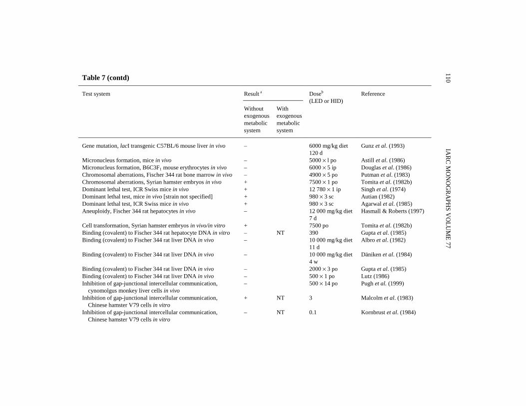
IARC M
ON
OG
RAPH
S VO
LUM
E 77110Table 7 (contd)
Result aTest system
Withoutexogenousmetabolicsystem
Withexogenousmetabolicsystem
Doseb
(LED or HID)Reference
Gene mutation, lacI transgenic C57BL/6 mouse liver in vivo – 6000 mg/kg diet120 d
Gunz et al. (1993)
Micronucleus formation, mice in vivo – 5000 × l po Astill et al. (1986)Micronucleus formation, B6C3F1 mouse erythrocytes in vivo – 6000 × 5 ip Douglas et al. (1986)Chromosomal aberrations, Fischer 344 rat bone marrow in vivo – 4900 × 5 po Putman et al. (1983)Chromosomal aberrations, Syrian hamster embryos in vivo + 7500 × 1 po Tomita et al. (1982b)Dominant lethal test, ICR Swiss mice in vivo + 12 780 × 1 ip Singh et al. (1974)Dominant lethal test, mice in vivo [strain not specified] + 980 × 3 sc Autian (1982)Dominant lethal test, ICR Swiss mice in vivo + 980 × 3 sc Agarwal et al. (1985)Aneuploidy, Fischer 344 rat hepatocytes in vivo – 12 000 mg/kg diet
7 dHasmall & Roberts (1997)
Cell transformation, Syrian hamster embryos in vivo/in vitro + 7500 po Tomita et al. (1982b)Binding (covalent) to Fischer 344 rat hepatocyte DNA in vitro – NT 390 Gupta et al. (1985)Binding (covalent) to Fischer 344 rat liver DNA in vivo – 10 000 mg/kg diet
11 dAlbro et al. (1982)
Binding (covalent) to Fischer 344 rat liver DNA in vivo – 10 000 mg/kg diet4 w
Däniken et al. (1984)
Binding (covalent) to Fischer 344 rat liver DNA in vivo – 2000 × 3 po Gupta et al. (1985)Binding (covalent) to Fischer 344 rat liver DNA in vivo – 500 × 1 po Lutz (1986)Inhibition of gap-junctional intercellular communication, cynomolgus monkey liver cells in vivo
– 500 × 14 po Pugh et al. (1999)
Inhibition of gap-junctional intercellular communication, Chinese hamster V79 cells in vitro
+ NT 3 Malcolm et al. (1983)
Inhibition of gap-junctional intercellular communication, Chinese hamster V79 cells in vitro
– NT 0.1 Kornbrust et al. (1984)
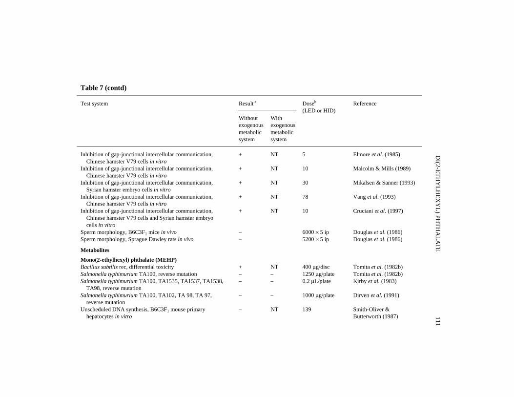
DI(2-ETH
YLH
EXY
L) PHTH
ALATE
111
Table 7 (contd)
Result aTest system
Withoutexogenousmetabolicsystem
Withexogenousmetabolicsystem
Doseb
(LED or HID)Reference
Inhibition of gap-junctional intercellular communication, Chinese hamster V79 cells in vitro
+ NT 5 Elmore et al. (1985)
Inhibition of gap-junctional intercellular communication, Chinese hamster V79 cells in vitro
+ NT 10 Malcolm & Mills (1989)
Inhibition of gap-junctional intercellular communication, Syrian hamster embryo cells in vitro
+ NT 30 Mikalsen & Sanner (1993)
Inhibition of gap-junctional intercellular communication, Chinese hamster V79 cells in vitro
+ NT 78 Vang et al. (1993)
Inhibition of gap-junctional intercellular communication, Chinese hamster V79 cells and Syrian hamster embryo cells in vitro
+ NT 10 Cruciani et al. (1997)
Sperm morphology, B6C3F1 mice in vivo – 6000 × 5 ip Douglas et al. (1986)Sperm morphology, Sprague Dawley rats in vivo – 5200 × 5 ip Douglas et al. (1986)
Metabolites
Mono(2-ethylhexyl) phthalate (MEHP)Bacillus subtilis rec, differential toxicity + NT 400 μg/disc Tomita et al. (1982b)Salmonella typhimurium TA100, reverse mutation – – 1250 μg/plate Tomita et al. (1982b)Salmonella typhimurium TA100, TA1535, TA1537, TA1538, TA98, reverse mutation
– – 0.2 μL/plate Kirby et al. (1983)
Salmonella typhimurium TA100, TA102, TA 98, TA 97, reverse mutation
– – 1000 μg/plate Dirven et al. (1991)
Unscheduled DNA synthesis, B6C3F1 mouse primary hepatocytes in vitro
– NT 139 Smith-Oliver &Butterworth (1987)

IARC M
ON
OG
RAPH
S VO
LUM
E 77112Table 7 (contd)
Result aTest system
Withoutexogenousmetabolicsystem
Withexogenousmetabolicsystem
Doseb
(LED or HID)Reference
Gene mutation, mouse lymphoma L5178Y cells, Tk locus in vitro
– – 0.3 μL/mL Kirby et al. (1983)
Sister chromatid exchange, Chinese hamster V79 cells in vitro + NT 25 Tomita et al. (1982b)Chromosomal aberrations in Syrian hamster embryo cells in vitro
– + 2.8 Tsutsui et al. (1993)
Cellular transformation in Syrian hamster embryo cells in vitro – (+) 56 Tsutsui et al. (1993)Cell transformation, Syrian hamster embryo cells, clonal assay + NT 23 Mikalsen et al. (1990)Cell transformation, C3H10T½ mouse cells – NT 417 Sanchez et al. (1987)Inhibition of gap-junctional intercellular communication, Chinese hamster V79 cells and Syrian hamster embryo cells in vitro
+ NT 28 Cruciani et al. (1997)
Unscheduled DNA synthesis, human primary hepatocytes in vitro
– NT 139 Butterworth et al. (1984)
DNA strand breaks, Wistar rat liver in vivo – NT 500 po × 14 Elliot & Elcombe (1987)
Mono(2-ethyl 5-hydroxyhexyl) phthalate (IX)Salmonella typhimurium TA100, TA102, TA98, TA97, reverse mutation
– – 1000 μg/plate Dirven et al. (1991)
Mono(2-ethyl 5-oxohexyl) phthalate (VI)Salmonella typhimurium TA100, TA102, TA98, TA97, reverse mutation
– – 1000 μg/plate Dirven et al. (1991)
Mono(5-carboxyl 2-ethylpentyl) phthalate (V)Salmonella typhimurium TA100, TA102, TA98, TA97, reverse mutation
– – 1000 μg/plate Dirven et al. (1991)

DI(2-ETH
YLH
EXY
L) PHTH
ALATE
113
Table 7 (contd)
Result aTest system
Withoutexogenousmetabolicsystem
Withexogenousmetabolicsystem
Doseb
(LED or HID)Reference
2-EthylhexanolBacillus subtilis rec, differential toxicity – NT 500 Tomita et al. (1982b)Salmonella typhimurium TA100, TA1535, TA1537, TA1538, TA98, reverse mutation
– – 1 μL/plate Kirby et al. (1983)
Gene mutation, mouse lymphoma L5178 cells, Tk locus in vitro – – 0.3 μL/mL Kirby et al. (1983)
Phthalic acidBacillus subtilis rec, differential toxicity – NT 500 Tomita et al. (1982b)
a +, positive; (+), weakly positive; –, negative; NT, not tested; ?, inconclusiveb LED, lowest effective dose; HID, highest ineffective dose; in-vitro tests, μg/mL; in-vivo tests, mg/kg bw/day; po, oral; ip, intraperitoneal; d,day; w, week; y, year; sc, subcutaneousc No oxidative damage in kidney DNAd Positive if followed by 5 h 0.16 μM 12-O-tetradecanoylphorbol 13-acetate
presence or absence of exogenous metabolic activation. It was also not mutagenic toEscherichia coli WP2 uvrA in the presence or absence of exogenous metabolic acti-vation. It did not induce gene mutations in Saccharomyces cerevisiae strain D7 in fourof five studies. It induced aneuploidy in a single study. Mitotic gene conversion wasinduced in two out of five studies with S. cerevisiae. In a single study, mitotic recom-bination was not induced in S. cerevisiae in the presence or absence of exogenousmetabolic activation. Neither mutation nor genetic crossing-over was induced inAspergillus nidulans cultured with di(2-ethylhexyl) phthalate in the absence of exo-genous metabolic activation in one study.
A small increase in somatic mutation frequency was reported in the eye-colourspot test with Drosophila melanogaster exposed to di(2-ethylhexyl) phthalate in thefeed but no effect was observed in two independent wing-spot tests. In a single study,mitotic recombination was not induced by di(2-ethylhexyl) phthalate; neither was sex-linked recessive lethal mutation induced in two studies in D. melanogaster treatedwith di(2-ethylhexyl) phthalate in the feed or by injection. When administered to D.melanogaster, di(2-ethylhexyl) phthalate and N-nitrosodiethylamine induced DNAdouble-strand breakage and DNA repair, although neither compound was active whenadministered alone.
DNA single-strand breaks were not induced by di(2-ethylhexyl) phthalate inprimary cultures of rat or Syrian hamster hepatocytes or in Chinese hamster ovary(CHO) cells. Unscheduled DNA synthesis was not induced in rat or mouse primaryhepatocytes exposed to di(2-ethylhexyl) phthalate. Only one of six studies reportedinduction of gene mutations at the Tk locus in mouse lymphoma L5178Y cells exposedto di(2-ethylhexyl) phthalate in the absence of an exogenous metabolic activationsystem. Ouabain-resistant mutants were not induced in mouse lymphoma or BALB/c-3T3 cells.
In one of two studies, di(2-ethylhexyl) phthalate induced a small increase in sisterchromatid exchange frequencies in Chinese hamster ovary cells cultured without butnot with exogenous metabolic activation. In other studies conducted only withoutmetabolic activation, it caused no increase in sister chromatid exchanges in eitherChinese hamster Don cells or rat liver RL4 cells. Di(2-ethylhexyl) phthalate did notinduce micronuclei in Chinese hamster ovary cells or in cultured rat hepatocytes,whereas the induction of micronuclei by di(2-ethylhexyl) phthalate in Syrian hamsterembryo cells has been reported.
Chromosomal aberrations were not induced by di(2-ethylhexyl) phthalate in any ofeight studies in various types of cultured cells in the absence of metabolic activation.Only three of these studies for chromosomal aberrations included an exogenousmetabolic activation system. Of these, one, using Syrian hamster embryo cells, foundan increase in aberration frequency. Weak effects were detected for the induction ofaneuploidy and mitotic division aberrations in Chinese hamster lung cells.
Di(2-ethylhexyl) phthalate induced cell transformation in the Syrian hamsterembryo clonal assay, the virally enhanced SA7/Syrian hamster embryo assay and
IARC MONOGRAPHS VOLUME 77114

RLV/Fischer rat assay without the addition of an exogenous metabolic activationsystem. In a single study, mouse JB6 epidermal cells were transformed by di(2-ethyl-hexyl) phthalate without activation and in one of two studies a weak response wasreported in the C3H10T½ cell transformation assay with di(2-ethylhexyl) phthalate ineither the absence or presence of exogenous metabolic activation. BALB/c-3T3 cellswere not transformed by di(2-ethylhexyl) phthalate with or without metabolic acti-vation. Di(2-ethylhexyl) phthalate inhibited gap-junctional intercellular communi-cation in Chinese hamster V79 cells in six of seven studies, but not in one study ofliver cells of cynomolgus monkeys in vivo. Di(2-ethylhexyl) phthalate treatment ofSyrian hamster embryo cells in a two-stage exposure with 12-O-tetradecanoylphorbol13-acetate resulted in superinduction of ornithine decarboxylase, an early event inmorphological transformation; no effect was seen after a one-stage treatment withdi(2-ethylhexyl) phthalate alone.
Di(2-ethylhexyl) phthalate did not increase the frequency of chromosomal aberra-tions in human lymphocytes in the absence of exogenous metabolic activation in vitro.In single studies with human lymphocytes in vitro, di(2-ethylhexyl) phthalate did notinduce unscheduled DNA synthesis or gene mutations at the TK or HPRT loci withoutexogenous metabolic activation, or aneuploidy in human fetal lung cells culturedwithout metabolic activation. One single study using human lymphocytes found thatdi(2-ethylhexyl) phthalate did not induce sister chromatid exchange, while anotherstudy found weak induction when the lymphocytes were co-cultured with rat livercells. A single study investigating the effect of di(2-ethylhexyl) phthalate on humanblood cells by the Comet assay reported induction of DNA damage but none in thepresence of exogenous metabolic activation.
Urine samples from rats treated by gavage with 15 daily doses of di(2-ethylhexyl)phthalate were not mutagenic to S. typhimurium strains TA100, TA1535, TA1537,TA1538 or TA98. In one study, the formation of 8-hydroxydeoxyguanosine (8-OH-dG) was measured as an indicator of DNA oxidative damage in liver and kidney ofrats exposed to di(2-ethylhexyl) phthalate in the diet for one to two weeks: 44–64%increased levels of 8-OH-dG were observed in liver but not kidney DNA. Similarresults were reported in a follow-up study in which rats received di(2-ethylhexyl)phthalate in the diet for one year. A significant increase in 8-OH-dG was measured inliver DNA at the one- and 12-month sampling times but not at three, six or ninemonths. Di(2-ethylhexyl) phthalate did not induce DNA strand breaks in rat liver orunscheduled DNA synthesis in rat or mouse liver following single or multiple oraltreatments, nor did it give rise to covalent DNA binding in rat hepatocytes in vitro orrat liver in vivo.
Di(2-ethylhexyl)phthalate did not increase the mutation frequency in the lacI geneof liver DNA isolated from female lacI transgenic mice given di(2-ethylhexyl)phthalate in the feed for 120 days. Micronuclei were not induced in mice fed withdi(2-ethylhexyl) phthalate. Di(2-ethylhexyl)phthalate did not induce chromosomalaberrations in rat bone marrow sampled 6 h after the last of five daily treatments
DI(2-ETHYLHEXYL) PHTHALATE 115

administered by gavage and it did not induce aneuploidy in hepatocytes of rats fed adiet containing di(2-ethylhexyl) phthalate for seven days. One study reported thatdi(2-ethylhexyl) phthalate was antimutagenic and co-recombinogenic in combinationwith N-ethyl-N-nitrosourea in the mouse spot test (Fahrig & Steinkamp-Zucht, 1996).Chromosomal aberrations and cell transformations were induced in Syrian goldenhamster embryos exposed transplacentally to di(2-ethylhexyl) phthalate for 24 h afterpregnant females were treated on day 11 of gestation. Di(2-ethylhexyl) phthalate wasreported to induce dominant lethal mutations in mice in three studies. Two re-eva-luations (Adler & Ashby, 1989; Ashby & Clapp, 1995) of these studies considered thatcytotoxicity can interfere with the recognition of a dominant lethal effect, so that thisreported mutagenic activity of di(2-ethylhexyl) phthalate is questionable. Di(2-ethyl-hexyl) phthalate did not induce changes in sperm morphology in mice or rats.
Metabolites
Five metabolites of di(2-ethylhexyl) phthalate, mono(2-ethylhexyl) phthalate,mono(2-ethyl-5-hydroxyhexyl) phthalate (IX), mono(2-ethyl-5-oxohexyl) phthalate(VI), mono(5-carboxy-2-ethylpentyl) phthalate (V) and 2-ethylhexanol, were tested ingenotoxicity assays. None induced mutations in S. typhimurium.
Negative results were also obtained in the Bacillus subtilis rec assay with mono(2-ethylhexyl) phthalate, 2-ethylhexanol and phthalic acid.
Unscheduled DNA synthesis was not induced in either mouse or human primaryhepatocyte cultures with mono(2-ethylhexyl) phthalate, and neither this metabolite nor2-ethylhexanol induced mutations in mouse lymphoma cells in vitro. Mono(2-ethyl-hexyl) phthalate induced sister-chromatid exchange in Chinese hamster V79 cells andchromosomal aberrations in Syrian hamster embryo cells. It also induced trans-formation in Syrian hamster embryo cells, but not in mouse C3H10T½ cells. Gap-junctional intercellular communication was inhibited by mono(2-ethylhexyl) phthalatein Syrian hamster embryo cells and in Chinese hamster V79 cells. As reported in anabstract (Baker et al., 1996), this function was also inhibited in rat and mousehepatocytes, but not in Syrian hamster or human hepatocytes.
In the single study with mono(2-ethylhexyl) phthalate, DNA strand breaks werenot induced in rat liver in vivo.
4.5 Mechanistic considerations
Significant species differences have been observed in the absorption and disposi-tion of di(2-ethylhexyl) phthalate. The peroxisome-proliferating effect of di(2-ethyl-hexyl) phthalate has been related most specifically in susceptible species to meta-bolites VI, IX and mono(2-ethylhexyl) phthalate; however, analysis of the dispositiondata does not provide an explanation for the observed species differences in di(2-ethylhexyl) phthalate-induced hepatic peroxisome proliferation.
IARC MONOGRAPHS VOLUME 77116

The weight of evidence for di(2-ethylhexyl) phthalate and for other rodent pero-xisome proliferators in general demonstrates that they do not act as direct DNA-damaging agents.
Chronic administration of peroxisome proliferators to rodents results in sustainedoxidative stress due to overproduction of peroxisomal hydrogen peroxide. This cantheoretically generate reactive oxygen species which can damage DNA and otherintracellular targets. The induction of peroxisomal fatty acid β-oxidation and increasesin peroxisomal volume density by di(2-ethylhexyl) phthalate in vivo under bioassayconditions (Marsman et al., 1988; Lake et al., 1987) supports this hypothesis.Supporting data on induction of oxidative stress (lipofuscin accumulation, 8-OH-dGin DNA) in rat liver by di(2-ethylhexyl) phthalate are available (Conway et al., 1989;Takagi et al., 1990a); however, there are no data for mouse liver.
Similarly, modulation of hepatocellular proliferation by peroxisome proliferatorshas been implicated in the mechanism of carcinogenesis. This can theoretically resultin increased levels of mutation by increasing the frequency of replicative DNAsynthesis as well as increasing the number of hepatocytes at risk. Furthermore,hepatocellular proliferation is probably involved in the promotion of growth of pre-neoplastic hepatocytes. There is clear evidence that di(2-ethylhexyl) phthalate causesacute and sustained hepatocellular proliferation under bioassay conditions whichresulted in liver tumours in rats (Marsman et al., 1988).
Marked species differences in hepatic peroxisome proliferation have been reported(Ashby et al., 1994; IARC, 1995; Lake, 1995a,b; Cattley et al., 1998). In biopsies fromhumans receiving hypolipidaemic drugs, there was no effect or changes were muchsmaller than those that would be produced in rodent hepatocytes at equivalent doselevels (Lake, 1995a,b; Cattley et al., 1998). While peroxisome proliferation may bereadily demonstrated in cultured rat and mouse hepatocytes, such effects are notobserved in hepatocytes from non-responsive species including guinea-pigs, primatesand humans. No study has yet compared the responsiveness to di(2-ethylhexyl) phtha-late of human versus rodent livers in vivo. Evidence from humans, rats and mice indi-cates that differences in disposition, including formation of active metabolites, are of aquantitative, not qualitative nature. Taken together, these disposition data do not providean explanation for the species differences in response to di(2-ethylhexyl) phthalate.However, several studies using human hepatocytes, unlike those with hepatocytes frommice and rats, have demonstrated that administration of di(2-ethylhexyl) phthalate meta-bolites does not lead to peroxisome proliferation in vitro. A growing body of evidenceconcerning the molecular basis of peroxisome proliferation, summarized below,indicates why human livers and hepatocytes would be expected to be refractory toinduction of peroxisome proliferation and carcinogenesis by di(2-ethylhexyl) phthalate.
Several lines of investigation have enhanced our understanding of the molecularbasis of peroxisome proliferation, with important implications for cancer risk andspecies differences. In rodent liver, increased organ weight, peroxisome proliferation,increased replicative DNA synthesis and induction of peroxisomal and microsomal fatty
DI(2-ETHYLHEXYL) PHTHALATE 117

acid-oxidizing enzymes require the expression of functional PPARα. This nuclearreceptor is a member of the steroid hormone receptor superfamily and binds to DNA asa heterodimer with the retinoid X receptor (RXR). Peroxisome proliferator responseelements (PPREs) have been found in genes for both peroxisomal and microsomal fattyacid-oxidizing enzymes (Lake, 1995b; Cattley et al., 1998). Studies with PPARα knock-out mice have demonstrated that all the above effects are lost in these mice (Lee et al.,1995; Peters et al., 1997a; Aoyama et al., 1998; Ward et al., 1998). Moreover, unlikewild-type mice, the potent peroxisome proliferator Wy-14,643 did not produce livertumours in PPARα-deficient mice (Peters et al., 1997a). In this study, WY-14,643 at0.1% in the diet or control diet was fed for 11 months to PPARα-deficient and wild-typemice and the livers were removed and examined. Of nine PPARα-deficient mice fedWY-14,643, three died and were lost to evaluation and one was euthanized and exa-mined when moribund before 11 months. WY-14,643 produced multiple hepatocellulartumours in 6/6 wild-type mice; in three of these mice, some of these tumours weremalignant. In contrast, the same treatment produced no tumours in PPARα-deficientmice (0/9), and no preneoplastic lesions were found by microscopy. No gross tumoursor microscopic evidence of preneoplasia were seen in either wild-type mice or PPARα-deficient mice on the control diet (incidence, 0/9 and 0/9, respectively). Taken together,these results demonstrate that peroxisome proliferation, related hepatic effects andcarcinogenesis induced by peroxisome proliferators are all absolutely dependent onfunctional PPARα.
Studies of PPARα activation in vitro have shown that di(2-ethylhexyl) phthalatemetabolites are capable of activating PPARα. A study of PPARα knock-out micetreated with di(2-ethylhexyl) phthalate in vivo demonstrated that the increased liverweights and induction of peroxisomal and microsomal enzymes are absolutelydependent on PPARα (Ward et al., 1998).
The species differences, particularly with respect to humans compared to rats andmice, can be potentially attributed to several aspects of PPARα-mediated regulation ofgene expression. These include the level of expression and functional capability ofPPARα, the presence or absence of active PPREs in the promoter region of specificgenes, and other aspects of interaction with transcriptional regulatory proteins.
Recent evidence confirms that species differences can involve more than one aspectof PPARα-mediated regulation of gene expression. The insensitivity of human liver torodent peroxisome proliferators is associated with low levels of expression of PPARα inhuman liver. Marked species differences in the expression of PPARα mRNA have beendemonstrated between rodent and human liver, with the latter expressing 1–10% of thelevels found in mouse or rat liver (Palmer et al., 1994; Tugwood et al., 1996; Palmeret al., 1998). Using a sensitive and specific immuno/DNA binding assay, Palmer et al.(1998) have shown that active PPARα protein is expressed at variable concentrations inhuman livers. The study compared 20 different human livers and found that those withthe highest levels of PPARα protein expression contained less than 10% of the level inmice. Most of the samples (13/20) contained no detectable PPARα activity, but did
IARC MONOGRAPHS VOLUME 77118

exhibit PPRE-binding activity that did not correspond to PPARα. All six subjects aged10 years or less lacked detectable PPARα activity. Such data support a ‘thresholdthrough competition’ concept. In most human samples studied, it was found that PPREsare mainly bound by other competing proteins that may block peroxisome proliferatorresponsiveness. In addition, the low levels of PPARα protein detected in human liverwere lower than those estimated from RNA analysis and this was explained by thefinding that a significant fraction of PPARα mRNA is mis-spliced in human liver. It ispossible that the low level of PPARα in human liver may explain, at least in part, theapparent lack of responsiveness of human hepatocytes to peroxisome proliferators. Insupport of this hypothesis, Palmer et al. (1998) have shown that transcriptionalresponses to peroxisome proliferators are not evident in the Huh cell line, whichcontains similar levels of PPARα and RXR to those found in intact human liver.However, increasing PPARα levels by heterologous expression in the Huh cell lineconferred responsiveness to peroxisome proliferators, suggesting that the low level ofendogenous PPARα is the major factor in the non-responsiveness of these cells.
The truncated human PPARα resulting from mRNA mis-splicing, identified byPalmer et al. (1998), has been further characterized by Gervois et al. (1999). Theyfound that the truncated PPARα mRNA accounted for 25–50% of total PPARαmRNA in 10 human liver samples, while no truncated PPARα mRNA was found inlivers of rats and mice. The truncated human PPARα mRNA was expressed in vitro,where it was shown to (a) fail to bind to PPRE, a necessary step for gene activationand (b) interfere with gene activation by expressed full-length human PPARα, in partdue to titration of coactivator CREB-binding protein, an additional element of trans-criptional regulation. Taken together, these results demonstrate that a certain fractionof human PPARα is probably truncated and inactive and may interfere with thefunction of any full-length human PPARα present in human hepatocytes.
Peroxisome proliferator responsiveness is unlikely to be a linear function of PPARαexpression levels. This may be due to competition for binding to PPREs by other trans-cription factors such as HNF4, ARP-1 and TR (Palmer et al., 1994; Miyamoto et al.,1997), binding of PPARα to other transcription factors (e.g., LXRα) leading toformation of non-functional heterodimers (Miyata et al., 1996), competition of PPARαwith other hormone receptors (e.g., TR) for dimerization with RXR (Juge-Aubry et al.,1995) or lack of co-activating proteins such as steroid receptor co-activator-1 (SRC-1)and PPAR-binding protein (PBP) (Zhu et al., 1997). Other human PPARs may competewith PPARα; for example PPAR* (hNUC1) can repress the activation of hPPARα (Jow& Mukherjee, 1995).
In addition to the role of receptor expression and other transcriptional regulators,differential species sensitivity to peroxisome proliferators could depend on gene-specific factors. In the case of peroxisomal acyl-coenzyme A oxidase, the promoterregions containing PPRE responsible for transcriptional activation of the rodent geneare not present in the promoter region of the human gene (Lambe et al., 1999;Woodyatt et al., 1999). It is possible that similar differences in promoter regions of
DI(2-ETHYLHEXYL) PHTHALATE 119

other genes could also help to explain differences between rodent and humanresponses to peroxisome proliferators.
The insensitivity towards peroxisome proliferators exhibited by human hepatocytesis reflected in guinea-pigs. Guinea-pigs are also refractory to the hepatic effects of rodentperoxisome proliferators (reviewed in Doull et al., 1999), including di(2-ethylhexyl)phthalate metabolites (Elcombe & Mitchell, 1986; Dirven et al., 1993c; Elcombe et al.,1996) and, like humans, express similar low levels of PPARα (Bell et al., 1998;Tugwood et al., 1998). Significant responses (peroxisome proliferation, induction offatty acid-oxidizing enzymes and stimulation of replicative DNA synthesis) believed tobe associated with hepatocarcinogenesis in rodents are not observed in humans orguinea-pigs. However, these species do exhibit hypolipidaemic responses when exposedto some rodent peroxisome proliferators (Lake, 1995a; Bell et al., 1998; Cattley et al.,1998). The hypolipidaemic response, which is not associated with any hypertrophic orhyperplastic response, has been attributed to PPARα-mediated regulation of genesencoding lipoprotein lipase and various lipoproteins. The existence of such a response,in spite of low levels of PPARα, may be explained by differences in the mechanism ofaction of PPARα in relation to these hypolipidaemic genes compared with those thatregulate hypertrophic or hyperplastic responses: the former may have a lower thresholdof activation and may require lower concentrations of receptors due to different bindingaffinities for different PPREs. Differences in the activation properties for differentPPRE-containing promoters have been demonstrated (Hsu et al., 1995).
In addition to results from guinea-pigs, recent studies with rabbits suggest a furtherpotential model for the refractory nature of human liver to peroxisome proliferation (Staels& Auwerx, 1998). In humans, fibrates increase plasma levels of high-density lipoprotein(HDL) via induction of human apo A-I gene expression. The fibrate effect on human apoA-I is mediated by PPARα that interacts with a PPRE in its promoter. In normal rabbits,plasma lipoprotein levels are not changed by fibrate treatment. However, fibrate treatmentof transgenic apo A-I rabbits results in increased plasma HDL and human apo A-I concen-trations due to induction of the human apoA-I transgene expression in the rabbit liver.Significantly, this induction of lipoprotein gene expression occurs without affecting liverweight or peroxisomal acyl-coenzyme A oxidase activity. This demonstrates that PPARα-mediated effects of fibrates on human lipoprotein gene expression can occur in the absenceof peroxisome proliferation.
In summary:1. The weight of evidence for di(2-ethylhexyl) phthalate and its metabolic
products demonstrates that they do not act as direct DNA-damaging agents.2. Di(2-ethylhexyl) phthalate produces liver tumours in rats and mice.3. Under conditions of the bioassays, di(2-ethylhexyl) phthalate induces
peroxisome proliferation and cell replication in liver that are characteristic ofa peroxisome proliferator in mice and rats.
4. Rodent peroxisome proliferators exercise their pleiotropic effects in liver dueto activation of PPARα. This process is essential for liver hypertrophy and
IARC MONOGRAPHS VOLUME 77120

hyperplasia and eventual hepatocarcinogenesis in response to peroxisomeproliferators.
5. Hepatic peroxisome proliferation has not been adequately evaluated in studiesof human livers following exposure to di(2-ethylhexyl) phthalate in vivo;however, the effect of treatment of human and mouse hepatocytes with di(2-ethylhexyl) phthalate metabolites which are active in rat hepatocytes, as wellas other peroxisome proliferators, indicate that humans can reasonably bepredicted to be refractory to induction of peroxisome proliferation and hepato-cellular proliferation by di(2-ethylhexyl) phthalate. The evidence indicatesthat the mechanism of peroxisome proliferation induced by di(2-ethylhexyl)phthalate in rat hepatocytes does not operate in humans.
6. The absence of a significant response of human liver to induction of peroxi-some proliferation and hepatocellular proliferation is explained by severalaspects of PPARα-mediated regulation of gene expression.
7. Overall, these findings indicate that the increased incidence of liver tumoursin mice and rats treated with di(2-ethylhexyl) phthalate results from a mecha-nism that does not operate in humans.
5. Summary of Data Reported and Evaluation
5.1 Exposure data
Di(2-ethylhexyl) phthalate is a liquid of low volatility, widely used as a plasticizerin flexible poly(vinyl chloride) products at concentrations of up to 40%, as well as ina number of other minor applications. Occupational exposure occurs mainly byinhalation as an aerosol during its manufacture and its use as a plasticizer in poly(vinylchloride) product manufacturing plants, at concentrations usually below 1 mg/m3.
Di(2-ethylhexyl) phthalate is ubiquitous in the general environment as a result ofits widespread use in poly(vinyl chloride) products. It is found in ambient air at levelsusually below 100 ng/m3. The highest levels of di(2-ethylhexyl) phthalate in foods arefound in milk products, meat and fish and in other products with a high fat content,where concentrations up to 10 mg/kg have been reported. The leaching of di(2-ethyl-hexyl) phthalate from flexible plastics used in medical devices, such as during dialysisand transfusion, can result in large direct exposures.
5.2 Human carcinogenicity data
One small study of workers in a di(2-ethylhexyl) phthalate production plant didnot show any excess of cancer mortality. However, this study did not have adequatepower to detect a potential excess risk.
DI(2-ETHYLHEXYL) PHTHALATE 121

5.3 Animal carcinogenicity data
Di(2-ethylhexyl) phthalate was tested for carcinogenicity by oral administration inthe diet in two experiments in mice and six experiments in rats. Hepatocellulartumours were produced consistently in both species.
In a number of initiation/promotion studies in strains of mice susceptible to livercarcinogenesis, administration of di(2-ethylhexyl) phthalate following administrationwith known carcinogens enhanced the incidences of hepatocellular preneoplastic foci,adenomas and carcinomas. In a number of similar studies in rats and in one study inhamsters, in general, no promoting activity of di(2-ethylhexyl) phthalate was demons-trated. No initiating activity of di(2-ethylhexyl) phthalate was found in the liver ofmice or rats. In two N-nitrosamine-initiation target organ models in rats, one showedenhancement of renal tubule tumours by di(2-ethylhexyl) phthalate, whereas the othershowed no promotion of urinary bladder tumours.
5.4 Other relevant data
The absorption and disposition of di(2-ethylhexyl) phthalate has been investigatedextensively in humans and laboratory animals. In all species studied, the compoundunderwent rapid metabolism, with the urine and faeces being the major routes ofexcretion. Following oral administration, the bulk of a di(2-ethylhexyl) phthalate dosewas absorbed as the monoester, mono(2-ethylhexyl) phthalate. This ester is alsoformed by esterases in the body following intravenous administration and is subject toextensive oxidative metabolism by the cytochrome P450 system.
The peroxisome-proliferating effects of di(2-ethylhexyl) phthalate in susceptiblespecies (e.g., rats and mice) have primarily been related to mono(2-ethylhexyl) phthalateand two other specific metabolites. However, while species differences have beenobserved in the absorption and disposition of di(2-ethylhexyl) phthalate, they do notprovide an explanation for the species differences in hepatic peroxisome-proliferatingactivity.
The literature on potential toxic effects of di(2-ethylhexyl) phthalate followinghuman exposure is limited. Taken together, the data indicate that di(2-ethylhexyl)phthalate does not cause observable toxicity following oral and intravenous exposure,but do not contribute information relevant to the evaluation of human carcinogenicity.
A considerable amount of information on the hepatic effects of orally administereddi(2-ethylhexyl) phthalate indicates that it causes hepatic peroxisome proliferation(ultrastructural effects and enzyme induction), hepatomegaly and increased replicativeDNA synthesis in rats and mice. At a lower magnitude in Syrian hamsters, enzymeinduction and hepatomegaly have been observed (ultrastructural effects andreplicative DNA synthesis have not been evaluated). Guinea-pigs, marmosets andcynomolgus monkeys evaluated under the same or similar experimental conditions didnot exhibit peroxisome proliferation responses. Studies of di(2-ethylhexyl) phthalate
IARC MONOGRAPHS VOLUME 77122

metabolites in primary rat, mouse and, to a lesser extent, Syrian hamster hepatocytecultures in vitro elicited markers of peroxisome proliferation, while the same orsimilar experimental conditions did not elicit markers of peroxisome proliferation inprimary cultures of either guinea-pig, rabbit, dog, cynomolgus monkey, marmoset or,most notably, human hepatocytes.
Hepatic peroxisome proliferation depends on a nuclear receptor, PPARα, tomediate these responses in mice, based on lack of response to peroxisome proliferatorsin PPARα-deficient mice. In one study with another peroxisome proliferator, WY-14,643, carcinogenesis was shown to be dependent on the same receptor. Oraladministration of di(2-ethylhexyl) phthalate failed to elicit markers of peroxisomeproliferation in PPARα-deficient mice, while the same treatment elicited this responsein normal mice. Metabolites of di(2-ethylhexyl) phthalate caused activation of PPARα-mediated gene expression in mammalian cell co-transfection assays. Differencesbetween responsive rodents and humans in various aspects of PPARα-mediatedregulation of gene expression are consistent with the lack of activity of di(2-ethylhexyl)phthalate metabolites in hepatocyte cultures from 12 people studied to date.
No data on reproductive and developmental effects in humans were available.Oral exposure of rats and mice to di(2-ethylhexyl) phthalate during organogenesis
caused malformations and fetal death. A study in knock-out mice suggested that thedevelopmental effects are not PPARα-mediated.
Irreversible testicular damage has been observed in male rat pups exposedprenatally and during suckling via maternal exposure to drinking water containing thecompound.
Oral exposure of adult rats and mice caused effects on fertility in males andfemales and serious effects on the testicles. Young animals were much more sensitiveto gonadal effects than adults and in some cases, the onset of occurrence of the testi-cular effects was earlier in young animals. Dose-dependent testicular effects were seenin young rats exposed to di(2-ethylhexyl) phthalate in the diet.
In one study using small groups of adult marmosets, oral exposure did not causetesticular toxicity at doses higher than those producing testicular effects in adult rats.
The Sertoli cells in the testes appear to be the main target of the testicular toxicity.Proposed mechanistic hypotheses relate to reduced testicular zinc levels, alteredhormonal status, altered metabolic function and altered follicle-stimulating hormonereactivity.
Di(2-ethylhexyl) phthalate has been studied extensively for its genotoxic effectsin a wide range of test systems, both in vitro and in vivo. The majority of these studiesdid not reveal any activity. No mutagenic activity was observed in bacteria. In fungi,all but two studies failed to show any evidence of recombinational events or mutation.A single study in yeast for aneuploidy was positive. Low levels of mutation wereinduced in Drosophila melanogaster in somatic cells in some studies, but no germ-cellmutations or DNA damage were induced in these insects. In cultured mammaliancells, no primary DNA damage, mutation, sister chromatid exchange or chromosomal
DI(2-ETHYLHEXYL) PHTHALATE 123

aberrations were induced (except in a single study for DNA strand breakage), whereastransformation of cells was induced in a number of different systems.
In vivo, neither covalent binding to DNA nor DNA strand breakage was inducedin several studies on rat liver, and unscheduled DNA synthesis was not induced in theliver of either rats or mice. Gene mutations were not induced in the liver of dosed micein a single study and there was no evidence for induction of chromosomal aberrationsin mice or rats. Aberrations were induced, however, in the embryos of dosed pregnantSyrian hamsters. Dominant lethal effects were reported to be induced in male mice,but re-evaluation of these data did not confirm this conclusion.
5.5 Evaluation
There is inadequate evidence in humans for the carcinogenicity of di(2-ethyl-hexyl) phthalate.
There is sufficient evidence in experimental animals for the carcinogenicity of di-(2-ethylhexyl) phthalate.
Overall evaluation
Di(2-ethylhexyl) phthalate is not classifiable as to its carcinogenicity to humans(Group 3).
In making its overall evaluation of the carcinogenicity to humans of di(2-ethyl-hexyl) phthalate, the Working Group took into consideration that (a) di(2-ethylhexyl)phthalate produces liver tumours in rats and mice by a non-DNA-reactive mechanisminvolving peroxisome proliferation; (b) peroxisome proliferation and hepatocellularproliferation have been demonstrated under the conditions of the carcinogenicitystudies of di(2-ethylhexyl) phthalate in rats and mice; and (c) peroxisome proliferationhas not been documented in human hepatocyte cultures exposed to di(2-ethylhexyl)phthalate nor in the liver of exposed non-human primates. Therefore, the mechanismby which di(2-ethylhexyl) phthalate increases the incidence of hepatocellular tumoursin rats and mice is not relevant to humans.
6. References
Abe, S. & Sasaki, M. (1977) Chromosome aberrations and sister chromatid exchanges inChinese hamster cells exposed to various chemicals. J. natl Cancer Inst., 58, 1635–1641
Adler, I.-L. & Ashby, J. (1989) The present lack of evidence for unique rodent germ-cellmutagens. Mutat. Res., 212, 55–66
Agarwal, D.K., Lawrence, W.H. & Autian, J. (1985) Antifertility and mutagenic effects in micefrom parenteral administration of di-2-ethylhexyl phthalate (DEHP). J. Toxicol. environ.Health, 16, 71–84
IARC MONOGRAPHS VOLUME 77124

Agarwal, D.K., Eustis, S., Lamb, J.C., IV, Reel, J.R. & Kluwe, W.M. (1986) Effects of di(2-ethylhexyl) phthalate on the gonadal pathophysiology, sperm morphology, and repro-ductive performance of male rats. Environ. Health Perspect., 65, 343–350
Agarwal, D.K., Lawrence, W.H., Turner, J.E. & Autian, J. (1989) Effects of parenteral di-(2-ethylhexyl)phthalate (DEHP) on gonadal biochemistry, pathology and reproductiveperformance of mice. J. Toxicol. environ. Health, 26, 39–59
Agency for Toxic Substances and Disease Registry (1989) Public Health Statement: Di(2-ethylhexyl) Phthalate, Athens, GA
Agency for Toxic Substances and Disease Registry (1993) Toxicological Profile: Di(2-ethyl-hexyl) Phthalate, Atlanta, GA, Department of Health and Human Services
Albro, P.W. (1986) Absorption, metabolism and excretion of di(2-ethylhexyl) phthalate by ratsand mice. Environ. Health Perspect., 65, 293–298
Albro, P.W. & Lavenhar, S.R. (1989) Metabolism of di(2-ethylhexyl)phthalate. Drug Metab.Rev., 21, 13–34
Albro, P.W. & Thomas, R.O. (1973) Enzymatic hydrolysis of di-(2-ethylhexyl) phthalate bylipase. Biochim. biophys. Acta, 360, 381–390
Albro, P.W., Thomas, R. & Fishbein, L. (1973) Metabolism of diethylhexyl phthalate by rats.Isolation and characterization of the urinary metabolites. J. Chromatogr., 76, 321–330
Albro, P.W., Hass, J.R., Peck, C.C., Odam, D.G., Corbett, J.T., Bailey, F.J., Blatt, H.E. &Barrett, B.B. (1981) Identification of the metabolites of di-(2-ethylhexyl) phthalate inurine from the African green monkey. Drug Metab. Dispos., 9, 223–225
Albro, P.W., Corbett, J.T., Schroeder, J.L., Jordan, S. & Matthews, H.B. (1982) Pharmaco-kinetics, interactions with macromolecules and species differences in metabolism ofDEHP. Environ. Health Perspect., 45, 19–25
Amacher, D.E. & Turner, G.N. (1985) Tests for gene mutational activity in the L5178Y/TKassay system. Prog. Mutat. Res., 5, 487–496
American Conference of Governmental Industrial Hygienists (ACGIH) (1999) TLVs andOther Occupational Exposure Values - 1999, Cincinnati, OH [CD-ROM]
American Society of Testing and Materials (1997) Standard Consumer Safety Specification onToy Safety (ASTM 963-96a), West Conshohocken, PA
Anderson, D., Yu, T.-W. & Hinçal, F. (1999) Effect of some phthalate esters in human cells inthe comet assay. Teratog. Carcinog. Mutag., 19, 275–280
Anon. (1996) Facts & figures for the chemical industry. Chem. Eng. News, 74, 38–79Anon. (1999) EU bans phthalates in young children’s mouthing toys. Environ. Watch Western
Europe, 8, 1–3Aoyama, T., Peters, J.M., Iritani, N., Nakajima, T., Furihata, K., Hashimoto, T. & Gonzalez,
F.J. (1998) Altered constitutive expression of fatty acid-metabolizing enzymes in micelacking the peroxisome proliferator-activated receptor α (PPARα). J. biol. Chem., 273,5678–5684
Arcadi, F.A., Costa, C., Imperatore, C., Marchese, A., Rapidisarda, A., Salemi, M., Trimarchi,G.R. & Costa, G. (1998) Oral toxicity of bis(2-ethylhexyl) phthalate during pregnancy andsuckling in Long-Evans rat. Food chem. Toxicol., 36, 963–970
Aristech Chemical Corp. (1992) PX-138 Di-2 Ethyl Hexyl Phthalate (DEHP), Pittsburgh, PAArni, P. (1985) Induction of various genetic effects in the yeast Saccharomyces cerevisiae
strain D7. Prog. Mutat. Res., 5, 217–224
DI(2-ETHYLHEXYL) PHTHALATE 125

Ashby, J. & Clapp, M.J.L. (1995) The rodent dominant lethal assay: a proposed format for datapresentation that alerts to pseudo-dominant lethal effects. Mutat. Res., 330, 209–218
Ashby, J., Brady, A., Elcombe, C.R., Elliott, B.M., Ishmael, J., Odum, J., Tugwood, J.D.,Kettle, S. & Purchase, I.F. (1994) Mechanistically-based human hazard assessment ofperoxisome proliferator-induced hepatocarcinogenesis. Hum. exp. Toxicol., 13 (Suppl. 2),S1–S117
Astill, B.D. (1989) Metabolism of DEHP: effects of prefeeding and dose variation, and compa-rative studies in rodents and the cynomolgus monkey (CMA studies). Drug Metab. Rev.,21, 35–53
Astill, B., Barber, E., Lington, A., Moran, E., Mulholland, A., Robinson, E. & Scheider, B.(1986) Chemical industry voluntary test program for phthalate esters: health effectsstudies. Environ. Health Perspect., 65, 329–336
Astill, B.D., Gingell, R., Guest, D., Hellwig, J., Hodgson, J.R., Kuettler, K., Mellert, W.,Murphy, S.R., Sielken, R.L. & Tyler, T.R. (1996) Oncogenicity testing of 2-ethylhexanolin Fischer 344 rats and B6C3F1 mice. Fund. appl. Toxicol., 31, 29–41
Atlas, E. & Giam, C.S. (1981) Global transportation of organic pollutants: ambient concen-trations in the remote marine atmosphere. Science, 211, 163–165
Autian, J. (1982) Antifertility effects and dominant lethal assays for mutagenic effects ofDEHP. Environ. Health Perspect., 45, 115–118
Baker, R.S.U. & Bonin, A.M. (1985) Tests with the Salmonella plate-incorporation assay.Prog. Mutat. Res., 5, 177–180
Baker, T.K., Kalimi, G.H., Lington, A.W., Isenberg, J.S., Klaunig, J.E. & Nikiforov, A.I. (1996)Gap junctional intercellular communication (GJIC) studies on 5 phthalate monoesters inhepatocytes of four species: implications for cancer risk assessment (Abstract). Toxico-logist, 30, 208
Barber, E.D., Astill, B.D., Moran, E.J., Schneider, B.F., Gray, T.J.B., Lake, B.G. & Evans, J.G.(1987) Peroxisome induction studies on seven phthalate esters. Toxicol. ind. Health, 3,7–22
Barrett, J.C. & Lamb, P.W. (1985) Tests with the Syrian hamster embryo cell transformationassay. Prog. Mutat. Res., 5, 623–628
Bauer, M.J. & Herrmann, R. (1997) Estimation of the environmental contamination by phthalicacid esters leaching from household wastes. Sci. total Environ., 208, 49–57
Bell, A.R., Savory, R., Horley, N.J., Choudhury, A.I., Dickins, M., Gray, T.J.B, Salter, A.M. &Bell, D.R. (1998) Molecular basis of non-responsiveness to peroxisome proliferators: theguinea-pig PPARα is functional and mediates peroxisome proliferator-inducedhypolipidaemia. Biochem. J., 332, 689–693
Boekelheide, K. (1993) Sertoli cell toxicants. In: Russell, L.D. & Griswold, M.D., eds, TheSertoli Cell, Clearwater, FL, Cache River Press, pp. 551–575
Bove, J.L., Dalven, P. & Kukreja, V.P. (1978) Airborne di-butyl and di(2-ethylhexyl)phthalateat three New York City air sampling stations. Int. J. environ. Chem., 5, 189–194
Bradley, M.O. (1985) Measurement of DNA single-strand breaks by alkaline elution in rathepatocytes. Prog. Mutat. Res., 5, 353–357
Brooks, T.M., Gonzalez, L.P., Calvert, R. & Parry, J.M. (1985) The induction of mitotic geneconversion in the yeast Saccharomyces cerevisiae strain JD1. Prog. Mutat. Res., 5,225–228
IARC MONOGRAPHS VOLUME 77126

Burg, R.V. (1988) Bis(2-ethylhexyl) phthalate. J. appl. Toxicol., 8, 75–78Butterworth, B.E., Bermudez, E., Smith-Oliver, T., Earle, L., Cattley, R., Martin, J., Popp, J.A.,
Strom, S., Jirtle, R. & Michalopoulos, G. (1984) Lack of genotoxic activity of di(2-ethyl-hexyl)phthalate (DEHP) in rat and human hepatocytes. Carcinogenesis, 5, 1329–1335
Butterworth, B.E., Smith-Oliver, O.T., Earle, L., Loury, D.J., White, R.D., Doolittle, D.J.,Working, P.K., Cattley, R.C., Jirtle, R., Michalopoulos, G. & Strom, S. (1989) Use ofprimary cultures of human hepatocytes in toxicology studies. Cancer Res., 49, 1075–1084
Calley, D., Autian, J. & Guess, W.L. (1966) Toxicology of a series of phthalate esters. J.pharm. Sci., 55, 158–162
Carere, A., Conti, G., Conti, L. & Crebelli, R. (1985) Assays in Aspergillus nidulans for theinduction of forward-mutation in haploid strain 35 and for mitotic nondisjunction,haploidization and crossing-over in diploid strain P1. Prog. Mutat. Res., 5, 307–312
Carls, N. & Schiestl, R.H. (1994) Evaluation of the yeast DEL assay with 10 compoundsselected by the International Programme on Chemical Safety for the evaluation of short-term tests for carcinogens. Mutat. Res., 320, 293–303
Castle, L., Gilbert, J. & Eklund, T. (1990) Migration of plasticizer from poly(vinylchloride)milk tubing. Food Addit. Contam., 7, 591–596
Cattley, R.C. & Glover, S.E. (1993) Elevated 8-hydroxydeoxyguanosine in hepatic DNA ofrats following exposure to peroxisome proliferators: relationship to carcinogenesis andnuclear localization. Carcinogenesis, 14, 2495–2499
Cattley, R.C., Conway, J.G. & Popp, J.A. (1987) Association of persistent proliferation andoxidative injury with hepatocarcinogenicity in female F-344 rats fed di(2-ethylhexyl)-phthalate for 2 years. Cancer Lett., 38, 15–22
Cattley, R.C., Smith-Oliver, T., Butterworth, B.E. & Popp, J.A. (1988) Failure of the peroxi-some proliferator WY-14,643 to induce unscheduled DNA synthesis in rat hepatocytesfollowing in vivo treatment. Carcinogenesis, 9, 1179–1183
Cattley, R.C., DeLuca, J., Elcombe, C., Fenner-Crisp, P., Lake, B.G., Marsman, D.S., Pastoor,T.A., Popp, J.A., Robinson, D.E., Schwetz, B., Tugwood, J. & Wahli, W. (1998) Doperoxisome proliferating compounds pose a hepatocarcinogenic hazard to humans? Regul.Toxicol. Pharmacol., 27, 47–60
Cautreels, W., Van Cauwenberghe, K.V. & Guzman, L.A. (1977) Comparison between theorganic fraction of suspended matter at a background and urban air station. Sci. totalEnviron., 8, 79–88
Chemical Information Services (1999) Directory of World Chemical Producers (Version99.1.0), Dallas, TX [CD-ROM]
Christensson, A., Ljunggren, L., Nilsson-Thorell, C., Arge, B., Diehl, U., Hagstam, K.-E. &Lundberg, M. (1991) In vivo comparative evaluation of hemodialysis tubing plasticizedwith DEHP and TEHTM. Int. J. artificial Organs, 14, 407–410
Chu, I., Villeneuve, D.C., Secours, V., Franklin, C., Rock, G. & Viau, A. (1978) Metabolismand tissue distribution of mono-2-ethylhexyl phthalate in the rat. Drug Metab. Dispos., 6,146–149
Chu, I., Dick, D., Bronaugh, R. & Tryphonas, L. (1996) Skin reservoir formation and bio-availability of dermally administered chemicals in hairless guinea pigs. Food chem.Toxicol., 34, 267–276
DI(2-ETHYLHEXYL) PHTHALATE 127

Cimini, A.M., Sulli, A., Stefanini, S., Serafini, B., Moreno, S., Rossi, L., Giorgi, M. & Ceru,M.P. (1994) Effects of di-(2-ethylhexyl)phthalate on peroxisomes of liver, kidney andbrain of lactating rats and their pups. Cell. mol. Biol., 40, 1063–1076
Cocchieri, R.A. (1986) Occurrence of phthalate esters in Italian packaged foods. J. FoodProtect., 49, 265–266
Cole, R.S., Tocchi, M., Wye, E., Villeneuve, D.C. & Rock, G. (1981) Contamination ofcommercial blood products by di-2-ethylhexyl phthalate and mono-2-ethylhexyl phthalate.Vox sang., 40, 317–332
Cole, R.H., Frederick, R.E., Healy, R.P. & Rolan, R.G. (1984) Preliminary findings of thepriority pollutant monitoring project of the nationwide urban runoff program. J. WaterPollut. Control Fed., 56, 898–908
Conway, J.G., Tomaszewski, K.E., Olson, M.J., Cattley, R.C., Marsman, D.S. & Popp, J.A.(1989) Relationship of oxidative damage to the hepatocarcinogenicity of the peroxisomeproliferators di(2-ethylhexyl)phthalate and Wy-14,643. Carcinogenesis, 10, 513–519
Cornu, M.C., Lhuguenot, J.C., Brady, A.M., Moore, R. & Elcombe, C.R. (1992) Identificationof the proximate peroxisome proliferator(s) derived from di(2-ethylhexyl) adipate andspecies differences in response. Biochem. Pharmacol., 43, 2129–2134
Council of Europe (1997) European Pharmacopoeia, 3rd Ed., Strasbourg, p. 153Crespi, C.L., Ryan, C.G., Seixas, G.M., Turner, T.R. & Penman, B.W. (1985) Tests for muta-
genic activity using mutation assays at two loci in the human lymphoblast cell lines TK6and AHH-1. Prog. Mutat. Res., 5, 497–516
Cruciani, V., Mikalsen, S.-O., Vasseur, P. & Sanner, T. (1997) Effects of peroxisome proli-ferators and 12-O-tetradecanoyl phorbol-13-acetate on intercellular communication andconnexin43 in two hamster fibroblast systems. Int. J. Cancer, 73, 240–248
Curto, K.A. & Thomas, J.A. (1982) Comparative effects of diethylhexyl phthlate or mono-ethylhexyl phthlate on male mouse and rat reproductive organs. Toxicol. appl. Pharmacol.,62, 121–125
Danford, N. (1985) Tests for chromosome aberrations and aneuploidy in the Chinese hamsterfibroblast cell line CH1-L. Prog. Mutat. Res., 5, 397–411
Daniel, J.W. & Bratt, H. (1974) The absorption, metabolism and tissue distribution of di(2-ethylhexyl) phthalate in rats. Toxicology, 2, 51–65
Däniken, A., Lutz, W.K., Jäckh, R. & Schlatter, C. (1984) Investigation of the potential forbinding of di(2-ethylhexyl)phthalate (DEHP) and di(2-ethylhexyl)adipate (DEHA) to liverDNA in vivo. Toxicol. appl. Pharmacol., 73, 373–387
Davey, E.W., Perez, K.T., Soper, A.E., Lackie, N.F., Morrison, G.E., Johnson, R.L. & Heltshe,J.F. (1990) Significance of the surface microlayer to the environmental fate of di(2-ethyl-hexyl)phthalate predicted from marine microcosms. Marine Chem., 31, 231–269
David, R.M., Moore, M.R., Cifone, M.A., Finney, D.C. & Guest, D. (1999) Chronic peroxi-some proliferation and hepatomegaly associated with the hepatocellular tumorigenesis ofdi(2-ethylhexyl)phthalate and the effects of recovery. Toxicol. Sci., 50, 195–205
Davis, B.J., Maronpot, R.R. & Heindel, J.J. (1994) Di-(2-ethylhexyl) phthalate suppressesestradiol and ovulation in cycling rats. Toxicol. appl. Pharmacol., 128, 216–223
DeLeon, I.R., Byrne, C.J., Peuler, E.A. Antoine, S.R., Schaeffer, J. & Murphy, R.C. (1986)Trace organic and heavy metal pollution in the Mississippi River. Chemosphere, 15,795–805
IARC MONOGRAPHS VOLUME 77128

Deisinger, P.J., Perry, L.G. & Guest, D. (1998) In vivo percutaneous absorption of [14C] DEHPfrom [14C]-DEHP plasticized polyvinyl chloride film in male Fischer 344 rats. Food chem.Toxicol., 36, 521–527
Deutsches Forschungsgemeinschaft (1999) List of MAK and BAT Values 1999 (Report No. 35),Weinheim, Wiley-VCH, p. 51
Dhalluin, S., Elias, Z., Cruciani, V., Bessi, H., Poirot, O., Rast, C., Gate, L., Pages, N., Tapiero,H., Vasseur, P. & Nguyen-Ba, G. (1998) Two-stage exposure of Syrian-hamster-embryocells to environmental carcinogens: superinduction of ornithine decarboxylase correlateswith increase of morphological transformation frequency. Int. J. Cancer, 75, 744–749
Dine, T., Luyckx, M., Cazin, M., Brunet, C., Cazin, J.C. & Goudaliez, F. (1991) Rapid determi-nation by high performance liquid chromatography of di-2-ethylhexyl phthalate in plasmastored in plastic bags. Biomed. Chromatogr., 5, 94–97
Dirven, H.A.A.M., Theuws, J.L.G, Jongeneelen, F.J. & Bos, R.P. (1991) Non-mutagenicity of 4metabolites of di(2-ethylhexyl)phthalate (DEHP) and 3 structurally related derivatives ofdi(2-ethylhexyl)adipate (DEHA) in the Salmonella mutagenicity assay. Mutat. Res., 260121–130
Dirven, H.A.A.M., van den Broek, P.H.H., Arends, A.M.M., Nordkamp, H.H., de Lepper,A.J.G.M., Henderson, P.Th. & Jongeneelen, F.J. (1993a) Metabolites of the plasticizerdi(2-ethylhexyl) phthalate in urine samples of workers in polyvinylchloride processingindustries. Int. Arch. occup. environ. Health, 64, 549–554
Dirven, H.A.A.M., van den Broek, P.H.H. & Jongeneelen, F.J. (1993b) Determination of fourmetabolites of the plasticizer di(2-ethylhexyl) phthalate in human urine samples. Int. Arch.occup. environ. Health, 64, 555–560
Dirven, H.A.A.M., van den Broek, P.H.H., Peeters, M.C.E., Peters, J.G.P., Mennes, W.C.,Blaauboer, B.J., Noordhoek, J. & Jongeneelen, F.J. (1993c) Effects of the peroxisomeproliferator mono(2- ethylhexyl)phthalate in primary hepatocyte cultures derived from rat,guinea pig, rabbit and monkey. Relationship between interspecies differences in biotrans-formation and peroxisome proliferating potencies. Biochem. Pharmacol., 45, 2425–2434
DiVincenzo, G.D., Hamilton, M.L., Mueller, K.R., Donish, W.H. & Barber, E.D. (1985)Bacterial mutagenicity testing of urine from rats dosed with 2-ethylhexanol derivedplasticizers. Toxicology, 34, 247–259
Diwan, B.A., Ward, J.M., Rice, J.M., Colburn, N.H. & Spangler, E.F. (1985) Tumor-promotingeffects of di(2-ethylhexyl)phthalate in JB6 mouse epidermal cells and mouse skin.Carcinogenesis, 6, 343–347
Dostal, L.A., Weaver, R.P. & Schwetz, B.A. (1987a) Transfer of di(2-ethylhexyl) phthalatethrough rat milk and effects on milk composition and the mammary gland. Toxicol. appl.Pharmacol., 91, 315–325
Dostal, L.A., Jenkins, W.L. & Schwetz, B.A. (1987b) Hepatic peroxisome proliferation andhypolipidemic effects of di(2-ethylhexyl)phthalate in neonatal and adult rats. Toxicol.appl. Pharmacol., 87, 81–90
Dostal, L.A., Chapin, R.E., Stefanski, S.A., Harris, M.W. & Schwetz, B.A. (1988) Testiculartoxicity and reduced Sertoli cell numbers in neonatal rats by di(2-ethylhexyl)phthalate andthe recovery of fertility as adults. Toxicol. appl. Pharmacol., 95, 104–121
DI(2-ETHYLHEXYL) PHTHALATE 129

Douglas, G.R., Blakey, D.H., Liu-lee, V.W., Bell, R.D.L. & Bayley, J.M. (1985) Alkalinesucrose sedimentation, sister-chromatid exchange and micronucleus assays in CHO cells.Prog. Mutat. Res., 5, 359–366
Douglas, G.R., Hugenholtz, A.P. & Blakey, D.H. (1986) Genetic toxicology of phthalate esters:mutagenic and other genotoxic effects. Environ. Health Perspect., 65, 255–262
Doull, J., Cattley, R., Elcombe, C., Lake, B.G., Swenberg, J., Wilkinson, C., Williams, G. & vanGemert, M. (1999) A cancer risk assessment of di(2-ethylhexyl)phthalate: application ofthe new US EPA Risk Assessment Guidelines. Regul. Toxicol. Pharmacol., 29, 327–357
Eagon, P.K., Chandar, N., Epley, M.J., Elm, M.S., Brady, E.P. & Rao, K.N. (1994) Di(2-ethylhexyl)phthalate-induced changes in liver estrogen metabolism and hyperplasia. Int. J.Cancer, 58, 736–743
Eisenreich, S.J., Looney, B.B. & Thornton, J.D. (1981) Airborne contaminants in the GreatLakes ecosystem. Environ. Sci. Technol., 15, 30–38
Ejlertsson, J. & Svensson, B.H. (1996) Degradation of bis(2-ethylhexyl)phthalate constituentsunder methanogenic conditions. Biodegradation, 7, 501–506
Ejlertsson, J., Alnervik, M., Jonsson, S. & Svensson, B.H. (1997) Influence of water solubility,side-chain degradability and side-chain structure on the degradation of phthalic acid estersunder methanogenic conditions. Environ. Sci. Technol., 31, 2761–2764
Elcombe, C.R. & Mitchell, A.M. (1986) Peroxisome proliferation due to di(2-ethylhexyl)phthalate (DEHP): species differences and possible mechanisms. Environ. Health Perspect.,70, 211–219
Elcombe, C.R., Bell, D.R., Elias, E., Hasmall, S.C. & Plant, N.J. (1996) Peroxisome proli-ferators: Species differences in response of primary hepatocyte cultures. Ann. N.Y. Acad.Sci., 804, 628–635
Eller, P.M., ed. (1994) Method 5020: Di(2-ethylhexyl) phthalate. In: NIOSH Manual of Ana-lytical Methods (NMAM) (DHHS (NIOSH) Publication No. 94-113), 4th Ed., Cincinnati,OH
Elliott, B.M. & Elcombe, C.R. (1987) Lack of DNA damage or lipid peroxidation measured invivo in the rat liver following treatment with peroxisomal proliferators. Carcinogenesis, 8,1213–1218
Elmore, E., Korytynski, E.A. & Smith, M.P. (1985) Tests with the Chinese hamster V79 inhi-bition of metabolic cooperation assay. Prog. Mutat. Res., 5, 597–612
Environmental Protection Agency (1984) Guidelines establishing test procedures for theanalysis of pollutants under the Clean Water Act. Final Rule and Interim Final rule andProposed Rule. Fed. Reg., 49, 43234–43312
Environmental Protection Agency (1995a) Method 525.2. Determination of organic compoundsin drinking water by liquid-solid extraction and capillary column gas chromatography/massspectrometry [Rev. 2.0]. In: Methods for the Determination of Organic Compounds inDrinking Water, Supplement III (EPA Report No. EPA-600/R-95/131; NTIS PB-216616),Cincinnati, OH, Environmental Monitoring Systems Laboratory
Environmental Protection Agency (1995b) Method 506. Determination of phthalate and adipateesters in drinking water by liquid-liquid extraction or liquid-solid extraction and gas chroma-tography with photoionization detection [Rev. 1.1]. In: Methods for the Determination ofOrganic Compounds in Drinking Water, Supplement III (EPA Report No. EPA-600/R-95/131;NTIS PB95-261616), Cincinnati, OH, Environmental Monitoring Systems Laboratory
IARC MONOGRAPHS VOLUME 77130

Environmental Protection Agency (1996) Method 8061A. Phthalate esters by gas chromato-graphy with electron capture detection (GC/ECD) [Rev 1]. In: Test Methods for EvaluatingSolid Waste - Physical/Chemical Methods (EPA No. SW-846), Washington DC, Office ofSolid Waste
Environmental Protection Agency (1998) Technical Factsheet on: Di(2-ethylhexyl)Phthalate (DEHP), Washington DC, Office of Ground Water and Drinking Water
Environmental Protection Agency (1999a) Methods for organic chemical analysis of municipaland industrial wastewater. Method 606 - Phthalate ester. US Code Fed. Regul., Title 40,Part 136, App. A, pp. 91-101
Environmental Protection Agency (1999b) Methods for organic chemical analysis ofmunicipal and industrial wastewater. Method 625 - Base/neutrals and acids. US Code Fed.Regul., Title 40, Part 136, App. A, pp. 202–228
Environmental Protection Agency (1999c) Method 1625 Revision B - Semivolatile organiccompounds by isotope dilution GC/MS. US Code Fed. Regul., Title 40, Part 136, App. A,pp. 286-306
Environmental Protection Agency (1999d) 1997 Toxic Release Inventory (EPA 745-F99-001),Washington, DC, Office of Pollution Prevention and Toxics
Environment Canada (1997) National Pollutant Release Inventory. Summary Report 1995,Ottawa, Minister of Public Works and Government Services Canada
Fahrig, R. & Steinkamp-Zucht, A. (1996) Co-recombinogenic and anti-mutagenic effects ofdiethylhexylphthalate, inactiveness of pentachlorophenol in the spot test with mice. Mutat.Res., 354, 59–67
Fatoki, O.S. & Vernon, F. (1990) Phthalate esters in rivers of the greater Manchester area, U.K.Sci. total Environ., 95, 227–232
Finnish Ministry of Social Affairs and Health (1998) Finnish Occupational Exposure Limits1998–Concentrations Known to be Harmful [http://www.occuphealth.fi/ttl/projekti/htp/english/oel_eng.htm]
Fishbein, L. (1992) Exposure from occupational versus other sources. Scand. J. Work Environ.Health, 18 (Suppl. 1), 5–16
Food and Drug Administration (1999) Food and drugs. US Code Fed. Regul., Title 21, Parts175.105, 175.300, 176.210, 177.1010, 177.1200, 178.3910, 181.27, pp. 138–165, 211–213,216–222, 227–230, 405–410, 424
Franke, S., Hildebrandt, S., Schwarzbauer, J., Link, M. & Franke, W. (1995) Organic com-pounds as contaminants of the Elbe River and its tributaries. Part II: GC/MS screening forcontaminants of the Elbe water. Fresenius J. anal. Chem., 353, 39–49
Fritzenschaf, H., Kohlpoth, M., Rusche, B. & Schiffmann, D. (1993) Testing of knowncarcinogens and noncarcinogens in the Syrian hamster embryo (SHE) micronucleus test invitro: correlations with in vivo micronucleus formation and cell transformation. Mutat.Res., 319, 47–53
Fujikawa, K., Ryo, H. & Kondo, S. (1985) The Drosophila reversion assay using the unstablezeste-white somatic eye color system. Prog. Mutat. Res., 5, 319–324
Ganning, A.E., Brunk, U. & Dallner, G. (1984) Phthalate esters and their effect on the liver.Hepatology, 4, 541–547
Ganning, A.E., Brunk, U., Edlund, C., Elhammer, Å. & Dallner, G. (1987) Effects of prolongedadministration of phthalate ester on the liver. Environ. Health Perspect., 73, 251–258
DI(2-ETHYLHEXYL) PHTHALATE 131

Ganning, A.E., Olsson, M.J., Brunk, U. & Dallner, G. (1991) Effects of prolonged treatmentwith phthalate ester on rat liver. Pharmacol. Toxicol., 68, 392–401
Garner, R.C. & Campbell, J. (1985) Tests for the induction of mutations to ouabain or 6-thio-guanine resistance in mouse lymphoma L5178Y cells. Prog. Mutat. Res., 5, 525–529
Garvey, L.K., Swenberg, J.A., Hamm, T.E., Jr & Popp, J.A. (1987) Di(2-ethylhexyl)phthalate:lack of initiating activity in the liver of female F-344 rats. Carcinogenesis, 8, 285–290
Gee, P., Sommers, C.H., Melick, A.S., Gidrol, X.M., Todd, M.D., Burris, R.B., Nelson, M.E.,Klemm, R.C. & Zeiger, E. (1998) Comparison of responses of base-specific Salmonellatester strains with the traditional strains for identifying mutagens: the results of a validationstudy. Mutat. Res., 412, 115–130
Gervois, P., Torra, I.P., Chinetti, G., Grötzinger, T., Dubois, G., Fruchart, J.-C., Fruchart-Najib,J., Leitersdorf, F. & Staels, B. (1999) A truncated human peroxisome proliferator-activatedreceptor α splice variant with dominant negative activity. Mol. Endocrinol., 13, 1535–1549
Ghassemi, M., Quinlivan, S. & Bachmaier, J. (1984) Characterization of leachates fromhazardous waste landfills. J. environ. Sci. Health, A19, 579–620
Giam, C.S. & Atlas, E. (1980) Accumulation of phthalate ester plasticizers in Lake Constancesediment. Naturwissenschaften, 67, 508–509
Giam, C.S. & Wong, M.K. (1987) Plasticizers in food. J. Food Protect., 50, 769–782Giam, C.S., Chan, H.S., Neff, G.S. & Atlas, E.L. (1978) Phthalate ester plasticizers: a new class
of marine pollutant. Science, 199, 419–421Giam, C.S., Atlas, E., Chan, H.S & Neff, G.S. (1980) Phthalate esters, PCB and DDT residues
in the Gulf of Mexico atmosphere. Atmos. Environ., 14, 65–69Giam, C.S., Atlas, E., Powers, M.A., Jr & Leonard, J.E. (1984) Phthalic acid esters. In:
Hutzinger, O., ed., The Handbook of Environmental Chemistry, Vol. 3, Part C, Anthro-pogenic Compounds, Berlin, Springer Verlag, pp. 67–142
Gilbert, J. (1994) The fate of environmental contaminants in the food chain. Sci. total Environ.,143, 103–111
Gray, T.J.B. & Butterworth, K.R. (1980) Testicular atrophy produced by phthalate esters. Arch.Toxicol., Suppl. 4, 452–455
Gray, T.J.B. & Gangolli, S.D. (1986) Aspects of the testicular toxicity of phthalate esters.Environ. Health Perspect., 65, 229–235
Gray, T.J.B, Butterworth, K.R., Gaunt, I.F., Grasso, P. & Gangolli, S.D. (1977) Short-termtoxicity study of di-(2-ethylhexyl) phthalate in rats. Food Cosmet. Toxicol., 15, 389–399
Gray, T.J.B, Beamand, J.A., Lake, B.G., Foster, J.R. & Gangolli, S.D. (1982) Peroxisome proli-feration in cultured rat hepatocytes produced by clofibrate and phthalate ester metabolites.Toxicol. Lett., 10, 273–279
Gray, T.J.B, Lake, B.G., Beamand, J.A., Foster, J.R. & Gangolli, S.D. (1983) Peroxisomaleffects of phthalate esters in primary cultures of rat hepatocytes. Toxicology, 28, 167–179
Gray, L.E., Jr, Wolf, C., Lambright, C., Mann, P., Price, M., Cooper, R.L. & Ostby, J. (1999)Administration of potentially antiandrogenic pesticides (procymidone, linuron, iprodione,chlozolinate, p,p′-DDE, and ketonazole) and toxic substances (dibutyl- and diethylhexylphthalate, PCB 169, and ethane dimethane sulphonate) during sexual differentiationproduces diverse profiles of reproductive malformations in the male rat. Toxicol. ind. Health,15, 94–118
IARC MONOGRAPHS VOLUME 77132

Greener, Y., Gillies, B., Wienkowski, D., Schmitt, D., Woods, E. & Youkilis, E. (1987)Assessment of the safety of chemicals administered intravenously in the neonatal rat. Tera-tology, 35, 187–194
Gruber, L., Wolz, G. & Piringer, O. (1998) [Analysis of phthalates in baby foods.] Dtsch.Lebensmittel-Rundschau, 94, 177–179 (in German)
Guidotti, M., Colasanti, G., Chinzari, M., Ravaioli, G. & Vitali, M. (1998) Investigation on thepresence of aromatic hydrocarbons, polycyclic aromatic hydrocarbons, persistent organo-chlorine compounds, phthalates and the breathable fraction of atmospheric particulates inthe air of Rieti urban area. Ann. Chim., 88, 419–427
Gulati, D.K., Sabharwal, P.S. & Shelby, M.D. (1985) Tests for the induction of chromosomalaberrations and sister chromatid exchanges in cultured Chinese hamster ovary (CHO)cells. Prog. Mutat. Res., 5, 413–426
Gunz, D., Shephard, S.E. & Lutz, W.K. (1993) Can nongenotoxic carcinogens be detected withthe lacI transgenic mouse mutation assay? Environ. mol. Mutag., 21, 209–211
Gupta, R.C., Goel, S.K., Earley, K., Singh, B. & Reddy, J.K. (1985) 32P-Postlabeling analysis ofperoxisome proliferator-DNA adduct formation in rat liver in vivo and hepatocytes in vitro.Carcinogenesis, 6, 933–936
Hagiwara, A., Tamano, S., Ogiso, T., Asakawa, E. & Fukushima, S. (1990) Promoting effectof the peroxisome proliferator, clofibrate, but not di(2-ethylhexyl)phthalate, on urinarybladder carcinogenesis in F344 rats initiated by N-butyl-N-(4-hydroxybutyl)nitrosamine.Jpn. J. Cancer Res., 81,1232–1238
Hansch, C., Leo, A. & Hoekman, D. (1995) Exploring QSAR: Hydrophobic, Electronic, andSteric Constants, Washington DC, American Chemical Society, p. 183
Hasmall, S.C. & Roberts, R.A. (1997) Hepatic ploidy, nuclearity, and distribution of DNAsynthesis: a comparison of nongenotoxic hepatocarcinogens with noncarcinogenic livermitogens. Toxicol. appl. Pharmacol., 144, 287–293
Hasmall, S.C., James, N.J,. Macdonald, N., West, D., Chevalier, S., Cosulich, S.C. & Roberts,R.A. (1999) Suppression of apoptosis and induction of DNA synthesis in vitro by thephthalate plasticisers monoethylhexylphthalate (MEHP) and diisononylphthalate (DINP):a comparison of rat and human hepatocytes in vitro. Arch. Toxicol., 73, 451–456
Hasmall, S.C., James, N.H., Macdonald, M., Soames, A.R. & Roberts, R.A. (2000) Speciesdifferences in response to diethylhexylphthalate (DEHP): suppression of apoptosis, induc-tion of DNA synthesis and PPARα-mediated gene expression. Arch. Toxicol., 74, 85–91
Hatch, G.G. & Anderson, T.M. (1985) Assays for enhanced DNA viral transformation ofprimary Syrian hamster embryo (SHE) cells. Prog. Mutat. Res., 5, 629–638
Hayashi, F., Tamura, H., Yamada, J., Kasai, H. & Suga, T. (1994) Characteristics of the hepato-carcinogenesis caused by dehydroepiandrosterone, a peroxisome proliferator, in male F-344rats. Carcinogenesis, 15, 2215–2219
Heindel, J.J. & Powell, C.J. (1992) Phthalate ester effects on rat Sertoli cell function in vitro:effects of phthalate side chain and age of animal. Toxicol. appl. Pharmacol., 115, 116–123
Hellwig, J., Freudenberger, H. & Jäckh, R. (1997) Differential prenatal toxicity of branchedphthalate esters in rats. Food Cosmet. Toxicol., 35, 501–512
Hildebrand, H., Schmidt, U., Kempka, G., Jacob, R., Ahr, H.J., Ebener, C., Goretzki, P.E. &Bader, A. (1999) An in-vitro model for peroxisome proliferation utilizing primaryhepatocytes in sandwich culture. Toxicol. In Vitro, 13, 265–273
DI(2-ETHYLHEXYL) PHTHALATE 133

Hollyfield, S. & Sharma, V.K. (1995) Organic contaminants and characteristics of sedimentsfrom Oso Bay, South Texas, USA. Environ. Geol., 25, 137–140
Howard, P.H., Banerjee, S. & Robillard, K.H. (1985) Measurement of water solubilities,octanol/water partition coefficients and vapour pressures of commercial phthalate esters.Environ. Toxicol. Chem., 4, 653–661
Hsu, M.H., Palmer, C.N., Griffin, K.J. & Johnson, E.F. (1995) A single amino acid change inthe mouse peroxisome proliferator-activated receptor alpha alters transcriptional responsesto peroxisome proliferators. Mol. Pharmacol., 48, 559–567
Huber, W.W., Grasl-Kraupp, B. & Schulte-Hermann, R. (1996) Hepatocarcinogenic potentialof di-(2-ethylhexyl) phthalate in rodents and its implications on human risk. Crit. Rev.Toxicol., 26, 365–481
IARC (1982) IARC Monographs on the Evaluation of the Carcinogenic Risk of Chemicals toHumans, Vol. 29, Some Industrial Chemicals and Dyestuffs, Lyon, IARCPress, pp. 269–294
IARC (1987) IARC Monographs on the Evaluation of Carcinogenic Risks to Humans, Suppl. 7,Overall Evaluations of Carcinogenicity: An Updating of IARC Monographs Volumes 1 to42, Lyon, IARCPress, p. 62
IARC (1995) Peroxisome Proliferation and its Role in Carcinogenesis (Technical ReportNo. 24), Lyon, IARCPress
Ikeda, G.J., Sapienza, P.P., Couvillion, J.L., Farber, T.M. & Van Loon, E.J. (1980) Comparativedistribution, excretion and metabolism of di-(2-ethylhexyl) phthalate in rats, dogs andminiature pigs. Food Cosmet. Toxicol., 18, 637–642
Inamoto, H., Ozaki, R., Matsuzaki, T., Wakui, M., Saruta, T. & Osawa, A. (1991) Incidence andmortality pattern of malignancy and factors affecting the risk of malignancy in dialysispatients. Nephron, 59, 611–617
Inge-Vechtomov, S.G., Pavlov, Y.I., Noskov, V.N., Repnevskaya, M.V., Karpova, T.S.,Khromov-Borisov, N.N., Chekuolene, J. & Chitavichus, D. (1985) Tests for genetic acti-vity in the yeast Saccharomyces cerevisiae: study of forward and reverse mutation, mitoticrecombination and illegitimate mating induction. Prog. Mutat. Res., 5, 243–255
IPCS (1985) Evaluation of short-term tests for carcinogenicity. Report of the InternationalProgramme on Chemical Safety’s collaborative study on in vitro assays. Prog. Mutat. Res.,5, 1–752
Ishidate, M., Jr & Odashima, S. (1977) Chromosome tests with 134 compounds on Chinesehamster cells in vitro – a screening for chemical carcinogens. Mutat. Res., 48, 337–354
Ishidate, M., Jr & Sofuni, T. (1985) The in vitro chromosomal aberration test using Chinesehamster lung (CHL) fibroblast cells in culture. Prog. Mutat. Res., 5, 427–432
Issemann, I. & Green, S. (1990) Activation of a member of the steroid hormone receptor super-family by peroxisome proliferators. Nature, 347, 645–650
Issemann, I., Prince, R.A., Tugwood, J.D. & Green, S. (1993) The peroxisome proliferator-activated receptor:retinoid X receptor heterodimer is activated by fatty acids and fibratehypolipidaemic drugs. J. mol. Endocrinol., 11, 37–47
Ito, N., Tsuda, H., Tatematsu, M., Inoue, T., Tagawa, Y., Aoki, T., Uwagawa, S., Kagawa, M.,Ogiso, T., Masui, T., Imaida, K., Fukushima, S. & Asamoto, M. (1988) Enhancing effect ofvarious hepatocinogens on induction of preneoplastic glutathione S-transferase placentalform positive foci in rats – an approach for a new medium-term bioassay system. Carcino-genesis, 9, 387–394
IARC MONOGRAPHS VOLUME 77134
Jaeger, R.J. & Rubin, R.J. (1970) Plasticizers from plastic devices: extraction, metabolism andaccumulation by biological systems. Science, 170, 460–461
Jaeger, R.J. & Rubin, R.J. (1972) Migration of a phthalate ester plasticizer from polyvinylchloride blood bags into stored human blood and its localization in human tissues. NewEngl. J. Med., 287, 1114–1118
Jain, G.C. & Joshi, S.C. (1991) Effects of plasticizer di-(2-ethyl hexyl) phthalate (DEHP) onreproductive function of mice. Z. Angew. Zool., 4, 465–470
James, N.H., Soames, A.R. & Roberts, R.A. (1998) Suppression of hepatocyte apoptosis andinduction of DNA synthesis by the rat and mouse hepatocarcinogen diethylhexyl phthalate(DEHP) and the mouse hepatocarcinogen 1,4-dichlorobenzene (DCB). Arch Toxicol., 72,784–790
Jeng, W.-L. (1986) Phthalate esters in marine sediments around Taiwan. Acta oceanogr.taiwan., 17, 61–68
Jow, L. & Mukherjee, R. (1995) The human peroxisome proliferator-activated receptor(PPAR) subtype NUC1 represses the activation of hPPAR α and thyroid hormonereceptors. J. biol. Chem., 270, 3836–3840
Juge-Aubry, A.C., Gorla-Bajszczak, A., Pernin, A., Lemberger, T., Wahli, W., Burger, A.G. &Meier, C.A. (1995) Peroxisome proliferator-activated receptor mediates cross-talk withthyroid hormone receptor by competition for retinoid X receptor. Possible role of a leucinezipper-like heptad repeat. J. biol. Chem., 270, 18117–18122
Kamrin, M.A. & Mayor, G.H. (1991) Diethyl phthalate: a perspective. J. clin. Pharmacol., 31,484–489
Karle, V.A., Short, B.L., Martin, G.R., Bulas, D.I., Getson, P.R., Luban, N.L.C., O’Brien, A.M.& Rubin, R.J. (1997) Extracorporeal membrane oxygenation exposes infants to the plasti-cizer, di(2-ethylhexyl)phthalate. Crit. Care Med., 25, 696–703
Kawai, K. (1998) Enhancement of the DNA damaging activity of N-nitrosodimethylamine bydi-(2-ethylhexyl)phthalate in somatic cells in vivo of Drosophila melanogaster. Biol.pharm. Bull., 21, 579–582
Keith, Y., Cornu, M.C., Canning, P.M., Foster, J., Lhuguenot, J.C. & Elcombe, C.R. (1992)Peroxisome proliferation due to di (2-ethylhexyl) adipate, 2-ethylhexanol and 2-ethyl-hexanic acid. Arch. Toxicol., 66, 321–326
Kelly, T.J., Mukund, R., Spicer, C.W. & Pollack, A.J. (1994) Concentrations and transfor-mations of hazardous air pollutants. Environ. Sci. Technol., 28, 378A–387A
Kirby, P.E., Pizzarello, R.F., Lawlor, T.E., Haworth, S.R. & Hodgson, J.R. (1983) Evaluationof di-(2-ethylhexyl)phthalate and its major metabolites in the Ames test and L5178Ymouse lymphoma mutagenicity assay. Environ. Mutag., 5, 657–663
Klimisch, H.-J., Gamer, A.O., Hellwig, J., Kaufmann, W. & Jäckh, R. (1992) Di-(2-ethylhexyl)phthlate: short-term repeated toxicity study including fertility assessment. Food chem.Toxicol., 30, 915–919
Kluwe, W.M., McConnell, E.E., Huff, J.E., Haseman, J.K., Douglas, J.F. & Hartwell, W.V.(1982) Carcinogenicity testing of phthalate esters and related compounds by the NationalToxicology Program and the National Cancer Institute. Environ. Health Perspect.,45,129–133
Kluwe, W.M., Haseman, J.K. & Huff, J.E. (1983) The carcinogenicity of di(2-ethylhexyl)phthalate (DEHP) in perspective. J. Toxicol. environ. Health, 1, 159–169
DI(2-ETHYLHEXYL) PHTHALATE 135

Kluwe, W.M., Huff, J.E., Matthews, H.B., Irwin, R. & Haseman, J.K. (1985) Comparativechronic toxicities and carcinogenic potentials of 2-ethylhexyl-containing compounds inrats and mice. Carcinogenesis, 6, 1577–1583
Kornbrust, D.J., Barfknecht, T.R. & Ingram, P. (1984) Effect of di(2-ethylhexyl)phthalate onDNA repair and lipid peroxidation in rat hepatocytes and on metabolic cooperation inChinese hamster V-79 cells. J. Toxicol. environ. Health, 13, 99–116
Kurata, Y., Kidachi, F., Yokoyama, M., Toyota, N., Tsuchitani, M. & Katoh, M. (1998)Subchronic toxicity of di(2-ethylhexyl)phthalate in common marmosets: lack of hepaticperoxisome proliferation, testicular atrophy, or pancreatic acinar cell hyperplasia. Toxicol.Sci., 42, 49–56
Kurokawa, Y., Takamura, N., Matushuma, Y., Imazawa, T. & Hayashi, Y. (1988) Promoting effectof peroxisome proliferators in two-stage rat renal tumorigenesis. Cancer Lett., 43, 145–149
Lake, B.G. (1995a) Peroxisome proliferation: Current mechanisms relating to nongenotoxiccarcinogenesis. Toxicol. Lett., 82/83, 673–681
Lake, B.G. (1995b) Mechanisms of hepatocarcinogenicity of peroxisome-proliferating drugsand chemicals. Ann. Rev. Pharmacol. Toxicol., 35, 483–507
Lake, B.G., Gangolli, S.D., Grasso, P. & Lloyd, A.G. (1975) Studies on the hepatic effects oforally administered di-(2-ethylhexyl) phthalate in the rat. Toxicol. appl. Pharmacol., 32,355–367
Lake, B.G., Gray, T.J.B, Foster, J.R., Stubberfield, C.R. & Gangolli, S.D. (1984) Comparativestudies on di-(2-ethylhexyl) phthalate-induced hepatic peroxisome proliferation in the ratand hamster. Toxicol. appl. Pharmacol., 72, 46–60
Lake, B.G., Gray, T.J.B. & Gangolli, S.D. (1986) Hepatic effects of phthalate esters and relatedcompounds—in vivo and in vitro correlations. Environ. Health Perspect., 67, 283–290
Lake, B.G., Kozlen, S.L., Evans, J.G., Gray, T.J.B., Young, P.J. & Gangolli, S.D. (1987) Effectof prolonged administration of clofibric acid and di(2-ethylhexyl)phthalate on hepaticenzyme activities and lipid peroxidation in the rat. Toxicology, 44, 213–228
Lamb, J.C., IV, Chapin, R.E., Teague, J., Lawton, A.D. & Reel, J.R. (1987) Reproductiveeffects of four phthalic acid esters in the mouse. Toxicol. appl. Pharmacol., 88, 255–269
Lambe, K.G., Woodyatt, N.J., Macdonald, N., Chevalier, S. & Roberts, R.A. (1999) Speciesdifferences in sequence and activity of the peroxisome proliferator response element(PPRE) within the acyl CoA oxidase gene promoter. Toxicol. Lett., 110, 119–127
Lawrence, W.H. (1978) Phthalate esters: the question of safety. Clin. Toxicol., 13, 89–139Lawrence, W.H. & Tuell, S.F. (1979) Phthalate esters: the question of safety—an update. Clin.
Toxicol., 15, 447–466Lawrence, N. & McGregor, D.B. (1985) Assays for the induction of morphological trans-
formation in C3H/10T-1/2 cells in culture with and without S9-mediated metabolic acti-vation. Prog. Mutat. Res., 5, 651–658
Lawrence, W.H., Malik, M., Turner, J.E., Singh, A.R. & Autian, J. (1975) A toxicologicalinvestigation of some acute, short-term, and chronic effects of administering di-2-ethylhexyl phthalate (DEHP) and other phthalate esters. Environ. Res., 9, 1–11
Lee, S.S.-T., Pineau, T., Drago, J., Lee, E.J., Owens, J.W., Kroetz, D.L., Fernandez-Salguero,P.M., Westphal, H. & Gonzalez, F.J. (1995) Targeted disruption of the α isoform of theperoxisome proliferator-activated receptor gene in mice results in abolishment of thepleiotropic effects of peroxisome proliferators. Mol. cell. Biol., 15, 3012–3022
IARC MONOGRAPHS VOLUME 77136
Lee, J., Richburg, J.H., Younkin, S.C. & Boekelheide, K. (1997) The Fas system is a key regu-lator of germ cell apoptosis in the testes. Endocrinology, 128, 2081–2088
Lewis, D.F.V. & Lake, B.G. (1993) Interaction of some peroxisome proliferators with themouse liver peroxisome proliferator-activated receptor (PPAR): a molecular modellingand quantitative structure-activity relationship (QSAR) study. Xenobiotica, 23, 79–96
Lewis, L.M., Flechtner, T.W., Kerkay, J., Pearson, K.H. & Nakamoto, S. (1978) Bis(2-ethyl-hexyl) phthalate concentrations in the serum of hemodialysis patients. Clin. Chem., 24,741–746
Liber, H.L. (1985) Mutation tests with Salmonella using 8-azaguanine resistance as the geneticmarker. Prog. Mutat. Res., 5, 213–216
Lide, D.R., ed. (1999) CRC Handbook of Chemistry and Physics, 79th Ed., Boca Raton, FL,CRC Press [CD-ROM]
Lide, D.R. & Milne, G.W.A. (1996) Properties of Organic Compounds on CDROM, Version5.0, Boca Raton, FL, CRC Press [CD-ROM]
Lindahl-Kiessling, K., Karlberg, I. & Olofsson, A.-M. (1989) Induction of sister chromatidexchanges by direct and indirect mutagens in human lymphocytes, co-cultured with intactrat liver cells. Effect of enzyme induction and preservation of the liver cells by freezing inliquid nitrogen. Mutat. Res., 211, 77–87
Liss, G.M. & Hartle, R.W. (1983) Health Hazard Evaluation Report, Badische Corporation,Kearny, New Jersey (Report No. HETA 82-032-1384), Cincinnati, OH, National Institutefor Occupational Safety and Health
Liss, G.M., Albro, P.W., Hartle, R.W. & Stringer, W.T. (1985) Urine phthalate determinationsas an index of occupational exposure to phthalic anhydride and di(2-ethylhexyl)phthalate.Scand. J. Work Environ. Health, 11, 381–387
Løkke, H. & Rasmussen, L. (1983) Phytotoxicological effects of di-(2-ethylhexyl)-phthalateand di-n-butyl-phthalate on higher plants in laboratory and field experiments. Environ.Pollut., 32, 179–199
Loprieno, N., Boncristiani, G., Forster, R. & Goldstein, B. (1985) Assays for forward mutationin Schizosaccharomyces pombe strain P1. Prog. Mutat. Res., 5, 297–306
Lutz, W.K. (1986) Investigation of the potential for binding of di(2-ethylhexyl) phthalate(DEHP) to rat liver DNA in vivo. Environ. Health Perspect., 65, 267–269
Malcolm, A.R. & Mills, L.J. (1989) Inhibition of gap-junctional intercellular communicationbetween Chinese hamster lung fibroblasts by di(2-ethylhexyl) phthalate (DEHP) andtrisodium nitrilotriacetate monohydrate (NTA). Cell Biol. Toxicol., 5, 145–153
Malcolm, A.R., Mills, L.J. & McKenna, E.J. (1983) Inhibition of metabolic cooperationbetween Chinese hamster V79 cells by tumor promoters and other chemicals. Ann. N.Y.Acad. Sci., 407, 448–450
Mallette, F.S. & von Haam, E. (1952) Studies on the toxicity and skin effects of compounds usedin the rubber and plastics industries. II. Plasticizers. Arch. ind. Hyg. occup. Med., 6, 231–236
Maloney, E.K. & Waxman, D.J. (1999) trans-Activation of PPARα and PPARγ by structurallydiverse environmental chemicals. Toxicol. appl. Pharmacol., 161, 209–218
Marsman, D.S., Cattley, R.C., Conway, J.G. & Popp, J.A. (1988) Relationship of hepaticperoxisome proliferation and replicative DNA synthesis to the hepatocarcinogenicity ofthe peroxisome proliferators di(2-ethylhexyl)phthalate and [4-chloro-6-(2,3-xylidino)-2-pyrimidinylthio]acetic acid (Wy-14,643) in rats. Cancer Res., 48, 6739–6744
DI(2-ETHYLHEXYL) PHTHALATE 137

Maruyama, H., Tanaka, T. & Williams, G.M. (1990) Effects of the peroxisome proliferator di(2-ethylhexyl)phthalate on enzymes in rat liver and on carcinogen-induced liver altered fociin comparison to the promoter phenobarbital. Toxicol. Pathol., 18, 257–267
Matsushima, T., Muramatsu, M. & Haresaku, M. (1985) Mutation tests on Salmonellatyphimurium by the preincubation method. Prog. Mutat. Res., 5, 181–186
Matthews, E.J., DelBalzo, T. & Rundell, J.O. (1985) Assays for morphological transformationand mutation to ouabain resistance of Balb/c-3T3 cells in culture. Prog. Mutat. Res., 5,639–650
Meek, M.E. & Chan, P.K.L. (1994) Bis(2-ethylhexyl)phthalate: evaluation of risks to healthfrom environmental exposure in Canada. Environ. Carcinog. Ecotoxicol. Rev., C12,179–194
Meek, M.E., Newhook, R., Liteplo, R.G. & Armstrong, V.C. (1994) Approach to assessment ofrisk to human health for priority substances under the Canadian Environmental ProtectionAct. Environ. Carcinog. Ecotoxicol. Rev., C12, 105–134
Mehta, R.D. & von Borstel, R.C. (1985) Tests for genetic activity in the yeast Saccharomycescerevisiae using strains D7-144, XV185-14c and RM52. Prog. Mutat. Res., 5, 271–284
Melnick, R.L., Morrissey, R.E. & Tomaszewski, K.E. (1987) Studies by the NationalToxicology Program on di(2-ethylhexyl)phthalate. Toxicol. ind. Health, 3, 99–118
Merkle, J., Klimisch, H.-J. & Jäckh, R. (1988) Developmental toxicity in rats after inhalationexposure of di-2-ethylhexylphthlate (DEHP). Toxicol. Lett., 42, 215–223
Mettang, T., Thomas, S., Kiefer, T., Fischer, F.-P., Kuhlmann, U., Wodarz, R. & Rettenmeier,A.W. (1996a) The fate of leached di(2-ethylhexyl)phthalate in patients undergoing CAPDtreatment. Peritoneal Dial. int., 16, 58–62
Mettang, T., Thomas, S., Kiefer, T., Fischer, F.-P., Kuhlmann, U., Wodarz, R. & Rettenmeier,A.W. (1996b) Uraemic pruritus and exposure to di(2-ethylhexyl) phthalate (DEHP) inhaemodialysis patients. Nephrol. Dial. Transplant., 11, 2439–2443
Mikalsen, S.-O. & Sanner, T. (1993) Intercellular communication in colonies of Syrian hamsterembryo cells and the susceptibility for morphological transformation. Carcinogenesis, 14,251–257
Mikalsen, S., Holen, I. & Sanner, T. (1990) Morphological transformation and catalase activityof Syrian hamster embryo cells treated with hepatic peroxisome proliferators, TPA andnickel sulphate. Cell Biol. Toxicol., 6, 1–13
Miripol, J.E., Garvin, P.J., Stern, I.J. & Wallin, R.F. (1975) Toxicity of Components of PlasticHaving Contact with Blood, Final Technical Progress Report, June 1972–March 1975(Contract No. NOI-HB-22990), Bethesda, MD, National Heart and Lung Institute
Mitchell, A.M., Lhuguenot, J.-C., Bridges, J.W. & Elcombe, C.R. (1985) Identification of theproximate peroxisome proliferator(s) derived from di(2-ethylhexyl) phthalate. Toxicol.appl. Pharmacol., 80, 23–32
Miyamoto, T., Kaneko, A., Kakizawa, T., Yajima, H., Kamijo, K., Sekine, R., Hiramatsu, K.,Nishii, Y., Hashimoto, T. & Hashizume, K. (1997) Inhibition of peroxisome proliferatorsignaling pathways by thyroid hormone receptor. Competitive binding to the responseelement. J. biol. Chem., 272, 7752–7758
Miyata, K.S., McCaw, S.E., Patel, H.V., Rachubinski, R.A. & Capone, J.P. (1996) The orphannuclear hormone receptor LXR α interacts with the peroxisome proliferator-activatedreceptor and inhibits peroxisome proliferator signaling. J. biol. Chem., 271, 9189–9192
IARC MONOGRAPHS VOLUME 77138

Mizuguchi, H., Kudo, N., Ohya, T. & Kawashima, Y. (1999) Effects of tiadenol and di-(2-ethyl-hexyl)phthalate on the metabolism of phosphatidylcholine and phosphatidylethanolaminein the liver of rats: comparison with clofibric acid. Biochem. Pharmacol., 57, 869–876
Mocchiutti, N.O. & Bernal, C.A. (1997) Effects of chronic di(2-ethylhexyl) phthalate intakeon the secretion and removal rate of triglyceride-rich lipoproteins in rats. Food chem.Toxicol., 35, 1017–1021
Möser, V.C., Cheek, B.M. & MacPhail, R.C. (1995) A multidisciplinary approach to toxico-logical screening: III. Neurobehavioral toxicity. J. Toxicol. environ. Health, 45, 173–210
Müller-Tegethoff, K., Kasper, P. & Müller, L. (1995) Evaluation studies on the in vitro rathepatocyte micronucleus assay. Mutat. Res., 335, 293–307
Murray, H.E., Ray, L.E & Giam, C.S. (1981) Phthalic acid esters, total DDTs and polychlori-nated biphenyls in marine samples from Galveston Bay, Texas. Bull. environ. Contam.Toxicol., 26, 769–774
Musial, C.J., Uthe, J.F., Sirota, G.R., Burns, B.G., Gilgan, M.W., Zitko, V. & Matheson. R.A.(1981) Di-n-hexyl phthalate (DHP), a newly identified contaminant in Atlantic herring(Clupea harengus larengus) and Atlantic mackerel (Scomber scombus). Can. J. Fisheriesaquat. Sci., 38, 856–859
Myhr, B., Bowers, L. & Caspary, W.J. (1985) Assays for the induction of gene mutations at thethymidine kinase locus in L5178Y mouse lymphoma cells in culture. Prog. Mutat. Res., 5,555–568
Nair, K.G.P., Deepadevi, K.V., Arun, P., Kumar, V.M., Santhosh, A., Lekshmi, L.R. & Kurup,P.A. (1998) Toxic effect of systemic administration of low doses of the plasticizer di-(2-ethyl hexyl) phthalate [DEHP] in rats. Indian J. exp. Biol., 36, 264–272
Narotsky, M.G. & Kavlock, R.J. (1995) A multidisciplinary approach to toxicologicalscreening. II. Developmental toxicity. J. environ. Toxicol. Health, 45, 145–151
Narotsky, M.G., Weller, E.A., Chinchilli, V.M. & Kavlock, R.J. (1995) Nonadditive develop-mental toxicity in mixtures of trichloroethylene, di(2-ethylhexyl)phthlate, and heptachlorin a 5 × 5 × 5 design. Fundam. appl. Toxicol., 27, 203–216
Nässberger, L., Arbin, A. & Östelius, J. (1987) Exposure of patients to phthalates frompolyvinyl chloride tubes and bags during dialysis. Nephron, 45, 286–290
National Institute of Standards and Technology (1998) NIST Chemistry WebBook (Data fromNIST Standard Reference Database 69 – November 1998 Release) Gaithersburg, MD
National Library of Medicine (1998) Hazardous Substances Data Bank (HSDB),Bethesda, MD [Record, No. 1345]
National Toxicology Program (1982) Carcinogen Bioassay of Di(2-ethylhexyl)phthalate (CASNo. 117-81-7) in F344 Rats and B6C3F1 Mice (Feed Study) (Technical Report Series No.217; DHEW Publication No. (NIH) 82-1773), Research Triangle Park, NC
National Toxicology Program (1991) NTP Chemical Repository: Di(2-ethylhexyl)Phthalate, Research Triangle Park, NC
National Toxicology Program (1997a) Diethylhexyl phthalate. Environ. Health Perspect., 105(Suppl. 1), 241–242
National Toxicology Program (1997b) Diethylhexyl phthalate, juvenile exposure, mouse.Environ. Health Perspect., 105 (Suppl. 1), 243–244
Nielsen, J., Åkesson, B. & Skerfving, S. (1985) Phthalate ester exposure—air levels and healthof workers processing polyvinylchloride. Am. ind. Hyg. Assoc. J., 46, 643–647
DI(2-ETHYLHEXYL) PHTHALATE 139

NOES (1999) National Occupational Exposure Survey 1981–83. Unpublished data as of July1999. Cincinnati, OH, Department of Health and Human Services, Public Health Service,Centers for Disease Control, National Institute for Occupational Safety and Health
Nohmi, T., Miyata, R., Yoshikawa, K. & Ishidate Jr, M. (1985) [Mutagenicity tests on organiccontaminants in city water and related compounds. I. Bacterial mutagenicity tests.] EiseiShikenjo Hokoku,103, 60–64 (in Japanese)
NTIS (National Technical Information Service) (1988) Reproduction and Fertility Evaluationof Diethylhexyl Phthalate (CAS No 117-81-7) in CD-1 Mice Exposed during Gestation(PB88-204300, 04-15-88), Springfield, VA
Obe, G., Hille, A., Jonas, R., Schmidt, S. & Thenhaus, U. (1985) Tests for the induction ofsister-chromatid exchanges in human peripheral lymphocytes in culture. Prog. Mutat. Res.,5, 439–442
Oberly, T.J., Bewsey, B.J. & Probst, G.S. (1985) Tests for the induction of forward mutation atthe thymidine kinase locus of L5178Y mouse lymphoma cells in culture. Prog. Mutat.Res., 5, 569–582
Occupational Safety and Health Administration (1999) Air contaminants. US Code. Fed.Regul., Title 29, Part 1910, Subpart 1910.1000, pp. 7–19
Oesterle, D. & Deml, E. (1988) Promoting activity of di(2-ethylhexyl)phthalate in rat liver focibioassay. J. Cancer Res. clin. Oncol., 114, 133–136
Øie, L., Hersoug, L.-G. & Madsen, J.Ø. (1997) Residential exposure to plasticizers and itspossible role in the pathogenesis of asthma. Environ. Health Perspect., 105, 972–978
Oishi, O. (1985) Reversibility of testicular atrophy induced by di(2-ethylhexyl) phthalate inrats. Environ. Res., 36, 160–169
Osumi, T. & Hashimoto, T. (1978) Enhancement of fatty acyl-CoA oxidizing activity in ratliver peroxisomes by di-(2-ethylhexyl)phthalate. J. Biochem. Tokyo, 83, 1361–1365
Page, B.D. & Lacroix, G.M. (1992) Studies into the transfer and migration of phthalate estersfrom aluminum foil-paper laminates to butter and margarine. Food Add. Contam., 9,197–212
Page, B.D. & Lacroix, G.M. (1995) The occurrence of phthalate ester and di-2-ethylhexyladipate plasticizers in Canadian packaging and food sampled in 1985-1989: a survey. FoodAddit. Contam., 12, 129–151
Palmer, C.N.A., Hsu, M.-H., Muerhoff, A.S., Griffin, K.J. & Johnson, E.F. (1994) Interactionof the peroxisome proliferator-activated receptor α with the retinoid X receptor α unmasksa cryptic peroxisome proliferator response element that overlaps an ARP-1-binding site inthe CYP4A6 promoter. J. biol. Chem., 269, 18083–18089
Palmer, C.N.A., Hsu, M.-H., Griffin, K.J., Raucy, J.L. & Johnson, E.F. (1998) Peroxisomeproliferator activated receptor-α expression in human liver. Mol. Pharmacol., 53, 14–22
Parmar, D., Srivastava, S.P., Srivastava, S.P. & Seth, P.K. (1985) Hepatic mixed functionoxidases and cytochrome P-450 contents in rat pups exposed to di(2-ethylhexyl) phthalatethrough mother’s milk. Drug Metab. Dispos., 13, 368–370
Parmar, D., Srivastava, S.P. & Seth, P.K. (1986) Effect of di(2-ethylhexyl)phthalate (DEHP) onspermatogenesis in adult rats. Toxicology, 42, 47–55
Parmar, D., Srivastava, S.P., Singh,G.B. & Seth, P.K. (1987) Effect of testosterone on the testi-cular atrophy caused by di(2-ethylhexyl)phthalate (DEHP). Toxicol. Lett., 36, 297–308
IARC MONOGRAPHS VOLUME 77140

Parmar, D., Srivastava, S.P., Singh, G.B. & Seth, P.K. (1995) Testicular toxicity of di(2-ethylhexyl) phthalate in developing rats. Vet. hum. Toxicol., 37, 310–313
Parry, E.M. (1985) Tests for the effects on mitosis and the mitotic spindle in Chinese hamsterprimary liver cells (CH1-L) in culture. Prog. Mutat. Res., 5, 479–485
Parry, J.M. & Eckardt, F. (1985) The detection of mitotic gene conversion, point mutation andmitotic segregation using the yeast Saccharomyces cerevisiae strain D7. Prog. Mutat. Res.,5, 261–269
Peck, C.C. & Albro, P.W. (1982) Toxic potential of the plasticizer di(2-ethylhexyl) phthalate inthe context of its disposition and metaboloism in primates and man. Environ. HealthPerspect., 45, 11–17
Peck, C.C., Odom, D.G., Friedman, H.I., Albro, P.W., Hass, J.R., Brady, J.T. & Jess, D.A.(1979) Di-2-ethylhexyl phalate (DEHP) and mono-2-ethylhexyl phthalate (MEHP)accumulation in whole blood and red cell concentrates. Transfusion, 19, 137–146
Persson, P.E., Penttinen, H. & Nuorteva, P. (1978) DEHP in the vicinity of an industrial areain Finland. Environ. Pollut., 16, 163–166
Peters, J.M., Cattley, R.C. & Gonzalez, F.J. (1997a) Role of PPAR α in the mechanism ofaction of the nongenotoxic carcinogen and peroxisome proliferator Wy-14,643.Carcinogenesis, 18, 2029–2033
Peters, J.M., Taubeneck, M.W., Keen, C.L. & Gonzalez, F.J. (1997b) Di(ethylhexyl) phthalateinduces a functional zinc deficiency that is independent of peroxisome proliferator-activated receptor. Teratology, 56, 311–316
Petersen, J.H. (1991) Survey of di-(2-ethylhexyl)phthalate plasticizer contamination of retailDanish milks. Food Addit. Contam., 8, 701–706
Peterson, J.C. & Freeman, D.H. (1982) Phthalate ester concentration variations in datedsediment cores from the Chesapeake Bay. Environ. Sci. Technol., 16, 464–469
Phillips, B.J., James, T.E.B. & Gangolli, S.D. (1982) Genotoxicity studies of di(2-ethylhexyl)phthalate and its metabolites in CHO cells. Mutat. Res., 102, 297–304
Plonait, S.L., Nau, H., Maier, R.F., Wittfoht, W. & Obladen, M. (1993) Exposure of newborninfants to di-(2-ethyl hexyl) phthalate and 2-ethyl hexanoic acid following exchangetransfusion with polyvinyl chloride catheters. Transfusion, 33, 598–605
Pollack, G.M., Buchanan, J.F., Slaughter, R.L., Kohli, R.K. & Shen, D.D. (1985) Circulatingconcentrations of di(2-ethylhexyl) phthalate and its de-esterified phthalic acid productsfollowing plasticizer exposure in patients receiving hemodialysis. Toxicol. appl.Pharmacol., 79, 257–267
Poon, R., Lecavalier, P., Mueller, R., Valli, V.E., Procter, B.G. & Chu, I. (1997) Subchronic oraltoxicity of di-n-octyl phthalate and di(2-ethylhexyl) phthalate in the rat. Food chem.Toxicol., 35, 225–239
Popp, J.A., Garvey, L.K., Hamm, T.E., Jr & Swenberg, J.A. (1985) Lack of hepaticpromotional activity by the peroxisomal proliferating hepatocarcinogen di(2-ethylhexyl)phthalate. Carcinogenesis, 6,141–144
Preston, M.R. & Al-Omran, L.A. (1989) Phthalate ester speciation in estuarine water,suspended particulates and sediments. Environ. Pollut., 62, 183–193
Priston, R.A.J. & Dean, B.J. (1985) Tests for the induction of chromosome aberrations,polyploidy and sister-chromatic exchanges in rat liver (RL4) cells. Prog. Mutat. Res., 5,387–395
DI(2-ETHYLHEXYL) PHTHALATE 141

Probst, G.S. & Hill, L.E. (1985) Tests for the induction of DNA-repair synthesis in primarycultures of adult rat hepatocytes. Prog. Mutat. Res., 5, 381–386
Pugh, G., Jr, Isenberg, J., Kamendulis, L., Clare, L., Brown, W., Lington, A., Ackley, D., Smith,J.H. & Klaunig, J. (2000) Absence of liver effects in cynomolgus monkeys treated withperoxisomal proliferators. Toxicol. Sci., 48 (in press)
Putman, D.L., Moore, W.A., Schechtman, L.M. & Hodgson, J.R. (1983) Cytogeneticevaluation of di-(2-ethylhexyl)phthalate and its major metabolites in Fischer 344 rats.Environ. Mutag., 5, 227–231
Rao, M.S., Usuda, N., Subbarao, V. & Reddy, J.K. (1987) Absence of γ-glutamyltranspeptidase activity in neoplastic lesions induced in the liver of male F-344 rats by di-(2-ethylhexyl)phthalate, a peroxisome proliferator. Carcinogenesis, 9, 1347–1350
Rao, M.S., Yeldandi, A.V. & Subbarao, V. (1990) Quantitative analysis of hepatocellular lesionsinduced by di(2-ethylhexyl)phthalate in F-344 rats. J. Toxicol. environ. Health, 30, 85–89
Ray, L.E., Murray, H.E. & Giam, C.S. (1983) Analysis of water and sediment from the NuecesEstuary/ Corpus Christi Bay (Texas) for selected organic pollutants. Chemosphere, 12,1039–1045
Reddy, J.K., Moody, D.E., Azarnoff, D.L. & Rao, M.S. (1976) Di-(2-ethylhexyl)phthalate: anindustrial plasticizer induces hypolipidemia and enhances hepatic catalase and carnitineacetyltransferase activities in rat and mice. Life Sci., 18, 941–945
Reddy, J.K., Reddy, M.K., Usman, M.I., Lalwani, N.D. & Rao, M.S. (1986) Comparison ofhepatic peroxisome proliferative effect and its implication for hepatocarcinogenicity ofphthalate esters, di(2-ethylhexyl) phthalate, and di(2-ethylhexyl) adipate with ahypolipidemic drug. Environ. Health Perspect., 65, 317–327
Rexroat, M.A. & Probst, G.S. (1985) Mutation tests with Salmonella using the plate-incorporation assay. Prog. Mutat. Res., 5, 201–212
Rhodes, C., Orton, T.C., Pratt, I.S., Batten, P.L., Bratt, H., Jackson, S.J. & Elcombe, C.R.(1986) Comparative pharmacokinetics and subacute toxicity of di(2-ethylhexyl) phthalate(DEHP) in rats and marmosets: extrapolation of effects in rodents to man. Environ. HealthPerspect., 65, 299–307
Richburg, J.H. & Boekelheide, K. (1996) Mono-(2-ethylhexyl) phthalate rapidly alters bothSertoli cell vimentin filaments and germ cell apoptosis in young rat testes. Toxicol. appl.Pharmacol., 137, 42–50
Ritsema, R., Cofino, W.P., Frintrop, P.C.M. & Brinkman, U.A.T. (1989) Trace-level analysis ofphthalate esters in surface water and suspended particulate matter by means of capillarygas chromatography with electron-capture and mass-selective detection. Chemosphere, 18,2161–2175
Ritter, E.J., Scott, W.J., Jr, Randall, J.L. & Ritter, J.M. (1987) Teratogenicity of di(2-ethylhexyl)phthlate, 2-ethylhexanol, 2-ethylhexanoic acid, and valproic acid, andpotentiation by caffeine. Teratology, 35, 41–46
Roberts, D.R. (1983) Summary Report NIOSH-EPA Interagency Agreement for the Assessmentof Human Health Effects from Exposure to Di-2-(Ethylhexyl) Phthalate, Cincinnati, OH,National Institute for Occupational Safety and Health
Robertson, I.G.C., Sivarajah, K., Eling, T.E. & Zeiger, E. (1983) Activation of some aromaticamines to mutagenic products by prostaglandin endoperoxide synthetase. Cancer Res., 43,476–480
IARC MONOGRAPHS VOLUME 77142

Rock, G., Secours, V.E., Franklin, C.A., Chu, I. & Villeneuve, D.C. (1978) The accumulationof mono-2-ethylhexylphthalate (MEHP) during storage of whole blood and plasma.Transfusion, 18, 553–558
Rock, G., Labow, R.S. & Tocchi, M. (1986) Distribution of di(2-ethylhexyl)phthalate andproducts in blood components. Environ. Health Perspect., 65, 309–316
Roth, B., Herkenrath, P., Lehmann, H.-J., Ohles, H.-D., Hömig, H.J., Benz-Bohm, G., Kreuder,J. & Younossi-Hartenstein, A. (1988) Di-(2-ethylhexyl) phthalate as plasticizer in PVCrespiratory tubing systems: indications of hazardous effects on pulmonary function inmechanically ventilated, preterm infants. Eur. J. Pediatr., 147, 41–46
Rubin, R.J. & Schiffer, C.A. (1976) Fate in humans of the plasticizer, di-2-ethylhexylphthalate, arising from transfusion of platelets stored in vinyl plastic bags. Transfusion, 16,330–335
Russell, D.J. & McDuffie, B. (1983) Analysis for phthalate esters in environmental samples:separation from PCBs and pesticides using dual column liquid chromatography. Int. J.environ. anal. Chem., 15, 165–183
Sadtler Research Laboratories (1980) The Sadtler Standard Spectra Collection, CumulativeIndex, Philadelphia, PA
Sanchez, J.H., Abernethy, D.J. & Boreiko, C.J. (1987) Lack of di-(2-ethylhexyl) phthalateactivity in the C3H/10T1/2 cell transformation system. Toxicol. In Vitro, 1, 49–53
Sanner, T. & Rivedal, E. (1985) Tests with the Syrian hamster embryo (SHE) celltransformation assay. Prog. Mutat. Res., 5, 665–671
Sasakawa, S. & Mitomi, Y. (1978) Di-2-ethylhexyl phthalate (DEHP) content of blood orblood components stored in plastic bags. Vox Sang., 34, 81–86
Saxena, D.K., Srivastava, S.P., Chandra, S.V. & Seth, P.K. (1985) Testicular effects of di-(2-ethylhexyl) phthalate (DEHP): histochemical and histopathological alterations. Ind.Health, 23, 191–198
Schmezer, P., Pool, B.L., Klein, R.G., Komitowski, D. & Schmahl, D. (1988) Various short-term and two long term studies with the plasticizer di(2-ethylhexyl)phthalate in the Syriangolden hamster. Carcinogenesis, 9, 37–43
Schmid, P. & Schlatter, C. (1985) Excretion and metabolism of di(2-ethylhexyl)-phthalate inman. Xenobiotica, 15, 251–256
Schwartz, H.E., Anzion, C.J.M., Van Vliet, H.P.M., Copius-Peereboom, J.W. & Brinkman,U.A.T. (1979) Analysis of phthalate in sediments from Dutch rivers by means of highperformance liquid chromatography. Int. J. environ. anal. Chem., 6, 133–134
Scott, R.C., Dugard, P.H., Ramsey, J.D. & Rhodes, C. (1987) In vitro absorption of some o-phthalate diesters through human and rat skin. Environ. Health Perspect., 74, 223–227
Shaffer, C.B., Carpenter, C.P. & Smyth, H.F., Jr (1945) Acute and subacute toxicity of di(2-ethylhexyl) phthalate with note upon its metabolism. J. ind. Hyg. Toxicol., 27, 130–135
Sharman, M., Read, W.A., Castle, L. & Gilbert, J. (1994) Levels of di-(2-ethylhexyl) phthalateand total phthalate esters in milk, cream, butter and cheese. Food Addit. Contam., 11,375–385
Shiota, K. & Mima, S. (1985) Assessment of the teratogenicity of di(2-ethylhexyl)phthalateand mono(2-ethylhexyl)phthalate in mice. Arch. Toxicol., 56, 263–266
Shiota, K. & Nishimura, H. (1982) Teratogenicity of di(2-ethylhexyl)phthalate (DEHP) and di-n-butyl phthalate (DBP) in mice. Environ. Health Perspect., 45, 65–70
DI(2-ETHYLHEXYL) PHTHALATE 143

Shneider, B., Schena, J., Truog, R., Jacobson, M. & Kevy, S. (1989) Exposure to di(2-ethylhexyl)phthalate in infants receiving extracorporeal membrane oxygenation (Letter tothe Editor). New Engl. J. Med., 320, 1563
Short, R.D., Robinson, E.C., Lington, A.W. & Chin, A.E. (1987) Metabolic and peroxisomeproliferation studies with di(2-ethylhexyl)phthalate in rats and monkeys. Toxicol. ind.Health, 3, 185–195
Siddiqui, A. & Srivastava, S.P. (1992) Effect of di(2-ethylhexyl)phthalate administration on ratsperm count and on sperm metabolic energetics. Bull. environ. Contam. Toxicol., 48,115–119
Singh, A.R., Lawrence, W.H. & Autian, J. (1972) Teratogenicity of phthalate esters in rats. J.pharm. Sci., 61, 51–54
Singh, A.R., Lawrence, W.H. & Autian, J. (1974) Mutagenic and antifertility sensitivities ofmice to di-2-ethylhexyl phthalate(DEHP) and dimethoxyethyl phthalate (DMEP). Toxicol.appl. Pharmacol., 29, 35–46
Singh, A.R., Lawrence, W.H. & Autian, J. (1975) Maternal–fetal transfer of 14C-di-2-ethylhexyl phthalate and 14C-diethyl phthalate in rats. J. pharm. Sci., 64, 1347–1350
Sjöberg, P.O.J., Bondesson, U.G., Sedin, E.G. & Gustafsson, J.R. (1985a) Exposure of newborninfants to plasticizers: plasma levels of di-(2-ethylhexyl) phthalate and mono-(2-ethylhexyl) phthalate during exchange transfusion. Transfusion, 25, 424–428
Sjöberg, P., Bondesson, U., Sedin, G. & Gustafsson, J. (1985b) Dispositions of di- and mono-(2-ethylhexyl) phthalate in newborn infants subjected to exchange transfusions. Eur. J.clin. Invest., 165, 430–436
Sjöberg, P., Bondesson, U. & Hammarlund, M. (1985c) Non-linearities in the pharmaco-kinetics of di-(2-ethylhexyl)phthalate and metabolites in male rats. Arch. Toxicol., 58,72–77
Sjöberg, P., Bondesson, U., Kjellen, L., Lindquist, N.G., Montin, G. & Plöen, L. (1985d)Kinetics of di-(2-ethylhexyl) phthalate in immature and mature rats and effect on testis.Acta pharmacol. toxicol., 56, 30–37
Sjöberg, P., Bondesson, U., Gray, T.J.B. & Plöen, L. (1986a) Effects of di-(2-ethylhexyl)phthalate and five of its metabolites on rat testis in vivo and in vitro. Acta pharmacol.toxicol., 58, 225-233
Sjöberg, P., Lindqvist, N.G. & Plöen, L. (1986b) Age-dependent response of the rat testes todi(2-ethylhexyl) phthalate. Environ. Health Perspect., 65, 237-242
Sjöberg, P., Bondesson, U. & Gustaffson, J. (1988) Metabolism of mono-(2-ethylhexyl)phthalate in fetal, neonatal and adult rat liver. Biol. Neonate, 53, 32–38
Smith-Oliver, T. & Butterworth, B.E. (1987) Correlation of the carcinogenic potential of di(2-ethylhexyl)phthalate (DEHP) with induced hyperplasia rather than with genotoxic activity.Mutat. Res., 188, 21–28
Srivastava, S., Awashi, V.K., Srivastava, S.P. & Seth, P.K. (1989) Biochemical alterations in ratfetal liver following in utero exposure to di(2-ethylhexyl)phthalate (DEHP). Indian J. exp.Biol., 27, 885–888
Staels, B. & Auwerx, J. (1998) Regulation of apo A-I gene expression by fibrates. Athero-sclerosis, 137 (S), 19-23
Staples, C.A., Werner, A.F. & Hoogheem, T.J. (1985) Assessment of priority pollutant concen-trations in the United States using STORET data base. Environ. Toxicol. Chem., 4, 131–142
IARC MONOGRAPHS VOLUME 77144
Staples, C.A., Peterson, D.R., Parkerton, T.F. & Adams, W.J. (1997) The environmental fate ofphthalate esters: a literature review. Chemosphere, 35, 667–749
Steiner, I., Scharf, L., Fiala, F. & Washüttl, J. (1998) Migration of di-(2-ethylhexyl) phthalatefrom PVC child articles into saliva and saliva stimulant. Food Addit. Contam., 15, 812–817
Stenchever, M.A., Allen, M.A., Jerominski, L. & Petersen, R.V. (1976) Effects of bis(2-ethylhexyl) phthalate on chromosomes of human leukocytes and human fetal lung cells. J.pharm. Sci., 65, 1648–1651
Stern, I.J., Miripol, J.E., Izzo, R.S. & Lueck, J.D. (1977) Physicochemical aspects of theextraction in blood and the disposition in rats of di-(2-ethylhexyl)phthalate plasticizer.Toxicol. appl. Pharmacol., 41, 507–522
Styles, J.A., Clay, P. & Cross, M.F. (1985) Assays for the induction of gene mutations at thethymidine kinase and the Na+/K+ ATPase loci in two different mouse lymphoma cell linesin culture. Prog. Mutat. Res., 5, 587–596
Suk, W.A. & Humphreys, J.E. (1985) Assay for the carcinogenicity of chemical agents usingenhancement of anchorage-independent survival of retrovirus-infected Fischer rat embryocells. Prog. Mutat. Res., 5, 673–683
Swedish Environmental Protection Agency (1996) Disposal of PVC Waste. Report on aGovernment Assignment (No. 4594), Stockholm
Takagi, A., Sai, K., Umemura, T., Hasegawa, R. & Kurokawa, Y. (1990a) Relationship betweenhepatic peroxisome proliferation and 8-hydroxydeoxyguanosine formation in liver DNAof rats following long-term exposure to three peroxisome proliferators; di(2-ethylhexyl)phthalate, aluminum clofibrate and simfibrate. Cancer Lett., 53, 33–38
Takagi, A., Sai, K., Umemura, T., Hasegawa, R. & Kurokawa, Y. (1990b) Significant increaseof 8-hydroxyguanosine in liver DNA of rats following short-term exposure to theperoxisome proliferators di(2-ethylhexyl)phthalate and di(2-ethylhexyl)adipate. Jpn. J.Cancer Res. (Gann), 81, 213–215
Tamura, H., Iida, T., Watanabe, T. & Suga, T. (1991) Lack of induction of hepatic DNA damageon long-term administration of peroxisome proliferators in male F-344 rats. Toxicologist,69, 55–62
Tan, G.H. (1995) Residue levels of phthalate esters in water and sediment samples from theKlang River Basin. Bull. environ. Contam. Toxicol., 54, 171–176
Tandon, R., Chowdhary, S.R., Seth, P.K. & Srivastava, S.P. (1990) Altered development oftestis of rat exposed to di(2-ethylhexyl) phthalate (DEHP) during lactation. J. environ.Biol., 11, 345–354
Tandon, R., Seth, P.K. & Srivastava, S.P. (1991) Effect of in utero exposure to di(2-ethylhexyl)phthlate on rat testes. Indian J. exp. Biol., 29, 1044–1046
Tandon, R., Paramar, D., Singh, G.B., Seth, P.K. & Srivastava, S.P. (1992) The influence of lowprotein diet on the testicular toxicity of di(2-ethylhexyl)phthalate. Vet. hum. Toxicol., 34,517–520
Teirlynck, O. & Belpaire, F. (1985) Disposition of orally administered di-(2-ethylhexyl)phthalate and mono-(2-ethylhexyl) phthalate in the rat. Arch. Toxicol., 57, 226–230
Thiess, A.M., Frentzel-Beyme, R. & Wieland, R. (1978) [Mortality study in workers exposedto di-2-ethylhexyl phtalate (DOP)]. In: Möglichkeiten und Grenzen des Biological Moni-toring. Arbeitsmedizinische Probleme des Dienstleistungsgewerbes. ArbeitsmedizinischesKolloquium, Stuttgart, A.W. Gentner, pp. 155–164 (in German)
DI(2-ETHYLHEXYL) PHTHALATE 145

Thomas, J.A. & Thomas, M.J. (1984) Biological effects of di-(2-ethylhexyl) phthalate andother phthalic acid esters. Crit. Rev. Toxicol., 13, 283–317
Thurén, A. (1986) Determination of phthalates in aquatic environments. Bull. environ. Contam.Toxicol., 36, 33–40
Thurén, A. & Larsson, P. (1990) Phthalate esters in the Swedish atmosphere. Environ. Sci.Technol., 24, 554–559
Tomita, I., Nakamura, Y., Yagi, Y. & Tutikawa, K. (1982a) Teratogenicity/fetotoxicity of DEHPin mice. Environ. Health Perspect., 45, 71–75
Tomita, I., Nakamura, Y., Aoki, N. & Inui, N. (1982b) Mutagenic/carcinogenic potential ofDEHP and MEHP. Environ. Health Perspect., 45, 119–125
Tomita, I., Nakamura, Y., Yagi, Y. & Tutikawa, K. (1986) Fetotoxic effects of mono-2-ethylhexyl phthlate (MEHP) in mice. Environ. Health Perspect., 65, 349–254
Towae, F.K., Enke, W., Jäckh, R. & Bhargava, N. (1992) Phthalic acid and derivatives. In:Elvers, B., Hawkins, S. & Schulz, G., eds, Ullmann’s Encyclopedia of IndustrialChemistry, Vol. A20, 5th rev. Ed., New York, VCH Publishers, pp. 181–211
Tsuchiya, K. & Hattori, K. (1976) Chromosomal study on human leucocytes cultures treatedwith phthalate acid ester. Rep. Hokkaido Inst. public Health, 26, 114
Tsutsui, T., Watanabe, E. & Barrett, J.C. (1993) Ability of peroxisome proliferators to inducecell transformation, chromosome aberrations and peroxisome proliferation in culturedSyrian hamster embryo cells. Carcinogenesis, 14, 611–618
Tugwood, J.D., Aldridge, T.C., Lambe, K.G., Macdonald, N. & Woodyatt, N.J. (1996) Peroxi-some proliferator-activated receptors: structures and function. Ann. N. Y. Acad. Sci., 804,252–265
Tugwood, J.D., Holden, P.R., James, N.H., Prince, R.A. & Roberts, R.A. (1998) A peroxisomeproliferator-activated receptor-alpha (PPARα) cDNA cloned from guinea-pig liverencodes a protein with similar properties to the mouse PPARα: implications for speciesdifferences in responses to peroxisome proliferators. Arch. Toxicol., 72, 169–177
Turner, J.H., Petricciani, J.C., Crouch, M.L. & Wenger, S. (1974) An evaluation of the effectsof diethylhexyl phthalate (DEHP) on mitotically capable cells in blood packs. Transfusion,14, 560–566
Tyl, R.W., Price, C.J., Marr, M.C. & Kimmel, C.A. (1988) Developmental toxicity evaluationof dietary di(2-ethylhexyl)phthalate in Fischer 344 rats and CD-1 mice. Fundam. appl.Toxicol., 10, 395–412
UNEP (United Nations Environment Programme) (1999) UNEP Chemicals (IRPTC) DataBank Legal File, Recommendations and Legal Mechanisms, Geneva, WHO [http://irptc.unep.ch/irptc]
Vainiotalo, S. & Pfäffli, P. (1990) Air impurities in the PVC plastics processing industry. Ann.occup. Hyg., 34, 585–590
Vang, O., Wallin, H., Doehmer, J. & Autrup, H. (1993) Cytochrome P450-mediated meta-bolism of tumour promoters modifies the inhibition of intercellular communication: amodified assay for tumour promotion. Carcinogenesis, 14, 2365–2371
Verschueren, K. (1996) Handbook of Environmental Data on Organic Chemicals, 3rd Ed.,New York, Van Nostrand Reinhold, pp. 866–873
Vitali, M., Leoni, V., Chiavarini, S. & Cremisini, C. (1993) Determination of 2-ethyl-1-hexanolas contaminant in drinking water. J. Assoc. off. anal. Chem. int., 76, 1133–1137
IARC MONOGRAPHS VOLUME 77146

Vogel, E.W. (1985) The Drosophila somatic recombination and mutation assay (SRM) usingthe white-coral somatic eye color system. Prog. Mutat. Res., 5, 313–317
Wams, T.J. (1987) Diethylhexylphthalate as an environmental contaminant—a review. Sci.total Environ., 66, 1–16
Ward, J.M., Rice, J.M., Creasia, D., Lynch, P. & Riggs, C. (1983) Dissimilar patterns of pro-motion by di(2-ethylhexyl)phthalate and phenobarbital of hepatocellular neoplasiainitiated by diethylnitrosamine in B6C3F1 mice. Carcinogenesis, 8, 1021–1029
Ward, J.M., Ohshima, M., Lynch, P. & Riggs, C. (1984) Di(2-ethylhexyl)phthalate but notphenobarbital promotes N-nitrosodiethylamine-initiated hepatocellular proliferativelesions after short-term exposure in male B6C3F1 mice. Cancer Lett., 24, 49–55
Ward, J.M., Diwan, B.A., Ohshima, M., Hu, H., Schuller, H.M. & Rice, J.M. (1986) Tumor-initiating and promoting activities of di(2-ethylhexyl)phthalate in vivo and in vitro.Environ. Health Perspect., 65, 279–291
Ward, J.M., Peters, J.M., Perella, C.M. & Gonzalez, F.J. (1998) Receptor and nonreceptor-mediated organ-specific toxicity of di(2-ethylhexyl)phthalate (DEHP) in peroxisomeproliferator-activated receptor α-null mice. Toxicol. Pathol., 26, 240–246
Weghorst, C.M., Devor, D.E., Henneman, J.R. & Ward, J.M. (1993/94) Promotion ofhepatocellular foci and adenomas by di(2-ethylhexyl) phthalate and phenobarbital inC3H/HeNCr mice following exposure to N-nitrosodiethylamine at 15 days of age. Exp.Toxicol. Pathol., 45, 423–431
WHO (1992) Diethylhexyl Phthalate (Environmental Health Criteria 131), Geneva, Inter-national Programme on Chemical Safety
WHO (1993) Guidelines for Drinking Water Quality, 2nd Ed., Vol. 1, Geneva, pp. 71–72Wilkinson, C.F. & Lamb, J.C. (1999) The potential health effects of phthalate esters in
children’s toys: a review and risk assessment. Regul. Toxicol. Pharmacol., 30, 140-155Williams, G.M., Tong, C. & Ved Brat, S. (1985) Tests with the rat hepatocyte primary culture/
DNA-repair test. Prog. Mutat. Res., 5, 341–345Williams, G.M., Maruyama, H. & Tanaka, T. (1987) Lack of rapid initiating, promoting or
sequential syncarcinogenic effects of di(2-ethylhexyl)phthalate in rat liver carcinogenesis.Carcinogenesis, 8, 875–880
Woodyatt, N.J., Lambe, K.G., Myers, K.A., Tugwood, J.D. & Roberts, R.A. (1999) The peroxi-some proliferator (PP) response element upstream of the human acyl CoA oxidase gene isinactive among a sample human population: significance for species differences inresponse to PPs. Carcinogenesis, 20, 369–372
Wurgler, F.E., Graf, U. & Frei, H. (1985) Somatic mutation and recombination test in wings ofDrosophila melanogaster. Prog. Mutat. Res., 5, 325–340
Yagi, Y., Nakamura, Y., Tomita, I., Tsuchikawa, K. & Shimoi, N. (1980) Teratogenic potentialof di- and mono-(2-ethylhexyl)phthalate in mice. J. environ. Pathol. Toxicol., 4, 533–544
Yoon, J.S., Mason, J.M., Valencia, R., Woodruff, R.C. & Zimmering, S. (1985) Chemicalmutagenesis testing in Drosophila. IV. Results of 45 coded compounds tested for theNational Toxicology Program. Environ. Mutag., 7, 349–367
Yoshikawa, K., Tanaka, A., Yamaha, T. & Kurata, H. (1983) Mutagenicity study of nine mono-alkyl phthalates and a dialkyl phthalate using Salmonella typhimurium and Escherichiacoli. Food chem. Toxicol., 21, 221–223
DI(2-ETHYLHEXYL) PHTHALATE 147

Zacharewski, T.R., Meek, M.D., Clemons, J.H., Wu, Z.F., Fielden, M.R. & Matthews, J.B.(1998) Examination of the in vitro and in vivo estrogenic activities of eight commercialphthalate esters. Toxicol. Sci., 46, 282–293
Zeiger, E. & Haworth, S. (1985) Tests with a preincubation modification of the Salmonella/microsome assay. Prog. Mutat. Res., 5, 187–199
Zeiger, E., Haworth, S., Mortelmans, K. & Speck, W. (1985) Mutagenicity testing of di(2-ethylhexyl)phthalate and related chemicals in Salmonella. Environ. Mutag., 7, 213–232
Zhu,Y., Qi, Ch., Jain, S., Rao, M.S. & Reddy, J.K. (1997) Isolation and characterization of PBP,a protein that interacts with peroxisome proliferator-activated receptor. J. biol. Chem., 272,25500–25506
Zimmering, S., Mason, J.M. & Valencia, R. (1989) Chemical mutagenesis testing in Droso-phila. VII. Results of 22 coded compounds tested in larval feeding experiments. Environ.mol. Mutag., 14, 245–251
IARC MONOGRAPHS VOLUME 77148