diagnosis of herpes simplex virus infection in a clinical setting by a
TRANSCRIPT
JOURNAL OF CLINICAL MICROBIOLOGY, Apr. 1989, p. 700-7040095-1137189/040700-05$02.00/0Copyright © 1989, American Society for Microbiology
Diagnosis of Herpes Simplex Virus Infection in a Clinical Setting bya Direct Antigen Detection Enzyme Immunoassay Kit
ANDRE DASCAL,* JACQUES CHAN-THIM, MARIE MORAHAN, JOSEPH PORTNOY, AND JACK MENDELSON
Department of Microbiology and Infectious Diseases, Sir Mortimer B. Davis-Jewish General Hospital, McGill University,3755 Cote Ste. Catherine Road, Montreal, Quebec H3T 1E2, Canada
Received 14 October 1988/Accepted 4 January 1989
A commercial 4-h direct herpes simplex virus (HSV) antigen detection enzyme immunoassay (EIA) kit (DuPont Herpchek) was evaluated by using 273 clinical specimens obtained in a hospital-based infectious diseasepractice. The EIA was compared with a standard culture method in which WI38 cells were inoculated within20 min of sample collection. Cultures were observed for 2 weeks, and positive findings were confirmed byfluorescein-labeled monoclonal antibody (FA) staining. The values for the overall HSV detection rate were
40.7% by the standard culture method and 41.4% by EIA. In eight cases, the EIA was positive, while theculture method was negative; however, clinical data and confirmatory blocking EIA suggested that a true HSVinfection was present. For six FA-confirmed, culture-positive samples, the direct EIA was negative; however,an EIA performed on the supernatants of these cultures was positive, suggesting that the failure of the EIA todetect these samples was not due to lack of strain specificity of the test. After confirmatory tests of standardculture and EIA discrepant results, the overall sensitivity of the test was 95.0% (113 of 119) and the specificitywas 100% (154 of 154).
Herpes simplex virus (HSV) is the viral agent mostcommonly isolated by the clinical laboratory (32). It isresponsible for a wide range of disease in a variety of settings(14). Standard cell culture (CC) techniques require a mini-mum of 18 to 24 h for viral isolation and up to 14 days for a
definitively negative result. This delay may make the man-
agement of infant delivery of a woman with herpes or thetreatment of a seriously ill immunocompromised patient lessthan optimal. Other aspects of CC viral isolation which makeit inconvenient include the need for specialized laboratoryspace and highly trained personnel, and the need for rapidtransport and inoculation of samples for optimal viral isola-tion yield (5, 32).
In order to circumvent some of these drawbacks of viralisolation, several immunologically based direct HSV antigendetection systems have been developed. These consist ofdirect immunofluorescence, immunoperoxidase staining,and enzyme immunoassays (EIAs) (2, 3, 12, 18, 20, 21, 23,26). Although these methods offer advantages in terms ofrapidity and ease of performance, clinical reports have failedto demonstrate adequate sensitivity and specificity to permitreplacement of CC viral isolation (12, 18, 20, 21, 23, 26).Another approach combines spin-amplified CC with immu-nofluorescence or immunoperoxidase staining for a morerapid diagnosis (11, 13, 17, 19, 22, 25, 27). Although thislatter approach offers some improvement over CC, it stillrequires CC expertise, is more expensive, and requires aminimum of 24 h. Owing to the continued need for improvedrapid diagnosis as outlined above, we evaluated a new
commercially available EIA kit and compared it with stan-dard CC viral isolation techniques for the identification ofHSV.
MATERIALS AND METHODS
Patient population. This institutionally approved studywas conducted at the Infectious Disease and Sexually Trans-mitted Diseases clinic of the Sir Mortimer B. Davis-Jewish
* Corresponding author.
General Hospital (McGill University), Montreal, Canada,between March 1988 and July 1988. The clinic is staffed bythree physicians who specialize in infectious diseases andmicrobiology (A.D., J.P., and J.M.). In all cases, a standardhistory was obtained and a physical exam was performed.Emphasis was placed on the sex and age of the patient, ageof the lesion at presentation, and location and stage of thelesion (4). Additional information about reproductive historyand status, and the use of systemic and topical acyclovir wasalso recorded. Duplicate swabs were simultaneously col-lected from patients with suspected lesions or from asymp-tomatic patients with previously proven HSV infections.One swab was used for CC, and the other was used for EIA.CC. Samples for viral culture were obtained by using
Culturettes (Marion Laboratories, Kansas City, Mo.) whichwere inoculated onto W138 cells (Connaught Laboratories,Willowdale, Ontario, Canada) within 20 min of sampling andoften immediately at the bedside (5, 8). Emphasis was placedon rapid CC inoculation to avoid loss of viral viability intransport (5, 6). Inoculated W138 cells were maintained inM-199 medium with Earle salts supplemented with 2% fetalcalf serum, L-glutamine, penicillin, streptomycin, and am-
photericin B in glass tubes (16 by 175 mm). CC tubes were
examined for any cytopathic effect (CPE) daily for 2 weeks.Positive cultures were confirmed and typed by immunofluo-rescence staining (FA) utilizing monoclonal reagents (SyvaDiagnostics, Palo Alto, Calif. [29]).EIA. The Herpchek direct HSV antigen test, a 4-h micro-
titer plate-based EIA (Du Pont Co., North Billerica, Mass.),was evaluated according to the instructions of the manufac-turer. Samples were collected using the Herptran collectionand transport pack included with the kit. The pack consistsof sterile cotton swabs and EIA transport medium (ETM)containing an antigen extraction agent. The swab containingthe specimen was placed directly into ETM, and for thepurposes of this study, stored at -80°C until tested. Fordetermining the presence of HSV antigen, samples con-
tained in ETM were added to strips of microwells coatedwith purified rabbit anti-HSV serum. Samples were run in
700
Vol. 27, No. 4
DIRECT EIA FOR HSV ANTIGEN 701
duplicate, except when the sample volume was insufficient.After a 2-h incubation and washing, biotinylated, mono-
clonal, HSV-specific detector antibody was added. The testdoes not distinguish between the two HSV types. Followinga 30-min incubation period and washing, streptavidin-horse-radish peroxidase was added and after a further incubationof 15 min, o-phenylenediamine substrate was applied.
After 1 h, the optical density (OD) of each well was
determined. A cutoff value was calculated by adding 0.09 tothe mean of the OD values of three negative controls. Thenet OD values reported in this paper were determined bysubtracting the cutoff value from the sample OD. In thisway, OD values from different runs are normalized so that 0and all positive numbers represent EIA-positive samples.
Additional testing of discrepant samples. The CC results ofsamples were not known to those carrying out EIA testing.When CC and EIA results were not in agreement, twoadditional tests were performed. If the EIA was positive, butCC was negative, an antibody blocking test was done. Thisconsisted of incubating a 100-,ul portion of the sample(contained in ETM) with 10 ,u1 of high-titer anti-HSV humanserum (Du Pont) for 30 min at room temperature. A second100-,ul portion of the sample was incubated with 10 ,ul ofHSV antibody-negative human serum (Du Pont) for the same
time period, and both portions of the sample were then run
in the standard EIA. Reduction of 50% or more in the sampleOD value by the HSV antiserum compared with that of thenegative serum was regarded as confirmation ofthe presence
of HSV antigen in the sample (24). In addition, clinicalinformation was reviewed to determine the likelihood of thepresence of an HSV infection at the time of sampling.For CC-positive, EIA-negative samples, the CC isolate
was tested by EIA in order to exclude the possibility that theEIA failed to detect certain clinical HSV strains. A positiveresult would eliminate this as the likely reason for the initialfailure of the EIA.
Statistical analysis. Sensitivity and specificity were calcu-lated by standard methods (9). Statistical analysis of EIAversus CC was performed by the McNemar test for thesignificance of changes and the distribution of crusted le-sions was analyzed by the chi-square test (30).
RESULTS
A total of 273 samples were obtained from 176 women and97 men. Of these, 235 were from genital lesions, 23 were
from orofacial lesions, and 15 were from other anatomicalsites. Of the 235 genital samples, 39 were from asymptomaticindividuals currently without lesions but with proven recur-
rent HSV in the past. The median age of the male populationwas 32, with a range from 18 to 82 years, whereas the medianage of the female group was 29, with a range from 16 to 88years.CC results. The overall viral isolation rate was 40.7% (111
of 273). Of the positive samples, 61.3% were from women.Thus, of the 97 male samples, 43 (44.3%) were CC positive,whereas 68 (38.6%) of the female samples were CC positive.Of 235 genital samples, 96 were positive by CC. Of the 23
orofacial samples, 11 were positive, as were 4 of 15 samplesfrom other sites. Of all vesicular, ulcerated, and crustedlesions studied, 68.8, 51.6, and 19.0%, respectively, were
positive by CC. An additional sample from an asymptomaticwoman was also positive. Of the CC-positive samples, 28(25.2%) yielded CPE after the first 24 h. However, 27(24.3%), 24 (21.6%), 15 (13.5%), and 17 (15.3%) of the CC-positive samples exhibited CPE 2, 3, 4 and 25 days after
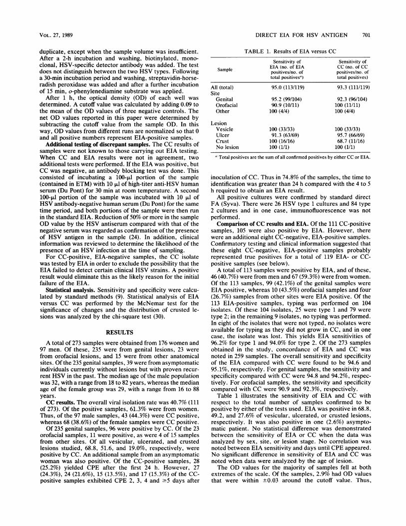
TABLE 1. Results of EIA versus CC
Sensitivity of Sensitivity of
Sample EIA (no. of EIA CC (no. of CCpositives/no. of positives/no. oftotal positives") total positives)
All (total) 95.0 (113/119) 93.3 (111/119)Site
Genital 95.2 (99/104) 92.3 (96/104)Orofacial 90.9 (10/11) 100 (11/11)Other 100 (4/4) 100 (4/4)
LesionVesicle 100 (33/33) 100 (33/33)Ulcer 91.3 (63/69) 95.7 (66/69)Crust 100 (16/16) 68.7 (11/16)No lesion 100 (1/1) 100 (1/1)
" Total positives are the sum of all confirmed positives by either CC or EIA.
inoculation of CC. Thus in 74.8% of the samples, the time toidentification was greater than 24 h compared with the 4 to 5h required to obtain an EIA result.
All positive cultures were confirmed by standard directFA (Syva). There were 26 HSV type 1 cultures and 84 type2 cultures and in one case, immunofluorescence was notperformed.Comparison of CC results and EIA. Of the 111 CC-positive
samples, 105 were also positive by EIA. However, therewere an additional eight CC-negative, EIA-positive samples.Confirmatory testing and clinical information suggested thatthese eight CC-negative, EIA-positive samples probablyrepresented true positives for a total of 119 EIA- or CC-positive samples (see below).A total of 113 samples were positive by EIA, and of these,
46 (40.7%) were from men and 67 (59.3%) were from women.Of the 113 samples, 99 (42.1%) of the genital samples wereEIA positive, whereas 10 (43.5%) orofacial samples and four(26.7%) samples from other sites were EIA positive. Of the113 EIA-positive samples, typing was performed on 104isolates. Of these 104 isolates, 25 were type 1 and 79 weretype 2; in the remaining 9 isolates, no typing was performed.In eight of the isolates that were not typed, no isolates wereavailable for typing as they did not grow in CC, and in onecase, the isolate was lost. This yields EIA sensitivities of96.2% for type 1 and 94.0% for type 2. Of the 273 samplesobtained in the study, concordance of EIA and CC wasnoted in 259 samples. The overall sensitivity and specificityof the EIA compared with CC were found to be 94.6 and95.1%, respectively. For genital samples, the sensitivity andspecificity compared with CC were 94.8 and 94.2%, respec-tively. For orofacial samples, the sensitivity and specificitycompared with CC were 90.9 and 92.3%, respectively.
Table 1 illustrates the sensitivity of EIA and CC withrespect to the total number of samples confirmed to bepositive by either of the tests used. EIA was positive in 68.8,49.2, and 27.6% of vesicular, ulcerated, or crusted lesions,respectively. It was also positive in one (2.6%) asympto-matic patient. No statistical difference was demonstratedbetween the sensitivity of EIA or CC when the data wasanalyzed by sex, site, or lesion stage. No correlation wasnoted between EIA sensitivity and days until CPE appeared.No significant difference in sensitivity of EIA and CC wasnoted when data were analyzed by the age of lesion.The OD values for the majority of samples fell at both
extremes of the scale. Of the samples, 2.9% had OD valuesthat were within +0.03 around the cutoff value. Thus,
VOL. 27, 1989

702 DASCAL ET AL.
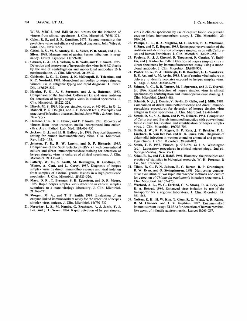
TABLE 2. Clinical and laboratory data on patients in whom discrepant CC and EIA results were noted
Age(days) of Stage EIA on ConfirmatoryDiscrepancY and Age Sex' lesion at Site of Treatment Net OD FA CC super- blocking Clinical datapatient no. (yr) time of lesion type natant EIA
sampling
CC positive, EIAnegative
1 37 M >7 Genital Ulcer None -0.024 2 + NA"' NA2 50 M 5 Genital Ulcer None -0.1 2 + NA NA3 40 F 3 Genital Ulcer None -0.1 2 + NA NA4 21 F 3 Genital Ulcer None -0.041 2 + NA NA5 24 F 9 Genital Ulcer None -0.092 2 + NA NA6 30 M 6 Orafacial Ulcer None -0.07 1 + NA NA
CC negative, EIApositive
1 24 M 3 Genital Crust None 0.177 NA NA Confirmed Known recurrent HSV withclinical episode suggestiveof HSV
2 48 F 7 Genital Ulcer Acyclovir 2.601 NA NA Confirmed Known recurrent HSV withclinical episode suggestiveof HSV
3 34 F >7 Genital Crust None 0.139 NA NA Confirmed Clinical episode suggestiveof HSV
4 26 M 4 Genital Crust None 0.757 NA NA Confirmed Clinical episode suggestiveof HSV; sexual partnerHSV positive
5 34 M >7 Genital Crust None 3.009 NA NA Confirmed Known recurrent HSV withclinical episode suggestiveof HSV
6 35 M 2 Genital Crust None 0.23 NA NA Confirmed Clinical episode suggestiveof HSV
7 31 M 4 Genital Crust None 0.245 NA NA Confirmed Known recurrent HSV withclinical episode suggestiveof HSV
8 48 M 3 Genital Crust None 2.988 NA NA Confirmed Known recurrent HSV withclinical episode suggestiveof HSV
M, Male; F. female."NA, Not applicable.
EIA-positive and -negative results were clearly distin-guished. Discrepant results were not clustered near the EIAcutoff value. Specifically, 78.1% of positive, vesicular le-sions and 60.3% of positive, ulcerated lesions had OD valuesof -2.0 compared with only 31.2% of positive, crustedlesions.
Discrepant results. Table 2 presents a detailed analysis ofthe 14 discrepant samples. There were six cases which wereCC positive but EIA negative. In all of these cases, theculture supernatant was positive on testing by EIA, suggest-ing that the failure of the direct assay is due to a reason otherthan lack of recognition of the particular clinical HSV strain.All six CC-positive EIA-negative samples were typed byFA; five were type 2, and one was type 1.Another group of discrepant results consisted of eight
CC-negative, EIA-positive samples. Table 2 shows the rel-evant clinical information and results of the blocking confir-matory EIA tests. Except for the one patient on acyclovir,all samples in this category were obtained from crustedlesions. This was significantly different from the total CC-positive sample population (P < 0.05). In six of these eightcases, the patient or the sexual partner had a previousculture-proven HSV infection. In the remaining two cases,the clinical history and physical exam strongly suggestedHSV infection as the diagnosis (Table 2).
Effect of acyclovir therapy. Eleven patients in the study
received acyclovir treatment. Four patients, three on sys-temic and one on topical acyclovir, were positive for HSVby both EIA and CC. One patient on systemic therapy wasEIA positive only. Six patients, four on systemic and two ontopical therapy, were HSV negative by both tests.
DISCUSSION
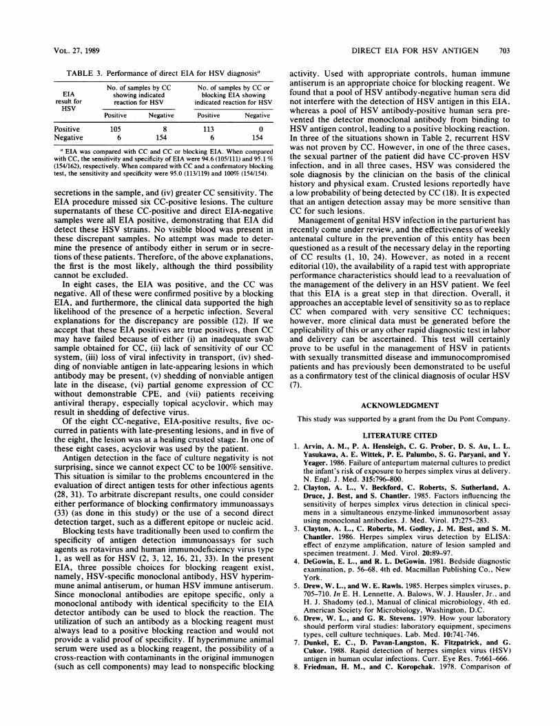
Our overall HSV isolation rate of 40.7% and specific ratesof 68.8, 51.6, and 19.0% for vesicular, ulcerated, and crustedlesions, respectively, compare favorably with previous re-ports on similar patient populations, indicating that our viralculture system had appropriate sensitivity (3, 12, 15, 18, 20,23). If the 'gold standard" is CC isolation, the overallsensitivity and specificity of the EIA, irrespective of theconfirmatory procedure, are 94.6% (105 of 111) and 95.1%,respectively. Taking the confirmatory tests into account, wehad a sensitivity of 95.0% (113 of 119) and a specificity of100% (154 of 154) (Table 3). Exclusion of the patientsreceiving acyclovir therapy did not alter the sensitivity andspecificity of this EIA.
Theoretical explanations to account for CC-positive, EIA-negative results include the following: (i) an inadequate swabsample obtained for EIA, (ii) failure of the monoclonalantibody to detect all HSV strains, (iii) interference in theassay by patient-derived HSV antibody present in blood or
J. CLIN. MICROBIOL.
DIRECT EIA FOR HSV ANTIGEN 703
TABLE 3. Performance of direct EIA for HSV diagnosis"
No. of samples by CC No. of samples by CC orEIA showing indicated blocking EIA showing
result for reaction for HSV indicated reaction for HSVHSV
Positive Negative Positive Negative
Positive 105 8 113 0Negative 6 154 6 154
a EIA was compared with CC and CC or blocking EIA. When comparedwith CC, the sensitivity and specificity of EIA were 94.6 (105/111) and 95.1 %(154/162), respectively. When compared with CC and a confirmatory blockingtest, the sensitivity and specificity were 95.0 (113/119) and 100% (154/154).
secretions in the sample, and (iv) greater CC sensitivity. TheEIA procedure missed six CC-positive lesions. The culturesupernatants of these CC-positive and direct EIA-negativesamples were all EIA positive, demonstrating that EIA diddetect these HSV strains. No visible blood was present inthese discrépant samples. No attempt was made to deter-mine the presence of antibody either in serum or in secre-tions of thèse patients. Therefore, of the above explanations,the first is the most likely, although the third possibilitycannot be excluded.
In eight cases, the EIA was positive, and the CC wasnegative. All of these were confirmed positive by a blockingEIA, and furthermore, the clinical data supported the highlikelihood of the presence of a herpetic infection. Severalexplanations for the discrepancy are possible (12). If weaccept that these EIA positives are true positives, then CCmay have failed because of either (i) an inadequate swabsample obtained for CC, (ii) lack of sensitivity of our CCsystem, (iii) loss of viral infectivity in transport, (iv) shed-ding of nonviable antigen in late-appearing lesions in whichantibody may be present, (v) shedding of nonviable antigenlate in the disease, (vi) partial genome expression of CCwithout demonstrable CPE, and (vii) patients receivingantiviral therapy, especially topical acyclovir, which mayresult in shedding of defective virus.Of the eight CC-negative, EIA-positive results, five oc-
curred in patients with late-presenting lesions, and in five ofthe eight, the lesion was at a healing crusted stage. In one ofthese eight cases, acyclovir was used by the patient.
Antigen detection in the face of culture negativity is notsurprising, since we cannot expect CC to be 100% sensitive.This situation is similar to the problems encountered in theevaluation of direct antigen tests for other infectious agents(28, 31). To arbitrate discrepant results, one could considereither performance of blocking confirmatory immunoassays(33) (as done in this study) or the use of a second directdetection target, such as a different epitope or nucleic acid.
Blocking tests have traditionally been used to confirm thespecificity of antigen detection immunoassays for suchagents as rotavirus and human immunodeficiency virus type1, as well as for HSV (2, 3, 12, 16, 21, 33). In the presentEIA, three possible choices for blocking reagent exist,namely, HSV-specific monoclonal antibody, HSV hyperim-mune animal antiserum, or human HSV immune antiserum.Since monoclonal antibodies are epitope specific, only amonoclonal antibody with identical specificity to the EIAdetector antibody can be used to block the reaction. Theutilization of such an antibody as a blocking reagent mustalways lead to a positive blocking reaction and would notprovide a valid proof of specificity. If hyperimmune animalserum were used as a blocking reagent, the possibility of across-reaction with contaminants in the original immunogen(such as cell components) may lead to nonspecific blocking
activity. Used with appropriate controls, human immuneantiserum is an appropriate choice for blocking reagent. Wefound that a pool of HSV antibody-negative human sera didnot interfere with the detection of HSV antigen in this EIA,whereas a pool of HSV antibody-positive human sera pre-vented the detector monoclonal antibody from binding toHSV antigen control, leading to a positive blocking reaction.In three of the situations shown in Table 2, recurrent HSVwas not proven by CC. However, in one of the three cases,the sexual partner of the patielit did have CC-proven HSVinfection, and in all three cases, HSV was considered thesole diagnosis by the clinician on the basis of the clinicalhistory and physical exam. Crusted lesions reportedly havea low probability of being detected by CC (18). It is expectedthat an antigen detection assay may be more sensitive thanCC for such lesions.Management of genital HSV infection in the parturient has
recently come under review, and the effectiveness of weeklyantenatal culture in the prevention of this entity has beenquestioned as a result of the necessary delay in the reportingof CC results (1, 10, 24). However, as noted in a recenteditorial (10), the availability of a rapid test with appropriateperformance characteristics should lead to a reevaluation ofthe management of the delivery in an HSV patient. We feelthat this EIA is a great step in that direction. Overall, itapproaches an acceptable level of sensitivity so as to replaceCC when compared with. very sensitive CC techniques;however, more clinical data must be ,generated before theapplicability of this or any other rapid diagnostic test in laborand delivery can be ascertained. This test will certainlyprove to be useful in the management of HSV in patientswith sexually transmitted disease and immunocompromisedpatients and has previously been demonstrated to be usefulas a confirmatory test of the clinical diagnosis of ocular HSV(7).
ACKNOWLEDGMENT
This study was supported by a grant from the Du Pont Company.
LITERATURE CITED1. Arvin, A. M., P. A. Hensleigh, C. G. Prober, D. S. Au, L. L.
Yasukawa, A. E. Wittek, P. E. Palumbo, S. G. Paryani, and Y.Yeager. 1986. Failure of antepartum maternal cultures to predictthe infant's risk of exposure to herpes simplex virus at delivery.N. Engl. J. Med. 315:796-800.
2. Clayton, A. L., V. Beckford, C. Roberts, S. Sutherland, A.Druce, J. Best, and S. Chantier. 1985. Factors influencing thesensitivity of herpes simplex virus detection in clinical speci-mens in a simultaneous enzyme-linked immunosorbent assayusing monoclonal antibodies. J. Med. Virol. 17:275-283.
3. Clayton, A. L., C. Roberts, M. Godley, J. M. Best, and S. M.Chantler. 1986. Herpes simplex virus detection by ELISA:effect of enzyme amplification, nature of lesion sampled andspecimen treatment. J. Med. Virol. 20:89-97.
4. DeGowin, E. L., and R. L. DeGowin. 1981. Bedside diagnosticexamination, p. 56-68, 4th ed. Macmillan Publishing Co., NewYork.
5. Drew, W. L., and W. E. Rawls. 1985. Herpes simplex viruses, p.705-710. In E. H. Lennette, A. Balows, W. J. Hausler, Jr., andH. J. Shadomy (ed.), Manual of clinical microbiology, 4th ed.American Society for Microbiology, Washington, D.C.
6. Drew, W. L., and G. R. Stevens. 1979. How your laboratoryshould perform viral studies: laboratory equipment, specimenstypes, cell culture techniques. Lab. Med. 10:741-746.
7. Dunkel, E. C., D. Pavan-Langston, K. Fitzpatrick, and G.Cukor. 1988. Rapid detection of herpes simplex virus (HSV)antigen in human ocular infections. Curr. Eye Res. 7:661-666.
8. Friedman, H. M., and C. Koropchak. 1978. Comparison of
VOL. 27, 1989
704 DASCAL ET AL.
WI-38, MRC-5, and IMR-90 cell strains for the isolation ofviruses from clinical specimens. J. Clin. Microbiol. 7:368-371.
9. Galen, R. S., and S. R. Gambino. 1975. Beyond normality: thepredictive value and efficacy of medical diagnosis. John Wiley &Sons, Inc., New York.
10. Gibbs, R. S., M. S. Amstey, R. L. Sweet, P. B. Mead, and J. L.Silver. 1988. Management of genital herpes infections in preg-nancy. Obstet. Gynecol. 71:779-780.
11. Gleaves, C. A., D. J. Wilson, A. D. Wold, and T. F. Smith. 1985.Detection and serotyping of herpes simplex virus in MRC-5 cellsby the use of centrifugation and monoclonal antibodies 16 hpostinoculation. J. Clin. Microbiol. 21:29-32.
12. Goldstein, L. C., L. Corey, J. K. McDougall, E. Tolentino, andR. C. Nowinski. 1983. Monoclonal antibodies to herpes simplexviruses: use in antigenic typing and rapid diagnosis. J. Infect.Dis. 147:829-837.
13. Hayden, F. G., A. S. Sorenson, and J. A. Bateman. 1983.Comparison of the lmmulok Cultureset kit and virus isolationfor detection of herpes simplex virus in clinical specimens. J.Clin. Microbiol. 18:222-224.
14. Hirsch, M. S. 1985. Herpes simplex virus, p. 945-951. In G. L.Mandell, P. G. Dot4glas, and J. E. Bennett (ed.), Principles andpractice of infectious diseases, 2nd ed. John Wiley & Sons, Inc.,New York.
15. Huntoon, C. S., R. F. House, and T. F. Smith. 1981. Recovery ofviruses from three transport media incorporated into cultur-ettes. Arch. Pathol. Lab. Med. 105:436-437.
16. Jackson, B. J., and H. H. Balfour, Jr. 1988. Practical diagnostictesting for human immunodeficiency virus. Clin. Microbiol.Rev. 1:124-138.
17. Johnson, F. B., R. W. Leavitt, and D. F. Richards. 1985.Comparison of the Scott Selecticult-HSV kit with conventionalculture and direct immunoperoxidase staining for detection ofherpes simplex virus in cultures of clinical specimens. J. Clin.Microbiol. 21:438-441.
18. Lafferty, W. E., S. Krofft, M. Remington, R. Giddings, C.Winter, A. Cent, and L. Corey. 1987. Diagnosis of herpessimplex virus by direct immunofluorescence and viral isolationfrom samples of external genital lesions in a high-prevalencepopulation. J. Clin. Microbiol. 25:323-326.
19. Mayo, D. R., T. Brennan, S. H. Egbertson, and D. R. Moore.1985. Rapid herpes simplex virus detection in clinical samplessubmitted to a state virology laboratory. J. Clin. Microbiol.21:768-771.
20. Morgan, M. A., and T. F. Smith. 1984. Evaluation of anenzyme-linked immunosorbent assay for the detection of herpessimplex virus antigen. J. Clin. Microbiol. 19:730-732.
21. Nerurkar, L. S., M. Namba, G. Brashears, A. J. Jacob, Y. J.Lee, and J. L. Sever. 1984. Rapid detection of herpes simplex
virus in clinical specimens by use of capture biotin streptavidinenzyme-linked immunosorbent assay. J. Clin. Microbiol. 20:109-114.
22. Phillips, L. E., R. A. Magliolo, M. L. Stehlik, P. A. Whiteman,S. Faro, and T. E. Rogers. 1985. Retrospective evaluation of theisolation and identification of herpes simplex virus with Culture-set and human fibroblasts. J. Clin. Microbiol. 22:255-258.
23. Pouletty, P., J. J. Chomel, D. Thouvenot, F. Catalan, V. Rabil-Ion, and J. Kadouche. 1987. Detection of herpes simplex virus indirect specimens by immunofluorescence assay using a mono-clonal antibody. J. Clin. Microbiol. 25:958-959.
24. Prober, C. G., P. A. Hensleigh, F. D. Boucher, L. L. Yasukawa,D. S. Au, and A. M. Arvin. 1988. Use of routine viral cultures atdelivery to identify neonates exposed to herpes simplex virus.N. Engl. J. Med. 318:887-891.
25. Salmon, V. C., R. B. Turner, M. J. Sperenza, and J. C. Overall,Jr. 1986. Rapid detection of herpes simplex virus in clinicalspecimens by centrifugation and immunoperoxidase staining. J.Clin. Microbiol. 23:683-686.
26. Schmidt, N. J., J. Dennis, V. Devlin, D. Gallo, and J. Mills. 1983.Comparison of direct immunofluorescence and direct immuno-peroxidase procedures for detection of herpes simplex virusantigen in lesion specimens. J. Clin. Microbiol. 18:445-448.
27. Sewell, D. S., S. A. Horn, and P. W. Dilbeck. 1984. Comparisonof Cultureset and Bartels immunodiagnostics with conventionaltissue culture for isolation and identification of herpes simplexvirus. J. Clin. Microbiol. 19:705-706.
28. Smith, J. W., R. F. Rogers, B. P. Katz, J. F. Brickler, P. L.Lineback, B. Van Der Pol, and R. B. Jones. 1987. Diagnosis ofchlamydial infection in women attending antenatal and gyneco-logic clinics. J. Clin. Microbiol. 25:868-872.
29. Smith, T. F. 1985. Viruses, p. 537-624. In J. A. Washington(ed.), Laboratory procedures in clinical microbiology, 2nd ed.Springer-Verlag, New York.
30. Sokal, R. R., and F. J. Rohlf. 1969. Biometry: the principles andpractice of statistics in biological research. W. H. Freeman &Co., San Francisco.
31. Tilton, R. C., F. N. Judson, R. C. Barnes, R. P. Grunninger,R. W. Ryan, and O. Steingrimsson. 1988. Multicenter compar-ative evaluation of two rapid microscopic methods and culturefor detection of Chlainvdia trachomnatis in patient specimens. J.Clin. Microbiol. 26:167-170.
32. Warford, A. L., W. G. Eveland, C. A. Strong, R. A. Levy, andK. A. Rekrut. 1984. Enhanced virus isolation by use of thetransporter for a regional laboratory. J. Clin. Microbiol. 19:561-562.
33. Yolken, R. H., H. W. Kim, T. Clem, R. G. Wyatt, A. R. Kalica,R. M. Chanock, and A. Z. Kapikian. 1977. Enzyme-linkedimmunosorbent assay (ELISA) for detection of human reovirus-like agent of infantile gastroenteritis. Lancet ii:263-267.
J. CLIN. MICROBIOL.