diagnosisofalcoholiccirrhosiswiththeright-to...
TRANSCRIPT
Various scintigraphic criteria for cirrhosis have beenproposed, including hepatomegaly, heterogeneous up
take, splenomegaly, bone-marrow or pulmonary uptake,early hepatic perfusion, and a high spleen-to-liver uptakeratio (1—5). However, none of these criteria have shown
a high degree of specificity on sensitivity for cirrhosis.Obviously, a highly sensitive and specific scintignaphiccriterion for cirrhosis would be very useful in determiningwhether a patient should have further diagnostic tests,which could include invasive procedures, such as pentoneoscopy on liven biopsy.
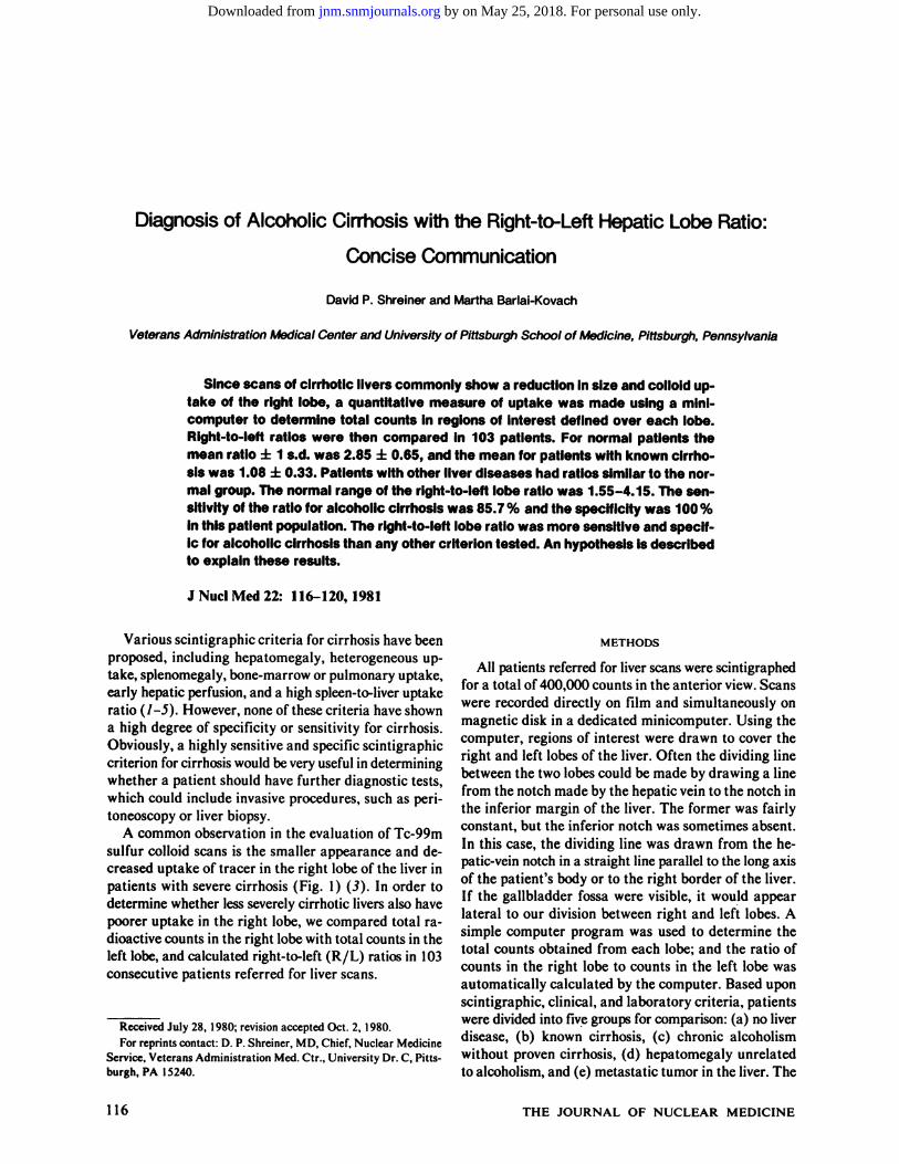
A common observation in the evaluation of Tc-99msulfur colloid scans is the smaller appearance and decreased uptake of tracer in the right lobe of the liven inpatients with severe cirrhosis (Fig. 1) (3). In order todetermine whether less severely cirrhotic livers also havepoorer uptake in the right lobe, we compared total radioactive counts in the night lobe with total counts in theleft lobe, and calculated right-to-left (R/L) ratios in 103consecutive patients referred for livenscans.
ReceivedJuly28,1980;revisionacceptedOct.2, 1980.For reprints contact: D. P. Shreiner, MD, Chief, Nuclear Medicine
Service,VeteransAdministration Med.Ctr., University Dr. C, Pittsburgh,PA 15240.
METHODS
All patients referred for livenscans were scintignaphedfor a total of 400,000 counts in the anterior view. Scanswere recorded directly on film and simultaneously onmagnetic disk in a dedicated minicomputer. Using the
computer, regions of interest were drawn to coven thenight and left lobes of the liver. Often the dividing linebetween the two lobes could be made by drawing a linefrom the notch made by the hepatic vein to the notch inthe inferior margin of the liven. The former was fairlyconstant, but the inferior notch was sometimes absent.In this case, the dividing line was drawn from the hepatic-vein notch in a straight line parallel to the long axisof the patient's body on to the night borden of the liver.If the gallbladder fossa were visible, it would appearlateral to our division between night and left lobes. Asimple computer program was used to determine the
total counts obtained from each lobe; and the ratio ofcounts in the right lobe to counts in the left lobe wasautomatically calculated by the computer. Based uponscintignaphic, clinical, and laboratory criteria, patientswere divided into five groups for comparison: (a) no liverdisease, (b) known cirrhosis, (c) chronic alcoholism
without proven cirrhosis, (d) hepatomegaly unrelatedto alcoholism, and (e) metastatic tumor in the liver. The
116 THE JOURNAL OF NUCLEAR MEDICINE
Diagnosis of AlcoholicCirrhosis with the Right-to--LeftHepatic Lobe Ratio:
Concise Communication
David P. Shrelner and Martha Barlal-Kovach
VeteransAdministration Medical Center and University of PfttsburghSchool of Medicine, Pittsburgh,Pennsylvania
Sincescansof cirrhotIcliverscommonlyshowa reductioninsize andcollolduptake of the right lobe, a quantitativemeasureof uptake was made usinga minicomputerto determinetotal countsin regionsof Interestdefinedover each lobe.Right-to-leftratios were then compared in 103 patIents.For normalpatientsthemean ratio ±I s.d. was 2.85 ±0.65, and the mean for patients with known cirrhosiswas t08 ±0.33. Patientswfthotherliverdiseaseshadratiossimilarto the normal group.The normalrangeof the right-to-leftloberatiowas 1.55-4.15. The sensitIvftyof the ratiofor alcoholiccirrhosiswas85.7% andthespecificitywas100%inthispatientpopulation.The right-to-leftloberatiowas moresensitiveandspecific foralcoholiccirrhosisthananyothercrfteriontested.Anhypothesisisdescribedto explainthese results.
I NuciMed22: 116—120,1981
by on May 25, 2018. For personal use only. jnm.snmjournals.org Downloaded from

Crfterion Sensitivity Specificity
ClINICAL SCIENCES
DIAGNOSTIC NUCLEAR MEDICINE
FIG. 1. Anterior liver-spleen scan (Ic-SC) of a patient with severecirrhosis.Uptakeof colloid is markedlyreducedInri@ithepaticlobe,and left lobe Is similar in size to right. Splenomegalyand intensesplenic uptake are also evident.
range of normal values was calculated as the mean ±2
s.d. of the group with no liver disease. The sensitivity[true positives + (true positives + false negatives)] wasdetermined from the group of cirrhotic patients. Thespecificity [true negatives —(true negatives + falsepositives)] was determined from the groups with nohistory of alcoholism, i.e., no liver disease, nonalcoholichepatomegaly, and metastatic tumor.
Liven biopsy was performed on all patients classified
as “knowncirrhosis,―except for two patients. These twohad overwhelming historical, clinical, and laboratoryevidence of alcoholic cirrhosis and were included in thegroup of known cinrhotics. When indicated biopsy wasdone with a Menghini needle, and the pathologic diag
nosis of “micronodularcirrhosis―was the criterion forinclusion in the group of known cirrhotics. Furthermore,a history of alcohol abuse was obtained from every patient in this group. Most of the patients in the nonalco
holic groups, and all patients in the group ofchnonic alcoholics without proven cirrhosis, did not have liver biopsy.
Seven blood tests (serum albumin, BUN, bilinubin,cholesterol, LDH, SGOT, SGPT) were made to provide
evidence of liver disease by assigning a unit value of 1 foreach abnormal test. Thus, if every test were abnormal,the total for that patient would be seven. If no test wereabnormal, a value of 0 would be assigned. Tests considered suggestive of cirrhosis were labeled positive whenthe serum albumin, BUN, on cholesterol were low, andwhen the bilirubin, LDH, SGOT, or SGPT were high.
Other criteria for cirrhosis that were studied included:(a) splenomegaly (spleen size >80 cm2 by scan), (b)early perfusion of the liver in the arterial phase of thenadionuclide angiogram, (c) heterogeneous uptake ofcolloid, (d) extnahepatic uptake (pulmonary or bonemarrow) of colloid, and (e) the spleen-to-liver uptakeratio. The spleen-to-liver ratio was determined by visual
inspection of posterior liver-spleen scans; it was considered abnormal if the count density in the spleen wasgreater than that in the liver.
RESULTS
Figure 2 is a bar graph showing the mean R/L ratio
NO LIVER CHRONIC HEPATOMEGALY METASTATICCIRRHOSIS ALCOHOLISM UNRELATEDTO TUMORDCSEASE
ALCOHOL
0C/)
+1U)0
0
>
z
FIG.2. Bar graph showing mean A/Lhepatlc lobe ratios for fivegroups of patIents. Numbersof patients (N)are indicated at baseof eachbar.NotethatmeanRILratiofor thegroupwithcirrhosisis signIfIcantly lower than for any other group (p < 0.0005).
±s.d.forthefivegroupsof patients.ThemeanR/L forpatients with no livendisease was 2.85 ±0.65, whereasin cirrhosis it was 1.08 ±0.33. The latter value was significantly lower than normal (p < 0.0005). The mean
R/L ratios of the other groups were not statisticallydifferent from normal.
For the purpose of determining the normal R/L ratio,the mean range for the R/L ±2 s.d. in the group withno liver disease was calculated as 1.55—4.15. Using thisrange as normal, the sensitivity of the R/L ratio test was
85.7%; the specificity was 100% (Table 1). Only threepatients with proven alcoholic cirrhosis had R/L ratiosin the normal range; two of these had ratios near thelower limit of normal and the third had had a porta-caval
shunt performed 16 yr previously, had abstained fromethanol, and had minimal evidence of cirrhosis at autopsy. If this patient were excluded from the group of
known cinrhotics, the sensitivity of the R/L ratio testwould be 90%.
The mean R/L ratio for the group with chronic al
TABLE1. CRITERIAFOR ALCOHOLICCIRRHOSIS
1. AlL hepaticloberatio2. Pulmonaryor marrow
uptake3. Splenomegaly4. Earlyhepatlcperfusion5. Bloodtestsofhepatlc
function6. Spleen/lIverratio
85.7% 100%81%(44%) 98.2%
57.1%47.6%52.4% 93%
52.4% 96.4%(81.3%)' (52.6%)'
‘NumbersInparenthesesreferto resultsbyotherInvestlgators(seetext).
Volume 22, Number 2 117
by on May 25, 2018. For personal use only. jnm.snmjournals.org Downloaded from

SHREINER AND BARLAI-KOVACH
found in two patients with hepatomegaly unrelated toalcoholism; this may represent a specificity of 96.4%.
DISCUSSION
The R/L hepatic lobe ratio has been shown to behighly specific (100%) and very sensitive (85.7%) foralcoholic cirrhosis. Except for liver biopsy, no othercombination of tests has this degree of specificity andsensitivity. The next-best criterion in this study was thepresence of pulmonary or marrow uptake, with a sensitivity of 81% for cirrhosis and a specificity of almost100%(Table 1).However,Prakash et al. founda sensitivity of only 44% for increased bone marrow uptake(Table 1) (4).
In our study the spleen-to-liver ratio had a sensitivityof only 52.4% for cirrhosis and a specificity of 96.4%(Table 1). Wilson and Keyes found a sensitivity of 81.3%and a specificity of 52.6% for the spleen-to-liver ratio(5).
The presence of splenomegaly by scan, on of earlyhepatic perfusion on the angiogram, were both of lowsensitivity for cirrhosis (<60%). Although calculationsof sensitivity and specificity were not done for heterogeneity ofuptake and hepatomegaly, these findings occurso frequently in liven scans that specificity must below.
In this study, seven blood tests of hepatic function wereevaluated. Since blood tests are not specific for hepaticdysfunction, each abnormal test was given a unit valueof 1and a laboratory “score―was obtained for each patient. Thus, while most patients with cirrhosis had atleast one abnormal blood test, many patients withouthepatic dysfunction also had abnormal tests. Patientswith cirrhosis tended to have more abnormal tests(higher scores) than others, but the sensitivity of thescore test for cirrhosis was only 52.4% (Table 1). It ispossible that some other combination of blood testswould be more sensitive—e.g., bilirubin > 10 mg%,serum albumin <2 g%, etc. If such a combination exists,more sensitive and specific for cirrhosis, the formula hasnot been recognized and is not in use clinically. The scoreused in this study took no account of more- on less-severedegrees of test abnormality, but considered the test asmerely normal on abnormal.
Among the population of patients at our medicalcenter, a decreased R/L ratio was highly specific foralcoholic cirrhosis. One patient with postnecrotic cmrhosis had a normal R/L ratio. Only three patients withproven alcoholic cirrhosis had normal R/L ratios, twonear the lower limit of normal. The third, with theporta-caval shunt and 16 yr of abstention, had onlyminimal evidence of cirrhosis at autopsy. This suggests
that reversal of cirrhosis and hepatic regeneration canoccur if ethanol consumptionis stopped.
We considered the possibility that a low R/L ratio
FIG.3. AnterIorliverscanof a patientwithprovenalcoholiccirrhosls, showing no obvious sclntI@'aphlccrfteria for cirrhosis, butwhohadalowR/Lratioof 1.05.
coholism without biopsy-proven cirrhosis was 2.42 (Fig.2); this lies within the normal range, but is the closestmean to the group with cirrhosis. This group includedthree patients with R/L ratios in the cirrhotic range(<1 .55), and, although liven biopsy was not performed,they probably had cirrhosis as judged by clinical andlaboratory criteria.
Two other patients were of interest because they hadno obvious scintigraphic criteria for cirrhosis, except anabnormally low R/L ratio. (Figure 3 shows the liver scanof one of these patients.) Both of them had alcoholiccirrhosis proven by liven biopsy and were included in thegroup of patients with known cirrhosis. One patient hadknown postnecnotic cirrhosis, with a normal R/L ratio.He had no history of alcoholism and was not includedin this study.
Since there is no single blood test, on combination ofthem, diagnostic of cirrhosis, seven liven function testswere used to assign a value of 0-7 to each patient in thisstudy (see Methods section). The mean value for thecirrhotic group was 3.5 ± 1 .8, and the mean values for
the nonalcoholic and no-liven-disease groups were L8 ±1.4 in each case. Thus, there was considerable overlapin the standard deviations of the means. If the normalrange is again assigned as the mean ±2 s.d. for the groupwith no liver disease, the “normal―range is 0—4.6;and11 of the 21 known cirrhotic patients would fall in the“normal―range for blood tests of liver function. Thus,the sensitivity of the blood tests was only 52.4%, and thespecificity was 93% (Table 1).
Splenomegaly by scan was found in 12 of 21 cirrhoticpatients. Sensitivity for this test was only 57.1%. Earlyperfusion of the liver on the nuclide angiogram was foundin ten of 21 cirrhotic patients, with a sensitivity of only47.6% for this test. Pulmonary on bone-marrow uptakeof the tracer occurred in 17 of 21 patients, with a sensitivity of 81.0%. One patient with nonalcoholic hepatomegaly of unknown cause also had pulmonary or marrowuptake; this may represent a specificity of9&i%. Elevenof 21 cirrhotic patients had high spleen-to-liven ratios(sensitivity = 52.4%). A high spleen-to-liver ratio was
118 THE JOURNAL OF NUCLEAR MEDICINE
by on May 25, 2018. For personal use only. jnm.snmjournals.org Downloaded from

CLINICAL SCIENCESDIAGNOSTIC NUCLEAR MEDICINE
regions of interest oven right and left lobes was considered, since anatomic markings are not always clear onliver scans. The right lobe is anatomically subdivided into
caudate and quadrate lobes, which are much smallerthan the remainder of the right lobe and occupy a position between the gallbladder fossa and the falciformligament. Anatomically, the division between the night
and left lobes is a line drawn from the notch of the he
patic vein along the medial borden of the porta hepatis.The division of the liver into night and left lobes, ascommonly used, does not correspond precisely with theportal venous circulation. The left portal vein suppliesthe anatomic left lobe, caudate lobe, and quadrate lobe;the right portal branch supplies the anatomic night lobeexclusive of the caudate and quadnate lobes.
Thus our arbitrary division of the liver scan into“night―and “left―lobes might well work against ourfinding of a low R/L in cirrhosis. Nevertheless, we havenot been able to change the results enough to alter theR/L ratio significantly. Any error one would be likelyto make would tend to raise the R/L ratio, since the dividing line would have to be drawn more laterally thanthe gallbladder fossa to decrease the ratio falsely. Furthermone, normal variations in liver structure may indude hypoplasia or aplasia of the left lobe, but the rightlobe is rarely, if ever, smaller than the left in congenitalvariations. Thus, it is possible to find a high R/L due to
normal variations, but a low ratio due to normal variation
would be most unlikely.In conclusion, a low R/L hepatic lobe ratio is highly
specific for alcoholic cirrhosis and is also very sensitive.In some patients a low R/L ratio may be the only scmtignaphic evidence of cirrhosis. Since the R/L ratio canbe obtained so easily from routine liver scans in anylaboratory with a modest computer, the test is necommended as a screening procedure for alcoholic cirrhosisbefore consideration for liver biopsy.
Studies are in progress to determine the distributionof labeled alcohol in the livenafter ingestion and to detenmine whether “streamlining―of blood flow occurs inthe portal vein in humans.
REFERENCES
1. BOYDRO, STADALNIK RC, BARNETr CA, et al: Quantitative hepatic scintiangiography. Clin Nucl Med 3: 478-484,1978
2. DRUMDE,BEARDJO:Liverscintigraphicfeaturesassociatedwith alcoholism. I Nucl Med 19: 154- 160, 1978
3. SIEGELBA, ed:Nuclear Radiology (secondseries),Chicago,TheAmericanCollegeof Radiology,1978,p 448
4. PRAKASHV, UN MS. KRISSJP: Liver scintigraphy in alcoholic liver disease.Clin Nucl Med 2: 308—309,1977
5. WILsoN GA, KEYES JW, JR: The significance of the liverspleenuptakeratio in liver scanning.J NucI Med 15:593-597,1974
6. KELLER M, SIROTA5, eds:Alcoholand Health: ReportfromtheSecretaryofHealth, Education,and Welfare.ChapterIV,New York, Charles Seribner's Sons, 1972,pp 113-1 14
____BLOODOFHIGHALCOHOL CONTENT
—BLOODOFLOWALCOHOL CONTENT
\1
PORTALVEIN \1@
SUPERIOR @.LI ‘@MESENTERIC —@-@ T@
VEIN @JJ
I iI I @n@ N1/ VEIN
I 1/I/
FiG.4. Portalcirculationof liver suggesting“streamlInIng―of bloodflow Inportalvein.Low-alcoholbloodfromspleen,stomach,andlargebowel tendsto “streamline―to the left, whereashI@-alcohoIblood from small intestine(superIormesenterlcveIn)tendstowardthe [email protected],bloodof hl@ieralcoholcontenttendsto bedistributedto rightportalbranchesandrighthepaticlobe.
could be caused by ascites, since ten of the 21 known
cirrhotic patients had ascites. The mean R/L ratio forthese patients was 1.18 ±0.35 (s.d.), and the mean R/Lfor the 11 patients without ascites was 0.99 ±0.31. Thisdifference is not statistically significant and suggests that
ascites did not account for the low R/L ratio.Other patients with liver disease should be studied to
determine whether this high specificity for alcoholiccirrhosis is substantiated. Diseases not available for studyin our investigation included biliary cirrhosis, chronicactive hepatitis, acute viral hepatitis, and c@ntainotheruncommon disorders.
An hypothesis was developed to explain the low R/Lratio in alcoholic cirrhosis. Figure 4 shows a diagram ofthe portal circulation and liven. After ingestion, most ofthe alcohol rapidly passes the pylorus and is absorbedfrom the small intestine (6). Blood high in alcohol iscarried from the small intestine through the superiormesenteric vein to the portal vein. “Streamlining―ofblood flow may occur at the junction of the superiormesentenic and inferior mesentenic, splenic, or gastricveins coming from the left side of the body, such thathigh-alcohol blood is “streamlined―to the right, whereasbloodoflowalcoholcontentfromthe inferiormesenteric,splenic, and gastric veins is “streamlined―to the left.Thus, more alcohol would be delivered to the right hepatic lobe than to the left. There is convincing experimental evidence for the “streamlining―of blood flow inthe portal vein in some animals (7,8), but this effect hasnot yet been demonstrated in man.
The possibility that errors could be made in defining
R
•4.\ GASTRICI VEINS
SPLENIC@—VEIN
Volume 22, Number 2 119
by on May 25, 2018. For personal use only. jnm.snmjournals.org Downloaded from

SHREINER AND BARLAI-KOVACH
7. HAHNPF,DONALDWD, GRIERRD.JR:Thephysiological 143:105-107,1945bilaterality of the portal circulation. Streamline flow of blood 8. HEATH T: Origin and distribution of portal blood in the sheep.into the liver asshownby radioactivephosphorus.Am J Physiol Am J Anat 122:95-105, 1968
EASTERN GREAT LAKES CHAPTERSOCIETY OF NUCLEAR MEDICINE
SECOND ANNUAL MEETING
May 15, 1981 The Prince of Wales Hotel Niagara-on-the-Lake, Ontario
The EasternGreat LakesChapterof SNM announcesitsSecondAnnual Meetingto be held May 15, 1981at The Princeof Wales Hotel, Niagara-on-the-Lake. Ontario, Canada.
The programwill includecontinuing educationcoursesto begiven by: Dr. MichaelLobergon “HepatobiliaryRadiopharmaceuticalsand Imaging―;Dr.Terry Mandelon “Krypton-81m GasGeneratorand Imaging―;and Dr.GünesEgëon “LymphNode Imaging.―
The technologistprogram will include in-depth lectureson “HepatitisTestingand InfectionControl―by Mr. Tom Diasand “ProcessorQuality Control―by John Blanowicz.
There will also be a presentationof contributed papers.Abstracts,typed single-spaced,not to exceed300words including title, author(s), and address, should be mailed to:
Azu Owunwanne, Ph.D.Univ. of Rochester Medical CenterDiv. of Nuclear Medicine, Box 620
601 Elmwood Ave.Rochester, NY 14642
Dedline for submIssionof abstracts Is March 6, 1981.
MIDEASTERN CHAPTERSOCIETY OF NUCLEAR MEDICINE
11thANNUAL MEETINGApril 9-11,1981 UniformedSsrvlcss Bethsd., Maryland
Univ•rsityof Health Srvic•s
ANNOUNCEMENT AND CALL FOR ABSTRACTS
TheScientificProgramCommitteeoftheMideasternChapteroftheSocietyofNuclearMedicinesolicitsthesubmlssionof abstractsfrom membersand nonmembersofthe Socletyof Nuclear Medicineforthe 1Ith AnnualMeetingtobeheld April 9—I1, 1981 In Bethesda, Maryland. The programwill Includesubmittedpapers, invitedspeakers,teachingsessions,andexhibits.
Abstractsshouldnotexceed300wordsandshouldcontainastatementofpurpose,themethodused,results,andconcluslons.The nameof the author presentingthe papermustbe underlined.
Original abstracts and four copies should be sent to:[email protected],M.D.Div. of Medical Imaging
Dept. of RadiologyBox 486
Univ. of Virginia Medical CenterCharlottesville,VA 22908
TheprogramwillbeapprovedforcredittowardtheAMAPhysician'sRecognitionAwardunderContinuingMedicalEducation,Category 1, through the Societyof Nuclear Medicine.For further Information concerning the program, write or telephone (804) 924-5201 the ProgramChairman listeda-boveor E.U.Buddemeyer,Sc.D.at (301)528-6890.
ABSTRACTSMUSTBERECEIVEDBYFEBRUARY1,1981.
120 THE JOURNAL OF NUCLEAR MEDICINE
by on May 25, 2018. For personal use only. jnm.snmjournals.org Downloaded from

1981;22:116-120.J Nucl Med. David P. Shreiner and Martha Barlai-Kovach CommunicationDiagnosis of Alcoholic Cirrhosis with the Right-to-Left Hepatic Lobe Ratio: Concise
http://jnm.snmjournals.org/content/22/2/116This article and updated information are available at:
http://jnm.snmjournals.org/site/subscriptions/online.xhtml
Information about subscriptions to JNM can be found at:
http://jnm.snmjournals.org/site/misc/permission.xhtmlInformation about reproducing figures, tables, or other portions of this article can be found online at:
(Print ISSN: 0161-5505, Online ISSN: 2159-662X)1850 Samuel Morse Drive, Reston, VA 20190.SNMMI | Society of Nuclear Medicine and Molecular Imaging
is published monthly.The Journal of Nuclear Medicine
© Copyright 1981 SNMMI; all rights reserved.
by on May 25, 2018. For personal use only. jnm.snmjournals.org Downloaded from