dickens, michael (2011) small molecule inhibitors of mdm2
TRANSCRIPT

Small Molecule Inhibitors of Mdm2 E3
Ubiquitin Ligase Activity
Michael Dickens, M.Chem, AMRSC
Thesis submitted to the University of Nottingham
for the degree of Doctor of Philosophy
March 2011

‘All truths are easy to understand once they are
discovered; the point is to discover them’
Galileo Galilei

I
Abstract
‘Small Molecule Inhibitors of Mdm2 E3 Ubiquitin Ligase Activity’
Half of cancers retain wild type p53 but have alterations in the pathwaysinvolved in p53 regulation. Murine double minute 2 (Mdm2) regulates p53 byacting as an E3 ubiquitin ligase, which tags p53 for degradation through theproteasome. A small molecule inhibitor, a 5-deazaflavin analogue, haspreviously been identified by high throughput screening to inhibit Mdm2 E3ubiquitin ligase activity, thereby reactivating apoptotic function of p53selectively in cancer cells.
Ninety 5-deazaflavin analogues have been synthesised by an optimizedexisting method and a novel method of synthesis, using the required 6-anilinouracil and 2-p-toluenesulfonyloxybenzaldehyde.The biological ability ofthe 5-deazaflavin analogues to act as inhibitors of Mdm2 E3 ubiquitin ligaseactivity to reactivate p53 has been ascertained. A new quantitative biologicalassay was developed, by scientists based at the Beatson Institute, for 5-deazaflavin compounds, showing excellent inhibition of Mdm2 E3 ubiquitinligase activity on the previous qualitative biological assay, to yield IC50 data.
The biological results have established a clear and logical structure-activityrelationship comprising of an electron-withdrawing hydrophobic substituent atthe nine position and the N10 phenyl being a prerequisite for activity as aMdm2 inhibitor. Also meta substitution of the N10 phenyl improves activityagainst Mdm2 E3 ubiquitin ligase activity. Hit optimization has occurred with10-(3-chlorophenyl)-9-trifluoromethyl-5-deazaflavin being thirty times moreactive than the previous identified hit compound, 10-(4-chlorophenyl)-7-nitro-5-deazaflavin.
Using the X-ray crystal structure of the Mdm2/MdmX heterodimer, animproved understanding of how Mdm2 acts as an E3 ubiquitin ligase isdescribed and used to form a hypothesis of how 5-deazaflavin analoguesfunction as inhibitors of Mdm2.
The work suggests the principle that small molecular weight compounds caninhibit E3 ubiquitin ligases as a possible anti-cancer therapy, and provide thefoundation and framework for additional studies and investigation in a new anddeveloping field of medicinal chemistry.
Supported by Cancer Research UK

II
Publications
Dickens, M. P.; Fitzgerald, R. F.; Fischer. P. M. Small-molecule inhibitors of
Mdm2 as new anticancer therapeutics. Semin. Cancer Biol. 2010, 20, 10-18

III
Acknowledgements
I would like to thank Prof Fischer and Dr Kellam, my project supervisors, for
their time, help, teaching, guidance and advice given throughout my time as a
PhD student. I would like to thank Prof Fischer’s and Dr Kellam’s research
groups for their practical advice and guidance given in the lab during my time
at the University of Nottingham. Also, I would like to thank my fellow
research colleagues in the University of Nottingham, Centre of Biomolecular
Sciences, C floor labs, for answering any questions I have had. I would like to
thank Chun Law who as a 4th year Pharmacy student, due to his skill in the lab,
allowed me to start writing my thesis.
I would like to express my gratitude to Dr Meagher and Dr Mezna at the
Beatson Institute for Cancer Research for undertaking the biological testing of
the compounds I synthesised. I am indebted to Prof Vousden at the Beatson
Institute for Cancer Research who managed and led the biological research as
well as the testing of my compounds.
I am grateful to Cancer Research UK for funding. I am also indebted to
Newcastle City Library for the use of the internet and the helpfulness of its
staff when writing up my thesis.
I would like to thank everyone who has proof read this thesis. A big thank you
to Charlotte Bell who has patiently helped and encouraged me while I was
writing this thesis. Finally, I would like to thank my friends and family for all
their support given to me over the last few years at University.

IV
Contents
Abstract I
Publications II
Acknowledgements III
Contents IV
Abbreviations X
Figures XIII
Tables XV
Compound Naming XVI
Introduction 1
Cancer 1
What Causes Cancer? 2
p53 4
p53 in Cell Cycle Arrest 6
p53 in Apoptosis 8
p53 in Senescence 9
p53 in Cancer 10
p53 in Other Diseases and Biological Functions 10
p53 Regulation 10
Mdm2 11
Mdm2 E3 Ubiquitin Ligase Activity 13
Other E3 Ubiquitin Ligases to p53 16
26S Proteasome 17
Small Molecule Inhibitors of 26S Proteasomal Degradation
17
Small Molecule Inhibitors of Mdm2 19
Inhibitors of Mdm2/p53 Protein-Protein Interaction 20
Mdm2 E3 Ubiquitin Ligase Activity 21
HLI Inhibitors of Mdm2 E3 Ubiquitin Ligase Activity 25
Aims 29
Synthesis 30
5-Deazaflavin Analogues to be Synthesised 30

V
Recap of Previous Research 30
Initial Plan 31
Development of 5-Deazaflavin Synthetic Route 34
Mechanism of the Yoneda Reaction of 5-Deazaflavin Synthesis
37
Conformation and Further Comparison of the Yoneda Method
of 5-Deazaflavin Synthesis 39
Synthesis of 5-Deazaflavin Analogue 40
Alternative Synthesis of 5-Deazaflavins Analogues 44
Diethylazodicarboxylate (DEAD) Method of 5-Deazaflavin
Synthesis 44
p-Toluenesulfonyloxy Method of 5-Deazaflavin Synthesis 46
Synthesis of 2-Fluoro-6-nitrobenzaldehyde, 82, Starting
Reagent 48
Synthesis of 2-Chloro-4-nitrobenzaldehyde, 88, Starting
Reagent 49
Biological Activity 51
Qualitative Biological Test 51
Biological Results 53
SAR 55
Quantitative Biological Test 56
Description 56
Biological Results 59
SAR 59
Summary of Biological Results 60
Medicinal Chemistry 62
10-Substituted-5-Deazaflavins Analogue Synthesis and Biological
Results 62
Rationale, Synthesis and Biological Testing 62
Biological Results 63
SAR 63
10-Pyridinyl-5-deazaflavin analogues 64
Summary of 10-Substituted-5-Deazaflavin Biological Results
64

VI
N10-Phenyl Substituent and Substitution Pattern 5-Deazaflavins
Analogues Synthesis and Biological Results 65
Biological Results 66
SAR 67
3-Substituted-5-Deazaflavin Analogues Synthesis and Biological
Results 68
9-Substituted-5-Deazaflavin Analogues Synthesis and Biological
Results 70
Rationale 70
Synthesis 71
Biological Results 74
Mode of Action Hypothesis 76
Introduction 76
Mdm2/MdmX RING Heterodimer 77
Mdm2 Homodimer 78
Mdm2 Monomer 79
Mdm2 Oligomer 79
Hypothesis 80
Conclusion 83
Future Work 85
Assay Calibration 85
Future Medicinal Chemistry Work 85
Topliss Tree 85
Third Position (N3) Position of 5-Deazaflavin 86
Five Position of 5-Deazaflavin 86
Proving the Mode of Action Hypothesis 87
Experimental 89
General Information 89
Synthesis of 6-Chlorouracil, 68 91
Synthesis of 6-(Phenylamino)pyrimidine-2,4(1H,3H)-dione, 69 92
Method A 92
Method B 92
Synthesis of 10-Phenylpyrimido[4,5-b]quinoline-2,4(3H,10H)-dione,
64, using 2-Halobenzaldeyhyde 93

VII
Method A 93
Method B 94
Method C 94
Method D 95
Synthesis of 6-(4-Chlorophenylamino)pyrimidine-2,4(1H,3H)-dione,
135 96
Synthesis of 6-(2-Fluorophenylamino)pyrimidine-2,4(1H,3H)-dione,
136 96
Synthesis of 10-(4-Chlorophenyl)pyrimido[4,5-b]quinoline-
2,4(3H,10H)-dione, 66 97
Method A 97
Method B 98
Synthesis of 10-(2-Fluorophenyl)pyrimido[4,5-b]quinoline-
2,4(3H,10H)-dione, 65 99
Method A 99
Method B 100
General Procedure to Synthesise 10-Phenylpyrimido[4,5-b]quinoline-
2,4(3H,10H)-dione Analogues using 2-Halobenzaldeyhyde 101
Synthesis of 2-p-Toluenesulfonyloxidebenzaldehyde, 78 127
Synthesis of 10-Phenylpyrimido[4,5-b]quinoline-2,4(3H,10H)-dione,
64, using 2-p-Toluenesulfonyloxidebenzaldehyde, 78 128
Method A 128
Method B 129
General Procedure to Synthesise 2-p-
Toluenesulfonyloxidebenzaldehyde Analogues 129
General Procedure to Synthesise 10-Phenylpyrimido[4,5-b]quinoline-
2,4(3H,10H)-dione Analogues using 2-p-
Toluenesulfonyloxidebenzaldehyde 131
Synthesis of 2-Fluoro-6-nitrobenzyl Bromide, 83 137
Synthesis of 2-Fluoro-6-nitrobenzyl Pyridium Bromide, 84 138
Synthesis of p-Nitrosodimethylaniline Hydrochloride, 85 139
Synthesis of N-(p-Dimethylaminobenzyl)-α-(6-fluoro-o-nitrophenyl)
Nitrone, 87 139
Synthesis of 2-Fluoro-6-nitrobenzaldehyde, 82 140

VIII
Synthesis of 2-Chloro-4-nitrobenzaldehyde, 88 140
General Procedure to Synthesise Nitro Analogues of 10-
Phenylpyrimido[4,5-b]quinoline-2,4(3H,10H)-dione 142
Synthesis of 6-Methylamino-1H-pyrimidine-2,4-dione, 96 146
Synthesis of 6-Benzylamino-1H-pyrimidine-2,4-dione, 97 147
General Procedure to Synthesise 10-Substituted-pyrimido[4,5-
b]quinoline-2,4(3H,10H)-dione Analogues 148
General Procedure to Synthesise 6-(2 or 3 or 4-Substituted-
phenylamino)pyrimidine-2,4(1H,3H)-dione Analogues 152
General Procedure to Synthesise 10-(2 or 3 or 4-Substituted- phenyl)-9-
substituted-pyrimido[4,5-b]quinoline-2,4(3H,10H)-dione Analogues
157
General Synthesis of 10-Phenyl-3-methylpyrimido[4,5-b]quinoline
2,4(3H,10H)-dione Analogues 173
General Synthesis of 10-Phenyl-3-ethylpyrimido[4,5-b]quinoline-
2,4(3H,10H)-dione Analogues 174
General Procedure to Synthesise 10-Phenyl-9-substituted-pyrimido[4,5-
b]quinoline-2,4(3H,10H)-dione Analogues 176
Synthesis of 2-p-Toluenesulfonyloxide-3-bromobenzaldehyde, 145
179
General Procedure to Synthesise 10-Phenyl-9-bromopyrimido[4,5-
b]quinoline-2,4(3H,10H)-dione Analogues using 2-p-
Toluenesulfonyloxide-3-bromobenzaldehyde, 145 180
Synthesis of 3-Nitrile-2-hydroxylbenzaldehyde, 132 182
Synthesis of 2-p-Toluenesulfonyloxide-3-nitrilebenzaldehyde, 146 183
General Procedure to Synthesise 10-Phenyl-9-nitrilepyrimido[4,5-
b]quinoline-2,4(3H,10H)-dione Analogues using 2-p-
Toluenesulfonyloxide-3-nitrilebenzaldehyde, 146 184
References 186
Appendix 210
Analytical HPLC Retention Times 210
Codes 213
Biological Results 217

IX
10-(3 or 4-Substituted-Phenyl)-(2, 3, 4 or 5)-Substituted-5-
Deazaflavin Analogues 217
10-Substituted-5-Deazaflavin Analogues 220
10-(2, 3 or 4-Substituted-Phenyl)-9-Substituted-5-Deazaflavin
Analogues 221
10-Phenyl-3-Substituted-5-Deazaflavin Analogues 223
10-(3 or 4- Substituted-Phenyl)- )-9-Substituted-5-Deazaflavin
Analogues 224
IC50 In Vitro Assay Data 1 225
Mdm2 Auto-ubiquitinylation IC50 In Vitro Assay Data 226
IC50 In Vitro Assay Data 2 227

X
Abbreviations
3J Coupling constants through 3 bonds during meta coupling
3T3 3 Day transfer, inoculum 3 x 10⁵ cells4J Coupling constants through 4 bonds due to meta coupling
aq Aqueous
ARF Alternate reading frame
ARF-BP1 ARF-Binding Protein 1
ATP Adenosine-5'-triphosphate
br s Broad singlet
calcd Calculated
CARP1/2 Caspases-8/10 associated RING proteins 1 and 2
CDCl3 Deuterated chloroform
CDK Cyclin-dependent kinase
CH Tertiary carbon
CH2 Secondary carbon
CH3 Primary carbon
COP1 Constitutively photomorphogenic 1
Cq Quaternary carbon
d Doublet
Daxx Death domain associated protein
dd Doublet of doublets
ddd Doublet of doublets of doublets
DEAD Diethylazodicarboxylate
dec Decomposed
DIBAL-H Di-isobutylaluminum hydride
DMF Dimethylformamide
DMSO Dimethyl sulfoxide
DMSO-d6 Deuterated dimethyl sulfoxide
DNA Deoxyribonucleic acid
DUBs Deubiquitinating enzymes
E1 Ubiquitin activating enzyme
E2 Ubiquitin conjugating enzyme
E3 Ubiquitin ligase
E6-AP E6-associated protein
ESI Electrospray ionisation
FDA Food and Drug Administration
FT-IR Fourier Transform Infra-Red
G1 Gap 1 phase of the cell cycle
G2 Gap 2 phase of the cell cycle
G76 Carboxyl-terminal glycine of ubiquitin
GST Glutathione S-transferase

XI
Hdm2 Human double minute 2
HECT Homologous to E6-AP carboxyl terminus
HLI Hdm2 ligase inhibitor
HPLC High performance liquid chromatography
HPV Human Papilloma Virus
HRMS High Resolution Mass Spectrum
IC50 Half maximal inhibitory concentration
IUPAC International Union of Pure and Applied Chemistry
J Coupling constants
K48 Ubiquitin subunits linked though lysine 48
kDa Kilo Dalton
L11 Ribosomal protein 11
L23 Ribosomal protein 23
L5 Ribosomal protein 5
M Mitosis phase of the cell cycle
m Multiplet
m.pt Melting point
MDa Mega dalton
Mdm2 Murine double minute 2
Mdm4 Murine double minute 4
MdmX MIPS digital media extension
mRNA Messenger ribonucleic acid
MTBPMdm2, transformed 3T3 cell double minute 2, p53 binding
protein
N3 Third position of the 5-deazaflavin pharmacophore
NMR Nuclear magnetic resonance
OTs p-Toluenesulfonyloxide method of 5-deazaflavin synthesis
p21 Protein 21
p53 Protein 53
PAI-1 Plasminogen activator inhibitor-1
Pd/C Palladium on carbon
PIRH2 p53 induced protein with RING-H2 domain
ppm Parts per million
PUMA p53 unregulated modulator of apoptosis protein
q Quadruplet
Redox Reduction and oxidation
RING Really Interesting New Gene
RPE Retinal Pigment Epithelium
S Synthesis phase of the cell cycle
s Singlet
SAR Structure-activity relationship
t Triplet
TMS Tetramethylsilane
TOPORS Human topoisomerase I and p53 binding protein

XII
TR3 Nerve growth factor IB
Ub Ubiquitinylation
UV Ultra violet light
δ Chemical shifts
π Substituent hydrophobicity constant
σ Hammet substituent constant
XIII
Figures
Figure 1. The three ways in which a proto-oncogene can be converted into
an oncogene 3
Figure 2. The cell cycle 7
Figure 3. Ubiquitinylation of p53 14
Figure 4. HECT E3 ubiquitin ligase mode of action in ubiquitinylation
15
Figure 5. The chemical structure of Bortezomib 18
Figure 6. The chemical structures of small molecule inhibitors of Mdm2
E3 ubiquitin ligase activity 24
Figure 7. The chemical structures of the HLI or 5-deazaflavin inhibitors
of Mdm2 E3 ubiquitin ligase activity 26
Figure 8. Chemical structure of 10-phenyl-5-deazaflavin 29
Figure 9. The labelled structure of 5-deazaflavin 30
Figure 10. The redox system of the 5-deazaflavin and flavin compounds
32
Figure 11. Synthesis of 5-deazaflavin analogues 35
Figure 12. Mechanism of base catalysed hydrolysis to produce 6-
chlorouracil, 68 36
Figure 13. The mechanism of 6-anilinouracil, 69, formation 36
Figure 14. Proposed mechanism of Yoneda reaction of 5-deazaflavin
synthesis 38
Figure 15. Another possible mechanism for the synthesis of 5-deazaflavin
39
Figure 16. The commercially unavailable 2-halobenzaldehyde reagents for
the Yoneda method of 5-deazaflavin synthesis 44
Figure 17. The DEAD synthesis of 5-deazaflavin 45
Figure 18. The mechanism of the DEAD method of 5-deazaflavin synthesis
46
Figure 19. The OTs method of 5-deazaflavin synthesis 47
Figure 20. The three commercially available 2-hydroxylbenzaldehydes
used to synthesise 5-deazaflavin analogues by the OTs method
48

XIV
Figure 21. The Synthesis of 2-Fluoro-6-nitrobenzaldehyde, 82 49
Figure 22. Synthesis of 2-chloro-4-nitrobenzaldehyde, 88 50
Figure 23. An example of the in vitro qualitative biological test on the
inhibition of p53 Ubiquitinylation for 31 52
Figure 24. An example of the cell based qualitative biological test on p53
reactivation and p21 up-regulation for 31 53
Figure 25. A diagram representing the new quantitative in vitro assay used
to obtain IC50 data for inhibition of p53 ubiquitinylation 57
Figure 26. Synthesis of 10-Substituted-5-deazaflavins analogues 62
Figure 27. Synthesis of 3-Substituted-5-deazaflavin analogues 69
Figure 28. The 9-Substituted-5-Deazaflavin Analogues, 124-131 71
Figure 29. Synthesis of 3-nitrile-2-hydroxylbenzaldehyde, 132 70
Figure 30. The generation of the reactive dichlorocarbene from chloroform
and sodium hydroxide for the synthesis of 3-nitrile-2-
hydrobenzaldehyde, 132 72
Figure 31. The mechanism of the Reimer-Tiemann reaction to synthesise
3-nitrile-2-hydro-benzaldehyde, 132 73
Figure 32. 10-(3-Chlorophenyl)-9-cyano-5-deazaflavin, 135 74
Figure 33. A ribbon representation of the lowest energy structure of Mdm2
RING domain 77
Figure 34. Structure of the Mdm2/MdmX RING domain heterodimer 78
Figure 35. The Mdm2 homodimer 79
Figure 36. Schematic representation of the RING domain C-terminus of
Mdm2 for the monomer and oligomer formation and role in
ubiquitinylation 80
Figure 37. The computer modelling top pose of HLI98C, 11 (grey CPK
sticks), superimposed onto the C-terminus tail (cyan CPK sticks
and cartoon) of Mdm2 in the hydrophobic groove/cleft of
MdmX (green CPK surface) 81
Figure 38. The synthetic pathway to produce the acylimidazolone
compound, 4 85

XV
Tables
Table 1. 5-Deazaflavin analogues to be synthesised and tested as
inhibitors of Mdm2 E3 ubiquitin ligase activity 33
Table 2. Comparing the use of 2-fluoro and 2-chloro benzaldehyde as
reagents for the synthesis of 5-deazaflavin analogues using the
Yoneda method 40
Table 3. Yields of the 6-trifluoromethyl-5-deazaflavin analogues at
different reaction times 42
Table 4. The in vitro results for the six 5-deazaflavin analogues, 23, 27,
39, 43, 47 & 51, that are more potent inhibitors of Mdm2 than
the previously identified hit compound, 10-(4-chlorophenyl)-7-
nitro-5-deazaflavin, 25 54
Table 5. The cell based qualitative biological results for p53 reactivation
and p21 up-regulation for the six 5-deazaflavin analogues, 23,
27, 39, 43, 47 & 51, that are more potent inhibitors of Mdm2
than the previously identified hit compound, 10-(4-
chlorophenyl)-7-nitro-5-deazaflavin, 25 55
Table 6. IC50 data for inhibiting Ubiquitinylation of p53 and Mdm2 auto-
Ubiquitinylation 59
Table 7. Biological results of 10-Substituted-5-deazaflavin analogues,
90-95, synthesised 63
Table 8. The biological results of the twenty two 5-deazaflavin
analogues, 98-119, synthesised to investigate the N10 phenyl
substituent and substituent position 66
Table 9. The link between compound activity as an inhibitor of Mdm2
E3 ubiquitin ligase activity to Log P 68

XVI
Compound Naming
In the main body of text the trivial names will be used for the compounds, for
ease of reading, but in the experimental section the International Union of Pure
and Applied Chemistry (IUPAC) systematic nomenclature will be used.
Hence in my thesis, the final compounds are called 5-deazaflavins but in the
experimental chapter these compounds are given their full name of 10-
phenylpyrimido[4,5-b]quinoline-2,4(3H,10H)-diones. The same nomenclature
system will be applied to the intermediates of 5-deazaflavin synthesis. For
example, in the main text of this thesis the intermediate is called 6-anilinouracil
but in the experimental section this compound is given its full name of 6-
(phenylamino)pyrimidine-2,4(1H,3H)-dione.
A final compound describes the last compound to be synthesised using a stated
reaction pathway that will be biologically tested as an inhibitor of Mdm2 E3
ubiquitin ligase activity.

1
Introduction
Cancer
Cancer is defined as a collection of diseases with the common feature of
uncontrolled and unregulated cell growth. Cancer cells grow regardless of
normal controls and have the ability to invade other tissue, either by direct
growth into adjacent tissue through invasion or by implantation into distant
sites by metastasis [1].
In the UK, cancer is the cause of more than a quarter (26%) of all deaths with
more than 284,000 people diagnosed with cancer each year [2]. There were
154,484 cancer deaths in the UK in 2007 [3]. The risk of developing cancer
tends to increase with age and in 2007, 76% of cases diagnosed in the UK were
in people aged over 65 [4-6]. This statistic proves that age is one of the biggest
pre-disposing risk factors in cancer.
Cancer is the second leading cause of death in America behind cardiovascular
disease. Overall 1 in 4 deaths in the USA are due to cancer with 1,479,350 new
cases of cancer predicted in 2009 [7]. Of that number, 38% or 562,340 people
are predicted to die of the disease [8].
The total economic burden of cancer to the US economy in 2008 was estimated
to be $192.4 billion, broken down as $72.1 billion on direct medical cost
including health spending and $120.4 billion on indirect cost of lost
productivity due to premature death [9].
Each year 10.9 million people worldwide are diagnosed with cancer and there
are 6.7 million deaths from the disease. It is estimated that there are 24.6

2
million people alive who have received a diagnosis of cancer in the last five
years [10].
Cancer is and will continue to be a global public health issue as the world
population increases and developed countries, such as the USA and the UK,
have ageing populations [11], the number of people diagnosed with cancer and
the scale of the problem will increase.
What Causes Cancer?
Cancer is caused by mutations to genes that encode for proteins involved in
cell growth, division, homeostasis and programmed cell death (apoptosis)
leading to unregulated and uncontrolled cell growth. In the majority of cancers,
multiple mutation events are required to transform a normal cell into a
cancerous cell [12, 13].
Genetic mutations found in cancer affect two classes of genes: oncogenes,
where a gain of function (or expression) mutation drives a cell towards cancer,
and tumour suppressor genes, where a loss of function (or expression) mutation
leads to cancer [14, 15].
Normal cellular genes that can be converted by mutation into oncogenes are
called proto-oncogenes. Proto-oncogenes are normal genes that code for
proteins that are vital for cell function, growth, repair, regulation and survival.
Mutations in proto-oncogenes create oncogenes which causes the gene to over
express itself, increasing the amount or activity of the protein that the gene
codes for [16]. There are three types of genetic mutation that can make a proto-
oncogene mutate into an oncogene [Figure 1] [17].

3
Figure 1. The three ways in which a proto-oncogene can be converted into an oncogene.The gene may be changed by a small scale alteration in sequence such as point mutation or bya large scale alteration such as partial deletion. Gene amplification events caused by errors inDNA replication may over express the cancer critical gene because of the presence of extracopies of the gene. Chromosome rearrangement that involves the breakage and rejoining of theDNA helix can cause cancer proto-oncogenes to mutate into oncogenes. These changes canoccur in adjacent control regions of the DNA so that the gene is simply expressed atconcentrations that are much higher than normal or the change can occur in the protein codingregion so as to yield a hyperactive protein [17, 18]. Taken from [17].
Tumour suppressor genes code for tumour suppressor proteins which generally
control cell growth or promote apoptosis. The function of these proteins can be
lost by mutations to the related tumour suppressor gene or to genes that code
for regulator or activating proteins of the tumour suppressor protein. The loss
of tumour suppressor protein function leads to increased cell growth with
decreased apoptosis of a cell and potentially cancer [19-22].
The complexity of cancer as a disease can be understood in a logical manner by
a small number of underlying principle or hallmarks [23]. The seven hallmarks
of cancer cells are:
1) Self sufficiency in growth signals.
2) Insensitivity to antigrowth signals.
3) Evading apoptosis.
4) Limitless replicative potential or evading senescence [24-26].
5) Sustained angiogenesis [27].
6) Tissue invasion and metastasis [28].
7) Genome instability [12, 13, 18].

4
Cancer cell formation is a multi-step process, which requires the occurrence of
many different mutations relating to the hallmarks of cancer [23], with each
genetic mutation guiding the progressive transformation of normal human cells
into cancerous cells. In other words cancer development continues in a similar
fashion to Darwinian evolution, with a succession of genetic changes, each
giving one or more type of growth advantage, from the hallmarks [23], leads to
the conversion of normal cells to cancerous cells [29]. Cancer is caused by the
propagation of genetic mutation from one cell to another until the cell has
achieved a growth advantage causing uncontrolled cell growth.
Genetic mutations occur regularly in the human body with an estimated rate of
one mutation for every twenty million gene cell divisions [30]. There are
approximately 1014 target cells in the average human being and a large number
of genes involved in regulating cell expansion, it is remarkable that cancer is
not more common. The small number of mutations yet the high number of
potential targets highlights the efficiency of the body’s antitumourigenic
mechanisms in protecting itself from genetic mutations [31]. Cancer only
prevails when these antitumourigenic mechanisms fail [32]. A protein highly
involved in the body’s antitumourigenic mechanisms is p53, whose primary
function is to prevent the propagation of genetic mutations.
p53
p53 is a 393 amino acid [33, 34] tumour suppressor protein known as the
‘Guardian of the Genome’[35] or ‘Cellular Gatekeeper’[36] because of its
critical role in coordinating cellular response to carcinogenic or genotoxic
stress. p53 functions as a transcription factor [37] which is a protein that binds

5
to specific DNA sequences and thereby controls the transcription of genetic
information from DNA to mRNA [38]. p53 plays a central role in the cell’s
defence against tumour development [35, 36, 39]. p53 induces cell cycle arrest
so that the cell can undergo genetic repair. If however the cell is damaged
beyond repair, p53 induces apoptosis [40-42].
p53 is named after its initially overestimated molecular mass of 53kDa. The
correct molecular mass of p53 is actually 43.7kDa . Initial overestimation was
due to the presence of proline rich regions that slows down p53 migration in
gels used to estimate its mass [43].
p53 was first discovered in 1979 as a cellular partner of simian virus 40 large
T-antigen, the oncoprotein of this tumour virus were found by several research
groups working independently [44-50]. Further research, in the 1980s, has seen
the cloning of p53 [51-56] and the understanding that p53 was not an oncogene
as first thought but a tumour suppressor protein [57-59]. The functions of p53
as a transcriptional factor [60-64] involved in apoptosis [65, 66], cell cycle
arrest [67] and senescence [68] were discovered in the 1990s. More recent
research work has shown new functions of p53 in metabolism [69] and embryo
implantation [70]. All of these discoveries contribute to the understanding of
p53 function in the human body. The next big step in p53 research will be the
transfer of the wealth of knowledge on p53 function into applications in cancer
treatment and prevention [43, 71].
p53 is activated by a variety of post-translational modifications which results in
p53 concentration to increase causing an increased ability of p53 to bind to
DNA, mediating transcriptional activation [72]. Transcriptional activation of

6
p53 can initiate cell cycle arrest allowing time for DNA repair, initiate removal
of the damaged cells by apoptosis or the prevention of the cell from growing at
all in senescence. The response to p53 depends partly on which p53 responsive
genes are activated following induction of p53. Many p53 inducible genes play
a role in mediating the different responses to p53 [73]. However p53 is
induced, the response is to prevent the propagation of genetic mutations by
either cell cycle arrest, apoptosis or senescence.
p53 in Cell Cycle Arrest
The cell cycle [Figure 2] is the orderly sequence of events by which cells
reproduce thus regulating cell growth. The cell cycle can be divided into 4
distinct phases; cells enter the cycle, due to external and internal growth
signals, at gap 1 phase (G1) and proceed around the cycle in a clockwise
direction. The G1 phase is required for cell growth and preparation of DNA
synthesis. The synthesis phase (S), is where DNA is replicated. The gap 2
phase (G2) is needed for cell growth and preparation for mitosis. Interphase
consists of the G1, S and G2 where the cell grows continuously. In the last
phase mitosis (M), the nucleus divides by mitosis then the cytoplasm divides
by cytokinesis. At the end of the cell cycle there are two genetically identical
daughter cells, so one new cell is produced hence cell division [17, 73-76].

7
Figure 2. The cell cycle. The cell grows continuously in interphase, which consists of 3phases: DNA replication is confined to the S phase; G1 is the gap between M phase and Sphase, while G2 is the gap between S phase and M phase. In M phase, the nucleus and then thecytoplasm divide. Taken from [17].
Progression through the cell cycle is tightly regulated by cell cycle checkpoints
that detect possible defects during DNA synthesis and chromosome
segregation. Regulation at these checkpoints ensures that critical events in a
particular phase of the cell cycle are completed before a new phase is initiated.
A variety of mechanisms are involved in the regulation of the checkpoints.
These mechanisms are controlled by proteins with the most important
group/class of regulator proteins being the cyclin-dependent kinases (CDKs).
CDKs allow progression through the different phases of the cell cycle by the
phosphorylation of substrates. Their kinase activity is dependent on the
presence of activating subunits known as cyclins. There are many different
CDKs and cyclins involved in the many different cell cycle checkpoints [74-
77].
p53 initiates cell cycle arrest by stimulating transcription of p21. p21 is a CDK
inhibitor that inhibits the CDK2/cyclin E and CDK2/cyclin A kinases,
preventing these kinases from promoting cell cycle progression at the G1/S and
S cell cycle check point [78].

8
Cell cycle arrest prevents the propagation of genetic mutation by allowing the
cell to undergo genetic repair. If however the cell is damaged beyond repair, as
previously mentioned, p53 induces apoptosis or depending on the type of DNA
damage, p53 induces senescence.
p53 in Apoptosis
Apoptosis is an active, tightly regulated, energy-dependent process of cellular
suicide that has been conserved throughout evolution [79]. The name
‘apoptosis’was first used in 1972, to refer to cells undergoing cell death with
defined morphological observations [80, 81]. Apoptotic cells undergo a typical
series of morphological changes; cell shrinkage, membrane blebbing, nuclear
chromatin condensation and DNA fragmentation [81, 82]. The cell breaks
down into cellular fragments, known as apoptotic bodies that are engulfed,
through phagocytosis [83].
The developmental role of apoptosis has been highly conserved throughout
evolution; for example the maturation of the lungs in foetuses [84]. Apoptosis
occurs in adult organisms to maintain normal cellular homeostasis. This
includes regulating a response to infectious agents [85] and eliminating cells
that have acquired DNA damage [86] or genetic mutation. Insufficient
apoptosis can lead to cancer [87].
p53 triggers apoptosis by directly activating transcription of a large array of
proteins involved in the activation, maintenance and progression of apoptosis
such as the p53 unregulated modulator of apoptosis protein (PUMA) [88].

9
p53 in Senescence
Senescence can be described as irreversible cycle arrest. The word senescence
derives from senex, a Latin word meaning old man or old age and was first
termed to describe cells that ceased to divide in culture [89]. Senescence is
characterised by the inability of cells to grow despite the presence of abundant
nutrients and by the maintenance of cell viability and metabolic activity [26].
Senescence in normal cells is caused by telomere shortening [90, 91] following
extensive cell division. Telomeres are basically the ends of linear
chromosomes [92]. Certain cells can undergo senescence independently of
telomere shortening [93-95] due to stress, with the nature of this type of
senescence not well understood [24-26].
Senescence is triggered through the p53 pathway similar to cell cycle arrest but
the maintenance and induction of senescence by p53 is not well understood, in
comparison to cell cycle arrest or apoptosis. One method by which p53
maintains senescence is through regulation of plasminogen activator inhibitor-1
(PAI-1) expression, which is a marker of senescent cells [68, 96]. p53
stabilizes PAI-1 mRNA through direct binding [97]. Senescent mouse and
human cells have been shown to escape senescence when PAI-1 is down-
regulated [98, 99].
Cells that fail to senesce and continue to grow despite dysfunctional telomeres
develop chromosomal mutations that can lead to cancer [100]. Cellular
senescence is an important tumour suppression mechanism, which plays a role
in the body’s defence against cancer formation [101].

10
p53 in Cancer
Loss of the tumour suppressor activity of p53 can lead to uncontrolled cell
growth and tumour formation causing cancer. The importance of p53 as a
tumour suppressor is highlighted by the fact that most, if not, all human
cancers show a loss of normal p53 function [39, 102], with 50% of these being
due to a direct mutation of the p53 gene [102, 103]. This leads to the
expression of mutant p53 protein, defective in its tumour suppression
properties [104]. The remaining 50% of human cancers retain wild type p53,
but have mutations to genes encoding for proteins involved in p53 regulation or
activation [105].
p53 in Other Diseases and Biological Functions
p53 senescence activity contributes to the development of insulin resistance in
diabetes [106]. p53 also plays a role in neuro-degeneration such as Alzheimer’s
[107], Parkinson’s [108] and Huntington’s [109] disease.
Recent research shows that p53 plays a role in other biological functions such
as metabolism [69], sun tanning [110], and contributes to ageing [111-113].
p53 can also promote aerobic respiration which becomes important for
endurance during exercise [114].
p53 Regulation
p53 has strong growth suppressive activity and therefore must be tightly
regulated to allow normal cells to live and grow. This is achieved to a large
extent by a protein called murine double minute 2 (Mdm2) [115, 116].

11
Mdm2
The Mdm2 gene was first discovered in DNA associated with paired acentric
chromatin bodies, termed double minutes, in spontaneously transformed mouse
3T3 fibroblasts [117]. The corresponding human protein is sometimes referred
to as Human double minute 2 (Hdm2) but in this thesis the abbreviation Mdm2
will be used regardless of species.
Mdm2 regulates p53 stability, function and concentration in three different
ways:
1) Mdm2 binds via protein-protein interactions to the N-terminal transcription
activation domain of p53 preventing transcription of p53 [118, 119].
2) Mdm2 is an E3 ubiquitin ligase, and thus promotes p53 degradation by
ubiquitin dependent proteasomal degradation [120-124].
3) Mdm2 causes nuclear export of p53 into the cytoplasm of the cell, moving
p53 away from its site of action [125].
Mdm2 is a negative regulator of p53 and forms an autoregulatory feedback
loop with p53 [126, 127]. Mdm2 regulates p53 protein levels [114, 115, 117-
122] while p53 transcribes Mdm2 [128, 129]. Mdm2 can also undergo auto-
ubiquitinylation thereby self targeting itself for degradation [130] with certain
kinases regulating this process [131]. Daxx (death domain associated protein)
regulates the switch between Mdm2 ubiquitinylation of p53 and Mdm2 auto-
ubiquitinylation [132]. Another protein, MTBP (Mdm2, transformed 3T3 cell
double minute 2, p53 binding protein), has been shown to promote Mdm2 E3
ubiquitin ligase activity causing p53 ubquitination and degradation in

12
unstressed cells [133, 134]. The E3 ubiquitin ligase activity of Mdm2 towards
p53 is significantly enhanced by heterodimerization with MdmX [135].
MdmX (also known as Mdm4) is a non redundant homologue of Mdm2 that
also regulates p53 [136] and is overexpressed in many cancers [137]. Unlike
Mdm2, however, MdmX expression is not regulated by p53 and MdmX is thus
not part of the negative feedback loop with p53. MdmX also lacks intrinsic
ubiquitin ligase activity but is itself a target for Mdm2 ubiquitinylation. It
forms heterodimers with Mdm2, which enhances the ability of Mdm2 to induce
p53 degradation [138]. MdmX binds p53 at the same site and with similar
affinity as Mdm2 and in so doing blocks p53 transcriptional activity.
The negative regulation of p53 by Mdm2 is interrupted in several different
ways depending on the nature of the genotoxic or non genotoxic stress. The
suppression of Mdm2 regulation of p53 allows normal functions of p53 to
resume hence inducing cell cycle arrest, apoptosis or senescence.
Most importantly, the functions of Mdm2 in p53 suppression are inhibited
upon association with the ARF protein [112, 132]. ARF is a tumour suppressor
protein which induces p53 mediated apoptosis by associating with Mdm2 to
inhibit the ubiquitinylation and degradation of p53 [115, 139]. Mdm2 is also
regulated by proteins involved in ribosome assembly and function such as L5,
L11 and L23 [140-143]. Furthermore, Mdm2 is regulated through post
translational modifications, including auto-ubiquitinylation [144] and multi-site
phosphorylations by a range of kinases, in particular the DNA damage-induced
kinases [139-145].

13
An example of post translational regulation of Mdm2 is by TR3 (Nerve Growth
Factor IB) a novel negative regulator of Mdm2 by mediating with p53. TR3
down regulates p53 transcription activity by blocking acetylation of p53 by
directly interacting with p53 but not with Mdm2 therefore inhibiting Mdm2
expression. Acetylation of p53 by TR3 down regulates transcriptional activity
of p53 while Mdm2 has been shown to be inactivated when acetylated [145].
Also TR3 protects p53 from Mdm2 induced proteasome degradation [146].
The ability of Mdm2 to act as an E3 ubiquitin ligase leading to p53 protein
degradation by ubiquitinylation is important in regulating p53 concentration
levels in the cell. So what is an E3 ubiquitin ligase and what is
ubiquitinylation?
Mdm2 E3 Ubiquitin Ligase Activity
Mdm2 E3 ubiquitin ligase activity is responsible for the attachment of
ubiquitin onto p53. Ubiquitin is a 76 amino acid protein of 8 kDa in size,
identified in 1975 [147]. Ubiquitin can be covalently attached to other proteins,
such as p53, in a process called ubiquitinylation [Figure 3] which is an
inducible and reversible process [148]. Ubiquitin tags a protein for degradation
into its constituent amino acids by the 26S proteasome in an ATP dependent
mechanism [149-151].
Ubiquitinylation of p53 involves three enzymes in a sequential reaction.
Ubiquitin activating enzyme (E1) binds to ubiquitin in an ATP dependent
manner, forming a thiol ester bond between its active site cysteine and the
carboxyl-terminal glycine (G76) of ubiquitin [152]. The activated ubiquitin is
then transferred from the E1-ubiquitin complex to ubiquitin conjugating

14
enzyme (E2) by transthioesterification forming a thioester bond between E2’s
active site cysteine and the G76 of ubiquitin. The final step involves Mdm2
which is already bound to p53 and acts as an E3 ubiquitin ligase. Mdm2 causes
ubiquitin to be directly transferred from E2 to p53, forming an isopeptide
linkage between the terminal G76 of ubiquitin and the ε amine group of an
internal lysine of p53 [153]. Once p53 has been mono-ubiquitinylated, a further
ubiquitin can be attached to the first ubiquitin to form a poly-ubiquitin chain by
the same process. A poly-ubiquitin chain of four or more ubiquitin subunits
linked though lysine 48 (K48) [154] is required to identify p53 for degradation
into its constituent amino acids by the 26S proteasome with ubiquitin being
recycled [149-151, 155-161].
Figure 3. Ubiquitinylation of p53. Where ubiquitin (ub, red), ubiquitin activating enzyme(E1, grey), ubiquitin conjugating enzyme (E2, yellow), Mdm2 (purple) acting as an E3ubiquitin ligase and p53 (green), the substrate, are shown. Adapted from [150].
E3 ubiquitin ligases provide the specificity and selectivity for substrate
recognition of the ubiquitinylation process. There are known to be 1000
different E3 ubiquitin ligases in the human body and around 20 different E2
enzymes while there is just a single E1 [150, 155]. E3 ubiquitin ligases can be
classified into two major types based on their domain structure and role in the
ubiquitinylation process.
Homologous to E6-AP carboxyl terminus (HECT) are a class of E3 ubiquitin
ligases that all function in a similar mannar. The first HECT domain E3

15
ubiquitin ligases was discovered during the identification of the E6-associated
protein (E6-AP) responsible for E6-dependent ubiquitinylation of p53 [162,
163] by the Human Papilloma Virus (HPV). Later studies showed homology
between the carboxyl terminal of E6-AP and a number of unrelated proteins
[164] functioning as E3 ubiquitin ligases, hence a new class of ligases was
discovered. The HECT domain is 350 amino acids long with a conserved
active site cysteine, 35 amino acids away from the carboxyl terminal [165].
HECT E3 ubiquitin ligases transfer ubiquitin from E2 to an active site cysteine
on itself, forming a thioester intermediate. This is then followed by transfer of
ubiquitin to the substrate [Figure 4] [166, 167].
Figure 4. HECT E3 ubiquitin ligase mode of action in ubiquitinylation. Where ubiquitin(ub, red), ubiquitin conjugating enzyme (E2, yellow), HECT E3 ubiquitin ligase (purple) andthe substrate (green) are shown. Adapted from [150].
The second type of E3 ubiquitin ligases are the Really Interesting New Gene
(RING) domain [168]. Mdm2 is one of these ligases. RING domain E3
ubiquitin ligases are structurally defined by active site histidine and cysteine
residues bound to two zinc atoms, in a cross branched system [169]. The NMR
solution structure of Mdm2 RING domain is known [170]. RING E3 ubiquitin
ligases allow ubiquitin to be transferred directly from E2 to substrate. In the
case of Mdm2, p53 is the substrate. Mdm2 can also undergo auto-
ubiquitinylation therefore self targeting itself for degradation [130].

16
Other E3 Ubiquitin Ligases to p53
Apart from Mdm2, other proteins can act as E3 ubiquitin ligases towards p53.
p53 induced protein with RING-H2 domain (PIRH2) is a protein that
negatively regulates p53 by binding to p53 and acting as a RING domain E3
ubiquitin ligase independently of Mdm2. Like Mdm2, PIRH2 is transcribed by
p53 causing an autoregulatory feedback loop that controls p53 function [171].
Constitutively photomorphogenic 1 (COP1) acts independently of Mdm2 as a
RING E3 ubiquitin ligase to p53 [172]. ARF-Binding Protein 1 (ARF-BP1)
acts as a HECT E3 ubiquitin ligase towards p53 [173]. Caspases-8/10
associated RING proteins 1 and 2 (CARP1/2) physically interact with and
ubiquitinate p53 independently of Mdm2, targeting it for degradation in the
absence of Mdm2 but more uniquely CAPR acts as an E3 ubiquitin ligase
towards serine 20 or 15-phosphorylated p53 [174]. p53 is phosphorylated at the
15 and 20 serine residues by DNA damage induced kinases due to genotoxic
stress, such as DNA damage or UV light damage. Also CARP acts as an E3
ubiquitin ligase towards caspases [175], which are a family of cysteine
proteases that plays essential roles in apoptosis. The human topoisomerase I
and p53 binding protein (TOPORS) also acts as an E3 ubiquitin ligase towards
p53 [176]. The roles of these E3 ubiquitin ligases in p53 regulation with or
without Mdm2 are not well understood, and there is no evidence that exists to
suggest that any of these p53 E3 ubiquitin ligases can replace Mdm2 in the
regulation of p53 stability [177].
Once Mdm2 has acted as an E3 ubiquitin ligase and attached four ubiquitin
proteins onto p53. p53 is then broken down by the 26S proteasome into its
constituent amino acids.

17
26S Proteasome
The 26S proteasome, also known as the proteasome holoenzyme, is a 2.5 MDa
multicatalytic protease that degrades polyubiquinated proteins to small
peptides. The structure of the 26S proteasome can be divided into two major
subcomplexes; the 20S core particle contains the protease subunits and the 19S
particle that regulates the function of the proteasome [178, 179]. The 20S core
particle is a barrel shaped structure made up of four stacked heptagonal rings
[180]. The two inner rings contain the proteolytic active sites facing inward
into the chamber. The 19S component is comprised of at least 18 different
subunits and can assemble at either end of the 20S, and is proposed to form a
lid and base substructure, which may specifically recognise ubiquitinylated
protein [181]. The mechanism of how the proteasome recognises ubiquinated
proteins has been partly described with the identification of an ubiquitin
binding subunit called Rpn10/Mcb1 located in the 19S component [182]. In
most cases the proteasome cleaves protein substrates into small peptides,
usually 3-22 amino acid residues in length [183]. The ubiquitin molecules are
cleaved off the protein by deubiquitinating enzymes (DUBs) (thiol proteases)
and recycled. There are at least five different structural classes of DUBs, and a
wide range of substrate specificities and functions have been reported [184].
Small Molecule Inhibitors of 26S Proteasomal Degradation
A drug named Bortezomib, 1, functions as a selective and reversible inhibitor
of the 26S proteasome thus inhibiting the degradation of proteins critically
involved in regulation of cell proliferation and survival [185]. Inhibition of the
26S proteasome prevents the degradation of key proteins and affects multiple
signalling cascades within the cell, ultimately leading to cell death by apoptosis

18
[186]. Bortezomib, 1, also known as VELCADE© (and originally known as
PS-341) is a first in class proteasome inhibitor for the treatment of multiple
myeloma, a form of cancer [187]. It received FDA approval in 2003 [188,
189]. Bortezomib, 1, is a modified dipeptidyl boronic acid analogue [Figure 5]
that binds reversibly and with high affinity to the 26S proteasome β-subunit
[190, 191], with other different inhibitors of the 26S proteasome currently
being researched [192].
N
NNH
OHN
O
BOH
OH
1
Figure 5. The chemical structure of Bortezomib, 1.
Bortezomib, 1, proves that the proteasome system and the ubiquitinylation
process can be targeted by drugs for the treatment of cancer. Furthermore a
compound called Ubistatin can inhibit proteasome recognition by blocking the
recognition of proteins with K48 linked poly ubiquitin chains for degradation
[193, 194]. Also an E1 ubiquitin activating ligase inhibitor, PYR-41, a
pyrazone derivative [195], has been discovered that blocks proteasomal
degradation of p53 leading to the reactivation of p53, and in transformed cells
containing wild type p53 causes apoptosis [195, 196]. The potential problems
of E1 inhibitors are their limited specificity as these inhibitors can affect any
protein undergoing ubiquitinylation within the cell.
These three examples Bortezonmib, 1, Ubistatins and PYR-41 show proof of
concept that small molecules can be used to inhibit the degradation of proteins
to cause a reactivation of the apoptotic response by p53 in the treatment of

19
cancer. Hence the inhibition of Mdm2 regulation of p53 could be a potential
drug target.
Small Molecule Inhibitors of Mdm2
Small molecule inhibitors of Mdm2 regulatory activity of p53 provide a
possible therapeutic target in the treatment of cancer. About a half of all
cancers retain wild-type p53 [197] and in these the normal regulation of p53 is
sometimes disrupted through direct overexpression of Mdm2 (in ca. 7% of
cancers [198]). Mdm2 overexpression due to gene amplification is especially
frequent (ca. 30%) in human osteogenic sarcomas and soft tissue sarcomas
[199].
Because of the central role of p53 in tumour suppression, non genotoxic
therapeutic strategies that activate p53 in one way or another are highly
desirable. Depending on p53 status this is achievable in various ways. For
example, proof-of-concept studies have shown that mutant p53 might be able
to be stabilised or otherwise reactivated pharmacologically [200-202]. In
tumours that retain a functional p53 pathway, on the other hand, preventing
p53 degradation is an attractive option.
Inhibiting the p53 regulatory activity of Mdm2 liberates stabilised p53 and
reactivates the p53 pathway to growth arrest and apoptosis. The increase in p53
levels would be seen in cancerous and non-cancerous cells. The selectivity of
p53 in inducing cell cycle arrest or apoptosis is questionable and needs to be
answered, but there is the suggestion that activation of p53 may cause tumour
specific cell death as transformed cells are more sensitive to p53 induced

20
apoptosis than their normal counterparts [203]. Mdm2 as a cancer drug target
has been validated by many studies [204-207].
There are two classes of compound that target Mdm2 to reactivate p53 that
have potential to be used in cancer therapy. The first class of compounds
inhibit Mdm2/p53 protein-protein interactions at the p53 N-terminal
transcription domain, while the second class inhibits Mdm2 E3 ubiquitin ligase
activity.
Inhibitors of Mdm2/p53 Protein-Protein Interaction
A large array of small molecule inhibitors of the Mdm2/p53 protein-protein
interaction have been discovered [208-213] because this interaction was
pioneering in proving that a protein-protein interaction could be targeted by
drugs. It was previously thought that protein–protein interactions could not be
effectively inhibited with drug-like small molecules [214]. The X-ray crystal
structure of a complex between the N-terminal domain of Mdm2 and a 12mer
peptide, encompassing residues 16-27 of the p53 transactivation domain,
showed that the bulk of the p53/Mdm2 interaction in fact involved just three
lipophilic residues of p53, buried in a well-defined hydrophobic surface cleft in
Mdm2, of a size that could clearly be fully occupied by a small molecule [118].
The Mdm2/p53 interaction was further studied with peptides [215-217] that
helped define the pharmacophore model and Mdm2 as a target [218], which
provided the platform for subsequent development of non peptide small
molecule inhibitors. The best known Mdm2/p53 class of inhibitors are the cis-
imidazoline derivatives called Nutlins [219] as these were the first potent and

21
selective inhibitors of the Mdm2/p53 protein-protein interaction [177] and have
progressed to early phase clinical trials [220].
Mdm2 E3 Ubiquitin Ligase Inhibitors
The first report on Mdm2 E3 ligase inhibitors dates back to 2002 and concerns
the arylsulfonamide, 2, bisarylurea, 3, and acylimidazolone, 4, compounds
[Figure 6] which were discovered in an Mdm2-mediated p53 ubiquitinylation
screen of a chemical library [221]. It was shown that all three compounds
behaved as simple reversible inhibitors of Mdm2 in vitro, that they bound to
Mdm2 in a mutually exclusive manner, and that inhibition was non competitive
with respect to both E2 and p53 substrates. Furthermore, the compounds were
selective, as they did not inhibit E3 ligases other than Mdm2, and,
interestingly, did not inhibit Mdm2 auto-ubiquitinylation.
It is known that while the isolated Mdm2 RING domain that includes the
extreme C-terminus of Mdm2 retains E3 ligase activity, ubiquitinylation of p53
by Mdm2 also requires the N-terminal domain, where the main p53 recruitment
site resides, as well as the central acidic domain, which contains a secondary
p53-binding site [222]. One could therefore imagine that the above compounds
might prevent p53 ubiquitinylation not at the level of the Mdm2 E3 catalytic
activity but by preventing p53 binding. A lack of effects of the compounds on
the physical interaction between Mdm2 and p53 was demonstrated, however,
suggesting that the mode of inhibition may be allosteric, perhaps by blocking a
structural rearrangement of Mdm2 necessary for p53 ubiquitinylation but not
for Mdm2 auto-ubiquitinylation [223].

22
Regardless of the mechanism of Mdm2 inhibition, the selectivity towards p53
ubiquitinylation as opposed to Mdm2 auto-ubiquitinylation by the
arylsulfonamide, 2, bisarylurea, 3, and acylimidazolone, 4, compounds would
be desirable from a therapeutic viewpoint, since inhibition of both activities
might lead to accumulation of Mdm2, which in turn would be expected to limit
inhibition of p53 ubiquitinylation and subsequent degradation. However, no
cellular or in vivo activity data were presented for these compounds, and
apparently there has not been any follow-up since the original report [214],
[221].
Another p53 selective Mdm2 E3 ligase inhibitor in the public domain concerns
a compound (of undisclosed structure) that was identified in a high-throughput
chemical library screen using more than 600,000 compounds in an Mdm2-
mediated p53 ubiquitinylation assay, as well as an Mdm2 auto-ubiquitinylation
counter-screen [224]. It was observed that although most of the numerous
screening hits identified showed similar activity in the p53 and auto-
ubiquitinylation assays, a few chemotypes displayed some selectivity. The
most selective compound (structure not given) inhibited p53 ubiquitinylation
with an IC50 value of 8 μM but was inactive at concentrations up to 100 μM in
the auto-ubiquitinylation assay.
Using the same high-throughput screen [224], five alkaloid extracts from the
sea squid (ascidian Lissoclinum cf. badium), collected from the coast of Papua
New Guinea, have been identified to inhibit Mdm2 E3 ubiquitin ligase activity.
The two previously known alkaloids diplamine B, 5 [225-227] [Figure 6], and
lissoclinidine B, 6 [228] [Figure 6], were shown to stabilize Mdm2 and p53 in

23
cells. Moreover, lissoclinidine B, 6, was shown to selectively kill wild-type
p53 expressing transformed cells [229]. Other alkaloids have been identified by
high-throughput screening to inhibit Mdm2 E3 ubiquitin ligase activity. Most
notably, NSC354961, 7 [Figure 6], has the potential to stabilize p53 but shows
significant toxicity to non-transformed cells, so should not be considered as a
potential drug candidate [195].
Sempervirine, 8 [Figure 6], another natural product, was discovered as an
inhibitor of Mdm2 E3 ubiquitin ligase activity in a high-throughput screen
[230]. Sempervirine, 8, was observed to inhibit both Mdm2-dependent p53
ubiquitinylation and Mdm2 auto-ubiquitinylation. Treatment of cancer cells
harbouring wild-type p53 with this compound induced stabilisation of p53 and
apoptosis. The structurally unusual [231] plant alkaloid sempervirine, 8, can be
extracted from a US coastal plant called Yellow Jessamine and has long been
known to possess anticancer activities [232], and perhaps inhibition of Mdm2
E3 ligase activity contributes to these.
Acridine derivatives, 9 [Figure 6], have been shown to stabilise p53 protein
levels by blocking p53 ubiquitinylation through a different mechanism to that
occurring following DNA damage p53 stabilisation. Acridine derivatives, 9,
induce p53 dependent cell death and induce p53 transcriptional activity in
tumour cells in vivo [233]. The mechanism of p53 ubiquitinylation inhibition is
unknown, and further tests need to be carried out to determine if the acridine
derivatives inhibit Mdm2 E3 ubiquitin ligase activity. Nevertheless, these
compounds are an example of how p53 ubiquitinylation can be inhibited by
small molecules.

24
F3C
CF3
OS
NH2
OO
H2N
NH
NH
NH
O
NH
NH2
NHHN
O
OOMe
Arylsulfonamide, 2 Bisarylurea, 3 Acylimidazolone, 4
N
N
Sempervirine, 8
N
HN
NRR
Acridine derivatives, 9
Where R= cyclic and non cyclic aliphatic systems
N
NS
O
HN
O
Diplamine, 5
NH
O
S
N
HN
O
HCF3CO2-
Lissoclinidine, 6
N
NH2
O
NN
N NH2H2N
NSC354961, 7
Figure 6. The chemical structures of small molecule inhibitors of Mdm2 E3 ubiquitinligase activity.
Computer modelling has been used to identify Mdm2 E3 ubiquitin ligase
inhibitors by virtual screening [234] but the Mdm2 RING domain X-ray crystal
structure is not known. Even though the NMR dimer structure of Mdm2 RING
domain is known [170], the protein biology and mode of action of Mdm2
RING domain is not understood well enough to produce accurate computer
models. At the moment, computer models are a very basic guide for searching
for Mdm2 E3 ubiquitin ligase inhibitors, whereas high throughput screening is
currently more useful in identifying potential Mdm2 E3 ubiquitin ligase
inhibitors [214]. After further biological research computer modelling could
become a more accurate tool for the future.
Reactivation of p53 by inhibiting Mdm2 E3 ubiquitin ligase activity is an
attractive therapeutic goal. Several examples or members of this new class of
potential anti-cancer therapeutic agents have been developed (as previously
25
described), but require hit optimisation to improve their potency and selectivity
due to the high concentrations required. The development of Mdm2 E3
ubiquitin ligase inhibitors is hampered by the biological complexity of the
ubiquitinylation process and there is a real need for more research to be carried
out. The lack of understanding of the Mdm2 RING domain mode of action as
an E3 ubiquitin ligase is also detrimental for the development of inhibitors
[214]. Researchers should not be down hearted; to quote a Journal of National
Cancer Institute news review. ‘‘Ligases are today where kinases were 10 to 15
years ago. It’s probably going to take a real visionary group that’s willing to
take huge risk… to make a breakthrough’’[235].
There is one more class of compounds that inhibit Mdm2 E3 ubiquitin ligase
activity that so far have not been mentioned. These are the HLI (Hdm2 ligase
inhibitor) compounds which form the background, framework and basis of my
work. The remainder of this thesis is devoted to the development of second
generation HLI compounds to inhibit Mdm2 E3 ubiquitin ligase activity to
reactivate p53.
HLI Inhibitors of Mdm2 E3 Ubiquitin Ligase Activity
A family of closely related 7-nitro-5-deazaflavin compounds called HLI98
compounds, 10-12 [Figure 7], have been identified as inhibitors of Mdm2 E3
ubiquitin ligase activity by high throughput screening of Mdm2 auto-
ubiquitinylation [236]. Using cell-based assays, the hit compound HLI98C, 11,
was demonstrated to inhibit selectively Mdm2 E3 ubiquitin ligase activity and
p53 ubiquitinylation, to increase Mdm2 and p53 protein levels, to reactivate
p53 function, and to induce p53 dependent apoptosis in cancer cells. On the

26
downside, these compounds have low potency and have clear p53 independent
toxicity. They do, however, succeed in showing proof of principle that small
molecules can inhibit Mdm2 E3 ubiquitin ligases, having potential for use as a
cancer therapy [237].
HN
N N
O
O
NO2 HN
N N
O
O
NO2
Cl
HN
N N
O
O
NO2
Cl
HLI98E, 10 HLI98C, 11 HLI98D, 12
HN
N N
O
O
Cl
Cl
HN
N N
O
O
Cl
N
N N
O
O
HN
N
HLI373, 1513 14
Figure 7. The chemical structures of the HLI or 5-deazaflavin inhibitors of Mdm2 E3ubiquitin ligase activity.
A potential problem of the HLI98 compounds, 10-12, results from the presence
of the nitro group which is susceptible to one electron reduction leading to
generation of the nitro anion radical. The planar heteroaromatic system of the
HLI98 compounds, 10-12 , can intercalate with DNA [238] and the presence of
the reactive radical can then result in cytotoxicity through DNA damage [239,
240].
Further literature research work investigated the ability of 5-deazaflavin
analogues to stabilise and activate p53. Results show that the nitro group
present in HLI98 compounds, 10-12, is not essential for 5-deazaflavins to

27
reactivate p53. Thus the 6-chloro-5-deazaflavin analogues, 13 & 14 [Figure 7],
were found to increase p53 levels to the same degree as HLI98 compounds, 10-
12. Removal of the 7-nitro group therefore reduces the risk of cytotoxicity,
without affecting activity. No other structure activity relationships could be
deduced from the biological data obtained [241].
Recently a more water soluble and potent 5-deazaflavin, HLI373, 15 [Figure
7], was discovered [242] which has a 5-dimethylaminopropylamino side chain
but lacks the 10-aryl group of HLI98 compounds, 10-12. HLI373, 15, was
determined to have high solubility and was more potent at inhibiting Mdm2-
p53 ubiquitinylation than the HLI98 compounds, 10-12, resulting in an
increase in Mdm2 and p53 protein levels, causing apoptosis. Furthermore,
HLI373, 15, was shown to induce apoptosis in a variety of different cancer cell
lines containing wild type p53 [243].
Previous research into 5-deazaflavins has investigated the redox chemistry of
the compound and its derivatives [244, 245]. The discovery of a naturally
occurring 8-hydro-5-deazaflavin (also called coenzyme F420), that functions as
a coenzyme with important electron transfer function [246] in anaerobic
methanogenic bacteria [247] generated the interest in 5-deazaflavin
compounds. F420 was first synthesised in 1970 by Cheng and co-workers [248].
Methanogenic bacteria produce methane as a by-product of metabolism in low
levels of oxygen [249] by a different method of energy production than most
other living organisisms. More recent research has used 5-deazaflavins, 5-
deazaflavin analogues and their derivatives to potentially inhibit protein
28
kinaseC (PKC) and tyrosine kinases as antiproliferative agents with antitumour
activity against different tumour cell lines [250-254].

29
Aims
To continue the work already underway in finding a small molecule inhibitor,
based on the 10-phenyl-5-deazaflavin template [Figure 8], of Mdm2 E3
ubiquitin ligase activity to reactivate p53, to be used as a potential therapy for
cancers that retains wild type p53.
HN
N N
O
O
R1 R2
R3
R4
R5R'1
R'2R'3
Figure 8. Chemical structure of 10-phenyl-5-deazaflavin.
The aim of the work was:
To build a more complete structure-activity relationship (SAR) of the 5-
deazaflavin pharmacophore as an inhibitor of Mdm2 E3 ubiquitin ligase
activity to reactivate p53.
To undertake hit optimisation of the 5-deazaflavin pharmacophore
investigated to produce a more potent inhibitor of Mdm2 E3 ubiquitin
ligase ability.
To gain an improved understanding of how Mdm2 acts as an E3 ubiquitin
ligase, to overcome a problem which hinders the development of ligase
inhibitors at present.
To gain a better understanding about the mode of action of how 5-
deazaflavin acts as an inhibitor of Mdm2 E3 ubiquitin ligase activity.

30
Synthesis
5-Deazaflavin Analogues to be Synthesised
Recap of Previous Research
Previous literature research had identified 5-deazaflavin analogues as inhibitors
of Mdm2 [237], 10-15, and had shown that the undesirable nitro group can be
replaced by a chloro group [241]. Limited research has investigated the SAR of
the 5-deazaflavin pharmacophore as an inhibitor of Mdm2 E3 ubiquitin ligase
activity.
From the previous research [237, 241], having a chloro group at the six
position of 5-deazaflavin [R2 - Figure 9] or a nitro group at the seven position
of 5-deazaflavin [R3 - Figure 9] with either a methyl or chloro group on the N10
phenyl [D Ring - Figure 9] provides 5-deazaflavins that inhibit Mdm2 E3
ubiquitin ligase activity.
HN
N NO
O R1 R2
R3
R4
R5R'1
R'2
R'3
1
2
34 5 6
7
8910
A B C
D
Figure 9. The labelled structure of 10-phenyl-5-deazaflavin. The blue numbers relate to thecarbon atom number and the green letters relate to the heterocyclic or homocyclic ring. Thesubstituents are labelled R1 to R5 on the exposed quinoline ring (C ring) and R’1 to R’3 on theN10 phenyl (D ring).

31
Initial Plan
The initial plan was to synthesise a library of closely related 5-deazaflavin
analogues, 16-66 [Table 1], to obtain biological information on how well these
compounds inhibit Mdm2 E3 activity. These compounds would provide
information of how the structure of the 5-deazaflavin template relates to
potency as an Mdm2 E3 ubiquitin ligase inhibitor. The 5-deazaflavin analogues
synthesised, 16-66, probe the different substituents and substitution pattern on
the exposed quinoline ring [C Ring - Figure 9] while keeping the substituents
on the N10 phenyl constant. To achieve this, the nitro, chloro, trifluoromethyl
and methyl groups were ‘moved around’the exposed quinoline ring whilst
keeping the N10 phenyl constant as unsubstituted, or being an ortho-fluoro or
para-chloro group [Table 1].
The nitro and chloro groups were chosen to be ‘moved around’the exposed
quinoline ring of 5-deazaflavin to investigate the discrepancy between position
and substituent from the previous research [237, 241]. The trifluoromethyl
group was chosen as an iso-electronic replacement group of the nitro group, as
it has similar electronic properties but without the undesirable effect of
undergoing one electron reduction which in 5-deazaflavin can be cytotoxic to
cells. Similar nitro-5-deazaflavin compounds with the nitro group at the six,
seven, eight or nine positions [R2, R3, R4 and R5 - Figure 9] which have long
chain alkyl groups at the N10 position have been shown to have cytotoxic
properties [255]. The cytotoxic effect of these compounds, however, was
eradicated when the nitro group was not present or replaced with the
isoelectronic replacement group, trifluoromethyl.

32
5-Deazaflavin compounds contain a redox system [Figure 10] where facile
hydride transfer reduction can occur producing the 1,5-dihydro species, which
in the presence of oxygen are readily oxidised back [256]. The electrophilic
nature of the system is undesirable due to the possible physiological redox
system interference leading to poor bioavailability and potential toxicity in the
body. Indeed, flavins which are structurally similar to 5-deazaflavin apart from
having a nitrogen atom at the five position [Figure 10] and are even more
redox reactive than 5-deazaflavin, have been shown to have antimalarial
activity with drug-like properties [257]. To reduce the redox system reactivity,
neutral or electron-donating substituents such as the methyl group in the
exposed quinoline ring of the 5-deazaflavin should make the compound more
stable, while electron-withdrawing substituents on the exposed quinoline ring
enhance the reactivity of the redox system [258]. Therefore a methyl
substituent on the exposed quinoline ring will stabilise the redox system of 5-
deazaflavin, and was chosen to see how the stabilisation of the redox system
will affect the compound’s ability to be an inhibitor of Mdm2 E3 ligase
activity.
HN
N N
X
O
O
HN
NH
N
X
O
O H
[Red]
[Ox]
Figure 10. The redox system of the 5-deazaflavin and flavin compounds. X is a carbonatom for the 5-deazaflavins compound or a nitrogen atom for the flavin compound. Red standsfor reduction. Ox stands for oxidation.
The ortho-fluoro and para-chloro were selected as substituents on the N10
phenyl as from previous work [241] these substituents were a common theme
in active inhibitors. The unsubstituted N10 phenyl series of compounds were

33
selected for synthesis as they could be used to compare the effect of no
substitution with substitution of the N10 phenyl of 5-deazaflavin compounds on
the inhibitors’activity on Mdm2 E3 ubiquitin ligase ability.
Analogues of 5-deazaflavin with no substituents on the exposed quinoline ring
but with the N10 phenyl constant as unsubstituted, or with an ortho-fluoro or
para-chloro group will be synthesised and tested. These 5-deazaflavin
analogues were synthesised and tested to see the effect of substituents on the
N10 phenyl and no substituents on the exposed quinoline ring has on the
inhibition of Mdm2.
Table 1
HN
N N
O
O
R2
R3
R4
R5R'1
R'3
Compound R2 R3 R4 R5 R’1 R’3
16 NO2
17 NO2
18 NO2
19 NO2
20 NO2 F21 *- NO2 F22 NO2 F23 NO2 F24 NO2 Cl25 *+ NO2 Cl26 NO2 Cl27 NO2 Cl28 Cl29 Cl30 Cl31 Cl32 *+ Cl F33 Cl F34 Cl F

34
Compound R2 R3 R4 R5 R’1 R’3
35 Cl F36 *- Cl Cl37 Cl Cl38 Cl Cl39 Cl Cl40 CF3
41 CF3
42 CF3
43 CF3
44 CF3 F45 *+ CF3 F46 CF3 F47 *- CF3 F48 CF3 Cl49 *- CF3 Cl50 CF3 Cl51 CF3 Cl52 Me53 Me54 Me55 Me56 Me F57 Me F58 Me F59 Me F60 Me Cl61 Me Cl62 Me Cl63 Me Cl6465 F66 Cl
Table 1. 5-Deazaflavin analogues to be synthesised and tested as inhibitors of Mdm2 E3ubiquitin ligase activity. *+ represents compound already reported to have been synthesised,tested and found to reactivate p53 [241]. *- represents a compound already reported to havebeen synthesised, tested and found not to reactivate p53 [241]. Empty squares representhydrogen atoms.
Development of 5-Deazaflavin Synthetic Route
To synthesise the desired analogues of 5-deazaflavin, a three step synthesis
pathway [Figure 11] was designed [241, 259-268]. The first step of this scheme
involves the reaction between 2,4,6-trichloropyrimidine, 67, and sodium
hydroxide by base-catalysed hydrolysis [269] to synthesise 6-chlorouracil, 68,
[Figure 12] [262, 266, 267]. The next reaction involves the corresponding

35
anilines, which are all commercially available, fusing with 6-chlorouracil, 68,
at melt temperature to give the desired 6-anilinouracil analogues [259, 260,
263, 265]. The final step of the process was to react the desired 6-anilinouracil
analogue with the commercially available desired substituted 2-
halobenzaldehyde by the Yoneda method of cyclisation condensation [241,
261, 265, 270], to synthesise the required 5-deazaflavin analogue.
HN
N N
R2O
O
R'1
R'3
HN
NH
NH
O
O
R'3
R2O
X
HN
NH
O
O Cl
NH2
R'1
R'2R'3
R3
R3
N
NCl
Cl
Cl
(a)(b)
R4
R5
R4
R5
(c)
R'1
6895% Yield
67
Figure 11. Synthesis of 5-deazaflavin analogues. (a) NaOH (aq) and 100 OC. (b) Δ. (c) DMFand 160 OC where X = F or Cl [241, 259-268].
The advantage of this method of 5-deazaflavin synthesis, in comparison to
other methods [270-274], is the high yields produced, good availability of
starting materials and the versatility of introducing substituents onto the
exposed quinoline and the N10 phenyl.

36
N
NCl
Cl
Cl
OHN
N
ClOH
Cl Cl
N
N
OH
Cl Cl
-Cl Proton TransferHN
N
O
Cl Cl
OH
HN
N
O
ClCl
HO
-ClHN
N
O
ClHO
Proton TransferHN
NH
O
O Cl
68
67
Figure 12. Mechanism of base catalysed hydrolysis to produce 6-chlorouracil, 68.
The trial synthesis of the totally unsubstituted 6-anilinouracil, 69, was initially
performed in the microwave reactor [259] and due to lower than predicted
yields, the separation methods of washing with diethyl ether [260] and
hydrochloric acid [259] were compared. The different separation techniques
had no effect on the yields which were approximately 50% for both.
Due to the low yields and the impracticality of using the microwave reactor for
larger volumes, 6-anilinouracil, 69, was synthesised, in a high yield, by reflux
conditions and using diethyl ether for separation. The mechanism for the
formation of 6-anilinouracil, 69, from 6-chlorouracil, 68, and aniline, 70, is by
nucleophilic substitution [Figure 13].
HN
NH
O
O NH2Cl
HN
NH
O
O NH
H HClClHN
NH
O
O NH
69
68
70
Figure 13. The mechanism of 6-anilinouracil, 69, formation.
The unsubstituted analogue, the 10-phenyl-5-deazaflavin, 64, was synthesised
by the Yoneda method of cyclisation condensation [241, 261, 265, 270] using
6-anilinouracil, 69, and 2-chlorobenzaldehyde in DMF. During the trial

37
synthesise of 10-phenyl-5-deazaflavin, 64, a new method of purification was
developed, using 95% dichloromethane and 5% methanol in dry column flash
chromatography instead of other methods of purification. The previous
methods of purification were recrystallisation from DMF [261, 265, 270] or no
purification at all but the addition of water to the reaction mixture with the 5-
deazaflavin precipitating out and being collected by filtration [241]. The dry
column flash chromatography method was more reliable than the previous two
purification methods. Another development discovered during the Yoneda
reaction was that a microwave reactor can be used, thus reducing the reaction
time from four hours under reflux, to thirty minutes in the microwave reactor.
Therefore the proposed pathway to synthesise 5-deazaflavins has been proven,
and there was now a general procedure to synthesise the further analogues of 5-
deazaflavin.
Mechanism of the Yoneda Reaction of 5-Deazaflavin Synthesis
The mechanism proposed by the Yoneda group involves the condensation of
the required 6-anilinouracil and 2-halobenzaldehyde to initially form the aryl-
bis(6-anilinouracil-5-yl) methane, 71a, which undergoes thermal fragmentation
to give both the 2-halobenzylidene-6-anilinouracil, 72, and the starting material
6-anilinouracil. Further cyclisation occurs to the 2-halobenzylidene-6-
anilinouracil, 72, by intramolecular nucleophilic attack to form the desired 5-
deazaflavin, [Figure 14] [261, 264, 268].

38
HN
NH
NHO
O
R'3
R'1
H O
R2
R3
R4
R5
X Condensation
HN
N NHO
O
R'3
R'1
R2
R3
R4
R5
X
NH
NH
O
OHN
R'1
R'3
H
HN
N NHO
O
R'1
R'3
X
R2 R3
R4
R5
HN
N N
O
O
R2
R3
R4
R5R'1
R'3
-HX
6-anilinouracil recycled
ThermalFramentation
71a71b; X= Br
72
Figure 14. Proposed mechanism of Yoneda reaction of 5-deazaflavin synthesis. Where X =F or Cl.
The evidence to support this is the intermediary o-bromophenylbis(6-anilino-3-
methyluracil-5-yl) methane, 71b, has been isolated and easily converted into
the 5-deazaflavin upon further heating [261]. In my opinion, there is little
evidence to support the proposed Yoneda mechanism. Furthermore, even if one
takes recycling of the 6-anilinouracil into account, the expected reaction would
involve two equivalents of the 6-anilinouracil to one equivalent of 2-
halobenzaldehyde but it does not. Actually there is a slight excess of the 2-
halobenzaldehyde required for the reaction. Another mechanism could involve
just a dehydration to form the intermediate 2-halobenzylidene-6-anilinouracil,
72 [Figure 15]. Whatever the mechanism, the Yoneda method of 5-deazaflavin

39
synthesis was successful for the synthesis of the most basic 5-deazaflavin
analogue required for the initial work plan.
HN
NH
NHO
O
R'3
R'1
H O
R2
R3
R4
R5
X
HN
N NHO
O
R'1
R'3
X
R2 R3
R4
R5
HN
N N
O
O
R1 R2
R3
R4
R5R'1
R'3
HN
N NHO
O
X
R2 R3
R4
R5
R'1
R'3
O
H
HN
N NHO
O
X
R2 R3
R4
R5
R'1
R'3
HO
H
-HX
Proton
Transfer
72
Figure 15. Another possible mechanism for the synthesis of 5-deazaflavin. Where X = F orCl.
Conformation and Further Comparison of the Yoneda Method of 5-
Deazaflavin Synthesis
The Yoneda reaction was further confirmed as a synthetic pathway by the
synthesis of 10-(4-chlorophenyl)-5-deazaflavin, 66, and 10-(2-fluorophenyl)-5-
deazaflavin, 65. During the synthesis of these two trial 5-deazaflavin
analogues, the method of heating by using a microwave reactor and
conventional reflux was compared. There was little effect on the yield of 5-
deazaflavin synthesised so either method of heating was used to synthesise the
required analogues of 5-deazaflavin depending on time constraints.
A further comparison of reagents was carried out investigating the difference
between using 2-fluoro- and 2-chloro-benzaldehydes synthesising the required
5-deazaflavin analogues with similar percentage yields after column
chromatography purification [Table 2]. Therfore both reagents will be used in
the Yoneda reaction to synthesise the required analogue.

40
Table 2
HN
NH
O
O NH
R'3
R'1
O
X
HN
N N
O
O
R'1
R'3
+
(a)
Compound X R’1 R’
3 Yield (%)64 Cl 9065 Cl F 9166 Cl Cl 4664 F 8165 F F 7066 F Cl 58
Table 2. Comparing the use of 2-fluoro and 2-chloro benzaldehyde as reagents for thesynthesis of 5-deazaflavin analogues using the Yoneda method. (a) DMF and 160 OC whereX = F or Cl. Empty squares represent hydrogen atoms. The yield of the reaction shown is afterpurification using flash chromatography with 95% dichloromethane and 5% methanol.
The Yoneda method of 5-deazaflavin synthesis has been successful in
synthesising three analogues of 5-deazaflavin, 64-66 required for the initial
plan to confirm SAR of the 5-deazaflavin template as potential inhibitors of
Mdm2 E3 ubiquitin ligase activity to reactivate p53. The synthesis process was
optimised and shown to work with the two possible reagents, the 2-chloro or 2-
fluoro benzaldehyde, that will be used in the next stage of work, the analogue
synthesis.
Synthesis of 5-Deazaflavin Analogue
The commercially available 2-halobenzaldehydes were reacted with the
required 6-anilinouracil intermediates by the Yoneda method to produce thirty
three 5-deazaflavin analogues, 17, 21, 25, 28-53, 56, 57, 60 & 61, from the
initial plan of action.
The para-chloro substituted N10 phenyl 5-deazaflavin analogues, 25, 36-39, 48-
51, 60 & 61, were synthesised with lower yields than the comparable

41
unsubstituted and ortho-fluoro N10 phenyl 5-deazaflavin analogues, 17, 21, 28-
35, 40-47, 52, 53, 56 & 57. This observation may be due to the electron-
withdrawing effect of the chloro group at the para position making the 2-
halobenzylidene-6-anilinouracil, 72, reaction transition state, anilino nitrogen,
less nucleophilic. Therefore, it does not react as well in the intramolecular
nucleophilic substitution reaction step. The ortho-fluoro 5-deazaflavin
derivatives, 32-35, 44-47, 56 & 57, do not follow this trend even though fluoro
is electron-withdrawing as the ortho position is not as activating as the para
position so does not affect the lone pairs on the nitrogen of the reaction
intermediate, 2-halobenzylidene-6-anilinouracil, 72.
The 6-methyl-5-deazaflavin analogues, 52, 56 & 60, were originally
synthesised in very low yield, and could not be tested as inhibitors of Mdm2
E3 ubiquitin ligase activity due to the small quantities synthesised. This could
be due to the electron-donating effect of the 6-methyl substituents on the
benzaldehyde causing the carbon on the carbonyl to be less susceptible to
nucleophilic attack. Another explanation for the low yields of the 6-methyl-5-
deazaflavin analogues, 52, 56 & 60, could be the methyl group size causes
steric hindrance to occur in the reaction intermediates of the Yoneda method.
To overcome the low yields, the reaction for these analogues was scaled up
using more reagents and repeated, which produced the 6-methyl-10-phenyl-5-
deazaflavin analogues, 52, 56 & 60, in sufficient amounts for testing.
The 6-trifluoromethyl-5-deazaflavin analogues, 40, 44 & 48, were synthesised
in low yields. The trifluoromethyl electron-withdrawing group at the six
position of the 2-halobenzaldehyde makes the carbonyl carbon more electro-

42
positive so the reaction of 5-deazaflavin synthesis would occur better/faster
than for the other analogues synthesised. A possibility of the low yields could
be steric hindrance by the trifluoromethyl group in the reaction intermediates,
or that the reaction was being overheated therefore degrading the 6-trifluoro-5-
deazaflavin analogues, 40, 44 & 48, through prolonged heating. Therefore a
small scale study was carried out investigating the reaction time of the 6-
trifluoro-5-deazaflavin analogues, 40, 44 & 48, and yield [Table 3]. The study
was inconclusive and not investigated further, as there were sufficient
quantities of 6-trifluoro-5-deazaflavin analogues, 40, 44 & 48, to test as
inhibitors of Mdm2 E3 ubiquitin ligase activity.
Table 3
HN
NH
O
O NH
R'3
R'1
O
F
HN
N N
O
O
R'1
R'3
+ CF3
CF3
(a)
Compound R’1 R’3 Time (min) Yield (%)44 F 60 2644 F 30 5244 F 20 1044 F 10 1848 Cl 360 1148 Cl 60 1248 Cl 30 1848 Cl 20 4048 Cl 10 1740 60 3040 30 1540 20 940 10 13
Table 3. Yields of the 6-trifluoromethyl-5-deazaflavin analogues at different reactiontimes. (a) DMF and 160 OC. Empty squares represent hydrogen atoms. The yield of thereaction shown is after purification using flash chromatography with 95% dichloromethane and5% methanol.

43
Another development was that the use of 3-chloro-2-fluorobenzaldehyde and
4-chloro-2-fluorobenzaldehyde as reagents in the Yoneda synthesis produced
the 9-chloro-5-deazaflavin, 31, 35 & 39, and 8-chloro-5-deazaflavin, 30, 34 &
38 analogues instead of the 9-fluoro or 8-fluoro 5-deazaflavin derivatives due
to the fluoride atom being displaced rather than the chloride atom. An
explanation of this observation is that fluorine atoms are more electronegative
than chlorine atoms and a decrease in the electron density at the carbon
fluorine bond results in faster attack by the internal nucleophile compared to
the carbon chlorine bond [241, 275, 276].
The fifteen remaining 5-deazaflavin analogues, 16, 18-20, 22, 23, 24, 26 27,
54, 55, 58, 59, 62 & 63, required to be synthesised for the initial plan to probe
the SAR of the 5-deazaflavin pharmacophore could not readily be synthesised
using the Yoneda method of synthesis. This is due to the fact that the 2-
halobenzaldehydes required [Figure 16] are not commercially available. A new
alternative method of 5-deazaflavin synthesis would have to be devised to
produce the remaining 5-deazaflavin analogues, or the individual 2-
halobenzaldehydes would have to be synthesised and used in the Yoneda
method, to produce the remaining 5-deazaflavin analogues, 16, 18, 20, 22-24,
26, 27, 54-59, 62, 63.

44
X
O
NO2X
O
NO2
X
O
O2N
X
O
X
O
Figure 16. The commercially unavailable 2-halobenzaldehyde reagents for the Yonedamethod of 5-deazaflavin synthesis. Where X = F, Cl or Br.
Alternative Synthesis of 5-Deazaflavin Analogues
Two alternative methods of synthesising the remaining 5-deazaflavin, 16, 18,
20, 22-24, 26, 27, 54-59, 62, 63, from the initial plan were investigated.
Diethylazodicarboxylate (DEAD) Method of 5-Deazaflavin Synthesis
The first is similar to the Yoneda method and actually devised by the same
research group uses diethylazodicarboxylate, 73 (DEAD), in an oxidative
cleavage reaction [Figure 17] [261, 264]. The mechanism of the reaction
[Figure 18] involves the formation of 6-phenylamino-5-benzylideneuracil, 74,
from the required 6-anilinouracil and benzaldehyde in the presence of acetic
acid. Then oxidative coupling of DEAD, 73, to 6-phenylamino-5-
benzylideneuracil, 74, followed by cyclisation, with the elimination of 6-
amino-5-(1,2-bisethoxycarbonylhydrazino)uracil, 75, to form the 1,5-dihydro-
5-deazaflavin, 76, which undergoes dehygrogenation by DEAD, 73, to produce
the desired 5-deazaflavin [261, 264]. The DEAD method’s major disadvantage
is that there is no selectivity of substituent position on the exposed quinoline
ring. Due to this potential problem and the success of the other alternative
method, this work was not continued.

45
HN
N N
O
O
R3
R'1
R'3
HN
NH
O
O NH
R3
NH
NH
HN
R'1
R'3 R'3
R'1
O
O
O
HN
NH
O
O NH
R'1
R'3
H+
DEAD
R2
R4
R5
R5
R2
R3
R4
R5
R4
R2
74
Figure 17. The DEAD synthesis of 5-deazaflavin. A potential alternative synthesis of theremaining fifteen 5-deazaflavin analogues from the initial plan [261, 264].

46
HN
NH
O
O
NH
R1
O
H+
HN
NH
O
O
NH
R1
NH
O
NH
O
HN
R1
N NR2
R2
HN
NH
O
O
N
R1
NH
O
NH
O
N
R1
NHN
R2
R2
H
HN
NH
O
O
NH
NR2
HNR2
R1
HN
NH
N
R1
O
OH
HN
NH
N
R1
O
O
HN
N N
R1
O
O
7473
75
76
Figure 18. The mechanism of the DEAD method of 5-deazaflavin synthesis. Where R1 = Phand R2 = CO2C2H5.
p-Toluenesulfonyloxy Method of 5-Deazaflavin Synthesis
The second method or p-toluenesulfonyloxy (shortened to OTs) method
[Figure 19] was trialed successfully for the synthesis of the unsubstituted 10-
phenyl-5-deazaflavin analogue, 64. In this method the p-toluenesulfonyloxy
group is used as the leaving group instead of a halogen in the Yoneda method.

47
HN
N N
R2O
O
R'1
R'3
HN
NH
NH
O
O
R'3
R2 O
OTs
HN
NH
O
O Cl
NH2
R'1
R'3
R3
R3
N
NCl
Cl
Cl
R4
R5
R4
R5
R'1
(a)
R2 O
OH
R3
R4
R5
(c)
(b)
(d)
6895% Yield
67
78; R2, R3, R4 & R5 = H - 98% Yield.79; R2, R3 & R4 = H, R5 = NO2 - 54% Yield.80; R2, R3 & R5 = H, R4 = Me - 59% Yield.81; R2, R3 & R4 = H, R5 = Me - 98% Yield.
77; R2, R3, R4 & R5 = H.
Figure 19. The OTs method of 5-deazaflavin synthesis. (a) NaOH (aq) and 100 OC. (b) Δ. (c) Acetone, Na2CO3, TsCl and 60 OC. (d) DMF and 160 OC.
For the trial synthesis, salicylaldehyde, 77, was stirred in acetone with sodium
carbonate to deprotonate the hydroxy group which then reacts with the added
p-toluenesulfonyl chloride to form 2-p-toluenesulfonyloxybenzaldehyde, 78.
This intermediate is used with 6-anilinouracil, 69, under the same conditions as
for the Yoneda method to synthesise 10-phenyl-5-deazaflavin, 64.
The three commercially available 2-hydroxybenzaldehydes [Figure 20] were
converted to the required 2-p-toluenesulfonyloxybenzaldehyde analogues, 79-
81, under the same conditions as the synthesis of 2-p-
toluenesulfonyloxybenzaldehyde, 78. The 2-p-
toluenesulfonyloxybenzaldehyde analogues, 79-81, were used with the
required 6-anilinouracil, under the same conditions as for the Yoneda method

48
to synthesise 10-phenyl-5-deazaflavin, 64, to form the eight 5-deazaflavin
analogues, 23, 27, 54-59, 62 & 63, for the initial plan of work.
HO
O
HO
O
HO
O
O2N
Figure 20. The three commercially available 2-hydroxybenzaldehydes used to synthesise5-deazaflavin analogues by the OTs method.
The six remaining 5-deazaflavin analogues from the initial work, 16, 18, 20,
22, 24 & 26, could not be synthesised using the OTs method as the 2-
hydroxybenzaldehydes required were not commercially available. Therefore,
the 2-halobenzaldehydes were synthesised to produce the required starting
materials of the Yoneda method to synthesise the remaining 5-deazaflavin
analogues, 16, 18, 20, 22, 24 & 26, for the initial plan.
Synthesis of 2-Fluoro-6-nitrobenzaldehyde Starting Reagent, 82
2-Fluoro-6-nitrobenzaldehyde, 82, was synthesised by the literature method
[Figure 21] [277-279]. Pure 2-fluoro-6-nitrobenzyl bromide, 83, was
synthesised by free radical halogenation using N-bromosuccinimide as the
halogen radical source. Nucleophilic substitution of 2-fluoro-6-nitrobenzyl
bromide, 83, by pyridine produced the bromide salt, 2-fluoro-6-nitrobenzyl
pyridinium bromide, 84. The reagent p-nitrosodimethylaniline hydrochloride,
85, is synthesized by concentrated hydrochloric acid reacting with sodium
nitrite to form the electrophile NO+, which then reacts with N,N-
dimethylaniline, 86, in a similar fashion to aromatic nitration of benzene with
the dimethylamino group being para directing. p-Nitrosodimethylaniline
hydrochloride, 85, and 2-fluoro-6-nitrobenzyl pyridinium bromide, 84, are
reacted by nucleophilic displacement to produce N-(p-dimethylaminobenzyl)-

49
α-(6-fluoro-o-nitrophenyl) nitrone, 87, which in the presence of concentrated
sulphuric acid undergoes intramolecular rearrangement to produce 2-fluoro-6-
nitrobenzaldehyde, 82.
NO2F NO2F
Br
NO2F
N Br
N
NO
HCl
N(c)
O2N F
N
O
NNO2F
O
(a) (b)
(d)(e)
8351% Yield
8491% Yield
8574% Yield
8773% Yield
82Yield 86%
86
Figure 21. The Synthesis of 2-Fluoro-6-nitrobenzaldehyde, 82. (a) N-bromosuccinimide,benzoyl peroxide, CCl4 and 80 OC. (b) EtOH, pyridine and 80 OC. (c) sodium nitrite and concHCl. (d) EtOH and NaOH. (e) Conc H2SO4 [277-279].
The 2-fluoro-6-nitrobenzaldehyde, 82, synthesised was then used in the
Yoneda method to produce the three 6-nitro-5-deazaflavin analogues, 16, 20 &
24, required for the initial plan of the work. Now only the 8-nitro-5-deazaflavin
analogues, 18, 22 & 26, were required to be synthesised to complete the initial
SAR study.
Synthesis of 2-Chloro-4-nitrobenzaldehyde Starting Reagent, 88
2-Chloro-4-nitrobenzaldehyde, 88, was synthesised by a literature method, by
reduction of the methyl ester derivative, methyl-2-chloro-4-nitrobenzoate, 89,
by di-isobutylaluminum hydride (or DIBAL-H) [280, 281] [Figure 22]. Di-
isobutylaluminum hydride acts as a source of hydrogen anions in the reduction.

50
O O
Cl
NO2
O
Cl
NO2
(a)
8817% Yield
89
Figure 22. Synthesis of 2-chloro-4-nitrobenzaldehyde, 88. (a) DIBAL, N2 atms, toluene,-78OC [280, 281].
Using the 2-chloro-4-nitrobenzaldehyde, 88, reagent, the remaining three 8-
nitro-5-deazaflavin analogues, 18, 22 & 26, were synthesised by the Yoneda
method thus finishing the synthesis stage of the planned initial work. The 5-
deazaflavin final compounds synthesised, 16-66, were tested as potential
inhibitors of Mdm2 E3 ubiquitin ligase activity to reactivate p53.
51
Biological Activity
The fifty 5-deazaflavin analogues, 16-66, synthesised for the initial plan were
sent to the Beatson Institute in Glasgow to obtain biological data.
Qualitative Biological Test
The biological test originally carried out on the compounds was an in vitro test
on the inhibition of p53 ubiquitinylation. The test can be described as having
E1, E2, Mdm2 (to act as an E3), p53, ATP (as ubiquitinylation is an energy
depent biochemical reaction) and ubiquitin present with a known concentration
of compound added in DMSO. Mdm2 was pre-bound to glutathione S-
transferase (GST) and tripeptide glutathione assay beads. After 1 hour, at 37 OC
under continuous shaking, the ubiquitinylation reaction was stopped by dipping
the assay into ice. The queched assay was washed, which removed the E1, E2,
unbound Mdm2 and free ubiquitin, and probed with a p53 identifying antibody
to produce a Western Blot for the tested compound yielding qualitative data as
excellent, good, reasonable or inactive [282] [Figure 23]. The qualitative
biological data were evaluated by a visual comparison of the Western Blot for
the tested compound with a negative and positive control. The negative control
contained the same proteins as before except the E2 and compound to be tested
were not present, so no ubiquitinylated or polyubiquitinylated forms of p53
could be seen. The positive control contains the same proteins as before except
the compound to be tested was not present, so the ubiquitinylated or
polyubiquitinylated forms of p53 could be seen [Figure 23]. p53 connected to a
chain, of up to four, ubiquitin subunits linked though lysine 48 [154] is referred
to as the polyubiquitinylated forms of p53.

52
Figure 23. An example of the in vitro qualitative biological test on the inhibition of p53ubiquitinylation for 31. The in vitro assay Western Blot of 31 at 250 μM, 100 μM and 50 μMconcentration compared with the negative control (no ubiquitinylation of p53 seen and labelledas - con) and the positive control (ubiquitinylated or polyubiquitinylated forms of p53 seen andlabelled as + con). A - represents no ubiquitinylated or polyubiquitinylated forms of p53therefore no ubiquitinylation has occurred. B - represents ubiquitinylated orpolyubiquitinylated forms of p53 therefore ubiquitinylation has occurred. All bands representp53 protein, as identified by an anti-p53 antibody, with p53 or either the monoubiquitinylatedor polyubiquitinylated, with different ubiquitin branching, forms of p53 (Ubn –p53) shown andlabeled. Compound 31 and the control blots shown from different Western Blots to easecomparison. At 250 μM concentration, the compound is stated as an excellent inhibitor. At100 μM concentration, the compound is stated as a reasonable inhibitor. At 50 μMconcentration, the compound is stated as inactive as an inhibitor of p53 ubiquitinylation [282].
The compounds were also tested on retinal pigment epithelial (RPE) cells to
investigate p53 reactivation. p53 and p21 protein levels were compared to a
control [241]. The control was a Western Blot of p53 and p21 protein of cells
without any compound to be tested added. Again qualitative data were obtained
and was deemed to be excellent, good and reasonable against the control
[Figure 24]. p21 is a down stream target of p53 involved in cell cycle arrest.
For the cell based assay, lower concentrations of the 5-deazaflavin analogue to
be tested were used, than for the in vitro assay as the pharmacological effects
occur at a lower concentration in the cell based assay than in the biochemical
p53 ubiquitinylation (in vitro) assay [237, 241].
Compound 31
p53
A B
Controls
+ con- con
Ubn - p53

53
Figure 24. An example of the cell based qualitative biological test on p53 reactivation andp21 up-regulation for 31. RPE cells were used with a concentration of 10 μM of compound.The control (labelled con) is a Western Blot of cells with no compound added. Both p53 andp21 protein levels are reactivated by 31 [241 & 282].
Biological Results
The results show that six 5-deazaflavin analogues, 23, 27, 39, 43, 47 & 51, are
more potent inhibitors of Mdm2 ubiquitinylation of p53 than the previously
identified hit compound, HLI98D, 12 [237] [Table 4] (See Appendix for full
table of results). Compound HLI98D, 12, was synthesised as 10-(4-
chlorophenyl)-7-nitro-5-deazaflavin, 25 (from now on just labelled as 25), and
a visual comparison of results confirm that these six compounds, 23, 27, 39,
43, 47 & 51, [Table 4] are more potent inhibitors. These six compounds, 23,
27, 39, 43, 47 & 51, also reactivate p53 and up-regulate p21 in the cell based
biological test [Table 5]. All the other 5-deazaflavin compounds, 16-22, 24-26,
28-38, 40-42, 44-46, 48-50 & 52-66, showed very low to no activity at
inhibiting Mdm2 ubiquitinylation of p53, reactivating p53 and up-regulating
p21.

54
Table 4
HN
N N
O
O
R3
R5R'1
R'3
Compound R3 R5 R'1 R'3Inhibition of Ub of p53 at
250 μM 100 μM 50 μM
43 CF3 +++ +++ ++
47 CF3 F +++ +++ +++
51 CF3 Cl +++ +++ +++
39 Cl Cl +++ ++ +
23 NO2 F +++ + -
27 NO2 Cl +++ ++ -
25 NO2 Cl ND + -
31 Cl +++ - -
Table 4. The in vitro results for the six 5-deazaflavin analogues, 23, 27, 39, 43, 47 & 51,that are more potent inhibitors of Mdm2 than the previously identified hit compound, 10-(4-chlorophenyl)-7-nitro-5-deazaflavin, 25. Empty squares represent hydrogen atoms. Ubstands for ubiquitinylation. Plusses and minuses indicates level of inhibition of p53ubiquitinylation at the stated concentration with the following representing; +++ excellent, ++good, + reasonable, - inactive. ND stands for not determined. Compound 31 results are shownto aid comparison of the tabulated results to the in vitro qualitative biological data provided[Figure 23] [282].

55
Table 5
HN
N N
O
O
R3
R5R'1
R'3
Compound R3 R5 R'1 R'3
Reactivation of p53at
Up-regulation of p21at
10 μM 5 μM 1 μM 10 μM 5 μM 1 μM43 CF3 +++ +++ ++ +++ ++ ++47 CF3 F +++ ++ + ++ ND ND51 CF3 Cl ND +++ ++ ND ND ++39 Cl Cl +++ ++ + +++ ++ ++23 NO2 F + + - ++ - -27 NO2 Cl + - - ++ ++ -25 NO2 Cl ++ - - ++ ND ND31 Cl + - - + ND ND
Table 5. The cell based qualitative biological results for p53 reactivation and p21 up-regulation for the six 5-deazaflavin analogues, 23, 27, 39, 43, 47 & 51, that are morepotent inhibitors of Mdm2 than the previously identified hit compound, 10-(4-chlorophenyl)-7-nitro-5-deazaflavin, 25. Empty squares represent hydrogen atoms. Plussesand minuses indicates level of reactivation of the protein at the stated concentration with thefollowing representing; +++ excellent, ++ good, + reasonable, - inactive. ND stands for notdetermined. Compound 31 results are shown to aid comparison of the tabulated results to thecell based qualitative biological data provided [Figure 24] [241 & 282].
SAR
A common feature of these six compounds, 23, 27, 39, 43, 47 & 51, is the
presence of an electron-withdrawing hydrophobic group at the nine position
[R5 - Figure 9] of 5-deazaflavin. Another SAR feature observed was that
substitution of the 5-deazaflavin template is required for activity as the
unsubstituted 10-phenyl-5-deazaflavin, 64, is inactive. Substitution on the
exposed quinoline ring at the nine position is a prerequisite for activity with the
5-deazaflavin analogues with no substitution on the exposed quinoline ring the
10-(4-chlorophenyl)-5-deazaflavin, 66, and 10-(2-fluorophenyl)-5-deazaflavin,
65, analogues or analogues with different substitution patterns on the exposed
quinoline ring, 16-22, 24-26, 28-38, 40-42, 44-46, 48-50, 52-66, being inactive.

56
All the methyl containing 5-deazaflavin analogues, 52-63, whatever the
position on the exposed quinoline ring and substituents on the N10 phenyl were
inactive. All the other 5-deazaflavin compounds, 16-22, 24-26, 28-38, 40-42,
44-46, 48-50, 64-66, synthesised showed very low to no activity.
Unfortunately, the qualitative nature of the biological data was starting to
hinder interpretation, as comparisons of the six most potent inhibitorsof Mdm2
E3 ubiquitin ligase, 23, 27, 39, 43, 47 & 51, could not be made.
Quantitative Biological Test
Description
A new biological in vitro assay which obtains quantitative data in the form of
IC50 (half maximal inhibitory concentration) for the inhibition of p53
ubiquitinylation by Mdm2, was developed by the collaborators at the Beatson
Institute.
IC50 represents the concentration of a drug that is required for 50% inhibition,
of a particular biological function, in vitro. The IC50 is a measure of the
effectiveness of a compound in inhibiting a biological or biochemical function
with the quantitative measure thus indicating how much of a particular
inhibitor is needed to inhibit a given biological process by 50% of control
[283].
To obtain IC50 data for inhibition of p53 ubiquitinylation, the new assay
contained E1, E2, Mdm2 (bound to GST and assay beads), p53, ATP and
fluorescently labelled ubiquitin as a tracer, with fluorescence measured [Figure
25] [282].

57
Figure 25. A diagram representing the new quantitative in vitro assay used to obtain IC50
data for inhibition of p53 ubiquitinylation. Where the E1 (blue square), E2 (cream square),Mdm2 (the E3, lilac square), GST (claret octagon), assay bead (purple circle), ubiquitin(yellow oval), fluorescently labelled ubiquitin (Fl-ub, green explosion connected to ubiquitin)and p53 (fluorescent pink) are shown. The graph shows the full biochemical reaction and twonegative controls, where ubiquitinylation could not occur do to the lack of E1 or E2 protein.The controls are preformed to calculate background fluorescents (or noise). The Western Blotof the washed and p53 labeled (by an anti-p53 antibody) assay is also shown for the fullreaction (A), no E1 (B) and no E2 (C) controls. Ubn –p53 stands for ubiquitin bound to p53 byeither the monoubiquitinylated or polyubiquitinylated (with different ubiquitin branching)forms. Fluorecent obtained was converted to % inhibition of Mdm2 and used to yield IC50 datadepending on whom carried out the biological assay (see data provided by the biologicalcollabators in the Appendix tittled IC50 In Vitro Assay Data 1, Mdm2 Auto-ubiquitinylationIC50 In Vitro Assay Data and IC50 In Vitro Assay Data 2). Diagram kindly provided by DrMezna from the Beatson Institute for Cancer Research [282].
The assay was performed two to four times per compound at six different
concentraions with % inhibition of Mdm2 determined from the fluorescent
signal. The IC50 value was obtained from a graph of average % inhibition of
Mdm2 against concentration of compound tested. The new assay was used to
obtain results for four of the six most active inhibitors of Mdm2 E3 ubiquitin
ligase activity, 39, 43, 47 & 51 [Table 6] (see data provided by the biological
collabators in the Appendix tittled IC50 In Vitro Assay Data 1). The two 9-
nitro-5-deazaflavin analogues, 23 & 27, were not selected for testing, as from
the past qualitative biological test or pre-screen from visual comparison these
compounds were not as active as the other four tested compounds, 39, 43, 47 &

58
51. Also for the two most active inhibitors of Mdm2, 39 & 51, the inhibition of
Mdm2 auto-ubiquitinylation IC50 data were obtained using a newly developed
assay [Table 6], similar to the assay used to gain data for inhibition of p53
ubiquitinylation [282]. The difference was that p53 was not present in the
Mdm2 auto-ubiquitinylation assay. The assay was performed four times per
compound at eight different concentraions with % inhibition of Mdm2 auto-
ubiquitinylation determined from the fluorescent signal. The IC50 for Mdm2
auto-ubiquitinylation value was obtained by using the average % inhibition of
Mdm2 auto-ubiquitinylation, in a graph, against concentration of compound
tested (see data provided by the biological collaborators in the Appendix titled
Mdm2 Auto-ubiquitinylation IC50 In Vitro Assay Data).

59
Biological ResultsTable 6
CompoundNumber
IC50 for Inhibition of Ub ofp53 (μM)
IC50 for Inhibition of Mdm2auto-Ub (μM)
43 27.8 ± 5.0 ND47 18.7 ± 3.5 ND39 11.9 ± 5.0 20.8 ± 6.251 8.0 ± 4.3 12.9 ± 7.225 75.0 ± ND ND
HN
N N
O
O
CF3
43
HN
N NO
O
F CF3
47
HN
N N
O
O
Cl
CF3
51
HN
N N
O
O
Cl
Cl
39
HN
N N
O
O
NO2
Cl
25
Table 6. IC50 data for inhibiting ubiquitinylation of p53 and Mdm2 auto-ubiquitinylation.Ub stands for ubiquitinylation and ND stands for not determined with compound structuresshown. For the IC50 raw data see Appendix titled IC50 In Vitro Assay Data 1 and Mdm2 Auto-ubiquitinylation IC50 In Vitro Assay Data.
The previously identified hit compound 10-(4-chlorophenyl)-7-nitro-5-
deazaflavin, 25, IC50 value was also obtained at 75 μM [Table 6], using the
newly developed assay. Therefore the current hit compound, 51, is 9.4 times
more active than the previously identified hit compound, 25.
SAR
Furthermore these results also prove that the undesirable nitro group present in
the HLI98 compounds, 10-12, [241] was not essential for 5-deazaflavins
function as inhibitors of Mdm2 E3 ubiquitin ligase activity. Removal of the 7-

60
nitro group therefore reduces the risk of off target cytotoxicity, without
affecting Mdm2 inhibitor activity and actually increasing inhibitor potency.
The results have further confirmed the previous SAR that an electron-
withdrawing hydrophobic group is required at the nine position of the 5-
deazaflavin. These results also show that substitution of the N10 phenyl
improves activity. The unsubstituted N10 phenyl analogue 10-phenyl-9-
trifluoromethyl-5-deazaflavin, 43, is the poorest inhibitor but with the addition
of a para-chloro group in 10-(4-chlorophenyl)-9-trifluoromethyl-5-deazaflavin,
51, inhibitor activity is increased three fold.
Summary of Biological Results
Analysis of these results allows a deduction of SAR, relating the structure
of the compounds to their ability to inhibit Mdm2 E3 ubquitin ligase
activity. Electron-withdrawing hydrophobic groups at the nine position
confer activity, as shown by the inactivity of the 9-methyl-5-deazaflavin
analogues, 52-63, and analogues with no or other substitution patterns on
the exposed quinoline ring, 16-22, 24-26, 28-38, 40-42, 44-46, 48-50, 64-
66. Substitution of the N10 phenyl improves activity. Clear hit optimization
was observed with the new hit compound 10-(4-chorophenyl)-9-
trifluoromethyl-5-deazaflavin, 51, being 9.4 times more potent than the
previously identified hit compound 10-(4-chlorophenyl)-7-nitro-5-
deazaflavin, 25.
To conclude, fifty 5-deazaflavin analogues, 16-66, have been synthesised
and tested. Six of these compounds, 23, 27, 39, 43, 47 & 51, were more
potent than the previous hit compound, 25. IC50 data were obtained for the

61
most promising compounds, 39, 43, 47 & 51, using a newly developed assay
which can be interpreted to give SAR. It has been shown that the nitro
group in HLI98D, 25, is not required for inhibitor activity, removing the
potential off target cytotoxic risk. The principle that small molecules can
inhibit E3 ubiquitin ligase has been confirmed.

62
Medicinal Chemistry
10-Substituted-5-Deazaflavin Analogue Synthesis and Biological Results
Rationale, Synthesis and Biological Testing
The previous work undertaken shows that substitution of the N10 phenyl
improves activity. The next set of 5-deazaflavin analogues, 90-95 were
synthesised to investigate if the phenyl group on N10 is a requirement for 5-
deazaflavin analogue activity as inhibitors of Mdm2.
To investigate the N10 positition of 5-deazaflavin, six 10-substituted-5-
deazaflavin analogues, 90-95 [Table 7], with hydrogen, methyl and benzyl
groups at the N10 position with unsubstitution or nine trifluoromethyl on the
exposed quinoline ring were synthesised by the Yoneda method [Figure 26].
Benzyl was chosen as a substituent at the N10 position to see how the addition
of flexibility around the nitrogen phenyl carbon bond affects activity.
HN
N N
R
O
O
HN
NH
NH
R
O
O
O
X
HN
NH
O
O Cl
N
NCl
Cl
Cl
(a) (b)
R5
R5
(c)
6895% Yield
RNH2
96; R = Me - 73% Yield.97; R = Bn - 65% Yield.67
Figure 26. Synthesis of 10-substituted-5-deazaflavins analogues. (a) NaOH (aq) and 100OC. (b) Δ. (c) DMF and 160 OC. Where R = H, Me or Bn group, R5 = H or CF3 and X = F or Cl[241, 259-268].

63
The two intermediates 6-methylaminouracil, 96, and 6-benzylaminouracil, 97,
were synthesised. These were then used to synthesise the six required 5-
deazaflavin analogues, 90-95.
The six 10-substituted-5-deazaflavin analogues, 90-95, were tested first in the
qualitative in vitro biological test of inhibition of p53 ubiquitinylation, as a pre-
screen at 250 µM concentration [Table 7]. If the compounds showed sufficient
activity, they were then tested in the quantitative biological test to obtain IC50
values as inhibitors of Mdm2 E3 ubiquitin ligase active [Table 7].
Biological ResultsTable 7
HN
N N
R6
O
O
R5
Compound R5 R6 Pre-Screen Inhibition of Ub of p53 (μM)90 Inactive ND91 CF3 H Inactive ND92 Me Inactive ND93 CF3 Me Inactive ND94 Bn Inactive ND95 CF3 Bn Active 129.2 ± ND
Table 7. Biological results of 10-substituted-5-deazaflavin analogues, 90-95, synthesised.Pre-screen data obtained using 250 µM concentration of compound and IC50 data for inhibitingubiquitinylation of p53 by Mdm2. Empty squares represent hydrogen atoms. Ub stands forubiquitinylation. ND stands for not determined.
SAR
All of the 10-substituted-5-deazaflavin analogues with the exposed quinoline
ring unsubstituted, 90, 92 & 94, were inactive, which is predictable as having
an electron-withdrawing hydrophobic group at the nine position is a
prerequisite for activity. An interestingly development, was that the N10
unsubstituted and methyl 9-trifluoromethyl-5-deazaflavin analogues, 91 & 93,
were inactive as inhibitors of Mdm2. The only active compound was 10-

64
benzyl-9-trifluoromethyl-5-deazaflavin, 95, with an IC50 of 129 μM which is
approximately 16 times less active than the current hit compound, 10-(4-
chlorophenyl)-9-trifluoromethyl-5-deazaflavin, 51. Also 10-benzyl-9-
trifluoromethyl-5-deazaflavin, 95, is four times less active than the comparable
10-phenyl-9-trifluoromethyl-5-deazaflavin, 43. These results show that the N10
phenyl is a requirement for activity, as shown by the inactivity of N10
unsubstituted and methyl 5-deazaflavin analogues, 91 & 93, and the low
potency of the N10 benzyl active compound, 95.
10-Pyridinyl-5-deazaflavin Analogues
The 10-pyridinyl-5-deazaflavin analogues were initially required to be
synthesised to probe the N10 position. The 6-(4-N-pyridinyl)-aminouracil
intermediate was synthesised, but when used in the Yoneda method of 5-
deazaflavin synthesis, no reaction occurred. A lot of time and effort was
invested to synthesise these analogues but due to the biological evidence
proving that the N10 phenyl was required for activity, this work was
discontinued.
Summary of 10-Substituted-5-Deazaflavin Biological Results
These biological results show that the N10 phenyl is required for activity as
well as an electron-withdrawing hydrophobic group at the nine position. The
importance of substituent and substituent position on the N10 phenyl affects
activity as an inhibitor of Mdm2 E3 ubiquitin ligase activity required
investigation.

65
N10-Phenyl Substituent and Substitution Pattern 5-Deazaflavins Analogues
Synthesis and Biological Results
From previous research work performed, the N10 phenyl group was shown to
be required for activity with different substituent and substituent position
causing an increase in activity as inhibitors of Mdm2. To probe which
substituent and position provides the best inhibitors, twenty two 5-deazaflavin
analogues, 98-119, [Table 8] were synthesised using the Yoneda method.
These analogues were tested by the biological collaborators using, firstly, the
qualitative in vitro biological test of inhibition for p53 ubiquitinylation at 250
µM concentration [Table 8]. If the compound showed activity, the quantitative
biological assay [282] was conducted to obtain IC50 data of p53
ubiquitinylation by Mdm2 [Table 8]. The assay was performed once per
compound at nine different concentrations with the fluorescence of the assay
measured. The IC50 values for inhibiting p53 ubiquitinylation by Mdm2 were
obtained from using a graph of fluorescence against concentration of
compound tested (see data provided by the biological collaborators in the
Appendix titled IC50 In Vitro Assay Data 2). The 50% fluorescence was
generated from the fluorescence signal of the negative control (no inhibitor,
therefore ubiquitinylation and polyubiquitinylation forms of p53 present)
minus the fluorescence signal of the positive control (no E1 present, therefore
no ubiquitinylation and polyubiquitinylation forms of p53 present) hence
removing background signal (noise).

66
Biological ResultsTable 8
HN
N N
O
O
R5R'1
R'2R'3
Table 8. The biological results of the twenty two 5-deazaflavin analogues, 98-119,synthesised to investigate the N10 phenyl substituent and substituent position. Pre-screendata obtained using 250µM concentration of compound and IC50 data for inhibitingubiquitinylation of p53 by Mdm2. Empty squares represent hydrogen atoms. ND stands for notdetermined. * represents 5-deazaflavins compounds already synthesised and tested but shownin the table to aid comparison. For the IC50 raw data see Appendix titled IC50 In Vitro AssayData 2.
Compound R5 R’1 R’2 R’3 Pre-Screen IC50 (μM)98 Cl Inactive ND99 Cl Inactive ND66* Cl Inactive ND65* F Inactive ND100 F Inactive ND101 F Inactive ND102 Me Inactive ND103 Me Inactive ND104 Me Inactive ND105 Cl Cl Inactive ND106 Cl Cl Active >10036* Cl Cl Active >10032* Cl F Inactive ND107 Cl F Active >250108 Cl F Inactive ND109 Cl Me Inactive ND110 Cl Me Inactive ND111 Cl Me Inactive ND112 CF3 Cl Inactive ND113 CF3 Cl Active 2.551* CF3 Cl Active 8.047* CF3 F Active 18.7114 CF3 F Active 13115 CF3 F Active 14116 CF3 Me Inactive ND117 CF3 Me Active 4.5118 CF3 Me Active 95119 CF3 Cl Cl Active 10.1

67
SAR
Observation of these results allowed a deduction of SAR, for inhibition of
Mdm2 E3 ubiquitin ligase activity. Again, these results further prove that the
presence of electron-withdrawing hydrophobic group at the nine position
confer activity, as shown by the inactivity of all of the unsubstituted exposed
quinoline ring 5-deazaflavin analogues, 98-104. In particular, the
trifluoromethyl group is the important substituent at the nine position for
inhibitor activity (as shown by 47, 51 & 114-118) with the chloro group less
active by comparison (as shown by 32, 36 & 105-108). The best position on the
N10 phenyl for inhibiting Mdm2 activity is the meta position. As shown by all
of the comparable methyl, fluoro and chloro substituted N10 phenyl 9-
trifluoromethy 5-deazaflavins analogue, 47, 51 & 114-118, the meta substituted
analogues, 113, 114 & 117, are the most active inhibitors of Mdm2. The best
substituent on the meta N10 phenyl for activity is the chloro group (as shown by
113), possibly due to the increased hydrophobic nature of the compounds
[Table 9]. There is a link between activity as an Mdm2 inhibitor and partition
coefficient (a measure of hydrophilicity, as illustrated in [Table 9]). 10-(3,4-
Dichlorophenyl)-9-trifluoromethyl-5-deazaflavin, 119, has chloro groups at the
meta and para positions of the N10 phenyl, is active but not as active as the
singly substituted meta- or para-chloro N10 phenyl 9-trifluoromethyl-5-
deazaflavin analogues 10-(3-chlorophenyl)-9-trifluoromethyl-5-deazaflavin,
113, and 10-(4-chlorophenyl)-9-trifluoromethyl-5-deazaflavin, 51.

68
Table 9
Compound Inhibition of Ub of p53 (μM) CLog P114 13 4.2117 4.5 4.5113 2.5 4.6
HN
N N
O
O
F
CF3
HN
N N
O
O
CF3
HN
N N
O
O
Cl
CF3
113117114
Table 9. The link between compound activity as an inhibitor of Mdm2 E3 ubiquitin ligaseactivity to Log P. CLog P calculated from Chem Draw Pro 10.0. With IC50 data for inhibitingubiquitinylation of p53. Ub stands for ubiquitinylation.
A noticeable exception to the SAR rule that the 9-trifluoromethyl group is a
prerequisite for activity, is for the ortho-chloro and ortho-methyl 9-
trifluoromethyl-5-deazaflavin analogues, 112 & 116, possibly due to the size of
the substituent at the ortho position forcing the 5-deazaflavins into a
conformation than cannot act in the site of action of inhibiting Mdm2. 10-(2-
Fluorophenyl)-9-trifluoromethyl-5-deazaflavin, 47 act as an inhibitor of Mdm2
(even with the ortho position substituted) possibly due to the smaller size of the
fluorine atom, leading to the compound having the required conformation to
act as an inhibitor.
These biological results also show clear hit optimization, with the current
hit compound, 10-(3-chlorophenyl)-9-trifluoromethyl-5-deazaflavin, 113,
being 30 times more active than the previously identified hit compound, 10-
(4-chlorophenyl)-7-nitro-5-deazaflavin, 25.
3-Substituted-5-Deazaflavin Analogues Synthesis and Biological Results
To investigate the role of the third position (N3) [Figure 9] of the 5-deazaflavin
pharmacophore, four 5-deazaflavin analogues, 120-123 [Figure 27], were

69
synthesized, containing a methyl or ethyl group at the N3 position with either
the exposed quinoline ring unsubstituted, 120 & 121, or 9-trifluoromethyl and
para-chloro substitution of the N10 phenyl, 122 & 123.
N
N N
O
O
R5
R
R'3
(a)HN
N N
O
O
R5
R'3
64; R5 & R'3 = H.51; R5 = CF3 & R'3 = Cl.
120; R = Me, R5 & R'3 = H - 33% Yield.121; R = Et, R5 & R'3 = H - 22% Yield.122; R = Me, R5 = CF3 & R'3 = Cl - 18% Yield.123; R = Et, R5 = CF3 & R'3 = Cl - 43% Yield.
Figure 27. Synthesis of 3-substituted-5-deazaflavin analogues. (a) ROH, NaOR, RI and Δ. Where R = Me or Et.
The N3 methyl and ethyl 5-deazaflavin analogues, 120 & 121, were inactive at
250 µM concentration on the qualitative in vitro biological assay as inhibitors
of Mdm2. Due to the fact of these analogues, 120 & 121, not having the
required trifluoromethyl group at the nine position. The N3 methyl or ethyl-10-
(4-chlorophenyl)-9-trifluoromethyl-5-deazaflavin analogues, 122 & 123, have
not, at the time of writing, been tested on the qualitative in vitro biological
assay.
These results will be interesting as they will establish if the N3 position is
required for inhibition of Mdm2 E3 ubiquitin ligase ability. If the N3 position is
not required for inhibitor activity, this would allow the addition of acidic and
basic solubilising groups to be added to the N3 position to improve the poor
aqueous solubility of the 5-deazaflavin compounds.

70
9-Substituted-5-Deazaflavin Analogues Synthesis and Biological Results
Rationale
At present having an electron-withdrawing and hydrophobic substituent at the
nine position of 5-deazaflavin is a prerequisite for inhibitor activity, with the
most active substituent being the trifluoromethyl group. The majority of 5-
deazaflavin analogues synthesised and tested contain an electron-withdrawing
and hydrophobic substituent at the nine position. Apart from the 9-methyl-5-
deazaflavin analogues, 52-63, which have electron-donating and hydrophobic
substituent and are inactive as inhibitors of Mdm2 E3 ubiquitin ligase activity.
Other substituents with different electronic and hydrophobic character at the
nine position of 5-deazaflavin require further investigation.
To this end, the fluoro and bromo substituents at the nine position of 5-
deazaflavin with the N10 phenyl either unsubstituted, 124 & 125 [Figure 28],
the least active substituent pattern, or bearing para-chloro, 126 & 127 [Figure
28], at the time the most active substituent pattern, were synthesised. These
compounds, 124-127 [Figure 28], will compare the three different halogen
atoms as substituents at the nine position of 5-deazaflavin on inhibitor activity
towards Mdm2. Using Craig plots [284] the hydroxy and cyano substituents
were selected at the nine position of 5-deazaflavin as these substituents have
different electronic and hydrophilic character to the previous substituents at the
nine position. A Craig plot compares the Hammet substituent constant (σ), a
measure of electronic effects of a substituent and the substituent
hydrophobicity constant (π) [285]. The hydroxy substituent is electron-
donating and hydrophilic while the cyano group is electron-withdrawing and
hydrophilic. These 5-deazaflavin analogues with different nine substituents

71
will have the N10 phenyl either unsubstituted, 128 & 129 [Figure 28], the least
active substituent pattern, or para-chloro, 130 & 131 [Figure 28], at the time of
synthesis the most active substituent pattern.
HN
N N
O
O
R5
R'3
124; R5 = F & R'3 = H.125; R5 = Br & R'3 = H.126; R5 = F & R'3 = Cl.127; R5 = Br & R'3 = Cl.128; R5 = OH & R'3 = H.129; R5 = CN & R'3 = H.130; R5 = OH & R'3 = Cl.131; R5 = CN & R'3 = Cl.
Figure 28. The 9-Substituted-5-Deazaflavin Analogues, 124-131.
Synthesis
The 9-hydroxy-5-deazaflavin and 9-fluoro-5-deazaflavin analogues, 128, 130,
124 & 126, were synthesised by the Yoneda method whilst the 9-bromo-5-
deazaflavin analogues, 125 & 127, were synthesised using the OTs method.
The synthesis of the 9-cyano-5-deazaflavin analogues, 129 & 131, was more
problematic as the starting material for the Yoneda or OTs methods were not
commercially available. Therefore the 3-cyano-2-hydroxy-benzaldehyde, 132,
starting material was synthesised by the Reimer-Tiemann Reaction [Figure 29]
[286, 287] in low yields.
NC
OH
NC
OH
O(a)
1323% Yield
133
Figure 29. Synthesis of 3-cyano-2-hydroxybenzaldehyde, 132. (a) NaOH, Chloroform, 70OC, mechanical stirring [286, 287].

72
The mechanism of the Reimer-Tiemann reaction involves the generation of the
dichlorocarbene from chloroform on treatment with sodium hydroxide [286].
The generation of the carbene occurs in the rate limiting step by unimolecular
elimination of a chloride ion from the trichloromethyl anion. The
dichlorocarbene formed reacts rapidly with water to form carbon monoxide
which slowly hydrolyses into sodium formate in the alkaline solution [286]
[Figure 30]. These two steps of the hydrolysis of carbene consumes sodium
hydroxide and compete with the next step of the reaction, the synthesis of 3-
cyano-2-hydroxybenzaldehyde, 132 [286].
CHCl3 NaOH H2ONa CCl3
CCl3 CCl2 Cl
CCl2 H2O
2 NaOH CO 2 NaCl 2 H2O
CO NaOH NaOCOH
HOCCHCl2
HOCCHCl2
Figure 30. The generation of the reactive dichlorocarbene from chloroform and sodiumhydroxide for the synthesis of 3-cyano-2-hydroxybenzaldehyde, 132.
The dichlorocarbene generated acts as an electrophile to the resonant forms of
the phenoxide anion of the 2-hydroxybenzonitrile, 133, leading to the synthesis
of the desired 3-cyano-2-hydroxybenzaldehyde, 132 [Figure 31] [286].

73
OH
NaOHNC
O
NC
O
NC
O
NC
O
NC
O
CN
CCl2
O
CCl2NC
H
O
NC
ClCl
OH
O
NCCl
OH
OH
NC
O
132
133
Figure 31. The mechanism of the Reimer-Tiemann reaction to synthesise 3-cyano-2-hydroxybenzaldehyde, 132.
The low yields of 3-cyano-2-hydro-benzaldehyde, 132, were seen because:
Neutralisation of the sodium hydroxide by the side products of
dichlorocarbene generation competes with the formation of 3-cyano-2-
hydrobenzaldehyde, 132 [286].
Side reactions occur with the phenoxide anion of the 2-
hydroxybenzononitrile leading to the synthesis of undesirable side products
and again competing with the synthesis of 3-cyano-2-
hydroxybenzaldehyde, 132 [286].
The problems of phase transfer, even with using mechanical stirring, as the
phenoxide anion substrate remains in the aqueous layer while the
dichlorocarbene is present in the organic layer of chloroform [286].
3-Cyano-2-hydroxybenzaldehyde, 132, was then used to synthesise the
required 9-cyano-5-deazaflavin analogues using the OTs method. 10-(4-
Chlorophenyl)-9-cyano-5-deazaflavin, 131, was synthesised to compare with
the other analogues investigating the nine position of the 5-deazaflavin, 124-
128 & 130, as inhibitors of Mdm2 E3 ubiquitin ligase activity. 10-(3-

74
Chlorophenyl)-9-cyano-5-deazaflavin, 134 [Figure 32], was synthesised to
compare with the most active compound 10-(3-chlorophenyl)-9-
trifluoromethyl-5-deazaflavin, 113. 10-(Phenyl)-9-cyano-5-deazaflavin, 129,
was not synthesised due to time constraints.
HN
N N
O
O
CF3
Cl
134
Figure 32. 10-(3-Chlorophenyl)-9-cyano-5-deazaflavin, 134.
Biological Results
Both of the 9-hydroxy-5-deazaflavin analogues, 128 & 130, synthesised were
inactive at inhibiting Mdm2 from the qualitative in vitro biological assay at 250
µM concentration. These results together with the inactivity of the 9-methyl-5-
deazaflavin analogues, 52-63, are proof that an electron-donating substituent at
the nine position of 5-deazaflavin do not produce active 5-deazaflavin
compounds towards Mdm2 and that electron-withdrawing groups at the nine
position are a prerequisite for activity. Both the 9-fluoro-5-deazaflavin
analogues, 124 & 126, were inactive. 10-Phenyl-9-bromo-5-deazaflavin, 125,
was inactive with IC50 data of greater than 100μM. 10-(4-Chlorophenyl)-9-
bromo-5-deazaflavin, 127, biological results has not yet been determined but it
would be interesting to compare against 10-(4-chlorophenyl)-9-chloro-5-
deazaflavin, 39, to see which halogen is the most active at the nine position of
5-deazaflavin as a Mdm2 inhibitor. The 9-cyano-5-deazaflavin analogues
synthesised, 131 & 134, at the time of writing have not yet been tested as
potential inhibitors of Mdm2 E3 ubiquitin ligase activity. These compounds,

75
131 & 134, would confirm what type of hydrophilic properties the nine
position substituent of 5-deazaflavin is required for inhibitor activity of Mdm2.
Because the cyano substituent is hydrophilic whilst the trifluoromethyl
substituent, which is present in the most active nine substituted 5-deazaflavin
compounds, 47, 51, 113-115, 117 & 118, is hydrophobic, with both
substituents being electron-withdrawing, is a prerequisite for activity against
Mdm2 activity.

76
Mode of Action Hypothesis
Introduction
The main problem in developing inhibitors of Mdm2 E3 ubiquitin ligase
activity is the lack of understanding of how Mdm2 acts as an E3 ubiquitin
ligase. Another major hindrance is the limited knowledge of 5-deazaflavin
analogues mode of action as inhibitors of Mdm2 E3 ubiquitin ligase activity.
Using an auto-ubiquitinylation assay, the RING domain of Mdm2 has been
shown to be inhibited by a 5-deazaflavin analogue, HLI98C, 11, to a similar
degree as the full length Mdm2 protein [237]. This suggests that the 5-
deazaflavin pharmacophore affects only the RING domain of Mdm2 to act as
an inhibitor of Mdm2 E3 ubiquitin ligase activity. The Mdm2 RING domain
structure has been characterised by NMR, and shows two zinc atoms in a
hydrophobic pocket bound to cysteine and histidine amino acid residues
[Figure 33] [170].

77
Figure 33. A ribbon representation of the lowest energy structure of Mdm2 RINGdomain. The diagram shows the distribution of regular secondary structure elements and thelocation of two Zn2+-binding sites. The side chains of the zinc ligands are shown as spheres,coloured black (C), blue (N) or yellow (S) Where C = cysteine and H = histidine with thenumber meaning the number of the amino acid of the Mdm2 protein. This Figure was preparedusing MOLMOL. Taken from [170].
Mdm2/MdmX RING Heterodimer
The recent publication of an Mdm2/MdmX RING domain heterodimer X-ray
crystal structure [288] [Figure 34] has lead to a better understanding of how
Mdm2 acts as an E3 ubiquitin ligase, and lead to the discovery of a proposed
mode of action hypothesis for the 5-deazaflavin analogues.
The Mdm2/MdmX RING domain heterodimer X-ray crystal structure [288]
shows the structural basis of the functional importance of the C-terminus of the
Mdm2 RING domain, which has been reported earlier [289]. The last 7 amino
acid residues of the C-terminus tail of Mdm2 RING domain are required for
Mdm2 E3 ubiquitin ligase activity and dimerisation [289]. Phenylalanine at
490 position of Mdm2 is shown to be an important residue for Mdm2 E3
ubiquitin ligase activity and dimerisation [289]. The C-terminus tail of Mdm2

78
can be seen to insert into a hydrophobic groove or cleft of MdmX, and vice
versa [Figure 34]. The Mdm2/MdmX heterodimer forms for the
ubiquitinylation of MdmX and is also involved in p53 ubiquitinylation [290] as
monomeric forms of Mdm2 are relatively ineffective E3 ubiquitin ligases [289-
291].
Figure 34. Structure of the Mdm2/MdmX RING domain heterodimer. (a) Cartoon diagramof the Mdm2/MdmX RING domain heterodimer structure. Mdm2 RING is shown in orangeand MdmX RING in yellow, with the zinc ions and coordinating residues shown as spheresand sticks, respectively. (b) Details of the heterodimer interface between the C terminus ofMdmX RING (yellow sticks), with b2 and the N terminus (top loop) of Mdm2 RING (orangesurface). Hydrogen bonds are shown as lines and the key residues that make cross dimercontacts are indicated. Taken from [289].
Mdm2 Homodimer
Mdm2 also forms a homodimer for auto-ubiquitinylation and is also involved
in p53 ubiquitinylation [290] as monomeric forms of Mdm2 are relatively
ineffective E3 ubiquitin ligases [289-291]. The Mdm2 homodimer [Figure 35]
is similar to the structure of Mdm2/MdmX RING domain heterodimer [Figure
32] with the C-terminus tail important for E3 ubiquitn ligase activity and
dimerisation inserted into a hydrophobic groove or cleft of the other Mdm2
subunit and vice-versa.

79
Figure 35. The Mdm2 homodimer. Both Mdm2 subunits labelled A and B and the zinc atomsin grey. The Mdm2 RING domain C-terminus in red fits into a hydrophobic groove/cleft of theother Mdm2 subunit and visa versa similar to the structure of Mdm2/MdmX RING domainheterodimer [Figure 34]. Taken from [170].
Mdm2 Monomer
Since Mdm2 can also function as a monomer as an E3 ubiquitin ligase, one can
imagine from the structure that the C terminus tail important for E3 ubiquitin
ligase activity can be inserted intramolecularly into its own hydrophobic
groove/cleft [Figure 36], as proposed by Poyurovsky et al. [289], forming a cis
C terminus RING interaction.
Mmd2 Oligomer
Mdm2 oligomers form by the RING domain C-terminus tail, important for E3
ubiquitin ligase activity and dimerisation, inserting into a hydrophobic groove
or cleft of another Mdm2 subunit and so on [Figure 36] [289], forming a trans
C terminus RING interaction. Results by Poyurovsky et al. [289] show that
Mdm2 RING domain can induce ubiquitinylation by providing a scaffold for
the reaction by forming supramolecular oligomers [Figure 36]. Mdm2 oligomer
structure may provide a binding surface for multiple E2s allowing ubiquitin
chain elongation required for ubiquitinylation [289].

80
Figure 36. Schematic representation of the RING domain C-terminus of Mdm2 for themonomer and oligomer formation and role in ubiquitinylation. Blue oval represents Mdm2with the tail respenting the RING domain C-terminus while the green oval represents E2. Theextreme C-terminus of Mdm2 is able to form either intramolecular interactions forming astable monomer that can interact with the required E2 or similar interactions with anotherMdm2 RING domain which can lead to oligomer formation. Adapted from [289].
Hypothesis
A possible mode of action of the 5-deazaflavin analogues as inhibitors of
Mdm2 E3 ubiquitin ligase activity could be to block this C terminus tail
hydrophobic groove/cleft interaction therefore, preventing the formation of the
Mdm2/MdmX heterodimer, the Mdm2 homodimer, the E3 ubiquitin ligase
active Mdm2 monomer and the Mdm2 oligomer. This seems reasonable since
we already know that the 5-deazaflavin compounds work at the level of the
RING domain [237] and this might explain why Mdm2 auto-ubiquitinylation is
also inhibited by these compounds.
Using the coordinates from the Mdm2/MdmX RING domain heterodimer X-
ray crystal structure [288] with computer modelling, HLI98C, 11, one of the
Active Mdm2Monomer/E2 Complex
Active Mdm2 oligomer/E2s Complex

81
previously identified hit compounds [237] was docked without defining a
binding site. The results were quite surprising insofar as they were all plausible
and highly scoring poses placed the ligand exactly in the hydrophobic
groove/cleft where the C-terminus tail in the complex structure is [Figure 37].
Figure 37. The computer modelling top pose of HLI98C, 11 (grey CPK sticks),superimposed onto the C-terminus tail (cyan CPK sticks and cartoon) of Mdm2 in thehydrophobic groove/cleft of MdmX (green CPK surface). Computer modelling kindlyproformed by Prof Fischer of the University of Nottingham, whom provided the diagram.
This is of course not conclusive but probably suggestive enough to test
experimentally. This could be done by seeing if the 5-deazaflavin compounds
are active as inhibitors of Mdm2 E3 ubiquitin ligase activity also block Mdm2
homodimerisation, Mdm2/MdmX heterodimerisation and Mdm2
oligomerisation; then one could be fairly certain that blocking of the C-
terminus tail hydrophobic groove/cleft interaction is occurring. A preliminary
biological test could be to test the active 5-deazaflavin compounds to see if
they inhibit MdmX ubiquitinylation as well as p53 ubiquitinylation by Mdm2

82
and Mdm2 auto-ubiquitinylation. Alternatively, collaboration with protein
chemists to determine the X-ray crystal structure of 5-deazaflavin in the C-
terminus tail hydrophobic groove/cleft interaction of the Mdm2/MdmX
heterodimer, Mdm2 homodimer, Mdm2 monomer or Mdm2 oligomer could be
carried out to prove this active site hypothesis.
When/If the active site hypothesis of the C-terminus tail hydrophobic
groove/cleft interaction inhibition by the 5-deazaflavin compounds to act as
inhibitors of Mdm2 E3 ubiquitin ligase activity is proven, this would lead to
the use of computer modelling in the drug discovery process. Computer aided
design of the 5-deazaflavin template could then be used to produce more potent
inhibitors of Mdm2 E3 ubiquitin ligase activity based on the 5-deazaflavin
pharmacophore. More likely, computer modelling could be used to find new
different classes of Mdm2 E3 ubiquitin ligase inhibitors.

83
Conclusion
To conclude I have successfully continued the research into finding a small
molecule inhibitor of Mdm2 E3 ubiquitin ligase activity to reactivate p53 based
on the 10-phenyl-5-deazaflavin template to be used as a potential therapy for
cancers that retain wild type p53.
Over 150 compounds have been synthesised with 90 being novel final
compounds based on the 10-phenyl-5-deazaflavin template which were
biologically tested as potential inhibitors of Mdm2 E3 ubiquitin ligase activity
to reactivate p53. A novel synthesis method of 5-deazaflavin using the required
6-anilinouracil and 2-p-toluenesulfonyloxybenzaldehyde was devised in the
process. A new quantitative biological assay was developed by scientists based
at the Beatson Institute for 10-phenyl-5-deazaflavin compounds, showing
excellent activity as inhibitors of Mdm2 E3 ubiquitin ligase activity on the
previous qualitative or pre-screen biological assay.
The work has established a clear and logical structure-activity relationship
between inhibitor activity and structure. An electron-withdrawing hydrophobic
substituent at the nine position is a prerequisite for activity, as shown by the
inactivity or poor activity of all 5-deazaflavins analogues tested without an
electron-withdrawing hydrophobic substituent at the nine position. The
hydrophobic nature of the substituent is to be confirmed after the biological
results have been obtained for the 9-cyano-5-deazaflavin analogues
synthesised, 131 & 134. The best substituent at the nine position of 5-
deazaflavin for potency as inhibitors of Mdm2 E3 ubiquitin ligase activity is
the trifluoromethyl group. The N10 phenyl is required for Mdm2 inhibitor

84
activity as shown by the inactivity of N10 hydrogen, methyl and benzyl 5-
deazaflavin analogues, 90-95. Substitution on the N10 phenyl improves activity
possibly due to increased hydrophilicity of compounds with the meta position
best for inhibitor activity. The best substituent at the N10 phenyl meta position
of 5-deazaflavin for potency as inhibitors of Mdm2 E3 ubiquitin ligase activity
is the chloro group. Further SAR will be deduced about the N3 position of the
5-deazaflavin pharmacophore once the biological results have been obtained
for 10-(4-chlorophenyl)-9-trifluoromethyl-3-methyl-5-deazaflavin, 122, and
10-(4-chlorophenyl)-9-trifluoromethyl-3-ethyl-5-deazaflavin, 123.
Hit optimization has been successful with the new hit compound, 10-(3-
chlorophenyl)-9-trifluoromethyl-5-deazaflavin, 113, being thirty times more
active than the previous identified hit compound 10-(4-chlorophenyl)-7-nitro-
5-deazaflavin, 25 [237].
Using the X-ray crystal structure of the Mdm2 MdmX heterodimer [Figure 34]
[288], an improved understanding of how Mdm2 acts as an E3 ubiquitin ligase
has been described and used to propose an mode of action hypothesis and
mode of action of the 5-deazaflavin based inhibitors of Mdm2 E3 ubiquitin
ligase activity. Once/If proven this hypothesis could, using computer
modelling, lead to further development of 5-deazaflavins analogues as Mdm2
inhibitors and possibly the discovery of new classes of inhibitors.
My PhD work has proven the principle that small molecules can inhibit E3
ubiquitin ligases. I believe that this work has provided the foundation for
additional studies by other scientists with continued external funding in a new
and developing field of medicinal chemistry.

85
Future Work
Assay Calibration
To further the research into inhibitors of Mdm2 E3 ubiquitin ligase activity, the
acylimidazolone compound, 4, previously shown to selectively inhibit Mdm2
E3 ubiquitin ligase activity, but not Mdm2 auto-ubiquitinylation, with a
published IC50 value [292] should be synthesised [Figure 38] [293-297] (work
carried out but not completed or discussed) and tested. The acylimidazolone
compound, 4, would then confirm and calibrate the newly developed
quantitative biological assay used to obtain IC50 data for p53 ubiquitinylation
by Mdm2 for the 5-deazaflavin analogues synthesised.
O
Br(a)
O
N3(b)
O
NH2
(c)HN
NH
O
O
O Cl
O
O
NHHN
O
(d)4
Figure 38. The synthetic pathway to produce the acylimidazolone compound, 4. (a) NaN3,acetone, stirring (b) H2, Pd/C, conc HCl and ethanol (c) KOCN (aq) and Δ (d) AlCl,nitrobenzene and Δ [293-297].
Future Medicinal Chemistry Work
Topliss Tree
Using the Topliss decision tree [298], more 5-deazaflavin analogues could be
synthesised, further probing the N10 phenyl substituent and substituent pattern.
In particular the meta-trifluoromethyl, meta-bromo, para-trifluoromethyl and
para-bromo substituents of the N10 phenyl-9-trifluoromethyl-5-deazaflavin

86
analogues should be synthesised. The Topliss decision tree is an operational
scheme or flow chart to aid analogue substituent design [298].
Third Position (N3) of 5-Deazaflavin
The biological results for 10-(4-chlorophenyl)-9-trifluoromethyl-3-methyl-5-
deazaflavin, 122, and 10-(4-chlorophenyl)-9-trifluoromethyl-3-ethyl-5-
deazaflavin, 123, will prove if the N3 position of 5-deazaflavin is required for
inhibitor activity of Mdm2 E3 ubiquitin ligase ability. If the N3 position of 5-
deazaflavin is not required for inhibitor activity, 5-deazaflavin analogues with
acidic solubilising groups (such as carboxylmethyl) and basic solubilising
groups (such as morpholine) at the N3 position should be synthesised (similar
to the other N3 5-deazaflavin analogues) to improve the poor solubility of the
5-deazaflavin compounds.
Five Position of 5-Deazaflavin
The role of substitution at the five position [R1 -Figure 9] of 5-deazaflavin for
activity as an inhibitor of Mdm2 E3 ubiquitin ligase activity requires further
investigation. As HLI373, 15 [242, 243], has a 5-dimethylaminopropylamino
side chain but lacks the N10 phenyl and an electron-withdrawing hydrophobic
substituent at the nine position, which from my work is a prerequisite for
activity as an inhibitor of Mdm2. HLI373, 15, has been shown to increase
Mdm2 and p53 protein levels, causing apoptosis in a variety of different cancer
cell lines [242, 243]. Also the acridine derivatives, 9, which are structurally
similar to 5-deazaflavins with substituents at the five position have been shown
to block p53 ubiquitinylation in an unknown manner [233]. Therefore 5-
deazaflavin analogues with the dimethylaminopropylamino,

87
diethylaminopropylamino, morpholinepropylamino and piperidinepropylamino
groups at the five position with m-chloro on the N10 phenyl and the 9-
trifluoromethyl should be synthesised [250, 299] and tested.
Proving the Mode of Action Hypothesis
A lot of further work to be undertaken is to prove the hypothesis for Mdm2
mode of action as an E3 ubiquitin ligase and the proposed mode of action
hypothesis of the C-terminus tail hydrophobic groove/cleft interaction of
Mdm2 inhibition by the 5-deazaflavin compounds. As from the speculative
computer modelling it is probably suggestive enough to test this theory
experimentally. A preliminary biological test that could be carried out is to test
the active 5-deazaflavin compounds ability to inhibit MdmX ubiquitinylation.
This experiment would be similar to the biological tests already performed for
p53 ubiquitinylation by Mdm2 and Mdm2 auto-ubiquitinylation. Another more
conclusive test of mode of action hypothesis would be to see if the 5-
deazaflavin active compounds block Mdm2 homodimerisation, Mdm2/MdmX
heterodimerisation and Mdm2 oligomerisation; then one could be fairly certain
that blocking of the C-terminus tail hydrophobic groove/cleft interaction of
Mdm2 and Mdm2/MdmX is occurring. Another potential test to confirm the
mode of action hypothesis would be to carry out binding studies of the active
5-deazaflavin compounds with the hydrophobic groove/cleft of Mdm2 which is
currently being researched by the biological collaborators. Alternatively,
through collaboration with protein chemists the X-ray crystal structure of 5-
deazaflavin in the C-terminus tail hydrophobic groove/cleft interaction of the
Mdm2/MdmX heterodimer, Mdm2 homodimer, Mdm2 monomer and Mdm2
oligomer could be carried out to prove this active site hypothesis.
88
If/When the active site hypothesis of 5-deazaflavin inhibition of Mdm2 E3
ubiquitin ligase activity is proven this would lead to the use of computer
modelling in the drug discovery process. Therefore computer aided medicinal
chemistry design could be carried out on the 5-deazaflavin template drug
design or more likely computer modelling will be used to find different classes
of Mdm2 E3 ubquitin ligase inhibitors

89
Experimental
General Information
Chemicals and solvents were purchased from standard suppliers and used
without further purification. Merck Kieselgel 60, 230-400 mesh, for flash
column chromatography was supplied by Merck KgaA (Darmstadt, Germany)
and deuterated solvents were purchased from Goss International Limited
(England) and Sigma-Aldrich Company Limited (England).
Reactions were monitored by thin layer chromatography on commercially
available pre-coated aluminium backed plates (Merck Kieselgel 60 F254).
Visualisation was by examination under UV light (254 and 366 nm). Organic
solvents were evaporated under reduced pressure at 40 °C (water bath
temperature). Flash chromatography was performed either using a classical
glass column or a Biotage Argonant Flash Master II or a Biotage Flash Master
Personal. Purification using preparative layer chromatography was carried out
using Fluka silica gel 60 PF254 containing gypsum (200 mm x 200 mm x 1
mm).
Melting points were recorded on a Gallenkamp Melting Point apparatus for 51,
68, 69, 87, 92, 93, 101, 113, 114, 104, 119, 121-124, 126, 132, 135, 136 and
138-145. All other compound melting points were obtained using a Bibby
Stuart Scientific Melting Point Apparatus SMP3. Literature melting points
obtained from research papers found using chemical structure searching
computer software, such as Sci-Finder Scholar. Where no literature melting
points are supplied, no compound data or melting point information was
available for that compound synthesised. 1H NMR spectra were recorded on a

90
Bruker-AV 400 at 400.13 MHz. 13C NMR spectra were recorded at 75 MHz.
Chemical shifts (δ) are recorded in ppm with reference to the chemical shift of
the deuterated solvent or an internal TMS standard. Coupling constants (J)
were recorded in Hz and the significant multiplicities described by singlet (s),
broad singlet (br s), doublet (d), triplet (t), quadruplet (q), multiplet (m),
doublet of doublets (dd) and doublet of doublets of doublets (ddd). 13C NMR
spectra peaks are described as quaternary (Cq), tertiary (CH), secondary (CH2)
and primary (CH3) identified by DEPT 135 and DEPT 90 NMR spectra. Mass
spectra (TOF ES+/-) were recorded on a Waters 2795 separation
module/micromass LCT platform and calibrated to within +/- 10 ppm with
theoretical mass values provided for comparison. FT-IR spectra were recorded
as KBr discs in the range of 4000 –500 cm-1. FT-IR spectra for compounds 40,
41, 44, 48, 64-66, 68, 69, 135 and 136, were obtained using an Avatar 360
Nicolet FT-IR spectrophotometer. All other compound FT-IR spectra were
obtained using a Thermo Scientific Nicoler IR200 FT-IR spectrophotometer.
Waters 2525 Binary Gradient Module was used for analytical HPLC with a
Waters 2487 Dual λ Absorbance UV Detector. Analytical HPLC was used to
confirm purity of greater than 95%, by chromatogram integration at 254 nm
wavelength, of final compounds using, System A and System B. All retention
times (tR) are quoted in minutes with percentage purity.
System A: Phenomenex Onyx Monolithic reverse phase C18 column (100 x
3.0 mm) with a flow rate of 3.00 mL/min and UV detection at 254 nm using
70% water and 30% acetonitrile over 10 min.

91
System B: Phenomenex Kromasil reverse phase C18 column (250 x 4.6 mm)
with a flow rate of 1.00 mL/min and UV detection at 254 nm using 50% water
and 50% methanol over 35 min.
Synthesis of 6-Chlorouracil, 68 [262, 266, 267, 269]
2, 4, 6-Trichloropyrimidine (25 g, 15.67 mL; 0.14 mol) was added to a warm
(temperature greater than 25 OC) solution of sodium hydroxide (21.86 g; 0.56
mol) in water (150 mL) under stirring and refluxed for 1 h. The hot suspension
of crystalline material was acidified with conc hydrochloric acid to pH 1. The
resulting mixture was cooled and the precipitate was collected by filtration.
The solid was purified by recrystallization from water to yield the pure 6-
chlorouracil, 68.
6-Chlorouracil, 68, (19.06 g, 0.13 mol, 95% yield) as a white solid with a
m.pt. of 295-298 OC (dec) (lit 300 OC dec) [262]; 1H NMR (400 MHz, DMSO-
d6): δ/ppm 5.75 (1H, s, C5-H), 11.31 (1H, br s, N1-H), 12.08 (1H, br s, N2-H);
13C NMR (75 MHz, DMSO-d6): δ/ppm 100.32 (CH), 145.19 (Cq), 150.78
(Cq), 163.15 (Cq); HRMS (ESI+): m/z [M + H]+ calcd for C4H4ClN2O2+
146.9956, found 146.9964; IR (KBr): 3095 (NH), 1728 (C=O), 1655 (C=O)
cm-1.
HN
NH
O
O Cl
68

92
Synthesis of 6-(Phenylamino)pyrimidine-2,4(1H,3H)-dione, 69 [259, 260,
263, 265, 300]
Method A
6-Chlorouracil, 68 (0.32 g; 2.19 mmol), was added to aniline (6.13 g, 6.00 mL;
65.84 mmol) and placed into a microwave reactor and irradiated for 20 min at
160 OC. The reaction mixture was cooled and diluted with diethyl ether which
caused separation of crystals which were filtered, washed with water and
recrystallised from ethanol to yield the pure 6-(phenylamino)pyrimidine-
2,4(1H,3H)-dione, 69.
6-(Phenylamino)pyrimidine-2,4(1H,3H)-dione, 69, (0.25 g, 1.25 mmol, 57%
yield) as an off white solid with a m.pt. of 324-326 OC (dec) (lit 325-327 OC
dec) [259]; 1H NMR (400 MHz, DMSO-d6): δ/ppm 4.68 (1H, s, C5-H), 7.12-
7.42 (5H, m, Ph-H), 8.26 (1H, s, N3-H), 10.18 (1H, s, N1-H), 10.47 (1H, s, N3-
H); 13C NMR (75 MHz, DMSO-d6): 76.25 (CH), 123.19 (CH), 125.16 (CH),
129.89 (CH) 138.36 (Cq), 151.32 (Cq), 152.68 (Cq), 164.84 (Cq); HRMS
(ESI+): m/z [M + H]+ calcd for C10H10N3O2+ 204.0768, found 204.0784; IR
(KBr): 3200 (NH), 3053 (NH), 1753 (C=O), 1604 (C=C) cm-1.
HN
NH
NH
O
O
69
Method B
6-Chlorouracil, 68 (0.50 g; 3.41 mmol), was added to aniline (9.53 g, 9.32 mL;
102.36 mmol) and refluxed for 30 min. The reaction mixture was cooled and
diluted with diethyl ether which caused separation of crystals which were

93
filtered, washed with water and recrystallised from ethanol to yield the pure 6-
(phenylamino)pyrimidine-2,4(1H,3H)-dione, 69.
6-(Phenylamino)pyrimidine-2,4(1H,3H)-dione, 69, (0.36 g, 1.79 mmol 53%
yield) as an off white solid; 1H NMR (400 MHz, DMSO-d6): δ/ppm 4.68 (1H,
s, C5-H), 7.16-7.40 (5H, m, Ph-H), 8.27 (1H, s, NH), 10.20 (1H, s, N1-H),
10.48 (1H, s, N3-H); HRMS (ESI+): m/z [M + H]+ calcd for C10H10N3O2+
204.0768, found 204.0785.
Synthesis of 10-Phenylpyrimido[4,5-b]quinoline-2,4(3H,10H)-dione, 64,
using 2-Halobenzaldehyde [241, 261, 264, 265, 268, 270]
Method A
A mixture of 6-(phenylamino)pyrimidine-2,4(1H,3H)-dione, 69 (69.75 mg;
0.34 mmol), and 2-chlorobenzaldehyde (58.09 mg, 46.54μL; 0.41 mmol), in
DMF (3 mL) was heated to 160 OC for 30 min in the microwave reactor.
Concentration of the solution under reduced pressure and dry column flash
chromatography using 95% dichloromethane and 5% methanol was carried out
to yield the pure 10-phenylpyrimido[4,5-b]quinoline-2,4(3H,10H)-dione, 64.
10-Phenylpyrimido[4,5-b]quinoline-2,4(3H,10H)-dione, 64, (90.3 mg, 0.31
mmol, 90% yield) as a yellow solid with a m.pt. of >350 OC (lit >330 OC)
[264]; 1H NMR (400 MHz, DMSO-d6): δ/ppm 6.71 (1H, d, J= 8.5 Hz, C9-H),
7.43 (2H, d, J= 7.1 Hz, Ph2-H), 7.51 (1H, t, J= 8.5 Hz, C8-H), 7.61-7.77 (4H,
m, C7-H & Ph-H), 8.24 (1H, d, J= 8.5 Hz, C6-H), 9.14 (1H, s, C5-H), 11.10
(1H, s, N3-H); 13C NMR (75 MHz, DMSO-d6): δ/ppm 112.31 (Cq), 115.68
(Cq), 118.08 (CH), 121.12 (Cq), 125.08 (CH), 128.02 (CH), 129.90 (CH),
130.68 (CH), 131.10 (CH), 135.27 (CH), 136.98 (Cq), 142.40(Cq), 143.27

94
(CH), 156.21 (Cq), 161.51 (Cq); HRMS (ESI+): m/z [M + H]+ calcd for
C17H12N3O2+ 290.0924, found 290.0922; IR (KBr): 3419 (NH), 1699 (C=O),
1667 (C=O), 1607 (C=C) cm-1; Anal. HPLC tR 0.91 min (99.5% pure - System
A), 3.55 min (100.0% pure - System B).
HN
N N
O
O
64
Method B
A mixture of 6-(phenylamino)pyrimidine-2,4(1H,3H)-dione, 69 (69.75 mg;
0.34 mmol), and 2-fluorobenzaldehyde (50.88 mg, 43.74 μL; 0.41 mmol), in
DMF (3 mL) was heated to 160 OC for 30 min in the microwave reactor.
Concentration of the solution under reduced pressure and dry column flash
chromatography using 95% dichloromethane and 5% methanol was carried out
to yield the pure 10-phenylpyrimido[4,5-b]quinoline-2,4(3H,10H)-dione, 64.
10-Phenylpyrimido[4,5-b]quinoline-2,4(3H,10H)-dione, 64, (80.5 mg, 0.27
mmol, 81% yield) as a yellow solid; 1H NMR (400 MHz, DMSO-d6): δ/ppm
6.71 (1H, d, J= 8.5 Hz, C9-H), 7.43 (2H, d, J= 7.1 Hz, Ph2-H), 7.50 (1H, t, J=
8.5 Hz, C8-H), 7.60-7.76 (4H, m, C7-H & Ph-H), 8.23 (1H, d, J= 8.5 Hz, C6-
H), 9.12 (1H, s, C5-H), 11.05 (1H, s, N3-H); HRMS (ESI+): m/z [M + H]+
calcd for C17H12N3O2+ 290.0924, found 290.0944.
Method C
A mixture of 6-(phenylamino)pyrimidine-2,4(1H,3H)-dione, 69 (69.75 mg;
0.34 mmol), and 2-chlorobenzaldehyde (58.09 mg, 46.54 μL; 0.41 mmol), in

95
DMF (3 mL) was refluxed for 4 h. Concentration of the solution under reduced
pressure and dry column flash chromatography using 95% dichloromethane
and 5% methanol was carried out to yield the pure 10-Phenylpyrimido[4,5-
b]quinoline-2,4(3H,10H)-dione, 64 .
10-Phenylpyrimido[4,5-b]quinoline-2,4(3H,10H)-dione, 64, (84.2 mg, 0.29
mmol, 84% yield) as a yellow solid; 1H NMR (400 MHz, DMSO-d6): δ/ppm
6.71 (1H, d, J= 8.5 Hz, C9-H), 7.43 (2H, d, J= 7.1 Hz, Ph2-H), 7.51 (1H, t, J=
8.5 Hz, C8-H), 7.60-7.76 (4H, m, C7-H & Ph-H), 8.23 (1H, d, J= 8.5 Hz, C6-
H), 9.12 (1H, s, C5-H), 11.05 (1H, s, N3-H); HRMS (ESI+): m/z [M + H]+
calcd for C17H12N3O2+ 290.0924, found 290.0922.
Method D
A mixture of 6-(phenylamino)pyrimidine-2,4(1H,3H)-dione, 69 (69.75 mg;
0.34 mmol), and 2-fluorobenzaldehyde (50.88 mg, 43.74 μL; 0.41 mmol), in
DMF (3 mL) was refluxed for 4 h. Concentration of the solution under reduced
pressure and dry column flash chromatography using 95% dichloromethane
and 5% methanol was carried out to yield the pure 10-Phenylpyrimido[4,5-
b]quinoline-2,4(3H,10H)-dione, 64.
10-Phenylpyrimido[4,5-b]quinoline-2,4(3H,10H)-dione, 64, (66.8 mg, 0.23
mmol, 67% yield) as a yellow solid; 1H NMR (400 MHz, DMSO-d6): δ/ppm
6.71 (1H, d, J= 8.5 Hz, C9-H), 7.43 (2H, d, J= 8.1 Hz, Ph2-H), 7.50 (1H, t, J=
8.5 Hz, C8-H), 7.60-7.76 (4H, m, C7-H & Ph-H), 8.23 (1H, d, J= 8.5 Hz, C6-
H), 9.12 (1H, s, C5-H), 11.06 (1H, s, N3-H); HRMS (ESI+): m/z [M + H]+
calcd for C17H12N3O2+ 290.0924, found 290.0896.

96
Synthesis of 6-(4-Chlorophenylamino)pyrimidine-2,4(1H,3H)-dione, 135
[259, 260, 263, 265, 300]
6-Chlorouracil, 68 (1.23 g; 8.41 mmol), was added to 4-chloroaniline (3.21 g;
25.23 mmol) and heated for 30 min at 180 OC. The reaction mixture was
cooled and diluted with diethyl ether which caused separation of crystals which
were filtered, washed with water and recrystallised from ethanol to yield the
pure 6-(4-chlorophenylamino)pyrimidine-2,4(1H,3H)-dione, 135.
6-(4-Chlorophenylamino)pyrimidine-2,4(1H,3H)-dione, 135, (1.05 g, 4.41
mmol, 53% yield) as an off white solid with a m.pt. of 345-347 OC (dec) (lit
344-346 OC dec) [241]; 1H NMR (400 MHz, DMSO-d6): δ/ppm 4.70 (1H, s,
C5-H), 7.21 (2H, d, AA’BB’system, J= 8.8 Hz, Ph2-H), 7.42 (2H, d, AA’BB’
system, J= 8.8 Hz, Ph3-H), 8.41 (1H, s, NH), 10.30 (1H, s, N1-H), 10.51 (1H,
s, N3-H); 13C NMR (75 MHz, DMSO-d6): δ/ppm 77.03 (CH), 124.61 (CH),
128.79 (Cq), 129.77 (CH), 137.56 (Cq), 151.34 (Cq), 152.40 (Cq), 164.86
(Cq); HRMS (ESI+): m/z [M + H]+ calcd for C10H9ClN3O2+ 238.0378, found
238.0350; IR (KBr): 3196 (NH), 3064 (NH), 1752 (CO), 1608 (C=C) cm-1.
NH
HN
NH
O
O
Cl135
Synthesis of 6-(2-Fluorophenylamino)pyrimidine-2,4(1H,3H)-dione, 136
[259, 260, 263, 265, 300]
6-Chlorouracil, 68 (0.66 g; 4.52 mmol), was added to 2-fluoroaniline (3.01 g,
2.61 mL; 25.23 mmol) and heated for 30 min at 180 OC. The reaction mixture

97
was cooled and diluted with diethyl ether which caused separation of crystals
which were filtered, washed with water and recrystallised from ethanol to yield
the pure 6-(2-Fluorophenylamino)pyrimidine-2,4(1H,3H)-dione, 136.
6-(2-Fluorophenylamino)pyrimidine-2,4(1H,3H)-dione, 136, (0.51 g, 2.31
mmol, 51% yield) as an off white solid with a m.pt. of 316-318 OC (dec); 1H
NMR (400 MHz, DMSO-d6): δ/ppm 4.45 (1H, s, C5-H), 7.20-7.43 (4H, m, Ph-
H), 8.19 (1H, s, NH), 10.32 (1H, s, N1-H), 10.52 (1H, s, N3-H); 13C NMR (75
MHz, DMSO-d6): δ/ppm 76.52 (CH), 116.83 (CH, d, J= 19.65 Hz), 125.54
(CH, d, J= 3.65 Hz), 125.69 (Cq, d, J= 11.91 Hz), 126.84 (CH), 127.69 (CH, d,
J= 7.45 Hz) 151.14 (Cq), 152.81 (Cq), 156.32 (Cq, d, J= 245.41 Hz), 164.73
(Cq); HRMS (ESI+): m/z [M + H]+ calcd for C10H9FN3O2+ 222.0673, found
222.0682; IR (KBr): 3204 (NH), 3055 (NH), 1733 (C=O), 1629 (C=C) cm-1.
NH
HN
NH
O
O
F
136
Synthesis of 10-(4-Chlorophenyl)pyrimido[4,5-b]quinoline-2,4(3H,10H)-
dione, 66 [241, 261, 264, 265, 268, 270]
Method A
A mixture of 6-(4-chlorophenylamino)pyrimidine-2,4(1H,3H)-dione, 135
(73.40 mg; 0.31 mmol), and 2-fluorobenzaldehyde (46.84 mg, 39.05 μL; 0.37
mmol), in DMF (3 mL), was heated to 160 OC for 30 min in the microwave
reactor. Concentration of the solution under reduced pressure and dry column
flash chromatography using 95% dichloromethane and 5% methanol was

98
carried out to yield the pure 10-(4-chlorophenyl)pyrimido[4,5-b]quinoline-
2,4(3H,10H)-dione, 66.
10-(4-Chlorophenyl)pyrimido[4,5-b]quinoline-2,4(3H,10H)-dione, 66,
(55.90 mg, 0.17 mmol, 56% yield) as a yellow solid with a m.pt. of >350 OC
(lit >300 OC) [264]; 1H NMR (400 MHz, DMSO-d6): δ/ppm 6.79 (1H, d, J= 8.8
Hz, C9-H), 7.44-7.55 (3H, m), 7.69-7.82 (3H, m), 8.23 (1H, d, J= 8.8 Hz, C6-
H), 9.13 (1H, s, C5-H), 11.01 (1H, br s, N3-H); 13C NMR (75 MHz, DMSO-
d6): δ/ppm 116.08 (Cq), 117.56 (CH), 121.47 (Cq), 124.95 (CH), 130.81 (CH),
130.98 (CH), 131.89 (CH), 134.51 (Cq), 135.61 (CH), 136.98 (Cq), 142.05
(Cq), 143.00 (CH), 156.82 (Cq), 159.20 (Cq), 162.34 (Cq); HRMS (ESI+): m/z
[M + H]+ calcd for C17H11ClN3O2+ 324.0534, found 324.0551; IR (KBr): 3421
(NH), 1701 (C=O), 1665 (C=O), 1609 (C=C) cm-1; Anal. HPLC tR 1.69 min
(100.0% pure - System A), 4.59 min (98.5% pure - System B).
HN
N N
O
O
Cl66
Method B
A mixture of 6-(4-chlorophenylamino)pyrimidine-2,4(1H,3H)-dione, 135
(73.40 mg; 0.31 mmol), and 2-chlorobenzaldehyde (52.10 mg, 41.75μL; 0.37
mmol), in DMF (3 mL) was heated to 160 OC for 30 min in the microwave
reactor. Concentration of the solution under reduced pressure and dry column
flash chromatography using 95% dichloromethane and 5% methanol was
99
carried out to yield the pure 10-(4-chlorophenyl)pyrimido[4,5-b]quinoline-
2,4(3H,10H)-dione, 66.
10-(4-Chlorophenyl)pyrimido[4,5-b]quinoline-2,4(3H,10H)-dione, 66, (45.5
mg, 0.14 mmol, 46% yield) as a yellow solid; 1H NMR (400 MHz, DMSO-d6):
δ/ppm 6.79 (1H, d, J= 8.8 Hz, C9-H), 7.47-7.54 (3H, m), 7.74-7.79 (3H, m),
8.24 (1H, d, J= 8.8 Hz, C6-H), 9.13 (1H, s, C5-H), 11.12 (1H, s, N3-H);
HRMS (ESI+): m/z [M + H]+ calcd for C17H11ClN3O2+ 324.0534, found
324.0528.
Synthesis of 10-(2-Fluorophenyl)pyrimido[4,5-b]quinoline-2,4(3H,10H)-
dione, 65 [241, 261, 264, 265, 268, 270]
Method A
A mixture of 6-(2-fluorophenylamino)pyrimidine-2,4(1H,3H)-dione, 136
(71.99 mg; 0.32 mmol), and 2-fluorobenzaldehyde (48.50 mg, 41.17 μL; 0.39
mmol), in DMF (3 mL) was heated to 160 OC for 30 min in the microwave
reactor. Concentration of the solution under reduced pressure and dry column
flash chromatography using 95% dichloromethane and 5% methanol was
carried out to yield the pure 10-(2-fluorophenyl)pyrimido[4,5-b]quinoline-
2,4(3H,10H)-dione, 65.
10-(2-Fluorophenyl)pyrimido[4,5-b]quinoline-2,4(3H,10H)-dione, 65,
(69.9mg, 0.22mmol, 70% yield) as a yellow solid with a m.pt. of 308-310 OC;
1H NMR (400 MHz, DMSO-d6): δ/ppm 6.82 (1H, d, J= 8.4 Hz, C9-H), 7.50-
7.65 (4H, m), 7.69-781 (2H, m), 8.25 (1H, d, J= 8.4 Hz, C6-H), 9.16 (1H, s,
C5-H), 11.16 (1H, s, N3-H); 13C NMR (75 MHz, DMSO-d6): δ/ppm 115.92
(Cq), 116.74 (CH), 117.67 (CH, d, J= 19.20 Hz), 121.42 (Cq), 124.98 (Cq, d,

100
J= 13.25 Hz), 125.33 (CH), 126.62 (CH, d, J= 3.75 Hz), 131.03 (CH), 132.21
(CH), 132.57 (CH, d, J= 10.05 Hz), 136.11 (CH), 141.38 (Cq), 143.49 (CH),
157.47 (Cq, d, J= 250.00 Hz), 156.83 (Cq), 158.90 (Cq), 162.16 (Cq); HRMS
(ESI+): m/z [M + H]+ calcd for C17H11FN3O2+ 308.0830, found 308.0846; IR
(KBr): 3444 (NH), 1705 (C=O), 1654 (C=O), 1611 (C=C) cm-1; Anal. HPLC tR
1.11 min (98.6% pure - System A), 3.72 min (97.6% pure - System B).
HN
N N
O
O
F
65
Method B
A mixture of 6-(2-fluorophenylamino)pyrimidine-2,4(1H,3H)-dione, 136
(71.99 mg; 0.32 mmol), and 2-chlorobenzaldehyde (48.50 mg, 41.17μL; 0.39
mmol), in DMF (3 mL) was heated to 160 OC for 30 min in the microwave
reactor. Concentration of the solution under reduced pressure and dry column
flash chromatography using 95% dichloromethane and 5% methanol was
carried out to yield the pure 10-(2-fluorophenyl)pyrimido[4,5-b]quinoline-
2,4(3H,10H)-dione, 65.
10-(2-Fluorophenyl)pyrimido[4,5-b]quinoline-2,4(3H,10H)-dione, 65, (91.0
mg, 0.29 mmol, 91% yield) as a yellow solid; 1H NMR (400 MHz, DMSO-d6):
δ/ppm 6.83 (1H, d, J= 8.4 Hz, C9-H), 7.50-7.66 (4H, m), 7.70-7.83 (2H, m),
8.27 (1H, d, J= 8.4 Hz, C6-H), 9.17 (1H, s, C5-H), 11.19 (1H, s, N3-H);
HRMS (ESI+): m/z [M + H]+ calcd for C17H11FN3O2+ 308.0830, found
308.0842.

101
General Procedure to Synthesise 10-Phenylpyrimido[4,5-b]quinoline-
2,4(3H,10H)-dione Analogues using 2-Halobenzaldehyde [241, 261, 264,
265, 268, 270]
To 1 equivalent of the desired 6-(phenylamino)pyrimidine-2,4(1H,3H)-dione
analogue in DMF was added 1.2 equivalents of the desired 2-halobenzaldehyde
(chloro or fluoro). The mixture was heated at 160 OC for 30 min in the
microwave reactor or refluxed for 4 h. Concentration of the solution under
reduced pressure and separation by dry column flash chromatography using
95% dichloromethane and 5% methanol to yield the pure desired 10-
phenylpyrimido[4,5-b]quinoline-2,4(3H,10H)-dione analogue.
a10-Phenyl-6-trifluoromethylpyrimido[4,5-b]quinoline-2,4(3H,10H)-dione,
40, (30.2 mg, 0.08 mmol, 30% yield) as a yellow solid with a m.pt. of 342-344
OC (dec); 1H NMR (400 MHz, DMSO-d6): δ/ppm 7.06 (1H, d, J= 8.0 Hz, C9-
H), 7.47 (2H, d, J= 7.4 Hz, Ph2-H), 7.64-7.76 (3H, m, Ph-H), 7.88 (1H, t, J=
8.0 Hz, C8-H), 7.96 (1H, d, J= 8.0 Hz, C7-H), 8.87 (1H, s, C5-H), 11.31 (1H,
s, N3-H); 13C NMR (75 MHz, DMSO-d6): δ/ppm 116.91 (Cq), 118.12 (Cq),
122.86 (CH), 123.14 (CH), 123.20 (Cq),), 128.78 (CH), 130.13 (CH), 130.89
(CH), 134.57 (CH), 135.55 (CH), 137.94 (Cq), 143.48 (Cq), 156.78 (Cq),
158.78 (Cq), 162.03 (Cq); HRMS (ESI+): m/z [M + H]+ calcd for
C18H11F3N3O2+ 358.0798, found 358.0833; IR (KBr): 3441 (NH), 1713 (C=O),
1679 (C=O), 1627 (C=C) cm-1; Anal. HPLC tR 2.30 min (97.4% pure - System
A), 5.45 min (100.0% pure - System B).

102
HN
N N
O
O
CF3
40
a microwaved for 1 h at 160 O C.
10-Phenyl-6-trifluoromethylpyrimido[4,5-b]quinoline-2,4(3H,10H)-dione,
40, (14.5 mg, 0.04 mmol, 15% yield) as a yellow solid; 1H NMR (400 MHz,
DMSO-d6): δ/ppm 7.05 (1H, d, J= 8.0 Hz, C9-H), 7.46 (2H, d, J= 7.68 Hz,
Ph2-H), 7.63-7.74 (3H, m, Ph-H), 7.87 (1H, t, J= 8.0 Hz, C8-H), 7.96 (1H, d,
J= 8.0 Hz, C7-H), 8.86 (1H, s, C5), 11.32 (1H, s, N3-H); HRMS (ESI+): m/z
[M + H]+ calcd for C18H11F3N3O2+ 358.0798, found 358.0798.
b10-Phenyl-6-trifluoromethylpyrimido[4,5-b]quinoline-2,4(3H,10H)-dione,
40, (14.5 mg, 0.04 mmol, 15% yield) as a yellow solid; 1H NMR (400 MHz,
DMSO-d6): δ/ppm 7.05 (1H, d, J= 8.0 Hz, C9-H), 7.46 (2H, d, J= 8.00 Hz,
Ph2-H), 7.63-7.74 (3H, m, Ph-H), 7.87 (1H, t, J= 8.0 Hz, C8-H), 7.96 (1H, d,
J= 8.0 Hz, C7-H), 8.86 (1H, s, C5-H), 11.32 (1H, s, N3-H); HRMS (ESI+): m/z
[M + H]+ calcd for C18H11F3N3O2+ 358.0798, found 358.0814.
b microwaved for 20 min at 160 OC.
c10-Phenyl-6-trifluoromethylpyrimido[4,5-b]quinoline-2,4(3H,10H)-dione,
40, (12.7 mg, 0.03 mmol, 13% yield) as a yellow solid; 1H NMR (400 MHz,
DMSO-d6): δ/ppm 7.06 (1H, d, J= 8.0 Hz, C9-H), 7.46 (2H, d, J= 7.01 Hz,
Ph2-H), 7.63-7.74 (3H, m, Ph-H), 7.87 (1H, t, J= 8.0 Hz, C8-H), 7.96 (1H, d,
J= 8.0 Hz, C7-H), 8.86 (1H, s, C5-H), 11.32 (1H, s, N3-H); HRMS (ESI+): m/z
[M + H]+ calcd for C18H11F3N3O2+ 358.0798, found 358.0800.

103
c microwaved for 10 min at 160 OC.
10-Phenyl-7-trifluoromethlypyrimido[4,5-b]quinoline-2,4(3H,10H)-dione,
41, (87.2 mg, 0.24 mmol, 87% yield) as a yellow solid with a m.pt. of 304-306
OC; 1H NMR (400 MHz, DMSO-d6): δ/ppm 6.87 (1H, d, J= 9.3 Hz, C9-H),
7.46 (2H, d, J= 7.1 Hz, Ph2-H), 7.63-7.74 (3H, m, Ph-H), 8.01 (1H, d, J= 9.3
Hz, C8-H), 8.73 (1H, s, C6-H), 9.23 (1H, s, C5-H), 11.22 (1H, s, N3-H); 13C
NMR (75 MHz, DMSO-d6): δ/ppm 117.57 (Cq), 118.88 (CH), 121.04 (Cq),
124.99 (Cq), 125.58 (Cq), 128.82 (CH), 129.34 (CH), 130.07 (CH), 130.84
(CH), 137.82 (CH), 142.48 (CH), 144.24 (Cq), 156.80 (Cq), 159.72 (Cq),
162.05 (Cq); HRMS (ESI+): m/z [M + H]+ calcd for C18H11F3N3O2+ 358.0798,
found 358.0791; IR (KBr): 3419 (NH), 1706 (CO), 1666 (CO), 1619 (C=C)
cm-1; Anal. HPLC tR 3.36 min (100.0% pure - System A), 6.30 min (99.6%
pure - System B).
HN
N N
O
O
CF3
41
10-Phenyl-8-trifluoromethylpyrimido[4,5-b]quinoline-2,4(3H,10H)-dione,
42, (66.7 mg, 0.18 mmol, 67% yield) as a yellow solid with a m.pt. of 347-348
OC (dec); 1H NMR (400 MHz, DMSO-d6): δ/ppm 6.82 (1H, s, C9-H), 7.49
(1H, d, J= 8.3 Hz, C7-H), 7.65-7.76 (3H, m, Ph-H), 7.84 (2H, d, J= 8.6 Hz,
Ph2-H), 8.48 (1H, d, J= 8.3 Hz, C6-H), 9.22 (1H, s, C5-H), 11.26 (1H, s, N3-
H); 13C NMR (75 MHz, DMSO-d6): δ/ppm 113.91 (CH), 118.46 (Cq), 120.58
(CH), 123.81 (Cq), 128.85 (CH), 130.27 (CH), 130.95 (CH), 133.52 (CH),
133.72 (Cq), 137.54 (Cq), 141.83 (CH), 142.01 (Cq), 156.76 (Cq), 159.50

104
(Cq), 162.02 (Cq); HRMS (ESI+): m/z [M + H]+ calcd for C18H11F3N3O2+
358.0798, found 358.0818; IR (KBr): 3417 (NH), 1706 (C=O), 1666 (C=O),
1616 (C=C) cm-1; Anal. HPLC tR 3.15 min (96.2% pure - System A), 6.02 min
(97.8% pure - System B).
N
HN
NO
O
CF3
42
10-Phenyl-9-trifluoromethylpyrimido[4,5-b]quinoline-2,4(3H,10H)-dione,
43, (23.2 mg, 0.06 mmol, 23% yield) as a yellow solid with a m.pt. of 312-314
OC; 1H NMR (400 MHz, DMSO-d6): δ/ppm 7.30-7.36 (2H, m, Ph2-H), 7.45-
7.51 (3H, m, Ph-H), 7.66 (1H, t, J= 7.7 Hz, C7-H), 8.18 (1H, dd, 4J= 1.4 Hz,
3J= 7.7 Hz, C8-H), 8.48 (1H, dd, 4J= 1.4 Hz, 3J= 7.7 Hz, C6-H), 9.11 (1H, s,
C5-H), 11.23 (1H, s, N3-H); 13C NMR (75 MHz, DMSO-d6): δ/ppm 116.79
(Cq), 120.91 (CH), 124.26 (Cq), 124.94 (CH), 128.44 (CH), 129.58 (CH),
129.90 (CH), 131.20 (CH), 136.79 (Cq), 139.46 (Cq), 139.67 (Cq), 143.32
(CH), 156.80 (Cq), 161.18 (Cq), 162.07 (Cq); HRMS (ESI+): m/z [M + H]+
calcd for C18H11F3N3O2+ 358.0798, found 358.0765; IR (KBr): 3424 (NH),
1716 (C=O), 1659 (C=O), 1618 (C=C) cm-1; Anal. HPLC tR 2.12 min (100.0%
pure - System A), 5.20 min (100.0% pure - System B).
HN
N N
O
O
CF3
43

105
a10-(2-Fluorophenyl)-6-trifluoromethylpyrimido[4,5-b]quinoline-
2,4(3H,10H)-dione, 44, (26.00 mg, 0.69 mmol, 26% yield) as a yellow solid
with a m.pt. of 299-300 OC; 1H NMR (400 MHz, DMSO-d6): δ/ppm 7.20 (1H,
d, J= 8.6 Hz, C9-H), 7.52-7.69 (3H, m, Ph-H), 7.72-7.81 (1H, m, Ph-H), 7.93
(1H, t, J= 8.6 Hz, C8-H), 8.00 (1H, d, J= 8.6 Hz, C7-H), 8.87 (1H, s, C5-H),
11.37 (1H, s, N3-H); 13C NMR (75 MHz, DMSO-d6): δ/ppm 116.86 (Cq),
117.82 (CH, d, J= 19.42 Hz), 118.02 (Cq), 122.10 (CH), 123.63 (CH), 124.59
(Cq), 124.72 (Cq), 126.82 (CH, d, J= 3.64 Hz), 130.88 (CH), 132.94 (CH, d,
J= 7.93 Hz), 135.29 (CH), 136.02 (CH), 147.52 (Cq), 156.60 (Cq), 157.59 (Cq,
d, J= 248.65 Hz), 158.43 (Cq), 161.73 (Cq); HRMS (ESI+): m/z [M + H]+ calcd
for C18H10F4N3O2+ 376.0704, found 376.0685; IR (KBr): 3433 (NH), 1720
(C=O), 1660 (C=O), 1625 (C=C); Anal. HPLC tR 2.55 min (100.0% pure -
System A), 5.87 min (99.5% pure - System B).
HN
N N
O
O
CF3
F
44
a microwaved for 1 h at 160 OC.
10-(2-Fluorophenyl)-6-trifluoromethylpyrimido[4,5-b]quinoline-
2,4(3H,10H)-dione, 44, (51.8 mg, 0.13 mmol, 10% yield) as a yellow solid; 1H
NMR (400 MHz, DMSO-d6): δ/ppm 7.19 (1H, d, J= 8.6 Hz, C9-H), 7.52-7.68
(3H, m, Ph-H), 7.72-7.79 (1H, m, Ph-H), 7.93 (1H, t, J= 8.6 Hz, C8-H), 8.00
(1H, d, J= 8.6 Hz, C6-H), 8.86 (1H, s, C5-H), 11.40 (1H, s, N3-H); HRMS
(ESI+): m/z [M + H]+ calcd for C18H10F4N3O2+ 376.0704, found 376.0725.

106
b10-(2-Fluorophenyl)-6-trifluoromethylpyrimido[4,5-b]quinoline-
2,4(3H,10H)-dione, 44, (10.0 mg, 0.02 mmol, 10% yield) as a yellow solid; 1H
NMR (400 MHz, DMSO-d6): δ/ppm 7.20 (1H, d, J= 8.6 Hz, C9-H), 7.53-7.68
(3H, m, Ph-H), 7.72-7.81 (1H, m, Ph-H), 7.93 (1H, t, J= 8.6 Hz, C8-H), 8.01
(1H, d, J= 8.6 Hz, C7-H), 8.86 (1H,s, C5-H), 11.40 (1H, s, N3-H); HRMS
(ESI+): m/z [M + H]+ calcd for C18H10F4N3O2+ 376.0704, found 376.0721.
b microwaved for 20 min at 160 OC.
c10-(2-Fluorophenyl)-6-trifluoromethylpyrimido[4,5-b]quinoline-
2,4(3H,10H)-dione, 44, (17.8 mg, 0.04 mmol, 18% yield) as a yellow solid; 1H
NMR (400 MHz, DMSO-d6): δ/ppm 7.20 (1H, d, J= 8.6 Hz, C9-H), 7.53-7.68
(3H, m, Ph-H), 7.72-7.81 (1H, m, Ph-H), 7.93 (1H, t, J= 8.6 Hz, C8-H), 8.01
(1H, d, J= 8.6 Hz, C7-H), 8.86 (1H,s, C5-H), 11.40 (1H, s, N3-H); HRMS
(ESI+): m/z [M + H]+ calcd for C18H10F4N3O2+ 376.0704, found 376.0718.
c microwaved for 10 min at 160 OC.
10-(2-Fluorophenyl)-7-trifluoromethylpyrimido[4,5-b]quinoline-
2,4(3H,10H)-dione, 45, (63.10 mg, 0.16 mmol, 63% yield) as a yellow sold
with a m.pt. of 308-309 OC; 1H NMR (400 MHz, DMSO-d6): δ/ppm 6.99 (1H,
d, J= 8.9 Hz, C9-H), 7.52-7.67 (3H, m, Ph-H), 7.71-7.78 (1H, m, Ph-H), 8.05
(1H, dd, 4J= 1.6 Hz, 3J= 8.9 Hz, C8-H), 8.75 (1H, d, 4J= 1.6 Hz, C6-H), 9.24
(1H, s, C5-H), 11.32 (1H, s, N3-H); 13C NMR (75 MHz, DMSO-d6): δ/ppm
117.39 (Cq), 117.78 (CH, d, J= 18.91 Hz), 118.11 (CH), 121.11 (CH), 124.36
(Cq, d, J= 13.59 Hz), 125.45 (Cq), 126.75 (CH, d, J= 3.14 Hz), 129.71 (CH),
130.91 (CH), 131.46 (Cq), 132.87 (CH, d, J= 7.74 Hz), 142.98 (CH), 143.37
(Cq), 156.73 (Cq), 157.48 (Cq, d, J= 250.50 Hz), 159.38 (Cq), 161.82 (Cq);
HRMS (ESI+): m/z [M + H]+ calcd for C18H10F4N3O2+ 376.0704, found

107
376.0727; IR (KBr): 3415 (NH), 1712 (C=O), 1655 (C=O), 1617 (C=C) cm-1;
Anal. HPLC tR 3.47 min (98.2% pure - System A), 6.74 min (96.8% pure -
System B).
HN
N N
O
O
CF3
F
45
10-(2-Fluorophenyl)-8-trifluoromethylpyrimido[4,5-b]quinoline-
2,4(3H,10H)-dione, 46, (39.10 mg, 0.10 mmol, 39% yield) as a yellow solid
with a m.pt. of 315-318 OC; 1H NMR (400 MHz, DMSO-d6): δ/ppm 6.90 (1H,
s, C9-H), 7.56 (1H, t, J= 7.6 Hz, Ph5-H), 7.62-7.69 (2H, m, Ph-H), 7.73-7.80
(1H, m, Ph-H), 7.89 (1H, d, J= 8.4 Hz, C7-H), 8.51 (1H, d, J= 8.4 Hz, C6-H),
9.23 (1H, s, C5-H), 11.34 (1H, s, N3-H); 13C NMR (75 MHz, DMSO-d6):
δ/ppm 112.97 (CH, d, J= 4.77 Hz), 117.88 (CH, d, J= 18.40 Hz), 118.28 (Cq),
121.10 (CH, d, J= 3.62 Hz), 123.86 (Cq), 124.29 (Cq, d, J= 13.29 Hz), 126.81
(CH, d, J= 3.52 Hz), 130.94 (CH), 133.06 (CH, d, J= 8.09 Hz), 133.93 (CH),
134.27 (Cq), 141.12 (Cq), 142.34 (CH), 156.63 (Cq), 157.58 (Cq, d, J= 251.92
Hz), 159.17 (Cq), 161.77 (Cq HRMS (ESI+): m/z [M + H]+ calcd for
C18H10F4N3O2+ 376.0704, found 376.0705; IR (KBr): 3423 (NH), 1708 (C=O),
1672 (C=O), 1622 (C=C) cm-1; Anal. HPLC tR 3.59 min (97.3% pure - System
A), 6.42 min (100.0% pure - System B).

108
HN
N NO
O
F
CF3
46
10-(2-Fluorophenyl)-9-trifluoromethylpyrimido[4,5-b]quinoline-
2,4(3H,10H)-dione, 47, (65.70 mg, 0.17 mmol, 66% yield) as a yellow solid
with a m.pt. of 292-293 OC; 1H NMR (400 MHz, DMSO-d6): δ/ppm 7.25-7.31
(2H, m, Ph-H), 7.42 (1H, t, J= 9.7 Hz, Ph5-H), 7.55-7.63 (1H, m, Ph-H), 7.69
(1H, t, J= 7.9 Hz, C7-H), 8.23 (1H, dd, 4J= 1.3 Hz, 3J= 7.9 Hz, C8-H), 8.52
(1H, dd, 4J= 1.3 Hz, 3J= 7.9 Hz, C6-H), 9.15 (1H, s, C5-H), 11.32 (1H, s, N3-
H); 13C NMR (75 MHz, DMSO-d6): δ/ppm 115.80 (CH, d, J= 20.14 Hz),
116.05 (CH), 123.89 (Cq), 124.64 (Cq), 126.75 (CH, d, J= 3.64 Hz), 129.70
(CH), 130.95 (CH), 131.14 (Cq), 131.99 (CH, d, J= 7.64 Hz), 135.36 (Cq),
136.91 (Cq), 139.57 (Cq), 143.28 (CH), 156.26 (Cq), 158.77 (Cq, d, J= 251.75
Hz), 159.99 (Cq), 161.45 (Cq); HRMS (ESI+): m/z [M + H]+ calcd for
C18H10F4N3O2+ 376.0704, found 376.0729; IR (KBr): 3418 (NH), 1703 (C=O),
1679 (C=O), 1631 (C=C) cm-1; Anal. HPLC tR 2.52 min (98.0% pure - System
A), 5.62 min (97.5% pure - System B).
HN
N NO
O
F CF3
47
d10-(4-Chlorophenyl)-6-trifluoromethylpyrimido[4,5-b]quinoline-
2,4(3H,10H)-dione, 48, (11.10 mg, 28.33 μmol, 11% yield) as a yellow solid
with a m.pt. of 304-306 OC; 1H NMR (400 MHz, DMSO-d6): δ/ppm 7.16 (1H,

109
d, J= 8.0 Hz, C9-H), 7.52 (2H, d, AA’BB’system, J= 8.8 Hz, Ph2-H), 7.80
(2H, d, AA’BB’system, J= 8.8 Hz, Ph3-H), 7.88 (1H, t, J= 8.0 Hz, C8-H),
7.97 (1H, d, J= 8.0 Hz, C7-H), 8.86 (1H, s, C5-H), 11.33 (1H, s, N3-H); 13C
NMR (75 MHz, DMSO-d6): δ/ppm 116.89 (Cq), 118.04 (Cq), 122.94 (CH),
123.27 (CH), 127.48 (Cq), 130.86 (CH), 130.99 (CH), 134.66 (CH), 134.83
(Cq), 135.64 (CH), 136.70 (Cq), 143.28 (Cq), 156.53 (Cq), 158.80 (Cq),
161.88 (Cq); HRMS (ESI+): m/z [M + H]+ calcd for C18H10ClF3N3O2+
392.0408, found 392.0439; IR (KBr): 3410 (NH), 1716 (C=O), 1670 (C=O),
1624 (C=C) cm-1; Anal. HPLC tR 3.98 min (100.0% pure - System A), 7.47
min (96.2% pure - System B).
HN
N N
O
O
CF3
Cl
48
d microwaved for 6 h at 160 OC.
e10-(4-Chlorophenyl)-6-trifluoromethylpyrimido[4,5-b]quinoline-
2,4(3H,10H)-dione, 48, (11.7 mg, 29.86 μmol, 12% yield) as a yellow solid;
1H NMR (400 MHz, DMSO-d6): δ/ppm 7.16 (1H, d, J= 8.0 Hz, C9-H), 7.52
(2H, d, AA’BB’system, J= 8.6 Hz, Ph2-H), 7.80 (2H, d, AA’BB’system, J=
8.6 Hz, Ph3-H), 7.88 (1H, t, J= 8.0 Hz, C8-H), 7.84 (1H, d, J= 7.0 Hz, C7-H),
8.86 (1H, s, C5-H), 11.33 (1H, s, N3-H); HRMS (ESI+): m/z [M + H]+ calcd
for C18H10ClF3N3O2+ 392.0408, found 392.0405.
e microwaved for 2 h at 160 OC.

110
10-(4-Chlorophenyl)-6-trifluoromethylpyrimido[4,5-b]quinoline-
2,4(3H,10H)-dione, 48, (18.1 mg, 0.04 mmol, 18% yield) as a yellow solid; 1H
NMR (400 MHz, DMSO-d6): δ/ppm 7.15 (1H, d, J= 8.0 Hz, C9-H), 7.51 (2H,
d, AA’BB’system, J= 8.6 Hz, Ph2-H), 7.79 (2H, d, AA’BB’system, J= 8.6
Hz, Ph3-H), 7.87 (1H, t, J= 8.0 Hz, C8-H), 7.96 (1H, d, J= 7.0 Hz, C7-H), 8.85
(1H, s, C5-H), 11.35 (1H, s, N3-H); HRMS (ESI+): m/z [M + H]+ calcd for
C18H10ClF3N3O2+ 392.0408, found 392.0423.
b10-(4-Chlorophenyl)-6-trifluoromethylpyrimido[4,5-b]quinoline-
2,4(3H,10H)-dione, 48, (40.3 mg, 0.10 mmol, 40% yield) as a yellow solid; 1H
NMR (400 MHz, DMSO-d6): δ/ppm 7.15 (1H, d, J= 8.0 Hz, C9-H), 7.51 (2H,
d, AA’BB’system, J= 8.6 Hz, Ph2-H), 7.79 (2H, d, AA’BB’system, J= 8.6
Hz, Ph3-H), 7.87 (1H, t, J= 8.0 Hz, C8-H), 7.96 (1H, d, J= 8.0 Hz, C7-H), 8.85
(1H, s, C5-H), 11.35 (1H, s, N3-H); HRMS (ESI+): m/z [M + H]+ calcd for
C18H10ClF3N3O2+ 392.0408, found 392.0429.
b microwaved for 20 min at 160 OC.
c10-(4-Chlorophenyl)-6-trifluoromethylpyrimido[4,5-b]quinoline-
2,4(3H,10H)-dione, 48, (40.3 mg, 0.10 mmol, 40% yield) as a yellow solid; 1H
NMR (400 MHz, DMSO-d6): δ/ppm 7.16 (1H, d, J= 8.0 Hz, C9-H), 7.52 (2H,
d, AA’BB’system, J= 8.6 Hz, Ph2-H), 7.80 (2H, d, AA’BB’system, J= 8.6
Hz, Ph3-H), 7.88 (1H, t, J= 8.0 Hz, C8-H), 7.97 (1H, d, J= 8.0 Hz, C7-H), 8.86
(1H, s, C5-H), 11.35 (1H, s, N3-H); HRMS (ESI+): m/z [M + H]+ calcd for
C18H10ClF3N3O2+ 392.0408, found 392.0440.
c microwaved for 10 min at 160 OC.

111
10-(4-Chlorophenyl)-7-trifluoromethlypyrimido[4,5-b]quinoline-
2,4(3H,10H)-dione, 49, (52.3 mg, 0.13 mmol, 53% yield) as a yellow solid
with a m.pt. of 342-343 OC (lit 342-343 OC) [241]; 1H NMR (400 MHz,
DMSO-d6): δ/ppm 6.96 (1H, d, J= 9.4 Hz, C9-H), 7.50-7.54 (2H, d, AA’BB’
system, J= 8.6 Hz, Ph2-H), 7.77-7.82 (2H, d, AA’BB’system, J= 8.6 Hz, Ph3-
H), 7.99 (1H, d, J= 9.4 Hz, C8-H), 8.73 (1H, s, C6-H), 9.22 (1H, s, C5-H),
11.24 (1H, s, N3-H); 13C NMR (75 MHz, DMSO-d6): δ/ppm 117.55 (CH),
118.92 (CH), 121.09 (Cq), 124.94 (Cq), 125.56 (Cq), 129.38 (CH), 130.90
(CH), 130.96 (CH), 134.82 (Cq), 136.68 (Cq), 142.58 (CH), 144.13 (Cq),
156.73 (Cq), 159.78 (Cq), 162.01 (Cq); HRMS (ESI+): m/z [M + H]+ calcd for
C18H10ClF3N3O2+ 392.0408, found 392.0416; IR (KBr): 3418 (NH), 1702
(C=O), 1685 (C=O), 1621 (C=C) cm-1; Anal. HPLC tR 7.15 min (97.5% pure -
System A), 9.10 min (100.0% pure - System B).
HN
N N
O
O
CF3
Cl49
10-(4-Chlorophenyl)-8-trifluoromethylpyrimido[4,5-b]quinoline-
2,4(3H,10H)-dione, 50, (70.80 mg, 0.18 mmol, 51% yield) as a yellow solid
with a m.pt. of 348-349 OC (dec); 1H NMR (400 MHz, DMSO-d6): δ/ppm 6.89
(1H, s, C9-H), 7.54 (2H, d, AA’BB’system, J= 8.7 Hz, Ph2-H), 7.81(2H, d,
AA’BB’system, J= 8.7 Hz, Ph3-H), 7.86 (1H, d, J= 8.2 Hz, C7-H), 8.49 (1H,
d, J= 8.2 Hz, C6-H), 9.22 (1H, s, C5-H), 11.28 (1H, s, N3-H); 13C NMR (75
MHz, DMSO-d6): δ/ppm 118.40 (CH), 120.69 (CH), 120.72 (CH), 122.36

112
(Cq), 123.96 (Cq), 130.94 (CH), 131.06 (CH), 133.59 (CH), 134.92 (Cq),
136.31 (Cq), 141.79 (Cq), 141.94 (CH), 156.67 (Cq), 159.58 (Cq), 161.97
(Cq); HRMS (ESI+): m/z [M + H]+ calcd for C18H10ClF3N3O2+ 392.0408, found
392.0374; IR (KBr): 3430 (NH), 1716 (C=O), 1683 (C=O), 1650 (C=C) cm-1;
Anal. HPLC tR 6.18 min (100.0% pure - System A), 8.60 min (97.4% pure -
System B).
HN
N N
O
O
Cl
CF3
50
10-(4-Chlorophenyl)-9-trifluoromethylpyrimido[4,5-b]quinoline-
2,4(3H,10H)-dione, 51, (28.3 mg, 0.07 mmol, 28% yield) as a yellow solid
with a m.pt. of 372-375 OC; 1H NMR (400 MHz, DMSO-d6): δ/ppm 7.39 (2H,
d, AA’BB’system, J= 8.6 Hz, Ph2-H), 7.56 (2H, d, AA’BB’system, J= 8.6
Hz, Ph3-H), 7.67 (1H, t, J= 7.7 Hz, C7-H), 8.20 (1H, dd, 4J= 1.3 Hz, 3J= 7.7
Hz, C8-H), 8.49 (1H, dd, 4J= 1.3 Hz, 3J= 7.7 Hz, C6-H), 9.11 (1H, s, C5-H),
11.25 (1H, s, N3-H); 13C NMR (75MHz, DMSO-d6): δ/ppm 116.76 (Cq),
118.70 (Cq), 124.38 (Cq), 124.98 (CH), 128.57 (CH), 133.16 (CH), 134.23
(Cq), 135.60 (CH), 137.03 (CH), 139.24 (Cq), 140.15 (Cq), 143.47 (CH),
156.69 (Cq), 161.14 (Cq), 161.97 (Cq); HRMS (ESI+): m/z [M + H]+ calcd for
C18H10ClF3N3O2+ 392.0408, found 392.0416; IR (KBr): 3483 (NH), 1715
(C=O), 1659 (C=O), 1618 (C=C) cm-1; Anal. HPLC tR 4.37 min (100.0% pure
- System A), 7.17 min (98.2% pure - System B).

113
HN
N N
O
O
Cl
CF3
51
10-Phenyl-6-chloropyrimido[4,5-b]quinoline-2,4(3H,10H)-dione, 28, (19.7
mg, 0.06 mmol, 20% yield) as a yellow solid with a m.pt. of 339-341 OC (lit
338-340 OC dec) [241]; 1H NMR (400 MHz, DMSO-d6): δ/ppm 6.69 (1H, dd,
4J= 1.2 Hz, 3J= 7.8 Hz, C9-H), 7.43 (2H, d, J= 7.3 Hz, Ph2-H), 7.62-7.74 (5H,
m, C8-H & C7-H & Ph-H), 9.03 (1H, s, C5-H), 11.25 (1H, s, N3-H); 13C NMR
(75 MHz, DMSO-d6): δ/ppm 117.23 (Cq), 117.27 (CH), 118.98 (Cq), 125.35
(CH), 128.82 (CH), 130.03 (CH), 130.79 (CH), 134.24 (Cq), 135.64 (CH),
137.02 (CH), 138.05 (Cq), 143.66 (Cq), 156.77 (Cq), 159.03 (Cq), 162.07
(Cq); HRMS (ESI+): m/z [M + H]+ calcd for C17H11ClN3O2+ 324.0534, found
324.0518; IR (KBr): 3419 (NH), 1707 (C=O), 1674 (C=O), 1613 (C=C) cm-1;
Anal. HPLC tR 1.49 min (98.4% pure - System A), 4.52 min (96.3% pure -
ystem B).
HN
N N
O
O
Cl
28
10-Phenyl-7-chloropyrimido[4,5-b]quinoline-2,4(3H,10H)-dione, 29, (65
mg, 0.20 mmol, 65% yield) as a yellow solid with a m.pt. of 295-296OC (dec);
1H NMR (400 MHz, DMSO-d6): δ/ppm 6.70 (1H, d, J= 9.1 Hz, C9-H), 7.43
(2H, d, J= 7.3 Hz, Ph2-H), 7.61-7.72 (3H, m, Ph-H), 7.75 (1H, dd, 4J= 2.5 Hz,

114
3J= 9.1 Hz, C8-H), 8.37 (1H, d, 4J= 2.5 Hz, C6-H), 9.09 (1H, s, C5-H), 11.16
(1H, s, N3-H); 13C NMR (75 MHz, DMSO-d6): δ/ppm 117.24 (Cq), 119.64
(CH), 122.47 (Cq), 128.80 (CH), 128.87 (Cq), 129.97 (CH), 130.23 (CH),
130.79 (CH), 135.03 (CH), 137.92 (Cq), 141.05 (Cq), 141.72 (CH), 156.82
(Cq), 159.08 (Cq), 162.17 (Cq); HRMS (ESI+): m/z [M + H]+ calcd for
C17H11ClN3O2+ 324.0534, found 358.0531; IR (KBr): 3420 (NH), 1702 (C=O),
1663 (C=O), 1614 (C=C) cm-1; Anal. HPLC tR 1.85 min (97.5% pure - System
A), 4.97 min (System B).
HN
N N
O
O
Cl
29
10-Phenyl-8-chloropyrimido[4,5-b]quinoline-2,4(3H,10H)-dione, 30, (35.5
mg, 0.10 mmol, 36% yield) as a yellow solid with a m.pt. of >350 OC (lit >360
OC) [241]; 1H NMR (400 MHz, DMSO-d6): δ/ppm 6.57 (1H, s, C9-H), 7.45
(2H, d, J= 7.1 Hz, Ph2-H), 7.58 (1H, dd, 4J= 1.5 Hz, 3J= 8.6 Hz, C7-H), 7.63-
7.74 (3H, m, Ph-H), 8.27 (1H, d, J= 8.6 Hz, C6-H), 9.13 (1H, s, C5-H), 11.16
(1H, s, N3-H); 13C NMR (75 MHz, DMSO-d6): δ/ppm 116.43 (Cq), 116.68
(CH), 120.30 (Cq), 125.21 (CH), 128.81 (CH), 130.13 (CH), 130.89 (Cq),
133.71 (CH), 137.67 (Cq), 139.84 (Cq), 142.20 (CH), 142.98 (Cq), 156.82
(Cq), 159.33 (Cq), 162.20 (Cq); HRMS (ESI+): m/z [M + H]+ calcd for
C17H11ClN3O2+ 324.0534, found 324.0565; IR (KBr): 3429 (NH), 1702 (C=O),
1663 (C=O), 1608 (C=C) cm-1; Anal. HPLC tR 1.68 min (96.2% pure - System
A), 4.82 min (97.5% pure - System B).

115
HN
N N
O
O Cl
30
10-Phenyl-9-chloropyrimido[4,5-b]quinoline-2,4(3H,10H)-dione, 31, (38.8
mg, 0.12 mmol, 39% yield) as a yellow solid with a m.pt. >350 OC; 1H NMR
(400 MHz, DMSO-d6): δ/ppm 7.37 (2H, m, Ph2-H), 7.45-7.54 (3H, m, Ph-H),
7.48 (1H, t, J= 7.8 Hz, C7-H), 7.83 (1H, dd, 4J= 1.5 Hz, 3J= 7.8 Hz, C8-H),
8.23 (1H, dd, 4J= 1.5 Hz, 3J= 7.8 Hz, C6-H), 9.10 (1H, s, C5-H), 11.18 (1H, s,
N3-H); 13C NMR (75 MHz, DMSO-d6): δ/ppm 116.42 (Cq), 121.22 (Cq),
124.57 (Cq), 125.70 (CH), 129.10 (CH), 129.45 (CH), 130.33 (CH), 132.34
(CH), 137.67 (Cq), 139.26 (CH), 139.62 (Cq), 143.26 (CH), 156.75 (Cq),
160.75 (Cq), 162.08 (Cq); HRMS (ESI+): m/z [M + H]+ calcd for
C17H11ClN3O2+ 324.0534, found 324.0513; IR (KBr): 3441 (NH), 1717 (C=O),
1659 (C=O), 1615 (C=C) cm-1; Anal. HPLC tR 1.53 min (97.2% pure - System
A), 4.59 min (100.0% pure - System B).
HN
N N
O
O
Cl
31
10-(2-Fluorophenyl)-6-chloropyrimido[4,5-b]quinoline-2,4(3H,10H)-dione,
32, (28.0 mg, 0.08 mmol, 28% yield) as a yellow solid with a m.pt. of 322-323
OC (lit 323-324 OC) [241]; 1H NMR (400 MHz, DMSO-d6): δ/ppm 6.82 (1H, d,
J= 8.4 Hz, C9-H), 7.51-7.66 (3H, m), 7.70-7.79 (3H, m), 9.04 (1H, s, C5-H),
11.34 (1H, s, N3-H); 13C NMR (75 MHz, DMSO-d6): δ/ppm 116.44 (CH),

116
117.05 (Cq), 117.75 (CH, d, J= 18.66 Hz), 118.98 (Cq), 124.84 (Cq, d, J=
12.92 Hz), 125.85 (CH), 126.71 (CH, d, J= 3.63 Hz), 130.90 (CH), 132.81
(CH, d, J= 7.51 Hz), 134.61 (Cq), 136.31 (CH), 137.61 (CH), 142.77 (Cq),
156.65 (Cq), 157.44 (Cq, d, J= 249.89 Hz), 158.74 (Cq), 161.81 (Cq); HRMS
(ESI+): m/z [M + H]+ calcd for C17H10ClFN3O2+ 342.0440, found 342.0414; IR
(KBr): 3464 (NH), 1710 (C=O), 1678 (C=O), 1620 (C=C) cm-1; Anal. HPLC tR
1.70 min (100.0% pure - System A), 4.89 min (96.8% pure - System B).
HN
N N
O
O
Cl
F
32
10-(2-Fluorophenyl)-7-chloropyrimido[4,5-b]quinoline-2,4(3H,10H)-dione,
33, (79.1 mg, 0.23 mmol, 79% yield) as a yellow solid with a m.pt. of 329-330
OC; 1H NMR (400 MHz, DMSO-d6): δ/ppm 6.84 (1H, d, J= 9.1 Hz, C9-H),
7.51-7.66 (3H, m, Ph-H), 7.71-7.77 (1H, m, Ph-H), 7.80 (1H, dd, 3J= 2.5 Hz,
4J= 9.1 Hz, C8-H), 8.41 (1H, d, 4J= 2.5 Hz, C6-H), 9.12 (1H, s, C5-H), 11.26
(1H, s, N3-H); 13C NMR (75 MHz, DMSO-d6): δ/ppm 117.05 (CH), 117.75
(CH, d, J= 19.07 Hz), 118.85 (CH), 122.46 (Cq), 124.72 (Cq, d, J= 13.64 Hz),
126.68 (CH), 129.30 (Cq), 130.67 (CH), 130.93 (CH), 132.75 (CH, d, J= 7.47
Hz), 135.54 (CH), 140.08 (Cq), 142.25 (CH), 157.72 (Cq), 157.57 (Cq, d, J=
251.58 Hz), 158.79 (Cq), 161.92 (Cq); HRMS (ESI+): m/z [M + H]+ calcd for
C17H10ClFN3O2+ 342.0440, found 342.0399; IR (KBr): 3427 (NH), 1707
(C=O), 1652 (C=O), 1615 (C=C) cm-1; Anal. HPLC tR 2.05 min (100.0% pure
- System A), 5.34 min (98.6% pure - System B).

117
HN
N N
O
O
Cl
F
33
10-(2-Fluorophenyl)-8-chloropyrimido[4,5-b]quinoline-2,4(3H,10H)-dione,
34, (48.3 mg, 0.14 mmol, 48% yield) as a yellow solid with a m.pt. of >350
OC; 1H NMR (400 MHz, DMSO-d6): δ/ppm 6.73 (1H, s, C9-H), 7.55 (1H, t, J=
7.4 Hz, Ph5-H), 7.61-7.67 (3H, m, C7-H & Ph-H), 7.72-7.79 (1H, m, Ph-H),
8.31 (1H, d, J= 8.5 Hz, C6-H), 9.17 (1H, s, C5-H), 11.25 (1H, s, N3-H); 13C
NMR (75 MHz, DMSO-d6): δ/ppm 115.90 (CH), 116.21 (Cq), 117.86 (CH, d,
J= 19.01 Hz), 120.29 (Cq), 124.43 (Cq, d, J= 13.26 Hz), 125.74 (CH), 126.75
(CH), 130.88 (CH), 132.90 (CH, d, J= 8.01 Hz), 134.07 (CH), 140.49 (Cq),
142.11 (Cq), 142.78 (CH), 156.75 (Cq), 157.32 (Cq, d, J= 250.76 Hz), 159.04
(Cq), 161.96 (Cq); HRMS (ESI+): m/z [M + H]+ calcd for C17H10ClFN3O2+
342.0440, found 342.0467; IR (KBr): 3432 (NH), 1700 (C=O), 1668 (C=O),
1608 (C=C) cm-1; Anal. HPLC tR 1.93 min (98.4% pure - System A), 5.10 min
(97.3% pure - System B).
HN
N N
O
O Cl
F
34
10-(2-Fluorophenyl)-8-chloropyrimido[4,5-b]quinoline-2,4(3H,10H)-dione,
35, (12.4 mg, 0.03 mmol, 12% yield) as a yellow solid with a m.pt. of 298-300
OC; 1H NMR (400 MHz, DMSO-d6): δ/ppm 7.34 (1H, t, J= 7.55 Hz, Ph5-H),
7.41-7.48 (2H, m, Ph-H), 7.52 (1H, t, J= 8.0 Hz, C7-H), 7.58-7.65 (1H, m, Ph-
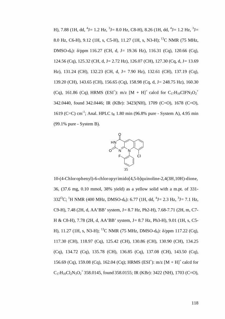
118
H), 7.88 (1H, dd, 4J= 1.2 Hz, 3J= 8.0 Hz, C8-H), 8.26 (1H, dd, 4J= 1.2 Hz, 3J=
8.0 Hz, C6-H), 9.12 (1H, s, C5-H), 11.27 (1H, s, N3-H); 13C NMR (75 MHz,
DMSO-d6): δ/ppm 116.27 (CH, d, J= 19.36 Hz), 116.31 (Cq), 120.66 (Cq),
124.56 (Cq), 125.32 (CH, d, J= 2.72 Hz), 126.07 (CH), 127.30 (Cq, d, J= 13.69
Hz), 131.24 (CH), 132.23 (CH, d, J= 7.90 Hz), 132.61 (CH), 137.19 (Cq),
139.20 (CH), 143.65 (CH), 156.65 (Cq), 158.98 (Cq, d, J= 248.75 Hz), 160.30
(Cq), 161.86 (Cq); HRMS (ESI+): m/z [M + H]+ calcd for C17H10ClFN3O2+
342.0440, found 342.0446; IR (KBr): 3423(NH), 1709 (C=O), 1678 (C=O),
1619 (C=C) cm-1; Anal. HPLC tR 1.80 min (96.8% pure - System A), 4.95 min
(99.1% pure - System B).
HN
N N
O
O
ClF
35
10-(4-Chlorophenyl)-6-chloropyrimido[4,5-b]quinoline-2,4(3H,10H)-dione,
36, (37.6 mg, 0.10 mmol, 38% yield) as a yellow solid with a m.pt. of 331-
332OC; 1H NMR (400 MHz, DMSO-d6): 6.77 (1H, dd, 4J= 2.3 Hz, 3J= 7.1 Hz,
C9-H), 7.48 (2H, d, AA’BB’system, J= 8.7 Hz, Ph2-H), 7.68-7.71 (2H, m, C7-
H & C8-H), 7.78 (2H, d, AA’BB’system, J= 8.7 Hz, Ph3-H), 9.01 (1H, s, C5-
H), 11.27 (1H, s, N3-H); 13C NMR (75 MHz, DMSO-d6): δ/ppm 117.22 (Cq),
117.30 (CH), 118.97 (Cq), 125.42 (CH), 130.86 (CH), 130.90 (CH), 134.25
(Cq), 134.72 (Cq), 135.78 (CH), 136.85 (Cq), 137.08 (CH), 143.50 (Cq),
156.69 (Cq), 159.08 (Cq), 162.04 (Cq); HRMS (ESI+): m/z [M + H]+ calcd for
C17H10Cl2N3O2+ 358.0145, found 358.0155; IR (KBr): 3422 (NH), 1703 (C=O),

119
1672 (C=O), 1607 (C=C) cm-1; Anal. HPLC tR 2.50 min (99.6% pure - System
A), 6.05 min (100.0% pure - System B).
HN
N N
O
O
Cl
Cl36
10-(4-Chlorophenyl)-7-chloropyrimido[4,5-b]quinoline-2,4(3H,10H)-dione,
37, (47.3 mg, 0.13 mmol, 47% yield) as a yellow solid with a m.pt. of >350
OC; 1H NMR (400 MHz, DMSO-d6): δ/ppm 6.80 (1H, d, J= 9.0 Hz, C9-H),
7.47-7.53 (2H, d, AA’BB’system, J= 8.4 Hz, Ph2-H), 7.72-7.79 (3H, m, C8-H
& Ph3-H), 8.38 (1H, d, 4J= 2.1 Hz, C6-H), 9.09 (1H, s, C5-H), 11.19 (1H, s,
N3-H); 13C NMR (75 MHz, DMSO-d6): δ/ppm 117.21 (Cq), 119.64 (CH),
122.48 (Cq), 128.97 (Cq), 130.28 (CH), 130.86 (CH), 130.90 (CH), 134.67
(Cq), 135.04 (CH), 136.72 (Cq), 140.80 (Cq), 141.79 (CH), 156.67 (Cq),
159.11 (Cq), 162.01 (Cq); HRMS (ESI+): m/z [M + H]+ calcd for
C17H10Cl2N3O2+ 358.0145, found 358.0152; IR (KBr): 3406 (NH), 1704 (C=O),
1665 (C=O), 1615 (C=C) cm-1; Anal. HPLC tR 3.42 min (100.0% pure -
System A), 6.90 min (98.6% pure - System B).
HN
N N
O
O
Cl
Cl37
10-(4-Chlorophenyl)-8-chloropyrimido[4,5-b]quinoline-2,4(3H,10H)-dione,
38, (22.7 mg, 0.06 mmol, 23% yield) as a yellow solid with a m.pt. of >350 OC

120
(lit >300 OC) [264]; 1H NMR (400 MHz, DMSO-d6): δ/ppm 6.70 (1H, d, 4J=
1.7 Hz, C9-H), 7.51 (2H, d, AA’BB’system, J= 8.6 Hz, Ph2-H), 7.60 (1H, dd,
4J= 1.7 Hz, 3J= 8.6 Hz, C7-H), 7.78 (2H, d, AA’BB’system, J= 8.6 Hz, Ph3-
H), 8.28 (1H, d, J= 8.6 Hz, C6-H), 9.13 (1H, s, C5-H), 11.61 (1H, s, N3-H);
13C NMR (75 MHz, DMSO-d6): δ/ppm 116.39 (Cq), 116.65 (CH), 120.32
(Cq), 125.37 (CH), 130.90 (CH), 131.02 (CH), 133.74 (CH), 134.78 (Cq),
136.44 (Cq), 140.05 (Cq), 142.29 (CH), 142.81 (Cq), 156.74 (Cq), 159.45
(Cq), 162.16 (Cq); HRMS (ESI+): m/z [M + H]+ calcd for C17H10Cl2N3O2+
358.0145, found 358.0146; IR (KBr): 3432 (NH), 1701 (C=O), 1664 (C=O),
1609 (C=C) cm-1; Anal. HPLC tR 3.54 min (100.0% pure - System A), 6.82
min (100.0% pure - System B).
HN
N N
O
O Cl
Cl38
10-(4-Chlorophenyl)-9-chloropyrimido[4,5-b]quinoline-2,4(3H,10H)-dione,
39, (14.6 mg, 0.04 mmol, 15% yield) as a yellow solid with a m.pt. of >350
OC; 1H NMR (400 MHz, DMSO-d6): δ/ppm 7.43-7.52 (3H, m, C7-H & Ph2-
H), 7.60 (2H, d, AA’BB’system, J= 8.8 Hz, Ph3-H), 7.85 (1H, dd, 4J= 1.5 Hz,
3J= 7.9 Hz, C8-H), 8.28 (1H, dd, 4J= 1.5 Hz, 3J= 7.9 Hz, C6-H), 9.09 (1H, s,
C5-H), 11.22 (1H, s, N3-H); 13C NMR (75 MHz, DMSO-d6): δ/ppm 116.48
(Cq), 121.00 (Cq), 124.59 (Cq), 125.77 (CH), 129.18 (CH), 132.22 (CH),
132.45 (CH), 134.12 (Cq), 137.53 (Cq), 138.57 (Cq), 139.25 (CH), 143.33
(CH), 156.63 (Cq), 160.81 (Cq), 162.04 (Cq); HRMS (ESI+): m/z [M + H]+

121
calcd for C17H10Cl2N3O2+ 358.0145, found 358.0156; IR (KBr): 3415 (NH),
1715 (C=O), 1655 (C=O), 1613 (C=C) cm-1; Anal. HPLC tR 3.10 min (100.0%
pure - System A), 6.34 min (97.2% pure - System B).
HN
N N
O
O
Cl
Cl
39
10-Phenyl-7-nitropyrimido[4,5-b]quinoline-2,4(3H,10H)-dione, 17, (66.1
mg, 0.19 mmol, 66% yield) as a yellow solid with a m.pt. of >350 OC (lit >360
OC) [241]; 1H NMR (400 MHz, DMSO-d6): δ/ppm 6.76 (1H, d, J= 9.4 Hz, C9-
H), 7.36 (2H, d, J= 8.0 Hz, Ph2-H), 7.53-7.65 (3H, m, Ph-H), 8.34 (1H, dd, 4J=
2.6 Hz, 3J= 9.4 Hz, C8-H), 9.13 (1H, d, 4J= 2.6 Hz, C6-H), 9.20 (1H, s, C5-H),
11.19 (1H, s, N3-H); 13C NMR (75 MHz, DMSO-d6): δ/ppm 118.05 (CH),
119.00 (Cq), 120.92 (Cq), 127.68 (CH), 128.75 (CH), 128.85 (CH), 130.19
(CH), 130.92 (CH), 137.75 (Cq), 142.62 (CH), 143.37 (Cq), 145.60 (Cq),
156.78 (Cq), 159.98 (Cq), 161.90 (Cq); HRMS (ESI+): m/z [M + H]+ calcd for
C17H11N4O4+ 335.0775, found 335.0776; IR (KBr): 3447 (NH), 1720 (C=O),
1659 (C=O), 1613 (C=C), 1521 (NO2), 1357 (NO2) cm-1; Anal. HPLC tR 0.92
min (99.1% pure - System A), 4.37 min (98.6% pure - System B).
HN
N N
O
O
NO2
17

122
10-(2-Fluorophenyl)-7-nitropyrimido[4,5-b]quinoline-2,4(3H,10H)-dione,
21, (88.9 mg, 0.25 mmol, 89% yield) as a yellow solid with a m.pt. of 326-327
OC; 1H NMR (400 MHz, DMSO-d6): δ/ppm 6.98 (1H, d, J= 9.4 Hz, C9-H),
7.53-7.69 (3H, m, Ph-H), 7.73-7.80 (1H, m, Ph-H), 8.48 (1H, dd, 4J= 2.3 Hz,
3J= 9.4 Hz, C8-H), 9.25 (1H, d, 4J= 2.3 Hz, C6-H), 9.33 (1H, s, C5-H), 11.38
(1H, s, N3-H); 13C NMR (75 MHz, DMSO-d6): δ/ppm 117.82 (CH, d, J= 19.02
Hz), 117.86 (Cq), 118.26 (CH), 120.99 (Cq), 124.54 (Cq, d, J= 13.34 Hz),
126.81 (CH, d, J= 3.56 Hz), 127.98 (CH), 129.44 (CH), 130.85 (CH), 132.99
(CH, d, J= 7.85 Hz), 143.07 (CH), 143.79 (Cq), 144.72 (Cq), 156.65 (Cq),
157.56 (Cq, d, J= 242.62 Hz), 159.58 (Cq), 161.65 (Cq); HRMS (ESI+): m/z
[M + H]+ calcd for C17H10FN4O4+ 353.0681, found 353.0671; IR (KBr): 3413
(NH), 1711 (C=O), 1685 (C=O), 1616 (C=C), 1488 (NO2), 1309 (NO2) cm-1;
Anal. HPLC tR 1.49 min (100.0% pure - System A), 4.60 min (97.2% pure -
System B).
HN
N N
O
O
NO2
F
21
10-(4-Chlorophenyl)-7-nitropyrimido[4,5-b]quinoline-2,4(3H,10H)-dione,
25, (49.8 mg, 0.13 mmol, 50% yield) as a yellow solid with a m.pt. of 343-344
OC (dec); 1H NMR (400 MHz, DMSO-d6): δ/ppm 6.95 (1H, d, J= 9.5 Hz, C9-
H), 7.52 (2H, d, AA’BB’system, J= 8.8 Hz, Ph2-H), 7.80 (2H, d, AA’BB’
system, J= 8.8 Hz, Ph3-H), 8.42 (1H, dd, 4J= 2.3 Hz, 3J= 9.5 Hz, C8-H), 9.22
(1H, d, 4J= 2.3 Hz, C6-H), 9.30 (1H, s, C5-H), 11.31 (1H, s, N3-H); 13C NMR
(75 MHz, DMSO-d6): δ/ppm 118.00 (Cq), 119.02 (CH), 120.96 (Cq), 127.66

123
(CH), 128.86 (CH), 130.82 (CH), 131.02 (CH), 134.88 (Cq), 136.56 (Cq),
142.69 (CH), 143.42 (Cq), 145.46 (Cq), 156.70 (Cq), 160.02 (Cq), 161.85
(Cq); HRMS (ESI+): m/z [M + H]+ calcd for C17H10ClN4O4+ 369.0385, found
369.0403; IR (KBr): 3423 (NH), 1709 (C=O), 1665 (C=O), 1614 (C=C), 1543
(NO2), 1335 (NO2) cm-1; Anal. HPLC tR 0.90 min (97.3% pure - System A),
5.80 min (100.0% pure - System B).
HN
N N
O
O
Cl
NO2
25
10-Phenyl-6-methylpyrimido[4,5-b]quinoline-2,4(3H,10H)-dione, 52,
(382.9 mg, 1.26 mmol, 48% yield) as a yellow solid with a m.pt. of 342-344 OC
(dec); 1H NMR (400 MHz, DMSO-d6): δ/ppm 2.75 (3H, s, Me), 6.55 (1H, d,
J= 8.8 Hz, C9-H), 7.33-7.43 (3H, m, C7-H & Ph2-H), 7.57-7.72 (4H, m, C8-H
& Ph-H), 9.02 (1H, s, C5-H), 11.09 (1H, s, N3-H); 13C NMR (75 MHz,
DMSO-d6): δ/ppm 18.15 (CH), 114.85 (Cq), 115.51 (CH), 119.88 (Cq), 125.61
(CH), 128.41 (CH), 129.34 (CH), 130.24 (CH), 134.87 (CH), 137.96 (Cq),
138.45 (CH), 139.15 (Cq), 142.42 (Cq), 156.42 (Cq), 158.30 (Cq), 162.06
(Cq); HRMS (ESI+): m/z [M + H]+ calcd for C18H14N3O2+ 304.1081, found
304.1085; IR (KBr): 3371 (NH), 1700 (C=O), 1651 (C=O), 1598 (C=C) cm-1;
Anal. HPLC tR 1.18 min (98.8% pure - System A), 4.03 min (97.7% pure -
System B).

124
HN
N N
O
O
52
10-Phenyl-7-methylpyrimido[4,5-b]quinoline-2,4(3H,10H)-dione, 53, (54.8
mg, 0.18 mmol, 55% yield) as a yellow solid with a m.pt. of 342-344 OC (dec);
1H NMR (400 MHz, DMSO-d6): δ/ppm 2.40 (3H, s, Me), 6.62 (1H, d, J= 8.8
Hz, C9-H), 7.41 (2H, d, J= 7.7 Hz, Ph2-H), 7.57 (1H, dd, 4J= 1.74 Hz, 3J= 8.8
Hz, C8-H), 7.60-7.72 (3H, m, Ph-H), 8.00 (1H, s, C6-H), 9.02 (1H, s, C5-H).
11.04 (1H, s, N3-H); 13C NMR (75 MHz, DMSO-d6): δ/ppm 20.64 (CH3),
115.99 (Cq), 117.50 (CH), 121.41 (Cq), 128.84 (CH), 129.77 (CH), 130.66
(CH), 130.84 (CH), 134.41 (Cq), 136.95 (CH), 138.18 (Cq), 140.46 (Cq),
142.51 (CH), 158.74 (Cq), 158.74 (Cq), 162.41 (Cq); HRMS (ESI+): m/z [M +
H]+ calcd for C18H14N3O2+ 304.1081, found 304.0836; IR (KBr): 3437 (NH),
1703 (C=O), 1649 (C=O), 1609 (C=C) cm-1; Anal. HPLC tR 1.37 min (96.2%
pure - System A), 4.24 min (96.8% pure - System B).
HN
N N
O
O
53
10-(2-Fluorophenyl)-6-methylpyrimido[4,5-b]quinoline-2,4(3H,10H)-
dione, 56, (129.6 mg, 0.40 mmol, 16% yield) as a yellow solid with a m.pt. of
309-310 OC; 1H NMR (400 MHz, DMSO-d6): 2.76 (3H, s, Me), 6.66 (1H, d, J=
8.0 Hz, C9-H), 7.40 (1H, d, J= 8.0 Hz, C7-H), 7.49-7.76 (5H, m, C8-H & Ph-
H), 9.05 (1H, s, C5-H), 11.19 (1H, s, N3-H); 13C NMR (75 MHz, DMSO-d6):

125
δ/ppm 19.04 (CH3), 115.01 (CH), 115.06 (Cq), 117.66 (CH, d, J= 19.01 Hz),
120.31 (Cq), 125.26 (Cq, d, J= 13.60 Hz), 126.11 (CH, d, J= 3.51 Hz), 126.53
(CH), 130.98 (CH), 132.54 (CH, d, J= 8.10 Hz), 135.95 (CH), 139.63 (CH),
140.17 (Cq), 142.02 (Cq), 156.81 (Cq), 157.43 (Cq, d, J= 247.76 Hz), 158.55
(Cq), 162.24 (Cq); HRMS (ESI+): m/z [M + H]+ calcd for C18H13FN3O2+
322.0986, found 322.0978; IR (KBr): 3467 (NH), 1702 (C=O), 1653 (C=O),
1597 (C=C) cm-1; Anal. HPLC tR 1.34 min (100.0% pure - System A), 4.28
min (99.0% pure - System B).
HN
N NO
O
F
56
10-(2-Fluorophenyl)-7-methylpyrimido[4,5-b]quinoline-2,4(3H,10H)-
dione, 57, (67.9 mg, 0.21 mmol, 68% yield) as a yellow solid with a m.pt. of
317-319 OC; 1H NMR (400 MHz, DMSO-d6): δ/ppm 2.42 (3H, s, Me), 6.74
(1H, d, J= 8.7 Hz, C9-H), 7.49-7.65 (4H, m, C8-H & Ph-H), 7.69-7.76 (1H, m,
Ph-H), 8.04 (1H, s, C6-H), 9.07 (1H, s, C5-H), 11.14 (1H, s, N3-H); 13C NMR
(75 MHz, DMSO-d6): δ/ppm 20.63 (CH3), 115.86 (Cq), 116.65 (CH), 117.65
(CH, d, J= 18.79 Hz), 121.42 (Cq), 125.06 (Cq, d, J= 13.71 Hz), 126.56 (CH,
d, J= 3.05 Hz), 130.98 (CH), 131.26 (CH), 132.50 (CH, d, J= 6.38 Hz), 134.95
(Cq), 137.50 (CH), 139.62 (Cq), 143.12 (CH), 156.77 (Cq), 157.61 (Cq, d, J=
248.01 Hz), 158.59 (Cq), 162.17 (Cq); HRMS (ESI+): m/z [M + H]+ calcd for
C18H13FN3O2+ 322.0986, found 322.0960; IR (KBr): 3423 (NH), 1705 (C=O),
1657 (C=O), 1609 (C=C) cm-1; Anal. HPLC tR 1.53 min (98.1% pure - System
A), 4.49 min (100.0% pure - System B).

126
HN
N N
O
O
F
57
10-(4-Chlorophenyl)-6-methylpyrimido[4,5-b]quinoline-2,4(3H,10H)-
dione, 60, (301.8 mg, 0.89 mmol, 30% yield) as a yellow solid with a m.pt. of
339-341 OC (dec); 1H NMR (400 MHz, DMSO-d6): δ/ppm 2.75 (3H, s, Me),
6.62 (1H, d, J= 7.9 Hz, C9-H), 7.37 (1H, d, J= 7.9 Hz, C7-H), 7.47 (2H, d,
AA’BB’system, J= 8.7 Hz, Ph2-H), 7.62 (1H, t, J= 7.9 Hz, C8-H), 7.76 (2H,
d, AA’BB’system, J= 8.7 Hz, Ph3-H), 9.02 (1H, s, C5-H), 11.11 (1H, s, N3-
H); 13C NMR (75 MHz, DMSO-d6): δ/ppm 19.01 (CH3), 115.30 (Cq), 115.93
(CH), 120.33 (Cq), 126.15 (CH), 130.79 (CH), 130.93 (CH), 134.47 (Cq),
135.45 (CH), 137.23 (Cq), 139.01 (CH), 139.68 (Cq), 142.68 (Cq), 156.79
(Cq), 158.82 (Cq), 162.46 (Cq); HRMS (ESI+): m/z [M + H]+ calcd for
C18H13ClN3O2+ 338.0691, found 338.0702; IR (KBr): 3424 (NH), 1701 (C=O),
1674 (C=O), 1599 (C=C) cm-1; Anal. HPLC tR 2.00 min (97.2% pure - System
A), 5.37 min (98.2% pure - System B).
HN
N NO
O
Cl60
10-(4-Chlorophenyl)-7-methylpyrimido[4,5-b]quinoline-2,4(3H,10H)-
dione, 61, (47.6 mg, 0.14 mmol, 48% yield) as a yellow solid with a m.pt. of
>350 OC; 1H NMR (400 MHz, DMSO-d6): δ/ppm 2.49 (3H, s, Me), 6.70 (1H,

127
d, J= 8.8 Hz, C9-H), 7.47 (2H, d, AA’BB’system, J= 8.6 Hz, Ph2-H), 7.58
(1H, dd, 4J= 1.9 Hz, 3J= 8.8 Hz, C8-H), 7.76 (2H, d, AA’BB’system, J= 8.6
Hz, Ph3-H), 8.01 (1H, s, C6-H), 9.03 (1H, s, C5-H), 11.07 (1H, s, N3-H); 13C
NMR (75 MHz, DMSO-d6): δ/ppm 20.68 (CH3), 116.01 (Cq), 117.48 (CH),
121.44 (Cq), 130.23 (Cq), 130.76 (CH), 130.93 (CH), 134.46 (Cq), 134.49
(Cq), 137.02 (CH), 137.04 (CH), 140.28 (Cq), 142.61 (CH), 156.74 (Cq),
158.85 (Cq), 162.36 (Cq); HRMS (ESI+): m/z [M + H]+ calcd for
C18H13ClN3O2+ 338.0691, found 338.0670; IR (KBr): 3434 (NH), 1701 (C=O),
1668 (C=O), 1608 (C=C) cm-1; Anal. HPLC tR 2.50 min (97.4% pure - System
A), 5.74 min (99.1% pure - System B).
HN
N N
O
O
Cl61
Synthesis of 2- p-Toluenesulfonyloxybenzaldehyde, 78 [301-303]
A mixture of salicylaldehyde (0.88 g, 0.76mL; 7.23 mmol,) and sodium
carbonate (3.06 g; 28.95 mmol), in acetone (20 mL) was stirred at room
temperature for 30 min under nitrogen atmosphere. Tosyl chloride (2.75 g;
14.47 mmol) dissolved in acetone (25 mL) was added to the reaction mixture
and refluxed for 5 h. Concentration of the solution under reduced pressure and
dry column flash chromatography using 66.6% petroleum ether and 33.3%
diethyl ether was carried out to yield the pure 2-p-
toluenesulfonyloxybenzaldehyde, 78.

128
2-p-Toluenesulfonyloxybenzaldehyde, 78 (1.96 g, 7.09 mmol, 98% yield) as
a white solid with a m.pt. of 66-68 OC; 1H NMR (400 MHz, DMSO-d6): δ/ppm
2.42 (3H, s, Me), 7.18 (1H, dd, 4J= 0.7 Hz, 3J= 7.8 Hz, C6-H), 7.48 (2H, d,
AA’BB’system, J= 8.0 Hz, Ph3-H), 7.52 (1H, ddd, 4J= 1.2 Hz, 3J= 7.8 Hz,
C5-H), 7.71-7.75 (3H, m, C4-H & Ph2-H), 7.81 (1H, dd, 4J= 1.2 Hz, 3J= 7.6
Hz, C3-H), 9.94 (1H, s, COH); 13C NMR (75 MHz, DMSO-d6): δ/ppm 21.66
(CH3), 124.14 (CH), 128.62 (CH), 128.88 (CH), 129.18 (CH), 129.44 (Cq),
130.84 (Cq), 130.90 (CH), 136.42 (CH), 147.00 (Cq), 150.51 (Cq), 187.92
(CH).
O
OS
O
O
78
Synthesis of 10-Phenylpyrimido[4,5-b]quinoline-2,4(3H,10H)-dione, 64,
using 2-p-Toluenesulfonyloxybenzaldehyde, 75 [241, 261, 264, 265, 268,
270]
Method A
A mixture of 6-(phenylamino)pyrimidine-2,4(1H,3H)-dione, 69 (70.2 mg; 0.34
mmol), and 2-p-toluenesulfonyloxybenzaldehyde, 78 (114.64 mg; 0.41 mmol),
in DMF (3 mL) was heated to 160 OC for 30 min in the microwave reactor.
Concentration of the solution under reduced pressure and dry column flash
chromatography using 95% dichloromethane and 5% methanol was carried out
to yield the pure 10-phenylpyrimido[4,5-b]quinoline-2,4(3H,10H)-dione, 64.
10-Phenylpyrimido[4,5-b]quinoline-2,4(3H,10H)-dione, 64, (58.7 mg, 0.20
mmol, 59% yield) as a yellow solid; 1H NMR (400 MHz, DMSO-d6): δ/ppm
6.71 (1H, d, J= 8.5 Hz, C9-H), 7.43 (2H, d, J= 7.1 Hz, Ph2-H), 7.50 (1H, t, J=

129
8.5 Hz, C8-H), 7.60-7.78 (4H, m, C7-H & Ph-H), 8.23 (1H, d, J= 8.5 Hz, C6-
H), 9.12 (1H, s, C5-H), 11.06 (1H, s, N3-H); HRMS (ESI+): m/z [M + H]+
calcd for C17H12N3O2+ 290.0924, found 290.0931.
HN
N N
O
O
64
Method B
A mixture of 6-(phenylamino)pyrimidine-2,4(1H,3H)-dione, 69 (70.2 mg; 0.34
mmol), and 2-p-toluenesulfonyloxybenzaldehyde, 78 (114.64 mg; 0.41 mmol),
in DMF (3 mL) was heated to 160 OC for 1 h in the microwave reactor.
Concentration of the solution under reduced pressure and dry column flash
chromatography using 95% dichloromethane and 5% Methanol was carried out
to yield the pure 10-phenylpyrimido[4,5-b]quinoline-2,4(3H,10H)-dione, 64.
10-Phenylpyrimido[4,5-b]quinoline-2,4(3H,10H)-dione, 64, (38.8 mg, 0.13
mmol, 39% yield) as a yellow solid; 1H NMR (400 MHz, DMSO-d6): δ/ppm
6.71 (1H, d, J= 8.5 Hz, C9-H), 7.43 (2H, d, J= 7.1 Hz, Ph2-H), 7.50 (1H, t, J=
8.5 Hz, C8-H), 7.60-7.76 (4H, m, C7-H & Ph-H), 8.23 (1H, d, J= 8.5 Hz, C6-
H), 9.12 (1H, s, C5-H), 11.06 (1H, s, N3-H); HRMS (ESI+): m/z [M + H]+
calcd for C17H12N3O2+ 290.0924, found 290.0922.
General Procedure to Synthesise 2-p-Toluenesulfonyloxybenzaldehyde
Analogues [301-303]
A mixture of 1 equivalent of the desired salicyladehyde and 4 equivalents of
sodium carbonate, in acetone was stirred at room temperature for 30 min under

130
nitrogen atmosphere. 2 Equivalents of tosyl chloride dissolved in acetone was
added to the reaction mixture and refluxed for 5 h. Concentration of the
solution under reduced pressure and dry column flash chromatography using
66.6% petroleum ether and 33.3% diethyl ether was carried out to yield the
pure desired 2-p-toluenesulfonyloxybenzaldehyde analogue.
2-p-Toluenesulfonyloxy-3-nitrobenzaldehyde, 79 (435.9 mg, 1.35 mmol,
54% yield) as a white solid with a m.pt. of 133-134 OC; 1H NMR (400 MHz,
CDCl3): δ/ppm 2.50 (3H, s, Me), 7.40 (2H, d, AA’BB’system, J= 8.3 Hz, Ph3-
H), 7.58 (1H, t, J= 8.0 Hz, C5-H), 7.78 (2H, d, AA’BB’system, J= 8.3 Hz,
Ph2-H), 8.15-8.22 (2H, m, C6-H & C4-H), 10.10 (1H, s COH); 13C NMR (75
MHz, CDCl3): δ/ppm 21.90 (CH3), 127.86 (CH), 128.98 (CH), 130.33 (Cq),
130.45 (CH), 130.72 (CH), 132.38 (Cq), 133.03 (CH), 142.72 (Cq), 144.41
(Cq), 147.35 (Cq), 185.63 (CH).
O
OS
O
ONO2
79
2-p-Toluenesulfonyloxy-4-methylbenzaldehyde, 80 (630.2 mg, 2.17 mmol,
59% yield) as a white solid with a m.pt. of 93-94 OC; 1H NMR (400 MHz,
DMSO-d6): δ/ppm 2.36 (3H, s, Me), 2.42 (3H, s, Me), 7.06 (1H, s, C3-H), 7.33
(1H, d, J= 7.8 Hz, C5-H), 7.48 (2H, d, AA’BB’system, J= 8.2 Hz, Ph3-H),
7.70 (1H, d, J= 7.8 Hz, C6-H), 7.76 (2H, d, AA’BB’system, J= 8.2 Hz, Ph2-
H), 9.83 (1H, s, COH); 13C NMR (75 MHz, DMSO-d6): δ/ppm 21.65 (CH3),
21.66 (CH3), 124.44 (CH), 126.96 (Cq), 128.86 (CH), 128.96 (CH), 129.31
(CH), 130.85 (CH), 130.90 (Cq), 146.95 (Cq), 147.74 (Cq), 150.51 (Cq),
187.45 (CH).

131
O
OS
O
O
80
2-p-Toluenesulfonyloxy-3-methylbenzaldehyde, 81 (1.04 g, 3.55 mmol, 98%
yield) as a white solid with a m.pt. of 68-69 OC; 1H NMR (400 MHz, DMSO-
d6): δ/ppm 2.03 (3H, s, Me), 2.45 (3H, s, Me), 7.44 (1H, t, J= 7.63 Hz, C5-H),
7.52 (2H, d, AA’BB’system, J= 8.2 Hz, Ph3-H), 7.62-7.70 (2H, m, C4-H &
C6-H), 7.79 (2H, d, AA’BB’system, J= 8.2 Hz, Ph2-H), 9.88 (1H, s, COH);
13C NMR (75 MHz, DMSO-d6): δ/ppm 16.11 (CH3), 21.69 (CH3), 126.63
(CH), 128.22 (CH), 128.78 (CH), 130.57 (Cq), 131.06 (CH), 131.46 (Cq),
133.56 (Cq), 138.13 (CH), 147.15 (Cq), 148.93 (Cq), 188.12 (CH).
O
OS
O
O
81
General Procedure to Synthesise 10-Phenylpyrimido[4,5-b]quinoline-
2,4(3H,10H)-dione Analogues using 2-p-Toluenesulfonyloxybenzaldehyde
[241, 261, 264, 265, 268, 270]
To 1 equivalent of the desired 6-(phenylamino)pyrimidine-2,4(1H,3H)-dione
analogue in DMF was added 1.2 equivalents of the desired 2-p-
toluenesulfonyloxybenzaldehyde. The mixture was heated at 160 OC for 30
min in the microwave reactor or refluxed for 4 h. Concentration of the solution
under reduced pressure and separation by dry column flash chromatography
using 95% dichloromethane and 5% methanol to yield the pure desired 10-
phenylpyrimido[4,5-b]quinoline-2,4(3H,10H)-dione analogue .

132
10-(2-Fluorophenyl)-9-nitropyrimido[4,5-b]quinoline-2,4(3H,10H)-dione,
23, (23.0 mg, 0.06 mmol, 23% yield) as a yellow solid with a m.pt. of 305-307
OC; 1H NMR (400 MHz, DMSO-d6): δ/ppm 7.25 (1H, t, J= 7.5 Hz, Ph5-H),
7.35-7.44 (2H, m, Ph-H), 7.56-7.74 (2H, m, C7-H & Ph-H), 8.17 (1H, d, J= 7.3
Hz, C8-H), 8.50 (1H, d, J= 7.3 Hz, C6-H), 9.19 (1H, s, C5-H), 11.36 (1H, s,
N3-H); 13C NMR (75 MHz, DMSO-d6): δ/ppm 116.72 (CH, d, J= 19.55 Hz),
117.13 (Cq), 124.05 (Cq), 125.11 (CH), 125.21 (Cq), 125.34 (Cq), 125.36
(CH), 130.03 (CH), 132.60 (CH), 133.12 (CH, d, J= 7.97 Hz), 136.17 (CH),
140.85 (Cq), 142.95 (CH), 156.53 (Cq), 158.52 (Cq, d, J= 252.37 Hz), 159.51
(Cq), 161.71 (Cq); HRMS (ESI+): m/z [M + H]+ calcd for C17H10FN4O4+
353.0681, found 353.0676; IR (KBr): 3425 (NH), 1712 (C=O), 1679 (C=O),
1619 (C=C), 1487 (NO2), 1328 (NO2) cm-1; Anal. HPLC tR 1.10 min (100.0%
pure - System A), 3.98 min (97.6% pure - System B).
HN
N N
O
O
NO2F
23
10-(4-Chlorophenyl)-9-nitropyrimido[4,5-b]quinoline-2,4(3H,10H)-dione,
27 (23.8 mg, 0.06 mmol, 24% yield) as a yellow solid with a m.pt. of 336-338
OC; 1H NMR (400 MHz, DMSO-d6): δ/ppm 7.41 (2H, d, AA’BB’system, J=
8.6 Hz, Ph2-H), 7.55 (2H, d, AA’BB’system, J= 8.6 Hz, Ph3-H), 7.64 (1H, t,
J= 7.8 Hz, C7-H), 8.13 (1H, d, J= 7.8 Hz, C8-H), 8.48 (1H, d, J= 7.8 Hz, C6-
H), 9.17 (1H, s, C5-H), 11.30 (1H, s, N3-H); 13C NMR (75 MHz, DMSO-d6):
δ/ppm 117.45 (Cq), 123.98 (Cq), 124.87 (CH), 129.21 (CH), 131.40 (CH),
132.03 (CH), 132.52 (Cq), 134,63 (Cq), 135.89 (CH), 136.24 (Cq), 141.09

133
(Cq), 142.62 (CH), 156.53 (Cq), 160.37 (Cq), 161.86 (Cq); HRMS (ESI+): m/z
[M + H]+ calcd for C17H10ClN4O4+369.0385, found 369.0419; IR (KBr): 3422
(NH), 1706 (C=O), 1672 (C=O), 1620 (C=C), 1427 (NO2), 1352 (NO2) cm-1;
Anal. HPLC tR 1.63 min (98.4% pure - System A), 4.75 min (100.0% pure -
System B).
HN
N N
O
O
NO2
Cl27
10-Phenyl-8-methylpyrimido[4,5-b]quinoline-2,4(3H,10H)-dione, 54, (64.7
mg, 0.21 mmol, 65% yield) as a yellow solid with a m.pt. of 299-301 OC; 1H
NMR (400 MHz, DMSO-d6): δ/ppm 2.32 (3H, s, Me), 6.48 (1H, s, C9-H), 7.35
(1H, d, J= 8.1 Hz, C7-H), 7.41 (2H, d, J= 7.2 Hz, Ph2-H), 7.59-7.72 (3H, m,
Ph-H), 8.12 (1H, d, J= 8.1 Hz, C6-H), 9.07 (1H, s, C5-H), 11.03 (1H, s, N3-H);
13C NMR (75 MHz, , DMSO-d6): δ/ppm 22.66 (CH3), 115.06 (Cq), 117.05
(CH), 119.58 (Cq), 126.56 (CH), 128.88 (CH), 129.81 (CH), 130.71 (CH),
131.73 (CH), 138.10 (Cq), 142.45 (Cq), 142.67 (CH), 146.79 (Cq), 156.91
(Cq), 159,19 (Cq), 162.49 (Cq); HRMS (ESI+): m/z [M + H]+ calcd for
C18H14N3O2+ 304.1081, found 304.1021; IR (KBr): 3433 (NH), 1709 (C=O),
1672 (C=O), 1608 (C=C) cm-1; Anal. HPLC tR 1.20 min (100.0% pure -
System A), 4.02 min (97.8% pure - System B).

134
HN
N N
O
O
54
10-Phenyl-9-methylpyrimido[4,5-b]quinoline-2,4(3H,10H)-dione, 55, (37.8
mg, 0.12 mmol, 38% yield) as a yellow solid with a m.pt. of 337-338 OC (dec);
1H NMR (400 MHz, DMSO-d6): δ/ppm 1.59 (3H, s, Me), 7.39-7.47 (3H, m,
C8-H & Ph2-H), 7.53-7.61 (4H, m, C7-H & Ph-H), 8.10 (1H, d, J= 7.9 Hz, C6-
H), 9.07 (1H, s, C5-H), 11.07 (1H, s, N3-H); 13C NMR (75 MHz, DMSO-d6):
δ/ppm 22.34 (CH3), 115.23 (Cq), 123.04 (Cq), 125.08 (CH), 127.13 (Cq),
129.24 (CH), 129.53 (CH), 130.64 (CH), 131.31 (CH), 140.55 (CH), 140.77
(Cq), 140.95 (Cq), 144.01 (CH), 156.84 (Cq), 160.62 (Cq), 162.35 (Cq);
HRMS (ESI+): m/z [M + H]+ calcd for C18H14N3O2+ 304.1081, found 304.1097;
IR (KBr): 3441 (NH), 1703 (C=O), 1665 (C=O), 1619 (C=C) cm-1; Anal.
HPLC tR 1.28 min (97.2% pure - System A), 14.40 min (97.9% pure - System
B).
HN
N N
O
O
55
10-(2-Fluorophenyl)-8-methylpyrimido[4,5-b]quinoline-2,4(3H,10H)-
dione, 58, (16.3 mg, 0.05 mmol, 16% yield) as a yellow solid with a m.pt. of
>350 OC; 1H NMR (400 MHz, DMSO-d6): δ/ppm 2.37 (3H, s, Me), 6.60 (1H,
s, C9-H), 7.39 (1H, d, J= 80 Hz, C7-H), 7,49-7.65 (3H, m, Ph-H), 7.69-7.77
(1H, m, Ph-H), 8.15 (1H, d, J= 8.0 Hz, C6-H), 9.10 (1H, s, C5-H), 11.10 (1H,

135
s, N3-H); 13C NMR (75 MHz, CDCl3): δ/ppm 114.17 (CH, d, J= 4.32 Hz),
118.07 (CH, d, J= 19.46 Hz), 119.12 (Cq), 122.01 (CH, d, J= 4.32 Hz), 123.48
(Cq), 124.73 (Cq, d, J= 12.96 Hz), 127.18 (CH, d, J= 4.62 Hz), 130.38 (CH),
133.26 (CH, d, J= 8.89 Hz), 134.23 (CH), 134.67 (Cq), 140.81 (Cq), 143.37
(CH), 156.84 (Cq), 158.78 (Cq, d, J= 263.14 Hz), 159.64 (Cq), 162.24 (Cq);
HRMS (ESI+): m/z [M + H]+ calcd for C18H13FN3O2+ 322.0986, found
322.0995; IR (KBr): 3442 (NH), 1707 (C=O), 1673 (C=O), 1608 (C=C) cm-1;
Anal. HPLC tR 1.34 min (100.0% pure - System A), 5.09 min (98.8% pure -
System B).
HN
N N
O
O
F
58
10-(2-Fluorophenyl)-9-methylpyrimido[4,5-b]quinoline-2,4(3H,10H)-
dione, 59, (53.1 mg, 0.16 mmol, 53% yield) as a yellow solid with a m.pt. of
287-290 OC; 1H NMR (400 MHz, DMSO-d6): δ/ppm 1.68 (3H, s, Me), 7.36-
7.54 (4H, m, C8-H & Ph-H), 7.62-7.70 (2H, m, C7-H & Ph-H), 8.13 (1H, d, J=
7.4 Hz, C6-H), 9.10 (1H, s, C5-H), 11.16 (1H, s, N3-H); 13C NMR (75 MHz,
DMSO-d6): δ/ppm 21.37 (CH3), 115.11 (Cq), 116.63 (CH, d, J= 19.5 6Hz),
123.03 (Cq), 125.40 (Cq), 125.50 (CH, d, J= 3.19 Hz), 126.28 (Cq), 128.52
(Cq, d, J= 13.26 Hz) 131.63 (CH), 131.84 (CH), 132.38 (CH, d, J= 8.08 Hz),
140.35 (Cq), 140.74 (CH), 144.51 (Cq), 156.81 (Cq), 158.84 (Cq, d, J= 249.14
Hz), 160.25 (Cq), 162.16 (Cq); m/z [M + H]+ calcd for C18H13FN3O2+
322.0986, found 322.0959; IR (KBr): 3442 (NH), 1712 (C=O), 1657 (C=O),

136
1619 (C=C) cm-1; Anal. HPLC tR 1.40 min (97.4% pure - System A), 15.64
min (96.8% pure - System B).
HN
N N
O
O
F
59
10-(4-Chlorophenyl)-8-methylpyrimido[4,5-b]quinoline-2,4(3H,10H)-
dione, 62, (24.3 mg, 0.08 mmol, 24% yield) as a yellow solid with a m.pt. of
346-347 OC (dec); 1H NMR (400 MHz, DMSO-d6): δ/ppm 2.36 (3H, s, Me),
6.57 (1H, s, C9-H), 7.36 (1H, d, J= 8.1 Hz, C7-H), 7.47 (2H, d, AA’BB’
system, J= 8.5 Hz, Ph2-H), 7.76 (2H, d, AA’BB’system, J= 8.5 Hz, Ph3-H),
8.12 (1H, d, J= 8.1 Hz, C6-H), 9.08 (1H, s, C5-H), 11.04 (1H, s, N3-H); 13C
NMR (75 MHz, DMSO-d6): δ/ppm 22.58 (CH3), 115.06 (Cq), 117.06 (CH),
119.64 (Cq), 126.66 (CH), 130.84 (CH), 130.99 (CH), 131.79 (CH), 134.46
(Cq), 136.95 (Cq), 142.29 (CH), 142.81 (Cq), 147.11 (Cq), 156.80 (Cq),
159.33 (Cq), 162.47 (Cq); m/z [M + H]+ calcd for C18H13ClN3O2+ 338.0691,
found 338.0718; IR (KBr): 3441 (NH), 1694 (C=O), 1663 (C=O), 1602 (C=C)
cm-1; Anal. HPLC tR 2.07 min (98.9% pure - System A), 8.40 min (99.8% pure
- System B).
HN
N N
O
O
Cl62

137
10-(4-Chlorophenyl)-9-methylpyrimido[4,5-b]quinoline-2,4(3H,10H)-
dione, 63, (26.4 mg, 0.07 mmol, 26% yield) as a yellow solid with a m.pt. of
>350 OC; 1H NMR (400 MHz, DMSO-d6): δ/ppm 1.67 (3H, s, Me), 7.42 (1H, t,
J= 7.6 Hz, C7-H), 7.51 (2H, d, AA’BB’system, J= 8.6 Hz, Ph2-H), 7.59-7.66
(3H, m, C8-H & Ph3-H), 8.11 (1H, d, J= 7.6 Hz, C6-H), 9.07 (1H, s, C5-H),
11.09 (1H, s, N3-H); 13C NMR (75 MHz, DMSO-d6): δ/ppm 22.81 (CH3),
115.25 (Cq), 123.04 (Cq), 125.18 (CH), 126.82 (Cq), 129.29 (CH), 131.38
(CH), 132.52 (CH), 134.11 (Cq), 139.81 (Cq), 140.56 (Cq), 140.60 (CH),
144.15 (CH), 156.75 (Cq), 160.68 (Cq), 162.26 (Cq); m/z [M + H]+ calcd for
C18H13ClN3O2+ 338.0691, found 338.0664; IR (KBr): 3428 (NH), 1715 (C=O),
1656 (C=O), 1617 (C=C) cm-1; Anal. HPLC tR 2.12 min (100.0% pure -
System A), 2.70 min (98.4% pure - System B).
HN
N N
O
O
Cl63
Synthesis of 2-Fluoro-6-nitrobenzyl Bromide, 83 [277, 279]
Benzoyl peroxide (2.10 mg; 0.09 mmol) was added to a mixture of 2-fluoro-6-
nitrotoluene (2.42 g; 1.90 mL; 15.64 mmol,) and N-bromosuccinimide (2.53 g;
14.22 mmol), in carbon tetrachloride (30 mL) and refluxed for 72 h. The
precipitate formed was hot filtered, collected and washed with hot carbon
tetrachloride (10 mL) and dry column flash chromatography using 90%
petroleum ether and 10% diethyl ether to yield the pure 2-fluoro-6-nitrobenzyl
bromide, 83.

138
2-Fluoro-6-nitrobenzyl bromide, 83, (1.68 g, 7.17 mmol, 51% yield) as a
white solid with a m.pt. of 55-56 OC; 1H NMR (400MHz, CDCl3): δ/ppm 4.82
(2H, s, CH2), 7.40 (1H, t, J= 8.6 Hz, C4-H), 7.49 (1H, m, C3-H), 7.89 (1H, d,
J= 8.6 Hz, C5-H); 13C NMR (75 MHz, CDCl3): δ/ppm 19.21 (CH2), 120.96
(Cq, d, J= 23.40 Hz), 121.20 (CH, d, J= 2.85 Hz), 121.74 (CH, d, J= 17.44
Hz), 130.16 (CH, d, J= 9.53 Hz), 148.78 (Cq), 160.87 (Cq, d, J= 253.61 Hz).
F
Br
NO2
83
Synthesis of 2-Fluoro-6-nitrobenzyl Pyridinium Bromide, 84 [277, 279]
2-Fluoro-6-nitrobenzyl bromide, 83 (1.40 g; 5.98 mmol), and pyridine (0.71 g,
0.70 mL; 8.97 mmol) was refluxed in ethanol (40 mL) for 3.5 h. The reaction
mixture was cooled in the fridge for 5 min. The precipitate formed was
collected by suction filtration, washed with ethanol and dry column flash
chromatography was carried out, using 80% dichloromethane and 20%
methanol, to yield the pure 2-fluoro-6-nitrobenzyl pyridium bromide, 84.
2-Fluoro-6-nitrobenzyl pyridinium bromide, 84, (1.68 g, 7.17 mmol, 91%
yield) as a white solid with a m.pt. of 59-60 OC; 1H NMR (400 MHz, CDCl3):
δ/ppm 6.7 (2H, s, CH2), 7.86-7.93 (2H, m), 8.11-8.21 (3H, m), 8.67 (1H, t, J=
7.6 Hz, C4-H), 9.10 (2H, d, J= 5.7 Hz, Py2-H); 13C NMR (75 MHz, CDCl3):
δ/ppm 54.28 (CH2, d, J= 4.43 Hz), 115.57 (Cq, d, J= 17.72 Hz), 122.45 (CH, d,
J= 3.06 Hz), 122.83 (CH, d, J= 23.56 Hz), 128.69 (CH), 133.72 (CH, d, J=
10.32 Hz), 145.33 (CH), 146.82 (CH), 149.99 (Cq, d, J= 4.05 Hz), 162.05 (Cq,
d, J= 253.66 Hz); m/z [M + H]+ calcd for C12H10FN2O2+ 233.0721, found
233.0705.

139
N
NO2F
Br
84
Synthesis of p-Nitrosodimethylaniline Hydrochloride, 85 [278]
To a mixture of N,N-dimethylamine (1.94 g, 2.03mL; 16.07 mmol) in conc
hydrochloric acid (30 mL), at 0-5 OC, was added sodium nitrite (1.16 g; 16.87
mmol) in water (50 mL) drop wise over 1 h. The reaction mixture was allowed
to rise to room temperature and stirred for a further 1 h with the resulting
yellow solid collected by suction filtration and washed with water (25 mL) to
yield the slightly impure p-Nitrosodimethylaniline hydrochloride, 85.
p-Nitrosodimethylaniline hydrochloride, 85, (2.21 g, 11.84 mmol, 74%
yield) as a yellow solid; 1H NMR (400 MHz, CDCl3): δ/ppm 3.52 (6H, s, Me),
7.11 (2H, d, AA’BB’system, J= 9.7 Hz, C2-H), 7.67 (2H, d, AA’BB’system,
J= 9.7 Hz, C3-H); m/z [M + H]+ calcd for C8H11N2O+ 151.0866, found
151.0858.
N
NO
HCl
85
Synthesis of N-(p-Dimethylaminobenzyl)-α-(6-fluoro-o-nitrophenyl)
Nitrone, 87 [277, 279]
p-Nitrosodimethylaniline hydrochloride, 85 (1.50 g; 4.79 mmol), was added to
2-fluoro-6-nitrobenzyl pyridinium bromide, 84 (1.07 g; 5.74 mmol), in ethanol
(70 mL) and cooled to 0-5 OC. Sodium hydroxide (0.40 g; 9.82 mmol) in

140
ethanol (70mL) was added dropwise over 30 min with the temperature
maintained at 5-10 OC under stirring. The reaction mixture was stirred for a
further 3 h at room temperature with the yellow precipitate formed filtered and
washed with ethanol to yield the pure N-(p-dimethylaminobenzyl)-α-(6-fluoro-
o-nitrophenyl) nitrone, 87.
N-(p-Dimethylaminobenzyl)-α-(6-fluoro-o-nitrophenyl) nitrone, 87, (1.06
g, 3.49 mmol, 73% yield) as a yellow solid with a m.pt. of 147-148 OC; 1H
NMR (400 MHz, CDCl3): δ/ppm 3.02 (6H, s, Me), 6.67 (2H, d, AA’BB’
system, J= 9.2 Hz, Ph3-H), 7.40 (1H, t, J= 8.4 Hz, C4-H), 7.48-7.55 (1H, m,
C3-H), 7.69 (2H, d, AA’BB’system, J= 9.2 Hz, Ph2-H), 7.84 (1H, d, J= 8.4
Hz, C5-H), 8.15 (1H, s, CH); 13C NMR (75 MHz, CDCl3): δ/ppm 40.33 (CH3),
111.18 (CH), 114.50 (Cq, d, J= 16.92 Hz), 120.05 (CH, d, J= 22.08 Hz),
120.32 (CH, d, J= 2.93 Hz), 120.91 (CH), 122.59 (CH), 130.43 (CH, d, J= 9.44
Hz), 136.84 (Cq), 149.05 (Cq), 151.87 (Cq), 160.62 (Cq, d, J= 254.19 Hz); m/z
[M + H]+ calcd for C15H15FN3O3+ 304.1092, found 304.1087.
F
NO2
N
O
N
87
Synthesis of 2-Fluoro-6-nitrobenzaldehyde, 82 [277, 279]
N-(p-Dimethylaminobenzyl)-α-(6-fluoro-o-nitrophenyl) nitrone, 87 (1.00 g;
3.29 mmol), was stirred in 3 molar concentration of sulfuric acid (80 mL) at
room temperature. After 30 min dichloromethane (50 mL) was added and
liquid extraction carried out. The organic layer was treated with magnesium
sulfate, filtered and dried to yield the pure 2-fluoro-6-nitrobenzaldehyde, 82.

141
2-Fluoro-6-nitrobenzaldehyde, 82, (0.47 g, 2.79 mmol, 86% yield) as a white
solid with a m.pt. of 62-63 OC; 1H NMR (400 MHz, CDCl3): δ/ppm 7.50 (1H,
t, J= 8.5 Hz, C4-H), 7.66-7.73 (1H, m, C3-H), 7.88 (1H, d, J= 8.5 Hz, C5-H),
10.31 (1H, s, COH); 13C NMR (75 MHz, CDCl3): δ/ppm 120.05 (CH, d, J=
3.61 Hz), 121.29 (CH, d, J= 16.22 Hz), 122.11 (Cq), 122.33 (Cq), 133.59 (CH,
d, J= 9.17 Hz), 160.80 (Cq, d, J= 260.25 Hz), 184.52 (CH); IR (KBr): 1704
(C=O), 1517 (NO2), 1310 (NO2) cm-1.
F NO2
O
82
Synthesis of 2-Chloro-4-nitrobenzaldehyde, 88 [280, 281]
Under a nitrogen atmosphere and dry conditions di-isobutylaluminum hydride
(5.10 mL, 1 molar solution in toluene) in dry toluene (50 mL) was added
slowly drop wise to methyl-2-chloro-4-nitrobenzoate (1.00 g; 4.63 mmol) in
dry toluene (100 mL) at a temperature range of -70 to -78 OC. After the
reaction mixture was stirred for a further 2 h at -70 to -78 OC, saturated
ammonium chloride solution (50mL) and dichloromethane (50ml) was added
and liquid extraction carried out with the organic dichloromethane layer treated
with magnesium sulfate and filtered. Concentration of the solution under
reduced pressure and separation by flash chromatography using a 66.6%
hexane and 33.3% ethyl acetate was performed to yield the pure 2-chloro-4-
nitrobenzaldehyde, 88.
2-Chloro-4-nitrobenzaldehyde, 88, (142.7 mg, 0.76 mmol, 17% yield) as a
white solid with a m.pt. of 57-60 OC; 1H NMR (400 MHz, CDCl3): δ/ppm 8.08
(1H, d, J= 8.5 Hz, C6-H), 8.30 (1H, dd, 4J= 2.2 Hz, 3J= 8.5 Hz, C5-H), 8.43

142
(1H, d, 4J= 2.2 Hz, C3-H), 10.36 (1H, s, COH); 13C NMR (75 MHz, CDCl3):
δ/ppm 123.19 (CH), 126.20 (CH), 131.44 (CH), 136.43 (Cq), 136.80 (Cq),
151.11 (Cq), 189.38 (CH); IR (KBr): 1719 (C=O), 1528 (NO2), 1336 (NO2)
cm-1.
O
Cl
NO2
88
General Procedure to Synthesise Nitro Analogues of 10-
Phenylpyrimido[4,5-b]quinoline-2,4(3H,10H)-dione [241, 261, 264, 265,
268, 270]
To 1 equivalent of the desired 6-(phenylamino)pyrimidine-2,4(1H,3H)-dione
analogue in DMF was added 1.2 equivalents of the desired 2-halobenzaldehyde
(chloro or fluoro). The mixture was heated at 160 OC for 30 min in the
microwave reactor or refluxed for 4 h. Concentration of the solution under
reduced pressure and separation by dry column flash chromatography using
95% dichloromethane and 5% methanol to yield the pure desired 10-
phenylpyrimido[4,5-b]quinoline-2,4(3H,10H)-dione analogue.
10-Phenyl-6-nitropyrimido[4,5-b]quinoline-2,4(3H,10H)-dione, 16, (66.50
mg, 0.18 mmol, 67% yield) as a yellow solid with a m.pt. of 348-349 OC (dec);
1H NMR (400 MHz, DMSO-d6): δ/ppm 7.08 (1H, d, J= 8.3 Hz, C9-H), 7.46
(2H, d, J= 7.0 Hz, Ph2-H), 7.63-7.75 (3H, m, Ph-H), 7.87 (1H, t, J= 8.3 Hz,
C8-H) 8.18 (1H, d, J= 8.3 Hz, C7-H), 9.20 (1H, s, C5-H), 11.30 (1H, s, N3-H);
13C NMR (75 MHz, DMSO-d6): δ/ppm 118.35 (Cq), 121.55 (CH), 123.27
(CH), 128.80 (CH), 130.01 (Cq), 130.18 (CH), 130.92 (CH), 134.36 (CH),

143
136.16 (CH), 137.89 (Cq), 140.29 (Cq), 142.95 (Cq), 156.74 (Cq), 159.96
(Cq), 161.87 (Cq); m/z [M + H]+ calcd for C17H11N4O4+ 335.0775, found
335.0783; IR (KBr): 3434 (NH), 1707 (C=O), 1677 (C=O), 1615 (C=C), 1551
(NO2), 1296 (NO2) cm-1; Anal. HPLC tR 2.43 min (98.3% pure - System A),
7.55 min (97.9% pure - System B).
HN
N N
O
O
NO2
16
10-Phenyl-8-nitropyrimido[4,5-b]quinoline-2,4(3H,10H)-dione, 18, (42.6
mg, 0.12 mmol, 71% yield) as a yellow solid with a m.pt. of 342-343 OC; 1H
NMR (400 MHz, DMSO-d6): δ/ppm 7.37 (1H, d, 4J= 1.9 Hz, C9-H), 7.49 (2H,
d, J= 7.0 Hz, Ph-H), 7.66-7.78 (3H, m, Ph-H), 8.23 (1H, dd, 4J= 1.9 Hz, 3J= 8.6
Hz, C7-H), 8.49 (1H, d, J= 8.6 Hz, C6-H), 9.21 (1H, s, C5-H), 11.27 (1H, s,
N3-H); 13C NMR (75 MHz, DMSO-d6): δ/ppm 112.36 (CH), 118.55 (CH),
119.12 (Cq), 125.20 (Cq), 128.89 (CH), 130.35 (CH), 130.97 (CH), 133.64
(CH), 137.45 (Cq), 141.45 (CH), 142.11 (Cq), 150.45 (Cq), 156.66 (Cq),
159.61 (Cq), 161.88; m/z [M + H]+ calcd for C17H11N4O4+ 335.0775, found
335.0764; IR (KBr): 3430 (NH), 1703 (C=O), 1667 (C=O), 1607 (C=C), 1539
(NO2), 1317 (NO2) cm-1; Anal. HPLC tR 1.28 min (99.8% pure - System A),
10.47 min (100.0% pure - System B).
HN
N N
O
O NO2
18

144
10-(2-Fluorophenyl)-6-nitropyrimido[4,5-b]quinoline-2,4(3H,10H)-dione,
20, (135.3 mg, 0.38 mmol, 10% yield) as a yellow solid with a m.pt. of 306-
308 OC; 1H NMR (400 MHz, DMSO-d6) δ/ppm 7.22 (1H, d, J= 8.2 Hz, C9-H),
7.52-7.68 (3H, m, Ph-H), 7.72-7.80 (1H, m, Ph-H), 7.92 (1H, t, J= 8.2 Hz, C8-
H), 8.22 (1H, d, J= 8.2 Hz, C7-H), 9.23 (1H, s, C5-H), 11.40 (1H, s, N3-H);
13C NMR (75 MHz, DMSO-d6): δ/ppm 114.01 (Cq), 117.85 (CH, d, J= 19.35
Hz), 118.09 (Cq), 122.05 (CH), 122.53 (CH), 124.67 (Cq, d, J= 13.64 Hz),
126.83 (CH, d, J= 3.46 Hz), 130.91 (CH), 132.99 (CH, d, J= 8.01 Hz), 135.05
(CH), 136.91 (CH), 142.02 (Cq), 148.21 (Cq), 156.55 (Cq), 157.49 (Cq, d, J=
250.29 Hz), 158.38 (Cq), 161.63 (Cq); m/z [M + H]+ calcd for C17H10FN4O4+
353.0681, found 353.0694; IR (KBr): 3442 (NH), 1707 (C=O), 1678 (C=O),
1620 (C=C), 1518 (NO2), 1289 (NO2) cm-1; Anal. HPLC tR 1.20 min (97.8%
pure - System A), 9.28 min (98.7% pure - System B).
HN
N N
O
O
NO2
F
20
10-(2-Fluorophenyl)-8-nitropyrimido[4,5-b]quinoline-2,4(3H,10H)-dione,
22, (23.6 mg, 0.06 mmol, 37% yield) as a yellow solid with a m.pt. of 308-310
OC; 1H NMR (400 MHz, DMSO-d6): δ/ppm 7.43 (1H, s, C9-H), 7.56-7.72 (3H,
m, Ph-H), 7.75-7.83 (1H, m, Ph-H), 8.27 (1H, dd, 4J= 2.0 Hz, 3J= 8.6 Hz, C7-
H), 8.52 (1H, d, J= 8.6 Hz, C6-H), 9.24 (1H, s, C5-H), 11.36 (1H, s, N3-H);
13C NMR (75 MHz, DMSO-d6): δ/ppm 111.51 (CH), 117.96 (CH, d, J= 17.58
Hz), 118.91 (Cq), 119.14 (CH), 124.23 (Cq, d, J= 13.18 Hz), 125.25 (Cq),
126.86 (CH, d, J= 2.93 Hz), 130.96 (CH), 133.17 (CH, d, J= 7.91 Hz), 134.05

145
(CH), 141.20 (Cq), 141.78 (CH), 150.89 (Cq), 156.56 (Cq), 157.61 (Cq, d, J=
250.48 Hz), 159.26 (Cq), 161.64 (Cq); m/z [M + H]+ calcd for C17H10FN4O4+
353.0681, found 353.0678; IR (KBr): 3420 (NH), 1711 (C=O), 1670 (C=O),
1628 (C=C), 1535 (NO2), 1342 (NO2) cm-1; Anal. HPLC tR 1.37 min (100.0%
pure - System A), 4.47 min (98.7% pure - System B).
HN
N N
O
O NO2
F
22
10-(4-Chlorophenyl)-6-nitropyrimido[4,5-b]quinoline-2,4(3H,10H)-dione,
24, (188.9 mg, 0.51 mmol, 20% yield) as a yellow solid with a m.pt. of 258-
260 OC; 1H NMR (400 MHz, DMSO-d6): δ/ppm 7.18 (1H, d, J= 8.2 Hz, C9-H),
7.51 (2H, d, AA’BB’system, J= 8.5 Hz, Ph2-H), 7.79 (2H, d, AA’BB’system,
J= 8.5 Hz, Ph3-H), 7.87 (1H, t, J= 8.2 Hz, C8-H), 8.19 (1H, d, J= 8.2 Hz, C7-
H), 9.19 (1H, s, C5-H), 11.32 (1H, s, N3-H); 13C NMR (75 MHz, DMSO-d6):
δ/ppm 113.86 (Cq), 118.27 (Cq), 121.64 (CH), 123.37 (CH), 130.87 (CH),
131.04 (CH), 134.43 (CH), 134.89 (Cq), 136.32 (CH), 136.68 (Cq), 142.77
(Cq), 148.05 (Cq), 156.58 (Cq), 158.77 (Cq), 161.82 (Cq); m/z [M + H]+ calcd
for C17H10ClN4O4+ 369.0385, found 369.0394; IR (KBr): 3400 (NH), 1715
(C=O), 1649 (C=O), 1614 (C=C), 1531 (NO2), 1347 (NO2) cm-1; Anal. HPLC
tR 1.75 min (98.7% pure - System A), 5.22 min (99.0% pure - System B).

146
HN
N N
O
O
NO2
Cl24
10-(4-Chlorophenyl)-8-nitropyrimido[4,5-b]quinoline-2,4(3H,10H)-dione,
26, (33.9 mg, 0.09 mmol, 51% yield) as a yellow solid with a m.pt. of 328-329
OC (dec); 1H NMR (400 MHz, DMSO-d6): δ/ppm 7.40 (1H, d, 4J= 1.9 Hz, C9-
H), 7.55 (2H, d, AA’BB’system, J= 8.6 Hz, Ph2-H), 7.83 (2H, d, AA’BB’
system, J=8.6 Hz, Ph3-H), 8.25 (1H, dd, 4J= 1.9 Hz, 3J= 8.6 Hz, C7-H), 8.49
(1H, d, J= 8.6 Hz, C6-H), 9.22 (1H, s, C5-H), 11.31 (1H, s, N3-H); 13C NMR
(75 MHz, DMSO-d6): δ/ppm 112.21 (CH), 118.66 (CH), 119.08 (Cq), 125.24
(Cq), 130.99 (CH), 131.09 (CH), 133.72 (CH), 135.00 (Cq), 136.22 (Cq),
141.36 (CH), 141.89 (Cq), 150.55 (Cq), 156.56 (Cq), 159.69 (Cq), 161.82
(Cq); m/z [M + H]+ calcd for C17H10ClN4O4+ 369.0385, found 369.0348; IR
(KBr): 3429 (NH), 1711 (C=O), 1654 (C=O), 1610 (C=C), 1521 (NO2), 1310
(NO2) cm-1; Anal. HPLC tR 2.13 min (97.4% pure - System A), 5.42 min
(100.0% pure - System B).
HN
N N
O
O NO2
Cl26
Synthesis of 6-(Methylamino)pyrimidine-2,4(1H,3H)-dione, 96
Methylamine (3.00 mL of 40% aqueous solution) was added to 6-chlorouracil,
68 (500.00 mg; 3.41 mmol), and was heated at 50 OC for 1 h in a microwave

147
reactor. Acetone (5 mL) was added to the reaction mixture with the white
precipitate formed, collected by suction filtration without further purification to
yield the impure desired 6-(methylamino)pyrimidine-2,4(1H,3H)-dione, 96.
6-(Methylamino)pyrimidine-2,4(1H,3H)-dione, 96, (0.35 g, 2.48 mmol, 73%
yield) as a white solid; 1H NMR (400 MHz, DMSO-d6) δ/ppm 2.62 (3H, s,
Me), 3.35 (1H, broad s, NH), 4.32 (1H, s, C5-H), 6.44 (1H, s, N1-H), 9.22 (1H,
broad s, N3-H); m/z [M + H]+ calcd for C5H8N3O2+ 142.0611, found 142.0597.
NH
HN
NH
O
O
96
Synthesis of 6-(Benzylamino)pyrimidine-2,4(1H,3H)-dione, 97 [259, 260,
263, 265, 300, 304]
Benzylamine (3.69 g, 3.77 mL; 34.52 mmol) was added to 6-chlorouracil, 68
(337.28 mg; 2.30 mmol), and was heated at 130 OC for 15 min in a microwave
reactor. The reaction mixture was cooled to room temperature and diluted with
diethyl ether (10 mL) resulting in the formation of a white precipitate. The
white precipitate was collected by suction filtration, washed with water and
recrystallised from ethanol to yield the pure desired 6-
(benzylamino)pyrimidine-2,4(1H,3H)-dione, 97.
6-(Benzylamino)pyrimidine-2,4(1H,3H)-dione, 97, (324.5 mg, 1.49 mmol,
65% yield) as a pale yellow solid with a m.pt. of 313-314 OC (lit 313-314 OC)
[304]; 1H NMR (400 MHz, DMSO-d6): δ/ppm 4.26 (2H, d, J= 5.8 Hz, CH2),
4.38 (1H, s, C5-H), 6.62 (1H, t, J= 5.8 Hz, NH), 7.24-7.42 (5H, m, Ph-H), 9.77
(1H, s, N1-H), 10.17 (1H, s, N3-H); 13C NMR (75 MHz, DMSO-d6): δ/ppm

148
45.40 (CH2), 73.91 (CH), 127.62 (CH), 127.68 (CH), 128.98 (CH), 138.42
(Cq), 151.29 (Cq), 154.48 (Cq), 164.62 (Cq); m/z [M + H]+ calcd for
C11H12N3O2+ 218.0924, found 218.0927; IR (KBr): 3259 (NH), 3085 (NH),
1728 (CO), 1658 (C=C) cm-1.
HN
NH
O
O NH
97
General Procedure to Synthesise 10-Substituted-pyrimido[4,5-b]quinoline-
2,4(3H,10H)-dione Analogues [241, 261, 264, 265, 268, 270]
To 1 equivalent of the desired 6-(Substituted-amino)pyrimidine-2,4(1H,3H)-
dione analogue in DMF was added 1.2 equivalents of the desired 2-
halobenzaldeyhyde (chloro or fluoro). The mixture was heated at 160 OC for 30
min in the microwave reactor or refluxed for 4 h. Concentration of the solution
under reduced pressure and separation by dry column flash chromatography
using 95% and dichloromethane 5% methanol to yield the pure desired 10-
substituted-pyrimido[4,5-b]quinoline-2,4(3H,10H)-dione analogues .
Pyrimido[4,5-b]quinoline-2,4(3H,10H)-dione, 90, (51.10 mg, 0.23 mmol,
26% yield) as a yellow solid with a m.pt. of >350 OC; 1H NMR (400 MHz,
DMSO-d6): δ/ppm 7.51-7.57 (1H, m, C9-H), 7.82-7.89 (2H, m, C7-H & C8-H),
8.15 (1H, d, J= 8.2 Hz, C6-H), 9.01 (1H, s, C5-H), 11.51 (1H, s, N3-H), 11.71
(1H, s, N10-H); 13C NMR (75 MHz, DMSO-d6): δ/ppm 111.52 (Cq), 125.10
(Cq), 125.65 (CH), 127.22 (CH), 130.33 (CH), 133.57 (CH), 139.50 (CH),
149.91 (Cq), 150.57 (Cq), 151.11 (Cq), 162.64 (Cq); m/z [M + H]+ calcd for
C11H8N3O2+ 214.0611, found 214.0617; IR (KBr): 3409 (NH), 3177 (NH),

149
1710 (C=O), 1622 (C=O), 1583 (C=C) cm-1; Anal. HPLC tR 0.72 min (100.0%
pure - System A), 3.05 min (98.9% pure - System B).
HN
N NH
O
O
90
9-Trifluoromethylpyrimido[4,5-b]quinoline-2,4(3H,10H)-dione, 91, (73.8
mg, 0.26 mmol, 74% yield) as a yellow solid; with a m.pt. of >350 OC; 1H
NMR (400 MHz, DMSO-d6): δ/ppm 7.69 (1H, t, J= 7.6 Hz, C7-H), 8.25 (1H,
d, J= 7.6 Hz, C8-H), 8.45 (1H, d, J= 7.6 Hz, C6-H), 9.14 (1H, s, C5-H), 11.64
(1H, s, N3-H), 11.80 (1H, s, N10-H); 13C NMR (75 MHz, DMSO-d6): δ/ppm
112.57 (Cq), 124.32 (CH), 125.65 (Cq), 131.76 (CH), 131.86 (Cq), 135.30
(CH), 139.94 (CH), 146.13 (Cq), 151.02 (Cq), 151.11 (Cq), 162.36 (Cq); m/z
[M + H]+ calcd for C12H7F3N3O2+ 282.0485, found 282.0505; IR (KBr): 3429
(NH), 3071 (NH), 1711 (C=O), 1654 (C=O), 1610 (C=C) cm-1; Anal. HPLC tR
1.59 min (99.2% pure - System A), 6.74 min (100.0% pure - System B).
HN
N NH
O
O
CF3
91
10-Methylpyrimido[4,5-b]quinoline-2,4(3H,10H)-dione, 92, (95.0 mg, 2.39
mmol, 48% yield) as a yellow solid with a m.pt. of 360-363 OC (lit >359 OC)
[254]; 1H NMR (400 MHz, DMSO-d6): δ/ppm 4.03 (3H, s, Me), 7.53 (1H, t, J=
7.5 Hz, C8-H), 7.89-7.97 (2H, m, C7-H & C9-H), 8.16 (1H, d, J= 7.5 Hz, C6-
H), 8.98 (1H, s, C5-H), 11.07 (1H, s, N3-H); 13C NMR (75 MHz, DMSO-d6):
δ/ppm 32.69 (CH3), 115.59 (Cq), 117.18 (CH), 121.34 (Cq), 124.82 (CH),
132.05 (CH), 135.77 (CH), 141.24 (Cq), 141.94 (CH), 156.90 (Cq), 157.98

150
(Cq) 162.54 (Cq); m/z [M + H]+ calcd for C12H10N3O2+ 228.0768, found
228.0771; IR (KBr): 3473 (NH), 1714 (C=O), 1624 (C=O), 1607 (C=C) cm-1;
Anal. HPLC tR 0.60 min (100.0% pure - System A), 4.05 min (97.8% pure -
System B).
HN
N N
O
O
92
*10-Methyl-9-trifluoromethylpyrimido[4,5-b]quinoline-2,4(3H,10H)-dione,
93, (3.39 mg, 0.01 mmol, 3% yield) as a yellow solid with a m.pt. of 318-320
OC; 1H NMR (400 MHz, DMSO-d6): δ/ppm 3.85 (3H, s, Me), 7.67 (1H, t, J=
7.8 Hz, C7-H), 8.35 (1H, d, J= 7.8 Hz, C8-H), 8.44 (1H, d, J= 7.8 Hz, C6-H),
9.00 (1H, s, C5-H), 11.59 (1H, s, N3-H); 13C NMR (75 MHz, DMSO-d6):
δ/ppm 45.11 (CH3), 116.56 (Cq), 124.10 (Cq), 124.45 (CH), 132.47 (Cq),
135.92 (CH), 137.03 (CH), 140.62 (Cq), 142.01 (CH), 157.01 (Cq), 160.74
(Cq), 162.19 (Cq); m/z [M + H]+ calcd for C13H9F3N3O2+ 296.0641, found
296.0643; IR (KBr): 3438 (NH), 1713 (C=O), 1664 (C=O), 1618 (C=C) cm-1;
Anal. HPLC tR 0.79 min (99.4% pure - System A), 7.95 min (99.6% pure -
System B).
HN
N N
O
O
CF3
93
* To fully purify 93, preparative HPLC was carried out using a Waters 2525Binary Gradient Module with a Waters 2487 Dual λ Absorbance Detector as the UV FT-IR detector. The system used was a Phenomenex Kromasil reversephase C18 column (250 x 21.2mm) with a flow rate of 14.10 mL/min and UVdetection at 254 nm using linear gradient of 70% water and 30% acetonitrileover 10 min.

151
10-Benzylpyrimido[4,5-b]quinoline-2,4(3H,10H)-dione, 94, (71.4 mg, 0.23
mmol, 71% yield) as a yellow solid with a m.pt. of 346-347 OC; 1H NMR (400
MHz, DMSO-d6): δ/ppm 6.00 (2H, s, CH2), 7.21-7.34 (5H, m, Ph-H), 7.49
(1H, t, J= 7.9 Hz, C7-H), 7.68 (1H, d, J= 7.9 Hz, C9-H), 7.82 (1H, ddd, 4J= 1.5
Hz, 3J= 7.9 Hz, C8-H), 8.20 (1H, dd, 4J= 1.5 Hz, 3J= 7.9 Hz, C6-H), 9.07 (1H,
s, C5-H), 11.15 (1H, s, N3-H); 13C NMR (75 MHz, DMSO-d6): δ/ppm 47.72
(CH2), 115.87 (Cq), 117.40 (CH), 121.74 (Cq), 124.91 (CH), 126.93 (CH),
127.74 (CH), 129.15 (CH), 132.32 (CH), 135.72 (CH), 136.02 (Cq), 140.46
(Cq), 142.51 (CH), 157.07 (Cq), 158.38 (Cq), 162.56 (Cq); m/z [M + H]+ calcd
for C18H14N3O2+ 304.1081, found 304.1065; IR (KBr): 3447 (NH), 1702
(C=O), 1652 (C=O), 1608 (C=C) cm-1; Anal. HPLC tR 1.17 min (100.0% pure
- System A), 12.82 min (99.2% pure - System B).
HN
N N
O
O
94
10-Benzyl-9-trifluoromethylpyrimido[4,5-b]quinoline-2,4(3H,10H)-dione,
95, (11.5 mg, 0.03 mmol, 12% yield) as a yellow solid with a m.pt. of 235-237
OC; 1H NMR (400 MHz, DMSO-d6): δ/ppm 5.87 (2H, s, CH2), 6.81 (2H, d, J=
6.7 Hz, Ph2-H), 7.13-7.21 (3H, m, Ph-H), 7.68 (1H, t, J= 7.8 Hz, C7-H), 8.31
(1H, dd,4 J= 1.3 Hz, 3J= 7.8 Hz, C8-H), 8.47 (1H, d, J= 7.8 Hz, C6-H), 9.02
(1H, s, C5-H), 11.26 (1H, s, N3-H); 13C NMR (75 MHz, DMSO-d6): δ/ppm
54.41 (CH2), 117.14 (Cq), 118.17 (Cq), 124.54 (Cq), 125.08 (CH), 126.70
(CH), 127.62 (CH), 128.86 (CH), 135.99 (CH), 136.69 (Cq), 137.34 (CH),
139.88 (Cq), 142.47 (CH), 156.73 (Cq), 160.83 (Cq), 162.02 (Cq); m/z [M +

152
H]+ calcd for C19H13F3N3O2+ 372.0954, found 372.0973; IR (KBr): 3416 (NH),
1706 (C=O), 1664 (C=O), 1615 (C=C) cm-1; Anal. HPLC tR 2.34 min (97.5%
pure - System A), 8.22 min (96.8% pure - System B).
HN
N N
O
O
CF3
95
General Procedure to Synthesise 6-(2 or 3 or 4-Substituted-
phenylamino)pyrimidine-2,4(1H,3H)-dione Analogues [259, 260, 263, 265,
300]
To 1 equivalent of 6-chlorouracil, 68, was added 6 equivalents of the desired
aniline and refluxed for 1.5 to 3 h. The reaction mixture was cooled and diluted
with diethyl ether which caused separation of crystals which were filtered,
washed with water and recrystallised from ethanol to yield the 6-(2 or 3 or 4-
substituted-phenylamino)pyrimidine-2,4(1H,3H)-dione analogue.
6-(2-Chlorophenylamino)pyrimidine-2,4(1H,3H)-dione, 137, (0.69g, 2.89
mmol, 69% yield) as a off white solid with a m.pt. of 322-324 OC (lit 321-323
OC) [241]; 1H NMR (400 MHz, DMSO-d6): δ/ppm 4.41 (1H, s, C5-H), 7.27
(1H, ddd, 4J= 1.5 Hz, 3J= 7.7 Hz, Ph5-H), 7.39 (1H, ddd, 4J= 1.5 Hz, 3J= 7.7
Hz, Ph4-H), 7.45 (1H, dd, 4J= 1.5, 3J= 7.7 Hz, Ph6-H), 7.57 (1H, dd, 4J= 1.5
Hz, 3J= 7.7 Hz, Ph3-H), 8.06 (1H, s, NH), 10.33 (1H, s, N1-H), 10.50 (1H, s,
N3-H), 13C NMR (75M Hz, DMSO-d6): δ/ppm 76.62 (CH), 127.24 (CH),
127.82 (CH), 128.67 (CH), 128.87 (Cq), 130.63 (CH), 134.94 (Cq), 151.14
(Cq), 152.60 (Cq), 164.67 (Cq); m/z [M + H]+ calcd for C10H9ClN3O2+

153
238.0378, found 238.0374, IR (KBr): 3200 (NH), 3048 (NH), 1728 (CO),
1628 (C=C) cm-1.
NH
HN
NH
O
O
Cl
137
6-(3-Chlorophenylamino)pyrimidine-2,4(1H,3H)-dione, 138, (467.2 mg,
1.96 mmol, 47% yield) as a off white solid with a m.pt. of 327-328 OC (lit 328-
329 OC) [241]; 1H NMR (400 MHz, DMSO-d6): δ/ppm 4.75 (1H, s, C5-H),
7.14-7.27 (3H, m, Ph-H), 7.38 (1H, t, J= 7.8 Hz, Ph5-H), 8.46 (1H, s, NH),
10.35 (1H, s, N1-H), 10.53 (1H, s, N3-H); 13C NMR (75 MHz, DMSO-d6):
δ/ppm 77.76 (CH), 121.22 (CH), 122.19 (CH), 124.50 (CH), 131.45 (CH),
134.02 (Cq), 140.36 (Cq), 151.35 (Cq), 152.15 (Cq), 164.83 (Cq); m/z [M +
H]+ calcd for C10H9ClN3O2+ 238.0378, found 238.0370; IR (KBr): 3157 (NH),
3044 (NH), 1734 (C=O), 1622 (C=C) cm-1.
NH
HN
NH
O
O
Cl138
6-(3-Fluorophenylamino)pyrimidine-2,4(1H,3H)-dione, 139, (1.19 g, 5.40
mmol, 60% yield) as a off white solid with a m.pt. of 320-322 OC (lit 321-322
OC) [241]; 1H NMR (400 MHz, DMSO-d6): δ/ppm 4.80 (1H, s, C5-H), 6.92-
7.06 (3H, m, Ph-H), 7.36-7.43 (1H, m, Ph-H), 8.47 (1H, s, NH), 10.30 (1H, s,
N1-H), 10.53 (1H, s, N3-H); 13C NMR (75 MHz, DMSO-d6): δ/ppm 77.74
(CH), 109.38 (CH, d, J= 24.31 Hz), 111.29 (CH, d, J= 20.98 Hz), 118.44 (CH,

154
d, J= 2.90 Hz), 131.48 (CH, d, J= 9.51 Hz), 140.60 (Cq, d, J= 10.96 Hz),
151.32 (Cq), 152.05 (Cq), 163.02 (Cq, d, J= 244.91 Hz), 164.85 (Cq); m/z [M
+ H]+ calcd for C10H9FN3O2+ 222.0673, found 222.0652; IR (KBr): 3248
(NH), 3042 (NH), 1752 (CO), 1616 (C=C) cm-1.
NH
HN
NH
O
O
F139
6-(4-Fluorophenylamino)pyrimidine-2,4(1H,3H)-dione, 140, (819.9 mg,
3.70 mmol, 82% yield) as a pale blue solid with a m.pt. of 338-340 OC (lit 339-
340 OC) [241]; 1H NMR (400 MHz, DMSO-d6): δ/ppm 4.54 (1H, s, C5-H),
7.18-7.27 (4H, m, Ph-H), 8.18 (1H, s, NH), 10.22 (1H, s, N1-H), 10.43 (1H, s,
N3-H); 13C NMR (75 MHz, DMSO-d63): δ/ppm 76.05 (CH), 116.57 (CH, d, J=
22.42 Hz), 126.02 (CH, d, J= 8.13 Hz), 134.57 (Cq, d, J= 2.90 Hz), 151.34
(Cq), 153.22 (Cq), 159.79 (Cq, d, J= 242.76 Hz), 164.77 (Cq); m/z [M + H]+
calcd for C10H9FN3O2+ 222.0673, found 222.0658; IR (KBr): 3196 (NH), 3090
(NH), 1752 (CO), 1611 (C=C) cm-1.
NH
HN
NH
O
O
F140
6-(2-Methylphenylamino)pyrimidine-2,4(1H,3H)-dione, 141, (557.5 mg,
2.56 mmol, 56% yield) as a off white solid with a m.pt. of 331-332 OC (lit 332-
333 OC) [305]; 1H NMR (400 MHz, DMSO-d6): δ/ppm 2.19 (3H, s, Me), 4.15

155
(1H, s, C5-H), 7,17-7.33 (4H, m, Ph-H), 7.78 (1H, s, NH), 10.18 (1H, s, N1-H),
10.35 (1H, s, N3-H); 13C NMR (75 MHz, DMSO-d6): δ/ppm 17.79 (CH3),
75.20 (CH), 126.65 (CH), 127.05 (CH), 127.31 (CH), 131.44 (CH), 134.19
(Cq), 136.12 (Cq), 151.27 (Cq), 153.53 (Cq), 164.70 (Cq); m/z [M + H]+ calcd
for C11H12N3O2+ 218.0924, found 218.0916; IR (KBr): 3175 (NH), 3038 (NH),
1735 (C=O), 1631 (C=C) cm-1.
NH
HN
NH
O
O
141
6-(3-Methylphenylamino)pyrimidine-2,4(1H,3H)-dione, 142, (707.4mg,
3.25mmol, 71% yield) as an off white solid with a m.pt. of 303-304 OC (lit
302-303 OC) [305]; 1H NMR (400 MHz, DMSO-d6): δ/ppm 2.30 (3H, s, Me),
4.68 (1H, s, C5-H), 6.95-7.02 (3H, m, Ph-H), 7.26 (1H, t, J= 7.5 Hz, Ph5-H),
8.16 (1H, s, NH), 10.10 (1H, s, N1-H), 10.43 (1H, s, N3-H); 13C NMR (75M
Hz, DMSO-d6): δ/ppm 17.78 (CH3), 75.21 (CH), 126.66 (CH), 127.07 (CH),
127.32 (CH), 131.45 (CH), 134.21 (Cq), 136.12 (Cq), 151.29 (Cq), 153.54
(Cq), 164.70 (Cq); m/z [M + H]+ calcd for C11H12N3O2+ 218.0924, found
218.0923; IR (KBr): 3211 (NH), 3053 (NH), 1734 (C=O), 1625 (C=C) cm-1.
NH
HN
NH
O
O
142
6-(4-Methylphenylamino)pyrimidine-2,4(1H,3H)-dione 143, (2.52 g, 11.61
mmol, 84% yield) as a off white solid with a m.pt. of 322-324 OC (lit 321-323

156
OC) [241]; 1H NMR (400 MHz, DMSO-d6): δ/ppm 2.28 (3H, s, Me), 4.59 (1H,
s, C5-H), 7.08 (2H, d, AA’BB’system, J= 8.1 Hz, Ph3-H), 7.18 (2H, d,
AA’BB’system, J= 8.1 Hz, Ph2-H), 8.10 (1H, s, NH), 10.09 (1H, s, N1-H),
10.41 (1H, s, N3-H); 13C NMR (75 MHz, DMSO-d6): δ/ppm 20.92 (CH3),
75.80 (CH), 123.58 (CH), 130.30 (CH), 134.63 (Cq), 135.58 (Cq), 151.30
(Cq), 152.99 (Cq), 164.81 (Cq); m/z [M + H]+ calcd for C11H12N3O2+ 218.0924,
found 218.0006, IR (KBr): 3197 (NH), 3030 (NH), 1759 (CO), 1596 (C=C)
cm-1.
NH
HN
NH
O
O
143
6-(3,4-dichlorophenylamino)pyrimidine-2,4(1H,3H)-dione, 144, (88.3 mg,
0.32 mmol, 9% yield) as a pale blue solid with a m.pt. of 333-335 OC (lit 334-
335 OC) [306]; 1H NMR (400 MHz, DMSO-d6): δ/ppm 4.78 (1H, s, C5-H),
7.19 (1H, dd, 4J= 2.5 Hz, 3J= 8.6 Hz, Ph6-H), 7.44 (1H, d, 4J= 2.5 Hz, Ph1-H),
7.59 (1H, d, J= 8.6 Hz, Ph5-H), 8.57 (1H, s, NH), 10.43 (1H, s, N1-H), 10.55
(1H, s, N3-H); 13C NMR (75 MHz, DMSO-d6): δ/ppm 78.40 (CH), 122.71
(CH), 123.97 (CH), 126.32 (Cq), 131.61(CH), 132.01 (Cq), 139.17 (Cq),
151.36 (Cq), 151.95 (Cq), 164.81 (Cq); m/z [M + H]+ calcd for C10H8Cl2N3O2+
271.9988, found 271.9983; IR (KBr): 3154 (NH), 3030 (NH), 1753 (C=O),
1652 (C=C) cm-1.

157
HN
NH
O
O NH
Cl
Cl
144
General Procedure to Synthesise 10-(2 or 3 or 4-Substituted-phenyl)-9-
sbstituted-pyrimido[4,5-b]quinoline-2,4(3H,10H)-dione Analogues [241,
261, 264, 265, 268, 270]
To 1 equivalents of the desired 6-(phenylamino)pyrimidine-2,4(1H,3H)-dione
analogue in DMF was added 1.2 equivalents of the desired 2-halobenzaldehyde
(chloro or fluoro). The mixture was heated at 160 OC for 30 min in the
microwave reactor or refluxed for 4 h. Concentration of the solution under
reduced pressure and separation by dry column flash chromatography using
95% dichloromethane and 5% methanol to yield the pure desired 10-
phenylpyrimido[4,5-b]quinoline-2,4(3H,10H)-dione analogue.
10-(2-Chlorophenyl)pyrimido[4,5-b]quinoline-2,4(3H,10H)-dione, 98, (70.2
mg, 0.21 mmol, 70% yield) as a yellow solid with a m.pt. of 347-348 OC (dec);
1H NMR (400 MHz, DMSO-d6): δ/ppm 6.69 (1H, d, J= 8.0 Hz, C9-H), 7.54
(1H, t, J= 8.0 Hz, C7-H), 7.62-7.72 (3H, m, Ph-H), 7.71 (1H, ddd, 4J= 1.4 Hz,
3J= 8.0 Hz, C8-H), 7.83-7.87 (1H, m, Ph-H), 8.26 (1H, dd, 4J= 1.4 Hz, 3J= 8.0
Hz, C6-H), 9.17 (1H, s, C5-H), 11.16 (1H, s, N3-H); 13C NMR (75 MHz,
DMSO-d6): δ/ppm 115.87 (Cq), 116.68 (CH), 121.40 (Cq), 125.31 (CH),
129.77 (CH), 131.06 (CH), 131.27 (CH), 131.61 (Cq), 131.96 (CH), 132.21
(CH), 135.18 (Cq), 136.11 (CH), 141.07 (Cq), 143.54 (CH), 156.90 (Cq),
158.59 (Cq), 162.21 (Cq); m/z [M + H]+ calcd for C17H11ClN3O2+ 324.0534,

158
found 324.0547; IR (KBr): 3421 (NH), 1702 (C=O), 1676 (C=O), 1614 (C=C)
cm-1; Anal. HPLC tR 1.17 min (98.6% pure - System A), 11.80 min (97.2%
pure - System B).
HN
N N
O
O
Cl
98
10-(2-Chlorophenyl)-9-chloropyrimido[4,5-b]quinoline-2,4(3H,10H)-dione,
105, (16.1 mg, 0.04 mmol, 16% yield) as a yellow solid with a m.pt. of 312-
313 OC (dec); 1H NMR (400 MHz, DMSO-d6): δ/ppm 7.49-7.54 (2H, m), 7.55-
7.62 (2H, m), 7.65-7.69 (1H, m), 7.89 (1H, dd, 4J= 1.3 Hz, 3J= 7.9 Hz, C8-H),
8.27 (1H, dd, 4J= 1.3 Hz, 3J= 7.9 Hz, C6-H), 9.15 (1H, s, C5-H), 11.28 (1H, s,
N3-H); 13C NMR (75 MHz, DMSO-d6): δ/ppm 116.24 (Cq), 120.37 (Cq),
124.35 (Cq), 126.01 (CH), 128.37 (CH), 129.89 (CH), 131.66 (CH), 132.06
(CH), 132.83 (CH), 133.50 (Cq), 136.71 (Cq), 137.01 (Cq), 139.44 (CH),
143.83 (CH), 156.60 (Cq), 160.02 (Cq), 161.82 (Cq); m/z [M + H]+ calcd for
C17H10Cl2N3O2+ 358.0145, found 358.0148; IR (KBr): 3434 (NH), 1702 (C=O),
1675 (C=O), 1618 (C=C) cm-1; Anal. HPLC tR 2.13 min (100.0% pure -
System A), 26.75 min (99.2% pure - System B).
HN
N N
O
O
Cl Cl
105
10-(2-Chlorophenyl)-9-trifloromethylpyrimido[4,5-b]quinoline-
2,4(3H,10H)-dione, 112, (10.7 mg, 0.02 mmol, 11% yield) as a yellow solid

159
with a m.pt. of 324-325 OC (dec); 1H NMR (400 MHz, DMSO-d6): δ/ppm
7.50-7.64 (3H, m), 7.66-7.73 (2H, m), 8.26 (1H, dd, 4J= 1.5 Hz, 3J= 7.8 Hz,
C8-H), 8.56 (1H, dd, 4J= 1.5 Hz, 3J= 7.8 Hz, C6-H), 9.16 (1H, s, C5-H), 11.32
(1H, s, N3-H); 13C NMR (75 MHz, DMSO-d6): δ/ppm 116.71 (Cq), 124.08
(Cq), 125.05 (CH), 127.36 (CH), 130.31 (CH), 131.86 9 (CH), 132.52 (Cq),
134.75 (CH), 135.94 (CH), 136.01 (Cq), 136.88 (Cq), 137.86 (CH), 139.92
(Cq), 143.80 (CH), 156.52 (Cq), 160.74 (Cq), 161.78 (Cq); m/z [M + H]+ calcd
for C18H10ClF3N3O2+ 392.0408, found 392.0449; IR (KBr): 3421 (NH), 1703
(C=O), 1679 (C=O), 1623 (C=C) cm-1; Anal. HPLC tR 2.88 min (97.8% pure -
System A), 34.63 min (98.0% pure - System B).
HN
N N
O
O
Cl CF3
112
10-(3-Chlorophenyl)pyrimido[4,5-b]quinoline-2,4(3H,10H)-dione, 99, (54.6
mg, 0.16 mmol, 55% yield) as a yellow solid with a m.pt. of 346-347 OC (dec);
1H NMR (400 MHz, DMSO-d6): δ/ppm 6.77 (1H, d, J= 7.9 Hz, C9-H), 7.43-
7.47 (1H, m, Ph-H), 7.51 (1H, t, J= 7.9 Hz, C7-H), 7.66 (1H, s, Ph2-H), 7.71-
7.78 (3H, m, C8-H & Ph-H), 8.23 (1H, d, J= 7.9 Hz, C6-H), 9.12 (1H, s, C5-
H), 11.10 (1H, s, N3-H); 13C NMR (75 MHz, DMSO-d6): δ/ppm 116.03 (Cq),
117.50 (CH), 121.43 (Cq), 124.99 (CH), 127.94 (CH), 129.13 (CH), 130.04
(CH), 131.89 (CH), 132.33 (CH), 134.74 (Cq), 135.67 (CH), 139.38 (Cq),
141.93 (Cq), 143.08 (CH), 156.83 (Cq), 159.13 (Cq), 162.30 (Cq); m/z [M +
H]+ calcd for C17H11ClN3O2+ 324.0534, found 324.0511; IR (KBr): 3422 (NH),

160
1703 (C=O), 1659 (C=O), 1614 (C=C) cm-1; Anal. HPLC tR 1.35 min (98.9%
pure - System A), 8.02 min (97.5% pure - System B).
HN
N N
O
O
Cl99
10-(3-Chlorophenyl)-9-chloropyrimido[4,5-b]quinoline-2,4(3H,10H)-dione,
106, (29.3 mg, 0.08 mmol, 30% yield) as a yellow solid, m.pt. >350 OC; 1H
NMR (400 MHz, DMSO-d6): δ/ppm 7.41-7.45 (1H, m, Ph-H), 7.49 (1H, t, J=
7.6 Hz, Ph5-H), 7.56 (1H, t, J= 8.1 Hz, C7-H), 7.60-7.64 (2H, m, Ph-H), 7.86
(1H, dd, 4J= 1.2 Hz, 3J= 8.1 Hz, C8-H), 8.25 (1H, dd, 4J= 1.2 Hz, 3J= 8.1 Hz,
C6-H), 9.10 (1H, s, C5-H), 11.21 (1H, s, N3-H); 13C NMR (75 MHz, DMSO-
d6): δ/ppm 116.39 (Cq), 120.84 (Cq), 124.56 (Cq), 125.82 (CH), 129.40 (CH),
129.60 (CH), 130.35 (CH), 130.62 (CH), 132.46 (CH), 133.31 (Cq), 137.39
(Cq), 139.32 (CH), 140.86 (Cq), 143.46 (CH), 156.62 (Cq), 160.75 (Cq),
161.96 (Cq); m/z [M + H]+ calcd for C17H10Cl2N3O2+ 358.0145, found
358.0151; IR (KBr): 3416 (NH), 1715 (C=O), 1655 (C=O), 1613 (C=C) cm-1;
Anal. HPLC tR 2.48 min (100.0% pure - System A), 8.60 min (98.3% pure -
System B).
HN
N N
O
O
Cl
Cl
106
10-(3-Chlorophenyl)-9-trifluoromethylpyrimido[4,5-b]quinoline-
2,4(3H,10H)-dione, 113, (54.6 mg, 0.16 mmol, 55% yield) as a yellow solid

161
with a m.pt. of 342-343 OC (dec); 1H NMR (400 MHz, DMSO-d6): δ/ppm 7.37
(1H, d, J= 7.7 Hz, Ph-H), 7.48-7.62 (3H, m, Ph-H), 7.68 (1H, t, J= 7.5 Hz, C7-
H), 8.21 (1H, d, J= 7.5 Hz, C8-H), 8.49 (1H, d, J= 7.5 Hz, C6-H), 9.11 (1H, s,
C5-H), 11.28 (1H, s, N3-H), 13C NMR (75 MHz, DMSO-d6): δ/ppm 116.76
(Cq), 124.45 (Cq), 125.03 (CH), 129.72 (CH), 130.04 (CH), 130.44 (CH),
131.19 (CH), 132.58 (Cq), 135.61 (CH), 135.66 (Cq), 137.06 (CH), 140.04
(Cq), 141.39 (Cq), 143.56 (CH), 156.70 (Cq), 161.16 (Cq), 161.95 (Cq); m/z
[M + H]+ calcd for C18H10ClF3N3O2+ 392.0408, found 392.0404; IR (KBr):
3421 (NH), 1706 (C=O), 1672 (C=O), 1623 (C=C) cm-1; Anal. HPLC tR 3.54
min (97.8% pure - System A), 8.69 min (98.9% pure - System B).
HN
N N
O
O
Cl
CF3
113
10-(3-Fluorophenyl)pyrimido[4,5-b]quinoline-2,4(3H,10H)-dione, 100,
(20.2 mg, 0.06 mmol, 20% yield) as a yellow solid with a m.pt. of 348-350 OC;
1H NMR (400 MHz, DMSO-d6): δ/ppm 6.78 (1H, d, J= 8.3 Hz, C9-H), 7.29-
7.33 (1H, m), 7.43-7.54 (3H, m), 7.71-7.78 (2H, m), 8.23 (1H, dd, 4J= 1.3 Hz,
3J= 8.3 Hz, C6-H), 9.13 (1H, s, C5-H), 11.09 (1H, s, N3-H); 13C NMR (75
MHz, DMSO-d6): δ/ppm 116.05 (Cq), 166.66 (CH, d, J= 24.05 Hz), 117.01
(CH, d, J= 20.76 Hz), 117.48 (CH), 121.43 (Cq), 124.97 (CH), 125.34 (CH, d,
J= 2.95 Hz), 131.87 (CH), 132.39 (CH, d, J= 9.06 Hz), 135.65 (CH), 139.46
(Cq, d, J= 10.25 Hz), 141.95 (Cq), 143.04 (CH), 156.81 (Cq), 159.10 (Cq),
162.29 (Cq), 163.40 (Cq, d, J= 244.81 Hz); m/z [M + H]+ calcd for
C17H17FN3O2+ 308.0830, found 308.0850; IR (KBr): 3501 (NH), 1696 (C=O),

162
1608 (C=O), 1561 (C=C) cm-1; Anal. HPLC tR 0.99 min (98.3% pure - System
A), 9.22 min (100.0% pure - System B).
HN
N N
O
O
F100
10-(3-Fluorophenyl)-9-chloropyrimido[4,5-b]quinoline-2,4(3H,10H)-dione,
107, (71.0 mg, 0.20 mmol, 71% yield) as a yellow solid with a m.pt. of >365
OC; 1H NMR (400 MHz, DMSO-d6): δ/ppm 7.28 (1H, d, J= 7.8 Hz, Ph6-H),
7.38-7.45 (2H, m, Ph-H), 7.49 (1H, t, J= 7.8 Hz, C7-H), 7.53-7.60 (1H, m, Ph-
H), 7.86 (1H, dd, 4J= 1.3 Hz, 3J= 7.8 Hz, C8-H), 8.25 (1H, dd, 4J= 1.3 Hz, 3J=
7.8 Hz, C6-H), 9.10 (1H, s, C5-H), 11.21 (1H, s, N3-H); 13C NMR (75 MHz,
DMSO-d6): δ/ppm 116.38 (Cq), 116.59 (CH, d, J= 21.21 Hz), 118.11 (CH, d,
J= 23.80 Hz), 120.94 (Cq), 124.55 (Cq), 125.82 (CH), 126.86 (CH, d, J= 2.93
Hz), 130.58 (CH, d, J= 8.79 Hz), 132.45 (CH), 137.43 (Cq), 139.33 (CH),
140.88 (Cq, d, J= 10.92 Hz), 143.42 (CH), 156.64 (Cq), 160.73 (Cq), 161.96
(Cq), 162.35 (Cq, d, J= 243.72 Hz); m/z [M + H]+ calcd for C17H10ClFN3O2+
342.0440, found 342.0453; IR (KBr): 3417 (NH), 1717 (C=O), 1658 (C=O),
1614 (C=C) cm-1; Anal. HPLC tR 1.63 min (97.0% pure - System A), 19.62
min (98.1% pure - System B).
HN
N N
O
O
F
Cl
107

163
10-(3-Fluorophenyl)-9-trifluoromethylpyrimido[4,5-b]quinoline-
2,4(3H,10H)-dione, 114, (64.09 mg, 0.17 mmol, 64% yield) as a yellow solid
with a m.pt. of 326-328 OC (lit 327-329 OC) [241]; 1H NMR (400 MHz,
DMSO-d6): δ/ppm 7.18 (1H, d, J= 7.9 Hz, Ph-H), 7.31-7.41 (2H, m, C7-H &
Ph-H), 7.49-7.55 (1H, m, Ph-H), 7.65-7.75 (1H, m, Ph-H), 8.20 (1H, dd, 4J=
1.3 Hz, 3J= 8.0 Hz, C8-H), 8.49 (1H, dd, 4J= 1.3 Hz, 3J= 8.0 Hz, C6-H), 9.11
(1H, s, C5-H), 11.27 (1H, s, N3-H); 13C NMR (75 MHz, DMSO-d6): δ/ppm
116.71 (CH, d, J= 21.10 Hz), 116.72 (Cq), 119.07 (CH, d, J= 24.58 Hz),
124.38 (Cq), 125.04 (Cq), 127.58 (Cq), 130.04 (CH, d, J= 8.77 Hz), 131.70
(CH), 132.17 (CH), 135.64 (CH), 137.01 (CH), 140.11 (Cq), 143.54 (CH),
156.72 (Cq), 161.11 (Cq), 161.59 (Cq, d, J= 243.57 Hz), 161.98 (Cq); m/z [M
+ H]+ calcd for C18H10F4N3O2+ 376.0704, found 376.0739; IR (KBr): 3422
(NH), 1716 (C=O), 1659 (C=O), 1619 (C=C) cm-1; Anal. HPLC tR 2.23 min
(98.7% pure - System A), 2.68 min (99.3% pure - System B).
HN
N N
O
O
F
CF3
114
10-(4-Fluorophenyl)pyrimido[4,5-b]quinoline-2,4(3H,10H)-dione, 101,
(45.8 mg, 0.14 mmol, 46% yield) as a yellow solid with a m.pt. of 344-346 OC
(dec); 1H NMR (400 MHz, DMSO-d6): δ/ppm 6.77 (1H, d, J= 8.2 Hz, C9-H),
7.46-7.57 (5H, m, C7-H & Ph-H), 7.74 (1H, ddd, 4J= 1.3 Hz, 3J= 8.2 Hz, C8-
H), 8.23 (1H, dd, 4J= 1.3 Hz, 3J= 8.2 Hz, C6-H), 9.12 (1H, s, C5-H), 10.86
(1H, s, N3-H); 13C NMR (75 MHz, DMSO-d6): δ/ppm 116.09 (Cq), 117.55
(CH, d, J= 4.51 Hz), 117.75 (CH), 121.47 (Cq), 124.90 (CH), 131.21 (CH, d,
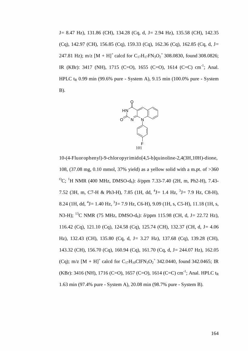
164
J= 8.47 Hz), 131.86 (CH), 134.28 (Cq, d, J= 2.94 Hz), 135.58 (CH), 142.35
(Cq), 142.97 (CH), 156.85 (Cq), 159.33 (Cq), 162.36 (Cq), 162.85 (Cq, d, J=
247.81 Hz); m/z [M + H]+ calcd for C17H17FN3O2+ 308.0830, found 308.0826;
IR (KBr): 3417 (NH), 1715 (C=O), 1655 (C=O), 1614 (C=C) cm-1; Anal.
HPLC tR 0.99 min (99.6% pure - System A), 9.15 min (100.0% pure - System
B).
HN
N N
O
O
F101
10-(4-Fluorophenyl)-9-chloropyrimido[4,5-b]quinoline-2,4(3H,10H)-dione,
108, (37.08 mg, 0.10 mmol, 37% yield) as a yellow solid with a m.pt. of >360
OC; 1H NMR (400 MHz, DMSO-d6): δ/ppm 7.33-7.40 (2H, m, Ph2-H), 7.43-
7.52 (3H, m, C7-H & Ph3-H), 7.85 (1H, dd, 4J= 1.4 Hz, 3J= 7.9 Hz, C8-H),
8.24 (1H, dd, 4J= 1.40 Hz, 3J= 7.9 Hz, C6-H), 9.09 (1H, s, C5-H), 11.18 (1H, s,
N3-H); 13C NMR (75 MHz, DMSO-d6): δ/ppm 115.98 (CH, d, J= 22.72 Hz),
116.42 (Cq), 121.10 (Cq), 124.58 (Cq), 125.74 (CH), 132.37 (CH, d, J= 4.06
Hz), 132.43 (CH), 135.80 (Cq, d, J= 3.27 Hz), 137.68 (Cq), 139.28 (CH),
143.32 (CH), 156.70 (Cq), 160.94 (Cq), 161.70 (Cq, d, J= 244.07 Hz), 162.05
(Cq); m/z [M + H]+ calcd for C17H10ClFN3O2+ 342.0440, found 342.0465; IR
(KBr): 3416 (NH), 1716 (C=O), 1657 (C=O), 1614 (C=C) cm-1; Anal. HPLC tR
1.63 min (97.4% pure - System A), 20.08 min (98.7% pure - System B).

165
HN
N N
O
O
F
Cl
108
10-(4-Fluorophenyl)-9-trifluoromethylpyrimido[4,5-b]quinoline-
2,4(3H,10H)-dione, 115, (45.50 mg, 0.12 mmol, 46% yield) as a yellow solid
with a m.pt. of >350 OC; 1H NMR (400 MHz, DMSO-d6): δ/ppm 7.29-7.36
(2H, m, Ph2-H), 7.38-7.45 (2H, m, Ph3-H), 7.67 (1H, t, J= 7.4 Hz, C7-H), 8.20
(1H, d, J= 7.4 Hz, C8-H), 8.49 (1H, d, J= 7.4 Hz, C6-H), 9.11 (1H, s, C5-H),
11.24 (1H, s, N3-H); 13C NMR (75 MHz, DMSO-d6): δ/ppm 115.41 (CH, d, J=
23.01 Hz), 116.76 (Cq), 118.60 (Cq), 118.91 (Cq), 124.34 (Cq), 124.88 (CH),
133.52 (CH, d, J= 9.33 Hz), 135.57 (CH), 136.97 (CH), 140.44 (Cq), 143.42
(CH), 156.75 (Cq), 162.00 (Cq), 161.31 (Cq), 162.27 (Cq, d, J= 247.93 Hz);
m/z [M + H]+ calcd for C18H10F4N3O2+ 376.0704, found 376.0719; IR (KBr):
3420 (NH), 1716 (C=O), 1660 (C=O), 1620 (C=C) cm-1; Anal. HPLC tR 2.25
min (100.0% pure - System A), 27.65 min (100.0% pure - System B).
HN
N N
O
O
F
CF3
115
10-(2-Methylphenyl)pyrimido[4,5-b]quinoline-2,4(3H,10H)-dione, 102,
(45.6 mg, 0.15 mmol, 46% yield) as a yellow solid with a m.pt. of 294-296 OC;
1H NMR (400 MHz, DMSO-d6): δ/ppm 2.42 (3H, s, Me), 6.73 (1H, d, J= 8.0
Hz, C9-H), 7.19-7.25 (2H, m, Ph-H), 7.44 (1H, m, Ph-H), 7.49 (1H, t, J= 7.4

166
Hz, Ph-H), 7.57 (1H, t, J= 8.0 Hz, C7-H), 7.73 (1H, ddd, 4J= 1.4 Hz, 3J= 8.0
Hz, C8-H), 8.21 (1H, dd, 4J= 1.4 Hz, 3J= 8.0 Hz, C6-H), 9.11 (1H, s, C5-H),
11.06 (1H, s, N3-H); 13C NMR (75 MHz, DMSO-d6): δ/ppm 21.33 (CH3),
116.05 (Cq), 117.66 (CH), 121.39 (Cq), 124.85 (CH), 125.80 (CH), 129.06
(CH), 130.45 (CH), 130.48 (CH), 131.78 (CH), 135.48 (CH), 138.06 (Cq),
140.39 (Cq), 142.22 (Cq), 142.83 (CH), 156.91 (Cq), 159.05 (Cq), 162,37
(Cq); m/z [M + H]+ calcd for C18H14N3O2+ 304.1081, found 304.1063; IR
(KBr): 3425 (NH), 1703 (C=O), 1654 (C=O), 1613 (C=C) cm-1; Anal. HPLC tR
1.27 min (100.0% pure - System A), 14.65 min (98.9% pure - System B).
HN
N N
O
O
102
10-(2-Methylphenyl)-9-chloropyrimido[4,5-b]quinoline-2,4(3H,10H)-dione,
109, (56.5 mg, 0.16 mmol, 57% yield) as a yellow solid with a m.pt. of 292-
293 OC (dec); 1H NMR (400 MHz, DMSO-d6): δ/ppm 2.36 (3H, s, Me), 7.18-
7.24 (2H, m, Ph-H), 7.31-7.36 (1H, m, Ph-H), 7.38-7.43 (1H, m, Ph-H), 7.47
(1H, t, J= 7.7 Hz, C7-H), 7.83 (1H, dd, 4J= 1.4 Hz, 3J= 7.7 Hz, C8-H), 8.23
(1H, dd, 4J= 1.4 Hz, 3J= 7.7 Hz, C6-H), 9.08 (1H, s, C5-H), 11.16 (1H, s, N3-
H); 13C NMR (75 MHz, DMSO-d6): δ/ppm 21.30 (CH3), 116.41 (Cq), 121.29
(Cq), 124.56 (Cq), 125.69 (CH), 127.65 (CH), 128.86 (CH), 130.08 (CH),
130.45 (CH), 132.32 (CH), 137.66 (Cq), 138.52 (Cq), 139.26 (CH), 139.48
(Cq), 143.21 (CH), 156.74 (Cq), 160.73 (Cq), 162.04 (Cq); m/z [M + H]+ calcd
for C18H13ClN3O2+ 338.0691, found 338.0718; IR (KBr): 3441 (NH), 1715

167
(C=O), 1656 (C=O), 1613 (C=C) cm-1; Anal. HPLC tR 2.20 min (97.6% pure -
System A), 32.08 min (96.5% pure - System B).
HN
N N
O
O
Cl
109
10-(2-Methylphenyl)-9-trifluoromethylpyrimido[4,5-b]quinoline-
2,4(3H,10H)-dione, 116, (24.0 mg, 0.6 mmol, 24% yield) as a yellow solid
with a m.pt. of 347-348 OC (dec); 1H NMR (400 MHz, DMSO-d6): δ/ppm 1.89
(3H, s, Me), 7.18 (1H, d, J= 7.6 Hz, Ph3-H), 7.28 (1H, ddd, 4J= 1.5 Hz, 3J= 7.6
Hz, Ph4-H), 7.33-7.43 (2H, m, Ph-H), 7.67 (1H, t, J= 7.7 Hz, C7-H), 8.22 (1H,
dd, 4J= 1.3 Hz, 3J= 7.7 Hz, C8-H), 8.51 (1H, dd, 4J= 1.3 Hz, 3J= 7.7 Hz, C6-
H), 9.12 (1H, s, C5-H), 11.23 (1H, s, N3-H); 13C NMR (75 MHz, DMSO-d6):
δ/ppm 18.32 (CH3), 116.89 (Cq), 117.99 (Cq), 118.62 (Cq), 124.19 (CH),
124.73 (Cq), 126.33 (CH), 129.92 (CH), 130.82 (CH), 131.19 (CH), 137.45
(CH), 137.78 (CH), 139.51 (Cq), 140.36 (Cq), 143.20 (CH), 156.86 (Cq),
160.61 (Cq), 162.10 (Cq); m/z [M + H]+ calcd for C19H13F3N3O2+ 372.0954,
found 372.0938; IR (KBr): 3421 (NH), 1703 (C=O), 1677 (C=O), 1623 (C=C)
cm-1; Anal. HPLC tR 2.68 min (98.5% pure - System A), 6.22 min (97.5% pure
- System B).
HN
N N
O
O
CF3
116

168
10-(3-Methylphenyl)pyrimido[4,5-b]quinoline-2,4(3H,10H)-dione, 103,
(20.8 mg, 0.06 mmol, 21% yield) as a yellow solid with a m.pt. of 328-329 OC
(dec); 1H NMR (400 MHz, DMSO-d6): δ/ppm 1.89 (3H, s, Me), 6.64 (1H, d,
J= 7.7 Hz, C9-H), 7.34-7.38 (1H, m, Ph-H), 7.47-7.58 (4H, m, C7-H & Ph-H),
7.74 (1H, ddd, 4J= 1.4 Hz, 3J= 7.7 Hz, C8-H), 8.25 (1H, dd, 4J= 1.4 Hz, 3J= 7.7
Hz, C6-H), 9.14 (1H, s, C5-H), 11.08 (1H, s, N3-H); 13C NMR (75 MHz,
DMSO-d6): δ/ppm 17.28 (CH3), 116.19 (Cq), 116.88 (CH), 121.54 (Cq),
125.08 (CH), 128.38 (CH), 128.70 (CH), 130.09 (CH), 131.98 (CH), 132.04
(CH),135.55 (Cq), 135.84 (CH), 137.01 (Cq), 141.30 (Cq), 142.94 (CH),
157.02 (Cq), 158.45 (Cq), 162.42 (Cq); m/z [M + H]+ calcd for C18H14N3O2+
304.1081, found 304.1095; IR (KBr): 3413 (NH), 1706 (C=O), 1655 (C=O),
1607 (C=C) cm-1; Anal. HPLC tR 1.12 min (97.8% pure - System A), 11.44
min (98.0% pure - System B).
HN
N N
O
O
103
10-(3-Methylphenyl)-9-chloropyrimido[4,5-b]quinoline-2,4(3H,10H)-dione,
110, (16.9 mg, 0.05 mmol, 17% yield) as a yellow solid with a m.pt. of 342-
343 OC; 1H NMR (400 MHz, DMSO-d6): δ/ppm 2.04 (3H, s, Me), 7.19 (1H,
dd, J= 1.0 Hz, J= 7.6 Hz, Ph4-H), 7.31 (1H, t, J= 7.6 Hz, Ph5-H), 7.36-7.45
(2H, m, Ph-H), 7.49 (1H, t, J= 7.8 Hz, C7-H), 7.84 (1H, dd, 4J= 1.4 Hz, 3J=
7.8 Hz, C8-H), 8.25 (1H, dd, 4J= 1.4 Hz, 3J= 7.8 Hz, C6-H), 9.11 (1H, s, C5-
H), 11.18 (1H, s, N3-H); 13C NMR (75 MHz, DMSO-d6): δ/ppm 18.10 (CH3),
116.53 (Cq), 120.90 (Cq), 124.58 (Cq), 125.74 (CH), 127.07 (CH), 129.38

169
(CH), 129.83 (CH), 130.65 (CH), 132.53 (CH), 137.26 (Cq), 137.61 (Cq),
139.02 (Cq), 139.23 (CH), 143.23 (CH), 156.87 (Cq), 159.97 (Cq), 162.10
(Cq); m/z [M + H]+ calcd for C18H13ClN3O2+ 338.0691, found 338.0653; IR
(KBr): 3417 (NH), 1712 (C=O), 1671 (C=O), 1619 (C=C) cm-1; Anal. HPLC tR
1.97 min (96.8% pure - System A), 24.47 min (97.2% pure - System B).
HN
N N
O
O
Cl
110
10-(3-Methylphenyl)-9-trifluoromethylpyrimido[4,5-b]quinoline-
2,4(3H,10H)-dione, 117, (38.6 mg, 0.10 mmol, 39% yield) as a yellow solid
with a m.pt. of 296-298 OC (dec); 1H NMR (400 MHz, DMSO-d6): δ/ppm 2.31
(3H, s, Me), 7.08 (1H, s, Ph2-H), 7.18 (1H, d, J= 7.6 Hz, Ph4-H), 7.29 (1H, d,
J= 7.6 Hz, Ph6-H), 7.36 (1H, t, J= 7.6 Hz, Ph5-H), 7.66 (1H, t, J= 7.6 Hz, C7-
H), 8.18 (1H, dd, 4J= 1.5 Hz, 3J= 7.6 Hz, C8-H), 8.47 (1H, dd, 4J= 1.5 Hz, 3J=
7.6 Hz, C6-H), 9.10 (1H, s, C5-H), 11.22 (1H, s, N3-H); 13C NMR (75 MHz,
DMSO-d6): δ/ppm 21.78 (CH3), 116.71 (Cq), 118.95 (Cq), 119.27 (Cq), 124.28
(Cq), 124.82 (CH), 128.12 (CH), 128.66 (CH), 130.10 (CH), 131.11 (CH),
135.51 (CH), 136.79 (CH), 137.96 (Cq), 140.49 (Cq), 143.29 (CH), 156.85
(Cq), 161.23 (Cq), 162.05 (Cq); m/z [M + H]+ calcd for C19H13F3N3O2+
372.0954, found 372.0954; IR (KBr): 3474 (NH), 1716 (C=O), 1657 (C=O),
1619 (C=C) cm-1; Anal. HPLC tR 3.00 min (100.0% pure - System A), 4.64
min (99.7% pure - System B).

170
HN
N N
O
O
CF3
117
10-(4-Methylphenyl)pyrimido[4,5-b]quinoline-2,4(3H,10H)-dione, 104,
(48.8 mg, 0.16 mmol, 49% yield) as a yellow solid with a m.pt. of 315-316 OC
(dec); 1H NMR (400 MHz, DMSO-d6): δ/ppm 2.46 (3H, s, Me), 6.76 (1H, d,
J= 8.2 Hz, C9-H), 7.29 (2H, d, AA’BB’system, J= 8.3 Hz, Ph3-H), 7.46-7.53
(3H, m, C7-H & Ph2-H), 7.73 (1H, ddd, 4J= 1.4 Hz, 3J= 8.2 Hz, C8-H), 8.22
(1H, dd, 4J= 1.4 Hz, 3J= 8.2 Hz, C6-H), 9.11 (1H, s, C5-H), 11.04 (1H, s, N3-
H); 13C NMR (75 MHz, DMSO-d6): δ/ppm 21.30 (CH3), 116.07 (Cq), 117.67
(CH), 121.41 (Cq), 124.86 (CH), 128.58 (CH), 131.16 (CH), 131.82 (CH),
135.43 (CH), 135.53 (Cq), 139.39 (Cq), 142.41 (Cq), 142.78 (CH), 156.87
(Cq), 159.19 (Cq), 162.43 (Cq); m/z [M + H]+ calcd for C18H14N3O2+ 304.1081,
found 304.1082; IR (KBr): 3437 (NH), 1698 (C=O), 1671 (C=O), 1611 (C=C)
cm-1; Anal. HPLC tR 1.34 min (98.6% pure - System A), 16.20 min (100.0%
pure - System B).
HN
N N
O
O
104
10-(4-Methylphenyl)-9-chloropyrimido[4,5-b]quinoline-2,4(3H,10H)-dione,
111, (19.0 mg, 0.05 mmol, 19% yield) as a yellow solid with a m.pt. of >350
OC; 1H NMR (400 MHz, DMSO-d6): δ/ppm 2.42 (3H, s, Me), 7.25 (2H, d,

171
AA’BB’system, J= 8.2 Hz, Ph3-H), 7.31 (2H, d, AA’BB’system, J= 8.2 Hz,
Ph2-H), 7.47 (1H, t, J= 7.7 Hz, C7-H), 7.82 (1H, dd, 4J= 1.4 Hz, 3J= 7.7 Hz,
C8-H), 8.22 (1H, dd, 4J= 1.4 Hz, 3J= 7.7 Hz, C6-H), 9.07 (1H, s, C5-H), 11.15
(1H, s, N3-H); 13C NMR (75 MHz, DMSO-d6): δ/ppm 21.38 (CH3), 116.39
(Cq), 121.40 (Cq), 124.57 (Cq), 125.63 (CH), 129.58 (CH), 130.09 (CH),
132.31 (CH), 137.05 (Cq), 137.83 (Cq), 138.88 (Cq), 139.22 (CH), 143.17
(CH), 156.76 (Cq), 160.88 (Cq), 162.16 (Cq); m/z [M + H]+ calcd for
C18H13ClN3O2+ 338.0691, found 338.0701; IR (KBr): 3418 (NH), 1717 (C=O),
1660 (C=O), 1615 (C=C) cm-1; Anal. HPLC tR 2.38 min (97.6% pure - System
A), 5.90 min (100.0% pure - System B).
HN
N N
O
O
Cl
111
10-(4-Methylphenyl)-9-trifluoromethylpyrimido[4,5-b]quinoline-
2,4(3H,10H)-dione, 118, (52.1 mg, 0.14 mmol, 52% yield) as a yellow solid
with a m.pt. of 344-346 OC (dec); 1H NMR (400 MHz, DMSO-d6): δ/ppm 2.38
(3H, s, Me), 7.19 (2H, d, AA’BB’system, J= 8.4 Hz, Ph3-H), 7.27 (2H, d,
AA’BB’system, J= 8.4 Hz, Ph2-H), 7.65 (1H, t, J= 7.9 Hz, C7-H), 8.17 (1H,
dd, 4J= 1.3 Hz, 3J= 7.9 Hz, C8-H), 8.47 (1H, dd, 4J= 1.3 Hz, 3J= 7.9 Hz, C6-
H), 9.09 (1H, s, C5-H), 11.21 (1H, s, N3-H); 13C NMR (75 MHz, DMSO-d6):
δ/ppm 21.27 (CH3), 116.72 (Cq), 118.94 (Cq), 124.30 (Cq), 124.80 (CH),
128.94 (CH), 130.92 (CH), 135.52 (CH), 136.79 (CH), 138.18 (Cq), 139.12
(Cq), 140.56 (Cq), 143.28 (CH), 156.80 (Cq), 161.29 (Cq), 162.07 (Cq); m/z
[M + H]+ calcd for C19H13F3N3O2+ 372.0954, found 372.0957; IR (KBr): 3438

172
(NH), 1706 (C=O), 1681 (C=O), 1622 (C=C) cm-1; Anal. HPLC tR 3.13 min
(100.0% pure - System A), 8.57 min (99.7% pure - System B).
HN
N N
O
O
CF3
118
10-(3,4-Dichlorophenyl)-9-trifluoromethylpyrimido[4,5-b]quinoline-
2,4(3H,10H)-dione, 119, (62.3 mg, 0.14 mmol, 62% yield) as a yellow solid
with a m.pt. of 320-322 OC; 1H NMR (400 MHz, DMSO-d6): δ/ppm 7.46 (1H,
dd, 4J= 1.1 Hz, 3J= 8.6 Hz, Ph6-H), 7.69 (1H, t, J= 7.8 Hz, C7-H), 7.73 (1H, d,
4J= 1.1 Hz, Ph2-H), 7.81 (1H, d, J= 8.6 Hz, Ph5-H), 8.23 (1H, dd, 4J= 1.6 Hz,
3J= 7.8 Hz, C8-H), 8.50 (1H, dd, 4J= 1.6 Hz, 3J= 7.8 Hz, C6-H), 9.12 (1H, s,
C5-H), 11.30 (1H, s, N3-H); 13C NMR (75 MHz, DMSO-d6): δ/ppm 116.71
(Cq), 118.33 (Cq), 121.67 (Cq), 124.46 (Cq), 125.13 (CH), 130.37 (CH),
130.94 (Cq), 132.28 (CH), 132.58 (Cq), 133.14 (CH), 135.71 (CH), 137.30
(CH), 139.84 (Cq), 143.71 (CH), 156.61 (Cq), 161.14 (Cq), 161.88 (Cq); m/z
[M + H]+ calcd for C18H9Cl2F3N3O2+ 426.0018, found 426.0034; IR (KBr):
3486 (NH), 1705 (C=O), 1655 (C=O), 1609 (C=C) cm-1; Anal. HPLC tR 6.40
min (99.4% pure - System A), 6.35 min (98.7% pure - System B).
HN
N N
O
O
CF3
Cl
Cl119

173
General Synthesis of 10-Phenyl-3-methylpyrimido[4,5-b]quinoline-
2,4(3H,10H)-dione Analogues
To 1 equivalent of the desired 10-phenylpyrimido[4,5-b]quinoline-
2,4(3H,10H)-dione analogue in methanol (20 mL) was added 2.05 equivalents
of sodium methoxide. After stirring for 30 min, 4 equivalents of methyl iodide
was added and refluxed for 24 h. Concentration of the solution under reduced
pressure and separation by preparative thin layer chromatography using 98%
dichloromethane and 2% methanol to yield the pure desired 10-phenyl-3-
methylpyrimido[4,5-b]quinoline-2,4(3H,10H)-dione analogue.
10-Phenyl-3-methylpyrimido[4,5-b]quinoline-2,4(3H,10H)-dione, 120,
(17.4 mg, 0.05 mmol, 33% yield) as a yellow solid with a m.pt. of >350 OC (lit
>360 OC) [271]; 1H NMR (400 MHz, DMSO-d6): δ/ppm 3.23 (3H, s, Me), 6.74
(1H, d, J= 8.2 Hz, C9-H), 7.40-7.78 (4H, m, C7-H & Ph-H), 7.62-7.73 (2H, m,
Ph2-H), 7.75 (ddd, 4J= 1.5 Hz, 3J= 8.2 Hz, C8-H), 8.28 (1H, dd, 4J= 1.5 Hz,
3J= 8.2 Hz, C6-H), 9.19 (1H, s, C5-H); 13C NMR (75 MHz, DMSO-d6): δ/ppm
28.01 (CH3), 115.60 (CH), 117.48 (Cq), 121.61 (Cq), 124.99 (CH), 128.81
(CH), 129.94 (CH), 130.82 (CH), 131.88 (CH), 135.58 (CH), 137.83 (Cq),
142.09 (Cq), 143.47 (CH), 156.30 (Cq), 157.62 (Cq), 161.94 (Cq); m/z [M +
H]+ calcd for C18H14N3O2+ 304.1081, found 304.1066; IR (KBr): 1637 (C=O),
1616 (C=O), 1566 (C=C) cm-1; Anal. HPLC tR 1.40 min (97.9% pure - System
A), 14.05 min (100.0% pure - System B).
N
N N
O
O
120

174
10-(4-Chlorophenyl)-9-trifluoromethyl-3-methylpyrimido[4,5-b]quinoline-
2,4(3H,10H)-dione, 122, (6.8 mg, 0.01 mmol, 22% yield) as a yellow solid
with a m.pt. of >360 OC; 1H NMR (400 MHz, DMSO-d6): δ/ppm 5.75 (3H, s,
Me), 7.39 (2H, d, AA’BB’system, J= 8.7 Hz, Ph2-H), 7.58 (2H, d, AA’BB’
system, J= 8.7 Hz, Ph3-H), 7.70 (1H, t, J= 7.8 Hz, C7-H), 8.23 (1H, dd, 4J= 1.4
Hz, 3J= 7.8 Hz, C8-H), 8.54 (1H, dd, 4J= 1.4 Hz, 3J= 7.8 Hz, C6-H), 9.18 (1H,
s, C5-H); 13C NMR (75 MHz, DMSO-d6): δ/ppm 21.08 (CH3), 115.84 (Cq),
118.34 (Cq), 123.98 (Cq), 125.12 (CH), 128.68 (CH), 133.86 (CH), 134.57
(Cq), 135.21 (CH), 137.77 (CH), 139.85 (Cq), 140.45 (Cq), 144.17 (CH),
157.04 (Cq), 161.84 (Cq), 162.27 (Cq); m/z [M + H]+ calcd for
C19H12ClF3N3O2+ 406.0565, found 406.0571; IR (KBr): 1700 (C=O), 1681
(C=O), 1620 (C=C) cm-1; Anal. HPLC tR 6.43 min (99.1% pure - System A),
5.12 min (96.8% pure - System B).
N
N N
O
O
CF3
Cl122
General Synthesis of 10-Phenyl-3-ethylpyrimido[4,5-b]quinoline-
2,4(3H,10H)-dione Analogues
To 1 equivalent of the desired 10-phenylpyrimido[4,5-b]quinoline-
2,4(3H,10H)-dione analogues in methanol (20 mL) was added 2.05 equivalents
of sodium ethoxide. After stirring for 30 min, 4 equivalents of ethyl iodide
were added and refluxed for 24 h. Concentration of the solution under reduced
pressure and separation by preparative thin layer chromatography using 98%

175
dichloromethane and 2% methanol to yield the pure desired 3-ethyl-10-
phenylpyrimido[4,5-b]quinoline-2,4(3H,10H)-dione analogue.
10-Phenyl-3-ethylpyrimido[4,5-b]quinoline-2,4(3H,10H)-dione, 121, (10.0
mg, 0.03 mmol, 18% yield) as a yellow solid with a m.pt. >360 OC; 1H NMR
(400 MHz, DMSO-d6): δ/ppm 1.11 (3H, t, J= 7.0 Hz, Me), 3.89 (2H, q, J= 7.0
Hz, CH2), 6.73 (1H, d, J= 8.0 Hz, C9-H), 7.43 (2H, d, J= 7.4 Hz, Ph2-H), 7.51
(1H, t, J= 8.0 Hz, C7-H), 7.61-7.80 (4H, m, C8-H & Ph-H), 8.27 (1H, dd, 4J=
1.2 Hz, 3J= 8.0 Hz, C6-H), 9.18 (1H, s, C5-H); 13C NMR (75 MHz, DMSO-
d6): δ/ppm 8.40 (CH3), 13.48 (CH2), 115.83 (CH), 118.05 (Cq), 122.16 (Cq),
125.57 (CH), 127.93 (CH), 130.09 (CH), 131.13 (CH), 131.77 (CH), 135.51
(CH), 138.03 (Cq), 142.25 (Cq), 143.67 (CH), 155.20 (Cq), 158.32 (Cq),
162.93 (Cq); m/z [M + H]+ calcd for C19H16N3O2+ 318.1237, found 318.1251;
IR (KBr): 1692 (C=O), 1638 (C=O), 1623 (C=C) cm-1; Anal. HPLC tR 1.99
min (100.0% pure - System A), 22.15 min (100.0% pure - System B).
N
N N
O
O
121
10-(4-Chlorophenyl)-9-trifluoromethyl-3-ethylpyrimido[4,5-b]quinoline-
2,4(3H,10H)-dione, 123, (9.3 mg, 0.02 mmol, 43% yield) as a yellow solid
with a m.pt. of 278-280 OC (dec); 1H NMR (400 MHz, DMSO-d6): δ/ppm 1.10
(3H, t, J= 7.9 Hz, Me), 3.87 (2H, q, J= 7.9 Hz, CH2), 7.39 (2H, d, AA’BB’
system, J= 8.6 Hz, Ph2-H), 7.57 (2H, d, AA’BB’system, J= 8.6 Hz, Ph3-H),
7.69 (1H, t, J= 7.8 Hz, C7-H), 8.22 (1H, dd, 4J= 1.4 Hz, 3J= 7.8 Hz, C8-H),
8.53 (1H, dd, 4J= 1.4 Hz, 3J= 7.8 Hz, C6-H), 9.17 (1H, s, C5-H); 13C NMR (75

176
MHz, DMSO-d6): δ/ppm 9.31 (CH3), 14.88 (CH2), 115.88 (Cq), 117.50 (Cq),
124.28 (Cq), 125.11 (CH), 128.52 (CH), 133.32 (CH), 134.67 (Cq), 136.16
(CH), 137.61 (CH), 139.47 (Cq), 140.94 (Cq), 144.28 (CH), 157.10 (Cq),
161.74 (Cq), 162.16 (Cq); m/z [M + H]+ calcd for C20H14ClF3N3O2+ 420.0721,
found 420.0692; IR (KBr): 1646 (C=O), 1611 (C=O), 1538 (C=C) cm-1; Anal.
HPLC tR 0.75 min (98.3% pure - System A), 19.62 min (99.4% pure - System
B).
N
N N
O
O
CF3
Cl123
General Procedure to Synthesise 10-Phenyl-9-Substituted-pyrimido[4,5-
b]quinoline-2,4(3H,10H)-dione Analogues [241, 261, 264, 265, 268, 270]
To 1 equivalent of the desired 6-(phenylamino)pyrimidine-2,4(1H,3H)-dione
analogue in DMF was added 1.2 equivalents of the desired 2-halobenzaldehyde
(chloro or fluoro). The mixture was heated at 160 OC for 30 min in the
microwave reactor or refluxed for 4 h. Concentration of the solution under
reduced pressure and separation by dry column flash chromatography using
95% dichloromethane and 5% methanol to yield the pure desired 10-
phenylpyrimido[4,5-b]quinoline-2,4(3H,10H)-dione analogue.
10-Phenyl-9-fluoropyrimido[4,5-b]quinoline-2,4(3H,10H)-dione, 124, (29.3
mg, 0.17 mmol, 29% yield) as a yellow solid with a m.pt. of 260-262 OC (dec);
1H NMR (400 MHz, DMSO-d6): δ/ppm 7.43-7.57 (6H, m, C8-H & Ph-H),
7.60 (1H, ddd, 4J= 1.5 Hz, 3J= 6.9 Hz, C7-H), 8.07 (1H, d, J= 6.9 Hz, C6-H),

177
9.12 (1H, s, C5-H), 11.14 (1H, s, N3-H); 13C NMR (75 MHz, DMSO-d6):
δ/ppm 116.78 (Cq), 122.61 (CH, d, J= 21.93 Hz), 123.84 (Cq), 125.42 (CH, d,
J= 8.01 Hz), 128.22 (CH, d, J= 4.70 Hz), 128.41 (CH, d, J= 3.77 Hz), 129.14
(CH), 129.41 (CH), 130.32 (Cq, d, J= 3.79 Hz), 140.98 (Cq, d, J= 3.88 Hz),
142.83 (CH, d, J= 2.21 Hz), 150.08 (Cq, d, J= 250.69 Hz), 156.69 (Cq), 159.98
(Cq), 162.10 (Cq); m/z [M + H]+ calcd for C17H11FN3O2+ 308.0830, found
308.0813; IR (KBr): 3410 (NH), 1703 (C=O), 1667 (C=O), 1613 (C=C) cm-1;
Anal. HPLC tR 1.02 min (99.5% pure - System A), 9.57 min (97.6% pure -
System B).
HN
N N
O
O
F
124
10-(4-Chlorophenyl)-9-fluoropyrimido[4,5-b]quinoline-2,4(3H,10H)-dione,
126, (20.6 mg, 0.06 mmol, 21% yield) as a yellow solid with a m.pt. of 268-
269 OC (dec); 1H NMR (400 MHz, DMSO-d6): δ/ppm 7.46-7.52 (3H, m, C7-H
& Ph2-H), 7.59-7.66 (3H, m, C8 & Ph3-H), 8.07 (1H, d, J= 7.8 Hz, C6-H),
9.12 (1H, s, C5-H), 11.16 (1H, s, N3-H); 13C NMR (75 MHz, DMSO-d6):
δ/ppm 116.76 (Cq), 122.65 (CH, d, J= 21.88 Hz), 123.84 (Cq), 125.49 (CH, d,
J= 8.02 Hz), 128.48 (CH, d, J= 3.47 Hz), 129.50 (CH), 130.15 (Cq, d, J= 4.53
Hz), 130.25 (CH, d, J= 4.47 Hz), 133.82 (Cq), 139.84 (Cq, d, J= 3.79 Hz),
142.97 (CH), 150.02 (Cq, d, J= 251.49 Hz), 156.64 (Cq), 160.05 (Cq), 162.08
(Cq); m/z [M + H]+ calcd for C17H10ClFN3O2+ 342.0440, found 342.0435; IR
(KBr): 3431 (NH), 1716 (C=O), 1657 (C=O), 1614 (C=C) cm-1; Anal. HPLC tR
1.82 min (99.8% pure - System A), 19.97 min (98.2% pure - System B).

178
HN
N N
O
O
F
Cl126
10-Phenyl-9-hydroxypyrimido[4,5-b]quinoline-2,4(3H,10H)-dione, 128,
(44.6 mg, 0.14 mmol, 45% yield) as a yellow solid with a m.pt. of 207-208 OC;
1H NMR (400 MHz, DMSO-d6): δ/ppm 7.09 (1H, dd, 4J= 1.2 Hz, 3J= 7.8 Hz,
C8-H), 7.28-7.34 (3H, m), 7.41-7.48 (3H, m), 7.65 (1H, dd, 4J= 1.2 Hz, 3J= 7.8
Hz, C6-H), 9.00 (1H, s, C5-H), 10.00 (1H, s, OH), 10.98 (1H, s, N3-H); 13C
NMR (75 MHz, DMSO-d6): δ/ppm 115.68 (Cq), 121.79 (CH), 122.63 (CH),
123.67 (Cq), 125.70 (CH), 128.06 (CH), 128.35 (CH), 128.70 (CH), 130.24
(Cq), 142.38 (Cq), 143.58 (Cq), 147.07 (Cq), 156.78 (Cq), 159.96 (Cq), 162.42
(Cq); m/z [M + H]+ calcd for C17H12N3O3+ 306.0873, found 306.0844; IR
(KBr): 3475 (NH), 1695 (C=O), 1650 (C=O), 1602 (C=C) cm-1; Anal. HPLC tR
0.63 min (100.0% pure - System A), 6.18 min (97.4% pure - System B).
HN
N N
O
O
OH
128
10-(4-Chlorophenyl)-9-hydroxypyrimido[4,5-b]quinoline-2,4(3H,10H)-
dione, 130, (17.50 mg, 0.05 mmol, 9% yield) as a yellow solid with a m.pt. of
316-318 OC; 1H NMR (400 MHz, DMSO-d6): δ/ppm 7.08 (1H, d, J= 7.6 Hz,
C8-H), 7.31 (1H, t, J= 7.6 Hz, C7-H), 7.37 (2H, d, AA’BB’system, J= 8.2 Hz,
Ph2-H), 7.50 (2H, d, AA’BB’system, J= 8.2 Hz, Ph3-H), 7.76 (1H, d, J= 7.6

179
Hz, C6-H), 8.99 (1H, s, C5-H), 10.16 (1H, s, OH), 11.00 (1H, s, N3-H); 13C
NMR (75 MHz, DMSO-d6): δ/ppm 115.68 (Cq), 121.84 (CH), 122.70 (CH),
123.69 (Cq), 125.79 (CH), 128.34 (CH), 130.05 (Cq), 130.64 (CH), 132.74
(Cq), 141.26 (Cq), 143.71 (CH), 146.95 (Cq), 156.70 (Cq), 159.95 (Cq),
162.34 (Cq); m/z [M + H]+ calcd for C17H11ClN3O3+ 340.0740, found
340.0713; IR (KBr): 3434 (NH), 1702 (C=O), 1655 (C=O), 1603 (C=C) cm-1;
Anal. HPLC tR 0.80 min (99.8% pure - System A), 10.85 min (100.0% pure -
System B).
HN
N N
O
O
OH
Cl130
Synthesis of 2-p-Toluenesulfonyloxy-3-bromobenzaldehyde, 145 [301-303]
A mixture of 2-hydroxy-3-bromobenzaldehyde (565.81 mg, 0.76 mL; 2.81
mmol) and sodium carbonate (2.31 g; 21.85 mmol) in acetone (20 mL) was
stirred at room temperature for 30 min under a nitrogen atmosphere. Tosyl
chloride (1.07 g; 5.62 mmol) dissolved in acetone (25 mL) was added to the
reaction mixture and refluxed for 5 h. Concentration of the solution under
reduced pressure and dry column flash chromatography using 66.6% petroleum
ether 33.3% and diethyl ether was carried out to yield the pure 2-p-
toluenesulfonyloxy-3-bromobenzaldehyde, 145.
2-p-Toluenesulfonyloxy-3-bromobenzaldehyde, 145 (0.82 g, 2.32 mmol,
82% yield) as a white solid with a m.pt. of 79-91 OC; 1H NMR (400 MHz,
DMSO-d6): δ/ppm 2.46 (3H, s, Me), 7.44-7.58 (3H, m), 7.77-7.88 (3H, m),

180
8.05 (1H, d, J= 7.3 Hz, C4-H), 9.86 (1H, s, COH); 13C NMR (75 MHz,
DMSO-d6): δ/ppm 21.74 (CH3), 118.58 (Cq), 128.40 (CH), 129.17 (CH),
129.94 (CH), 131.02 (CH), 131.28 (Cq), 132.40 (Cq), 139.98 (CH), 147.44
(Cq), 147.65 (Cq), 187.53 (CH).
O
Br
OS
O
O
145
General Procedure to Synthesise 10-Phenyl-9-bromopyrimido[4,5-
b]quinoline-2,4(3H,10H)-dione Analogues using 2-p-Toluenesulfonyloxy-3-
bromobenzaldehyde, 145 [241, 261, 264, 265, 268, 270]
To 1 equivalent of the desired 6-(phenylamino)pyrimidine-2,4(1H,3H)-dione
analogue in DMF was added 1.2 equivalents of the 2-p-toluenesulfonyloxy-3-
bromobenzaldehyde, 145. The mixture was heated at 160 OC for 30 min in the
microwave reactor or refluxed for 4 h. Concentration of the solution under
reduced pressure and separation by dry column flash chromatography using
95% dichloromethane and 5% methanol to yield the pure desired 10-
phenylpyrimido[4,5-b]quinoline-2,4(3H,10H)-dione analogue.
10-Phenyl-9-bromopyrimido[4,5-b]quinoline-2,4(3H,10H)-dione, 125, (29.3
mg, 0.07 mmol, 29% yield) as a yellow solid with a m.pt. of >350 OC; 1H
NMR (400 MHz, DMSO-d6): δ/ppm 7.37-7.42 (3H, m), 7.51-7.55 (3H, m),
8.06 (1H, dd, 4J= 1.5 Hz, 3J= 7.8 Hz, C8-H), 8.27 (1H, dd, 4J= 1.5 Hz, 3J= 7.8
Hz, C6-H), 9.06 (1H, s, C5-H), 11.16 (1H, s, N3-H); 13C NMR (75 MHz,
DMSO-d6): δ/ppm 109.28 (Cq), 116.31 (Cq), 124.76 (Cq), 126.06 (CH),
129.11 (CH), 129.63 (CH), 131.02 (Cq), 131.15 (CH), 132.87 (CH), 138.75
(CH), 138.88 (Cq), 143.19 (CH), 156.73 (Cq), 160.73 (Cq), 162.06 (Cq); m/z

181
[M + H]+ calcd for C17H11BrN3O2+ 368.0029, found 367.9998; IR (KBr): 3413
(NH), 1717 (C=O), 1658 (C=O), 1614 (C=C) cm-1; Anal. HPLC tR 1.55 min
(98.5% pure - System A), 21.07 min (99.2% pure - System B).
HN
N N
O
O
Br
125
10-(4-Chlorophenyl)-9-bromopyrimido[4,5-b]quinoline-2,4(3H,10H)-
dione, 127, (36.4 mg, 0.09 mmol, 36% yield) as a yellow solid with a m.pt. of
>350 OC; 1H NMR (400 MHz, DMSO-d6): δ/ppm 7.40 (1H, t, J= 7.7 Hz, C7-
H), 7.44 (2H, d, AA’BB’system, J= 8.7 Hz, Ph2-H), 7.61 (2H, d, AA’BB’
system, J= 8.7 Hz, Ph3-H), 8.08 (1H, dd, 4J= 1.5 Hz, 3J= 7.7 Hz, C8-H), 8.27
(1H, dd, 4J= 1.5 Hz, 3J= 7.7 Hz, C6-H), 9.06 (1H, s, C5-H), 11.19 (1H, s, N3-
H); 13C NMR (75 MHz, DMSO-d6): δ/ppm 109.12 (Cq), 116.30 (Cq), 124.76
(Cq), 126.15 (CH), 129.18 (CH), 132.95 (CH), 132.96 (CH), 134.33 (Cq),
137.72 (Cq), 138.72 (Cq), 143.19 (CH), 143.34 (CH), 156.68 (Cq), 160.84
(Cq), 162.04 (Cq); m/z [M + H]+ calcd for C17H10BrClN3O2+ 401.9639, found
401.9613; IR (KBr): 3439 (NH), 1714 (C=O), 1655 (C=O), 1610 (C=C) cm-1;
Anal. HPLC tR 2.73 min (97.6% pure - System A), 4.17 min (100.0% pure -
System B).
HN
N N
O
O
Br
Cl127

182
Synthesis of 3-Cyano-2-hydroxybenzaldehyde, 132 [286, 287]
To a warm solution of sodium hydroxide (40.29 g; 1007.38 mmol) in water
(250 mL) was added 2-hydroxybenzonitrile (15.00 g; 125.92 mmol) under
mechanical stirring. After the addition the temperature was raised to 70 OC and
chloroform (30.06 g, 20.31mL; 251.81 mmol) was added slowly drop wise
over 1 h. The reaction mixture temperature was kept at 70 OC with mechanical
stirring for a further 3 h. After heating, the reaction mixture was cooled to
room temperature and acidified by sulfuric acid to pH 1. Diethyl ether (300
mL) was added and liquid extraction carried out with the diethyl ether layer
treated with magnesium sulfate and filtered. Concentration of the solution
under reduced pressure and separation by dry column flash chromatography
using 95% dichloromethane and 5% methanol was carried out to yield the pure
3-cyano-2-hydroxy-benzaldehyde, 132.
3-Cyano-2-hydroxybenzaldehyde, 132 (0.47 g, 3.22 mmol, 3% yield) as a
white solid with a m.pt. of 114-115 OC (lit 113-114 OC) [287]; 1H NMR (400
MHz, DMSO-d6): δ/ppm 7.20 (1H, t, J= 7.8 Hz, C5-H), 8.01-8.06 (2H, m),
10.14 (1H, s, COH), 11.73 (1H, br s, OH); 13C NMR (75 MHz, DMSO-d6):
δ/ppm 102.25 (Cq), 116.02 (Cq), 121.07 (CH), 123.31 (Cq), 137.26 (CH),
140.36 (CH), 162.04 (Cq), 194.25 (CH); IR (KBr): 3411 (OH), 2235 (CN),
1668 (C=O)cm-1.
O
OH
CN132

183
Synthesis of 2-p-Toluenesulfonyloxy-3-cyanobenzaldehyde, 146 [301-303]
A mixture of 3-Cyano-2-hydroxybenzaldehyde, 132 (250.00 mg; 1.69 mmol),
and sodium carbonate (720.45 mg; 6.79 mmol) in acetone (50 mL) was stirred
at room temperature for 30 min under nitrogen atmosphere. Tosyl Chloride
(647.86 mg; 3.39 mmol) dissolved in acetone (25 mL) was added to the
reaction mixture and refluxed overnight. Concentration of the solution under
reduced pressure and dry column flash chromatography using 66.6% petroleum
ether and 33.3% diethyl ether was carried out to yield the pure 2-p-
toluenesulfonyloxy-3-cyanobenzaldehyde, 146.
2-p-Toluenesulfonyloxy-3-cyanobenzaldehyde, 146 (481.2 mg, 1.59 mmol,
94% yield) as a white solid with a m.pt. of 84-86 OC; 1H NMR (400 MHz,
DMSO-d6): δ/ppm 2.46 (3H, s, Me), 7.55 (2H, d, AA’BB’system, J= 8.5 Hz,
Ph3-H), 7.75 (1H, t, J= 7.6 Hz, C5-H), 7.82 (2H, d, AA’BB’system, J= 8.5
Hz, Ph2-H), 8.15 (1H, dd, J=1.76 Hz, J= 7.6 Hz, C6-H), 8.28 (1H, dd, J= 1.76
Hz, J= 7.6 Hz, C4-H), 9.84 (1H, s, COH); 13C NMR (75 MHz, DMSO-d6):
δ/ppm 21.76 (CH3), 109.73 (Cq), 114.46 (Cq), 129.27 (CH), 129.55 (CH),
129.95 (Cq), 131.22 (Cq), 131.25 (CH), 133.78 (CH), 140.01 (CH), 148.06
(Cq), 150.68 (Cq), 186.55 (CH).
O
O
CN
SO
O
146

184
General Procedure to Synthesise 10-Phenyl-9-cyanopyrimido[4,5-
b]quinoline-2,4(3H,10H)-dione Analogues using 2-p-Toluenesulfonyloxy-3-
cyanobenzaldehyde, 146 [241, 261, 264, 265, 268, 270]
To 1 equivalent of the desired 6-(phenylamino)pyrimidine-2,4(1H,3H)-dione
analogue in DMF was added 1.2 equivalents of 2-p-toluenesulfonyloxy-3-
cyanobenzaldehyde, 146. The mixture was heated at 160 OC for 30 min in the
microwave reactor or refluxed for 4 h. Concentration of the solution under
reduced pressure and separation by flash chromatography using 95%
dichloromethane 5% methanol to yield the pure desired 10-
phenylpyrimido[4,5-b]quinoline-2,4(3H,10H)-dione analogue .
10-(3-Chlorophenyl)-9-cyanopyrimido[4,5-b]quinoline-2,4(3H,10H)-dione,
134, (96.0 mg, 0.27 mmol, 64% yield) as a yellow solid with a m.pt. of >350
OC; 1H NMR (400 MHz, DMSO-d6): δ/ppm 7.53 (1H, d, J= 8.1 Hz, Ph6-H),
7.61-7.66 (2H, m), 7.72-7.76 (2H, m), 8.23 (1H, dd, 4J= 1.5 Hz, 3J= 7.8 Hz,
C8-H), 8.55 (1H, dd, 4J= 1.5 Hz, 3J= 7.8 Hz, C6-H), 9.16 (1H, s, C5-H), 11.28
(1H, s, N3-H); 13C NMR (75 MHz, DMSO-d6): δ/ppm 101.06 (Cq), 114.89
(Cq), 117.10 (Cq), 122.87 (Cq), 124.90 (CH), 129.74 (CH), 130.76 (CH),
131.44 (CH), 131.78 (CH), 134.32 (Cq), 137.80 (CH), 139.07 (Cq), 141.37
(Cq), 142.98 (CH), 143.66 (CH), 156.55 (Cq), 160.12 (Cq), 161.90 (Cq); m/z
[M + H]+ calcd for C18H10ClN4O2+ 349.0687, found 349.0627; IR (KBr): 3420
(NH), 2213 (CN), 1703 (C=O), 1664 (C=O), 1619 (C=C) cm-1; Anal. HPLC tR
0.90 min (99.5% pure - System A), 6.78 min (98.7% pure - System B).

185
HN
N N
O
O
Cl
CN
134
10-(4-Chlorophenyl)-9-cyanopyrimido[4,5-b]quinoline-2,4(3H,10H)-dione,
131, (37.2 mg, 0.10 mmol, 25% yield) as a yellow solid with a m.pt. of >350
OC; 1H NMR (400 MHz, DMSO-d6): δ/ppm 7.58 (2H, d, AA’BB’system, J=
8.7 Hz, Ph2-H), 7.62 (1H, t, J= 7.7 Hz, C7-H), 7.68 (2H, d, AA’BB’system,
J= 8.7 Hz, Ph3-H), 8.23 (1H, dd, 4J= 1.5 Hz, 3J= 7.7 Hz, C8-H), 8.54 (1H, dd,
4J= 1.5 Hz, 3J= 7.7 Hz, C6-H), 9.15 (1H, s, C5-H), 11.26 (1H, s, N3-H); 13C
NMR (75 MHz, DMSO-d6): δ/ppm 101.15 (Cq), 114.87 (Cq), 117.12 (Cq),
122.88 (Cq), 124.85 (CH), 130.23 (CH), 132.75 (CH), 135.99 (Cq), 136.81
(Cq), 137.69 (CH), 141.47 (Cq), 142.95 (CH), 143.56 (CH), 156.56 (Cq),
160.23 (Cq), 161.88 (Cq); m/z [M + H]+ calcd for C18H10ClN4O2+ 349.0687,
found 349.0627; IR (KBr): 3416 (NH), 2198 (CN), 1704 (C=O), 1666 (C=O),
1621 (C=C) cm-1; Anal. HPLC tR 0.92 min (100.0% pure - System A), 4.92
min (100.0% pure - System B).
HN
N N
O
O
CN
Cl131

186
References
1. Pecorino, L. Molecular Biology of Cancer. Oxford University Press,Oxford. 2nd ed. 2005.
2. Incidence - UK, in CancerStats. 2007. Cancer Research UK. Fromhttp://info.cancerresearchuk.org/prod_consump/groups/cr_common/@nre/@sta/documents/generalcontent/018070.pdf as of 27th Sept 2010.
3. Latest UK Cancer Incidence and Mortality Summary - numbers. 2010.Cancer Research UK. Fromhttp://info.cancerresearchuk.org/prod_consump/groups/cr_common/@nre/@sta/documents/generalcontent/crukmig_1000ast-2735.pdf as of27th Sept 2010.
4. Registrar General Annual Report 2007. Northern Ireland Statistics andResearch Agency. 2009.
5. Registrar General's Annual Report 2007. General Register Office forScotland. 2009.
6. Mortality statistics. Deaths registered in 2007. Office of NationalStatistics. 2009.
7. Cancer Facts & Figures 2009. American Cancer Society. Atlanta.American Cancer Society. 2009.
8. Jemal, A. R. S.; Ward, E.; Hao, Y.; Xu, J.; Thun, M. J. Cancer statistics2009. Ca. Cancer. J. Clin. 2009, 59, 225-49.
9. Cancer Trends Progress Report - 2007 Update, National CancerInstitute, NIH, DHHS, Bethesda, MD. 2007.
10. Worldwide Cancer, in CancerStats. 2005, Cancer Research UK. Fromhttp://info.cancerresearchuk.org/prod_consump/groups/cr_common/@nre/@sta/documents/generalcontent/cr_043265.pdf as of 27th Sept 2010.
11. Executive Summary, in World Population Prospects. The 2008Revision. The Department of Economic and Social Affairs of theUnited Nations Secretariat. 2009.
12. Loeb, L. A.; Loeb, K. R.; Anderson, J. P. Multiple mutations andcancer. Proc. Natl. Acad. Sci. U. S. A. 2003, 100, 776-81.
13. Loeb, K. R.; Loeb, L. A. Significance of multiple mutations in cancer.Carcinogenesis. 2000, 21, 379-85.
14. King, R. J. B. Cancer Biology. Oxford University Press, PearsonEducation Limited. 2nd ed. 2000.

187
15. Kleinsmith, L. Principles of Cancer Biology. Garland Science, PearsonInternational Edition, 1st ed. 2005.
16. Polsky, D.; Cordon-Cardo, C. Oncogenes in melanoma. Oncogene.2003, 22, 3087-91.
17. Alberts, B.; Lewis, J.; Raff, M.; Roberts, K.; Walter, P. MolecularBiology of The Cell. New York, Garland Science. 4th ed. 2002.
18. Lengauer, C.; Kinzler, K. W.; Vogelstein, B. Genetic instabilities inhuman cancers. Nature. 1998, 396, 643-49.
19. Marshall, C. J. Tumor suppressor genes. Cell. 1991, 64, 313-26.
20. Yeo, C. J. Tumor suppressor genes: A short review. Surgery. 1999, 125,363-66.
21. Vile, R. Tumour suppressor genes: Loss of these genes may causecancer. Br. Med. J. 1989, 298, 1335-36.
22. Swale, V. J.; Quinn, A. G. Tumour suppressor genes. Clin. Exp.Dermatol. 2000, 25, 231-35.
23. Hanahan, D.; Weinberg, R. A. The hallmarks of cancer. Cell. 2000,100, 57-70.
24. Evan, G. I.; di Fagagna, F. D. Cellular senescence: hot or what? Curr.Opin. Genet. Dev. 2009, 19, 25-31.
25. Zuckerman, V.; Wolyniec, K.; Sionov, R. V.; Haupt, S.; Haupt, Y.Tumour suppression by p53: the importance of apoptosis and cellularsenescence. Am. J. Pathol. 2009, 219, 3-15.
26. Campisi, J.; di Fagagna, F. D. Cellular senescence: when bad thingshappen to good cells. Nat. Rev. Mol. Cell Biol. 2007, 8, 729-40.
27. Bergers, G.; Benjamin, L. E. Tumorigenesis and the angiogenic switch.Nat. Rev.Cancer. 2003, 3, 401-10.
28. Meyer, T.; Hart, I. R. Mechanisms of tumour metastasis. Eur. J.Cancer. 1998, 34, 214-21.
29. Nowell, P. C. Clonal evolution of tumor cell populations. Science.1976, 194, 23-28.
30. Oller, A. R.; Rastogi, P.; Morgenthaler, S.; Thilly, W. G. A statisticalmodel to estimate variance in long term low dose mutation assays -Testing of the model in a human lymphoblastoid mutation assay. Mutat.Res. 1989, 216, 149-61.

188
31. Evan, G. I.; Vousden, K. H. Proliferation, cell cycle and apoptosis incancer. Nature. 2001, 411, 342-48.
32. Evan, G.; Littlewood, T. A matter of life and cell death. Science. 1998,281, 1317-22.
33. Agrawal, A.; Yang, J.; Murphy, R. F. Regulation of thep14ARF/Mdm2/p53 pathway: An overview in breast cancer. Exp. Mol.Pathol. 2006, 81, 115-22.
34. Hainaut, P.; Soussi, T.; Shomcr, T. Database of p53 gene somaticmutations in human tumors and cell lines: Updated compilation andfuture prospects. Nucleic Acids Res. 1997, 25, 151-57.
35. Lane, D. P. Cancer: p53, guardian of the genome. Nature. 1992, 358,15-16.
36. Levine, A. J. p53, The cellular gatekeeper for growth and division. Cell.1997, 88, 323-31.
37. Millau, J. F.; Bastien, N.; Drouin, R. p53 transcriptional activities: Ageneral overview and some thoughts. Mutat. Res. 2009, 681, 118-33.
38. Latchman, D. S. Transcription factors: An overview. Int. J. Biochem.Cell Biol. 1997, 29, 1305-12.
39. Vogelstein, B.; Lane, D.; Levine, A. J. Surfing the p53 network.Nature. 2000, 408,. 307-10.
40. Vousden, K. H. p53: Death star. Cell. 2000, 103, 691-94.
41. Symonds, H.; Krall, L.; Remington, L.; Saenz-Robels, M.; Lowe, S.;Jacks, T.; van Dyke, T. p53 dependemt apoptosis suppresses tumorgrowth and progression in vivo. Cell. 1994, 78, 703-11.
42. Schmitt, C. A.; McCurrach, M. E.; de Stanchina, E.; Lowe, S. W.Dissecting p53 tumor suppressor functions in vivo. Cancer Cell. 2002,1, 289-98.
43. Levine, A. J.; Oren, M. The first 30 years of p53: growing ever morecomplex. Nat. Rev. Cancer. 2009, 9, 749-58.
44. Lane, D. P.; Crawford, L. V. T antigen is bound to a host protein inSV40 transformed cells. Nature. 1979, 278, 261-63.
45. Linzer, D. I. H.; Levine, A. J. Characterization of a 54K dalton cellularSV40 tumor antigen present in SV40 transformed cells and uninfectedembryonal carcinoma cells. Cell. 1979, 17, 43-52.

189
46. Kress, M.; May, E.; Cassingena, R.; May, P. Simian virus 40transformed cells express new species of proteins precipitable by antisimian virus 40 tumor serum. J. Virol. 1979, 31, 472-83.
47. Melero, J. A.; Stitt, D. T.; Mangel, W. F.; Carroll, R. B. Identificationof new polypeptide species (48-55K) immunoprecipitable by antiserumto purified large T antigen and present in SV40 infected and SV40transformed cells. Virology. 1979, 93, 466-80.
48. Smith, A. E.; Smith, R.; Paucha, E. Characterization of different tumorantigens present an cells transformed by simian virus 40. Cell. 1979,18, 335-46.
49. Deleo, A. B.; Jay, G.; Appella, E.; Dubois, G. C.; Law, L. W.; Old, L. J.Detection of a transformation related antigen in chemically inducedsarcomas and other transformed cells of the mouse. Proc. Natl. Acad.Sci. U. S. A. 1979, 76, 2420-24.
50. Rotter, V.; Witte, O. N.; Coffman, R.; Baltimor, D. Abelson murineleukemia virus induced tumors elicit antibodies against a host cellprotein, p50. J. Virol. 1980, 36, 547-55.
51. Chumakov, P. M.; Iotsova, V. S.; Georgiev, G. P. Isolation of plasmidclone containing sequences of messenger RNA for murine non viral Tantigene. Dokl. Akad. Nauk SSSR. 1982, 267, 1272.
52. Oren, M.; Levine, A. J. Molecular cloning of a CDNA specific for themurine p53 cellular tumor antigen. Proc. Natl. Acad. Sci. U. S. A. 1983,80, 56-59.
53. Harlow, E. Molecular cloning and in vitro expression of a CDNA clonefor human cellular tumor antigen p53. Mol. Cell. Biol. 1985, 5, 1601-10.
54. Pennica, D.; Goeddal, D. V.; Hayflick, J. S.; Reich, N. C.; Anderson, C.W.; Levine, A. J. The amino acid sequence of murine p53 determinedfrom a C-DNA clone. Virology. 1984, 134, 477-82.
55. Leppard, K.; Totty, N.; Waterfield, M.; Harlow, E.; Jenkins, J.;Crawford, L. Purification and partial amino acid sequence analysis ofthe cellular tumor antigen, p53, from mouse SV40 transformed cells.EMBO J. 1983, 2, 1993-99.
56. Jenkins, J. R.; Rudge, K.; Currie, G. A. Cellular immortalization by aCDNA clone encoding the transformation associated phosphoproteinp53. Nature. 1984, 312, 651-54.
57. Baker, S. J.; Fearon, E. R.; Nigro, J. M. Chromosome 17 deletions andp53 gene mutations in colorectal carcinomas. Science. 1989, 244, 217-21.

190
58. Eliyahu, D.; Raz, A.; Gruss, P.; Givol, D.; Oren, M. Wild type p53 caninhibit oncogene mediated focus formation. Proc. Natl. Acad. Sci. U. S.A. 1989, 86, 8763-67.
59. Finlay, C. A.; Hinds, P. W.; Levine, A. J. The p53 proto oncogene canact as a suppressor of transformation. Cell. 1989, 57, 1083-93.
60. Bargonetti, J.; Friedman, P.; Kern, S.; Vogelstein, B.; Prives, C. Wildtype but not mutant p53 immunopurified proteins bind to sequencesadjacent to the SV40 origin of replication. Cell. 1991, 65, 1083-91.
61. Kern, S. E.; Kinzler, K. W.; Bruskin, A.; Jarosz, D.; Friedman, P.Identification of p53 as a sequence specidic DNA bindinf protein.Science. 1991, 252, 1708-11.
62. El-Deiry, W. S.; Kern, S.; Pietenpol, J. A.; Kinzler, K. W.; Vogelstein,B. Definition of a consensus binding site for p53. Nat. Genet. 1992, 1,45-49.
63. Funk, W. D.; Pak, D. T.; Karas, R. H.; Wright, W. E.; Shay, J. W. Atranscriptionally active DNA binding site for human p53 proteincomplexes. Mol. Cell. Biol. 1992, 12, 2866-71.
64. Farmer, G.; Bargonetti, J.; Zhu, H.; Friedman, P.; Prywes, R.; Rives, C.Wild type p53 activates transcription in vitro. Nature. 1992, 358, 83-86.
65. Yonishrouach, E.; Resnitzky, D.; Lotem, J.; Sachs, L.; Kimchi, A.;Oren, M. Wild type p53 induces apoptosis of myeloid leulemic cellsthat is inhibited by interleukin 6. Nature. 1991, 352, 345-47.
66. Shaw, P.; Bavey, R.; Tardy, S.; Sahli, R.; Sordat, B.; Costa, J. Inductionof apoptosis by wild type p53 in a human colon tumor derived cell line.Proc. Natl. Acad. Sci. U. S. A. 1992, 89, 4495-99.
67. Diller, L.; Kassel, J.; Nelson, C. E. p53 functions as a cell cycle controlprotein in osteosarcomas. Mol. Cell. Biol. 1990, 10, 5772-81.
68. Serrano, M.; Lin, A. W.; McCurrach, M. E.; Beach, D.; Lowe, S. W.Oncogenic ras provokes premature cell senescence associated withaccumulation of p53 and p16(INK4a). Cell. 1997, 88, 593-602.
69. Vousden, K. H.; Ryan, K. M. p53 and metabolism. Nat. Rev.Cancer.2009, 9, 691-700.
70. Hu, W.; Feng, Z.; Teresky, A. K.; Levine, A. J. p53 regulates maternalreproduction through LIF. Nature. 2007, 450, 721-28.
71. Hainaut, P.; Wiman, K. G. 30 years and a long way into p53 research.Lancet Oncol. 2009, 10, 913-19.

191
72. Maki, C. G.; Howley, P. M. Ubiquitination of p53 and p21 isdifferentially affected by ionizing and UV radiation. Mol. Cell. Biol.1997, 17, 355-63.
73. Vousden, K.; Lu, X. Live or let die: The cell's response to p53. Nat.Rev. Cancer. 2002, 2, 594-604.
74. Berckmans, B.; De Veylder, L. Transcriptional control of the cell cycle.Curr. Opin. Plant Biol. 2009, 599-605.
75. Malumbres, M.; Barbacid, M. Cell cycle, CDKs and cancer: a changingparadigm. Nat. Rev. Cancer. 2009, 9.
76. Johnson, D. G.; Walker, C. L. Cyclins and cell cycle checkpoints.Annu. Rev. Pharmacol. Toxicol. 1999, 39, 295-312.
77. King, K. L.; Cidlowski, J. A. Cell cycle regulation and apoptosis. Annu.Rev. Physiol. 1998, 60, 601-17.
78. Vermeulen, K.; Van Bockstaele, D. M.; Berneman, Z. N. The cellcycle: a review of regulation, deregulation and therapeutic targets incancer. Cell. Prolif. 2003, 36, 131-49.
79. Yuan, J. Evolutionary conservation of a genetic pathway ofprogrammed cell death. J. Cell. Biochem. 1996, 60, 4-11.
80. Kerr, J. F.; Wyllie, A. H.; Currie, A. R. Apoptosis: a basic biologicalphenomenon with wide ranging implications in tissue kinetics. Br. J.Cancer. 1972, 26, 239-57.
81. Kerr, J. F. History of the events leading to the formulation of theapoptosis concept. Toxicology. 2002, 471-4.
82. Danial, N. N.; Korsmeyer, S. J. Cell death: critical control points. Cell.2004, 116, 205-19.
83. Savill, J.; Fadok, V. Corpse clearance defines the meaning of cell death.Nature. 2000, 407, 784-8.
84. Scavo, L. M.; Ertsey, R.; Chapin, C. J.; Allen, L.; Kitterman, J. A.Apoptosis in the development of rat and human fetal lungs. Am. J.Respir. Cell. Mol. Biol. 1998, 18, 21-31.
85. Shibata, S.; Kyuwa, S.; Lee, S. K.; Toyoda, Y.; Goto, N. Apoptosisinduced in mouse hepatitis virus infected cells by a virus specific CD8+cytotoxic T lymphocyte clone. J. Virol. 1994, 68, 7540-5.
86. Schwarz, A.; Kulms, D. Ultraviolet B induced apoptosis ofkeratinocytes: evidence for partial involvement of tumor necrosis factor

192
alpha in the formation of sunburn cells. J. Invest. Dermatol. 1995, 104,922-7.
87. Strasser, A.; O'Connor, L.; Dixit, V. M. Apoptosis signaling. Annu.Rev. Biochem. 2000, 69, 217-45.
88. Nakano, K.; Vousden, K. H. PUMA, a novel proapoptotic gene, isinduced by p53. Mol. Cell. 2001, 7, 683-94.
89. Hayflick, L.; Moorhead, P. S. Serial cultivation of human diploid cellstrains. Exp. Cell Res. 1961, 25, 585.
90. Hemann, M. T.; Strong, M. A.; Hao, L. Y.; Greider, C. W. The shortesttelomere, not average telomere length, is critical for cell viability andchromosome stability. Cell. 2001, 107, 67-77.
91. Harley, C. B.; Futcher, A. B.; Greider, C. W. Telomeres shorteningduring aging of human fibroblasts. Nature. 1990, 345, 458-60.
92. Blackburn, E. H. Structure and function of telomeres. Nature. 1991,350, 569-73.
93. Herbig, U.; Jobling, W. A.; Chen B. P.; Chen D. J.; Sedivy J. M.Telomere shortening triggers senescence of human cells through apathway involving ATM, p53, and p21(CIP1), but not p16(INK4a).Mol. Cell. 2004, 14, 501-13.
94. Itahana, K.; Zou, Y.; Itahana, Y.; Martinez, J. L.; Beausejour, C.;Jacobs, J. J. L.; van Lohuizen, M.; Band, V.; Campisi, J.; Dimri, G. P.Control of the replicative life span of human fibroblasts by p16 and thepolycomb protein Bmi-1. Mol. Cell. Biol. 2003, 23, 389-401.
95. Huschtscha, L. I.; Noble, J. R.; Neumann, A. A.; Moy, E. L.; Barry, P.;Melki, J. R.; Clark, S. J.; Reddel, R. R. Loss of p16(INK4) expressionby methylation is associated with lifespan extension of humanmammary epithelial cells. Cancer Res. 1998, 58, 3508-12.
96. Mu, X. C.; Higgins, P. J. Differential growth state dependent regulationof plasminogen activator inhibitor type 1 expression in senescentIMR90 human diploid fibroblasts. J. Cell. Physiol. 1995, 165, 647-57.
97. Shetty, S.; Shetty, P.; Idell, S.; Velusamy, T.; Bhandary, Y. P.; Shetty,R. S. Regulation of plasminogen activator inhibitor 1 expression bytumor suppressor protein p53. J. Biol. Chem. 2008, 283, 19570-80.
98. Kortlever, R. M.; Higgins, P. J.; Bernards, R. Plasminogen activatorinhibitor 1 is a critical downstream target of p53 in the induction ofreplicative senescence. Nat. Cell Biol. 2006, 8, 877-915.

193
99. Kortlever, R. M.; Bernards, R. Senescence, wound healing and cancer -The PAI-1 connection. Cell Cycle. 2006, 5, 2697-703.
100. Artandi, S. E.; de Pinho, R. A. A critical role for telomeres insuppressing and facilitating carcinogenesis. Curr. Opin. Genet. Dev.2000, 10, 39-46.
101. Campisi, J. Cancer - Suppressing cancer: The importance of beingsenescent. Science. 2005, 309, 886-87.
102. Meek, D. W. Tumour suppression by p53: a role for the DNA damageresponse? Nat. Rev. Cancer. 2009, 9, 714-23.
103. Carvajal, D.; Tovar, C.; Yang, H.; Vu, B. T.; Heimbrook, D. C.;Vassilev, L. T. Activation of p53 by Mdm2 antagonists can protectproliferating cells from mitotic inhibitors. Cancer Res. 2005, 65, 1918-24.
104. Selinova, G. Mutant p53: The loaded gun. Curr. Opin. Investig. Drugs2. 2001, 1136-41.
105. Balint, E.; Vousden, K. Activation of the p53 tumour suppressorprotein. Acta Biochim. Biophys. 2002, 47-59.
106. Minamino, T.; Orimo, M.; Shimizu, I.; Kunieda, T.; Yokoyama, M.;Ito, T.; Nojima, A.; Nabetani, A.; Oike, Y.; Matsubara, H.; Ishikawa,F.; Komuro, I. A crucial role for adipose tissue p53 in the regulation ofinsulin resistance. Nature Medicine. 2009. 15, 1082-1140.
107. Culmsee, S. Molecular insights into mechanisms of the cell deathprogram: role in the progression of neurodegenerative disorders. Curr.Alzheimer Res. 2006, 269-83.
108. Bretaud, S.; Allen, C.; Ingham, P. W.; Bandmann, O. p53 dependentneuronal cell death in a DJ-1 deficient zebrafish model of Parkinson'sdisease. J. Neurochem. 2007, 100, 1626-35.
109. Bae, B. I.; Xu, H.; Igarashi, S.; Fujimuro, M.; Agrawal, N.; Taya, Y.;Hayward, S. D.; Moran, T. H.; Snyder, S. H.; Sawa, A. p53 mediatescellular dysfunction and behavioral abnormalities in Huntington'sdisease. Neuron. 2005, 47, 29-41.
110. Cui, R. T.; Widlund, H. R.; Feige, E.; Lin, J. Y.; Wilensky, D. L.; Igras,V. E.; D'Orazio, J.; Fung, C. Y.; Schanbacher, C. F.; Granter, S. R.;Fisher, D. E. Central role of p53 in the suntan response and pathologichyperpigmentation. Cell. 2007, 128, 853-64.
111. Ungewitter, E.; Scrable, H. Antagonistic pleiotropy and p53. Mech.Ageing Dev. 2009, 130, 10-17.

194
112. Feng, Z. H.; Hu, W. W.; Rajagopal, G.; Levine, A. J. The tumorsuppressor p53 - Cancer and aging. Cell Cycle. 2008, 7, 842-47.
113. Matheu, A.; Maraver, A.; Serrano, M. The Arf/p53 pathway in cancerand aging. Cancer Res. 2008, 68, 6031-34.
114. Matoba, S.; Kang, J. G.; Patino, W. D.; Wragg, A.; Boehm, M.;Gavrilova, O.; Hurley, P. J.; Bunz, F.; Hwang, P. M. p53 regulatesmitochondrial respiration. Science. 2006, 312, 1650-53.
115. Michael, D.; Oren, M. The p53/Mdm2 module and the ubiquitinsystem. Semin. Cancer Biol. 2003, 49-58.
116. Chen, J. D.; Wu, X.; Lin, J.; Levine, A. J. Mdm2 inhibits the G(1) arrestand apoptosis functions of the p53 tumor suppressor protein. Mol. Cell.Biol. 1996. 16,. 2445-52.
117. Cahilly-Snyder, L.; Yang-Feng, T.; Francke, U.; George, D. L.Molecular analysis and chromosomal mapping of amplified genesisolated from a transformed mouse 3T3 cell line. Somat. Cell. Mol.Genet. 1987, 13, 235-44.
118. Kussie, P. H.; Gorina, S.; Marechal, V.; Elenbaas, B.; Moreau, J.;Levine, A. J.; Pavletich, N. P. Structure of the Mdm2 oncoproteinbound to the p53 tumor suppressor transactivation domain. Science.1996, 274, 948-53.
119. Momand, J.; Zambetti, G. P.; Olson, D. C.; George, D.; Levine, A. J.The Mdm2 oncogene product forms a complex with the p53 protein andinhibits p53 mediated transactivation. Cell. 1992, 69, 1237-45.
120. Moll, U. M.; Petrenko, O. The Mdm2/p53 interaction. Mol. Cancer Res.2003, 1, 1001-08.
121. Haupt, Y.; Maya, R.; Kazaz, A.; Oren, M. Mdm2 promotes the rapiddegradation of p53. Nature. 1997, 387, 296-99.
122. Stommel, J. M.; Wahl, G. M. Accelerated Mdm2 autodegradationinduced by DNA damage kinases is required for p53 activation. EMBOJ. 2004, 23, 1547-56.
123. Honda, R.; Tanaka, H.; Yasuda, H. Oncoprotein Md,2 is a ubiquitinligase E3 for tumor suppressor p53. FEBS Lett. 1997, 420, 25-27.
124. Coutts, A. S.; Adams, C. J.; la Thangue, N. B. p53 ubiquitination byMdm2: A never ending tail? DNA Repair. 2009, 8, 483-90.
125. Meek, D. W. The p53 response to DNA damage. DNA Repair. 2004,3,1049-56.

195
126. Wu, X.; Bayle, J. H.; Olson, D.; Levine A. J. The p53 Mdm2autoregulatory feedback loop. Genes Dev. 1993, 7, 1126-32.
127. Iwakuma, T.; Lozano, G. Mdm2, an introduction. Mol. Cancer Res.2003, 1, 993-1000.
128. Perry, M. E. The Mdm2 gene is induced in response to UV light in ap53 dependent manner. Proc. Natl. Acad. Sci. U. S. A. 1993, 90, 11623-27.
129. Barak, Y.; Juven, T.; Haffner, R.; Oren, M. Mdm2 expression isinduced by wild type p53 activity. EMBO J. 1993, 12, 461-68.
130. Fang, S.; Ludwigs, R.; Vousden, K.; Weissman, A. Mdm2 is a RINGfinger dependent ubiquitin protein ligase for itself and p53. J. Biol.Chem. 2000, 275, 8945-51.
131. Feng, J. H.; Tamaskovic, R.; Yang, Z. Z.; Brazil, D. P.; Merlo, A.;Hess, D.; Hemmings, B. A. Stabilization of Mdm2 via decreasedubiquitination is mediated by protein kinase B/Akt dependentphosphorylation. J. Biol. Chem. 2004, 279, 35510-17.
132. Ronai, Z. Balancing Mdm2 - a Daxx/HAUSP matter. Nat. Cell Biol.2006, 8, 790-91.
133. Brady, M.; Vlatkovic, N.; Boyd, M. T. Regulation of p53 and Mdm2activity by MTBP. Mol. Cell. Biol. 2005, 25, 545-53.
134. Boyd, M. T.; Vlatkovic, N.; Haines, D. S. A novel cellular protein(MTBP) binds to Mdm2 and induces a G1 arrest that is suppressed byMdm2. J. Biol. Chem. 2000, 275, 31883-90.
135. Singh, R.; Lyappan, S.; Scheffner, M. Hetero oligomerization withMdmX rescues the ubiquitin/Nedd8 ligase activity of RING fingermutants of Mdm2. J. Biol. Chem. 2007, 282, 10901-7.
136. Shvarts, A.; Steegenga, W. T.; Riteco, N. MdmX: A novel p53 bindingprotein with some functional properties of Mdm2. EMBO J. 1996, 15,5349-57.
137. Ramos, Y. F.; Stad, R.; Attema, J.; Peltenburg, L. T. C.; van der Eb, A.J.; Jochemsen, A. G. Aberrant expression of HdmX proteins in tumorcells correlates with wild type p53. Cancer Res. 2001, 61, 1839-42.
138. Linares, L. K.; Hengstermann, A.; Ciechanover, A.; Muller, S.;Scheffner, M. HdmX stimulates Hdm2 mediated ubiquitination anddegradation of p53. Proc. Natl. Acad. Sci. U. S. A. 2003, 100, 12009-14.

196
139. Gallagher, S. J.; Kefford, R. F.; Rizos, H. The ARF tumour suppressor.Int. J. Biochem. Cell Biol. 2006, 38, 1637-41.
140. Dai, D. S.; Jin, Y.; Sun, X.; Zhang, Y.; Grossman, S.; Lu, H.Regulation of the Mdm2/p53 pathway by ribosomal protein L11 involesa post ubiquitination mechanism. J. Biol. Chem. 2006, 281, 24304-13.
141. Jin, A.; Itahana, K.; O'Keefe, K.; Zhang, Y. Inhibition of Hdm2 andactivation of p53 by ribosomal protein L23. Mol. Cell. Biol. 2004, 24,7669-80.
142. Zhang, Y. P.; Wolf, G. W.; Bhat, K.; Jin, A.; Allio, T.; Burkhart, W. A.;Xiong, Y. Ribosomal protein L11 negatively regulates oncoproteinMdm2 and mediates a p53 dependent ribosomal stress checkpointpathway. Mol. Cell. Biol. 2003, 23, 8902-12.
143. Lohrum, M.; Ludwig, R. L.; Kubbutat, M. H. G.; Hanlon, M.; Vousden,K. H. Regulation of Hdm2 activity by the ribosomal protein L11.Cancer Cell. 2003, 3, 577-87.
144. Fang, S.; Jensen, J. P.; Ludwig, R. L.; Vousden, K. H.; Weissman, A.M. Mdm2 is a RING finger dependent ubiquitin protein ligase for itselfand p53. J. Biol. Chem. 2000, 275, 8945-51.
145. Wang, X. J.; Taplick, J.; Geva, N.; Oren, M. Inhibition of p53degradation by Mdm2 acetylation. FEBS Lett. 2004, 561, 195-201.
146. Zhao, B. X.; Chen, H. Z.; Lei, N. Z.; Li, G. D.; Zhao, W. X.; Zhan, Y.Y.; Liu, B.; Lin, S. C.; Wu, Q. p53 mediates the negative regulation ofMdm2 by orphan receptor TR3. EMBO J. 2006, 25, 5703-15.
147. Schlesinger, D. H.; Goldstein, G.; Niall, H. D. The complete amino acidsequence of ubiquitin, an adenylate cyclase stimulating polypeptideprobably universal in living cells. Biochemistry. 1975, 14, 2214-8.
148. Shan, J. Dissecting Roles of Ubiquitination in the p53 Pathway. InUbiquitin System in Health and Disease, Jentsch, S.; Haendler, B. 2009.127-36.
149. Hoeller, D.; Hecker, C. M.; Dikic, I. Ubiquitin and ubiquitin likeproteins in cancer pathogenesis. Nat. Rev. Cancer. 2006, 6, 776-88.
150. Fang, S.; Weissman, A. M. A field guide to ubiquitylation. Cell. Mol.Life Sci. 2004, 1546-61.
151. Burger, A. M.; Seth, A. K. The ubiquitin mediated protein degradationpathway in cancer: therapeutic implications. Eur. J. Cancer. 2004,2217-29.

197
152. Hershko, A.; Heller, H.; Elias, S.; Ciechanover, A. Components ofubiquitin protein ligase system. Resolution, affinity purification, androle in protein breakdown. J. Biol. Chem. 1983, 258, 8206-14.
153. Breitschopf, K.; Bengal, E.; Ziv, T.; Admon, A.; Ciechanover, A. Anovel site for ubiquitination: the N terminal residue, and not internallysines of MyoD, is essential for conjugation and degradation of theprotein. EMBO J. 1998, 17, 5964-73.
154. Chau, V.; Tobias, J. W.; Bachmair, A.; Marriott, D.; Ecker, D. J. Amultiubiquitin chain is confined to specific lysine in a targeted shortlived protein. Science. 1989, 243, 1576-83.
155. Sun, Y. E3 ubiquitin ligases as cancer targets and biomarkers.Neoplasia. 2006, 645-54
156. Pickart, C. M. Ubiquitin enters the new millennium. Mol. Cell. 2001, 8,499-504.
157. Pickart, C. M. Mechanisms underlying ubiquitination. Annu. Rev.Biochem. 2001, 70, 503-33.
158. Hershko, A.; Ciechanover, A.; Varshavsky, A. The ubiquitin system.Annu. Rev. Biochem. 1998, 67, 425-79.
159. Ciechanover, A. The ubiquitin proteasome pathway: on protein deathand cell life. EMBO J. 1998, 17, 7151-60.
160. Nakayama, K. I.; Nakayama, K. Ubiquitin ligases: cell cycle controland cancer. Nat. Rev. Cancer, 2006, 6, 369-81.
161. Ardley, H. C.; Robinson, P. A. E3 ubiquitin ligases. In Essays inBiochemistry, Vol 41: The Ubiquitin Proteasome System. Portland PressLtd: London. 2005, 15-30.
162. Scheffner, M.; Huibregtse, J. M.; Howley, P. M. Identification of ahuman ubiquitin conjugating enzyme that mediates the E6-APdependent ubiquitination of p53. Proc. Natl. Acad. Sci. U. S. A. 1994,91, 8797-801.
163. Scheffner, M.; Huibregtse, J. M.; Vierstra, R. D.; Howley, P. M. TheHPV-16 E6 and E6-AP complex functions as a ubiquitin protein ligasein the ubiquitination of p53. Cell. 1993, 75, 495-505.
164. Huibregtse, J. M.; Scheffner, M.; Beaudenon, S.; Howley, P. M. Afamily of proteins structurally and functionally related to the E6-APubiquitin protein ligase. Proc. Natl. Acad. Sci. U. S. A. 1995, 92, 2563-67.

198
165. Weissman, A. M. Themes and variations on ubiquitylation. Nat. Rev.Mol. Cell Biol. 2001, 2,169-78.
166. Schwarz, S. E.; Rosa, J. L.; Scheffner, M. Characterization of humanhect domain family members and their interaction with UbcH5 andUbcH7. J. Biol. Chem. 1998, 273, 12148-54.
167. Scheffner, M.; Nuber, U.; Huibregtse, J. M. Protein ubiquitinationinvolving an E1/E2/E3 enzyme ubiquitin thioester cascade. Nature.1995, 373, 81-83.
168. Joazeiro, C.; Weissman, A. M. RING finger proteins: Mediators ofubiquitin ligase activity. Cell. 2000, 102, 549-52.
169. Saurin, A. J.; Borden, K.; Boddy, M. N.; Freemont P. S. Does this havea familiar RING? Trends Biochem. Sci. 1996, 21, 208-14.
170. Kostic, M.; Martinez-Yamout, M.; Dyson, H.; Wright, P. Solutionstructure of the Hdm2 C2H2C4 RING, a domain critical forubquitination of p53. J. Mol. Biol. 2006, 363, 433-50.
171. Leng, R. P.; Lin, Y.; Ma, W.; Wu, H.; Lemmers, B. Pirh2, a p53induced ubiquitin protein ligase, promotes p53 degradation. Cell. 2003,112, 779-91.
172. Dornan, D.; Wertz, I.; Shimizu, H.; Arnott, D.; Frantz, G. D.; Dowd, P.;O'Rourke, K. The ubiquitin ligase COP1 is a critical negative regulatorof p53. Nature. 2004, 429, 86-92.
173. Chen, D. L.; Kon, N.; Li, M.; Zhang, W.; Qin, J.; Gu, W.ARF/BP1/mule is a critical mediator of the ARF tumor suppressor.Cell. 2005, 121, 1071-83.
174. Yang, W. S.; Rozan, L. M.; McDonald, E. R; Navaraj, A.; Liu, J. J.;Matthew, E. M.; Wang, W, G.; Dicker, D. T.; El-Deiry, W. S. CARPsare ubiquitin Ligases that promote Mdm2 independent p53 andphospho-p53(ser20) degradation. In 13th International p53 Workshop.New York, NY. 2006.
175. Yang, W.; El-Deiry, W. S. CARPs are E3 ligases that target apicalcaspases and p53. Cancer Biol. Ther. 2007, 6, 1676-83.
176. Rajendra, R.; Malegaonkar, D.; Pungaliya, P.; Marshall, H.; Rasheed,Z.; Brownell, J.; Liu, L. F.; Lutzker, S.; Saleem, A.; Rubin, E. H.Topors functions as an E3 ubiquitin ligase with specific E2 enzymesand ubiquitinates p53. J. Biol. Chem. 2004, 279, 36440-44.
177. Vassilev, L. T. Mdm2 inhibitors for cancer therapy. Trends Mol. Med.2007, 13, 23-31.

199
178. Hoffman, L.; Pratt, G.; Rechsteiner, M. Multiple forms of the 20 Smulticatalytic and the 26 S ubiquitin/ATP dependent proteases fromrabbit reticulocyte lysate. J. Biol. Chem. 1992, 267, 22362-8.
179. de Martino, G.; Slaughter, C. The proteasome, a novel proteaseregulated by multiple mechanisms. J. Biol. Chem. 1999, 274, 22123–26.
180. Groll, M.; Ditzel, L.; Löwe, J.; Stock, D.; Bochtler, M.; Bartunik, H.D.; Huber, R. Structure of 20S proteasome from yeast at 2.4 Aresolution. Nature. 1997, 386, 463-71.
181. Glickman, M. H.; Rubin, D. M.; Fried, V. A.; Finley, D. The regulatoryparticle of the Saccharomyces cerevisiae proteasome. Mol. Cell. Biol.1998, 18, 3149-62.
182. van Nocker, S.; Deveraux, Q.; Rechsteiner, M.; Vierstra, R. D. Themultiubiquitin chain binding protein Mcb1 is a component of the 26Sproteasome in Saccharomyces cerevisiae and plays a nonessential,substrate specific role in protein turnover. Mol. Cell. Biol. 1996, 16,6020-8.
183. Kisselev, A. F.; Akopian, T. N.; Woo, K. M.; Goldberg, A. L. The sizesof peptides generated from protein by mammalian 26 and 20 Sproteasomes. Implications for understanding the degradativemechanism and antigen presentation. J. Biol. Chem. 1999, 274, 3363-71.
184. Nijman, S. M.; Luna-Vargas, M. P.; Velds, A.; Brummelkamp, T. R.;Dirac, A. M.; Sixma, T. K.; Bernards, R. A. A genomic and functionalinventory of deubiquitinating enzymes. Cell. 2005, 123, 773-86.
185. Hayden, P. J.; Mitsiades, C. S.; Anderson, K. C.; Richardson, P. G.From the bench to the bedside: emerging new treatments in multiplemyeloma. Target Oncol. 2008, 3, 19-29.
186. Miasari, M.; Puthalakath, H.; Silke, J. Ubiquitylation and cancerdevelopment. Curr. Cancer Drug Targets, 2008, 8, 118-23.
187. McConkey, D. J.; Zhu, K. Mechanisms of proteasome inhibitor actionand resistance in cancer. Drug Resist. Updat. 2008, 11, 164-79.
188. Bross, P. F.; Kane, R.; Farrell, A. T.; Abraham, S.; Benson, K.; Brower,M. E. Approval summary for bortezomib for injection in the treatmentof multiple myeloma. Clin. Cancer Res. 2004, 10, 3954-64.
189. Twombly, R. First proteasome inhibitor approved for multiplemyeloma. J. Natl. Cancer Inst. 2003, 95, 845-45.

200
190. Curran, M. P.; McKeage, K. Bortezomib: A review of its use in patientswith multiple myeloma. Drugs. 2009, 69, 859-88.
191. Richardson, P. G.; Mitsiades, C.; Hideshima, T.; Anderson, K. C.Bortezomib: Proteasome inhibition as an effective anticancer therapy.Annu. Rev. Med. 2006, 57, 33-47.
192. Orlowski, R. Z.; Kuhn, D. J. Proteasome inhibitors in cancer therapy:Lessons from the first decade. Clin. Cancer Res. 2008, 14, 1649-57.
193. Yang, Y.; Wang, H.; Hou, D.; Alan, O. Targeting the ubiquitinproteasome system for cancer therapy. Cancer Sci. 2008, 100, 24-28.
194. Verma, R.; Peters, N. R.; D'Onofrio, M. Ubistatins inhibit proteasomedependent degradation by binding the ubiquitin chain. Science. 2004,306, 117-20.
195. Weissman, A. M. Inhibiting Hdm2 and ubiquitin activating enzyme:Targeting the ubiquitin conjugating system in cancer. In UbiquitinSystem in Health and Disease, Jentsch, S.; Haendler, B. 2009,171-90.
196. Yang, Y.; Kitagaki, J.; Dai, R. M. Inhibitors of ubiquitin activatingenzyme (E1), a new class of potential cancer therapeutics. Cancer Res.2007, 67, 9472-81.
197. Vousden, K. H. Activation of the p53 tumor suppressor protein.Biochim. Biophys. Acta. 2002, 14, 47-59.
198. Momand, J.; Warren, W.; Eeles, R. A.; Ponder, B. A.; Easton, D. F.;Averill, D.; Ponder, M. A.; Anderson, K.; Evans, A. M.; de Mars, R.;Love, R. The Mdm2 gene amplification database. Nucleic Acids Res.1998, 26, 3453-9.
199. Bartel, F. Amplification of the Mdm2 gene, but not expression of splicevariants of Mdm2 MRNA, is associated with prognosis in soft tissuesarcoma. Int. J. Cancer. 2001, 95, 168-75.
200. Boeckler, F. M.; Joerger, A. C.; Jaggi, G.; Rutherford, T. J.; Veprintsev,D. B.; Fersht, A. R. Targeted rescue of a destabilized mutant of p53 byan in silico screened drug. Proc. Natl. Acad. Sci. U. S. A. 2008, 105,10360-5.
201. Joerger, A. C.; Ang, H. C.; Fersht, A. R. Structural basis forunderstanding oncogenic p53 mutations and designing rescue drugs.Proc. Natl. Acad. Sci. U. S. A. 2006, 103, 15056-61.
202. Myers, M. C.; Wang, J.; Iera, J. A.; Bang, J. K.; Hara, T.; Saito, S.;Zambetti, G. P.; Appella, D. H. A new family of small molecules toprobe the reactivation of mutant p53. J. Am. Chem. Soc. 2005, 127,6152-53.

201
203. Lane, D. P.; Lain, S. Therapeutic exploitation of the p53 pathway.Trends. Mol. Med. 2002, 8, S38-S42.
204. Klein, C.; Vassilev, L. T. Targeting the p53/Mdm2 interaction to treatcancer. Br. J. Cancer. 2004, 91, 1415-19.
205. Chene, P. Inhibiting the p53/Mdm2 interaction: An important target forcancer therapy. Nat. Rev. Cancer. 2003, 3, 102-09.
206. Chene, P.; Fuchs, J.; Bohn, J.; Garcia-Echeverria, C.; Furet, P.; Fabbro,D. A small synthetic peptide, which inhibits the p53/Hdm2 interaction,stimulates the p53 pathway in tumour cell lines. J. Mol. Biol. 2000, 299,245-53.
207. Bottger, A.; Bottger, V.; Sparks, A.; Liu, W. L.; Howard, S. F.; Lane,D. P. Design of a synthetic Mdm2 binding mini protein that activatesthe p53 response in vivo. Curr. Biol. 1997, 7, 860-69.
208. Shangary, S.; Wang, S. Targeting the Mdm2/p53 interaction for cancertherapy. Clin. Cancer Res. 2008, 14, 5318-24.
209. Hu, C.; Hu, Y. Small molecule inhibitors of the p53/Mdm2. Curr. Med.Chem. 2008, 15, 1720-30.
210. Domling, A. Small molecular weight protein/protein interactionantagonists: an insurmountable challenge? Curr. Opin. Chem. Biol.2008, 12, 281-91.
211. Dey, A.; Verma, C. S.; Lane, D. P. Updates on p53: modulation of p53degradation as a therapeutic approach. Br. J. Cancer. 2008, 98, 4-8.
212. Berg, T. Small molecule inhibitors of protein/protein interactions. CurrOpin. Drug. Discov. Devel. 2008, 11, 666-74.
213. Deng, J. X.; Dayam, R.; Neamati, N. Patented small molecule inhibitorsof p53/Mdm2 interaction. Expert. Opin. Ther. Pat. 2006, 16, 165-88.
214. Dickens, M. P.; Fitzgerald, R.; Fischer, P. M. Small molecule inhibitorsof Mdm2 as new anticancer therapeutics. Semin. Cancer Biol. 2009.
215. Fischer, P. M. Peptide, peptidomimetic, and small molecule antagonistsof the p53/Hdm2 protein/protein interaction. Int. J. Pept. Res. Ther.2006, 12, 3-19.
216. Hardcastle, I. R. Inhibitors of the Mdm2/p53 interaction as anticancerdrugs. Drugs Future. 2007, 32, 883-96.
217. Murray, J. K.; Gellman, S. H. Targeting protein/protein interactions:lessons from p53/Mdm2. Biopolymers. 2007, 88, 657-86.

202
218. Chene, P.; Fuchs, J.; Carena, I. A small synthetic peptide, whichinhibits the p53/Hdm2 interaction, stimulates the p53 pathway intumour cell lines. J. Mol. Biol. 2000, 299, 245-53.
219. Vassilev, L. T.; Vu, B. T.; Graves, B. In vivo activation of the p53pathway by small molecule antagonists of Mdm2. Science. 2004, 303,844-48.
220. Shangary, S.; Wang, S. M. Small molecule inhibitors of the Mdm2/p53protein/protein interaction to reactivate p53 function: A novel approachfor cancer therapy. Annu. Rev. Pharmacol. Toxicol. 2009, 49, 223-41.
221. Lai, Z.; Yang, T.; Kim, Y. B.; Sielecki, T. M.; Diamond, M. A.; Strack,P.; Rolfe, M.; Caligiuri, M.; Benfield, P. A.; Auger, K. R.; Copeland, R.A. Differentiation of Hdm2 mediated p53 ubiquitination and Hdm2autoubiquitination activity by small molecular weight inhibitors. Proc.Natl. Acad. Sci. U. S. A. 2002, 99, 14734-9.
222. Ma, J.; Martin, J. D.; Zhang, H.; Auger, K. R.; Ho, T. F.; Kirkpatrick,R. B.; Grooms, M. H. A second p53 binding site in the central domainof Mdm2 is essential for p53 ubiquitination. Biochemistry. 2006, 45,9238-45.
223. Lai, Z. H.; Ferry, K. V.; Diamond, M. A.; Wee, K. E.; Kim, Y. B.; Ma,J. H.; Yang, T.; Benfield, P. A.; Copeland, R. A.; Auger, K. R. HumanMdm2 mediates multiple mono ubiquitination of p53 by a mechanismrequiring enzyme isomerization. J. Biol. Chem. 2001, 276, 31357-67.
224. Murray, M. F.; Jurewicz, A. J.; Martin, J. D.; Ho, T. F.; Zhang, H.;Johanson, K. O.; Kirkpatrick, R. B.; Ma, J. H.; Lor, L. A.; Thrall, S. H.;Schwartz, B. A high throughput screen measuring ubiquitination of p53by human Mdm2. J. Biomol. Screen. 2007, 12, 1050-8.
225. Charyulu, G. A.; McKee, T. C.; Ireland, C. M. Diplamine, A cyto-toxicpolyaromatic alkaloid from the tunicate diplosoma SP. TetrahedronLett. 1989, 30, 4201-02.
226. Ciufolini, M. A.; Shen, Y. C. Total Synthesis of Cystodytin-J,Diplamine and Shermilamine-B. Tetrahedron Lett. 1995, 36, 4709-12.
227. Szczepankiewicz, B. G.; Heathcock, C. H. Total Synthesis ofDiplamine, A Cytotoxic Pyridoacridine Alkaloid from a PacificTunicate. J. Org. Chem. 1994, 59, 3512-13.
228. Appleton, D. R.; Pearce, A. N.; Lambert, G.; Babcock, R. C.; Copp, B.R. Isodiplamine, cystodytin K and lissoclinidine: novel bioactivealkaloids from the New Zealand ascidian Lissoclinum notti.Tetrahedron. 2002, 58, 9779-83.

203
229. Clement, J. A.; Kitagaki, J.; Yang, Y.; Saucedo, C. J.; O'Keefe, B. R.;Weissman, A. M.; Mckee, T. C.; McMahon, J. B. Discovery of newpyridoacridine alkaloids from Lissoclinum cf. badium that inhibit theubiquitin ligase activity of Hdm2 and stabilize p53. Bioorg. Med.Chem. 2008, 16, 10022-28.
230. Sasiela, C. A.; Stewart, D. H.; Kitagaki, J.; Safiran, Y. J.; Yang, Y.Identification of inhibitors for Mdm2 ubiquitin ligase activity fromnatural product extracts by a novel high throughputelectrochemiluminescent screen. J. Biomol. Screen. 2008, 13, 229-37.
231. Woodward, R. B.; Witkop, B. The structure of sempervirine. J. Am.Chem. Soc. 1949, 71, 379.
232. Beljanski, M. S.; Beljanski, M. Three alkaloids as selective destroyersof cancer cells in mice. Synergy with classic anticancer drugs.Oncology. 1986, 43, 198-203.
233. Wang, W. G.; Ho, W. C.; Dicker, D. T.; MacKinnon, C.; Winkler, J.D.; Marmorstein, R.; El-Diery, W. S. Acridine derivatives activate p53and induce tumor cell death through Bax. Cancer Biol. Ther. 2005, 4,893-98.
234. Hu, Z.; Yang, Y.; Southerland, W. M. Computer modeling study ofsmall molecular inhibitors of ubiquitin activating enzyme (E1).Abstracts of Papers, 233rd ACS National Meeting, Chicago, IL, UnitedStates, March 25-29th, 2007. COMP-184.
235. Garber, K. Missing the target: Ubiquitin ligase drugs stall. J. Natl.Cancer Inst. 2005, 97.
236. Weissman, A. M. Deazaflavin compounds and methods of use thereof.2004, (United States Dept. of Health and Human Services, USA; IgenInternational, Inc.; NASA; Bioveris Corporation). Application:WOWO. 46.
237. Yang, Y.; Jensen, J.; Pierre, S.; Medaglia, M.; Davydov, I.; Safiran, Y.;Oberoi, P.; Kenten, J.; Phillips, A.; Weissman, A.; Vousden, K. Smallmolecule inhibitors of Hdm2 ubiquitin ligase activity stabilize andactivate p53 in cells. Cancer Cell. 2005, 7, 547-59.
238. Burgstaller, P.; Hermann, T.; Huber, C.; Westhof, E.; Famulok, M.Isoalloxazine derivatives promote photocleavage of natural RNAs atG.U base pairs embedded within helices. Nucleic Acids Res. 1997, 25,4018-27.
239. Kawamoto, T.; Ikeuchy, Y.; Hiraki, J.; Eikyu, Y.; Shimizu, K.;Tomishima, M.; Bessho, K.; Yoneda, F.; Mikata, Y.; Nishida, M.;Ikehara, K.; Sasaki, T. Synthesis and evaluation of nitro 5-deazaflavins

204
as novel bioreductive antitumor agents. Bioorg. Med. Chem. Lett. 1995,5, 2109-14.
240. Kawamoto, T.; Ikeuchy, Y.; Hiraki, J.; Eikyu, Y.; Shimizu, K.;Tomishima, M.; Bessho, K.; Yoneda, F.; Mikata, Y.; Nishida, M.;Ikehara, K.; Sasaki, T. Evaluation of differential hypoxic cytotoxicityand electrochemical studies of nitro 5-deazaflavins. Bioorg. Med.Chem. Lett. 1995, 5, 2115-18.
241. Wilson, J. M.; Henderson, G.; Black, F.; Sutherland, A.; Ludwig, R. L.;Vousden, K. H.; Robins, D. J. Synthesis of 5-deazaflavin derivativesand their activation of p53 in cells. Bioorg. Med. Chem. 2007, 15, 77-86.
242. Weissman, A.; Yang, Y. Highly soluble pyrimidoquinolinedionecompounds for treating cancer. 2007, (Department of Health andHuman Services, USA). Application: WOWO. 55.
243. Kitagaki, J.; Pommier, Y.; Yang, Y.; Weissman, A. Targeting tumorcells expressing p53 with a water soluble inhibitor of Hdm2. Mol.Cancer. Ther. 2008, 7.
244. Yoneda, F.; Tanaka, K. The biofunctional chemistry of 5-deazaflavinsand related compounds. Med. Res. Rev. 1987, 7, 477-506.
245. Kokel, F. Synthesis and properties of 5-deazaflavins. Chemistry andBiochemistry of Flavoenzymes, 1991, 1.
246. Tzeng, S. F.; Wolfe, R. S.; Bryant, M. P. Factor 420 dependent pyridinenucleotide linked hydrogenase system of methanobacteriumruminantium. J. Bacteriol. 1975, 121, 184-91.
247. Eirich, L. D.; Vogels, G. D.; Wolfe, R. S. Proposed structure forcoenzyme F420 from methanobacterium. Biochemistry. 1978, 17, 4583-93.
248. O'Brian, D. E.; Weinstoc, L. T.; Cheng, C. C. Synthesis of 10-deazariboflavin and related 2,4-dioxopyrimido[4,5-b]quinolines. J.Heterocycl. Chem. 1970, 7.
249. Deppenmeier, U. Redox driven proton translocation in methanogenicArchaea. Cell. Mol. Life Sci. 2002, 59, 1513-33.
250. Shrestha, A. R.; Ali, H. I.; Ashida, N.; Nagamatsu, T. Antitumorstudies. Part 5: Synthesis, antitumor activity, and molecular dockingstudy of 5-(monosubstituted amino)-2-deoxo-2-phenyl-5-deazaflavins.Bioorg. Med. Chem. 2008, 16, 9161-70.
251. Ali, H. I.; Tomita, K.; Akaho, E.; Kunishima, M.; Kawashima, Y.;Yamagishi, T.; Ikeya, H.; Nagamatsu, T. Antitumor studies. Part 2:

205
Structure activity relationship study for flavin analogs includinginvestigations on their in vitro antitumor assay and docking simulationinto protein tyrosine kinase. Eur. J. Med. Chem. 2008, 43, 1376-89.
252. Ali, H. I.; Ashida, N.; Nagamatsu, T. Antitumor studies. Part 4: Design,synthesis, antitumor activity, and molecular docking study of novel 2-substituted 2-deoxoflavin-5-oxides, 2-deoxoalloxazine-5-oxides, andtheir 5-deaza analogs. Bioorg. Med. Chem. 2008, 16, 922-40.
253. Ali, H. I.; Tomita, K.; Akaho, E.; Kambara, H.; Miura, S.; Hayakawa,H.; Ashida, N.; Kawahima, Y.; Yamagishi, T.; Ikeya, H.; Yoneda, F.;Nagamatsu, T. Antitumor studies. Part 1: Design, synthesis, antitumoractivity, and AutoDock study of 2-deoxo-2-phenyl-5-deazaflavins and2-deoxo-2-phenylflavin-5-oxides as a new class of antitumor agents.Bioorg. Med. Chem. 2007, 15, 242-56.
254. Ali, H. I.; Ashida, N.; Nagamat-Sua, T. Antitumor studies. Part 3:Design, synthesis, antitumor activity, and molecular docking study ofnovel 2-methylthio-, 2-amino-, and 2-(N-substituted amino)-10-alkyl-2-deoxo-5-deazaflavins. Bioorg. Med. Chem. 2007, 15, 6336-52.
255. Kawamoto, T.; Ikeuchi, Y.; Hiraki, J.; Eikyu, Y.; Shimizu, K.;Tomishima, M.; Bessho, K.; Yoneda, F.; Mikata, Y.; Nishida, M.;Ikehara, K. Evaluation of differential hypoxic cytotoxicity andelectrochemical studies of nitro 5-deazaflavins. Bioorg. Med. Chem.Lett. 1995, 5, 2115-18.
256. Hemmerich, P.; Massey, V.; Fenner, H. Flavin and 5-deazaflavin: achemical evaluation of 'modified' flavoproteins with respect to themechanisms of redox biocatalysis. FEBS lett. 1977, 84, 5-21.
257. Cowden, W. B.; Clark, I. A.; Hunt, N. H. Flavins as potentialantimalarials. 1. 10-(halophenyl)-3-methylflavins. J. Med. Chem. 1988,31, 799-801.
258. Link, P. A. J.; van der Plas, H. C.; Mueller, F. Spectroscopy andphotochemistry of 8-substituted 5-deazaflavins. J. Photochem.Photobiol. A. Chem. 1987, 45, 557-63.
259. Fang, W.; Cheng, Y.; Cherng, Y. Synthesis of substituted uracils by thereactions of halouracils with selenium, sulfur, oxygen and nitrogennucleophiles under focused microwave irradiation. Tetrahedron. 2005,61, 3107-13.
260. Lee, I.; Lee, J.; Gong, Y. Microwave assisted facile one stepcarbamoylation of 6-aminouracils. Synthesis. 2005, 16, 2713-17.
261. Yoneda, F.; Kokel, B. Synthesis and properties of 5-deazaflavins.Chem. Biochem. Flavoenzymes. 1991, 1, 121-69.

206
262. Ishikawa, I.; Melik-Ohanjanian, R.; Takayangi, H.; Mizuno, Y.; Ogura,H. Synthesis and X-Ray analysis of 1-benyl-6-chlorouracil.Heterocycles. 1990, 31, 1641-46.
263. Shinkai, S.; Kuwahara, I.; Miyamoto, M.; Yamaguchi, T.; Manabe, O.Coenzyme models. Part 45. Synthesis of atropisomeric flavins and theirnovel redox induced racemisation. J. Chem. Soc. Perkin Trans. 1988,313-19.
264. Nagamatsu, T.; Hashiguchi, Y.; Yoneda, F. A new, general andconvenient synthesis of 5-deazaflavins (5-deazaisoalloxazines) and bis-(5-deazaflavin-10-yl)alkanes. J. Chem. Soc. Perkin Trans 1. 1984, 561-65.
265. Koshiro, A. A new synthesis of 10-arylisoalloxazines (10-arylflavins).J. Heterocycl. Chem. 1979, 16, 1365-67.
266. Cresswell, R.; Woods, H. The biosynthesis of pteridines. Part I. Thesynthesis of riboflavin. J. Chem. Soc. 1960, 4768.
267. Davoll, J.; Evans, D. The synthesis of 9-glycitylpurines, 3-glycityl-[1,2,3]-triazolo[d]-pyrimidines, 8-glycitylpteridines, and 10-glycitylbenzo[g]pteridines including riboflavin and riboflavin 2-imine.J. Chem. Soc. 1960, 5041.
268. Nagamatsu, T.; Higuchi, M.; Yoneda, F. A new, general and convenientsynthesis of 5-deazaflavins (5-deazaisoalloxazines). J. Chem. Soc.Chem. Commun. 1982, 1085-86.
269. Murray, P.; Lockyer, S.; Williams, K.; Stratford, I.; Jaffar, M.;Freeman, S. Synthesis and enzymatic evaluation of pyridiniumsubstituted uracil derivatives as novel inhibitors of thymidinephosphorylase. Bioorg. Med. Chem. 2002, 10, 525-30.
270. Yoneda, F.; Mizumoto, S.; Ito, R. Synthesis of 5-deazaflavins. J. Chem.Soc. Perkin I. 1976, 1805-08.
271. Yoneda, F.; Tsukuda, K.; Hirayama, F.; Uekama, K.; Koshiro, A.Synthesis of 10-arylpyrimido[4,5-b]quinoline-2,4(3H,10H)diones (10-aryl-5-deazaflavins) and their use in oxidations of alcohols an amines.Chem. Pharm. Bull. 1980, 3049-56.
272. Yoneda, F. Syntheses of 5-deazaflavins. Meth. Enzymol. 1980, 66, 267-77.
273. Fenner, H.; Bauch, W. Synthesis and properties of 5-deazaflaviniumsalts. Arch. Pharm. 1978, 311, 196-204.

207
274. Chen, X.; Tanaka, K.; Yoneda, F. A new synthetic approach to 5-deazaflavin and 5-deaza-10-thiaflavin. Chem. Pharm. Bull. 1990, 38,612-15.
275. Loudon, J. The mobility of groups tn chloronitrodiphenylsulphones. J.Chem. Soc, 1941, 772.
276. Suhr, H. Uber diazoverbindungen .20. Struktur isomerer diazotate dashantzschsche methyldiazota. Chem. Ber. 1963, 97, 3268.
277. Kalir, A. o-Nitrobenzaldehyde. Organic. Synth. 1966, 46, 81.
278. Bennet, G. 2,4-Dinitrobenzaldehyde, Organic. Synth. 1943, 2, 223.
279. Kanaoka, Y.; Ikeuchi, Y.; Kawamoto, T.; Bessho, K.; Akimoto, N.;Mikata, Y.; Nishida, M.; Yano, S.; Sasaki, T.; Yoneda, F. Synthesis andevaluation of nitro 5-deazaflavinpyrrolecarboxamide(s) hybridmolecules as novel DNA targeted bioreductive antitumor agents.Bioorg. Med. Chem. 1998, 6, 301-14.
280. Zakharkin, L. I.; Khorlina, I. M. Reduction of esters of carboxylic acidsinto aldehydes with diisobutylaluminium hydride. Tetrahedron. Lett.1962, 14, 619-20.
281. Moody, L. Experimental Organic Chemistry: Principles and Practice.Oxford University Press. 1989, 498-501.
282. Davydov, I. V.; Fang, S.; Fearnhead, H. O.; Jensen, J. P.; Kenten, J. H.;Oberoi, P.; Safiran, Y. J.; Vousden, K. H.; Weissman, A. M.; Woods,D. Assay for ubiquitin ligase activity: High throughput screen forinhibitors of Hdm2. J. Biomol. Screen. 2004, 9, 695-703.
283. Silverman, R. The Organic Chemistry of Drug Design and DrugAction. Academic Press. 1992.
284. Craig, P. N. Interdependence between physical parameters andselection of substituent groups for correlation studies. J. Med. Chem.1971, 14, 680.
285. Patrick, G. An Introduction to Medicinal Chemistry. Oxford UniversityPress. 3rd ed. 2005.
286. Wynberg, H.; Meijer, E. W. The Reimer-Tiemann reaction. OrganicReactions. 1982, 28, 1-36.
287. Ferguson, L. N.; Calvin, M. Substituted salicylaldehydes andderivatives. J. Am. Chem. Soc. 1950, 72, 4324-24.
288. Linke, K.; Mace, P. D.; Smith, C. A.; Vaux, D. L.; Silke, J.; Day, D. Y.Structure of the Mdm2/MdmX RING domain heterodimer reveals

208
dimerization is required for their ubiquitylation in trans. Cell. Death.Differ. 2008, 15, 841-48.
289. Poyurovsky, M. V.; Priest, C.; Kentsis, A.; Borden, K. L.; Pan, Z. Q.;Pavletich, N.; Prives, C. The Mdm2 RING domain C terminus isrequired for supramolecular assembly and ubiquitin ligase activity.EMBO J. 2007, 26, 90-101.
290. Wade, M.; Wahl, G. M. Targeting Mdm2 and Mdmx in cancer therapy:Better living through medicinal chemistry? Mol. Cancer. Res. 2009, 7,1-11.
291. Uldrijan, S.; Pannekoek, W. J.; Vousden, K. H. An essential function ofthe extreme C terminus of Mdm2 can be provided by MdmX. E.M.B.O.J. 2007, 26, 102-12.
292. Lai, Z.; Kim, Y.; Sielecki, T.; Diamond, M.; Strack, P.; Rolfe, M.;Caligiuri, M.; Benfield, P.; Auger, K.; Copeland, R. Differentiation ofHdm2 mediated p53 ubiquitination and Hdm2 autoubiquitinationactivity by small molecular weight inhibitors. Proc. Natl. Acad. Sci. U.S. A. 2002, 99, 14734-39.
293. Misra, R. N.; Xiao, H. Y.; Kim, K. S.; Lu, S.; Han, W. C.; Barbosa, S.A.; Hunt, J. T.; Rawlins, D. B. N-(cycloalkylamino)acyl-2-aminothiazole inhibitors of cyclin-dependent kinase 2. N-[5-[5-(1,1-dimethylethyl)-2-oxazolyl]methyl]thio]-2-thiazolyl]-4-piperidinecarboxamide (BMS-387032), a highly efficacious andselective antitumor agent. J. Med. Chem. 2004, 47, 1719-28.
294. Loksha, Y. M.; Pedersen, E. B.; El-Barbary, A. A.; El-Badawi, M. A.;Nielsen, C. Studying the synthesis of 4-tert-butyl-1,3-dihydroimidazol-2-ones and their corresponding thiones. J. Heterocycl. Chem. 2003, 40,593-99.
295. Loksha, Y. M.; Pedersen, E. B.; El-Barbary, A. A.; El-Badawi, M. A.;Nielsen, C. Synthesis of imidazoles as novel Emivirine and S-DABOanalogues. J. Heterocycl. Chem. 2002, 39, 375-82.
296. Jackman, M.; Klenk, M.; Fishburn, B.; Tullar, B. F.; Archer, S. Thepreparation of some substituted thiohydantoins and thiomidazoles. J.Am. Chem. Soc. 1948, 70, 2884-86.
297. Schnettler, R. A.; Dage, R. C.; Grisar, J. M. 4-Aroyl-1,3-dihydro-2H-imidazol-2-ones, a new class of cardiotonic agents. J. Med. Chem.1982, 25, 1477-81.
298. Topliss, J. G. Utilization of operational schemes for analog synthesis indrug design. J. Med. Chem. 1972, 15, 1006.

209
299. Kimachi, T.; Yoneda, F.; Sasaki, T. New synthesis of 5-amino-5-deazaflavin derivatives by direct coupling of 5-deazaflavins andamines. J. Heterocycl. Chem. 1992, 29, 763-65.
300. Baker, B. Irreversible enzyme inhibitors. CIV. Inhibitors of thymidinephosphorylase. VIII. Further studies on hydrophobic bonding with 6-substituted uracils. J. Med. Chem. 1967, 9, 1109-13.
301. Tan, J. H.; Zhang, Q. X.; Huang, Z. S.; Chen, Y.; Wang, D.; Gu, L. Q.;Wu, J. Y. Synthesis, DNA binding and cytotoxicity of new pyrazoleemodin derivatives. Eur. J. Med. Chem. 2006, 41, 1041-47.
302. Kostakis, I. K.; Magiatis, P. Pouli, N.; Marakos, P.; Skaltsounis, A. L.;Pratsinis, H.; Leonce, S.; Pierre, A. Design, synthesis, andantiproliferative activity of some new pyrazole fused amino derivativesof the pyranoxanthenone, pyranothioxanthenone, and pyranoacridonering systems: A new class of cytotoxic agents. J. Med. Chem. 2002, 45,2599-609.
303. Bentley, T. W. Kinetic and spectroscopic characterization of highlyreactive methanesulfonates. Leaving group effects for solvolyses andcomments on geminal electronic effects influencing SN1 reactivity. J.Chem. Soc. Perkin Trans. 2. 1994, 12, 2531-8.
304. Baker, B. R.; Rzeszota. W. Irreversible enzyme inhibitors .104.Inhibitors of thymidine phosphorylase .8. Further studies onhydrophobic bonding with 6-substituted uracils. J. Med. Chem. 1967,10, 1109.
305. Baker, B. R.; Rzeszota. W. Irreversible enzyme inhibitors .121.Thymidine phosphorylase .9. On nature and dimensions of hydrophobicbonding region. J. Med. Chem. 1968, 11, 639.
306. Wright, G. E.; Brown, N. C. Inhibitors of bacillus subtilis DNApolymerase III 6-anilinouracils and 6-(alkylamino)uracils. J. Med.Chem. 1980, 23, 34-38.

210
Appendix
Analytical HPLC Retention Times
Waters 2525 Binary Gradient Module was used for analytical HPLC with a
Waters 2487 Dual λ Absorbance UV Detector. Analytical HPLC was used to
confirm purity of greater than 95%, by chromatogram intergration at 254 nm
wavelength, of final compounds using System A and System B. All retention
times are quoted in minutes.
System A: Phenomenex Onyx Monolithic reverse phase C18 column (100 x
3.0mm) with a flow rate of 3.00 mL/min and UV detection at 254 nm using
70% water and 30% acetonitrile over 10 minutes.
System B: Phenomenex Kromasil reverse phase C18 column (250 x 4.6 mm)
with a flow rate of 1.00 mL/min and UV detection at 254 nm using 50% water
and 50% methanol over 35 min.
CompoundNumber
System A System BRetention Time
(min)Purity (%)
Retention Time(min)
Purity (%)
64 0.91 99.5 3.55 10066 1.69 100.0 4.59 98.565 1.11 98.6 3.72 97.640 2.30 97.4 5.45 100.041 3.36 100.0 6.30 99.642 3.15 96.2 6.02 97.843 2.12 100.0 5.20 100.044 2.55 100.0 5.87 99.845 3.47 98.2 6.74 96.846 3.59 97.3 6.42 100.047 2.52 98.0 5.62 97.548 3.98 100.0 7.47 96.249 7.15 97.5 9.10 100.050 6.18 100.0 8.60 97.451 4.37 100.0 7.17 98.2

211
CompoundNumber
System A System BRetention Time
(min)Purity (%)
Retention Time(min)
Purity (%)
28 1.49 98.4 4.52 96.329 1.85 97.5 4.97 98.430 1.68 96.2 4.82 97.531 1.53 97.2 4.59 100.032 1.70 100.0 4.89 96.833 2.05 100.0 5.34 98.634 1.93 98.4 5.10 97.335 1.80 96.8 4.95 99.136 2.50 99.6 6.05 100.037 3.42 100.0 6.90 98.638 3.54 100.0 6.82 100.039 3.10 100.0 6.34 97.217 0.92 99.1 4.37 98.621 1.49 100.0 4.60 97.225 0.90 97.3 5.80 100.052 1.18 98.8 4.03 97.753 1.37 96.2 4.24 96.856 1.34 100.0 4.28 99.057 1.53 98.1 4.49 100.060 2.00 97.2 5.37 98.261 2.50 97.4 5.74 99.123 1.10 100.0 3.98 97.627 1.63 98.4 4.75 100.054 1.20 100.0 4.02 97.855 1.28 97.2 14.40 97.958 1.34 100.0 5.09 98.859 1.40 97.4 15.64 96.862 2.07 98.9 8.40 99.863 2.12 100.0 2.70 98.416 2.43 98.3 7.55 97.918 1.28 99.8 10.47 100.020 1.20 97.8 9.28 98.722 1.37 100.0 4.47 98.724 1.75 98.7 5.22 99.026 2.13 97.4 5.42 100.090 0.72 100.0 3.05 98.991 1.59 99.2 6.74 100.092 0.60 100.0 4.05 97.893 0.79 99.4 7.95 99.694 1.17 100.0 12.82 99.295 2.34 97.5 8.22 96.898 1.17 98.6 11.80 97.2105 2.13 100.0 26.75 99.2112 2.88 97.8 34.63 98.099 1.35 98.9 8.02 97.5

212
CompoundNumber
System A System BRetention Time
(min)Purity (%)
Retention Time(min)
Purity (%)
106 2.48 100.0 8.60 98.3113 3.54 97.8 8.69 98.9100 0.99 98.3 9.22 100.0107 1.63 97.0 19.62 98.1114 2.23 98.7 2.68 99.3101 0.99 99.6 9.15 100.0108 1.63 97.4 20.08 98.7115 2.25 100.0 27.65 100.0102 1.27 100.0 14.65 98.9109 2.20 97.6 32.08 96.5116 2.68 98.5 6.22 97.5103 1.12 97.8 11.44 98.0110 1.97 96.8 24.47 97.2117 3.00 100.0 4.64 99.7104 1.34 98.6 16.20 100.0111 2.38 97.6 5.90 100.0118 3.13 100.0 8.57 99.7119 6.40 99.4 6.35 98.7120 1.40 97.9 14.05 100.0122 6.43 99.1 5.12 96.8121 1.99 100.0 22.15 100.0123 0.75 98.3 19.62 99.4124 1.02 99.5 9.57 97.6126 1.82 99.8 19.97 98.2128 0.63 100.0 6.18 97.4130 0.80 99.8 10.85 100.0125 1.55 98.5 21.07 99.2127 2.73 97.6 4.17 100.0134 0.90 99.5 6.78 98.7131 0.92 100.0 4.92 100.0

213
Codes
Overleaf is a table of codes used throughout my work.
The thesis number is the number given to the compound as the compound
appears in this PhD thesis. The compound number is the unique number for
that chemical. The reaction number relates to a specific reaction or batch which
can be found in my lab book by the lab book page. The data number connects
the hardcopy print out of characterisation data with the location in the data
folder.
I have included this information so that in the future anyone can find the
compounds, look at the characterisation data or repeat the reaction by looking
in my lab book.
All compounds synthesised are stored in sample vials labelled with compound
number and reaction number in boxes labelled Michael Dickens in the Centre
for Biomolecular Sciences C floor cold room. The lab books and data folders
have been handed to my supervisor, Prof Fischer for safe keeping.

214
ThesisNumber
CompoundNumber
ReactionNumber
DataNumber
Lab BookPage
68 MPD001 RMPD004 1 769 MPD002 - Method A
MPD002 - Method BRMPD006RMPD008
35
1215
64 MPD003 - Method AMPD003 - Method BMPD003 - Method CMPD003 - Method D
RMPD018RMPD049RMPD014RMPD013
103087
31802523
135 MPD004 RMPD019 11 33136 MPD005 RMPD028 13 5466 MPD006 - Method A
MPD006 - Method BRMPD030RMPD051
1431
6082
65 MPD007 - Method AMPD007 - Method B
RMPD031RMPD050
1529
6181
40 MPD009 - 1 HMPD009 - 30 MinMPD009 - 20 MinMPD009 - 10 Min
RMPD037RMPD061RMPD062RMPD063
18414243
68939495
41 MPD012 RMPD040 22 7142 MPD017 RMPD045 27 7643 MPD019 RMPD122 84 15644 MPD011 - 1 H
MPD011 - 30 MinMPD011 - 20 MinMPD011 - 10 Min
RMPD038RMPD055RMPD056RMPD057
20353637
69868788
45 MPD013 RMPD116 81 14946 MPD018 RMPD046 34 7747 MPD020 RMPD115 80 14848 MPD010 - 6 H
MPD010 - 2 HMPD010 - 30 MinMPD010 - 20 MinMPD010 - 10 Min
RMPD039RMPD035RMPD058RMPD059RMPD060
2119383940
7066899091
49 MPD014 RMPD042 24 7350 MPD036 RMPD093 65 12851 MPD037 RMPD081 60 11328 MPD033 RMPD077 61 10929 MPD024 RMPD068 48 10030 MPD027 RMPD101 72 13531 MPD030 RMPD104 74 13832 MPD034 RMPD078 62 11033 MPD025 RMPD099 70 13334 MPD028 RMPD072 55 10435 MPD031 RMPD106 75 13936 MPD035 RMPD110 76 14337 MPD026 RMPD100 71 13438 MPD029 RMPD073 56 10539 MPD032 RMPD076 58 108
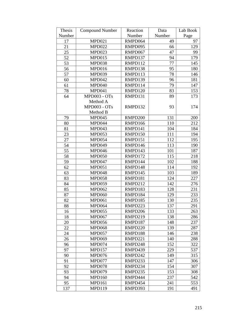
215
ThesisNumber
Compound Number ReactionNumber
DataNumber
Lab BookPage
17 MPD021 RMPD064 49 9721 MPD022 RMPD095 66 12925 MPD023 RMPD067 47 9952 MPD015 RMPD137 94 17953 MPD038 RMPD112 77 14556 MPD016 RMPD138 95 18057 MPD039 RMPD113 78 14660 MPD042 RMPD139 96 18161 MPD040 RMPD114 79 14778 MPD041 RMPD120 83 15364 MPD003 - OTs
Method AMPD003 - OTs
Method B
RMPD131
RMPD132
89
93
173
174
79 MPD045 RMPD200 131 20080 MPD044 RMPD166 110 21281 MPD043 RMPD141 104 18423 MPD053 RMPD150 111 19427 MPD054 RMPD151 112 19554 MPD049 RMPD146 113 19055 MPD046 RMPD143 101 18758 MPD050 RMPD172 115 21859 MPD047 RMPD144 102 18862 MPD051 RMPD148 114 19263 MPD048 RMPD145 103 18983 MPD058 RMPD181 124 22784 MPD059 RMPD212 142 27685 MPD062 RMPD183 128 23187 MPD060 RMPD184 129 23382 MPD061 RMPD185 130 23588 MPD064 RMPD223 137 29116 MPD055 RMPD206 133 26318 MPD067 RMPD219 138 28620 MPD056 RMPD187 148 23722 MPD068 RMPD220 139 28724 MPD057 RMPD188 146 23826 MPD069 RMPD221 140 28896 MPD074 RMPD248 152 32297 MPD157 RMPD439 229 53790 MPD076 RMPD242 149 31591 MPD077 RMPD233 147 30692 MPD078 RMPD234 154 30793 MPD079 RMPD235 153 30894 MPD160 RMPD444 237 54295 MPD161 RMPD454 241 553137 MPD119 RMPD393 191 491

216
ThesisNumber
Compound Number ReactionNumber
DataNumber
Lab BookPage
138 MPD120 RMPD394 192 492139 MPD121 RMPD395 197 493140 MPD122 RMPD396 193 492141 MPD123 RMPD440 230 538142 MPD124 RMPD441 231 539143 MPD125 RMPD399 196 497144 MPD154 RMPD433 232 53198 MPD126 RMPD403 204 501105 MPD127 RMPD404 199 526112 MPD128 RMPD405 203 50399 MPD129 RMPD406 205 504106 MPD130 RMPD407 206 505113 MPD131 RMPD408 202 506100 MPD132 RMPD409 207 507107 MPD133 RMPD410 208 508114 MPD134 RMPD411 209 509101 MPD135 RMPD412 210 510108 MPD136 RMPD413 211 511115 MPD137 RMPD414 212 512102 MPD138 RMPD415 213 513109 MPD139 RMPD416 215 514116 MPD143_2 RMPD446 238 544103 MPD141 RMPD418 217 516110 MPD142 RMPD419 218 517117 MPD140 RMPD417 216 515104 MPD144 RMPD421 220 521111 MPD145 RMPD422 221 520118 MPD146 RMPD423 222 521119 MPD162 RMPD448 234 546120 MPD083 RMPD243 155 316122 MPD165 RMPD456 257 555121 MPD087 RMPD252 158 326123 MPD166 RMPD457 261 556124 MPD147 RMPD424 201 522126 MPD148 RMPD425 223 523128 MPD149 RMPD426 224 524130 MPD150 RMPD451 240 549145 MPD155 RMPD434 228 532125 MPD158 RMPD442 236 540127 MPD159 RMPD443 235 541132 MPD176 RMPD582 303 690146 MPD198 RMPD584 305 692134 MPD199 RMPD585 306 693131 MPD200 RMPD586 307 694

217
Biological Results
10-(3 or 4-Substituted-Phenyl)-(2, 3, 4 or 5)-Substituted-5-Deazaflavin Analogues
HN
N N
O
O
R1 R2
R3
R4
R5R'1
R'2R'3
Thesis No R2 R3 R4 R5 R’1 R’2 R’3 Pre-Screen IC50 (μM)64 H H H H H H H Inactive ND66 Cl Inactive ND65 F Inactive ND40 CF3 Inactive ND41 CF3 Inactive ND42 CF3 Inactive ND43 CF3 Active 27.844 CF3 F Inactive ND45 CF3 F Inactive ND46 CF3 F 100-250μM ND47 CF3 F Active 18.748 CF3 Cl 100-250μM ND
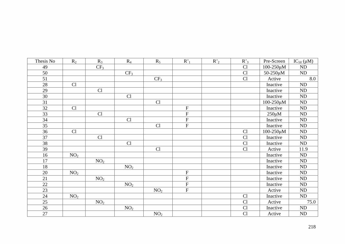
218
Thesis No R2 R3 R4 R5 R’1 R’2 R’3 Pre-Screen IC50 (μM)49 CF3 Cl 100-250μM ND50 CF3 Cl 50-250μM ND51 CF3 Cl Active 8.028 Cl Inactive ND29 Cl Inactive ND30 Cl Inactive ND31 Cl 100-250μM ND32 Cl F Inactive ND33 Cl F 250μM ND34 Cl F Inactive ND35 Cl F Inactive ND36 Cl Cl 100-250μM ND37 Cl Cl Inactive ND38 Cl Cl Inactive ND39 Cl Cl Active 11.916 NO2 Inactive ND17 NO2 Inactive ND18 NO2 Inactive ND20 NO2 F Inactive ND21 NO2 F Inactive ND22 NO2 F Inactive ND23 NO2 F Active ND24 NO2 Cl Inactive ND25 NO2 Cl Active 75.026 NO2 Cl Inactive ND27 NO2 Cl Active ND

219
Thesis No R2 R3 R4 R5 R’1 R’2 R’3 Pre-Screen IC50 (μM)52 Me Inactive ND53 Me Inactive ND54 Me Inactive ND55 Me Inactive ND56 Me F Inactive ND57 Me F Inactive ND58 Me F Inactive ND59 Me F Inactive ND60 Me Cl Inactive ND61 Me Cl Inactive ND62 Me Cl 100-250μM ND63 Me Cl Inactive ND

220
10-Substituted-5-Deazaflavin Analogues
HN
N N
R6
O
O
R2
R3
R4
R5
R1
Thesis No R5 R6 Pre-Screen IC50 (μM)90 H H Inactive ND91 CF3 H Inactive ND92 Me Inactive ND93 CF3 Me Inactive ND94 Bn Inactive ND95 CF3 Bn Active 129.2

221
10-(2, 3 or 4-Substituted-Phenyl)-9-Substituted-5-Deazaflavin Analogues
HN
N N
O
O
R1 R2
R3
R4
R5R'1
R'2R'3
Thesis No R5 R’1 R’2 R’3 Pre-Screen IC50 (μM)98 Cl Inactive ND99 Cl Inactive ND114 CF3 Cl Inactive ND113 CF3 Cl Active 2.5105 Cl Cl Inactive ND106 Cl Cl Active >100100 F Inactive ND101 F Inactive ND114 CF3 F Active 13115 CF3 F Active 14107 Cl F Active >250108 Cl F Inactive ND102 Me Inactive ND103 Me Inactive ND

222
Thesis No R5 R’1 R’2 R’3 Pre-Screen IC50 (μM)104 Me Inactive ND116 CF3 Me Inactive ND117 CF3 Me Active 4.5118 CF3 Me Active 95109 Cl Me Inactive ND11 Cl Me Inactive ND111 Cl Me Inactive ND119 CF3 Cl Cl Active 10.15147 F Cl Inactive ND148 Cl F Cl Inactive ND

223
10-Phenyl-3-Substituted-5-Deazaflavin Analogues
N
N N
O
O
R2
R3
R4
R5
R1
RN
R'3
Thesis No RN R5 R’3 Pre-Screen IC50 (μM)
120 Me Inactive ND121 Et Inactive ND122 Me CF3 Cl ND* ND*123 Et CF3 Cl ND* ND*

224
10-(3 or 4-Substituted-Phenyl)-9-Substituted-5-Deazaflavin Analogues
HN
N N
O
O
R1 R2
R3
R4
R5R'1
R'2R'3
* This compound was the previously identified lead compound. ND = Not Determined. ND* = Not Determined with result waiting for testing.Empty squares represent hydrogen atoms.
Thesis No R5 R’2 R’
3 Pre-Screen IC50 (μM)128 OH Inactive ND130 OH Cl Inactive ND125 Br Active >100127 Br Cl ND* ND*124 F Inactive ND126 F Cl Inactive ND134 CN Cl ND* ND*131 CN Cl ND* ND*

225
IC50 In Vitro Assay Data 1
Below is the in vitro assay data provided for inhibition of Mdm2 E3 ubiquitin ligase activity for compounds 39 (MPD032), 43 (MPD019), 47(MPD020) & 51 (MPD037).
M P D 02 0u M 0 .0 9 .0 1 8 .0 36 .0 71 .4 1 43 .0 2 86 .0
e xp t 2 0 .0 26 .1 42 .0 71 .1 82 .6 78 .9 90 .5e xp t 3 0 .0 36 .6 48 .2 67 .6 83 .2 85 .4 88 .1a v erag e 0 .0 31 .4 45 .1 69 .4 82 .9 82 .1 89 .3 18 .7S T D E V 0 .0 7 .4 4 .4 2 .5 0 .4 4 .6 1 .7
MP D 019 % Inh ibition of p53 Ub
u M0 80 160 240
% Inh ib ition
0
10
20
30
40
50
60
70
80
90
MP D 020 % Inhib ition of p53 Ub
uM0 80 160 240
% Inh ib ition
0
10
20
30
40
50
60
70
80
90
Inhibition of Ub of p53 (in-vitro assay spiked with fluoroescent-Ub)
MP D 032 % Inhib ition of p53 Ub
uM0 80 160 240
% Inh ib ition
0
10
20
30
40
50
60
70
80
90
100M P D 032uM 0 .0 9 .0 1 8 .0 36 .0 71 .4 1 43 .0 2 86 .0
ex 2 0 .0 40 .7 47 .9 65 .6 82 .5 86 .4 93 .4ex 3 0 .0 44 .3 63 .3 74 .1 85 .2 90 .1 95 .6ex 4 0 .0 50 .6 63 .9 82 .2 86 .2 93 .7 94 .4ex 5 0 .0 35 .0 64 .9 78 .2 91 .1 93 .5 97 .7
av erag e 0 .0 42 .6 60 .0 75 .0 86 .3 90 .9 95 .3 11 .9S T D E V 0 .0 6 .5 8 .1 7 .1 3 .6 3 .4 1 .8
M P D 019u M 0 .0 9 .0 1 8 .0 36 .0 71 .4 1 43 .0 2 86 .0
e xp t 2 0 .0 3 .2 41 .5 57 .8 79 .4 81 .2 82 .8e xp t 4 0 .0 25 .9 34 .7 61 .5 81 .2 83 .2 88 .8 IC 50a v erag e 0 .0 14 .5 38 .1 59 .6 80 .3 82 .2 85 .8 27 .8S T D E V 0 .0 16 .0 4 .8 2 .6 1 .3 1 .4 4 .3
XLfit4 model 652Hyperbolic InhibitionModel.
fit = (C+(A*(1-(x/(B+x)))))inv = ((B*((A+C)-y))/(y-C))Res = (y-fit)
M P D 0 3 7 % Inh ib ition o f p 5 3 U b
uM0 80 1 60 240
% Inh ib ition
0
10
20
30
40
50
60
70
80
90
100
M P D 0 3 7
u M 0 .0 9 .0 1 8 .0 3 6 .0 7 1 .4 1 4 3 .0 2 8 6 .0
e x p t 2 0 .0 3 4 .3 7 0 .5 7 5 .6 8 5 .6 9 1 .5 9 6 .9
e x p t 3 0 .0 4 6 .0 7 9 .0 7 9 .0 9 0 .9 9 2 .7 9 5 .9
e x p t 4 0 .0 5 0 .7 7 3 .6 8 3 .9 8 9 .8 9 6 .2 9 7 .4
e x p t 5 0 .0 5 9 .0 8 0 .3 8 7 .2 9 0 .6 9 5 .3 9 4 .3
a v e ra g e 0 .0 4 7 .5 7 5 .8 8 1 .4 8 9 .2 9 3 .9 9 6 .1 8 . 0
S T D E V 0 .0 1 0 .3 4 .6 5 .2 2 .4 2 .2 1 .4

226
Mdm2 Auto-ubiquitinylation IC50 In Vitro Assay Data
Below is the in vitro assay data provided for inhibition of Mdm2 auto-ubiquitinylation for compounds 39 (MPD032) & 51 (MPD037).
MDM2 auto-Ub Inhibition #32: expt's 1,2,3,4
uM0 80 160 240
%Inhibition
-20
0
20
40
60
80
100
IC50 20.8 M
MDM2 auto-Ub Inhibition #32: expt's 1,2,3,4
uM0 80 160 240
%Inhibition
-20
0
20
40
60
80
100
IC50 20.8 M
1.82.65.49.55.28.05.711.30.0STDEV
96.494.886.275.744.820.310.5-8.20.0average
97.693.588.778.744.015.69.9-1.70.0’’expt 4
97.897.287.171.050.611.613.0-24.60.0’’expt 3
96.596.690.887.646.428.22.80.00.0’’expt 2
93.991.778.465.638.225.916.3-6.40.0%Inhibitionexpt 1
M286.0143.071.436.018.09.04.52.20.0MPD032
MDM2 auto-Ub Inhibition #37: expt's 1,2,3,4
uM0 80 160 240
%Inhibition
0
10
20
30
40
50
60
70
80
90
100
IC50 12.9 M
MDM2 auto-Ub Inhibition #37: expt's 1,2,3,4
uM0 80 160 240
%Inhibition
0
10
20
30
40
50
60
70
80
90
100
IC50 12.9 M
1.92.31.53.54.519.014.010.60.0STDEV
96.294.690.885.461.635.018.410.40.0average
97.095.791.388.465.237.29.012.30.0’’expt 4
97.095.591.985.963.660.938.224.20.0’’expt 3
97.496.291.286.955.018.28.15.50.0’’expt 2
93.491.188.680.362.623.618.4-0.50.0%Inhibitionexpt 1
M286.0143.071.436.018.09.04.52.20.0MPD037
Inhibition of mdm2 auto-

227
IC50 In Vitro Assay Data 2
Below is the in vitro assay data provided for inhibition of Mdm2 E3 ubiquitin ligase activity for compounds 106 (MPD130), 113 (MPD131), 107(MPD133), 114 (MPD134), 115 (MPD137), 117 (MPD140), 116 (MPD143) & 118 (MPD146).
106 (MPD130) IC50 > 100 µM

228
113 (MPD131) IC50 = 2.5 µM

229
107 (MPD133) IC50 > 250 µM

230
114 (MPD134) IC50 = 13 µM

231
115 (MPD137) IC50 = 14 µM

232
117 (MPD140) IC50 = 4.5 µM

233
116 (MPD143) IC50 = 9.5 µM

234
118 (MPD146) IC50 = 10.11 µM