supplemental information caspase-2-mediated cleavage of mdm2
TRANSCRIPT

Molecular Cell, Volume 43
Supplemental Information
Caspase-2-Mediated Cleavage of Mdm2 Creates
a p53-Induced Positive Feedback Loop
Trudy G. Oliver, Etienne Meylan, Gregory P. Chang, Wen Xue, James R. Burke, Timothy J. Humpton,
Diana Hubbard, Arjun Bhutkar, and Tyler Jacks
SUPPLEMENTAL EXPERIMENTAL PROCEDURES Antibodies Caspase 2 (11B4, 1:500), Pidd (Anto-1, 1:500), and RAIDD (AT100, 1:1000) were obtained from Alexis. Parp (46D11, 1:1000), Myc-tag (2272, 1:1000), and pro-Caspase-3 (#9665, clone 8G10, 1:1000) were obtained from Cell Signaling Technologies. Nemo (FL-419, 1:1000), p21 (F-5, 1:100), p65 (sc-372, 1:1000) and p53 (FL-393, for IP) were obtained from Santa Cruz. Mdm2 N-terminal (IF2, 1:200), Mdm2 (ab6, 1:100), Mdm2 (ab3, 1:50), and p53 (DO-1, 1:2000) were obtained from Calbiochem. Flag-Tag (F3165, 1:1000) and Actin (A2066, 1:1000) were obtained from Sigma. MdmX (A300-287A, 1:1000) was obtained from Bethyl Labs. HA-tag antibodies were obtained from Covance (MMS-101P, 1:1000). Constructs and reagents pCMV-myc3-HDM2 (Yue Xiong), pCMV-HDM2 C464A, and pCMVTAG-Nemo (Wu et al, 2006) are available from Addgene. Caspase-2 constructs (wt, C320A, Ac152 wt and Ac152 C320A) were kindly provided by M. Guha and D. Altieri. MSCV-PIDD, MSCV-PIDD point-mutants (S446A, F582A, and S446A/F582A), and N-terminal Flag-tagged RAIDD were kindly provided by A. Tinel and J. Tschopp. Mdm2 D367A and D367E were cloned by site-directed mutagenesis using the following primers from (Stratagene Quikchange): Mdm2 D367A FOR gaagagggctttgatgttcctgcttgtaaaaaaactatagtgaatg Mdm2 D367A REV cattcactatagtttttttacaagcaggaacatcaaagccctcttc Mdm2 D367E FOR gctgaagagggctttgatgttcctgagtgtaaaaaaactatagt Mdm2 D367E REV actatagtttttttacactcaggaacatcaaagccctcttcagc along with the parental pCMV-myc3-HDM2 vector according to the manufacturer’s instructions (QuikChange Lightening, Stratagene) and verified by DNA sequencing. Truncated Mdm2 p60 was generated by PCR of the parental pCMV-myc3-HDM2 vector with the following primers. Underline represents BamH1 site; bold represents the premature stop codon introduced by PCR. BamH1-HDM2-p60-For: 5’-GTT CGA GGA TCC GTG TCT AGA GAA TTC ACC ATG GTG AG-3’ BamH1-HDM2-p60-Rev: 5’-CCA GAC GGA TCC TAC TAA TCA GGA ACA TCA AAG CCC TCT-3’

Gel-extracted BamH1 digests were cloned back into the BamH1 site of the parental vector, pCMV-myc3-HDM2. Clones in the correct orientation with the Myc-tag were verified by DNA sequencing and selected for subsequent experiments. Hairpins designed to human Caspase-2 (shCasp2 #1): TGCTGTTGACAGTGAGCGCCCCAACTTCCCTGTTCTTTAATAGTGAAGCCACAGATGTATTAAAGAACAGGGAAGTTGGGATGCCTACTGCCTCGGA were obtained from Invitrogen, and PCR amplified with: shSub1: CTA AAG TAG CCC CTT GAA TTC CGA GGC AGT AGG CA shSub2: CAG AAG GCT CGA GAA GGT ATA TTG CTG TTG ACA GTG AGC G and digested with EcoR1/Xho1 and cloned into MSCV-IRES-GFP-Puro (EcoR1/Xho1) and verified by sequencing. Recombinant human Caspase-2 and Caspase-3 were obtained from Alexis (ALX-201-057, ALX-201-059), now Enzo Life Sciences. Human recombinant Mdm2 was obtained from Calbiochem (#444146). Cisplatin, doxorubicin and neocarzinostatin were obtained from Sigma. Cisplatin was dissolved in PBS and used within one hour of preparation. Doxorubicin was prepared in sterile water. Real time RT-PCR For gene expression analysis by real time RT-PCR, RNA was isolated by TRIzol (Invitrogen) as described (Oliver et al., 2010) and 1 µg of total RNA was converted to cDNA using iScript cDNA synthesis kit (Bio-Rad). Real time RT-PCR was performed using gene specific primers and Sybr Green Supermix (Bio-rad) in a 20 μl reaction in triplicate on a Bio-rad CFX96 Real Time PCR machine. Analysis was performed using Bio-rad CFX Manager software and expression values were based on 10-fold serial dilutions of standards and normalized to actin levels. Primers for real time RT-PCR
Human
ACTIN-F CCAACCGCGAGAAGATGA
ACTIN-R CCAGAGGCGTACAGGGATAG
CASP2-F2 GCCTTCTGTGAAGCCTTGCACTCC
CASP2-R2 TCCACATCCCCTCCAGAGCGA
LRDD/PIDD-F1 TCTGACACGGTGGAGATGTTCG
LRDD/PIDD-R1 AGGTGCGAGTAGAAGACAAAGCAG
p21-F2 GAGGATGCGTGTTCGCGGGT
p21-R2 GCTGCTCGCTGTCCACTGGG
p53-F1 ACGACGGTGACACGCTTCCC
p53-R1 GGGACGGCAAGGGGGACAGA
RAIDD-F2 CCGCCCCAAAGATACGTGGTTGC
RAIDD-R2 CCCTGACCCAGGGAAACTCCTGT
CASP3-F1 GCGGTTGTAGAAGAGTTTCGTGAGT
CASP3-R1 CCGAGTGTGAGCAGGGCTCG
Mouse
Actin-F2 TATTGGCAACGAGCGGTTCC
Actin-B2 GGCATAGAGGTCTTTACGGATGTC
Lrdd/Pidd-F1 TCGCTGTCGTGAGGTAGTTG
Lrdd/Pidd-R1 GAGAAGTGCTCCCTCTGGTG
p21-F TCCAGACATTCAGAGCCACA
p21-R ACGAAGTCAAAGTTCCACCG
Quantitative western blotting Quantitative western blot was performed by running total cell lysates on nitrocellulose membranes, transferred as previously indicated, and blocked in 1:2 parts (Odyssey blocking buffer:PBS) for 1 hr. Membranes were washed 7X for 5 min in PBS-T, incubated with primary antibodies in blocking buffer overnight, washed again, and incubated in fluorescent goat anti-rabbit-IgG-680 or anti-mouse-IgG-800 secondary antibodies (LI-COR, IRDye, 1:10,000) for 1 hr. Membranes were dried for 2 hrs or overnight and analyzed using an Odyssey Imaging System (LI-COR Biosciences). p53 protein levels were normalized to ACTIN.

Cell viability assays For viability assays, cells were seeded in triplicate (6x10³/well) in opaque 96 wp and treated the next day with increasing doses of cisplatin at 0, 1, 5, 10, 20, 50, 100, and 200 µM. After 48 hrs of treatment, cell viability was measured using CellTiter Glo (Promega) on a luminometer. Normalized, transformed dose response curves were generated and analyzed using GraphPad Prism to determine IC50. Cell cycle analysis Cells were harvested with trypsin, washed twice in PBS, and 1 x 10^6 cells were fixed in ice-cold 70% ethanol overnight. Cells were washed 2X in PBS/1% BSA, treated with 500 μg/mL RNase for 30 min at 37C, then stained with 50 μg/mL propidium iodide overnight at 4C. Cells were analyzed on a BD FACScalibur flow cytometer and cell cycle analysis was performed using FlowJo software. Annexin-V Apoptosis Assay U2OS cells transfected with siRNAs for 24 hrs were treated with or without 500 nM doxorubicin in 6-well plates and harvested after 48-72 hrs. Following manufacturer’s instructions (BD Pharmingen), cells were stained with Annexin-V-APC and DAPI and analyzed using a FACS LSR II HTS flow cytometer (BD Biosciences). Live, apoptotic and dead cells were quantified using FlowJo software. Bioinformatics methods Mdm2 protein conservation. Sequences from the following species were used to test conservation of the Mdm2 cleavage site across evolutionary time (Figure 3C): Human (gi: 89993689), Chimp (gi:114645764 ), Mouse (gi:1209699), Rat (gi:157821747), Hamster (GenBank: AAC52425.1), Dog (gi: 50978798), Pig (gi: 157073220), Cow (gi: 149642933), Frog (gi: 160420277), Zebrafish (gi: 205361189). Flanks of 15 amino acids on both sides of the conserved DVPD sequence were selected (34 amino acid subsequences) and aligned using ClustalW2 [Ref. below]. The conservation plot was generated using Jalview v2.5.1 [Ref. below].

Figure S1. Related to Figure 1 A
Cisplatin treatment induces PIDD expression in p53 wild-type NSCLC cell lines. Indicated human NSCLC cell lines that are p53 wild-type (orange bars) or null/mutant (black bars) were treated with 0, 1, 5, 10, or 20 μM cisplatin (increasing dosage indicated by solid triangles) and harvested after 24 hrs. PIDD mRNA expression levels were determined by real time RT-PCR in triplicate and normalized to ACTIN levels. Fold change in PIDD levels relative to untreated cells are shown. Data from three p53 wild-type lines were previously reported in Oliver et al., Genes and Dev, 2010. Error bars represent mean +/- SD.
B
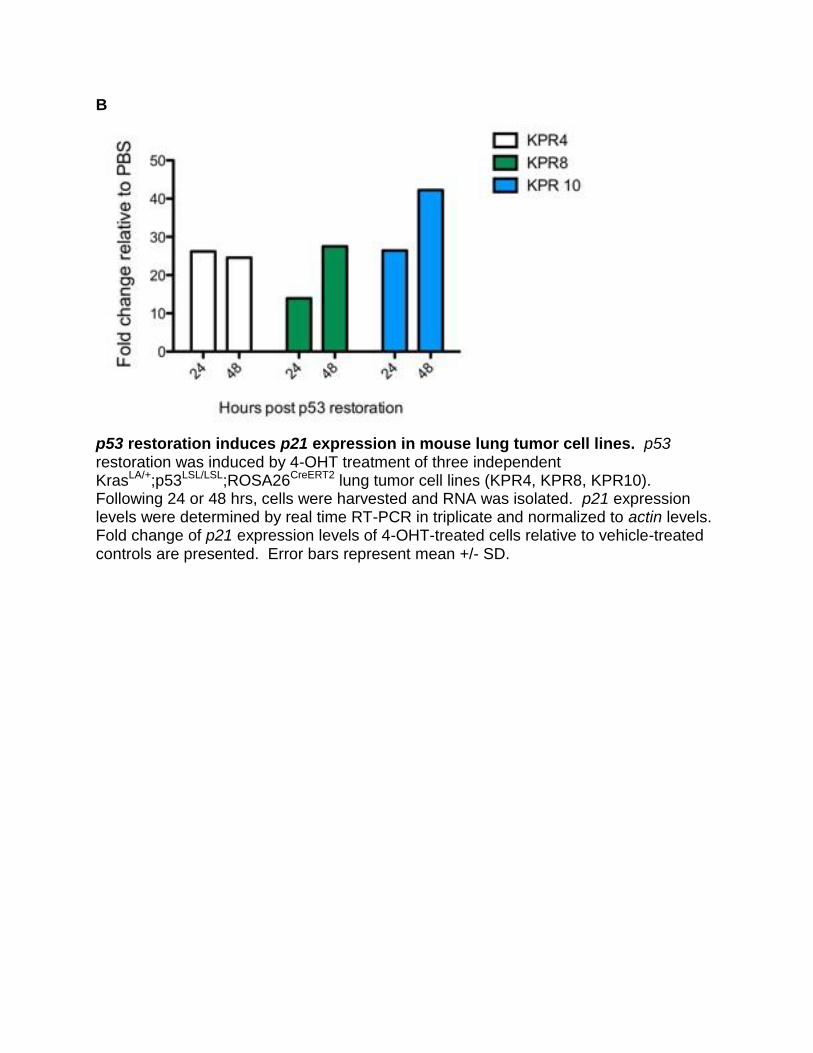
p53 restoration induces p21 expression in mouse lung tumor cell lines. p53 restoration was induced by 4-OHT treatment of three independent KrasLA/+;p53LSL/LSL;ROSA26CreERT2 lung tumor cell lines (KPR4, KPR8, KPR10). Following 24 or 48 hrs, cells were harvested and RNA was isolated. p21 expression levels were determined by real time RT-PCR in triplicate and normalized to actin levels. Fold change of p21 expression levels of 4-OHT-treated cells relative to vehicle-treated controls are presented. Error bars represent mean +/- SD.
C
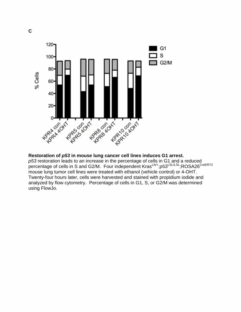
Restoration of p53 in mouse lung cancer cell lines induces G1 arrest. p53 restoration leads to an increase in the percentage of cells in G1 and a reduced percentage of cells in S and G2/M. Four independent KrasLA/+;p53LSL/LSL;ROSA26CreERT2 mouse lung tumor cell lines were treated with ethanol (vehicle control) or 4-OHT. Twenty-four hours later, cells were harvested and stained with propidium iodide and analyzed by flow cytometry. Percentage of cells in G1, S, or G2/M was determined using FlowJo.

D
PIDD expression in KRAS mutant NSCLC cell lines. Immunoblot of nuclear (n) or cytoplasmic (c) fractions from cells expressing Flag-tagged PIDD (+) or vector control (-) with antibodies to Flag (PIDD), Nemo (cytoplasmic loading control) and Parp (nuclear loading control). Full-length PIDD (FL), PIDD-C (C) or PIDD-CC (CC) cleavage fragments are indicated by black arrowheads. Top panel includes p53 wild-type lines: H460, SW1573, A549. Bottom panel includes p53 null and/or mutant lines: Calu-1, H23, COR-L23, H2009. Note: A427 cells, the only p53 wild-type line without significant growth rate reduction upon PIDD expression, did not exhibit cisplatin-resistance, had low Caspase-2 expression levels compared to other cell lines, and lacked Mdm2 cleavage (data not shown).

E
PIDD expression does not promote p53 stability or p21 expression in p53 null/mutant lung cancer cell lines. Nuclear (n) and cytoplasmic (c) cell lysates from MSCV-PIDD (+) or vector control cells (-), analyzed by western blotting with p53 (DO-1) or p21 (Santa Cruz) antibodies. Loading controls are included in Supp Fig S1D. Positive control (+) for the p21 blot is included on the bottom right panel.

Figure S2. PIDD Overexpression Promotes Cleavage of Caspase-2 as Previously Described, Related to Figure 2 A549 cells were infected with MSCV-PIDD (+) or vector control (-) viruses and selected in puromycin. Total cell lysates were analyzed by Western blotting with antibodies to PIDD or pro-Caspase-2. Cleavage fragments of PIDD and Caspase-2 indicated by arrowheads.

Figure S3. Caspase-2 Promotes Cleavage of Mdm2, Whereas Nemo Does Not, Related to Figure 3 293T cells were transfected with vectors encoding Myc-tagged Mdm2 in the presence of no cDNA (Control), truncated active Caspase-2 (C2) or Nemo. Nuclear (n) and cytoplasmic (c) cell fractions were analyzed by immunoblot for Myc tag (Mdm2) and p53. Top panel: Expression of active Caspase-2 (C2) promotes cleavage of Mdm2 in 293T cells, whereas expression of Nemo does not. Bottom panel: Full-length Mdm2 promotes p53 modifications (brackets, indicative of ubiquitination) whereas cleaved Mdm2 significantly reduces p53 modifications (C2 lane).
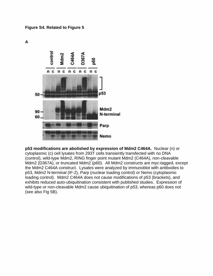
Figure S4. Related to Figure 5 A
p53 modifications are abolished by expression of Mdm2 C464A. Nuclear (n) or cytoplasmic (c) cell lysates from 293T cells transiently transfected with no DNA (control), wild-type Mdm2, RING finger point mutant Mdm2 (C464A), non-cleavable Mdm2 (D367A), or truncated Mdm2 (p60). All Mdm2 constructs are myc-tagged, except the Mdm2 C464A construct. Lysates were analyzed by immunoblot with antibodies to p53, Mdm2 N-terminal (IF-2), Parp (nuclear loading control) or Nemo (cytoplasmic loading control). Mdm2 C464A does not cause modifications of p53 (brackets), and exhibits reduced auto-ubiquitination consistent with published studies. Expression of wild-type or non-cleavable Mdm2 cause ubiquitination of p53, whereas p60 does not (see also Fig 5B).

B
Expression of Mdm2 p60 does not promote ubiquitination of p53. U2OS cells were transiently transfected with no DNA (ctrl), wild-type Mdm2 (WT), or Mdm2 p60 (p60). Twenty-four hours later, cells were harvested and nuclear (n) and cytoplasmic (c) protein lysates were analyzed for Mdm2, p53, Parp (nuclear control) and Nemo (cytoplasmic control). Short exposure (SE) for p53 demonstrates that p60 expression increases endogenous p53 levels; long exposure (LE) shows reduced p53 modifications and increased p53 levels compared to wild-type Mdm2.

C
Expression of RAIDD reduces p53 modifications by promoting Mdm2 cleavage. Nuclear (n) or cytoplasmic (c) cell lysates from 293T cells transiently transfected with eGFP (control), Myc-tagged Mdm2 (Mdm2) or Myc-tagged non-cleavable Mdm2 D367A (D367A) in the presence (+) or absence (-) of Flag-tagged RAIDD. Lysates were analyzed by immunoblot with antibodies to p53 (DO-1) or Parp (nuclear loading control). RAIDD reduces ubiquitination of p53 (bracket) in the presence of wild-type Mdm2, but not in the presence of non-cleavable Mdm2. Figure 3E demonstrates that RAIDD promotes cleavage of Mdm2. See also Figure 3E for additional loading controls.
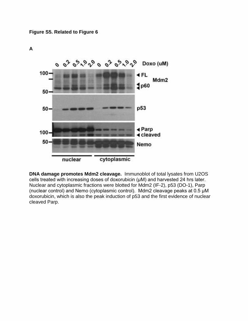
Figure S5. Related to Figure 6
A
DNA damage promotes Mdm2 cleavage. Immunoblot of total lysates from U2OS cells treated with increasing doses of doxorubicin (μM) and harvested 24 hrs later. Nuclear and cytoplasmic fractions were blotted for Mdm2 (IF-2), p53 (DO-1), Parp (nuclear control) and Nemo (cytoplasmic control). Mdm2 cleavage peaks at 0.5 μM doxorubicin, which is also the peak induction of p53 and the first evidence of nuclear cleaved Parp.

B
Doxorubicin treatment induces PIDD expression. Real time RT-PCR for human PIDD expression in U2OS cells treated with 100 or 500 nM doxorubicin or vehicle control for 24 hrs. PIDD levels analyzed in triplicate and normalized to ACTIN. Fold-change relative to vehicle-treated control. Error bars represent mean +/- SEM.

C
Inhibition of the Caspase-2-PIDDosome prevents Mdm2 cleavage following double-strand breaks. U2OS cells were transfected with control (CON), Casp2 (C2), PIDD, or Casp3 (C3) siRNAs for 24 hrs, split, and damaged with (+) or without (-) 500ng/mL neocarzinostatin (NCS). After eight hours, cells were washed, incubator for an additional 32 hrs, and then collected for protein lysates. Total cell lysates were analyzed by immunoblot for Actin and Mdm2.

D
PIDD expression does not appear to promote MdmX cleavage. H460 or SW1573 cells were infected with MSCV viruses containing control (-) or PIDD (+) cDNAs and selected in puromycin. Nuclear (n) and cytoplasmic (c) cell lysate fractions were analyzed by immunoblot for MdmX and Parp (nuclear loading control).

Figure S6. Related to Figure 7
A
Inhibition of the Caspase-2-PIDDosome reduces Mdm2 cleavage and p53 levels following DNA damage. Top panel: U2OS cells were transfected with control (siCON), Casp2 or PIDD siRNAs for 24 hrs, split, and damaged with 500 nM doxorubicin. After eight hours, cells were washed and collected at various time points from 0-72 hrs for protein lysates. Total cell lysates were analyzed by immunoblot for Actin (loading control) and Mdm2. Bottom panel: Quantitative western blot was performed using the LI-COR Odyssey system for Actin and p53. p53 levels were normalized to Actin levels for each sample.

B
Inhibition of the Caspase-2-PIDDosome impacts the maintenance of p53 levels following DNA damage. U2OS cells were transfected with control, Casp2 or PIDD siRNAs for 24 hrs, split, and damaged with 500nM doxorubicin. After eight hours, cells were washed and collected at various time points from 0-72 hrs for protein lysates. Total cell lysates were analyzed by quantitative western blot using the LI-COR Odyssey system for Actin and p53. p53 levels were normalized to Actin levels per sample. Error bars represent mean +/- SD. siCONTROL, n = 4. Combined siCasp2 (n=2)/ siPIDD (n=2), n = 4. At 48 hrs, * p < 0.05; at 56 hrs, ** p < 0.005.

SUPPLEMENTAL REFERENCES ClustalW: Larkin M.A., Blackshields G., Brown N.P., Chenna R., McGettigan P.A., McWilliam H., Valentin F., Wallace I.M., Wilm A., Lopez R., Thompson J.D., Gibson T.J. and Higgins D.G. (2007). ClustalW and ClustalX version 2. Bioinformatics 2007 23(21): 2947-2948. JalView: Waterhouse, A.M., Procter, J.B., Martin, D.M.A, Clamp, M. and Barton, G. J. (2009). "Jalview Version 2 - a multiple sequence alignment editor and analysis workbench". Bioinformatics 25 (9) 1189-1191. Wu CJ, Conze DB, Li T, Srinivasula SM, Ashwell JD. (2006). NEMO is a sensor of Lys 63-linked polyubiquitination and functions in NF-kappaB activation. Nat Cell Biol. Apr.
8(4):398-406.