differential posttranslational processing confers intraspecies
TRANSCRIPT
INFECTION AND IMMUNITY,0019-9567/99/$04.0010
Feb. 1999, p. 760–771 Vol. 67, No. 2
Copyright © 1999, American Society for Microbiology. All Rights Reserved.
Differential Posttranslational Processing Confers Intraspecies Variationof a Major Surface Lipoprotein and a Macrophage-Activating
Lipopeptide of Mycoplasma fermentansMICHAEL J. CALCUTT,1 MARY F. KIM,1 ARTHUR B. KARPAS,2
PETER F. MUHLRADT,3 AND KIM S. WISE1*
Department of Molecular Microbiology and Immunology, School of Medicine, University of Missouri—Columbia,Columbia, Missouri 652121; Laboratory of Developmental and Molecular Immunity, National Institute of
Child Health and Human Development, Bethesda, Maryland 20892-27202; and ImmunobiologyResearch Group, Gesellschaft fur Biotechnologische Forschung mbH,
D-38124 Braunschweig, Germany3
Received 28 August 1998/Returned for modification 6 October 1998/Accepted 10 November 1998
The malp gene of Mycoplasma fermentans is shown to occur in single copy but to encode two discretetranslated forms of lipid-modified surface protein that can be differentially expressed on isolates within thisspecies: MALP-2, a 14-amino-acid (2-kDa) lipopeptide with potent macrophage-stimulatory activity (P. F.Muhlradt, M. Kiess, H. Meyer, R. Sussmuth, and G. Jung, J. Exp. Med. 185:1951–1958, 1997), and MALP-404,an abundant, full-length (404-amino-acid) surface lipoprotein of 41 kDa, previously designated P41 (K. S.Wise, M. F. Kim, P. M. Theiss, and S.-C. Lo, Infect. Immun. 61:3327–3333, 1993). The sequences, transcripts,and translation products of malp were compared between clonal isolates of strains PG18 (known to expressP41) and II-29/1 (known to express high levels of MALP-2). Despite conserved malp DNA sequences containingfull-length open reading frames and expression of full-length monocistronic transcripts in both isolates,Western blotting using a monoclonal antibody (MAb) to the N-terminal MALP-2 peptide revealed markeddifferences in the protein products expressed. Whereas PG18 expressed abundant MALP-404 with detectableMALP-2, II-29/1 revealed no MALP-404 even in samples containing a large comparative excess of MALP-2.Colony immunoblots with the MAb showed uniform surface expression of MALP-2 in II-29/1 populations. Asecond MAb to an epitope of MALP-404 outside the MALP-2 sequence predictably failed to stain II-29/1colonies but uniformly stained PG18 populations. Collectively, these results provide evidence for novel post-transcriptional (probably posttranslational) processing pathways leading to differential intraspecies expres-sion of a major lipoprotein, and a potent macrophage-activating lipopeptide, on the surface of M. fermentans.In the course of this study, a striking conserved motif (consensus, TD-G--DDKSFNQSAWE--), designated SLA,was identified in MALP-404; this motif is also distributed among selected lipoproteins and species from diversebacterial genera, including Bacillus, Borrelia, Listeria, Mycoplasma, and Treponema. In addition, malp was shownto flank a chromosomal polymorphism. In eight isolates of M. fermentans examined, malp occurred upstreamof an operon encoding the phase-variable P78 ABC transporter; but, in three of these isolates, a newlydiscovered insertion sequence, IS1630 (of the IS30 class), was located between these genes.
Lipid-modified translation products expressed on the sur-face of mycoplasmas are increasingly recognized as an impor-tant class of proteins contributing to fundamental biologicaland pathogenic processes of these organisms, including adap-tive surface variation now widely observed in this group ofwall-less procaryotes (3, 4, 28, 61) and modification of assortedhost cell functions, characteristic of bacterial modulins (20).First, surface lipoproteins representing abundant mycoplasmacoat proteins (4, 27, 50, 66), adhesins (5, 16, 55, 57, 65), andtransporters (52) have been shown to undergo rapid, heritablealteration in expression (phase variation) or structure, due tounderlying mutational instabilities directly associated with cor-responding genes. Genes encoding products with these diversefunctions can occur in single copy or as families of relatedsequences distributed in the limited genome of these organ-isms. To date, mutations associated with genes encoding pri-
mary gene products are the most prevalent mechanisms knownto determine population diversity involving mycoplasma sur-face lipoprotein expression or structural variation. However,evidence has also been presented for unidentified factors thatindirectly affect other surface properties in a phase-variablemanner, such as the accessibility of specific surface epitopes onproteins that are continually expressed (51, 53). It is likely thatadditional and novel mechanisms may be employed by myco-plasmas to generate surface diversity in populations, whichappears to be a common theme in the survival of these obligateparasitic pathogens. One type of variation not previously re-ported in mycoplasmas is the use of posttranslational pathwaysto generate alternative forms of primary lipoprotein geneproducts.
A second major impact of mycoplasmal lipoproteins lies inthe potent immunomodulatory activities associated with thelipid-modified N-terminal region of these processed mem-brane proteins (33, 35, 36, 38). Due to the relatively largenumber (relative to the total gene content) and diversity oflipoproteins in mycoplasmas, documented by genomic se-quencing (17, 21) and experimental evidence (11, 56), and thelack of other components such as lipopolysaccharide or cell
* Corresponding author. Mailing address: Department of MolecularMicrobiology and Immunology, School of Medicine, M616 MedicalSciences Building, University of Missouri—Columbia, Columbia, MO65212. Phone: (573) 882-8138. Fax: (573) 882-4287. E-mail: [email protected].
760
on April 9, 2019 by guest
http://iai.asm.org/
Dow
nloaded from
wall constituents in these organisms, lipid-modified proteinsare likely to be a primary element responsible for the immu-nomodulatory role of mycoplasmas that has been extensivelydocumented in their respective animal hosts or in vitro (12, 29,44). In this regard, the activities, biogenesis, and intraspeciesvariation of lipoproteins and corresponding lipopeptides areimportant features to be understood in the analysis of myco-plasmal pathogenesis.
Past studies of the lipoproteins and related products in My-coplasma fermentans in our laboratories (11, 33, 35, 36, 38, 43,51–53, 59) and by others (15, 18, 26, 40, 41) have contributedto the understanding of mycoplasmal surface variation andimmunomodulatory activities associated with these surfacemembrane components. In the present report, we document anovel form of surface variation that involves a previously re-ported major lipoprotein (P41) and a potent macrophage-ac-tivating lipopeptide (MALP-2) of M. fermentans. P41 is anabundant amphiphilic lipoprotein, first defined (59) in the typestrain PG18 by detergent fractionation and metabolic labelingwith [3H]palmitate and [35S]cysteine and shown by monospe-cific antibody (Ab) to be present also in the strain Incognitus(sb51 isolate). Subsequently, this protein has been identified asa common target of the Ab response to M. fermentans in thehuman host (22). MALP-2 is a lipopeptide isolated from M.fermentans. It has been rigorously purified, shown to have anexceptionally high macrophage-activating activity (in the pmo-lar range), and structurally solved (33). Significantly, theMALP-2 lipopeptide contains only two fatty acid chains. Thisfeature was shown to be compatible with a characteristicS-(2,3-di-O-acyloxypropyl) cysteinyl group at the Cys residue inthe 11 position (11 Cys residue) generated after cleavage ofa typical procaryotic prolipoprotein (6); but, it is highly dis-tinctive in that a third fatty acid normally linked in othereubacteria by an amide bond to the free amino group of Cys isabsent. This is particularly intriguing in light of recent genomicanalysis (17, 21) that has failed to identify an ortholog of thebacterial apolipoprotein N-acyl transferase (47) that is respon-sible for this final acylation reaction, in the two mycoplasmagenome sequences reported to date.
This report defines the single gene, malp, that encodes boththe P41 and MALP-2 products of M. fermentans and reveals anovel mechanism operating in mycoplasmas that determinesthe relative expression of these two components on variantswithin the species, and possibly within clonal populations. Thepotential role of this variation in determining antigenic andimmunomodulatory properties of propagating populations isdiscussed. In the course of this study, a highly distinctive pro-tein sequence motif distributed selectively among individuallipoproteins of diverse bacterial genera is defined, and amarked chromosomal polymorphism associated with the malpgene is shown to occur in M. fermentans isolates, due to thepresence or absence of a newly defined insertion-like element,IS1630.
MATERIALS AND METHODS
Bacteria, plasmids, and growth conditions. M. fermentans PG18 (clone 39) wasgrown in modified Hayflick medium as described elsewhere (59). The cloned M.fermentans isolate II-29/1 was propagated in GBF-3 medium as described previ-ously (33). Mycoplasma colonies were grown by plating liquid cultures on re-spective solid medium containing 1% (for PG18) or 0.7% (for II-29/1) Nobleagar (Difco Laboratories, Detroit, Mich.). Escherichia coli DH10B (Gibco BRL,Grand Island, N.Y.) and strains containing plasmid pZero2.1 (Invitrogen, Carls-bad, Calif.) and its derivatives were grown at 37°C in Luria-Bertani medium,supplemented with 50 mg of kanamycin per ml when appropriate. The speciesMycoplasma hyorhinis SK76 (42), Mycoplasma hominis 1620 (64), and Myco-plasma arginini G230 (provided by Richard DelGiudice) were propagated inHayflick medium supplemented as described in the respective references.
Synthesis of a MALP-2-specific probe. With the exception of oligonucleotidesMALP-F1 and MALP-R1, purchased from Gibco BRL, the oligonucleotidesused in this study were synthesized on a model 3948 nucleic acid synthesis andpurification system (Applied Biosystems, Inc. [ABI], Foster City, Calif.) by theUniversity of Missouri Molecular Biology Program DNA Core Facility. Inosine(I)-containing degenerate oligonucleotides MALP-F1 [59 TG(A/G) GGIAA(C/T) AA(C/T) GA(C/T) GA(A/G) 39] and MALP-R1 [59 (C/T)TT (C/T)TC(C/T)TT (A/G)AA IGA IAT (A/G)TT 39] were synthesized based on theMALP-2 peptide sequence (33). These two primers (25 pmol each) were used ina standard PCR to generate a MALP-2-derived 42-bp product from genomicDNA (50 ng) from M. fermentans II-29/1. The thermocycle parameters included30 cycles of 94°C for 1 min, 40°C for 1 min, and 72° for 1 min. A gel slicecontaining this amplicon was excised from a 3% (wt/vol) low-melting-pointagarose gel (FMC BioProducts, Rockland, Maine) and melted at 68°C for 10min, and a fraction was used as the template in a second PCR amplification usingthe same primers and conditions, but in the presence of digoxigenin-11-labeleddUTP (DIG-dUTP). The resulting DIG-labeled PCR product was used as aprobe for Southern analysis and E. coli colony hybridization at 42°C understandard conditions, as outlined by the product supplier (Boehringer Mannheim,Indianapolis, Ind.).
PCR amplification and chromosomal linkage of the malp gene in M. fermen-tans strains. The oligonucleotides MALP-F2 (59 TTG AGA TAT TTA AGCAAA ATA TCT A 39) and MALP-R2 (59 ATT TTC CAG CAT TTT TTT GAT39) were used to amplify the entire MALP-404 protein coding sequence fromgenomic DNA from various M. fermentans strains, kindly provided by S.-C. Lo,Armed Forces Institute of Pathology, Washington, D.C. These included DNApreparations from strains K7, MT-2, M39A, M70B, SK5, and Incognitus. DNAtemplate (10 ng) was used in a standard PCR consisting of 35 cycles of 94°C for1 min, 50°C for 1 min, and 72°C for 1.5 min. The resulting PCR amplicons werepurified by using QiaQuick spin columns (Qiagen, Santa Clarita, Calif.) andsequenced directly with primers MALP-F2 and MALP-R2 and with two addi-tional internal primers.
To determine the linkage of malp to the downstream operon p63-p58-p35-p78encoding the P78 ABC transporter (52) and to characterize the interveningregion, PCR was performed under standard conditions with oligonucleotidesMALP-F3 (59 AAT TTA CCA CAC ATC ACC TGT T 39) and P63-R (59 GGTCGG GTT CAT ATA GTC CAA 39) on genomic DNA samples from various M.fermentans strains.
Inverse PCR methods. Inverse PCR was used to isolate DNA sequencesupstream of the malp gene from M. fermentans PG18. Briefly, approximately 500ng of genomic DNA was digested with either BglII or EcoRI, purified by theQiaQuick (Qiagen) spin column procedure, and concentrated in vacuo. Dilutionsof the DNA sample were self-ligated in a 20-ml volume with 1 U of T4 DNAligase for 3 to 4 h at room temperature. Samples from the ligation reaction werethen amplified under standard PCR conditions using primers MALP-F3 andORF3-R1 (59 TAA TAG CAT GAC GAG CTT CAT 39), for the 1.6-kb frag-ment from the ligation of EcoRI-generated DNA fragments, and primersMALP-F4 (59 CTC AGA CCA AGG TAT GAT TCA A 39) and ORF2-R1 (59CTC CAT TAT TTG AGT TGA TTG GAA 39), for amplifying the 1.6-kbfragment from the ligation of BglII fragments (shown in Fig. 2). The resultingamplicons were purified and sequenced directly by primer walking.
DNA preparation and hybridization methods. Genomic DNA from M. fer-mentans PG18, M. hyorhinis SK76, and M. arginini G230 was prepared as de-scribed previously (62). Small-scale preparations of genomic DNA from M.fermentans II-29/1 were prepared by using a Wizard genomic DNA isolation kit(Promega, Madison, Wis.) in accordance with the supplied instructions. ForSouthern hybridization, genomic DNA was digested to completion with eitherNheI, PstI, or EcoRI, resolved by electrophoresis in 0.7% (wt/vol) agarose gels,transferred to positively charged nylon membranes (Boehringer Mannheim), andhybridized with a denatured 42-nucleotide (nt) DIG-labeled probe. Hybridiza-tion and washing steps were carried out at 42°C (because of the small size anddegenerate nature of the probe) in standard buffer solutions described in theguide provided by the manufacturer (Boehringer Mannheim). Hybridizing bandswere detected by using nonradioactive detection methods, as described previ-ously (64). Colony hybridization of transformants with DIG-labeled probes wascarried out by following the recommendations of the manufacturer of the non-radioactive detection system (Boehringer Mannheim).
Cloning of the DNA sequence encoding MALP-2. NheI-digested chromosomalDNA from M. fermentans PG18 and II-29/1 was ligated to XbaI-digested pZero2.1cloning vector under conditions recommended by the supplier. Following trans-formation into competent E. coli DH10B cells (Gibco BRL), kanamycin-resistanttransformants were screened by standard colony hybridization methods with theDIG-labeled 42-bp MALP-2 probe, as described for Southern analysis. Multiplehybridizing clones were isolated for both the 4.1-kb NheI fragment from strainPG18 and the 2.7-kb fragment from strain II-29/1. The nucleotide sequence foreach of these NheI fragments was determined by using a combination of specificdeletion subclones (generated from restriction sites present within the clonedregion and the vector polylinker) and sequence-generated oligonucleotide primers.
DNA sequencing and computer analysis. DNA sequencing reactions wereperformed by the University of Missouri Molecular Biology Program DNA CoreFacility by using Taq dye terminators and a Prism 377 automated DNA se-
VOL. 67, 1999 GENESIS OF A MYCOPLASMA SURFACE LIPOPEPTIDE 761
on April 9, 2019 by guest
http://iai.asm.org/
Dow
nloaded from
quencer (ABI). DNA and protein sequences were analyzed by using the GCGsoftware package (Genetics Computer Group, Madison, Wis.).
RNA extraction and analysis. Total RNA was extracted from mycoplasmasharvested by centrifugation from 15-ml late-logarithmic-phase cultures of M.fermentans, using an RNeasy kit (Qiagen). The RNA (5 mg) was denatured andseparated by electrophoresis through formaldehyde-containing agarose gels, bystandard procedures (46). The RNA was then transferred to a positively chargednylon membrane (Boehringer Mannheim) by capillary blotting, immobilized byUV cross-linking, and incubated with a denatured DIG-labeled DNA probederived from the MALP-404 coding sequence (using high-stringency hybridiza-tion conditions recommended by the membrane supplier [Boehringer Mann-heim]). This 536-nt probe was prepared by PCR using oligonucleotides MALP-2F5 (59 TAT TAG GAT TGA GTC CTA TTG CT 39) and MALP-2 R3 (59 TTAATC AAC TTG CAA TTG CAT A 39) and corresponds to the region betweenamino acid residues 216 to 1160 of MALP-404. Following hybridization, themembrane was washed and developed by using standard procedures for chemi-luminescent detection of DIG-labeled molecules (Boehringer Mannheim).
Peptide synthesis and antipeptide antibodies. Synthetic peptides were pre-pared as previously described (11, 63) on a model 432A peptide synthesizer(ABI) by using standard Fmoc (9-fluorenylmethylcarbonyl) chemistries. Twosynthetic peptides, CGNNDESNISFKEK (the MALP-2 peptide sequence lo-cated at the N terminus of the predicted MALP-404 lipoprotein) and CKEAIKMFKELPEDFVKYINSDKALKDGNK (an internal sequence near the C ter-minus of MALP-404) were synthesized and coupled through an appended N-terminal Cys residue to keyhole limpet hemocyanin (KLH), using a hapten-carrier conjugation kit (Pierce Chemical Co., Rockford, Ill.). Abs to the MALP-2peptide were generated in BALB/c mice that were injected intraperitoneallythree times at weekly intervals with 20 to 50 mg of KLH-peptide emulsified inincomplete Freund adjuvant. Preimmune serum and serum samples taken atleast 1 week after the last injection were used at a dilution of 1:200 for immu-nostaining.
Hybridoma construction and screening. Monoclonal Ab (MAb) 4444H7.A{immunoglobulin G1(k) [IgG1(k)]} to MALP-404 and MAb 4443H2.C[IgG2b(k)] to P76 were generated by fusion of the myeloma cell line SP2/0 withsplenocytes from BALB/c mice immunized with freeze-thaw-disrupted wholeorganisms of M. fermentans PG18 (51). MAb F208C2B1 [IgG2b(k)] to theMALP-2 peptide was generated by methods described previously (10). Briefly, 9months after injection with the KLH–MALP-2 peptide conjugate (describedabove), a mouse was injected intraperitoneally with 25 mg of the same conjugatedissolved in phosphate-buffered saline. Three days later, splenocytes were fusedwith the murine myeloma cell line NS-1 (ATCC TIB 18) and hybridomas wereselected. Fusions and selection were performed at the University of Missouri—Columbia Molecular Biology Program Cell and Immunobiology Core Facility.
An enzyme-linked immunosorbent assay using immobilized synthetic peptideswas used to screen MAbs, as described previously (10). Briefly, a saturatingamount of biotinylated peptide was immobilized in 96-well plates (Immulon 2;Dynatech Laboratories, Inc., Chantilly, Va.) that had been previously coated with0.5 mg of neutral avidin (NeutrAvidin; Pierce Chemical Co.) per well andblocked with phosphate-buffered saline containing 3% bovine serum albumin(fraction V; Fisher Scientific, Fairlawn, N.J.). A biotinylated peptide comprisingan irrelevant sequence in VlpA (10) was used as a negative control to ensure Abspecificity. Peroxidase-conjugated, goat anti-mouse IgG (whole molecule; Cap-pel Organon Teknika, Durham, N.C.) was used as the secondary Ab to select forbound MAb. The MAb isotype was determined by Western immunostaining withsecondary Abs specific for murine heavy- and light-chain subclasses (SouthernBiotechnology Associates, Inc., Birmingham, Ala.).
Detergent fractionation, PAGE, immunoblotting, and immunoprecipitation.Triton X-114 (TX-114) phase fractionation was performed as previously de-scribed (60), using 1 mM 4-(2-aminoethyl)-benzenesulfonyl fluoride hydrochlo-ride (Pefabloc SC; Boehringer Mannheim) as a protease inhibitor. Sodium do-decyl sulfate (SDS)-polyacrylamide gel electrophoresis (PAGE) and Westernblotting of proteins were performed as described previously (42, 53). Myco-plasma colony immunoblotting was performed as described before (60).
To resolve low-molecular-weight proteins and the MALP-2 lipopeptide, aTris-Tricine buffer system (48) was used with modifications (2), employing a 10%resolving gel and a 4% stacking gel. The bromophenol blue dye front was run towithin 1 cm of the end of the 11-cm gel. Prestained protein molecular sizestandards (2.8- to 43-kDa range; Gibco BRL) were used. Western blotting ofthese gels was performed by using polyvinylidene difluoride (PVDF)-type mem-branes with a pore size of 0.1 to 0.2 mm (ABI). Electrophoretic transfer wasperformed in transfer buffer (48 mM Tris, 39 mM glycine, 0.04% SDS, 20%[vol/vol] methanol [pH 8.6]) at 4°C for 1 h at a current of 0.7 to 0.8 A in aTransphor electrophoresis unit (TE52X; Hoeffer Scientific Instruments, SanFrancisco, Calif.). The membrane was blocked overnight at 4°C with TS buffer(42) containing 0.05% Tween 20 detergent and 10% newborn calf serum. Mem-branes were immunostained as described above.
Immunoprecipitation was carried out as described previously (58) by using adetergent phase preparation from M. fermentans PG18 adjusted to 0.05% TX-114 and the precipitating MAb 4444H7.A immobilized on immunosorbentSepharose 4B beads conjugated to affinity-purified goat antibody to mouse IgG(Cappel Organon Teknika, Durham, N.C.).
Nucleotide sequence accession numbers. The sequences reported in this paperare deposited in GenBank under accession number AF100324 for the malpgenomic region from strain PG18 and accession numbers AF099209 (II-29/1),AF099210 (Incognitus), AF099211 (SK5), AF099212 (MT-2), AF099213 (K7),AF099214 (M39A), and AF099215 (M70B) for the malp genes from the M.fermentans isolates indicated in parentheses.
RESULTS
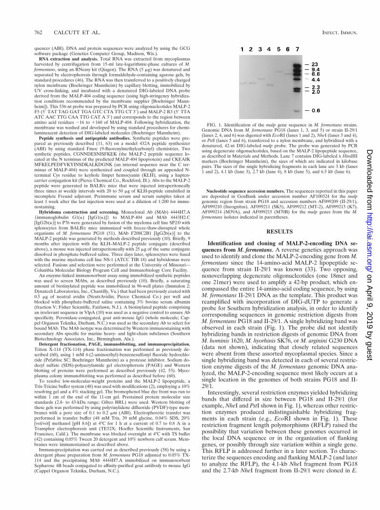
Identification and cloning of MALP-2-encoding DNA se-quences from M. fermentans. A reverse genetics approach wasused to identify and clone the MALP-2-encoding gene from M.fermentans since the 14-amino-acid MALP-2 lipopeptide se-quence from strain II-29/1 was known (33). Two opposing,nonoverlapping degenerate oligonucleotides (one 18mer andone 21mer) were used to amplify a 42-bp product, which en-compassed the entire 14-amino-acid coding sequence, by usingM. fermentans II-29/1 DNA as the template. This product wasreamplified with incorporation of DIG-dUTP to generate aprobe for Southern hybridization analysis, in order to identifycorresponding sequences in genomic restriction digests fromM. fermentans PG18 and II-29/1. A single hybridizing band wasobserved in each strain (Fig. 1). The probe did not identifyhybridizing bands in restriction digests of genomic DNA fromM. hominis 1620, M. hyorhinis SK76, or M. arginini G230 DNA(data not shown), indicating that closely related sequenceswere absent from these assorted mycoplasmal species. Since asingle hybridizing band was detected in each of several restric-tion enzyme digests of the M. fermentans genomic DNA ana-lyzed, the MALP-2-encoding sequence most likely occurs at asingle location in the genomes of both strains PG18 and II-29/1.
Interestingly, several restriction enzymes yielded hybridizingbands that differed in size between PG18 and II-29/1 (forexample, NheI and PstI shown in Fig. 1), whereas other restric-tion enzymes produced indistinguishable hybridizing frag-ments in each strain (e.g., EcoRI shown in Fig. 1). Theserestriction fragment length polymorphisms (RFLP) raised thepossibility that variation between these genomes occurred inthe local DNA sequence or in the organization of flankinggenes, or possibly through size variation within a single gene.This RFLP is addressed further in a later section. To charac-terize the sequences encoding and flanking MALP-2 (and laterto analyze the RFLP), the 4.1-kb NheI fragment from PG18and the 2.7-kb NheI fragment from II-29/1 were cloned in E.
FIG. 1. Identification of the malp gene sequence in M. fermentans strains.Genomic DNA from M. fermentans PG18 (lanes 1, 3, and 5) or strain II-29/1(lanes 2, 4, and 6) was digested with EcoRI (lanes 1 and 2), NheI (lanes 3 and 4),or PstI (lanes 5 and 6), transferred to a nylon membrane, and hybridized with adenatured, 42-nt DIG-labeled malp probe. The probe was generated by PCRusing degenerate oligonucleotides, based on the MALP-2 lipopeptide sequence,as described in Materials and Methods. Lane 7 contains DIG-labeled l HindIIImarkers (Boehringer Mannheim), the sizes of which are indicated in kilobasepairs. The sizes of the single hybridizing fragments in each lane are 3 kb (lanes1 and 2), 4.1 kb (lane 3), 2.7 kb (lane 4), 8 kb (lane 5), and 6.5 kb (lane 6).
762 CALCUTT ET AL. INFECT. IMMUN.
on April 9, 2019 by guest
http://iai.asm.org/
Dow
nloaded from
coli, as described in Materials and Methods, and the nucleotidesequence of each fragment was determined.
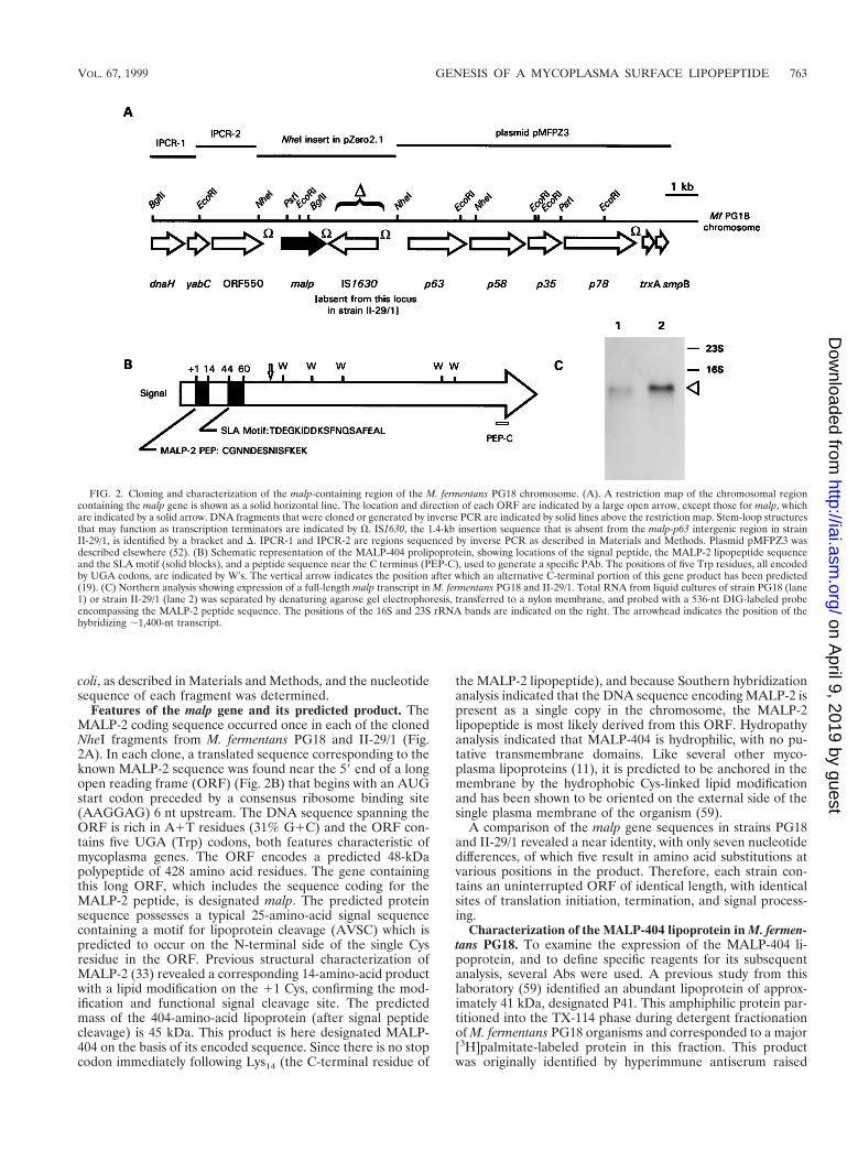
Features of the malp gene and its predicted product. TheMALP-2 coding sequence occurred once in each of the clonedNheI fragments from M. fermentans PG18 and II-29/1 (Fig.2A). In each clone, a translated sequence corresponding to theknown MALP-2 sequence was found near the 59 end of a longopen reading frame (ORF) (Fig. 2B) that begins with an AUGstart codon preceded by a consensus ribosome binding site(AAGGAG) 6 nt upstream. The DNA sequence spanning theORF is rich in A1T residues (31% G1C) and the ORF con-tains five UGA (Trp) codons, both features characteristic ofmycoplasma genes. The ORF encodes a predicted 48-kDapolypeptide of 428 amino acid residues. The gene containingthis long ORF, which includes the sequence coding for theMALP-2 peptide, is designated malp. The predicted proteinsequence possesses a typical 25-amino-acid signal sequencecontaining a motif for lipoprotein cleavage (AVSC) which ispredicted to occur on the N-terminal side of the single Cysresidue in the ORF. Previous structural characterization ofMALP-2 (33) revealed a corresponding 14-amino-acid productwith a lipid modification on the 11 Cys, confirming the mod-ification and functional signal cleavage site. The predictedmass of the 404-amino-acid lipoprotein (after signal peptidecleavage) is 45 kDa. This product is here designated MALP-404 on the basis of its encoded sequence. Since there is no stopcodon immediately following Lys14 (the C-terminal residue of
the MALP-2 lipopeptide), and because Southern hybridizationanalysis indicated that the DNA sequence encoding MALP-2 ispresent as a single copy in the chromosome, the MALP-2lipopeptide is most likely derived from this ORF. Hydropathyanalysis indicated that MALP-404 is hydrophilic, with no pu-tative transmembrane domains. Like several other myco-plasma lipoproteins (11), it is predicted to be anchored in themembrane by the hydrophobic Cys-linked lipid modificationand has been shown to be oriented on the external side of thesingle plasma membrane of the organism (59).
A comparison of the malp gene sequences in strains PG18and II-29/1 revealed a near identity, with only seven nucleotidedifferences, of which five result in amino acid substitutions atvarious positions in the product. Therefore, each strain con-tains an uninterrupted ORF of identical length, with identicalsites of translation initiation, termination, and signal process-ing.
Characterization of the MALP-404 lipoprotein in M. fermen-tans PG18. To examine the expression of the MALP-404 li-poprotein, and to define specific reagents for its subsequentanalysis, several Abs were used. A previous study from thislaboratory (59) identified an abundant lipoprotein of approx-imately 41 kDa, designated P41. This amphiphilic protein par-titioned into the TX-114 phase during detergent fractionationof M. fermentans PG18 organisms and corresponded to a major[3H]palmitate-labeled protein in this fraction. This productwas originally identified by hyperimmune antiserum raised
FIG. 2. Cloning and characterization of the malp-containing region of the M. fermentans PG18 chromosome. (A). A restriction map of the chromosomal regioncontaining the malp gene is shown as a solid horizontal line. The location and direction of each ORF are indicated by a large open arrow, except those for malp, whichare indicated by a solid arrow. DNA fragments that were cloned or generated by inverse PCR are indicated by solid lines above the restriction map. Stem-loop structuresthat may function as transcription terminators are indicated by V. IS1630, the 1.4-kb insertion sequence that is absent from the malp-p63 intergenic region in strainII-29/1, is identified by a bracket and D. IPCR-1 and IPCR-2 are regions sequenced by inverse PCR as described in Materials and Methods. Plasmid pMFPZ3 wasdescribed elsewhere (52). (B) Schematic representation of the MALP-404 prolipoprotein, showing locations of the signal peptide, the MALP-2 lipopeptide sequenceand the SLA motif (solid blocks), and a peptide sequence near the C terminus (PEP-C), used to generate a specific PAb. The positions of five Trp residues, all encodedby UGA codons, are indicated by W’s. The vertical arrow indicates the position after which an alternative C-terminal portion of this gene product has been predicted(19). (C) Northern analysis showing expression of a full-length malp transcript in M. fermentans PG18 and II-29/1. Total RNA from liquid cultures of strain PG18 (lane1) or strain II-29/1 (lane 2) was separated by denaturing agarose gel electrophoresis, transferred to a nylon membrane, and probed with a 536-nt DIG-labeled probeencompassing the MALP-2 peptide sequence. The positions of the 16S and 23S rRNA bands are indicated on the right. The arrowhead indicates the position of thehybridizing ;1,400-nt transcript.
VOL. 67, 1999 GENESIS OF A MYCOPLASMA SURFACE LIPOPEPTIDE 763
on April 9, 2019 by guest
http://iai.asm.org/
Dow
nloaded from
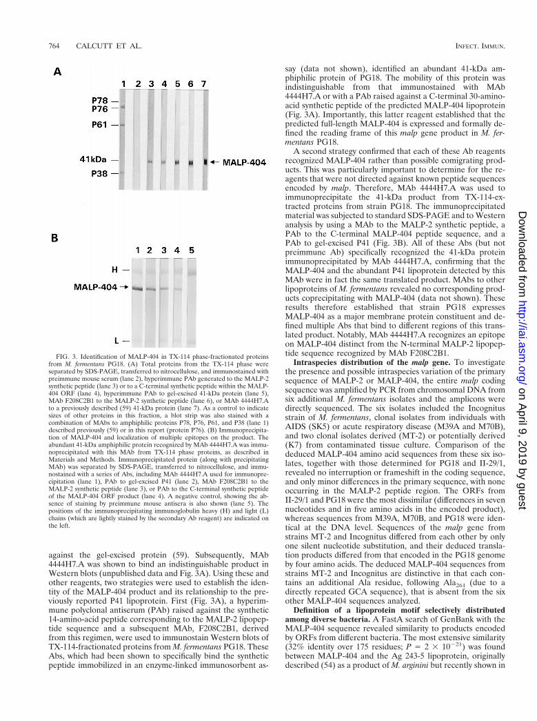
against the gel-excised protein (59). Subsequently, MAb4444H7.A was shown to bind an indistinguishable product inWestern blots (unpublished data and Fig. 3A). Using these andother reagents, two strategies were used to establish the iden-tity of the MALP-404 product and its relationship to the pre-viously reported P41 lipoprotein. First (Fig. 3A), a hyperim-mune polyclonal antiserum (PAb) raised against the synthetic14-amino-acid peptide corresponding to the MALP-2 lipopep-tide sequence and a subsequent MAb, F208C2B1, derivedfrom this regimen, were used to immunostain Western blots ofTX-114-fractionated proteins from M. fermentans PG18. TheseAbs, which had been shown to specifically bind the syntheticpeptide immobilized in an enzyme-linked immunosorbent as-
say (data not shown), identified an abundant 41-kDa am-phiphilic protein of PG18. The mobility of this protein wasindistinguishable from that immunostained with MAb4444H7.A or with a PAb raised against a C-terminal 30-amino-acid synthetic peptide of the predicted MALP-404 lipoprotein(Fig. 3A). Importantly, this latter reagent established that thepredicted full-length MALP-404 is expressed and formally de-fined the reading frame of this malp gene product in M. fer-mentans PG18.
A second strategy confirmed that each of these Ab reagentsrecognized MALP-404 rather than possible comigrating prod-ucts. This was particularly important to determine for the re-agents that were not directed against known peptide sequencesencoded by malp. Therefore, MAb 4444H7.A was used toimmunoprecipitate the 41-kDa product from TX-114-ex-tracted proteins from strain PG18. The immunoprecipitatedmaterial was subjected to standard SDS-PAGE and to Westernanalysis by using a MAb to the MALP-2 synthetic peptide, aPAb to the C-terminal MALP-404 peptide sequence, and aPAb to gel-excised P41 (Fig. 3B). All of these Abs (but notpreimmune Ab) specifically recognized the 41-kDa proteinimmunoprecipitated by MAb 4444H7.A, confirming that theMALP-404 and the abundant P41 lipoprotein detected by thisMAb were in fact the same translated product. MAbs to otherlipoproteins of M. fermentans revealed no corresponding prod-ucts coprecipitating with MALP-404 (data not shown). Theseresults therefore established that strain PG18 expressesMALP-404 as a major membrane protein constituent and de-fined multiple Abs that bind to different regions of this trans-lated product. Notably, MAb 4444H7.A recognizes an epitopeon MALP-404 distinct from the N-terminal MALP-2 lipopep-tide sequence recognized by MAb F208C2B1.
Intraspecies distribution of the malp gene. To investigatethe presence and possible intraspecies variation of the primarysequence of MALP-2 or MALP-404, the entire malp codingsequence was amplified by PCR from chromosomal DNA fromsix additional M. fermentans isolates and the amplicons weredirectly sequenced. The six isolates included the Incognitusstrain of M. fermentans, clonal isolates from individuals withAIDS (SK5) or acute respiratory disease (M39A and M70B),and two clonal isolates derived (MT-2) or potentially derived(K7) from contaminated tissue culture. Comparison of thededuced MALP-404 amino acid sequences from these six iso-lates, together with those determined for PG18 and II-29/1,revealed no interruption or frameshift in the coding sequence,and only minor differences in the primary sequence, with noneoccurring in the MALP-2 peptide region. The ORFs fromII-29/1 and PG18 were the most dissimilar (differences in sevennucleotides and in five amino acids in the encoded product),whereas sequences from M39A, M70B, and PG18 were iden-tical at the DNA level. Sequences of the malp gene fromstrains MT-2 and Incognitus differed from each other by onlyone silent nucleotide substitution, and their deduced transla-tion products differed from that encoded in the PG18 genomeby four amino acids. The deduced MALP-404 sequences fromstrains MT-2 and Incognitus are distinctive in that each con-tains an additional Ala residue, following Ala261 (due to adirectly repeated GCA sequence), that is absent from the sixother MALP-404 sequences analyzed.
Definition of a lipoprotein motif selectively distributedamong diverse bacteria. A FastA search of GenBank with theMALP-404 sequence revealed similarity to products encodedby ORFs from different bacteria. The most extensive similarity(32% identity over 175 residues; P 5 2 3 10221) was foundbetween MALP-404 and the Ag 243-5 lipoprotein, originallydescribed (54) as a product of M. arginini but recently shown in
FIG. 3. Identification of MALP-404 in TX-114 phase-fractionated proteinsfrom M. fermentans PG18. (A) Total proteins from the TX-114 phase wereseparated by SDS-PAGE, transferred to nitrocellulose, and immunostained withpreimmune mouse serum (lane 2), hyperimmune PAb generated to the MALP-2synthetic peptide (lane 3) or to a C-terminal synthetic peptide within the MALP-404 ORF (lane 4), hyperimmune PAb to gel-excised 41-kDa protein (lane 5),MAb F208C2B1 to the MALP-2 synthetic peptide (lane 6), or MAb 4444H7.Ato a previously described (59) 41-kDa protein (lane 7). As a control to indicatesizes of other proteins in this fraction, a blot strip was also stained with acombination of MAbs to amphiphilic proteins P78, P76, P61, and P38 (lane 1)described previously (59) or in this report (protein P76). (B) Immunoprecipita-tion of MALP-404 and localization of multiple epitopes on the product. Theabundant 41-kDa amphiphilic protein recognized by MAb 4444H7.A was immu-noprecipitated with this MAb from TX-114 phase proteins, as described inMaterials and Methods. Immunoprecipitated protein (along with precipitatingMAb) was separated by SDS-PAGE, transferred to nitrocellulose, and immu-nostained with a series of Abs, including MAb 4444H7.A used for immunopre-cipitation (lane 1), PAb to gel-excised P41 (lane 2), MAb F208C2B1 to theMALP-2 synthetic peptide (lane 3), or PAb to the C-terminal synthetic peptideof the MALP-404 ORF product (lane 4). A negative control, showing the ab-sence of staining by preimmune mouse antisera is also shown (lane 5). Thepositions of the immunoprecipitating immunoglobulin heavy (H) and light (L)chains (which are lightly stained by the secondary Ab reagent) are indicated onthe left.
764 CALCUTT ET AL. INFECT. IMMUN.
on April 9, 2019 by guest
http://iai.asm.org/
Dow
nloaded from
studies from our laboratory (13) to be the P47 lipoprotein ofM. hyorhinis, to which the originally described feature of tumormetastasis promotion in a nude mouse model (54) has nowbeen ascribed. The lipoprotein P39 (also designated BmpB) ofBorrelia burgdorferi also contains an extended region of simi-larity to MALP-404 (27% identity over 180 amino acids; P 52 3 1028). In addition to the modest similarity exhibited overextended regions of these two lipoproteins, a 19-amino-acidregion of striking local sequence conservation was shared byMALP-404 and, surprisingly, several additional lipoproteinsfrom diverse bacteria (Fig. 4). This sequence, designated theSLA (selective lipoprotein-associated) motif, was found inMALP-404, P47, and BmpB ORFs, as well as ORFs encodingputative lipoproteins of unknown function from Bacillus sub-tilis, Treponema pallidum, Mycoplasma capricolum, and Listeriamonocytogenes. Interestingly, the conserved motif is similarlypositioned in all of these lipoproteins, occurring from 17 to 43residues downstream of the predicted N-terminal lipid-modi-fied 11 Cys residue of each ORF. The significance of this motifis currently unknown, but the conservation of its sequence andrelative position argues for some hitherto-unknown functionassociated with the specific lipoproteins containing it. Theseselected lipoproteins are not ubiquitous in mycoplasmas orother bacteria, since the motif could not be identified in thecomplete genome sequences from the mycoplasmas M. geni-talium or M. pneumoniae, the gram-negative bacteria, E. coli,Haemophilus influenzae, or Helicobacter pylori, or any of thesequenced archeons.
Organization and IS1630-associated polymorphism of themalp region of the M. fermentans chromosome. Sequence anal-ysis of the 4.1- and 2.7-kb NheI fragments containing the malpgenes of strains PG18 and II-29/1, respectively, revealed thatthe RFLP observed in Southern analysis (Fig. 1) is not due todifferences in the malp DNA sequences but instead resultsfrom the presence of a newly discovered insertion sequence(IS)-like element immediately downstream of the malp gene inPG18 (Fig. 2A), which is absent from this corresponding re-gion of the genome in strain II-29/1. Interestingly, this element,designated IS1630, is oriented in the opposite direction fromall other genes in this region of the M. fermentans PG18 chro-mosome (Fig. 2A). IS1630 is distinct from the IS-like element
described previously in M. fermentans by Lo and coworkers(23) and is related to a characteristic group typified by IS30 ofE. coli (8). Since IS1630 is approximately 1.4 kb in size, itsabsence from the NheI fragment from II-29/1 accounts for theobserved 1.4-kb size difference between the cloned 2.7- and4.1-kb NheI fragments from II-29/1 and PG18, respectively, aswell as the 1.4-kb size difference between the respective PstIfragments from these strains that hybridize with a MALP-2-specific probe (Fig. 1 and 2A). IS1630 is not restricted to themalp region of the M. fermentans chromosome, as Southernanalysis revealed that the IS element is present in multiplecopies in the genomes of both strains PG18 and II-29/1 (datanot shown).
Analysis of the sequence downstream of malp indicated thatthis NheI fragment overlaps a genomic region containing fourgenes, p63-p58-p35-p78, that comprise an ABC transporteroperon (52). Further PCR analysis using forward and reversedprimers in the malp and p63 gene regions, respectively (Fig.2A), confirmed the presence of this gene organization in bothstrains PG18 and II-29/1. Application of this PCR analysis tothe additional six isolates of M. fermentans revealed (data notshown) that in all cases, malp was linked to p63 and that IS1630was present in the intergenic region in two of the six strainstested, M39A and M70B. In contrast, IS1630, although de-tected by PCR in the genome of all M. fermentans isolates usedin this study (data not shown), was absent from this intergenicregion in the remaining four strains, Incognitus, MT-2, SK5,and K7. The presence of identical malp DNA sequences andequivalent malp-IS1630 linkages between strains M39A,M70B, and PG18 suggests that these three strains may be veryclosely related. This notion is consistent with the results ofCampo et al. (9) from Southern analysis of these three strains,using restriction polymorphisms associated with an unrelatedIS element.
To determine further the genomic context in the region ofmalp in strain PG18, inverse PCR and DNA sequencing wereused to characterize three ORFs 59 to the malp gene. In ad-dition, sequencing of a previously isolated p78-containing plas-mid clone (52) was carried out to identify two ORFs 39 to p78.Each of the five additional ORFs identified in the contiguous15-kb region shown in Fig. 2A had identifiable orthologs inother mycoplasmas, but none was predicted to be a lipoproteinor other translocated surface protein. The genes were dnaH,yabC, and orf550 upstream of malp and trxA and smpB down-stream of p78. The presence of flanking genes encoding thedelta subunit of DNA polymerase III (dnaH) and thioredoxin(trxA) indicated that the genomic region surrounding malp ischromosomal. The closest homologs and other features of theORFs from this chromosomal region are summarized in Table1. Interestingly, the malp gene and IS1630 are the only se-quences present in this chromosomal region that do not haveorthologous counterparts in the complete published genomesequences of Mycoplasma genitalium (17) and Mycoplasmapneumoniae (21).
Immunological detection of the MALP-2 lipopeptide andintraspecies variation in the levels of malp-encoded products.The data presented above indicate that the MALP-2 lipopep-tide is expressed from a larger ORF that encodes MALP-404,an abundant TX-114 phase membrane surface lipoprotein ofM. fermentans PG18 (59). In a previous report (33), the potentmacrophage-stimulating activity attributed to the purifiedMALP-2 lipopeptide was shown to vary between PG18 and thehigh producer, isolate II-29/1. To further examine this variabil-ity between isolates, we sought to directly monitor the expres-sion of the MALP-2 lipopeptide and MALP-404 lipoprotein,using specific MAb reagents. By employing Tris-Tricine gels
FIG. 4. Amino acid sequence alignment of the SLA motif of various lipopro-teins. SLA motif-containing lipoproteins were identified and aligned by theFastA search program (GCG). The sequences of MALP-404 from M. fermentans,P47 from M. hyorhinis (13) (GenBank accession no. D16674), P20 deducedlipoprotein from M. capricolum (Z33368), p3A1 protein from L. monocytogenes(S80336), YufN from B. subtilis (Z93937), P39 or BmpB from Borrelia burgdorferi(L24194 and U49938, respectively), and TmbC lipoprotein from T. pallidum(X57836) are shown. Amino acid residues present in at least six of the sevenlipoproteins are highlighted with a black background, and residues conservedamong five of the lipoproteins are indicated with an asterisk. A consensus SLAmotif sequence was derived from the seven sequences and is shown below thesequence alignment. The numbers in parentheses to the left indicate the numberof residues between the putative lipid-modified 11 Cys residue and the firstamino acid of the motif. Those in parentheses to the right indicate the numberof residues between the last residue of the motif and those encoded by the stopcodon of the ORF.
VOL. 67, 1999 GENESIS OF A MYCOPLASMA SURFACE LIPOPEPTIDE 765
on April 9, 2019 by guest
http://iai.asm.org/
Dow
nloaded from
for resolution of the lipopeptide, PVDF membranes to bindthe lipopeptide after electrotransfer, and F208C2B1, the po-tent MAb to the MALP-2 synthetic peptide, a strong immu-nostaining band, which migrated close to the established (33)2-kDa mass of MALP-2, could be readily detected in totalprotein extracted from either strain PG18 or strain II-29/1,(Fig. 5). As expected, the MAb to the MALP-2 peptide alsoimmunostained the 41-kDa polypeptide in strain PG18, repre-senting MALP-404. Surprisingly, no MALP-404 could be de-tected in total cellular protein (Fig. 5, lanes 1 to 5) or in theTX-114 phase protein fraction (data not shown) of II-29/1. Thelack of staining of MALP-404 in II-29/1 is not due to differ-ences in the epitope recognized by the MAb, since theMALP-2 lipopeptide sequence deduced from the DNA se-
quence is identical in each strain. In additional experiments,MALP-404 could not be detected in total protein extracts ofII-29/1 with MAb 4444H7.A, polyclonal anti-P41, or the PAbraised against the C-terminal synthetic peptide of MALP-404(data not shown). These data suggest that under the conditionsemployed, the only product of the malp gene detectable inII-29/1 is the MALP-2 lipopeptide.
To compare the relative ratios of MALP-2 to MALP-404between strains PG18 and II-29/1, a series of twofold dilutionsof total protein from each strain was subjected to SDS-PAGEand Western analysis with the MAb to MALP-2. The results(Fig. 5) show that whereas MALP-404 can be detected in themost-dilute protein sample from strain PG18 shown (2.5 mg oftotal protein), MALP-404 is not detected in extracts fromII-29/1, even in samples containing eightfold-greater amountsof MALP-2. Therefore, the relative steady-state ratio ofMALP-404 to MALP-2 is dramatically lower in II-29/1 (ifMALP-404 is expressed at all) than in PG18. Furthermore, thisanalysis revealed no additional polypeptide species containingthe MALP-2 peptide in either strain, indicating that alternativeforms or possible intermediates of processing are either absentor occur at undetectable levels.
Distribution of malp products in clonal populations. Al-though the mycoplasmal isolates analyzed in this study hadbeen cloned during isolation, the distribution of malp transla-tion products was examined further to assess the possibility ofdifferential expression of MALP-2 and MALP-404 withinpropagating clonal populations. In particular, Western blotanalysis of the two strains was uninformative in resolvingwhether all of the cells in the broth culture from PG18 expressMALP-2 and MALP-404 or whether variation in the expres-sion pattern within the population occurred during growth inliquid medium. Similarly, the actual proportion of cells in theII-29/1 population that are producing MALP-2 could not bedetermined by such analysis. To address these questions, aportion of the broth cultures used for Western analysis wasplated onto solid medium and the resulting colonies were an-alyzed by colony immunoblotting with the MAb to the N-terminal MALP-2 peptide, F208C2B1, or MAb 4444H7.A,which recognizes a distinct epitope on MALP-404 outside theregion of this N-terminal peptide and is therefore operation-ally specific for the full-length product identified previously(Fig. 3 and 5). As shown in Fig. 6, colonies derived from the
FIG. 5. Strain variation in the relative levels of MALP-2 and MALP-404.Total proteins from late-logarithmic-phase cultures of M. fermentans II-29/1(lanes 1 to 5) or PG18 (lanes 7 to 9) were separated through a Tris-Tricine gel,transferred to a PVDF membrane, and immunostained with MAb F208C2B tothe MALP-2 peptide sequence as described in Materials and Methods. Twofolddilutions of each protein sample were analyzed over a range that includedequivalent immunostaining of the MALP-2 lipopeptide. The most concentratedsample from PG18 (lane 7) contained 10 mg of protein. Lane 6 contains low-molecular-weight markers (Gibco BRL), the sizes of which (in kilodaltons) areindicated on the left. The positions of the MALP-2 lipopeptide and the 41-kDaMALP-404 are indicated on the right. Lanes indicated by stars contain samplesof the two strains with approximately the same amount of immunostainedMALP-2.
TABLE 1. Products of ORFsa in chromosomal region of malp in M. fermentans PG18
ORF product designation Size (amino acids) Closest homolog (identity)b MG homologc MP homologd Motifse
DnaH 305 DnaH, B. subtilis (21/256) MG007 D12_orf253 NDf
YabC 236 HI1654, H. influenzae (42/223) MG056 D09_orf276 UPF0011ORF 550 550 MG423, M. genitalium (28/550) MG423/139 C12_orf561 NDMALP-404 428 P47, M. hyorhinis (32/175) ND ND LP 1 SLATnpA IS1630 387 Transposase IS30, E. coli (27/349) ND ND NDP63g 601 YufO, B. subtilis (38/597) MG119 A65_orf572 ATPP58g 537 MG120, M. genitalium (27/434) MG120 A65_orf517 NDP35g 327 MG121, M. genitalium (35/321) MG121 A65_orf311 NDP78g 681 ND ND ND LPTrxA 104 TrxA, Cyanidium sp. (32/96) MG124 A65_orf102 NDSmpB 147 SmpB, E. faecalis (40/147) MG059 D09_orf147 ND
a Relative position and orientation of each ORF are indicated in Fig. 2.b Basis for homology is given as number of identical amino acid residues/total number of amino acid residues in region (FastA).c MG homolog is from the M. genitalium database; designations refer to a system described previously (17).d MP homolog is from the M. pneumoniae database; designations refer to a system described previously (21).e Motif abbreviations: UPF0011, uncharacterized protein family 0011 (as defined in the GCG Motif program); LP, lipoprotein signal peptide and cleavage motif; SLA,
selective lipoprotein-associated motif (this work); ATP, ATP binding site.f ND, none detected.g ORF products previously characterized (52).
766 CALCUTT ET AL. INFECT. IMMUN.
on April 9, 2019 by guest
http://iai.asm.org/
Dow
nloaded from
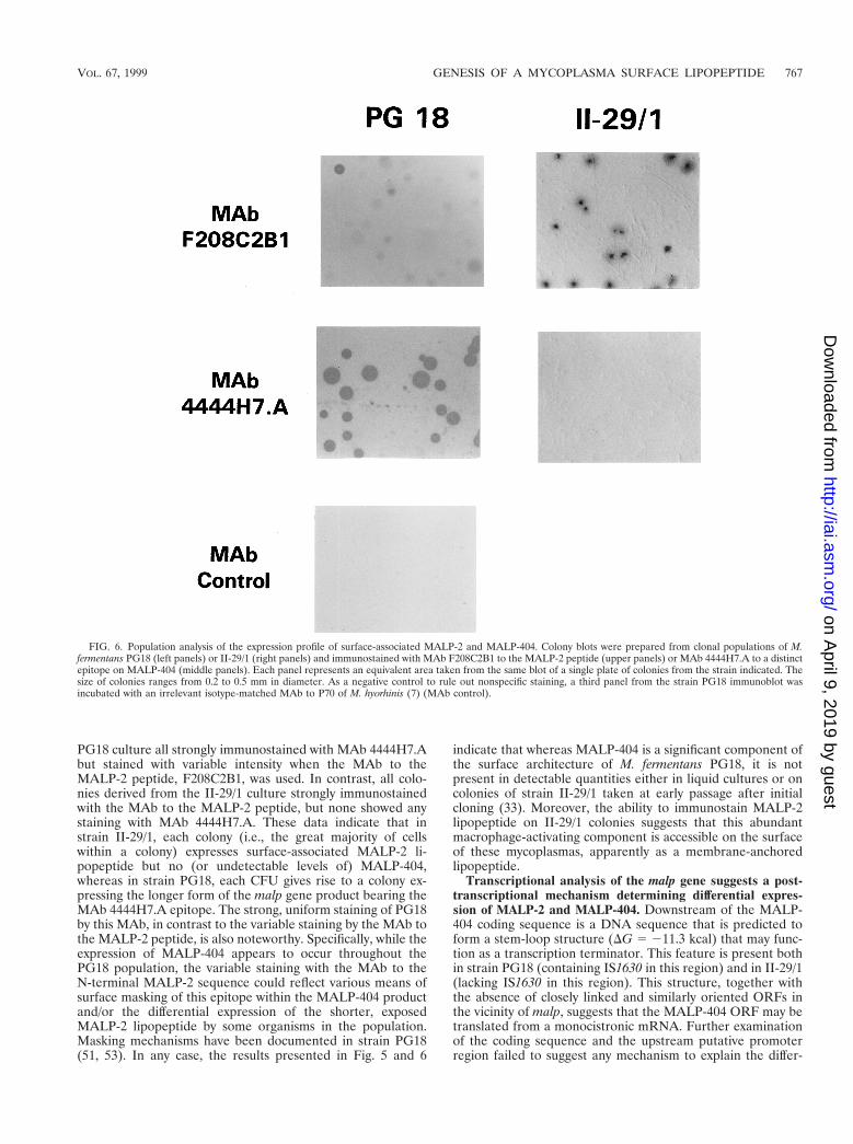
PG18 culture all strongly immunostained with MAb 4444H7.Abut stained with variable intensity when the MAb to theMALP-2 peptide, F208C2B1, was used. In contrast, all colo-nies derived from the II-29/1 culture strongly immunostainedwith the MAb to the MALP-2 peptide, but none showed anystaining with MAb 4444H7.A. These data indicate that instrain II-29/1, each colony (i.e., the great majority of cellswithin a colony) expresses surface-associated MALP-2 li-popeptide but no (or undetectable levels of) MALP-404,whereas in strain PG18, each CFU gives rise to a colony ex-pressing the longer form of the malp gene product bearing theMAb 4444H7.A epitope. The strong, uniform staining of PG18by this MAb, in contrast to the variable staining by the MAb tothe MALP-2 peptide, is also noteworthy. Specifically, while theexpression of MALP-404 appears to occur throughout thePG18 population, the variable staining with the MAb to theN-terminal MALP-2 sequence could reflect various means ofsurface masking of this epitope within the MALP-404 productand/or the differential expression of the shorter, exposedMALP-2 lipopeptide by some organisms in the population.Masking mechanisms have been documented in strain PG18(51, 53). In any case, the results presented in Fig. 5 and 6
indicate that whereas MALP-404 is a significant component ofthe surface architecture of M. fermentans PG18, it is notpresent in detectable quantities either in liquid cultures or oncolonies of strain II-29/1 taken at early passage after initialcloning (33). Moreover, the ability to immunostain MALP-2lipopeptide on II-29/1 colonies suggests that this abundantmacrophage-activating component is accessible on the surfaceof these mycoplasmas, apparently as a membrane-anchoredlipopeptide.
Transcriptional analysis of the malp gene suggests a post-transcriptional mechanism determining differential expres-sion of MALP-2 and MALP-404. Downstream of the MALP-404 coding sequence is a DNA sequence that is predicted toform a stem-loop structure (DG 5 211.3 kcal) that may func-tion as a transcription terminator. This feature is present bothin strain PG18 (containing IS1630 in this region) and in II-29/1(lacking IS1630 in this region). This structure, together withthe absence of closely linked and similarly oriented ORFs inthe vicinity of malp, suggests that the MALP-404 ORF may betranslated from a monocistronic mRNA. Further examinationof the coding sequence and the upstream putative promoterregion failed to suggest any mechanism to explain the differ-
FIG. 6. Population analysis of the expression profile of surface-associated MALP-2 and MALP-404. Colony blots were prepared from clonal populations of M.fermentans PG18 (left panels) or II-29/1 (right panels) and immunostained with MAb F208C2B1 to the MALP-2 peptide (upper panels) or MAb 4444H7.A to a distinctepitope on MALP-404 (middle panels). Each panel represents an equivalent area taken from the same blot of a single plate of colonies from the strain indicated. Thesize of colonies ranges from 0.2 to 0.5 mm in diameter. As a negative control to rule out nonspecific staining, a third panel from the strain PG18 immunoblot wasincubated with an irrelevant isotype-matched MAb to P70 of M. hyorhinis (7) (MAb control).
VOL. 67, 1999 GENESIS OF A MYCOPLASMA SURFACE LIPOPEPTIDE 767
on April 9, 2019 by guest
http://iai.asm.org/
Dow
nloaded from
ential expression (between PG18 and II-29/1) of the MALP-2lipopeptide and MALP-404, either at the DNA or transcrip-tional levels. Nevertheless, to directly characterize the malptranscript in each strain, total RNA was extracted from cul-tures of strain PG18 or early-passage II-29/1 and subjected toNorthern analysis using a 536-nt probe encompassing the por-tion of MALP-404 between amino acid residues 216 to 1160.The results of this analysis (Fig. 2C) revealed a single hybrid-izing band of approximately 1.4 kb in each strain. This isconsistent with the translation of MALP-404 from a monocis-tronic mRNA and suggests that the IS1630 does not affect theoverall size of the malp transcript. In conjunction with thefull-length ORFs present in PG18 and II-29/1, these resultsfurther support a posttranscriptional and probably posttrans-lational mechanism that dictates the strain differences in ex-pression of the MALP-2 versus MALP-404 products from thisgene.
DISCUSSION
This study identifies a single gene encoding the immuno-modulatory MALP-2 lipopeptide and the 41-kDa MALP-404surface lipoprotein of M. fermentans and documents key fea-tures of translation and processing of these products, as well asmarked differences in their resultant patterns of expression ondifferent isolates of this species. The structure, topology, andvariable posttranslational processing of malp-encoded prod-ucts, and the polymorphic nature of the malp chromosomallocus, all underscore a potentially novel role for this mycoplas-mal system in adaptation and pathogenesis in the human host.
Based on previous evidence (33, 59) and the results of thepresent study, the two malp-encoded translation products arepredicted and demonstrated to be surface associated and lipidmodified, based on colony immunoblot analysis, detergentphase partitioning, direct structural determination (for MALP-2), and [3H]palmitate labeling and accessibility to surface pro-teolysis (for MALP-404). The malp gene sequence revealed asignal peptide and a lipoprotein cleavage site, AVSC, verysimilar to those targeting membrane translocation and acyla-tion of other mycoplasma lipoproteins (11, 56). Matrix-associ-ated laser desorption/ionization time of flight mass spectrom-etry of MALP-2 indicated that this signal was cleaved and thatthe acylation of MALP-2 was atypical for procaryotes in thatthe N-linked acylation was absent. The ability to directly se-quence a product likely to be related to MALP-404 (discussedbelow) implies that the full-length lipoprotein may have thesesame N-terminal features (19). A further example of macroph-age-stimulatory lipoproteins and lipopeptides with two O-es-ter-linked fatty acids and a free N terminus on the 11 Cyscomes from recent studies with M. hyorhinis (34). It is conceiv-able that lipoproteins from mollicutes of the genus Myco-plasma may generally contain only two acyl groups on the 11Cys. This could have general ramifications in affecting proper-ties of lipoproteins that determine their ability to interact withand partition into the single mycoplasmal lipid bilayer mem-brane, as well as in their solubility and micellar formation, allpossibly related to the potency of their immunomodulatoryactivity.
Colony immunoblot analysis of PG18 indicated that virtuallyall or a great majority of cells express MALP-404. Since West-ern blot analysis confirmed that MALP-2 is present in PG18, atleast some cells must produce both products. The pathway forthe biogenesis of MALP-2 in PG18 is not known, but since onlyfull-length mRNA species could be detected from malp, themechanism must operate co- or posttranslationally, althoughmechanisms hitherto unknown in procaryotes, such as RNA
editing, cannot be formally discounted. Two quite distinctpathways can be envisioned. In one model, the MALP-2 li-popeptide is derived by processing of the larger MALP-404product. In this case, the full-length 41-kDa polypeptide istranslated, lipid modified, and then cleaved by signal peptidaseII to remove the signal peptide, prior to the separate process-ing event(s) that generate MALP-2. A slight variation on thismodel entails processing of the lipid-modified 41-kDa mole-cule to MALP-2, prior to signal peptide cleavage. In eitherscenario, processing to MALP-2 must occur after translocationthrough the membrane. An alternative cotranslational modelinvokes a cytoplasmic processing apparatus that acts upon thetranslation product (perhaps as the nascent peptide) prior tomembrane translocation, lipid modification, and signal peptidecleavage. In neither model is it clear what determines the sizeof the final surface-associated MALP-2 product or whether aprecise site of processing is recognized. The simplest prospec-tive candidate for the processing step is an endoprotease, spe-cifically cleaving at a single site C-terminal of Lys14 (the ter-minal residue in MALP-2). This is consistent with a lack ofdetectable intermediates, which might otherwise be antici-pated if nonspecific proteolytic cleavage occurred.
In contrast to the two translation products seen in M. fer-mentans PG18, only the MALP-2 lipopeptide was detected inextracts of the clonal isolate II-29/1 despite the presence of afull-length mRNA. This result defines an important, novelform of variation, wherein the absence of a major antigen, butprevalence of an immunomodulatory lipopeptide, occurs onthe mycoplasma surface as a result of posttranslational modi-fication. Although the precise mechanism and steps of biosyn-thesis are not known, it is useful to speculate that strain vari-ability in the levels of MALP-404 and MALP-2 may reflectdifferent extents of processing, possibly due to (i) differentialaccess to, or presence of, a specific protease; (ii) different levelsof components that specifically inhibit such a protease, or thatinteract with the substrate in a way to render it resistant toprocessing to the MALP-2 form; or (iii) differences in theefficiency of processing this specific lipoprotein through thesecretory apparatus of the cell, thereby leading to differencesin degradation of the larger product during transit. These rep-resent likely and testable hypotheses.
Posttranslational processing adds a novel level of complexityto our understanding of mycoplasma lipoproteins and surfaceantigenic variation within a species; however, the extent towhich this mechanism causes intraspecies variation has yet tobe explored. Reports suggest that a full-length MALP-404protein may be expressed in strain Incognitus (59) and in twoM. fermentans-contaminated human cell lines (19, 31) (seebelow), but the presence of the shorter MALP-2 product wasnot assessed in these studies. That variation in posttransla-tional processing underlies further complexities in propagatingpopulations is suggested by our recent observation (25) thatupon increased passage of II-29/1 clones in liquid culture,variants that express MALP-404 arise, as measured both byWestern and colony immunoblotting with MAb 4444H7.A, thereagent specific for the longer form. It is not yet knownwhether these clonal variants are capable of reversing thisphenotypic switch (the hallmark of phase variation), but if so,this would confer a phase-variable phenotype based on differ-ential processing of a primary gene product. Although theprecise mechanism that results in the emergence of the MALP-404-expressing variants is not known, our initial PCR analysisindicates that the malp gene organization is maintained in suchpopulations, without an insertion of IS1630. This result rulesout any correlation between the presence or absence of amalp-linked copy of IS1630 and the predominant translation
768 CALCUTT ET AL. INFECT. IMMUN.
on April 9, 2019 by guest
http://iai.asm.org/
Dow
nloaded from
product that is expressed. While intraclonal variation ofMALP-404 expression is yet to be fully characterized, twoimportant technical points are addressed by the emergence ofMALP-404 in propagating populations of II-29/1. First, theimmunostaining of these mycoplasmas with MAb 4444H7.Ademonstrates that the cognate epitope is present in the MALP-404 product encoded by the malp gene of this strain and thatthe lack of positive staining of the colony immunoblot shown inFig. 5 is not due to amino acid substitutions affecting the MAb4444H7.A epitope. Second, such populations formally demon-strate that the strain and the conditions used for cultivation donot lead to any generalized proteolytic degradation of surfaceproteins, including MALP-404. Thus, the biogenesis ofMALP-2 is unlikely to be a result of generalized proteolyticactivity. It may be of interest to note that in at least one cloneof another mycoplasma species, M. hyorhinis, truncated formsof the variable lipoprotein VlpA exist in addition to the longerforms of this Vlp (34).
Two recent studies reporting genes with sequences similar tomalp are noteworthy, in the context of assigning immunologi-cal roles to MALP-404 or its various regions and as a caution-ary note regarding the attribution of well-known immunolog-ical properties of whole mycoplasmas in tissue culture to thisspecific translation product. In one study (19, 24), a proteinassociated with (and initially thought to be derived from) thehuman HL60 promyelocytic cell line was subsequently recog-nized as a gene product of M. fermentans (19) to which theproperty of promoting proliferation and activation of HL60cells was attributed. Interestingly, the gene sequence, derivedfrom a cDNA library of infected cell cultures, predicted aproduct that resembled the N-terminal region of MALP-404but reflected another reading frame product following aminoacid position 90 (see Fig. 2), due to the absence of a base in theDNA sequence corresponding to this position. Our study re-vealed no such alternative form of a malp gene product in eightstrains examined, and the bona fide reading frame for MALP-404 was confirmed by its immunostaining with an Ab to ourpredicted C-terminal peptide sequence. In a second series ofstudies, Matsumoto et al. incorrectly identified the MALP-404gene sequence as a human cell product in which UGA codonswere suggested to encode selenocysteine and proposed that acomplement-activating property found in cell culture superna-tants was associated with this soluble protein product (31).These authors recently reassigned the origin of the gene as M.fermentans (30, 49), based on its high sequence similarity withthe sequence reported by Hall et al. (19). Although not for-mally identifying their tissue culture contaminants as M. fer-mentans, Matsumoto et al. also suggested that the gene andcorresponding antigen were represented in several culturesdisplaying complement-activating properties (30). Finally,these authors showed Southern hybridization of DNA fromcontaminated lines that generate multiple sizes of restrictionfragments (30). Whether this represents malp-linked polymor-phisms associated with multiple contaminants of M. fermen-tans, or possibly other mycoplasmal species, is not clear. Over-all, these reports underscore the need for extremely rigorousdefinition of (i) the activities measured, (ii) the source of thecomponents with the activity, (iii) the physical nature and stateof the moiety endowing the property, and (iv) high levels ofpurity (for authentic or recombinant products) to avoid con-founding modulatory compounds present in most eubacteria,including multiple mycoplasmal species (20). With these strictrequirements in mind, we suggest that the lipid moiety ofMALP-2 is the critical element in macrophage activation andthat the solubility and differential expression of MALP-2 andMALP-404 may provide critically discrete physical forms of
this product that could affect the antigenic properties and theimmunomodulatory properties of M. fermentans. We have notruled out other activities associated with the protein sequenceof MALP-404, but currently see no definitive evidence for suchactivities.
One of the most intriguing aspects of the MALP-404 se-quence is the SLA motif. A cursory examination of the knownbinding motifs of human major histocompatibility complexmolecules (39) reveals that the SLA sequence contains thenonapeptide motif -G------F, which could be presented to Tcells by the human class I major histocompatibility complexmolecule HLA-B*5101, provided that this peptide can be gen-erated from SLA after appropriate processing by the antigen-presenting cells. Incidentally, there exists a rare multifacetedautoimmune disease, Behcet’s disease, which is strongly HLA-B51 associated in many ethnic groups (14, 32). The etiology ofBehcet’s disease is unknown, but infectious agents have beendiscussed as a possible cause from early on, as recently re-viewed (45). It is tempting to speculate that this rheumaticautoimmune disease may be triggered in susceptible B51-pos-itive patients by protein antigens from quite different genera ofeubacteria expressing the conserved SLA sequence. Whetherthis motif is associated with an immunological activity per se ormight serve other functions in the organism, such as a recog-nition motif for proteolytic processing to MALP-2, analogousto other motifs involved in regulatory proteolysis (1, 37), iscurrently under investigation.
ACKNOWLEDGMENTS
We thank Shyh-Ching Lo, Joseph Tully, and Richard DelGiudice forproviding mycoplasmal isolates and DNA preparations and Jeong Imfor preparation of peptides and conjugates.
This work was supported in part by U.S. Public Health Service grantAI32219 (to K.S.W.) from the National Institute of Allergy and Infec-tious Diseases and by grant Mu 672/2-2 (to P.F.M.) from the DeutscheForschungsgemeinschaft.
REFERENCES
1. Alley, M. R., J. R. Maddock, and L. Shapiro. 1993. Requirement of thecarboxyl terminus of a bacterial chemoreceptor for its targeted proteolysis.Science 259:1754–1757.
2. Applied Biosystems. 1991. ProBlott™ applications in SDS-PAGE, electro-blotting and protein sequencing, p. 1–12. User bulletin no. 42. AppliedBiosystems, Inc., Foster City, Calif.
3. Behrens, A., M. Heller, H. Kirchhoff, D. Yogev, and R. Rosengarten. 1994. Afamily of phase- and size-variant membrane surface lipoprotein antigens(Vsps) of Mycoplasma bovis. Infect. Immun. 62:5075–5084.
4. Bhugra, B., L. R. Voelker, N. Zou, H. Yu, and K. Dybvig. 1996. Mechanismof antigenic variation in Mycoplasma pulmonis: interwoven, site-specificDNA inversions. Mol. Microbiol. 18:703–714.
5. Boesen, T., J. Emmersen, L. T. Jensen, S. A. Ladefoged, P. Thorsen, S.Birkelund, and G. Christiansen. 1998. The Mycoplasma hominis vaa genedisplays a mosaic gene structure. Mol. Microbiol. 29:97–110.
6. Braun, V., and H. C. Wu. 1994. Lipoproteins, structure, function, biosynthe-sis and model for protein export, p. 319–341. In J.-M. Ghuysen and R.Hakenbeck (ed.), New comprehensive biochemistry. Elsevier Science, Am-sterdam, The Netherlands.
7. Bricker, T. M., M. J. Boyer, J. Keith, R. Watson-McKown, and K. S. Wise.1988. Association of lipids with integral membrane surface proteins of My-coplasma hyorhinis. Infect. Immun. 56:295–301.
8. Calcutt, M. J., and K. S. Wise. Unpublished results.9. Campo, L., P. Larocque, T. La Malfa, W. D. Blackburn, and H. L. Watson.
1998. Genotypic and phenotypic analysis of Mycoplasma fermentans strainsisolated from different host tissues. J. Clin. Microbiol. 36:1371–1377.
10. Citti, C., M. F. Kim, and K. S. Wise. 1997. Elongated versions of Vlp surfacelipoproteins protect Mycoplasma hyorhinis escape variants from growth-in-hibiting host antibodies. Infect. Immun. 65:1773–1785.
11. Cleavinger, C. M., M. F. Kim, J. H. Im, and K. S. Wise. 1995. Identificationof mycoplasma membrane proteins by systematic TnphoA mutagenesis of arecombinant library. Mol. Microbiol. 18:283–293.
12. Cole, B. C., Y. Naot, E. J. Stanbridge, and K. S. Wise. 1985. Interactions ofmycoplasmas and their products with lymphoid cells in vitro, p. 203–257. InS. Razin and M. F. Barile (ed.), The mycoplasmas, vol. 4. Mycoplasma
VOL. 67, 1999 GENESIS OF A MYCOPLASMA SURFACE LIPOPEPTIDE 769
on April 9, 2019 by guest
http://iai.asm.org/
Dow
nloaded from
pathogenicity. Academic Press, Inc., New York, N.Y.13. Droesse, M., and K. S. Wise. Unpublished results.14. Falk, K., O. Rotzschke, M. Takiguchi, V. Gnau, S. Stevanovic, G. Jung, and
H. G. Rammensee. 1995. Peptide motifs of HLA-B51, -B52 and -B78 mol-ecules, and implications for Behcet’s disease. Int. Immunol. 7:223–228.
15. Feng, S.-H., and S.-C. Lo. 1994. Induced mouse spleen B-cell proliferationand secretion of immunoglobulin by lipid-associated membrane proteins ofMycoplasma fermentans incognitus and Mycoplasma penetrans. Infect. Im-mun. 62:3916–3921.
16. Forsyth, M. H., M. E. Tourtellotte, and S. J. Geary. 1992. Localization of animmunodominant 64 kDa lipoprotein (LP 64) in the membrane of Myco-plasma gallisepticum and its role in cytadherence. Mol. Microbiol. 6:2099–2106.
17. Fraser, C. M., J. D. Gocayne, O. White, M. D. Adams, R. A. Clayton, R. D.Fleischmann, C. J. Bult, A. R. Kerlavage, G. Sutton, J. M. Kelley, J. L.Fritchman, J. F. Weidman, K. V. Small, M. Sandusky, J. Fuhrmann, D.Nguyen, T. R. Utterback, D. M. Saudek, C. A. Phillips, J. M. Merrick, J. F.Tomb, B. A. Dougherty, K. F. Bott, P. C. Hu, T. S. Lucier, S. N. Peterson,H. O. Smith, C. A. Hutchison III, and J. C. Venter. 1995. The minimal genecomplement of Mycoplasma genitalium. Science 270:397–403.
18. Garcia, J., B. Lemercier, S. Roman-Roman, and G. Rawadi. 1998. A Myco-plasma fermentans-derived synthetic lipopeptide induces AP-1 and NF-kBactivity and cytokine secretion in macrophages via the activation of MAPKpathways. J. Biol. Chem. 273:34391–34398.
19. Hall, R. E., S. Agarwal, D. P. Kestler, J. A. Cobb, K. M. Goldstein, and N. S.Chang. 1996. cDNA and genomic cloning and expression of the P48 mono-cytic differentiation/activation factor, a Mycoplasma fermentans gene prod-uct. Biochem. J. 319:919–927.
20. Henderson, B., S. Poole, and M. Wilson. 1996. Bacterial modulins: a novelclass of virulence factors which cause host tissue pathology by inducingcytokine synthesis. Microbiol. Rev. 60:316–341.
21. Himmelreich, R., H. Hilbert, H. Plagens, E. Pirkl, B.-C. Li, and R. Herr-mann. 1996. Complete sequence analysis of the genome of the bacteriumMycoplasma pneumoniae. Nucleic Acids Res. 24:4420–4449.
22. Hoffman, R. W., F. X. O’Sullivan, K. R. Schafermeyer, T. L. Moore, D.Roussell, R. Watson-McKown, M. F. Kim, and K. S. Wise. 1997. Myco-plasma infection and rheumatoid arthritis: analysis of their relationship usingimmunoblotting and an ultrasensitive polymerase chain reaction detectionmethod. Arthritis Rheum. 40:1219–1228.
23. Hu, W. S., R. Y. Wang, R. S. Liou, J. W. Shih, and S. C. Lo. 1990. Identifi-cation of an insertion-sequence-like genetic element in the newly recognizedhuman pathogen Mycoplasma incognitus. Gene 93:67–72.
24. Kestler, D. P., S. Agarwal, and R. E. Hall. 1995. Up-regulation of cytokinemRNA in human monocytes and myeloid cell lines by the differentiation/activation factor p48. Immunology 86:463–468.
25. Kim, M. F., and K. S. Wise. Unpublished results.26. Kostyal, D. A., G. H. Butler, and D. H. Beezhold. 1994. A 48-kilodalton
Mycoplasma fermentans membrane protein induces cytokine secretion byhuman monocytes. Infect. Immun. 62:3793–3800.
27. Lysnyansky, I., R. Rosengarten, and D. Yogev. 1996. Phenotypic switching ofvariable surface lipoproteins in Mycoplasma bovis involves high-frequencychromosomal rearrangements. J. Bacteriol. 178:5395–5401.
28. Markham, P. F., M. D. Glew, K. G. Whithear, and I. D. Walker. 1993.Molecular cloning of a member of the gene family that encodes pMGA, ahemagglutinin of Mycoplasma gallisepticum. Infect. Immun. 61:903–909.
29. Marshall, A. J., R. J. Miles, and L. Richards. 1995. The phagocytosis ofmycoplasmas. J. Med. Microbiol. 43:239–250.
30. Matsumoto, M., M. Nishiguchi, S. Kikkawa, H. Nishimura, S. Nagasawa,and T. Seya. 1998. Structural and functional properties of complement-activating protein M161Ag, a Mycoplasma fermentans gene product thatinduces cytokine production by human monocytes. J. Biol. Chem. 273:12407–12414.
31. Matsumoto, M., J. Takeda, N. Inoue, T. Hara, M. Hatanaka, K. Takahashi,S. Nagasawa, H. Akedo, and T. Seya. 1997. A novel protein that participatesin nonself discrimination of malignant cells by homologous complement.Nat. Med. 3:1266–1270.
32. Mizuki, N., S. Ohno, H. Ando, L. Chen, G. D. Palimeris, E. Stavropoulos-Ghiokas, M. Ishihara, K. Goto, S. Nakamura, Y. Shindo, K. Isobe, N. Ito,and H. Inoko. 1997. A strong association between HLA-B*5101 and Behcet’sdisease in Greek patients. Tissue Antigens 50:57–60.
33. Muhlradt, P. F., M. Kiess, H. Meyer, R. Sussmuth, and G. Jung. 1997.Isolation, structure elucidation, and synthesis of a macrophage stimulatorylipopeptide from Mycoplasma fermentans acting at picomolar concentration.J. Exp. Med. 185:1951–1958.
34. Muhlradt, P. F., M. Kiess, H. Meyer, R. Sussmuth, and G. Jung. 1998.Structure and specific activity of macrophage-stimulating lipopeptides fromMycoplasma hyorhinis. Infect. Immun. 66:4804–4810.
35. Muhlradt, P. F., H. Meyer, and R. Jansen. 1996. Identification of S-(2,3-dihydroxypropyl)cystein in a macrophage-activating lipopeptide from Myco-plasma fermentans. Biochemistry 35:7781–7786.
36. Muhlradt, P. F., and U. Schade. 1991. MDHM, a macrophage-stimulatoryproduct of Mycoplasma fermentans, leads to in vitro interleukin-1 (IL-1),
IL-6, tumor necrosis factor, and prostaglandin production and is pyrogenic inrabbits. Infect. Immun. 59:3969–3974.
37. Popham, P. L., T. W. Hahn, K. A. Krebes, and D. C. Krause. 1997. Loss ofHMW1 and HMW3 in noncytadhering mutants of Mycoplasma pneumoniaeoccurs post-translationally. Proc. Natl. Acad. Sci. USA 94:13979–13984.
38. Quentmeier, H., E. Schmitt, H. Kirchhoff, W. Grote, and P. F. Muhlradt.1990. Mycoplasma fermentans-derived high-molecular-weight material in-duces interleukin-6 release in cultures of murine macrophages and humanmonocytes. Infect. Immun. 58:1273–1280.
39. Rammensee, H. G., T. Friede, and S. Stevanovic. 1995. MHC ligands andpeptide motifs: first listing. Immunogenetics 41:178–228.
40. Rawadi, G., V. Ramez, B. Lemercier, and S. Roman-Roman. 1998. Activationof mitogen-activated protein kinase pathways by Mycoplasma fermentansmembrane lipoproteins in murine macrophages: involvement in cytokinesynthesis. J. Immunol. 160:1330–1339.
41. Rawadi, G., S. Roman-Roman, M. Castedo, V. Dutilleul, S. Susin, P. Mar-chetti, M. Geuskens, and G. Kroemer. 1996. Effects of Mycoplasma fermen-tans on the myelomonocytic lineage. Different molecular entities with cyto-kine-inducing and cytocidal potential. J. Immunol. 156:670–678.
42. Rosengarten, R., and K. S. Wise. 1991. The Vlp system of Mycoplasmahyorhinis: combinatorial expression of distinct size variant lipoproteins gen-erating high-frequency surface antigenic variation. J. Bacteriol. 173:4782–4793.
43. Ruschmeyer, D., H. Thude, and P. F. Muhlradt. 1993. MDHM, a macro-phage-activating product of Mycoplasma fermentans, stimulates murine mac-rophages to synthesize nitric oxide and become tumoricidal. FEMS Immu-nol. Med. Microbiol. 7:223–230.
44. Ruuth, E., and F. Praz. 1989. Interactions between mycoplasmas and theimmune system. Immunol. Rev. 112:133–160.
45. Sakane, T. 1997. New perspective on Behcet’s disease. Int. Rev. Immunol.14:89–96.
46. Sambrook, J., E. F. Fritsch, and T. Maniatis. 1989. Molecular cloning: alaboratory manual, 2nd ed. Cold Spring Harbor Laboratory, Cold SpringHarbor, N.Y.
47. Sankaran, K., and H. C. Wu. 1994. Lipid modification of bacterial proli-poprotein. Transfer of diacylglyceryl moiety from phosphatidylglycerol.J. Biol. Chem. 269:19701–19706.
48. Schagger, H., and G. von Jagow. 1987. Tricine-sodium dodecyl sulfate-polyacrylamide gel electrophoresis for the separation of proteins in the rangefrom 1 to 100 kDa. Anal. Biochem. 166:368–379.
49. Seya, T., N. A. Begum, and M. Matsumoto. 1998. Mycoplasma origin oftumor cell protein. Nat. Med. 4:133.
50. Simmons, W. L., C. Zuhua, J. I. Glass, J. W. Simecka, G. H. Cassell, andH. L. Watson. 1996. Sequence analysis of the chromosomal region aroundand within the V-1-encoding gene of Mycoplasma pulmonis: evidence forDNA inversion as a mechanism for V-1 variation. Infect. Immun. 64:472–479.
51. Theiss, P., A. Karpas, and K. S. Wise. 1996. Antigenic topology of the P29surface lipoprotein of Mycoplasma fermentans: differential display ofepitopes results in high-frequency phase variation. Infect. Immun. 64:1800–1809.
52. Theiss, P., and K. S. Wise. 1997. Localized frameshift mutation generatesselective, high-frequency phase variation in a surface lipoprotein encoded bya mycoplasma ABC transporter operon. J. Bacteriol. 179:4013–4022.
53. Theiss, P. M., M. F. Kim, and K. S. Wise. 1993. Differential protein expres-sion and surface presentation generates high-frequency antigenic variation inMycoplasma fermentans. Infect. Immun. 61:5123–5128.
54. Ushio, S., K. Iwaki, M. Taniai, T. Ohta, S. Fukuda, K. Sugimura, and M.Kurimoto. 1995. Metastasis-promoting activity of a novel molecule, Ag243-5, derived from mycoplasma, and the complete nucleotide sequence.Microbiol. Immunol. 39:393–400.
55. Washburn, L. R., K. E. Weaver, E. J. Weaver, W. Donelan, and S. Al-Sheboul. 1998. Molecular characterization of Mycoplasma arthritidis variablesurface protein MAA2. Infect. Immun. 66:2576–2586.
56. Wieslander, Å., M. J. Boyer, and H. Wroblewski. 1992. Membrane andprotein structure, p. 93–112. In J. Maniloff, R. N. McElhaney, L. R. Finch,and J. B. Baseman (ed.), Mycoplasmas: molecular biology and pathogenesis.American Society for Microbiology, Washington, D.C.
57. Wilton, J. L., A. L. Scarman, M. J. Walker, and S. P. Djordjevic. 1998.Reiterated repeat region variability in the ciliary adhesin gene of Myco-plasma hyopneumoniae. Microbiology 144:1931–1943.
58. Wise, K. S., and M. F. Kim. 1987. Major membrane surface proteins ofMycoplasma hyopneumoniae selectively modified by covalently bound lipid. J.Bacteriol. 169:5546–5555.
59. Wise, K. S., M. F. Kim, P. M. Theiss, and S.-C. Lo. 1993. A family ofstrain-variant surface lipoproteins of Mycoplasma fermentans. Infect. Immun.61:3327–3333.
60. Wise, K. S., M. F. Kim, and R. Watson-McKown. 1995. Variant membraneproteins, p. 227–241. In S. Razin and J. G. Tully (ed.), Molecular anddiagnostic procedures in mycoplasmology. Academic Press, New York, N.Y.
61. Wise, K. S., D. Yogev, and R. Rosengarten. 1992. Antigenic variation, p.473–490. In J. Maniloff, R. N. McElhaney, L. R. Finch, and J. B. Baseman(ed.), Mycoplasmas: molecular biology and pathogenesis. American Society
770 CALCUTT ET AL. INFECT. IMMUN.
on April 9, 2019 by guest
http://iai.asm.org/
Dow
nloaded from

for Microbiology, Washington, D.C.62. Yogev, D., R. Watson-McKown, M. A. McIntosh, and K. S. Wise. 1991.
Sequence and TnphoA analysis of a Mycoplasma hyorhinis protein with mem-brane export function. J. Bacteriol. 173:2035–2044.
63. Yogev, D., R. Watson-McKown, R. Rosengarten, J. H. Im, and K. S. Wise.1995. Increased structural and combinatorial diversity in an extended familyof genes encoding Vlp surface proteins of Mycoplasma hyorhinis. J. Bacteriol.177:5636–5643.
64. Zhang, Q., and K. S. Wise. 1996. Molecular basis of size and antigenic
variation of a Mycoplasma hominis adhesin encoded by divergent vaa genes.Infect. Immun. 64:2737–2744.
65. Zhang, Q., and K. S. Wise. 1997. Localized reversible frameshift mutation inan adhesin gene confers a phase-variable adherence phenotype in myco-plasma. Mol. Microbiol. 25:859–869.
66. Zheng, X., L. J. Teng, H. L. Watson, J. I. Glass, A. Blanchard, and G. H.Cassell. 1995. Small repeating units within the Ureaplasma urealyticum MBantigen gene encode serovar specificity and are associated with antigen sizevariation. Infect. Immun. 63:891–898.
Editor: V. A. Fischetti
VOL. 67, 1999 GENESIS OF A MYCOPLASMA SURFACE LIPOPEPTIDE 771
on April 9, 2019 by guest
http://iai.asm.org/
Dow
nloaded from