posttranslational modification, folding, assembly, and death 1.covalent modifications 1)proteolytic...
Post on 22-Dec-2015
216 views
TRANSCRIPT
Posttranslational modification, folding, assembly, and death
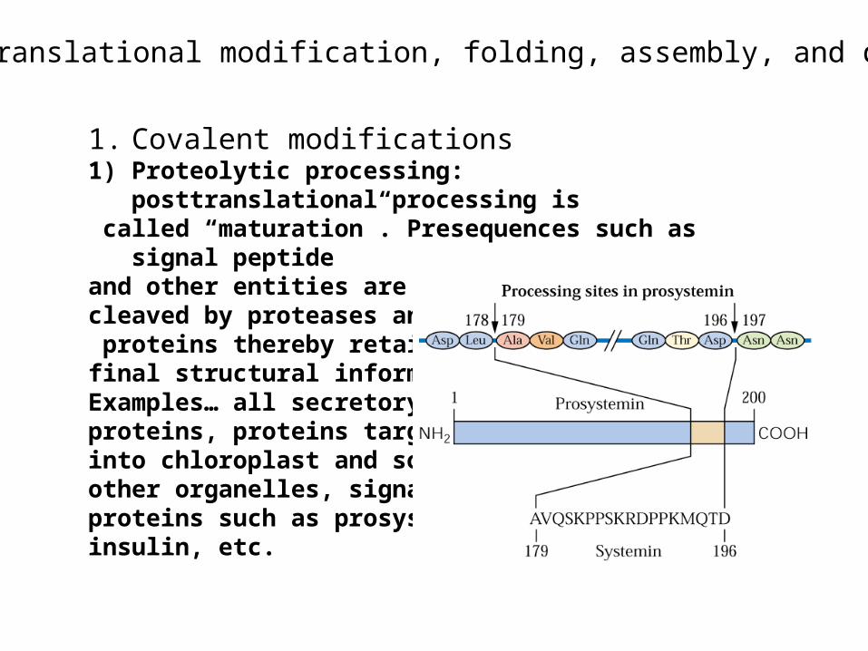
1. Covalent modifications1) Proteolytic processing: posttranslational processing is called “maturation”. Presequences such as signal peptide and other entities are cleaved by proteases and proteins thereby retain their final structural information. Examples… all secretory proteins, proteins targeted into chloroplast and some other organelles, signaling proteins such as prosystemin, insulin, etc.
2) Protein phosphorylationArguably the most important posttranslational modification of proteins in eukaryotic cells. Which amino acid residues are phosphorylated?Protein kinases and phosphatases. Their names and substrate specificity. Plants vs. animals—differences will be further covered in signaling lectures
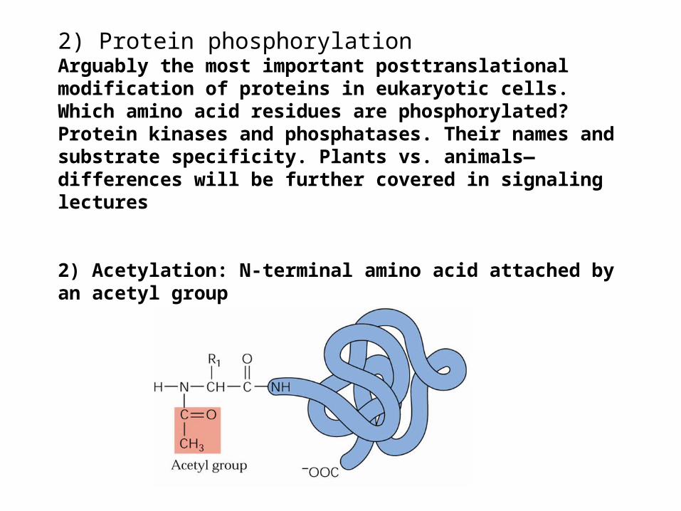
2) Acetylation: N-terminal amino acid attached by an acetyl group
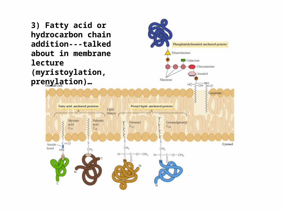
3) Fatty acid or hydrocarbon chain addition---talked about in membrane lecture (myristoylation, prenylation)…
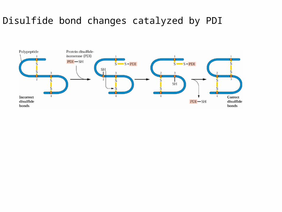
4) Disulfide bond changes catalyzed by PDI
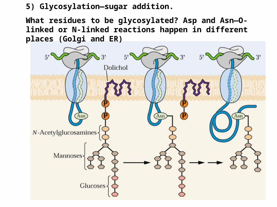
5) Glycosylation—sugar addition.
What residues to be glycosylated? Asp and Asn—O-linked or N-linked reactions happen in different places (Golgi and ER)

2. Protein foldingThe AA sequence of protein - 3_D structure1) Spontaneous or assisted process? Evidence?2) Molecular chaperones : proteins that bind and stabilize
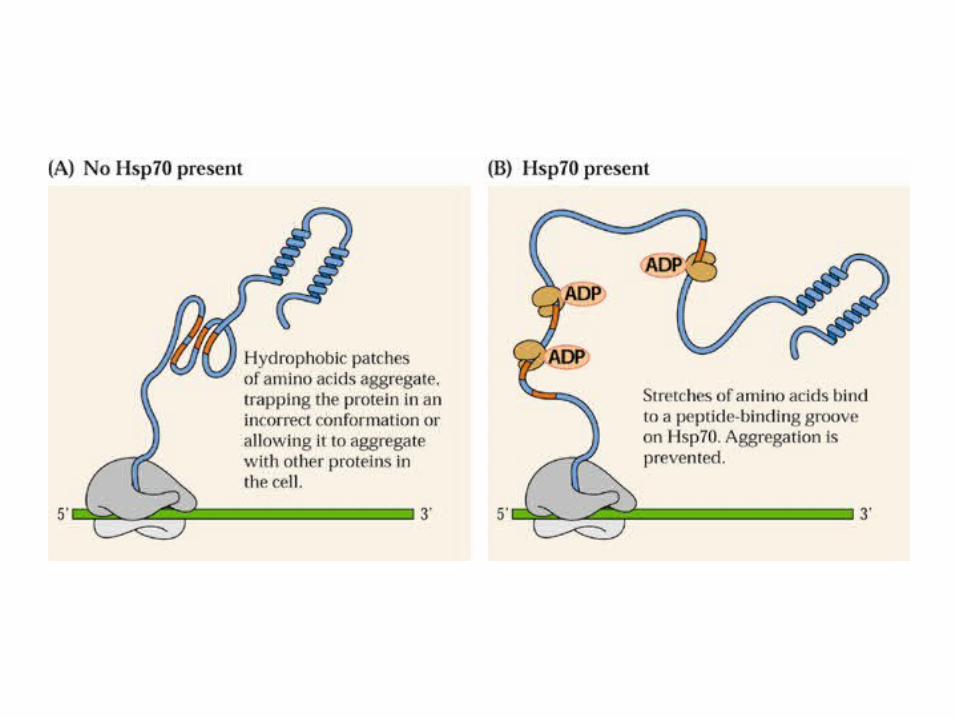
other proteins and help them fold/assemble properly (can be folding of one protein and assembly of multiple proteins). Heat shock protein story:
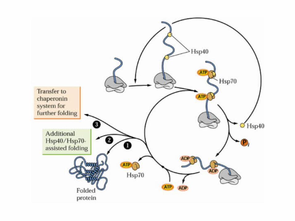
Two major types: type I includes hsp70---bind and prevent misfolding of the substrate proteins (can also unfold proteins)---cytosol, chloroplast, mitochondria.
Type II also referred to chaperonins such as hsp60 (GroEL in bacteria) family proteins that form large complex like a beer barrel that brew the substrate proteins with the help of “lid” protein such as cpn10 in the chloroplast (or GroES in bacteria).
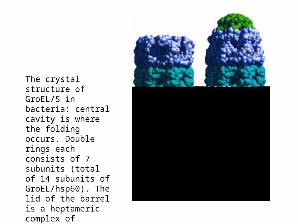
The crystal structure of GroEL/S in bacteria: central cavity is where the folding occurs. Double rings each consists of 7 subunits (total of 14 subunits of GroEL/hsp60). The lid of the barrel is a heptameric complex of GroES/cpn10.
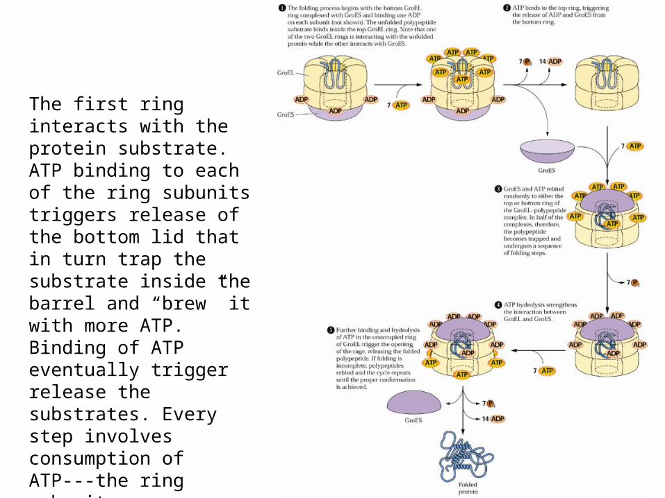
The first ring interacts with the protein substrate. ATP binding to each of the ring subunits triggers release of the bottom lid that in turn trap the substrate inside the barrel and “brew” it with more ATP. Binding of ATP eventually trigger release the substrates. Every step involves consumption of ATP---the ring subunits are ATPases.
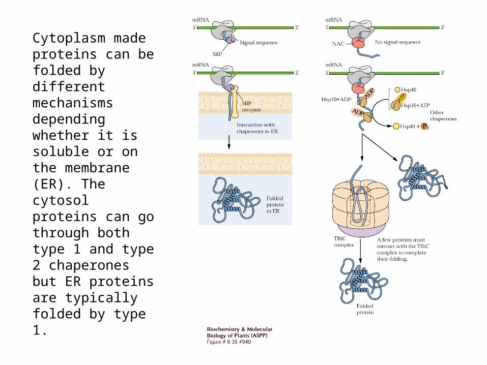
Cytoplasm made proteins can be folded by different mechanisms depending whether it is soluble or on the membrane (ER). The cytosol proteins can go through both type 1 and type 2 chaperones but ER proteins are typically folded by type 1.

3) Protein foldases---enzymes that catalyze foldingPDI—protein disulfide isomerase catalyze correct formation of S-S_bonds as described earlier.
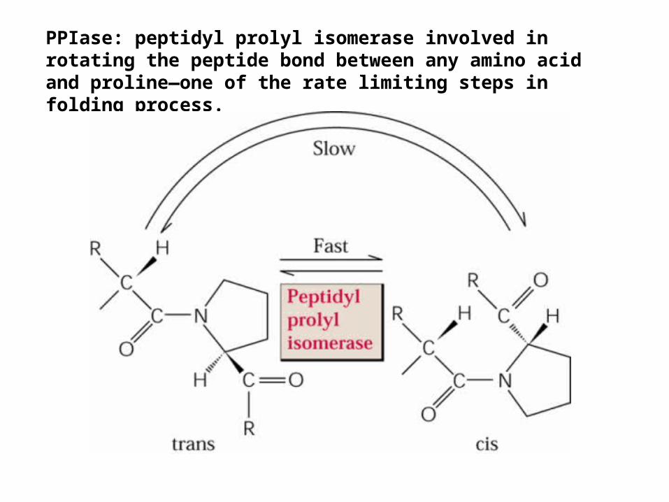
PPIase: peptidyl prolyl isomerase involved in rotating the peptide bond between any amino acid and proline—one of the rate limiting steps in folding process.
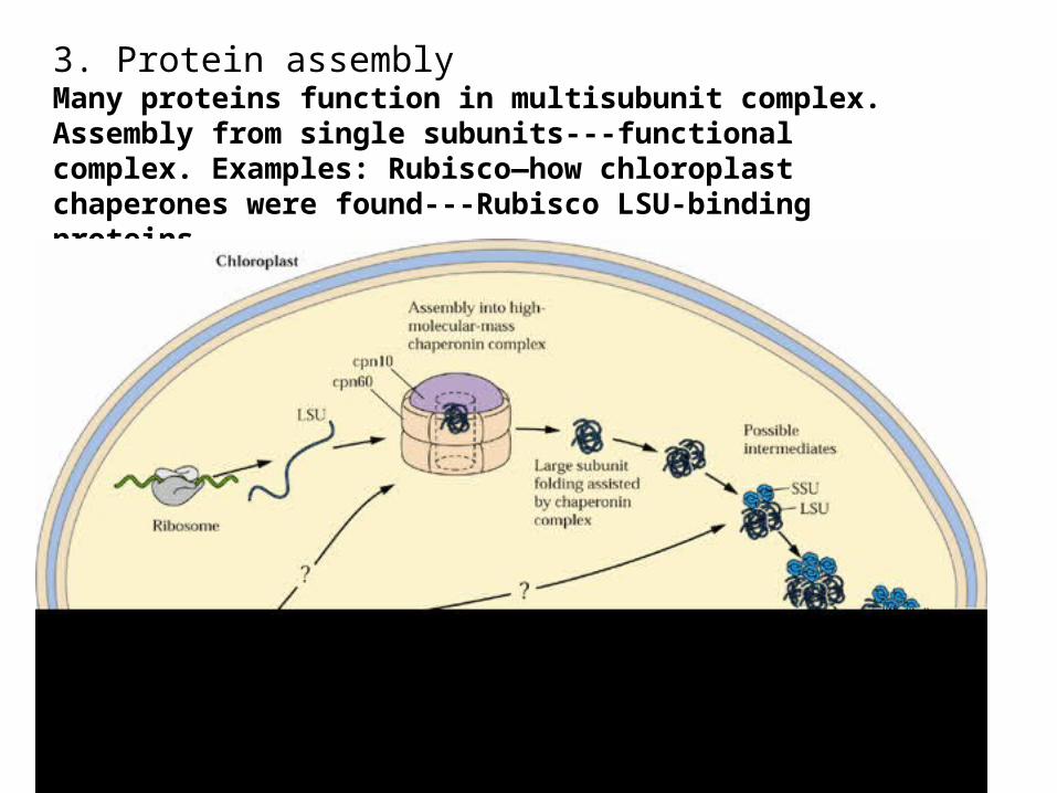
3. Protein assemblyMany proteins function in multisubunit complex. Assembly from single subunits---functional complex. Examples: Rubisco—how chloroplast chaperones were found---Rubisco LSU-binding proteins.
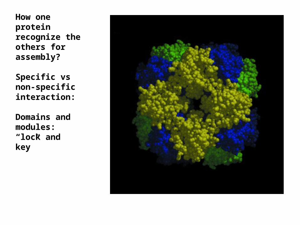
How one protein recognize the others for assembly?
Specific vs non-specific interaction:
Domains and modules: “lock and key”

4. Protein degradation—end of the life cycle1) Biologically very important: Examples, a regulatory protein
works when it is needed. Degradation is a way to remove the “function”. Such function can be positive or negative…
2) Where degradation occurs: many compartments inside the cell.
3) What enzymes:a large number of proteases, some cleave a specific bond some chew the entire protein.
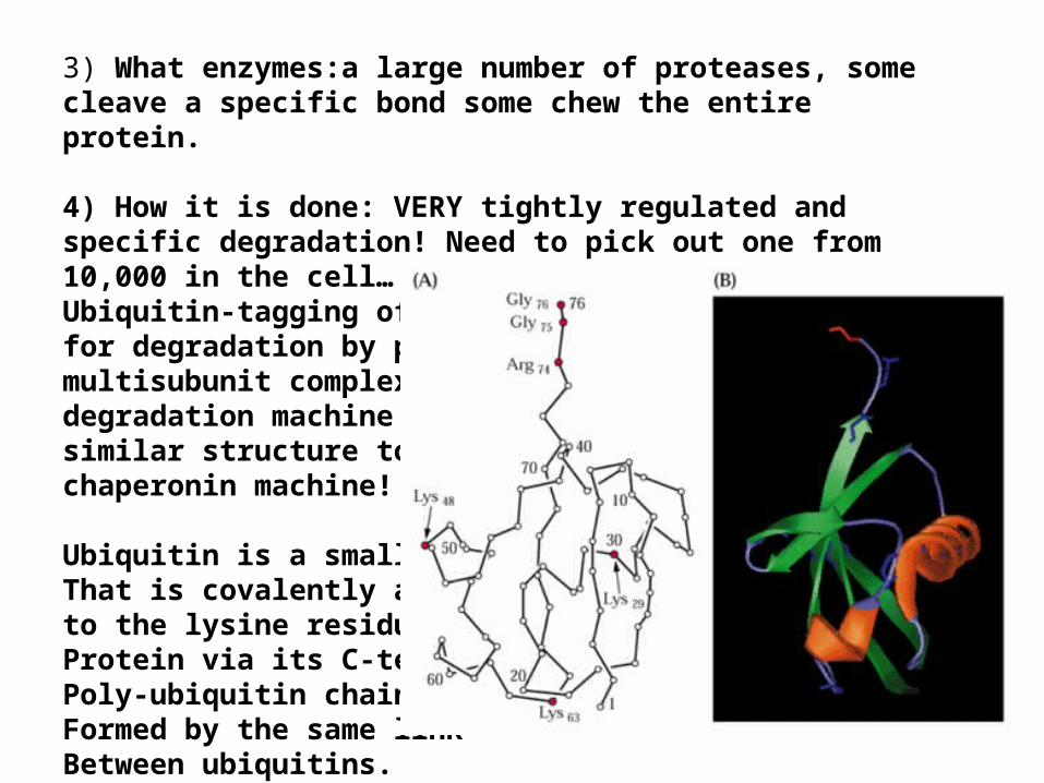
4) How it is done: VERY tightly regulated and specific degradation! Need to pick out one from 10,000 in the cell…Ubiquitin-tagging of proteins: serves as signal for degradation by proteasome, a large multisubunit complex---degradation machine with similar structure to chaperonin machine!
Ubiquitin is a small protein That is covalently attached to the lysine residue of a Protein via its C-terminus. Poly-ubiquitin chain can beFormed by the same link Between ubiquitins.
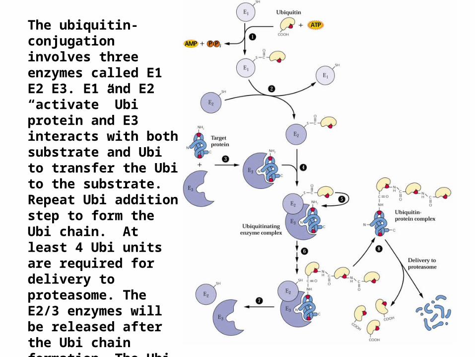
The ubiquitin-conjugation involves three enzymes called E1 E2 E3. E1 and E2 “activate” Ubi protein and E3 interacts with both substrate and Ubi to transfer the Ubi to the substrate. Repeat Ubi addition step to form the Ubi chain. At least 4 Ubi units are required for delivery to proteasome. The E2/3 enzymes will be released after the Ubi chain formation. The Ubi chain will be recognized by the proteasome and the substrate is sent for degradation but Ubi is “spit” out of the proteasome for reuse.