digest - university of pittsburgh fall 2014 ... high risk for developing wound complications after a...
TRANSCRIPT

dige
stFall 2014
Affiliated with the University of Pittsburgh School of Medicine, UPMC is
ranked among the nation’s best hospitals by U.S. News & World Report.
By David C. Whitcomb, MD, PhD
UPMC has earned an international reputation for advanced pancreatic clinical care and groundbreaking pancreas research, especially related to genetics and technical procedures. An integration of translational research and technical advances, along with clinical expertise from multiple specialties into new clinical care models, occurs in UPMC’s multidisciplinary Pancreas Center of Excellence (PCOE). We share self-designated academic PCOE status with many other
academic medical centers throughout the United States. However, standard minimum requirements for truly “qualified” centers of excellence remain unclear and undeveloped. PCOE programs, equipment, expertise, and the demonstration of quality care and excellent outcomes need further definition.
Patient advocacy groups such as the National Pancreas Foundation (NPF) and others can direct patients and families to qualified regional pancreas centers. These health nonprofits have recognized the need for qualified PCOEs to be assessed by independent and objective criteria.
Academic physician scientists at UPMC are working internally and with other colleagues to develop novel, automated patient assessment instruments, electronic medical record
continued on Page 10
What Is a PCOE? Do You Need One?
Division of Gastroenterology, Hepatology, and Nutrition
accreditation Statement: The University of Pittsburgh School of Medicine is accredited by the Accreditation Council for Continuing Medical Education (ACCME) to provide continuing medical education for physicians.
The University of Pittsburgh School of Medicine designates this enduring material for a maximum of 0.5 AMA PRA Category 1 Credits™. Each physician should only claim credit commensurate with the extent of their participation in the activity. Other health care professionals are awarded .05 continuing education units (CEU), which are equivalent to 0.5 contact hours.
Disclosures: Doctors Blaney, Dasyam, Mounzer, O’Keefe, Papachristou, Razzak, Singhi, Yadav, and Zureikat have reported no relationships with entities producing health care goods or services.
Dr. Greer has received research support from AbbVie Pharmaceuticals. Dr. Whitcomb has received research support from the NIH, is a consultant for AbbVie Pharmaceuticals, Novartis, and Millennium Pharmacy Systems, and is a stockholder in Ambry Genetics™.
Instructions: To take the CME evaluation and receive credit, please visit UPMCPhysicianResources.com/GI and click on UPMC Digest Fall 2014.
In This Issue 1 What Is a PCOE? Do You Need One?
2 Robot Cyst-Gastrostomy and Pancreatic Debridement
3 The Importance of Nutrition in Chronic Pancreatitis Patients
4 PRSS1 Hereditary Pancreatitis: Lessons From Pathology
5 Autoimmune Pancreatitis: A Great Masquerader
6 Lifestyle Choices That Impact the Pancreas
7 Role of Imaging in Necrotizing Pancreatitis
8 Nutritional Support in Acute Pancreatitis: Pancreatic Rest and How to Avoid TPN
1 1 Managing Chronic Pancreatitis: Our Multidisciplinary Case-by-Case Approach
13 The Complex Nature (and Nurture) of Recurrent Acute Pancreatitis
14 What Is This?
15 Welcoming New Faculty / PancreasFest 2015

D I G E S T — FA L L 2 0 1 4
Robotic Cyst-Gastrostomy and Pancreatic Debridement for Walled-off Pancreatic NecrosisBy Amer H. Zureikat, MD, FACS
Walled-off pancreatic necrosis (WOPN) is an infrequent
but serious sequela of acute necrotizing pancreatitis. This
complication typically requires operative drainage into the
stomach (cyst-gastrostomy) with debridement of the necrotic
pancreatic tissue. The procedure is traditionally done via a
laparotomy and associated with significant morbidity, but
surgeons at UPMC are now using minimally invasive techniques
to treat these collections in the hope of reducing blood loss,
postoperative pain, morbidity, and length of hospital stay.
A 32-year-old male with no history of alcohol or cigarette abuse
was referred with a WOPN following an attack of acute necrotizing
pancreatitis in 2011. His past medical history was significant for
systemic lupus erythematosus (SLE) (treated with chronic
steroids), insulin-dependent diabetes mellitus (IDDM), and a
cholecystectomy. The etiology of the pancreatitis was deemed
to be secondary to SLE and/or chronic steroid use, because all
other investigations (including genetic defects PRSS1, SPINK1,
and CFTR) were negative. The collection measured 13 cm and
was connected to another pelvic collection (12 cm) via a tract
(Figures 1 and 2). This young patient was deemed a good candidate
for a robotic-assisted operation, especially because he was at
high risk for developing wound complications after a laparotomy.
The patient underwent a successful robotic cyst-gastrostomy,
pancreatic cyst wall biopsy, pancreatic debridement (Figure 3),
and drainage and debridement of the pelvic collection.
Intraoperatively, the tract joining the pancreatic and pelvic
collections was identified, clipped, and severed. The operation
was performed entirely in minimally invasive fashion in less than
four hours with minimal blood loss (30 mL). Final pathology
revealed a fibrotic cyst wall with no malignancy. The patient was
discharged home six days postoperatively with no complications,
and he was able to resume his daily physical activities within two
weeks of discharge.
The robotic approach is being utilized increasingly in many
pancreatic surgeries at UPMC, including Whipple procedures,
central, distal, and total pancreatectomies, Frey and Puestow
procedures, and pancreatic debridement and cyst-gastrostomies.
This robotic approach allows complex pancreatic operations to be
performed in a purely minimally invasive fashion while maintaining
adherence to the principles of open pancreatic surgery.
Reference:
Zureikat AH, Moser AJ, Boone BA, Bartlett DL, Zenati M, Zeh HJ III.
250 Robotic Pancreatic Resections: Safety and Feasibility. Ann Surg.
2013 Oct;258(4): 554-62 PMID:24002300.
Dr. Zureikat is assistant professor of surgery in the UPMC Division of Surgical Oncology. He also serves as the co-director of the UPMC Pancreatic Cancer Center.
Figure 1. Walled-off pancreatic necrosis (WOPN) connected to a second pancreatic enzyme-rich collection in the pelvis. Note the connecting tract between the two collections.
Figure 2. Axial images showing the pancreatic collection with necrotic debris within it.
Figure 3. Robotic-assisted pancreatic debridement after the creation of the cyst-gastrostomy: the extraction of necrotic pancreatic debris from the WOPN using robotic graspers. The stereotactic vision and wristed instruments allow meticulous and comprehensive pancreatic debridement.
2
Affiliated with the University of Pittsburgh School of Medicine, UPMC is ranked among the nation’s best hospitals by U.S. News & World Report.

The Importance of Nutrition in Chronic Pancreatitis PatientsBy Julia B. Greer, MD, MPH
Chronic pancreatitis is a progressive inflammatory disease characterized by irreversible structural changes and gradual fibrotic replacement of the pancreas. This parenchymal fibrosis leads to diminished exocrine and endocrine function of the gland. Not surprisingly, the majority of chronic pancreatitis patients will develop maldigestion and malabsorption of varying degrees due to the loss of exocrine function. Weight loss is also very common in these patients and has multiple contributing causes, including maldigestion, fear of eating due to pain (sitophobia), delayed gastric emptying, anorexia, nausea, and vomiting.
Levels of serum proteins, including albumin, prealbumin, and retinol-binding protein, are often significantly lower in chronic pancreatitis patients than in healthy individuals, reflecting protein-calorie malnutrition.1, 2 Malabsorption frequently results in malnutrition and micronutrient deficiencies, and may elevate markers of oxidative stress and inflammation. Chronic pancreatitis patients may have diminished levels of nutrients such as glutathione, zinc, and selenium, as well as vitamins B12, C, D, and E. Micronutrient deficits compromise critical bodily enzymatic and immune functions, thereby compounding health issues. A recent systematic review demonstrated that most individuals with chronic pancreatitis have osteopathy, and one in four has osteoporosis.3
Pancreatic enzyme replacement therapy (PERT) is the most effective means of treating malabsorption in chronic pancreatitis patients.4 However, many clinicians do not prescribe PERT until overt symptoms, such as weight loss and steatorrhea (more than 15 g/day fecal fat), have developed. Many patients underdose or take enzymes before they begin eating, although PERT is most effective when taken in conjunction with a meal and right after eating.5 Some patients require larger doses of PERT than others, with or without gastric acid suppression, to achieve adequate nutrient absorption.5
There are few published investigations of malnutrition in chronic pancreatitis,6 and most studies are small in size and are of limited statistical significance.1 Currently, our group at UPMC is conducting a large-scale, adequately powered study to measure serum levels of micronutrients and markers of oxidative stress in 300 chronic pancreatitis patients and 300 individuals without pancreatic disease. Study participants are derived from the North American Pancreatitis Studies (NAPS). Our initial goal is to show that micronutrient deficiencies exist in a significantly greater proportion of the chronic pancreatitis patients than controls. Secondarily, we will assess how disease duration and comorbidities, such as diabetes mellitus and alcoholism, may affect these same measures of nutritional status and oxidative stress. Finally, it will be possible to correlate deficiencies with quality of life variables, such as degree of pain. This data will
enable us to design an interventional study of PERT in specific subsets of chronic pancreatitis patients, such as those whose primary etiologic factor is heavy alcohol consumption. The variables that we are evaluating are listed in Table 1.
References:1 Schrader H, Menge BA, Belyaev O, et al. Amino Acid Malnutrition in
Patients With Chronic Pancreatitis and Pancreatic Carcinoma. Pancreas.
2009 May;38(4):416-21. PMID: 19169171.2 Lasztity N, Biro L, Nemeth E, et al. Protein Status in Pancreatitis —
Transthyretin Is a Sensitive Biomarker of Malnutrition in Acute and
Chronic Pancreatitis. Clinical Chemistry & Laboratory Medicine. 2002
Dec;40(12):1320-24. PMID: 12553437.3 Duggan SN, Smyth ND, Murphy A, et al. High Prevalence of Osteoporosis in
Patients With Chronic Pancreatitis: A Systematic Review and Meta-analysis.
Clinical Gastroenterology & Hepatology. 2014 Feb;12(2):219-28. PMID: 238563359.4 Layer P, Keller J, Lankisch PG. Pancreatic Enzyme Replacement Therapy.
Current Gastroenterology Reports. 2001 Apr;3(2):101-8. PMID: 11276376.5 Dominguez-Munoz JE, Iglesias-Garcia J, Vilarino-Insua M, et al. 13C-Mixed
Triglyceride Breath Test to Assess Oral Enzyme Substitution Therapy in
Patients With Chronic Pancreatitis. Clinical Gastroenterology & Hepatology.
2007 Apr;5(4):484-8. PMID: 17445754.6 Trolli PA, Conwell DL, Zuccaro G, Jr. Pancreatic Enzyme Therapy and
Nutritional Status of Outpatients With Chronic Pancreatitis. Gastroenterology
Nursing. 2001 Mar-Apr;24(2):84-7. PMID: 11847733.
Dr. Greer is assistant professor of medicine with the Division of Gastroenterology, Hepatology, and Nutrition. She has written two health-related cookbooks, The Anti-Cancer Cookbook and The Anti-Breast Cancer Cookbook: How to Cut Your Risk With the Most Powerful Cancer-Fighting Foods.
Table 1. Nutrition in Chronic Pancreatitis
Biomarker What Blood Levels Indicate
Pre-albumin Protein-calorie malnutrition
Retinol Binding Protein Protein-calorie malnutrition
Vitamin D (25-hydroxy vitamin D3) Fat malabsorption, bone mineral density
Vitamin E (α-tocopherol) Fat malabsorption, potential oxidative stress
Vitamin B12 (cobalamin) Malabsorption, potential nervous system dysfunction
Tumor Necrosis Factor Alpha (TNF-α)
Inflammation
Interleukin-6 Inflammation
C-Reactive Protein Inflammation
Apolipoprotein-CII Fat malabsorption
Glutathione Protein-calorie malnutrition, immune function
Zinc Mineral malabsorption, immune function
Osteocalcin Bone mineral density
3UPMC Division of Gastroenterology, Hepatology, and Nutrition
UPMCPhysicianResources.com/GI For consults and referrals, please call UPMC’s 24-hour physician OnDemand service at 1-866-884-8579.

D I G E S T — FA L L 2 0 1 4
PRSS1 Hereditary Pancreatitis: Lessons From PathologyBy Aatur D. Singhi, MD, PhD
Hereditary pancreatitis is a rare, heritable form of chronic
pancreatitis. It is an autosomal dominant disorder with an
80 percent penetrance and variable expressivity. Patients
with hereditary pancreatitis suffer from recurrent episodes of
acute pancreatitis, and the majority progress to having chronic
pancreatitis. The disease usually begins in early childhood, but
onset can vary from infancy to the sixth decade of life.
By microsatellite linkage analysis, David Whitcomb, MD, PhD, and
colleagues discerned the gene responsible for 80 percent of
hereditary pancreatitis families on the
long arm of chromosome 7 (7q35).1
Subsequently, mutations within the
cationic trypsinogen gene, also referred
to as serine protease 1 (PRSS1), were
identified as the underlying defect by
candidate gene approach.2 Several
mutations within both the coding
sequence and introns of PRSS1 have been
described, but the R122H and N29I
mutations are the most prevalent. The
discovery of PRSS1 mutations has allowed
for the development of genetic testing
and, consequently, better classification of
the etiology of chronic pancreatitis within
a subset of patients and their families.
Further, a major motivation for identifying
patients with hereditary pancreatitis is the
increased risk of developing pancreatic
cancer. Recent studies have found a lifetime
risk of pancreatic cancer of approximately
40 percent at 70 years of age.
Data on the histopathologic findings of
PRSS1 hereditary pancreatitis have been
limited. Practical difficulties in obtaining
biopsies from the pancreas have
prevented research during the early
stages of the disease. In addition, studies
obtaining specimens at later disease
stages from patients undergoing surgery
or from autopsies are lacking, although a
report of two young adults with PRSS1
hereditary pancreatitis describes
histologic findings as indistinguishable from those seen in the
setting of nonhereditary forms of chronic pancreatitis (e.g.,
pancreatic atrophy and prominent fibrosis).3
A review of pancreatic pathology from PRSS1 patients seen at
UPMC argues the contrary.4 Patients with PRSS1 mutations
develop a clinicopathologic form of pancreatitis that involves
progressive pancreatic lipomatous atrophy. In children, most
often the pancreas is grossly normal, but microscopically, there is
variation in pancreatic lobular size and shape (Figure 1A). While
the central portions of the pancreas display parenchymal loss
and are accompanied by loose fibrosis, the periphery of the
pancreas is remarkable for replacement by mature adipose
tissue. These changes are more developed in younger adults,
where fatty replacement seems to extend from the periphery to
the central portion of the pancreas (Figure 1B). With older
patients, the pancreas shows marked
atrophy and extensive replacement by
mature adipose tissue with scattered
islets of Langerhans and rare acinar
epithelium (Figure 1C). In one patient,
the extensive fatty replacement
mimicked a mass lesion within the head
of the pancreas.
While lipomatous atrophy of the
pancreas is a frequent occurrence in the
general population and is seen with
increasing age, it typically presents
focally and not as extensively as in PRSS1
patients.5 Other conditions associated
with fatty replacement of the pancreas
include Cushing’s syndrome, steroid
therapy, malnutrition, and viral infections.
Genetic syndromes, such as Shwachman-
Diamond, Johanson-Blizzard, and cystic
fibrosis, are also associated with
lipomatous atrophy. Of note, the most
common etiology of pancreatitis in
children is cystic fibrosis, an autosomal
recessive disorder caused by CFTR. CFTR
mutations result in thick inspissated
secretions and mucus plugs within the
pancreatic ducts, and ductal obstruction
has been postulated to be responsible for
lipomatous atrophy. However, how a
similar phenomenon could be envisioned
in PRSS1 patients is not entirely clear. Figure 1. (a) Microscopic variation in pancreatic lobular size and shape in children. (B) Fatty replacement extend ing from periphery to central portion of the pancreas in a young adult. (C) Marked atrophy and extensive replace ment by mature adipose tissue with scattered islets of Langerhans and acinar epithelium in older adults.
continued on Page 16
4
Affiliated with the University of Pittsburgh School of Medicine, UPMC is ranked among the nation’s best hospitals by U.S. News & World Report.

Figure 1. Pancreatic mass and abnormal peri-pancreatic lymph node on EUS.
Autoimmune Pancreatitis: A Great MasqueraderBy Rawad Mounzer, MD
A 75-year-old male with past medical history of COPD, coronary
artery disease, nephrolithiasis, and a new onset of diabetes
presented to clinic for the evaluation of a 10-month history of
diarrhea and weight loss. He reported having three to four greasy
stools per day with nocturnal symptoms, and he had lost 40 pounds
since symptom onset. He denied taking any new medications
and has made several dietary modifications with no significant
improvement. He denied any recent travel history. He reported
no sick contacts but did endorse drinking well water at home.
The patient had undergone an upper endoscopy and colonoscopy
at an outside facility, and biopsies taken during those procedures
showed no abnormalities. Normal blood work at presentation was
ascertained from a CBC, TSH, ESR, and CRP in addition to a CEA
and CA 19-9. However, the patient’s serum IgG4 level was highly
elevated. Stool studies for infectious etiologies of diarrhea were
unrevealing, and he tested positive on a fecal fat screen. A CT scan
of the abdomen and pelvis revealed no significant abnormalities.
EUS was performed,
which showed
diffuse parenchymal
abnormalities,
including
echogenicity and
hyperechoic foci
throughout the
pancreas with a
vague mass-like
appearance
measuring 3 cm in
the head of the
pancreas plus
several abnormal peri-pancreatic lymph nodes (Figure 1). FNA of the
mass was negative for malignant cells and revealed a polymorphous
lymphoid cell population. The patient was diagnosed with type 1
autoimmune pancreatitis (AIP) and was started on pancreatic
enzyme replacement in addition to a prednisone taper, resulting in
almost complete resolution of his diarrhea.
AIP is divided into two subtypes, which differ in both clinical
presentation and histologic findings. Type 1 has a male predom-
inance, and the majority of patients are more than 50 years of
age. The classic presentation of type 1 is obstructive jaundice
with a mass-like lesion noted on abdominal imaging, resembling
pancreatic cancer. New onset of diabetes and weight loss may also
present, which raises concern for malignancy. Serum IgG4 levels
are elevated in these patients, and histologic evaluation of the
pancreas shows lymphoplasmacytic sclerosing pancreatitis. AIP
is thought to be a part of a IgG4-related systemic disease, which
may involve other organs, including the biliary tree, salivary glands,
kidneys, and retroperitoneum.
Type 2 disease usually presents in a younger age group and
has a fairly equal gender distribution. Type 2 also can present
as an obstructive pancreatic mass or as acute pancreatitis in
approximately one-third of patients. Serologic evaluation is
usually negative for IgG4, with histology revealing granulocytic
epithelial lesions. Despite the absence of other organ involvement,
this subtype is associated with inflammatory bowel disease in
nearly one-third of patients.1 The main diagnostic criteria of AIP
are divided into five categories, known as the HISORt criteria:
• Histology (H)
• Imaging (I)
• Serology (S)
• Other organ involvement (OOI)
• Response to steroids therapy (Rt)2
Treatment of AIP consists primarily of corticosteroids, which may
alleviate the need for biliary stenting in patients with obstructive
jaundice and can help to restore pancreatic exocrine function.
Moreover, response to corticosteroid therapy aids in confirming an
AIP diagnosis.3 Disease relapse rates range from 30 to 50 percent
in type 1, with most relapses occurring within the first few years
of diagnosis. Relapse in type 2 disease is rare.2
References:1 Park DH, Kim MH, Chari ST. Recent Advances in Autoimmune Pancreatitis.
Gut. 2009;58(12):1680-9. PMID: 19240063.2 Sah RP, Chari ST. Autoimmune Pancreatitis: An Update on Classification,
Diagnosis, Natural History, and Management. Curr Gastroenterol Rep.
2012;14(2):95-105. PMID: 22350841.3 Sugumar A, Chari ST. Diagnosis and Treatment of Autoimmune Pancreatitis.
Curr Opin Gastroenterol. 2010;26(5):513-8. PMID: 20693897.
Dr. Mounzer graduated from the division’s Gastroenterology Fellowship Program in July 2014 and is currently receiving advanced therapeutic endoscopy training at the University of Colorado.
5UPMC Division of Gastroenterology, Hepatology, and Nutrition
UPMCPhysicianResources.com/GI For consults and referrals, please call UPMC’s 24-hour physician OnDemand service at 1-866-884-8579.

D I G E S T — FA L L 2 0 1 4
Lifestyle Choices That Impact the PancreasBy Dhiraj Yadav, MD, MPH
Many common gastrointestinal diseases are associated with
particular lifestyle factors. For some, such as gastroesophageal
reflux disease and constipation, lifestyle modification is an
important component of management. The lifestyle factor
typically associated with pancreatic disease is heavy alcohol
consumption. Moreover, physicians often believe that after initial
presentation with alcohol-related pancreatitis, patients will
inevitably continue to drink and have progressive disease.
Research in the past few years has identified several lifestyle
factors other than alcohol that impact pancreatic disease.
Alcohol is the second-most common cause of acute pancreatitis
and the most common risk factor for chronic pancreatitis. The
attributable risk of alcohol to the burden of chronic pancreatitis
in the United States is about 40 percent. Contrary to what was
formerly taught, research has shown that in patients with alcohol-
related pancreatitis abstinence can substantially reduce the
risk of abdominal pain,
recurrences, hospitalizations,
and progression to chronic
pancreatitis. Alcohol also can
modify the risk and progression
of pancreatitis of any etiology.
Therefore, all patients with
recurrent acute or chronic
pancreatitis should be
counseled (repeatedly, if necessary) to avoid all alcohol
consumption. Even without data support, social drinking may
be appropriate for patients who have had only one episode of
mild acute pancreatitis from a cause that can be eliminated
(e.g., gallstones, medications, etc.).
Smoking is now recognized as a strong risk factor for all forms
of pancreatitis, as well as pancreatic cancer. Patients who drink
alcohol are more likely to be smokers. While physicians may
routinely counsel pancreatitis patients to quit drinking, they
may neglect counseling for smoking cessation. Tobacco use is
responsible for 25 percent of all cases of chronic pancreatitis
and pancreatic cancer each, making smoking cessation the best
option available currently to reduce the risk of pancreatic cancer.
Therefore, smoking cessation counseling also should be included
in the management of pancreatitis patients.
A healthy diet and regular exercise also may reduce the risk of
pancreatitis. A high-fat diet and lack of exercise are associated
with metabolic syndrome. Abdominal (central) obesity, elevated
fasting blood sugar levels, and high serum triglycerides are
important components of metabolic syndrome. Although not a
direct cause of acute pancreatitis, obesity increases the risk of
gallstone formation, which in turn is the most common cause
of acute pancreatitis. Therefore, the rising rate of obesity in the
United States is believed to be an important contributor to the
increasing incidence of acute pancreatitis observed within the
past two to three decades. Obesity is a strong determinant of
experiencing more severe disease for acute pancreatitis patients,
and obesity also increases the risk of diabetes. In recent studies,
diabetes was shown to independently increase the risk of acute
pancreatitis by two to threefold. Appropriate management of
diabetes can reduce pancreatitis risk.
Severe hypertriglyceridemia (i.e., serum triglyceride levels of
1000 mg/dl or higher) is an uncommon, under-recognized cause of
acute and recurrent acute pancreatitis. In genetically predisposed
subjects (conditions not too uncommon in the general population,
such as familial combined hyperlipidemia, which has a prevalence of
1:200, and familial hypertriglyceridemia, which has a prevalence
of 1:500), serum triglyceride levels typically range between 250
and 1000 mg/dl. In these subjects, poorly controlled diabetes and
alcohol consumption can precipitate severe hypertriglyceridemia
and can increase the risk of pancreatitis. Many of these patients
require frequent hospitalizations, often due to poor control of
these secondary risk factors. Therefore, a critical component of
management is patient education. Dietary and lifestyle modifications
(i.e., strictly following a low-fat diet and avoidance of alcohol and
tobacco), tight control of diabetes, and when needed, medications,
can be used to control triglyceride levels and may virtually eliminate
the risk of future pancreatitis episodes.
Awareness among physicians and the general public about the
role of lifestyle factors that impact the pancreas is therefore very
important for prevention and treatment of pancreatic diseases.
Reference:
Yadav D, Lowenfels AB. The Epidemiology of Pancreatitis and Pancreatic
Cancer. Gastroenterology. 2013;144:1252-61. PMID: 23622135.
Dr. Yadav is associate professor of medicine with the Division of Gastroenterology, Hepatology, and Nutrition.
… a critical component
of management is
patient education.
6
Affiliated with the University of Pittsburgh School of Medicine, UPMC is ranked among the nation’s best hospitals by U.S. News & World Report.

Role of Imaging in Necrotizing PancreatitisBy Anil K. Dasyam, MD
Acute pancreatitis has a varied prognosis, ranging from complete
recovery to death. Associated morbidity and mortality are higher
in the presence of pancreatic and/or peripancreatic necrosis,
especially if the pancreas is infected.
Contrast-enhanced CT scan (CECT) supports the diagnosis
of acute pancreatitis (Figure 1). After the first week, CECT is
a vital tool to assess for complications such as pancreatic/
peripancreatic necrosis, peripancreatic fluid collections (PFC),
vascular thrombosis, and pseudoaneurysms. Based on the revised
Atlanta classification criteria, CECT accurately characterizes
PFCs as acute peripancreatic fluid collection, pseudocyst,
acute necrotic collection, or walled-off necrosis.
MRI with or without contrast is comparable to a CT in diagnosing
pancreatitis and its complications. MRI is superior to CECT for the
identification of choledocholithiasis, non-liquefied components in
PFCs, and the presence of pancreatic duct disruption early in the
course of disease.
The presence of gas in pancreatic or peripancreatic necrosis is a
strong suggestion of infection. In the settings of a new onset of
sepsis, systemic inflammatory response syndrome, or organ
failure, presumed infected pancreatic necrosis may be confirmed
with image-guided percutaneous tissue sampling.
Prompt surgical debridement has been the historical mainstay
of treatment of infected pancreatic necrosis. More recently,
disease treatment has dramatically shifted toward conservative
management and minimally invasive procedures, such as
image-guided percutaneous catheter drainage (PCD), endo -
scopic drainage, and minimally invasive surgical procedures
(e.g., video-assisted retroperitoneal drainage) to delay or
avoid open surgical necrosectomy.
PCD is performed with ultrasound or CT guidance and is
indicated for infected necrotic collections in a septic, unstable
patient. Ideally, PCD should be delayed until the collections are
walled off. PCD is not needed for sterile necrotic collections,
except when they are associated with intractable abdominal pain
or mechanical obstruction of the bowel or biliary tract. PCD is a
safe procedure with a high technical success rate and is
associated with improved clinical outcomes.
References:1 Freeman ML, Werner J, van Santvoort HC, et al. International
Multidisciplinary Panel of Speakers and Moderators. Interventions for
Necrotizing Pancreatitis: Summary of a Multidisciplinary Consensus
Conference. Pancreas. 2012 Nov;41(8):1176–94. PMID: 23086243.2 Banks P, Bollen T, Dervenis C, et al. Acute Pancreatitis Classification Working
Group. Classification of Acute Pancreatitis–2012: Revision of the Atlanta
Classification and Definitions by International Consensus. Gut. 2013
Jan;6291):102-11. PMID: 23100216.3 Arvanitakis M, Delhaye M, De Maertelaere V, et al. Computed Tomography
and Magnetic Resonance Imaging in the Assessment of Acute Pancreatitis.
Gastroenterology. 2004 Mar;126(3):715–23. PMID: 14988825.4 van Santvoort HC, Besselink MG, Bakker OJ, et al. A Step-Up Approach
or Open Necrosectomy for Necrotizing Pancreatitis. New Engl J Med.
2010;362:1491–1502.5 Baudin G, Chassang M, Gelsi E, et al. CT-Guided Percutaneous Catheter
Drainage of Acute Infectious Necrotizing Pancreatitis: Assessment of
Effectiveness and Safety. AJR Am J Roentgenol. 2012 Jul; 199(1):192-9.
PMID: 22733912.
Dr. Dasyam is assistant professor of medicine with the Department of Radiology’s Abdominal Imaging Division at UPMC.
Figure 1. Axial contrast-enhanced CT scan image showing a percutaneous pigtail drainage catheter within a heterogeneous infected pancreatic necrotic collection. Note the nonenhancing necrotic parenchyma in the pancreatic neck (arrow).
7UPMC Division of Gastroenterology, Hepatology, and Nutrition
UPMCPhysicianResources.com/GI For consults and referrals, please call UPMC’s 24-hour physician OnDemand service at 1-866-884-8579.
D I G E S T — FA L L 2 0 1 4
Nutritional Support in Acute Pancreatitis: Pancreatic Rest and How to Avoid TPNBy Stephen J.D. O’Keefe, MD, MSc
The nutritional management of patients with severe acute pancreatitis is one of the most complex situations encountered in medicine, consuming an enormous amount of health care costs. The reasons are threefold:
• Acute pancreatitis (AP) is one of the most catabolic diseases in the ICU. Consequently, the rate of the body’s nutrient loss is higher than in the majority of other acute conditions, making patients “nutritionally at risk.”
• Feeding can stimulate the pancreas and thus exacerbate the disease process.
• The function of the upper GI tract is impaired by extrinsic compression from the pancreatic inflammatory mass or from fluid collections, leading to gut failure with obstruction and vomiting, thereby increasing aspiration risk.
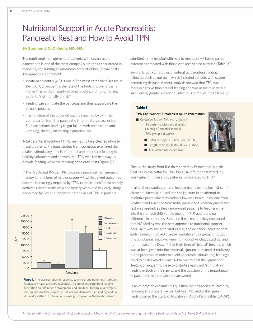
Total parenteral nutrition (TPN) seemed to be a clear solution to these problems. Previous studies from our group examined the relative stimulatory effects of enteral and parenteral feeding in healthy volunteers and showed that TPN was the best way to provide feeding while maintaining pancreatic rest (Figure 1).1
In the 1980s and 1990s, TPN became a universal management therapy for any form of mild or severe AP, while patient outcomes became increasingly impaired by “TPN complications,” most notably catheter-related septicemia and hyperglycemia. A key early study performed by Sax et al. showed that the use of TPN in patients
admitted to the hospital with mild to moderate AP had impaired outcomes compared with those who received no nutrition (Table 1).2
Several larger RCT studies of enteral vs. parenteral feeding followed, such as our own, which included patients with severe necrotizing disease. A meta-analysis showed that TPN was more expensive than enteral feeding and was associated with a significantly greater number of infectious complications (Table 2).3
Finally, the study from Russia reported by Petrov et al. put the final nail in the coffin for TPN, because it found that mortality was higher in those study patients randomized to TPN.3
In all of these studies, enteral feeding had taken the form of semi-elemental formula infused into the jejunum in an attempt to minimize pancreatic stimulation. However, two studies, one from Scotland and a second from India, questioned whether pancreatic rest was needed, as they randomized patients to feeding either into the stomach (NG) or the jejunum (NJ) and found no difference in outcomes. Based on these results, they concluded that NG feeding was the best approach to nutritional support, because it was easier to start earlier, and evidence indicated that early feeding improved disease resolution.4 Our group criticized this conclusion, since we knew from our physiologic studies,1 and from those of the Dutch,5 that their form of “jejunal” feeding, which was at best given into the proximal jejunum, remained stimulatory to the pancreas. In order to avoid pancreatic stimulation, feedings need to be delivered at least 40 to 60 cm past the ligament of Treitz. Consequently, these two studies had used “stimulatory” feeding in both of their arms, and the question of the importance of pancreatic rest remained unanswered.
In an attempt to evaluate this question, we designed a multicenter, randomized comparative trial between NG and distal jejunal feeding called the Study of Nutrition in Acute Pancreatitis (SNAP).
Table 1
TPN Can Worsen Outcomes in acute Pancreatitis
■ Controlled Study: TPN vs. IV fluids:2
— 54 patients with mild disease (average Ranson’s score 1)
— TPN group did worse
■ Catheter sepsis 11% vs. 2%, p<0.01
■ Length of hospital stay 16 vs. 10 days
■ TPN arm more expensive
22500Placebo
Intravenous
Oral
Duodenal
Elemental
20000
17500
15000
12500
Amylase
IU/h
10000
7500
5000
2500
0
Figure 1. Amylase secretion in response to enteral and parenteral nutrition. Relative amylase secretory responses to enteral and parenteral feeding, illustrating no difference between oral and duodenal feeding of a complex diet, an intermediate response to duodenal elemental diet feeding, and no stimulatory effect of intravenous feeding compared with placebo saline.1
8
Affiliated with the University of Pittsburgh School of Medicine, UPMC is ranked among the nation’s best hospitals by U.S. News & World Report.

To start, we designed a form of enteral feeding that avoided pancreatic stimulation. Recognizing that there had to be a “null point” somewhere between the duodenum and ileum where feeding was no longer stimulatory (note: feeding into the ileum actually inhibits pancreatic secretion), we fed volunteers at progressive distances past the ligament of Treitz, namely 20, 40, 80, 105, and 120 cm. The pattern of the secretory responses during 360 minutes of feeding demonstrates no significant secretory response to mid-distal jejunal feeding, because secretion rates were no different from those measured during fasting or IV feeding (Figure 2).6 Examination of gut peptide responses supported the suggestion that pancreatic secretion might have been suppressed by induction of the ileal brake, with substantial increases in plasma glucagon like peptide-1 and peptide YY, but not cholecystokinin. Our results were consistent with those of Vu et al., who established that the infusion of a mixed polymeric liquid diet at normal tube feeding rates (i.e., 1.6 kcal/min) into the proximal jejunum stimulated basal trypsin secretion four-fold, whereas infusion 60 cm below the ligament of Treitz had no stimulatory effect over three hours.6 Based on the results of our study, we now had a form of enteral feeding, termed distal jejunal feeding or DJ, that avoided pancreatic stimulation and could be used in our proposed NG vs. DJ feeding study.
Only 26 patients completed the SNAP study, and the study was closed before total recruitment was achieved due to slow enrollment. With such a small number of participants, the question of pancreatic rest could not be addressed. That is, we needed to show exacerbation of the disease, which was very difficult in a patient who was already critically ill and on a ventilator. However, our analysis revealed important information about best practice feeding techniques such as “feeding failure,” defined as failure to achieve a feeding rate of more than 10 percent of goal for a 48-hour period, which occurred in 0/14 DJ feeding patients and in 6/11 NG patients. Feeding failure in the NG group was primarily due to nausea, vomiting, or gastric residual volumes of more than 500 ml/4h, which is an indication of gastric outlet obstruction that necessitated a crossover to DJ feeding. As a result, the quantity of feed delivered was significantly higher in DJ patients, leading to greater success for enteral feeding via this route. It is now our standard of practice to place double-lumen feeding tubes by transnasal endoscopy as early in the disease as possible. These tubes feed 40 cm past the ligament of Treitz and decompress the stomach proximally.
Figure 2. The relative pancreatic stimulatory effects of enteral and parenteral (IV) feeding on pancreatic secretion in normal healthy volunteers showing that distal jejunal (>40 cm past the ligament of Treitz) feeding had no stimulatory response.6
continued on Page 16
continued from Page 8
Table 2Random Effects Model of Relative Risk (95% confidence interval) of Infections associated With Enteral Feeding Compared With Parenteral Nutrition
Influence of Feed Complexity and Feeding Positionon Trypsin Secretory Response
Study Enteral Total Parenteral Relative Risk Weight Relative Risk (95% CI random) % (95% CI random)
Abou-Assi 1/26 9/27 7.7 0.12 (0.02 to 0.85)
Gupta 0/8 2/9 3.7 0.22 (0.01 to 4.04)
Kalfarentzos 5/18 10/20 41.2 0.56 (0.23 to 1.32)
McClave 2/16 2/16 9.2 1.00 (0.16 to 6.26)
Olah 5/41 13/48 34.6 0.45 (0.18 to 1.16)
Windsor 0/16 3/16 3.7 0.16 (0.01 to 2.87)
Total (95% CI) 13/125 39/138 100.0 0.45 (0.26 to 0.78)
Test for heterogeneity: χ2 = 3.71, df=5, P=0.59
Test for overall effect: z=2.85, P=0.0040.01 0.1 1 10 100Favors enteral Favors totalnutrition parenteral nutrition
9UPMC Division of Gastroenterology, Hepatology, and Nutrition
UPMCPhysicianResources.com/GI For consults and referrals, please call UPMC’s 24-hour physician OnDemand service at 1-866-884-8579.

D I G E S T — FA L L 2 0 1 4
What Is a PCOE? continued from Page 1
interfaces, information management systems, and analytical tools,
all complemented by quality indicators to address questions of
qualifications and competency. Initial challenges for our group will
focus on chronic pancreatitis, because this illness requires a true
multidisciplinary approach
to achieve optimal care
for patients. The PCOE
process for UPMC, as well
as other models, was
reviewed this summer
at PancreasFest 2014, at
which academic physician
leaders from across the
U.S. discussed PCOE
objectives and process
implementation during
a dedicated, half-day
program. Physicians from
more than 20 well-
established academic
pancreas centers participated in this PCOE discussion to identify
unanticipated problems and opportunities. This process was
observed by representatives from the NPF and other groups,
who functioned as independent monitors.
The goal for academic PCOE programs is to provide every medical
center and practice with guidelines to develop and maintain the
best possible care for patients with pancreatic diseases. PCOEs
are not intended to be exclusive or elitist organizations. Instead,
PCOEs will provide assurance that high-quality, cutting-edge,
evidence-based care linked to quality indicators and known
outcomes is available to guide patient choices. In addition
to offering standard high-quality care to patients, academic
PCOEs provide a classroom for advanced education; a research
laboratory for translational research; a testing ground for new
medications, instruments, and treatments; and a last resort for
the most complex and challenging of patients. Such critically
important functions may be seen as “money losers,” so system
financial margins may threaten the existence of academic PCOEs
in the future. The time for better models of care is now.
Rapid and dynamic changes have magnified our understanding
of chronic pancreatitis etiologies, complications, outcomes, and
treatments. We need to implement and measure treatment
advancements to provide better guidance to all physicians. Our
understanding of the role of susceptibility genes, modifier genes,
smoking, alcohol, pain subtypes, type 3c diabetes mellitus, nutrition,
and cancer risk have changed dramatically over the past 20 years.
Different approaches and measures of disease progression are
needed.1,2 The acceptance of total pancreatectomy with islet
autotransplantation (TP-IAT) is also growing, from implementation
at only two major U.S. centers only a few years ago to at least
18 centers performing these procedures today. A workshop
sponsored by the National Institute of Diabetes and Digestive
and Kidney Diseases (NIDDK) was held immediately before
PancreasFest 2014 to address gaps in knowledge and methods for
TP-IAT patient evaluation, and TP-IAT guidelines were developed
at PancreasFest 2014.3
The key to a successful academic PCOE program includes
acceptance and continued evaluation of new recommendations,
a collaborative process among major centers, and demonstration
of superb outcomes. Support of such centers also requires an
economic model in which stakeholders (i.e., patients, patient
advocacy groups, educational institutions, government, industry,
and philanthropy) understand the value of an academic PCOE
to address unique and unmet needs, and to provide appropriate
levels of support.
The annual PancreasFest conference has become a venue to
advance these ideas due to its robust history of major academic
pancreas programs working collaboratively on multicenter studies.
PancreasFest also provides an opportunity for subspecialty
programs and leaders to partner with colleagues and contribute
efforts to markedly improve patient care.
If you or your colleagues would like to participate in the PCOE,
please contact me or PCOE co-coordinator, Darwin Conwell, MD,
MS, Ohio State University. PCOE meetings will occur throughout
the year, and we welcome your input.
Dr. Whitcomb is the Giant Eagle Foundation Professor of Cancer Genetics and serves as chief for the Division of Gastroenterology, Hepatology, and Nutrition. He co-chairs PancreasFest, an international subspecialty pancreas symposium for physicians and scientists.
References:1 Nawaz H, Mounzer R, Yadav D, et al. Revised Atlanta and Determinant-
Based Classification: Application in a Prospective Cohort of Acute
Pancreatitis Patients. Am J Gastroenterol. 2013 Dec;108(12):1911-7.
PMID: 24126632.
2 Rickels MR, Bellin M, Toledo FG, et al. PancreasFest Recommendation
Conference Participants. Detection, Evaluation and Treatment of Diabetes
Mellitus in Chronic Pancreatitis: Recommendations From PancreasFest
2012. Pancreatology. 2013 Jul-Aug;13(4):336-42. PMID: 23890130.
3 Bellin MD, Freeman ML, Gelrud A, et al. PancreasFest Recommendation
Conference Participants. Total Pancreatectomy and Islet Autotransplantation
in Chronic Pancreatitis: Recommendations from PancreasFest. Pancreatology.
2014 Jan-Feb;14(1):27-35. PMID: 24555976.
… PCOEs will provide assurance that high-quality, cutting-edge, evidence-based care linked to quality indicators and known outcomes is available to guide patient choices.
10
Affiliated with the University of Pittsburgh School of Medicine, UPMC is ranked among the nation’s best hospitals by U.S. News & World Report.
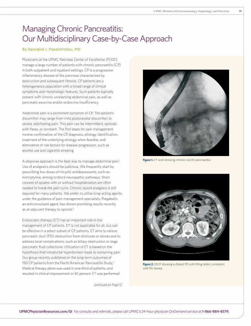
Figure 1. CT scan showing chronic calcific pancreatitis.
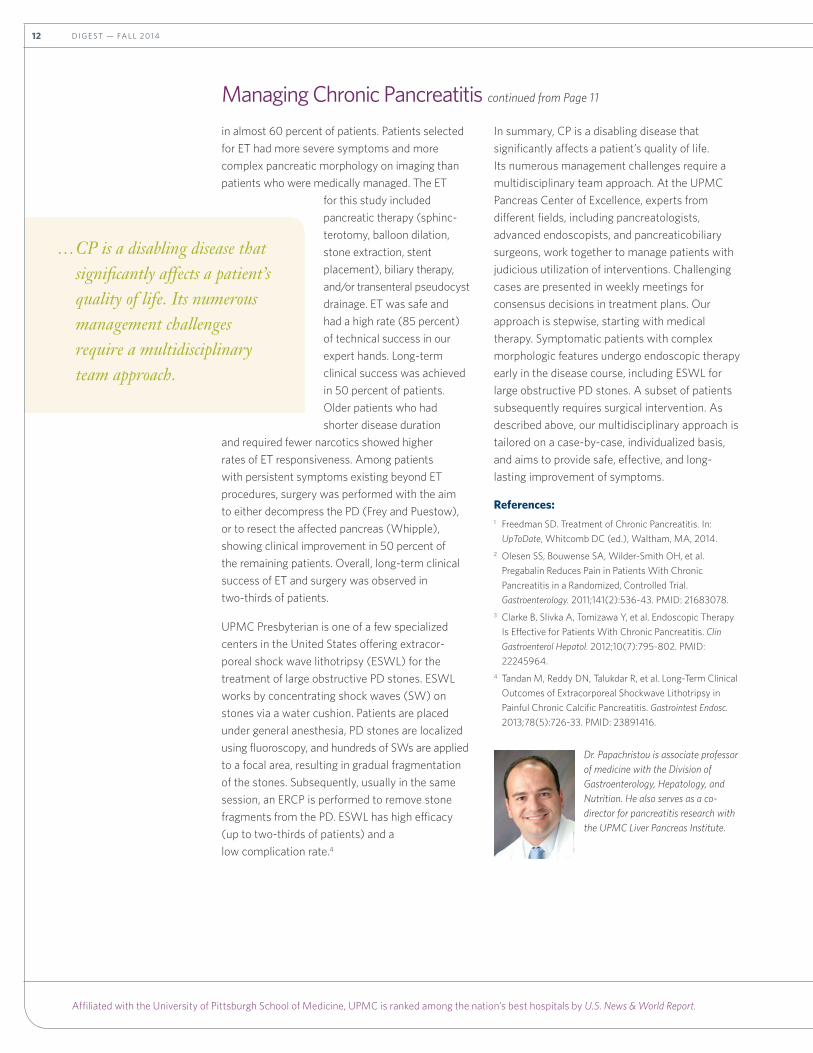
Figure 2. ERCP showing a dilated PD with filling defect consistent with PD stones.
Managing Chronic Pancreatitis: Our Multidisciplinary Case-by-Case ApproachBy Georgios I. Papachristou, MD
Physicians at the UPMC Pancreas Center of Excellence (PCOE)
manage a large number of patients with chronic pancreatitis (CP)
in both outpatient and inpatient settings. CP is a progressive
inflammatory disease of the pancreas characterized by
destruction and subsequent fibrosis. CP patients are a
heterogeneous population with a broad range of clinical
symptoms and morphologic features. Such patients typically
present with chronic unrelenting abdominal pain, as well as
pancreatic exocrine and/or endocrine insufficiency.
Abdominal pain is a prominent symptom of CP. The patient’s
discomfort may range from mild postprandial discomfort to
severe, debilitating pain. This pain can be intermittent, episodic
with flares, or constant. The first steps for pain management
involve confirmation of the CP diagnosis, etiology identification,
treatment of the underlying etiology when feasible, and
elimination of risk factors for disease progression, such as
alcohol use and cigarette smoking.
A stepwise approach is the best way to manage abdominal pain.1
Use of analgesics should be judicious. We frequently start by
prescribing low doses of tricyclic antidepressants, such as
nortriptyline, aiming to block neuropathic pathways. Short
courses of opiates with or without hospitalization are often
needed to break the pain cycle. Chronic opioid analgesia is still
required for many patients. We prefer to utilize long-acting agents
under the guidance of pain management specialists. Pregabalin,
an anticonvulsant agent, has shown promising results recently
as an adjuvant therapy to opioids.2
Endoscopic therapy (ET) has an important role in the
management of CP patients. ET is not applicable for all, but can
be effective in a select subset of CP patients. ET aims to relieve
pancreatic duct (PD) obstruction from strictures or stones and to
address local complications, such as biliary obstruction or large
pancreatic fluid collections. Utilization of ET is based on the
hypothesis that intraductal hypertension leads to worsening pain.
Our group recently published on the long-term outcomes of
150 CP patients from the North American Pancreatitis Study.3
Medical therapy alone was used in one-third of patients, and
resulted in clinical improvement in 30 percent. ET was performed
continued on Page 12
11UPMC Division of Gastroenterology, Hepatology, and Nutrition
UPMCPhysicianResources.com/GI For consults and referrals, please call UPMC’s 24-hour physician OnDemand service at 1-866-884-8579.
D I G E S T — FA L L 2 0 1 4
Managing Chronic Pancreatitis continued from Page 11
in almost 60 percent of patients. Patients selected
for ET had more severe symptoms and more
complex pancreatic morphology on imaging than
patients who were medically managed. The ET
for this study included
pancreatic therapy (sphinc-
terotomy, balloon dilation,
stone extraction, stent
placement), biliary therapy,
and/or trans enteral pseudocyst
drainage. ET was safe and
had a high rate (85 percent)
of technical success in our
expert hands. Long-term
clinical success was achieved
in 50 percent of patients.
Older patients who had
shorter disease duration
and required fewer narcotics showed higher
rates of ET responsiveness. Among patients
with persistent symptoms existing beyond ET
procedures, surgery was performed with the aim
to either decompress the PD (Frey and Puestow),
or to resect the affected pancreas (Whipple),
showing clinical improvement in 50 percent of
the remaining patients. Overall, long-term clinical
success of ET and surgery was observed in
two-thirds of patients.
UPMC Presbyterian is one of a few specialized
centers in the United States offering extracor-
poreal shock wave lithotripsy (ESWL) for the
treatment of large obstructive PD stones. ESWL
works by concentrating shock waves (SW) on
stones via a water cushion. Patients are placed
under general anesthesia, PD stones are localized
using fluoroscopy, and hundreds of SWs are applied
to a focal area, resulting in gradual fragmentation
of the stones. Subsequently, usually in the same
session, an ERCP is performed to remove stone
fragments from the PD. ESWL has high efficacy
(up to two-thirds of patients) and a
low complication rate.4
In summary, CP is a disabling disease that
significantly affects a patient’s quality of life.
Its numerous management challenges require a
multidisciplinary team approach. At the UPMC
Pancreas Center of Excellence, experts from
different fields, including pancreatologists,
advanced endoscopists, and pancreaticobiliary
surgeons, work together to manage patients with
judicious utilization of interventions. Challenging
cases are presented in weekly meetings for
consensus decisions in treatment plans. Our
approach is stepwise, starting with medical
therapy. Symptomatic patients with complex
morphologic features undergo endoscopic therapy
early in the disease course, including ESWL for
large obstructive PD stones. A subset of patients
subsequently requires surgical intervention. As
described above, our multidisciplinary approach is
tailored on a case-by-case, individualized basis,
and aims to provide safe, effective, and long-
lasting improvement of symptoms.
References:1 Freedman SD. Treatment of Chronic Pancreatitis. In:
UpToDate, Whitcomb DC (ed.), Waltham, MA, 2014.2 Olesen SS, Bouwense SA, Wilder-Smith OH, et al.
Pregabalin Reduces Pain in Patients With Chronic
Pancreatitis in a Randomized, Controlled Trial.
Gastroenterology. 2011;141(2):536-43. PMID: 21683078.3 Clarke B, Slivka A, Tomizawa Y, et al. Endoscopic Therapy
Is Effective for Patients With Chronic Pancreatitis. Clin
Gastroenterol Hepatol. 2012;10(7):795-802. PMID:
22245964.4 Tandan M, Reddy DN, Talukdar R, et al. Long-Term Clinical
Outcomes of Extracorporeal Shockwave Lithotripsy in
Painful Chronic Calcific Pancreatitis. Gastrointest Endosc.
2013;78(5):726-33. PMID: 23891416.
Dr. Papachristou is associate professor of medicine with the Division of Gastroenterology, Hepatology, and Nutrition. He also serves as a co-director for pancreatitis research with the UPMC Liver Pancreas Institute.
… CP is a disabling disease that significantly affects a patient’s quality of life. Its numerous management challenges require a multidisciplinary team approach.
12
Affiliated with the University of Pittsburgh School of Medicine, UPMC is ranked among the nation’s best hospitals by U.S. News & World Report.

The Complex Nature (and Nurture) of Recurrent Acute PancreatitisBy Anthony Razzak, MD
Gastroenterology Fellow, Year III
A 27-year-old medical student of Pakistani
descent was referred to clinic for evaluation of
recurrent acute pancreatitis of unclear etiology.
She experienced her first episode one year
prior, and described severe epigastric pain
associated with nausea and vomiting.
She denied alcohol use and was not taking
any medications or supplements. Acute
pancreatitis was diagnosed with a lipase of greater than
10,000 U/L. Her liver function tests were normal. An abdominal
ultrasound was negative for gallbladder pathology or biliary ductal
dilation. A CT scan with IV contrast revealed acute interstitial
pancreatitis without evidence of chronic inflammatory changes.
Metabolic parameters, calcium, and triglycerides were within
normal limits. She was managed conservatively and recovered
without complications.
Six months later, she experienced a second,
similar episode of radiating abdominal pain
with vomiting that required hospitalization. Her
admission lipase was again noted to be markedly
elevated (greater than 22,000 U/L) with normal
liver function tests. Acute pancreatic interstitial
inflammation was noted on a contrast-enhanced
CT, and she was diagnosed with recurrent acute
pancreatitis (RAP). A magnetic resonance chol-
angio pan creatography (MRCP) was performed
and sug gested findings consistent with pancreas
divisum, a congenital anomaly that can contribute
to recurrent acute pancreatic injury.1 She recovered without
difficulties and was referred to UPMC for outpatient endoscopic
retrograde pancreatography (ERP) with possible intervention.
During the ERP, her dorsal pancreatic duct appeared to
communicate with the ventral duct, discounting the presence of
divisum. Fluoroscopically, her pancreatic duct appeared prominent
and contained noncalcified stones in the pancreatic head, which
precluded passage of a wire to facilitate stone removal.
The patient presented with no problems or complaints during her
follow-up clinic visit. She denied any family history of pancreatitis
or cystic fibrosis. She did not endorse any clinical manifestations
of pancreatic exocrine or endocrine insufficiency. A thorough
serologic evaluation to investigate her recurrent acute and
suspected early chronic pancreatitis (CP) was initiated. She was
found to be homozygous for the N34S variant of the serine
protease inhibitor Kazal type 1 (SPINK1) gene, a well-known
contributor to RAP and CP.2,3
RAP/CP syndrome is thought to be a complex multifactorial
disorder, involving genetic susceptibility with environmental stress
and stimuli, which appears largely related to pathologic intra-
pancreatic trypsin activation and subsequent pancreatic injury.4-6
The list and mechanisms leading to RAP/CP continue to grow.7
Genetic tests are available for SPINK1, cationic trypsinogen
gene (PRSS1), and some CFTR mutations, but these tests may
identify only a fraction of the susceptibilities associated with
RAP/CP. The cost of testing is high, and the medical, personal,
and ethical implications of testing should invite
discussion with a genetic counselor.2
SPINK1 encodes a secretory trypsin inhibitor
that protects against prematurely activated
intra-pancreatic trypsin.5,8 When mutated, its
pathologic role in pancreatitis is not direct but
related to an impaired defense against trypsin
activation from a variety of other pancreatitis
susceptibility factors. It has been implicated in
tropical pancreatitis, a syndromic pancreas-
related process afflicting populations in Asia and
Africa.9 The South Asian ancestral history of this
patient and lack of family history are consistent
with the complex genetic risk inheritance patterns for SPINK1-
associated pancreatitis.
Current management options available for RAP/CP are limited.
Scant evidence suggests that antioxidant therapy (vitamins
A, C, E, and selenium) may help to prevent oxidative parenchymal
injury and pain.10 The role of total pancreatectomy with islet
autotransplantation (TP-IAT) is unclear but may lessen the
insulin requirements and narcotic needs of younger patients.11,12
For most patients, pancreatic enzyme replacement, insulin,
and opiate-based analgesia remain mainstay therapies for
disease-related complications.
continued on Page 14
13UPMC Division of Gastroenterology, Hepatology, and Nutrition
UPMCPhysicianResources.com/GI For consults and referrals, please call UPMC’s 24-hour physician OnDemand service at 1-866-884-8579.

D I G E S T — FA L L 2 0 1 4
Recurrent Acute Pancreatitis continued from Page 13
The remainder of our patient’s evaluation was negative, including
CFTR and PRSS1 mutation analyses. She did not drink or smoke,
and was without any obvious environmental exposures. Our
finding of homozygous SPINK1 risk alleles provided an etiologic
diagnosis and facilitated discussion regarding clinical expectations,
prognosis, and management options. To date, she has not
endorsed evidence of exocrine or endocrine insufficiency, and
thus remains a candidate for TP-IAT evaluation at UPMC.12
References:1 Bertin C, Pelletier AL, Vullierme MP, et al. Pancreas Divisum Is Not a Cause of
Pancreatitis by Itself but Acts as a Partner of Genetic Mutations. Am J
Gastroenterol. 2012 Feb;107(2):311-7. PMID: 22158025.2 Solomon S, Whitcomb DC. Genetics of Pancreatitis: An Update for Clinicians
and Genetic Counselors. Curr Gastroenterol Rep. 2012 Apr;14(2):112-7. PMID:
22314809.3 LaRusch J, Solomon S, Whitcomb DC. Pancreatitis Overview. GeneReviews®
[Internet]. Seattle (WA): University of Washington, Seattle; 1993-2014.
2014 Mar 13. PMID: 24624459.4 Whitcomb DC, Gorry MC, Preston RA, et al. Hereditary Pancreatitis Is
Caused by a Mutation in the Cationic Trypsinogen Gene. Nat Genet.
1996;14:141-5. PMID: 8841182.
5 Witt H, Luck W, Hennies HC, et al. Mutations in the Gene Encoding the
Serine Protease Inhibitor, Kazal Type 1 Are Associated With Chronic
Pancreatitis. Nat Genet. 2000;25:213-6. PMID: 10835640.6 Sharer N, Schwarz M, Malone G, et al. (1998) Mutations of the Cystic
Fibrosis Gene in Patients With Chronic Pancreatitis. N Engl J Med.
1998;339:645-52. PMID: 9725921.7 Whitcomb DC. Genetic Risk Factors for Pancreatic Disorders. Gastroenterology.
2013 Jun;144(6):1292-302. PMID: 23622139.8 Threadgold J, Greenhalf W, Ellis I, et al. The N34S Mutation of SPINK1 (PSTI)
Is Associated With a Familial Pattern of Idiopathic Chronic Pancreatitis but
Does Not Cause the Disease. Gut. 2002 May;50(5):675-81. PMID: 11950815.9 Schneider A, Suman A, Rossi L, et al. SPINK1/PSTI Mutations Are Associated
With Tropical Pancreatitis and Type II Diabetes Mellitus in Bangladesh.
Gastroenterology. 2002 Oct;123(4):1026-30. PMID: 12360464.10 Uomo G, Talamini G, Rabitti PG. Antioxidant Treatment in Hereditary
Pancreatitis: A Pilot Study on Three Young Patients. Dig Liver Dis.
2001;33(1):58. PMID: 11303976.11 Bellin MD, Carlson AM, Kobayashi T, et al. Outcome After Pancreatectomy and
Islet Autotransplantation in a Pediatric Population. J Pediatr Gastroenterol Nutr.
2008;47(1):37. PMID: 18607267.12 Bellin MD, Freeman ML, Gelrud A. Total Pancreatectomy and Islet
Autotransplantation in Chronic Pancreatitis: Recommendations From
PancreasFest. Pancreatology. 2014 Jan-Feb;14(1):27-35. PMID: 24555976.
What Is This?Elizabeth J. Blaney, MD Clinical Assistant Professor of MedicineDivision of Gastroenterology, Hepatology, and Nutrition
A 51-year-old African-American female with a history of multiple myeloma has recurrent admissions for abdominal
pain consistent with pancreatitis. Pancreatic enzymes are normal, and a CT was normal one year ago. Sequential
images over the three months since symptoms began are shown below.
Compare your answer to Dr. Blaney’s on Page 15.
14
Affiliated with the University of Pittsburgh School of Medicine, UPMC is ranked among the nation’s best hospitals by U.S. News & World Report.

University of Pittsburgh • Division of Gastroenterology, Hepatology and Nutrition
PancreasFest 2015Applying Research Discoveries in Pancreatitis and
Pancreatic Cancer to Patient-Centered Care
Collaborative Medicine to Advance Knowledge in Pancreatic DiseasesPancreasFest 2015 is an annual pancreas research and clinical conference designed for gastroenterologists, surgeons, researchers, other physicians, and interested medical professionals. Lectures and discussion groups will mix with investigative research meetings to further the multidisciplinary understanding and treatment of pancreatic diseases. Special surgery programs will be held on Saturday, July 25th.
Course Directors
David C. Whitcomb, MD, PhDRandall E. Brand, MDHerbert J. Zeh III, MD
Location
The University Club123 University Place • Pittsburgh, PA 15213
Hotel
Wyndham Pittsburgh University Center100 Lytton Avenue • Pittsburgh, PA 15213
July 22 – 24, 2015 • Pittsburgh, Pennsylvania
Welcoming New FacultyThe Division of Gastroenterology, Hepatology, and Nutrition
is proud to welcome the following new faculty members:
Elizabeth J. Blaney, MD
Clinical Assistant Professor of Medicine
Practicing gastroenterology at the
Magee-Womens Hospital of UPMC
Naudia N. Jonassaint, MD, MHS
Assistant Professor of Medicine
Practicing hepatology at the
UPMC Center for Liver Diseases
Vinod K. Rustgi, MD, MBA
Professor of Medicine
Clinical Director of Hepatology
Medical Director for Liver Transplantation
Save the DatePancreasFest 2015July 22–24, 2015
Pittsburgh, Pennsylvania
PancreasFest is an annual research
and clinical conference designed
for gastroenterologists, surgeons, researchers, other physicians,
and interested medical professionals. Lectures, discussion
groups, and investigative research meetings will further
participants’ multidisciplinary understanding of the treatment
of pancreatic diseases.
David Whitcomb, MD, PhD, Named “Gastroenterologist to Know”
David C. Whitcomb, MD, PhD, has been
named to the 2014 listing of “160 Gastro-
enterologists to Know,” an honor designated
by Becker’s ASC Review. Dr. Whitcomb was
recognized for his leadership, research, and
business development as a division chief
and pancreas genetics subspecialist.
What Is This? continued from Page 14
This patient had biopsy-proven anaplastic myeloma. In spite of
multiple lines of chemotherapy, she developed a diffusely
infiltrating plasmacytoma involving the entire pancreas. Biliary
obstruction ensued, and she expired from prepyloric gastric
perforation, likely related to tumor ingrowth.
Pancreatic plasmacytoma is a rare condition, with approximately
26 cases reported in the literature, and should be considered in
the differential diagnosis of pancreatic mass in patients with
multiple myeloma. Patients typically present with jaundice and
abdominal pain. Radiographic characteristics include
homogenous enhancement of a solid mass or diffuse
enlargement, though findings are nonspecific and mimic other
pancreatic neoplasms. Endoscopic ultrasound with biopsy is
often the diagnostic modality of choice. Anaplastic myeloma is
an aggressive variant of myeloma, which is associated with
extensive extramedullary disease, portending a poor prognosis
as seen in this case.
References:
Hue SS, Azhar R. Plasmacytoma of the Pancreas: An Unusual Manifestation
of Multiple Myeloma. Singapore Medical Journal. 2013 May; 54(5): e105-107.
Smith A, Hal H, Frauenhoffer E. Extramedullary Plasmacytoma of the
Pancreas: A Rare Entity. Case Reports in Radiology. 2012; 2012: 798264.
Foucar K, Raber M, Foucar E, Barlogie B, Sandler CM, Alexanian R.
Anaplastic Myeloma With Massive Extramedullar Involvement: Report of
Two Cases. Cancer. 1983; 51: 166–174.
15UPMC Division of Gastroenterology, Hepatology, and Nutrition
UPMCPhysicianResources.com/GI For consults and referrals, please call UPMC’s 24-hour physician OnDemand service at 1-866-884-8579.

© 2014 UPMC USNW413365 HM/SM 1 1/14
UPMC Division of Gastroenterology, Hepatology, and Nutrition
EDITORS Julia B. Greer, MD, MPH Janet R. Harrison, MD Matthew G. Warndorf, MD Joy Jenko Merusi, Ma
ADDRESS CORRESPONDENCE TO:
Joy Jenko [email protected]
Centers of Excellence
• Pancreas and Biliary Diseases
• Inflammatory Bowel Disease
– Visceral Inflammation and Pain (VIP) – Gastrointestinal Dermatology Clinic
• Liver Diseases
• Neurogastroenterology and Motility Diseases
• Intestinal Health and Nutrition Support
• Gastrointestinal Cancer Prevention and Treatment
• Women’s Digestive Health
A world-renowned health care provider and insurer, Pittsburgh-based UPMC is inventing new models of accountable, cost-effective, patient-centered care. It provides more than $887 million a year in benefits to its communities, including more care to the region’s most vulnerable citizens than any other health care institution. The largest nongovernmental employer in Pennsylvania, UPMC integrates more than 62,000 employees, 22 hospitals, 400 doctors’ offices and outpatient sites, a 2.3-million-member health insurance division, and international and commercial operations. Affiliated with the University of Pittsburgh Schools of the Health Sciences, UPMC is ranked among the nation’s best hospitals, and No. 1 in Pennsylvania, by U.S. News & World Report. For more information, go to UPMC.com.
Furthermore, how the histopathologic findings contribute to chronic pain in PRSS1 patients remains enigmatic. As genetic testing for hereditary pancreatitis becomes more widespread, future studies should provide greater insight into the pathophysiology of this debilitating disease.
Dr. Singhi is assistant professor of pathology in the Division of Anatomical Pathology at UPMC.
References:1 Whitcomb DC, Preston RA, Aston CE, et al. A Gene for
Hereditary Pancreatitis Maps to Chromosome 7q35. Gastroenterology 1996;110:1975-80. PMID: 8964426.
2 Whitcomb DC, Gorry MC, Preston RA, et al. Hereditary Pancreatitis Is Caused by a Mutation in the Cationic Trypsinogen Gene. Nat Genet 1996;14:141-5. PMID: 8841182.
3 Felderbauer P, Stricker I, Schnekenburger J, et al. Histo-pathological Features of Patients With Chronic Pancreatitis Due to Mutations in the PRSS1 Gene: Evaluation of BRAF and KRAS2 Mutations. Digestion 2008;78:60-5. PMID: 18946221.
4 Singhi AD, Pai RK, Kant JA, et al. The Histopathology of PRSS1 Hereditary Pancreatitis. Am J Surg Pathol 2014;38:346-53. PMID: 24525505.
5 Walters MN. Adipose Atrophy of the Exocrine Pancreas.
J Pathol Bacteriol 1966;92:547-57. PMID: 5964381.
PRSS1 Hereditary Pancreatitis continued from Page 4
Nutritional Support in Acute Pancreatitis continued from Page 9
This process maintains gut function, prevents ileus and bacterial overgrowth, and decompresses the obstructed proximal gut, decreasing aspiration risk.7
References:1 O’Keefe SJ, Lee RB, Anderson FP, Gennings C, Abou-Assi
S, Clore J, Heuman D, Chey W. Physiological Effects of Enteral and Parenteral Feeding on Pancreaticobiliary Secretion in Humans. Am J Physiol Gastrointest Liver Physiol. 2003;284(1):G27-36. PMID: 12488233.
2 Marik PE, Zaloga GP. Meta-Analysis of Parenteral Nutrition Versus Enteral Nutrition in Patients With Acute Pancreatitis. BMJ. 2004;328(7453):1407. PMID: 15175229.
3 Petrov MS, Kukosh MV, Emelyanov NV. A Randomized Controlled Trial of Enteral Versus Parenteral Feeding in Patients With Predicted Severe Acute Pancreatitis Shows a Significant Reduction in Mortality and in Infected Pancreatic Comp li ca-tions With Total Enteral Nutrition. Dig Surg. 2006;23(5-6):336-44; discussion 344-5. PMID: 17164546.
4 Hegazi R, Raina A, Graham T, Rolniak S, Centa P, Kandil H, O’Keefe SJ. Early Jejunal Feeding Initiation and Clinical
Outcomes in Patients With Severe Acute Pancreatitis. JPEN J Parenter Enteral Nutr. 2011 Mar;35(2):276. PMID: 21224435.
5 Vu MK, van der Veek PP, Frölich M, Souverijn JH, Biemond I, Lamers CB, Masclee AA. Does Jejunal Feeding Activate Exocrine Pancreatic Secretion? Eur J Clin Invest. 1999 Dec;29(12):1053-9. PMID: 10583454.
6 Kaushik N, Pietraszewski M, Holst JJ, O’Keefe SJ. Enteral Feeding Without Pancreatic Stimulation. Pancreas. 2005 Nov;31(4):353-9. PMID: 16258370.
7 O’Keefe S, Rolniak S, Raina A, Graham T, Hegazi R, Centa-Wagner P. Enteral Feeding Patients With Gastric Outlet Obstruction. Nutr Clin Pract. 2012 Feb;27(1):76-81. PMID: 22307492.
Dr. O’Keefe is professor of medicine with the Division of Gastroenterology, Hepatology, and Nutrition. He treats patients within UPMC’s Clinical Nutrition Support Service.
200 Lothrop Street
Pittsburgh, PA 15213-2582
Nonprofit Org.U.S. Postage
PAIDPittsburgh, PA
Permit No. 3834