digestive processes 1. ingestion 2. propulsion 3. mechanical digestion 4. chemical digestion 5....
TRANSCRIPT
Digestive Processes
1. ingestion
2. propulsion
3. mechanical digestion
4. chemical digestion
5. absorption
6. defecation
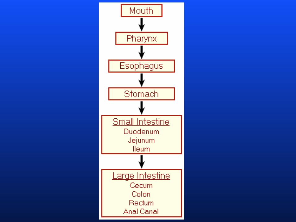
Digestive system organs
• Alimentary canal or GI tract
1. mouth
2. pharynx
3. esophagus
4. stomach
5. small intestine
6. large intestine
Digestive system organs
• Accessory glands and organs
1. teeth
2. tongue
3. salivary glands
4. gallbladder
5. pancreas
6. liver
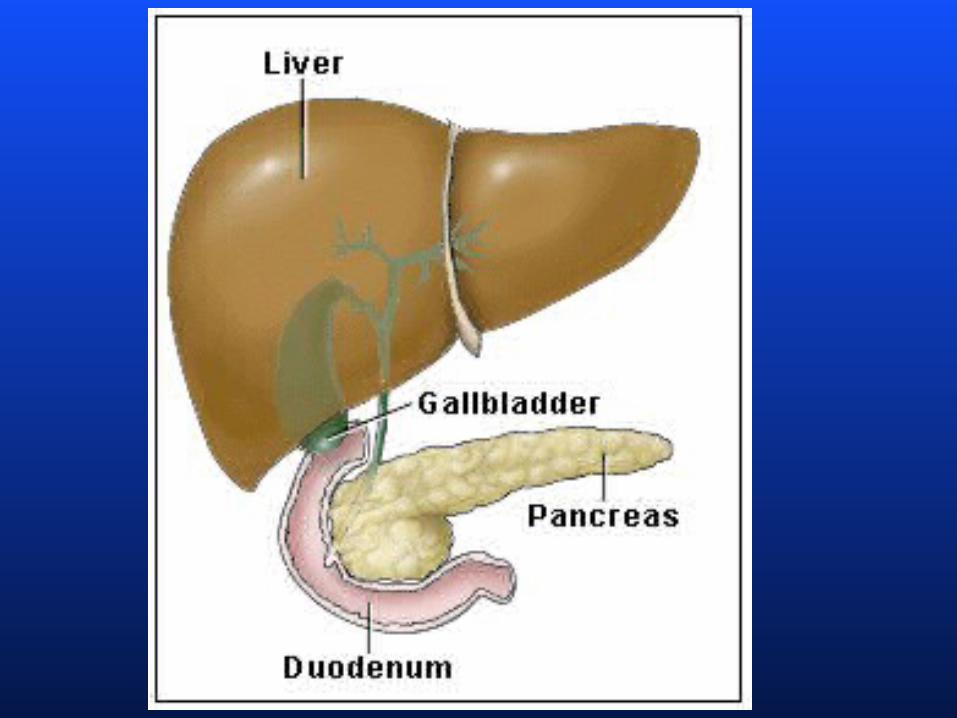
Anatomy of the organs of the alimentary canal (GI tract)
Most digestive systems reside within the
abdominopelvic cavity. The organs are
covered by a visceral and parietal peritoneum
separated by the peritoneal cavity. The
mesentery is a piece of fused double layered
membrane connecting the peritoneal
membranes and provides a route for
conducting blood vessels lymphatics and
nerves to the digestive viscera some parts of
these membranes are giving special names
such as greater and lesser omenta.
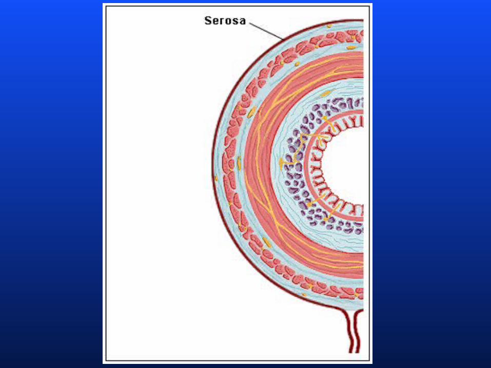
Anatomy of the organs of the alimentary canal (GI tract)
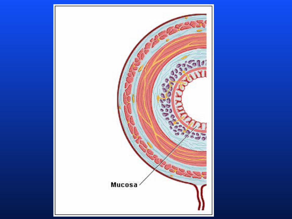
From the esophagus to the anal canal, the walls
of every organ are made up of 4 basic tunics
from the lumen outward these layers are :
1.mucosa
2. submucosa
3. muscularis externa
4. serosa (or adventitia)
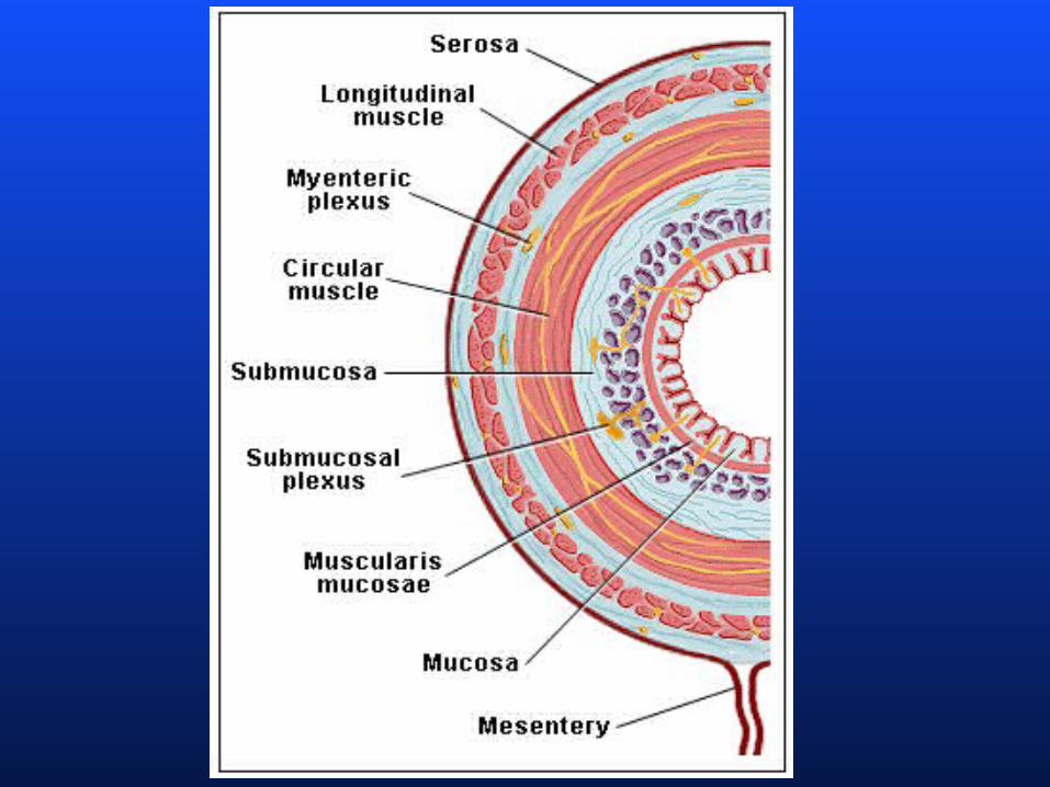
Anatomy of the organs of the alimentary canal (GI tract)
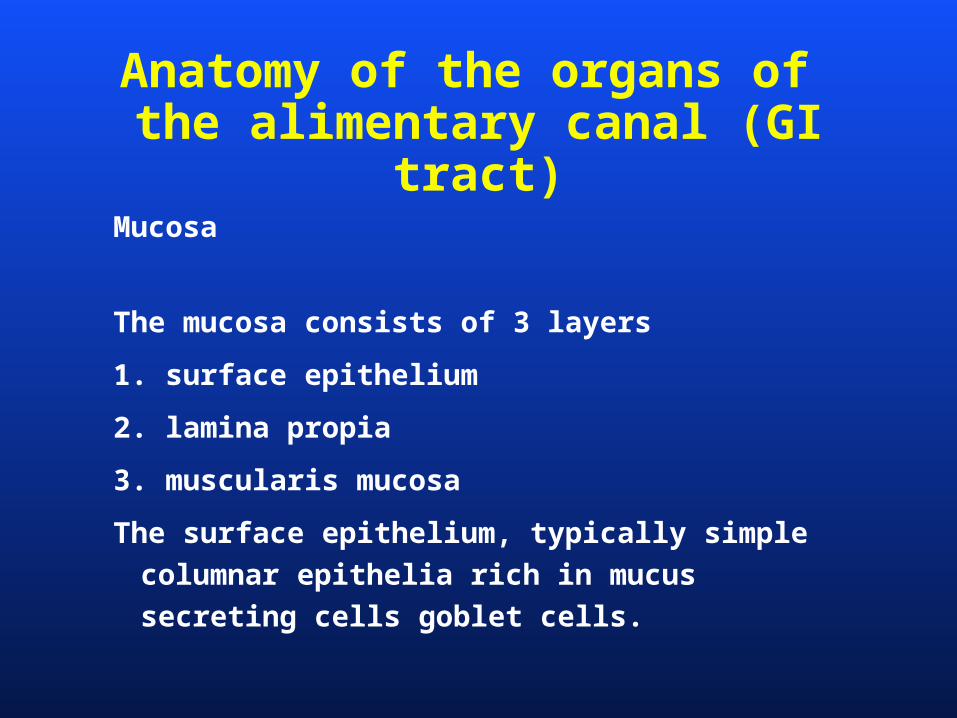
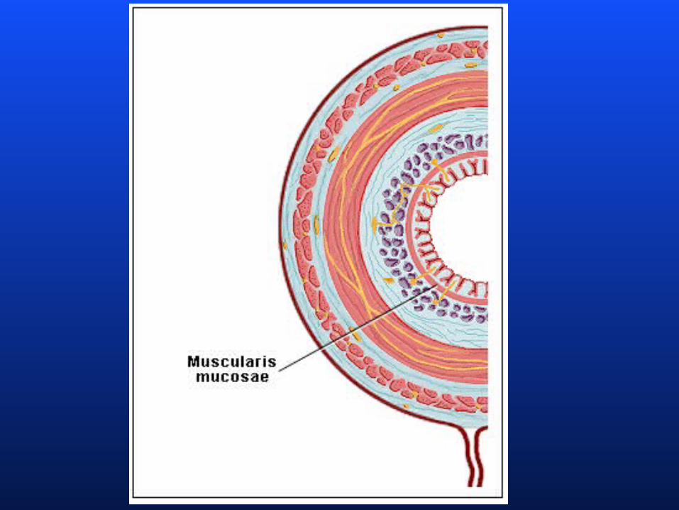
Mucosa
The mucosa consists of 3 layers
1. surface epithelium
2. lamina propia
3. muscularis mucosa
The surface epithelium, typically simple columnar
epithelia rich in mucus secreting cells goblet
cells.
Anatomy of the organs of the alimentary canal (GI tract)
Underlain the surface epithlium there is a small
amount of connective tissue made of areolar
connective tissue called the lamina propria
The lamina propia contains capillaries which
nourish the epithelia and function in
absorption. It also contains lymph nodes
important in defense against bacteria and
other pathogens.
Anatomy of the organs of the alimentary canal (GI tract)
Underlain the lamina propia there is a scant
smooth muscle layer called the muscularis
mucosae. The muscularis mucosae produces
local movements and make small folds vastly
increasing the surface area.
Anatomy of the organs of the alimentary canal (GI tract)
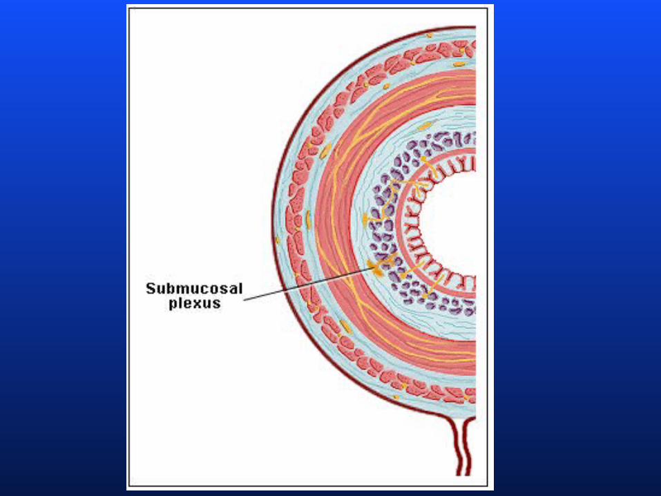
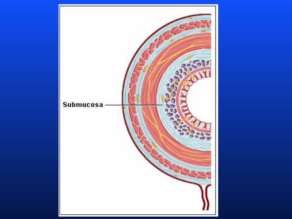
Submucosa
Made of dense connective contains a rich blood
supply, lymphatic, elastic fibers and nerves.
Contains the submucosal plexus which is
part of the intrinsic nerve supply of the GI
tract.
Anatomy of the organs of the alimentary canal (GI tract)
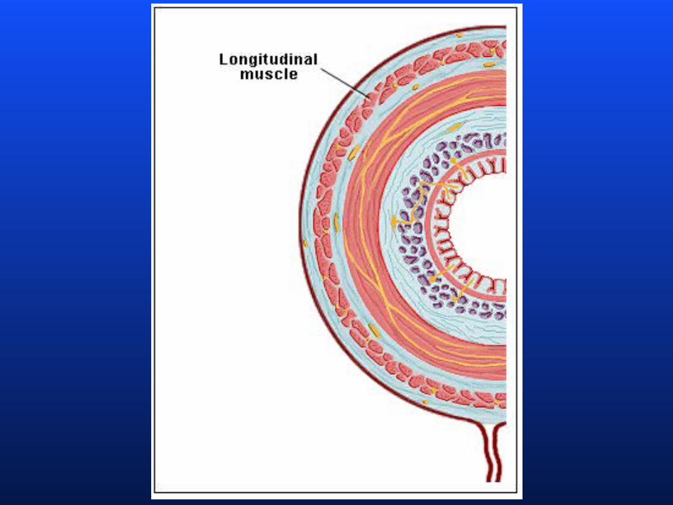
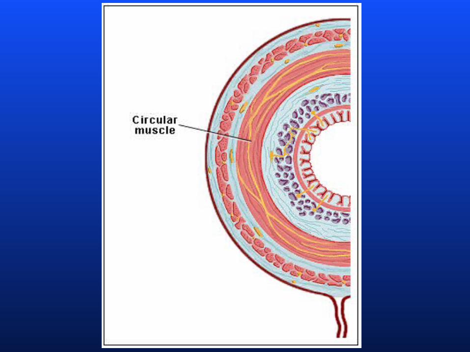
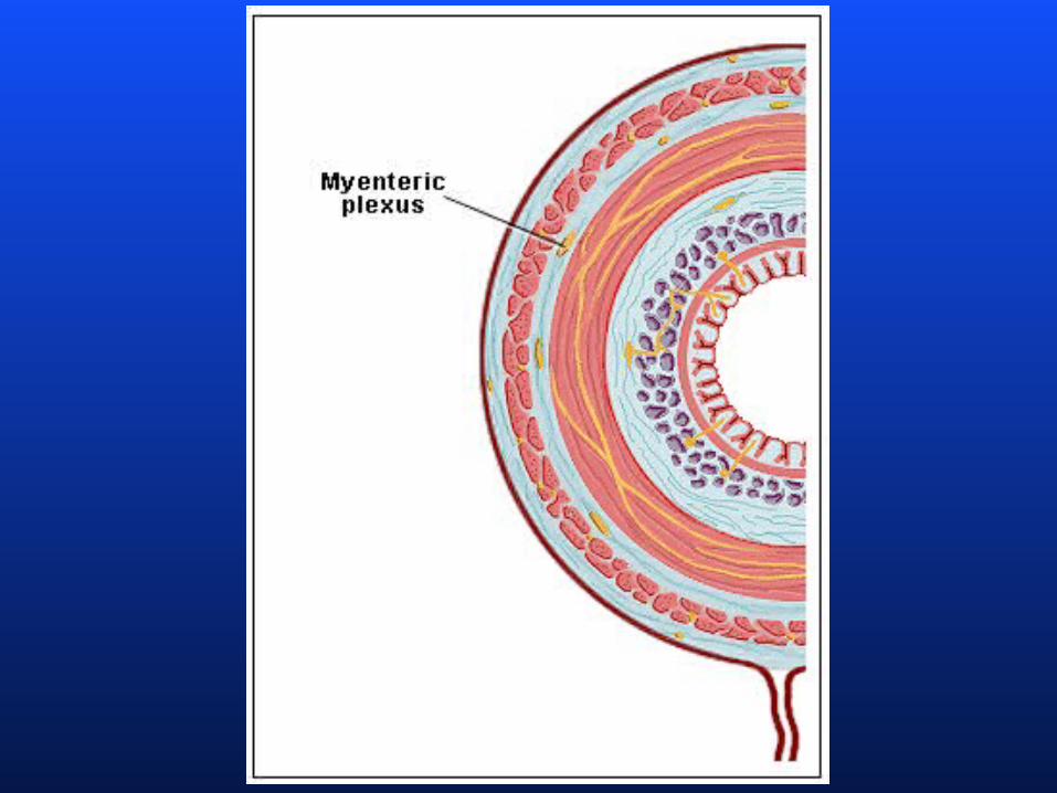
Muscularis externa
The muscularis externa is responsible for
segmentation and peristalsis typically has an
inner circular layer and outer longitudinal
layers of smooth muscle. In many places the
circular layer thickens to form sphincters that
act as valves to control food passage. The
myenteric plexuses the second intrinsic nerve
supply of the GI tract lays between the circular
and longitudinal smooth muscle layers.
Anatomy of the organs of the alimentary canal (GI tract)
Serosa-
The outermost protective is formed of aerolar
connective tissue and mesothelium a single
layer of squamouss epithelial tissue. The serosa
contains the subserous plexus the third
autonomic plexus . In the esophagus the serosa
is replaced by an adventitia which is an ordinary
fibrous connective tissue. Retroperitoneal
organs have both a serosa and an adventitia
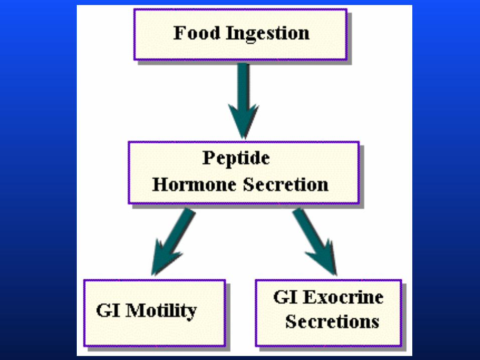
Basic functional concepts
1. All processes in GI tract are geared to control
the lumen to maximize the digestive
processess. The lumen is actually outside the
body
How ??
By reflex arcs which involve neural and
hormonal componenets. and stimuli sensed by
sensors mechano and chemo
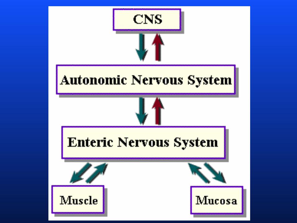
Basic functional concepts
2. Local control - short and long reflexes. Short
reflexes are mediated entirely by the enteric
plexuses inn response to GI tract stimuli.
Long reflexes initiated by stimuli arising
within or outside the GI tract and involve CNS
centers and extrinsic autonomic nerves.

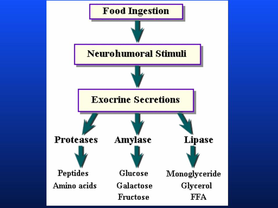
Digestion and absorption
Carbohydrates
Broken down to monosaccharides: glucose,
fructose and galactose. Starting in mouth
with salivary amylase, a little in the stomach
before acid destroys it and finishing in the
small intestine with pancreatic amylase.
Absorbed in the first segment of small
intestine by an active transport system which
can not be saturated
Digestion and absorption
Minerals and water
Water is highly diffusable and 95% of ingested
is absorbed by passive diffusion in small
intestine. Na+, K+, active transport Na, K
ATPase pump. Other minerals such as
calcium and potassium and trace minerals
such as iron and zinc are also absorbed in the
small intestine
Digestion and absorption
Protein
First to fragments in stomach by pepsin and
small intestine by trypsin and chymotrypsin.
Further by pancreatic carboxypeptidase and
brush border aminopeptidase. Transported by
secondary active transport linked to Na+.
Small # of small intact proteins can cross the
small intestine walls- passive immunity in
infants
Digestion and absorption
Fat-
Quite complex digestion process. Fat are
insoluble in water and therefore arrived at the
small intestine as a TG fat droplet. They get
emulsify by bile salts then lipase goes to
work on their surface splitting TG into 2 free
fa and a monoglyceride and make micelles.
Digestion and absorption
Fat (continued)
Micelles get absorbed in the small intestine cell
wall and during their passage the
monoglycerides and free fa’s combine to
form TG which are wrapped in a membrane
in the rough ER called chylomicrons which
get absorbed by the lacteal not the capillaries
and end up in the general circulation
Digestion and absorption
Vitamins
Fat soluble are solubilized in chylomicrons.
Most soluble vitamins are absorbed by
diffusion or carrier mediated transport with
the exception of B12 which is a very large
molecule and requires to bind to intrinsic
factor released by parietal cells absorbed in
the ileum.