digestive system - site title :: anita plagge · 2010-05-02 · digestive system general overview...
TRANSCRIPT

1
Digestive SystemDigestive System
General OverviewGoal of the Digestive System is to:
provide the cells of body with the nutrients required to do their job…be largely self reliant (autonomic)provide defense against ingested pathogensremove waste products
General OverviewThe processes of digestion that allow this to happen (not necessarily in order) are:
IngestionMotility (mixing & propulsion)Digestion
MechanicalChemical
SecretionAbsorptionDefecation

2
General Overview
Structural Organization of the Digestive System – Gross Anatomical
Organs of the alimentary canal (GI-Tract)Mouth to Anus & everything in between that materials pass through.
Accessory organs/structuresSalivary glands, pancreas, liver, gallbladderAid in the processing of nutrients
General OverviewStructural Organization of the Digestive
System – Histology of the GI-Tract
Serosa (visceral peritoneum)
Muscularismyenteric plexus
Submucosasubmucosal plexus
Mucosa
The Anatomy & Physiology of Digestion
Starting from the oral cavity:an examination of the structures and function of each portion of the GI tract with
accessory structures included.
3
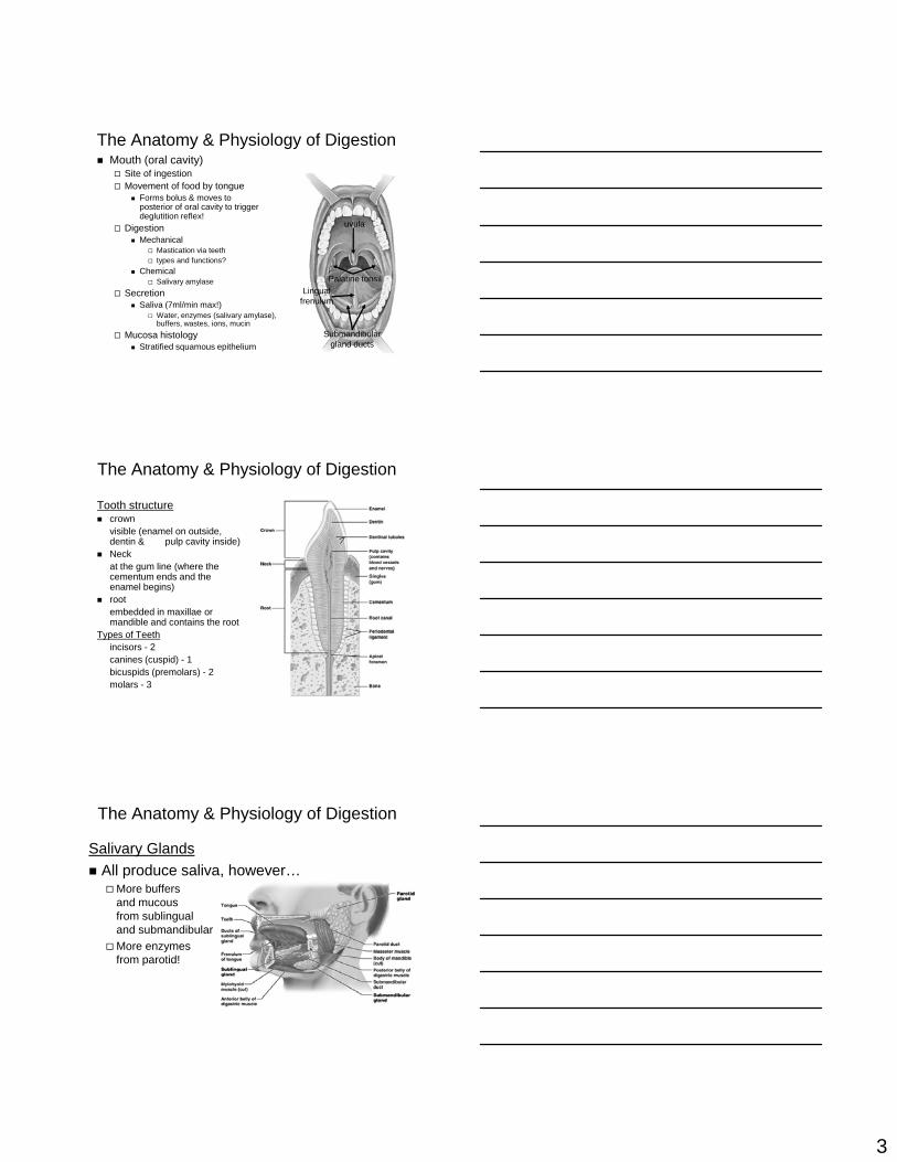
The Anatomy & Physiology of DigestionMouth (oral cavity)
Site of ingestionMovement of food by tongue
Forms bolus & moves toposterior of oral cavity to triggerdeglutition reflex!
DigestionMechanical
Mastication via teethtypes and functions?
ChemicalSalivary amylase
SecretionSaliva (7ml/min max!)
Water, enzymes (salivary amylase),buffers, wastes, ions, mucin
Mucosa histologyStratified squamous epithelium
uvula
Lingual frenulum
Palatine tonsil
Submandibular gland ducts
The Anatomy & Physiology of Digestion
Tooth structurecrownvisible (enamel on outside, dentin & pulp cavity inside)Neckat the gum line (where the cementum ends and the enamel begins)rootembedded in maxillae or mandible and contains the root
Types of Teethincisors - 2canines (cuspid) - 1bicuspids (premolars) - 2molars - 3
Salivary GlandsAll produce saliva, however…
More buffers and mucous from sublingual and submandibularMore enzymes from parotid!
The Anatomy & Physiology of Digestion

4
The Anatomy & Physiology of Digestion
Oropharynx & laryngopharynxfood (liquid & solid) & air pathwaystill lined with stratified squamouscontains tonsils (pharyngeal, palatal, lingual)muscles move food into esophagus
EsophagusMuscular tube (upper 1/3 is skeletal muscle, rest is smooth & involuntary)Stratified squamous liningMucous secretionUpper and lower esophageal sphincters define start and end of esophagusFunction: deglutition (swallowing)
The Pharynx & Esophagus
The Anatomy & Physiology of DigestionDeglutition
Initially voluntarily, continues automaticallyVoluntary process
The oral phaseformation and movement of bolus into pharynx Soft palate elevates (prevents intrusion into nasopharynx)
The pharyngeal phaseInitiates the swallowing reflex:
Larynx elevates, epiglottis moves down to prevent bolus movement into glottis!Pharyngeal muscles move bolus through the Upper Esophageal Sphincter (UES) and into the esophagus
Involuntary processThe esophageal phase
Peristalsis propels food to the stomachBolus must pass through the Lower Esophageal Sphincter (LES)
5
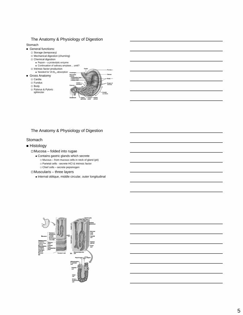
The Anatomy & Physiology of DigestionStomach
General functions:Storage (temporary)Mechanical digestion (churning)Chemical digestion
Pepsin – a proteolytic enzymeContinuation of salivary amylase… until?
Intrinsic factor productionNeeded for Vit B12 absorption
Gross AnatomyCardiaFundusBodyPylorus & Pyloric sphincter
The Anatomy & Physiology of Digestion
StomachHistology
Mucosa – folded into rugaeContains gastric glands which secrete
Mucous – from mucous cells in neck of gland (pit)Parietal cells - secrete HCl & intrinsic factorChief cells – secrete pepsinogen
Muscularis – three layersInternal oblique, middle circular, outer longitudinal
6
The Anatomy & Physiology of Digestion
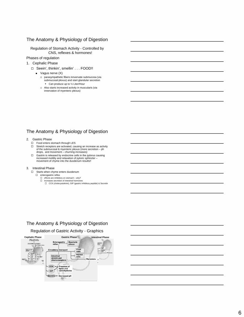
Regulation of Stomach Activity - Controlled by CNS, reflexes & hormones!
Phases of regulation1. Cephalic Phase
Seein’, thinkin’, smellin’ . . . FOOD!!Vagus nerve (X)
parasympathetic fibers innvervate submucosa (via submucosal plexus) and start glandular secretion
Can produce up to ½ Liter/HourAlso starts increased activity in muscularis (via innervation of myenteric plexus)
2. Gastric PhaseFood enters stomach through LESStretch receptors are activated, causing an increase as activity of the submucosal & myenteric plexus (more secretion – ph drops, and movement – churning increases)Gastrin is released by endocrine cells in the pylorus causing increased motility and relaxation of pyloric sphincter –movement of chyme into the duodenum results!!
3. Intestinal PhaseStarts when chyme enters duodenum
enterogastric reflexeffects are inhibitory on stomach – why?Increases secretion of intestinal hormones
CCK (cholecystokinin), GIP (gastric inhibitory peptide) & Secretin
The Anatomy & Physiology of Digestion
Regulation of Gastric Activity - Graphics
The Anatomy & Physiology of Digestion
Cephalic Phase Gastric Phase Intestinal Phase
7
The Anatomy & Physiology of Digestion
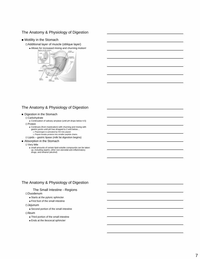
Motility in the StomachAdditional layer of muscle (oblique layer)
Allows for increased mixing and churning motion!
The Anatomy & Physiology of Digestion
Digestion in the StomachCarbohydrate
Continuation of salivary amylase (until pH drops below 4.5)Protein
Continues (from mastication) with churning and mixing with gastric juices until pH has dropped to 2 and below…
Pepsinogen is activated by HCl into pepsinPepsin breaks proteins into smaller peptide chains
Lipids – gastric lipase (milk fat digestion begins)Absorption in the Stomach
Very littlesmall amounts of certain lipid-soluble compounds can be taken up, including aspirin, other non-steroidal anti-inflammatory drugs, and ethanol (alcohol)
The Anatomy & Physiology of Digestion
DuodenumStarts at the pyloric sphincterFirst foot of the small intestine
JejunumSecond portion of the small intestine
IleumThird portion of the small intestineEnds at the ileocecal sphincter
The Small Intestine - Regions

8
The Anatomy & Physiology of DigestionThe Small Intestine – The Wall
Visible circular folds are present (plicae circulares)
Forces chyme to mix and spiral as it movesVilli present throughout the mucosa
Though more at the duodenum, less at the ileumEach villus contains a lacteal (lymphatic capillary) – why?At base of villus is an intestinal gland
Some mucous (duodenal region mainly) secretedBuffers secreted
Lined with simple columnar epithelial cells with microvilli
Microvilli dramatically increase surface area for digestion and absorption of nutrientsWater also enters lumen through the mucosa
Almost 2 Liters/day of intestinal juice is produced in the small intestine!
The Anatomy & Physiology of Digestion
The Anatomy & Physiology of Digestion
MotilitySegmentation
Alternate constriction of circular muscles only
PeristalsisCauses a forward spiral movement of chyme
Due to plicae circulares
Hormonal issuesEnterogastric reflex – speeds up movement in all areas of small intestineGastroileal reflex – relaxation of ileocecal sphincter due to gastrin (from stomach), increases movement into large intestine
The Small Intestine

9
The Anatomy & Physiology of Digestion
Control of secretion of enzymes into the duodenumUnder parasympathetic control (starts in cephalic phase)
Under hormonal controlGastrin
↑ secretion of enzymes in stomachSecretin
↑ secretion of pancreas (buffers) & liver (bile)↓ gastric secretion
CCK (cholecystokinin)↓ feeling of hunger, slows stomach motility & gastrin secretionRelaxes hepatopancreatic sphincter (allows bile in SI)↑ production of pancreatic enzymesContracts galllbladder
GIP (Gastric Inhibitory Peptide)Release of insulin by beta cells of pancreatic islets (islets of Langerhans)
The Small Intestine
The Anatomy & Physiology of Digestion
Digestion (chemical) in the Small IntestineProteins
via pancreatic enzymes (like the stomach, activated in the lumen of the small intestine)
Trypsin, Chymotrypsin & carboxypeptidaseAct like molecular scissors, cutting proteins in chains of aa’s and also taking off individual aa’s.
CarbohydratesReduced by enzymatic action (pancreatic amylase & enzymatic action in microvilli) to absorbable units
Glucose, Galactose & Fructose
LipidsEmulsified by bile secretions & digested by pancreatic lipase
The Small Intestine
The Anatomy & Physiology of DigestionPancreatic Anatomical Features

10
The Anatomy & Physiology of Digestion
Largest visceral organ (3 ½ lbs) Four lobes
Right lobe (largest & mainly in rt. Hypochondriac region)Left lobeCaudate lobeQuadrate lobe
The Liver - Features
The Anatomy & Physiology of Digestion
Bile produced in liverTransported via hepatic ducts (right & left) to common hepatic ductIf not needed, stored in gallbladder via cystic ductCystic duct joins hepatic duct to make common bile duct which empties into duodenum
The Gallbladder & Ducts
The Anatomy & Physiology of Digestion
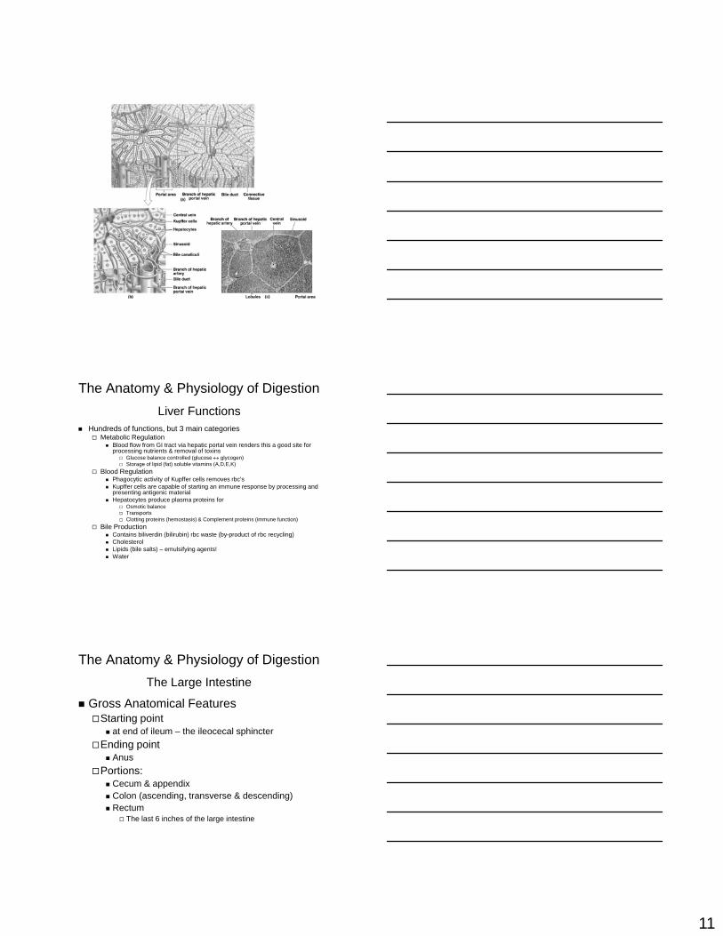
Lobes of liver consist of many lobules (small functional units)
Each lobule containsHepatocytes (main cells of liver)Kupffer Cells – macrophages in the lobuleBlood vessels
Blood from hepatic portal veinBlood from hepatic arteriesSinusoids
enlarged capillaries lined with hepatocytes & Kupffer cellsCentral Vein – in middle of lobule
Bile canaliculi transport bile away from lobule via bile ducts
Liver Histology
11
The Anatomy & Physiology of Digestion
Hundreds of functions, but 3 main categoriesMetabolic Regulation
Blood flow from GI tract via hepatic portal vein renders this a good site for processing nutrients & removal of toxins
Glucose balance controlled (glucose ↔ glycogen)Storage of lipid (fat) soluble vitamins (A,D,E,K)
Blood RegulationPhagocytic activity of Kupffer cells removes rbc’sKupffer cells are capable of starting an immune response by processing and presenting antigenic materialHepatocytes produce plasma proteins for
Osmotic balanceTransportsClotting proteins (hemostasis) & Complement proteins (immune function)
Bile ProductionContains biliverdin (bilirubin) rbc waste (by-product of rbc recycling)CholesterolLipids (bile salts) – emulsifying agents!Water
Liver Functions
The Anatomy & Physiology of Digestion
Gross Anatomical FeaturesStarting point
at end of ileum – the ileocecal sphincterEnding point
AnusPortions:
Cecum & appendixColon (ascending, transverse & descending)Rectum
The last 6 inches of the large intestine
The Large Intestine

12
The Anatomy & Physiology of Digestion
Layers of the WallMucosa
Large quantity of goblet cellsNo villi
MuscularisCircular muscle forms pouches = haustraLongitudinal muslce forms a band = taenia coli
SerosaVisceral peritoneum forms mesenteries to attach colon to abdominal wall.
The Large Intestine
The Anatomy & Physiology of Digestion
FunctionsAbsorption
water!1500 mls of substance enters daily, only 200 mls leaves1.3 L/day reabsorbed!
Other:Bile salts, bilirubin (unintentional, modified & excreted by kidney later), toxins – if present (from bacterial action)Vitamins
Vitamin K – required for proper clottingBiotin – required for glucose metabolismPantothenic Acid (B5) – required for some hormones and neurotransmitters synthesis
The Large Intestine
13
The Anatomy & Physiology of Digestion
FunctionsMovement
Haustral churningSequential contraction of haustral pockets
Mass movement (peristalsis)In response to gastrin (gastric phase & intestinal phase)Creates urge to defecate as fecal matter is moved into rectum (initiates defecation reflex)
Defecation – 2 positive feedback loops!!Stretch receptors in rectum (when stretched) – starts process
increase activity in sigmoid colon and rectumThis moves feces towards the anus, stretching the rectum and anal canal
Parasympathetic motor neurons are activated, initiating mass movement!Voluntary Aspect – control over external anal sphincter – yeah!
The Large Intestine
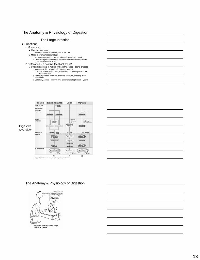
Digestive Overview
The Anatomy & Physiology of Digestion