digital and manual rotation of the persistent occiput
TRANSCRIPT
Journal of Midwifery &Women’s Health www.jmwh.orgClinical Rounds
CEUDigital and Manual Rotation of the Persistent OcciputPosterior FetusChristina Elmore1, CNM, MSN , Kelly McBroom2, CNM, PhD, Jessica Ellis1, CNM, PhD
Abstract: Persistent fetal occiput posterior (OP) position is a topic of interest with implications for intrapartum management. Although studiesreport a low incidence of persistent OP position, anecdotal evidence suggests an increase in prevalence given changes in maternal demographics.Clinicians are often familiar with interventions such as position changes and the use of props and a rebozo to address persistent OP position inearly labor; however, midwives remain uncomfortable with the techniques of digital and manual rotation. This article reviews current evidenceand recommendations for the management of persistent OP position in the second stage of labor. Further research is needed to guide clinicianson the optimal timing and techniques for digital and manual rotation.J Midwifery Womens Health 2020;65:387–394 c© 2020 by the American College of Nurse-Midwives.
Keywords: persistent occiput posterior, digital and manual rotation, midwifery
CASE SUMMARY
A.B., a 39-year-old gravida 1, para 0 presented to the laborand birth unit at 40 weeks’ 5 days’ gestation in early activelabor. Her health history included abody mass index (BMI)of 38, but her pregnancy was otherwise uncomplicated.Thefirst stage of labor progressed slowly and was noteworthyfor significant back pain and coupling contractions. Givenconcerns for labor dystocia and maternal exhaustion, themidwife supported her through several position changes,including hands and knees, side lunging, slow dancing, andthe use of an exercise ball. Additionally, the midwife useda rebozo between contractions to optimize fetal position.After 6 hours of labor support, A.B. elected for epiduralanalgesia. She was placed in an exaggerated Sims positionwith apeanut ball prop between her legs to promote sponta-neous rotation while resting. She reached complete cervicaldilatation at midnight after an hour of rest.
After approximately 60 minutes of pushing with min-imal descent despite position changes and strong mater-nal effort, the midwife found the fetal position to be directocciput posterior (OP) during a sterile cervical examina-tion. The midwife reviewed concern for possible arrest oflabor, and A.B. was given the option for continued push-ing or manual rotation.The midwife counseled on the risksof no intervention, which included possible cesarean birthor instrumental birth, increased risk of perineal trauma,and surgical complications should spontaneous birth notoccur. The risks and benefits of manual rotation were alsoreviewed, which included maternal and fetal intolerance of
1Birthcare Healthcare, University of Utah’s College of Nursing,Salt Lake City, Utah2Swedish Medical Center, Kaiser Permanente WashingtonMidwives, Seattle University, Seattle, WashingtonCorrespondenceChristina ElmoreEmail: [email protected] Elmore https://orcid.org/0000-0003-4799-1473
rotation, fetal injury, cord prolapse, cervical laceration, andpotential for unsuccessful rotation. A.B. met privately withher partner and gave verbal consent for manual rotation.
In preparation for the procedure, the nurse placed aFoley catheter to empty A.B.’s bladder. The charge nurseand consulting physician were notified and available shouldcomplications arise. The midwife used bedside ultrasoundto confirm OP position and the fetal back location (left) toaid with the direction of rotation. A.B. reported adequatepain control with her epidural analgesia. She was placedin a right side-lying position with her left leg supportedto maximize pelvic diameter. The fetal heart rate patternwas determined to be category 1 prior to rotation.The mid-wife inserted her right hand with the thumb down until shecould feel the fetal ears and chin. She gently grasped thefetal head and rotated clockwise along the short arc dur-ing 2 contractions. Fetal heart monitoring showed a vari-able deceleration during the subsequent contraction. Theneonate was born with the next push and noted to have agood tone and a strong cry. The examination of the new-born head showed no signs of injury.Maternal bleeding wasminimal, and A.B.’s perineum was intact. The midwife leftthe family in stable condition to complete documentationof a successful birth.
INTRODUCTION
Persistent OP position has been defined as “ . . . when the oc-ciput remains in the posterior quadrants of the pelvis untildelivery.”1(p 696) This definition does not distinguish betweenthe physiologically normalOP position and the pathologicallypersistent OP position. Diagnosis of persistent OP position isoften made retrospectively depending on whether or not la-bor followed a normal curve.1 Therefore, clinicians should usesound judgmentwhen differentiating between physiologicallynormal OP and pathologically persistent OP position, espe-ciallywith regard to implications for labor progress. In the firststage of labor, management should include measures to sup-port spontaneous rotation, including position changes. In thesecond stage of labor, interventions such as digital or manual
1526-9523/09/$36.00 doi:10.1111/jmwh.13118 c© 2020 by the American College of Nurse-Midwives 387

rotation may be considered. The purpose of this article is tohighlight key considerations for digital and manual rotation.As such, the authors hope midwives will have increased con-fidence regarding when and how to utilize this intervention.
RISK FACTORS AND CLINICAL IMPLICATIONS
Risk Factors for Persistent Occiput Posterior
The prevalence of OP position at the onset of labor rangesfrom 15% to 20%, with persistence into the second stage of la-bor ranging from 5% to 8%.1,2 Clinicians report an anecdotalincrease in prevalence in recent years; however, more researchis needed to evaluate current prevalence rates and associatedrisk factors.
Maternal risk factors for persistentOPpositioning includea narrowpubic arch,3 BMI greater than 30,1 nulliparity,4 race,3and maternal age greater than 35 years.4,5 The correlation be-tween use of epidural analgesia and persistent OP position isa topic of debate.1,3,4,6 A 2011 meta-analysis of 4 randomizedcontrolled trials (RCTs) representing 673 women found mal-position at birth was higher in women with epidural analge-sia use during labor, although the findings were not statisti-cally significant (relative risk 1.40; 95% CI, 0.98-1.99).6 Addi-tionally, the correlation between labor induction and persis-tent OP position is inadequately studied.3 Finally, fetal con-tributions to the prevalence of persistent OP position remainunclear; however, higher fetal weight and a larger fetal headcircumference are associated with persistent OP position, alonger second stage of labor, and higher rates of operativevaginal birth (forceps or vacuum assistance).7,8 Multiple riskfactors often exist simultaneously; for example, advancedma-ternal age, nulliparity, BMI greater than 30 kg/m2, and the useof epidural analgesia were noted in this clinical case presenta-tion. Therefore, it can be difficult to ascertain which factorsare most influential on the fetal position.
Clinical Implications of Persistent Occiput Posterior
Lack of spontaneous resolution of OP positioning is corre-lated with clinical implications impacting both maternal andneonatal outcomes. Persistent OP position has been shown toincrease the maternal risk for postpartum hemorrhage, post-partum infection, operative vaginal birth, intraamniotic infec-tion (formerly chorioamnionitis) and the degree of perineallaceration, including anal sphincter injury.2,5,9 Fetal risks forunresolved persistent OP position may include increased ad-mission to neonatal intensive care unit, a low 5-minute Ap-gar score, and meconium-stained fluid;4,10,11 however, theseassociations did not persist in studies that were adjusted forconfounders.12 Findings are also mixed regarding the impactof persistent OP position on risk of fetal acidemia10,13 and riskfor hypoxic-ischemic encephalopathy.14
Persistent OP position is also associated with prolongedlabor and arrest of labor, which are leading indications for op-erative vaginal birth and cesarean birth.1,5 Given the knownrisks with cesarean birth, reduction of the rates of primarycesarean birth has been a national priority since 2014.15,16The use of manual and digital rotation is a reasonable man-agement tool to reduce the incidence of operative vaginal
and/or cesarean birth5,11,12,15,17 and may be a promisingintervention to promote physiologic birth.
MANAGEMENT CONSIDERATIONS
Confirmation of Fetal Position
Midwives often utilize Leopold’s maneuvers for evaluation ofthe fetal position. Additionally, the evaluation of fetal posi-tion includes the palpation of the fetal fontanelles via cervi-cal examination. The anterior fontanelle is surrounded by thefrontal sutures and 2 coronal sutures (Figure 1).18 The pos-terior fontanelle in comparison is surrounded by the bilaterallambdoid sutures (Figure 1).18 However, studies evaluating in-trapartum care provider reliability have shown poor accuracyfor diagnosis of OP position based on palpation of the pos-terior or anterior fontanelle even among skilled clinicians.19The difficulty of palpating suture lines and fontanelles is es-pecially challenging after physiologic molding, which causesthe sutures to override and fontanelles to be less palpable, aswell as when caput is present. In these cases, some cliniciansuse the fetal ear position to evaluate fetal head position; how-ever, this method also has poor reliability.
Although the use of ultrasound is not a routine standardof care, the use of bedside ultrasound to confirm the loca-tion of the fetal back may be a valuable tool in the handsof trained practitioners to confirm the fetal position. A 2017RCT demonstrated the use of bedside ultrasound to confirmhead orientation and spine location significantly increasedsuccessful rotation (P � .001) and spontaneous vaginal birth(P = .01).20 Additional literature highlighted the increased ac-curacy of bedside ultrasound to detect asynclitism, poor flex-ion, and occiput transverse presentation, as well as use of theoccipital-spinal angle to predict the likelihood of spontaneousvaginal birth.19 The full utility of ultrasound to guide manualrotation, including clinician training, has been inadequatelyreported in current literature. Therefore, when clinically ap-propriate, the use of ultrasound by trained clinicians may bebeneficial but should not be mandatory prior to attemptedrotation.
Before Rotation: Use of Position Changes and Props
Appropriate management of persistent OP position dependson the timing of diagnosis and the degree of disruption in thenatural labor progress. Historically, midwives, labor nurses,and labor support doulas have utilized position changes to as-sist with fetal rotation in the first stage of labor.25 Key positionsto address posterior fetal position include hands and knees,lunges, side-lying, and exaggerated Sims.21 There is limitedevidence that position changes facilitate the spontaneous res-olution of persistent OP positioning.
An RCT from France (2013) evaluated interventions topromote anterior rotation from OP position among womenin labor with a single fetus in documented OP position(N = 220).The women in the control group were placed indorsal recumbent position, whereas women in the interven-tion group were assigned to 3 different postures includinghands and knees position (fetal head at −5 to −3 station), astrict lateral recumbent position on the same side as the fetalspine with the inferior leg folded (fetal head −2 to 0 station),
388 Volume 65, No. 3, May/June 2020

Coronal Suture Anterior Fontanelle
Posterior Fontanelle
Occipital Bone
FrontalBone
FrontalBone
Parietal BoneParietal Bone
Lambdoidal Suture
Sagi�al Suture
Coronal Suture
Frontal Suture
Lambdoidal Suture
Figure 1. Landmarks of the Fetal Skull Used to Determine Fetal Position
and finally a lateral recumbent position in which the upperleg was supported (fetal head station � 0). These researchersfound there were no significant differences in the rates ofspontaneous rotation (anterior rotation, 78.2% in controlgroup vs 76.4% in the intervention group; P = .75).22 A 2016RCT from Switzerland (N = 439) also found no statisticallysignificant differences in the rates of spontaneous rotationamong women using the hands and knees position (for atleast 10 minutes) compared with usual care: one hour afterrandomization, 12% of fetuses in the control group wereocciput anterior (OA) compared with 17% of the fetuses inthe intervention group (P = .13).23 However, this study foundthere was an increase in patient-reported comfort with use ofhands and knees positioning (P = .02).23 Additionally, a 2016RCT (N = 322) evaluating the impact of lateral recumbentpositioning found the rates of OA position did not differbetween the control and intervention groups (21.9% vs 21.6%,respectively; P = .887).13 In contrast, a 2018 RCT from Spain(N = 120) found the use of modified Sims position duringthe active phase of labor significantly improved spontaneousrotation from persistent OP position to OA position (51% vs22%) compared with women using their position of choice(P = .001), as well as increased rates of spontaneous vaginalbirth (84.7% vs 68.3%, P = .04).24 More research is neededto address the utility of maternal positioning during labor tooptimize spontaneous fetal rotation.
In addition to position changes, the intrapartum careprovider can use props such as bed stirrups, side tables, pil-lows, and peanut-shaped balls to encourage pelvic floor relax-ation, maximize pelvic diameter, and promote spontaneousfetal rotation to OA position, especially in women usingepidural analgesia (Figure 2). Another tool to consider is a re-bozo, which is a wide shawl that was originally used byMayanmidwives inMexico. A rebozo is used to facilitate pelvic rock-ing and encourage fetal rotation through relaxation of pelvic
musculature.25 The effectiveness of the rebozo and alternativeprops to promote spontaneous rotation has not been formallystudied.25 When spontaneous rotation does not occur by theonset of the second stage of labor, the diagnosis of persistentOP position and/or labor dystocia can be made. At this time,digital or manual rotation is an appropriate intervention toconsider.1,26
Informed Consent
During any intervention, including digital or manual ro-tation, it is important to assess patient safety and clinicalappropriateness and for the patient to provide informedconsent prior to implementation. For digital or manualrotation, key elements of informed consent include thepotential for maternal discomfort, fetal intolerance of theintervention, fetal injury, cord prolapse, cervical lacerations,and unsuccessful rotation (Table 1). There are limited datato guide clinicians on the prevalence of these risks; however,experts in both the United States and Canada state that“digital and manual rotation is considered safe with minimalrisk of maternal or fetal injury.”27(p e74) Contraindications todigital and manual rotation are similar to those of vaginalbirth, including cord prolapse, excessive bleeding, concernsfor maternal or fetal clinical stability, and the need to expeditebirth.1
Preparation for Rotation
After ensuring that manual or digital rotation is safe andclinically appropriate, confirming fetal position, and ob-taining informed consent, the intrapartum care providershould consider measures to promote successful rotation.These include optimizing maternal comfort, maximizingpelvic diameter, assessing fetal well-being, and notifying thecollaborating physician (Table 1).
Journal of Midwifery &Women’s Health � www.jmwh.org 389

A
C
B
D
Figure 2. Positions and Props to Encourage Spontaneous Fetal Rotation from Occiput Posterior to Occiput Anterior PositionA) Side stirrups. B) Side table with pillow. C) Peanut ball. D) Close-up of peanut ball.
Source: Photographs by Christina Elmore, CNM, MSN.
In order to optimize bothmaternal comfort and pelvic di-ameter, several interventions may be considered. Cliniciansmay consider emptying the patient’s bladder and bowel whenappropriate. Medication to promote maternal comfort duringthe procedure may be offered, including nitrous oxide, pu-dendal block, or epidural analgesia. A 2014 study found a sig-nificant difference in transverse pelvic diameter with positionchanges, such as kneeling squat position (P � .0001), whichsupports historical studies on the changes of pelvic diameterin upright positions.28 Therefore, positioning the patient in amanner that increases the pelvic diameter may also promotesuccessful rotation (Figure 2).
Evaluating fetal well-being prior to and immediately af-ter the procedure is a critical component of safe rotation. Itis common for rotational maneuvers to cause some degree oftransient change in fetal heart rate pattern, most notably vari-able decelerations. Careful consideration should be taken re-garding clinical appropriateness of rotation in the context ofa category II fetal heart rate pattern. Finally, notification ofthe support staff, such as the charge nurse and collaboratingphysician, prior to the implementation of rotation is anotherkey step of preparation.
Timing of Rotation
The ideal timing of digital and/or manual rotation remainsa source of debate. In a prospective analysis in Israel (2005),61 women with term fetus in confirmed OP position wereassigned to either digital rotation (n = 31) or no interven-tion (n = 30), with a significant decrease in duration of sec-ond stage of labor found in the digital rotation group (P �.003).15 A separate RCT (N = 65) compared early manual ro-tation (n = 33) with routine care (n = 32) and found a sig-nificant decrease in the second stage of labor in the groupundergoing early manual rotation (P = .04).29 Current evi-dence to guide clinicians on the use of prophylactic rotationversus rotation after a diagnosis of dystocia has been made islimited.30 Although some experts promote prophylactic rota-tion prior to the time of diagnosis of dystocia,29 others pro-mote a more cautious approach that allows time for potentialspontaneous rotation.1 Another consideration in the timingof rotation includes whether to perform rotation with11,31,32or between contractions.1 Cliniciansmay considermanual ro-tation after 30 to 60 minutes of pushing for multiparous andprimiparous women, respectively.1 More or less time may beconsidered depending on whether or not the patient is using
390 Volume 65, No. 3, May/June 2020
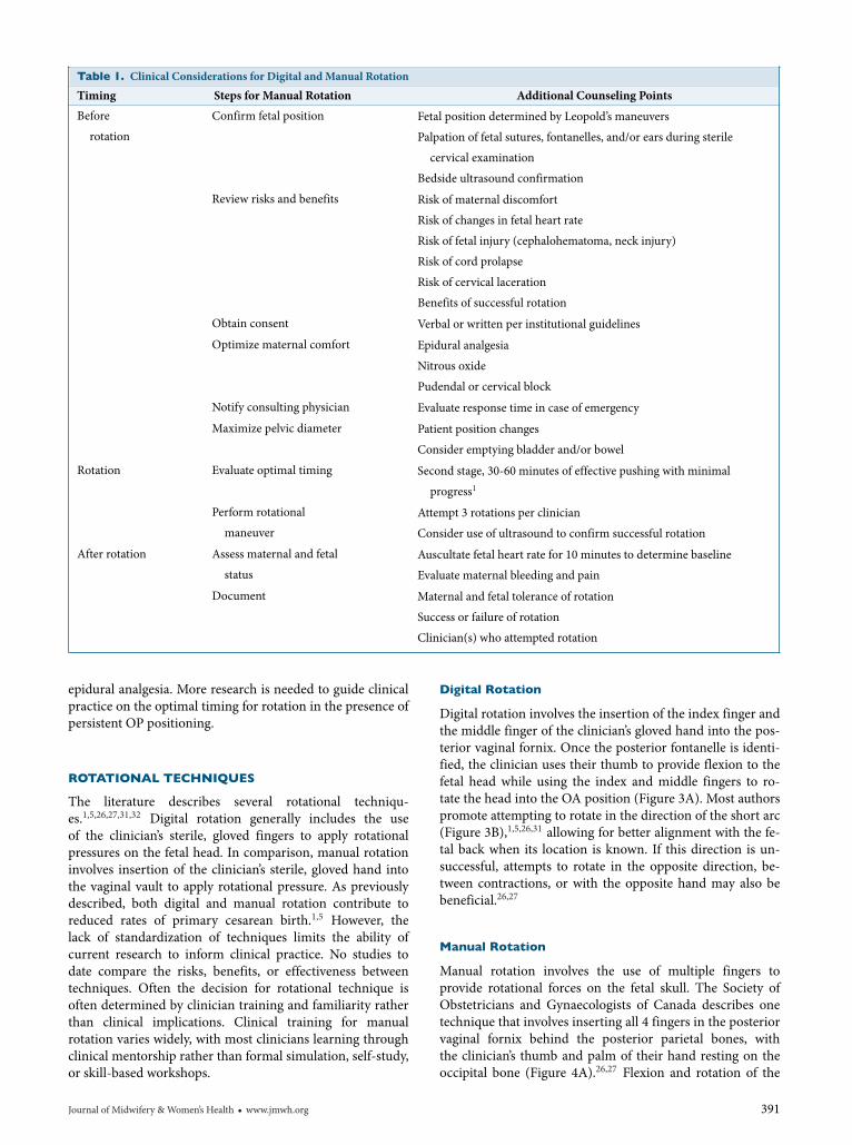
Table 1. Clinical Considerations for Digital and Manual Rotation
Timing Steps for Manual Rotation Additional Counseling Points
Before
rotation
Confirm fetal position Fetal position determined by Leopold’s maneuvers
Palpation of fetal sutures, fontanelles, and/or ears during sterile
cervical examination
Bedside ultrasound confirmation
Review risks and benefits Risk of maternal discomfort
Risk of changes in fetal heart rate
Risk of fetal injury (cephalohematoma, neck injury)
Risk of cord prolapse
Risk of cervical laceration
Benefits of successful rotation
Obtain consent Verbal or written per institutional guidelines
Optimize maternal comfort Epidural analgesia
Nitrous oxide
Pudendal or cervical block
Notify consulting physician Evaluate response time in case of emergency
Maximize pelvic diameter Patient position changes
Consider emptying bladder and/or bowel
Rotation Evaluate optimal timing Second stage, 30-60 minutes of effective pushing with minimal
progress1
Perform rotational
maneuver
Attempt 3 rotations per clinician
Consider use of ultrasound to confirm successful rotation
After rotation Assess maternal and fetal
status
Auscultate fetal heart rate for 10 minutes to determine baseline
Evaluate maternal bleeding and pain
Document Maternal and fetal tolerance of rotation
Success or failure of rotation
Clinician(s) who attempted rotation
epidural analgesia. More research is needed to guide clinicalpractice on the optimal timing for rotation in the presence ofpersistent OP positioning.
ROTATIONAL TECHNIQUES
The literature describes several rotational techniqu-es.1,5,26,27,31,32 Digital rotation generally includes the useof the clinician’s sterile, gloved fingers to apply rotationalpressures on the fetal head. In comparison, manual rotationinvolves insertion of the clinician’s sterile, gloved hand intothe vaginal vault to apply rotational pressure. As previouslydescribed, both digital and manual rotation contribute toreduced rates of primary cesarean birth.1,5 However, thelack of standardization of techniques limits the ability ofcurrent research to inform clinical practice. No studies todate compare the risks, benefits, or effectiveness betweentechniques. Often the decision for rotational technique isoften determined by clinician training and familiarity ratherthan clinical implications. Clinical training for manualrotation varies widely, with most clinicians learning throughclinical mentorship rather than formal simulation, self-study,or skill-based workshops.
Digital Rotation
Digital rotation involves the insertion of the index finger andthe middle finger of the clinician’s gloved hand into the pos-terior vaginal fornix. Once the posterior fontanelle is identi-fied, the clinician uses their thumb to provide flexion to thefetal head while using the index and middle fingers to ro-tate the head into the OA position (Figure 3A). Most authorspromote attempting to rotate in the direction of the short arc(Figure 3B),1,5,26,31 allowing for better alignment with the fe-tal back when its location is known. If this direction is un-successful, attempts to rotate in the opposite direction, be-tween contractions, or with the opposite hand may also bebeneficial.26,27
Manual Rotation
Manual rotation involves the use of multiple fingers toprovide rotational forces on the fetal skull. The Society ofObstetricians and Gynaecologists of Canada describes onetechnique that involves inserting all 4 fingers in the posteriorvaginal fornix behind the posterior parietal bones, withthe clinician’s thumb and palm of their hand resting on theoccipital bone (Figure 4A).26,27 Flexion and rotation of the
Journal of Midwifery &Women’s Health � www.jmwh.org 391

A B
Figure 3. Digital Rotation TechniqueA) Clinician places the index and middle finger of their right hand over the occipital bone and the thumb placed over the parietal bone. B) Theclinician then flexes the head and using the index and middle fingers to rotate the head along the short arc into the occiput anterior position.
Source: Photographs by Christina Elmore, CNM, MSN.
A B
Figure 4. Manual Rotation, Technique Described by the Society of Obstetricians and Gynaecologists of CanadaA) The palm of the clinician’s hand is placed over the occipital bone, and the thumb is placed over the parietal bone. B) The clinician then flexes thehead and using their hand to rotate the head along the short arc into the occiput anterior position.
Source: Photographs by Christina Elmore, CNM, MSN.
fetal head is achieved by cupping the head and aligning theocciput with the fetal spine (Figure 4B).
An alternative method is a technique originally describedby Tarnier and Chantreuil,31,32 which involves the insertionof all 4 fingers along the side of the fetal head to the level ofthe ear or chin (Figure 5A). Depending on pelvic space, thesize of the fetal head, and the size of the clinician’s hand, thethumb is placed next to the fingers or on the opposing parietalbone (Figure 5A). Gentle pressure is then used to flex the fetalhead and rotate the fetal head to the OA position (Figure 5B).No studies have compared the risks or benefits of a choiceof hand or direction of rotation. Additionally, no researchhas examined the risks or benefits between the techniquesdescribed by the Society of Obstetricians and Gynaecolo-gists of Canada and Tarnier and Chantreuil, nor have therebeen comparisons of rotation with or between contractions.
More research is needed to guide clinical practice on thesetechniques.
Predictors of Success
The success rates of digital and manual rotation in the lit-erature range from 47% to 93%.5,15,17,31,33 Predictors of suc-cessful rotation include maternal age less than 35 years andmultiparity.5 Additionally, Desbriere et al (2013) found thatsuccess of rotation increased when the maternal BMI wasless than 28 kg/m2 at time of birth (P = .05) and with par-ity greater than or equal to 2 (P = .02).22 A study by Mas-truo et al (2017) found that the use of ultrasound to identifythe head and spinal alignment increased the success of ro-tation (P � .001). Finally, Bertholdt et al (2018) found thatmanual rotations were more successful after fetal engagement
392 Volume 65, No. 3, May/June 2020

A B
Figure 5. Manual Rotation, Technique Described by Tarnier and ChantreuilA) The clinician’s hand is placed over the side of the fetus’ head with fingertips at the level of the chin or ears, and the thumb is placed over the oppositeparietal bone. B) The clinician then flexes the head and using their hand to rotate the head along the short arc into the occiput anterior position.
Source: Photographs by Christina Elmore, CNM, MSN.
during spontaneous labor.34 Studies to date examine primar-ily hospital-based clinicians, with no data comparing suc-cess rates between intrapartum care provider types or in theout-of-hospital setting. Success rates may also be influencedby clinician training and familiarity with different rotationaltechniques.
Risks for unsuccessful rotation include multiple attemptsto rotate prior to 10 cm cervical dilatation (odds ratio [OR],3.4; 95% CI, 1.3-8.6).31 Additionally, 2 studies found thatsuccessful rotation was decreased after the diagnosis of failureto progress (OR, 3.3; 95% CI, 1.2-8.5;31 OR, 2.01; 95% CI,1.1-3.9).34 One study found that up to 3 attempts at manualrotation were reasonable; however, no success was found inthe fourth or fifth attempts.31 The study did not differentiatebetween the number of attempts made between multipleclinicians.
SPECIAL CONSIDERATIONS
Individualization of care is a hallmark of midwifery, and clini-cians are often tasked with the application of vague guidelinesto unique clinical situations. For example, a multiparouswoman whose cervix is not fully dilatated or with a categoryII fetal heart rate tracing may or may not be an appropriatecandidate for rotation. Considerations such as the clinician’saccess to emergency resources, the fetal oxygen reserves, andthe strength of maternal pushing potential, for example, mayaid in determining the appropriateness of digital or manualrotation. Although the emphasis of this article has been onpersistent OP position, many of these concepts are equallyapplicable in the context of occiput transverse position and/orin the context of fetal asynclitism.31 Finally, current studieshave focused on hospital-based midwives and obstetricians.Clinicians practicing in out-of-hospital settings or otherremote settings should use caution before implementing in-terventions that have the potential to cause maternal or fetaldistress. In such settings, there should be specific guidelinesand plans for transfer of care. Additionally, there is a need
for clinical training prior to the implementation of advancedmaneuvers such as digital or manual rotation.
IMPLICATIONS FOR MIDWIFERY PRACTICE
Digital and manual rotation techniques have the potential tofacilitate vaginal birth and decrease the cesarean birth rate.The American College of Obstetricians and Gynecologistsand the Society for Maternal-Fetal Medicine promote manualrotation as “reasonable to consider prior to operative vaginalbirth or cesarean birth.”16 Additionally, experts from Canadareport that manual rotation represents “little or no increasedrisk to the pregnant woman or fetus.”27 Although no po-sition is statement available from the American College ofNurse-Midwives, manual rotation is described in Varney’sMidwifery as a reasonable intervention that may reduce theincidence of OP birth with rare complications, so long as ap-propriate clinical considerations (fetal station, stage of labor,diagnosis of arrest) are determined.26(pp 990-991)
The goal of this article is to provide tools and guidancefor clinical practice. Manual rotation techniques can also bepracticed in high- or low-tech simulation environments to in-crease clinician confidence. Physician collaboration may beuseful in select clinical contexts, such as difficult rotation,concerns for fetal tolerance of rotation, possible operativebirth, or rotational forceps. With continued training and col-laboration, midwives will gain confidence in the critical skillsof digital and manual rotation.
CONCLUSION
Many lessons are highlighted in the case presented above,including optimal midwifery management of persistent OPfetal position during labor and birth. Management of OPpositioning requires early identification of fetal malposition;thorough patient counseling on risks, benefits, and alterna-tives; and implementation of interventions in a systematicfashion. Although there is a clear need for additional research
Journal of Midwifery &Women’s Health � www.jmwh.org 393

to inform best practices, increasing both knowledge andaccess to clinician training has the potential to improve theutilization and success of this clinical skill.
ACKNOWLEDGMENTS
The authors would like to thank the University of Utah’s Col-lege of Nursing and Seattle University College of Nursingfor support during production of the manuscript, in partic-ular the midwives and patients of Birthcare Healthcare andSwedish Medical Center.
CONFLICT OF INTEREST
The authors have no conflicts of interest to disclose.
REFERENCES
1.Barth WH, Jr. Persistent occiput posterior. Obstet Gynecol.2015;125(3):695-709.
2.Cheng YW, Hubbard A, Caughey AB, Tager IB. The association be-tween persistent fetal occiput posterior position and perinatal out-comes: an example of propensity score and covariate distance match-ing. Am J Epidemiol. 2010;171(6):656-663.
3.Cheng YW, Shaffer BL, Caughey AB. The association between per-sistent occiput posterior position and neonatal outcomes. Obstet Gy-necol. 2006;107(4):837-844.
4.Ponkey SE, Cohen AP, Heffner LJ, Lieberman E. Persistent fe-tal occiput posterior position: obstetric outcomes. Obstet Gynecol.2003;101(5 pt 1):915-920.
5.Shaffer BL, Cheng YW, Vargas JE, Caughey AB. Manual rotation toreduce caesarean delivery in persistent occiput posterior or transverseposition. J Matern Fetal Neonatal Med. 2011;24(1):65-72.
6.Anim-Somuah M, Smyth RM, Cyna AM, Cuthbert A. Epidural ver-sus non-epidural or no analgesia for pain management in labour.Cochrane Database Syst Rev. 2018;(5):CD000331.
7.GardbergM, Laakkonen E, SalevaaraM. Intrapartum sonography andpersistent occiput posterior position: a study of 408 deliveries. ObstetGynecol. 1998;91(5 pt 1):746-749.
8.Yagel O, Cohen SM, Lipschuetz M, et al. Higher rates of operative de-livery and maternal and neonatal complications in persistent occiputposterior position with a large head circumference: a retrospective co-hort study. Fetal Diagn Ther. 2018;44(1):51-58.
9.Hirsch E, Elue R, Wagner A, et al. Severe perineal laceration duringoperative vaginal delivery: the impact of occiput posterior position. JPerinatol. 2014;34(12):898-900.
10.Dahlqvist K, Jonsson M. Neonatal outcomes of deliveries in occiputposterior position when delayed pushing is practiced: a cohort study.BMC Pregnancy Childbirth. 2017;17(1):377.
11.Le Ray C, Deneux-Tharaux C, Khireddine I, Dreyfus M, Vardon D,Goffinet F. Manual rotation to decrease operative delivery in posterioror transverse positions. Obstet Gynecol. 2013;122(3):634-640.
12.Shaffer BL, Cheng YW, Vargas JE, Laros RK Jr, Caughey AB. Manualrotation of the fetal occiput: predictors of success and delivery. Am JObstet Gynecol. 2006;194(5):e7-e9.
13.Le Ray C, Lepleux F, De La Calle A, et al. Lateral asymmetric decu-bitus position for the rotation of occipito-posterior positions: multi-center randomized controlled trial EVADELA. Am J Obstet Gynecol.2016;215(4):511.e1-e7.
14.TempestN,McGuinnessN, Lane S,HapangamaDK.Neonatal andma-ternal outcomes of successful manual rotation to correct malpositionof the fetal head; a retrospective and prospective observational study.PLoS One. 2017;12(5):e0176861.
15.Reichman O, Gdansky E, Latinsky B, Labi S, Samueloff A. Dig-ital rotation from occipito-posterior to occipito-anterior decreasesthe need for cesarean section. Eur J Obstet Gynecol Reprod Biol.2008;136(1):25-28.
16.Caughey AB, Cahill AG, Guise JM, Rouse DJ. Safe prevention of theprimary cesarean delivery. Am J Obstet Gynecol. 2014;210(3):179-193.
17.Sen K, Sakamoto H, Nakabayashi Y, et al. Management of the occiputposterior presentation: a single institute experience. J Obstet GynaecolRes. 2013;39(1):160-165.
18.Powers GD. The passenger: fetus. In: Posner GD, Dy J, Black AY, JonesGD, eds.Oxorn-Foote Human Labor and Birth. 6 ed. New York, NY:McGraw-Hill; 2013:55-64.
19.Bellussi F, Ghi T, Youssef A, et al. The use of intrapartum ultrasoundto diagnose malpositions and cephalic malpresentations. Am J ObstetGynecol. 2017;217(6):633-641.
20.Masturzo B, Farina A, Attamante L, et al. Sonographic evaluation ofthe fetal spine position and success rate of manual rotation of the fe-tus in occiput posterior position: a randomized controlled trial. J ClinUltrasound. 2017;45(8):472-476.
21.Simkin P. The fetal occiput posterior position: state of the science anda new perspective. Birth. 2010;37(1):61-71.
22.Desbriere R, Blanc J, Le Du R, et al. Is maternal posturing during la-bor efficient in preventing persistent occiput posterior position?A ran-domized controlled trial.Am J Obstet Gynecol. 2013;208(1):60.e1-e8.
23.Guittier MJ, Othenin-Girard V, de Gasquet B, Irion O, Boulvain M.Maternal positioning to correct occiput posterior fetal position dur-ing the first stage of labour: a randomised controlled trial. BJOG.2016;123(13):2199-2207.
24.Bueno-Lopez V, Fuentelsaz-Gallego C, Casellas-Caro M, et al. Effi-ciency of the modified Sims maternal position in the rotation of per-sistent occiput posterior position during labor: a randomized clinicaltrial. Birth. 2018;45(4):385-392.
25.Cohen SR, Thomas CR. Rebozo technique for fetal malposition in la-bor. J Midwifery Womens Health. 2015;60(4):445-451.
26.Kane-Low L, Osborne K. Second stage of labor and birth. In: King TL,Brucker MC, Osborne K, Jevitt CM, eds. Varney’s Midwifery. 6th ed.Burlington, MA: Jones & Bartlett; 2019:985-991.
27.Cargill YM, MacKinnon CJ. Guidelines for operative vaginal birth:SCOG clinical practice guideline no. 148. J Obstet Gynaecol Can.2018;40(2):e74-e80.
28.ReitterA,Daviss BA, Bisits A, et al. Does pregnancy and/or shifting po-sitions create more room in a woman’s pelvis? Am J Obstet Gynecol.2014;211(6):662.e1-e9.
29.Broberg J, Rees S, Jacob S, et al. A randomized controlled trial of pro-phylactic early manual rotation of the occiput posterior fetal head atthe beginning of the second stage of labor vs. expectant managementin nulliparas. Am J Obstet Gynecol. 2016;214(suppl 1):S63.
30.Phipps H, de Vries B, Hyett J, Osborn DA. Prophylactic manual ro-tation for fetal malposition to reduce operative delivery. CochraneDatabase Syst Rev. 2014;(12):CD009298.
31.Le Ray C, Serres P, Schmitz T, Cabrol D, Goffinet F. Manualrotation in occiput posterior or transverse positions: risk factorsand consequences on the cesarean delivery rate. Obstet Gynecol.2007;110(4):873-879.
32.Verhaeghe C, Parot-Schinkel E, Bouet PE, et al. The impact of man-ual rotation of the occiput posterior position on spontaneous vaginaldelivery rate: study protocol for a randomized clinical trial (RMOS).Trials. 2018;19(1):109.
33.Guerby P, Allouche M, Simon-Toulza C, Vayssiere C, Parant O, VidalF. Management of persistent occiput posterior position: a substantialrole of instrumental rotation in the setting of failed manual rotation. JMatern Fetal Neonatal Med. 2018;31(1):80-86.
34.Bertholdt C, Gauchotte E, Dap M, Perdriolle-Galet E, Morel O. Pre-dictors of successful manual rotation for occiput posterior positions.Int J Gynaecol Obstet. 2019;144(2):210-215.
Continuing education units (CEUs) are available forthis article. To obtain CEUs online, please visit www.jmwhce.org. A CEU form that can be mailed or faxed isavailable in the print edition of this issue.
394 Volume 65, No. 3, May/June 2020