digital imaging to evaluate root system architectural ... · associated with soil biotic factors...
TRANSCRIPT
Phytobiomes Journal • 2019 • 3:102-111 https://doi.org/10.1094/PBIOMES-12-18-0062-R
RESEARCH
Digital Imaging to Evaluate Root System Architectural ChangesAssociated with Soil Biotic Factors
Chakradhar Mattupalli,1,2 Anand Seethepalli,1 Larry M. York,1,† and Carolyn A. Young1,†
1Noble Research Institute, LLC, 2510 Sam Noble Parkway, Ardmore, OK 734012San Luis Valley Research Center, Colorado State University, 0249 East Road 9 North, Center, CO 81125
Accepted for publication 26 March 2019.
ABSTRACT
Root system architecture is critical for plant growth, which isinfluenced by several edaphic, environmental, genetic, and bioticfactors including beneficial and pathogenic microbes. Studyingroot system architecture and the dynamic changes that occurduring a plant’s lifespan, especially for perennial crops growingover multiple growing seasons, is still a challenge because of thenature of their growing environment. We describe the utility of animaging platform called RhizoVision Crown to study root systemarchitecture of alfalfa, a perennial forage crop threatened byPhymatotrichopsis root rot (PRR) disease. Phymatotrichopsisomnivora is the causal agent of PRR disease that reduces alfalfastand longevity. During the lifetime of the stand, PRR disease ringsenlarge and the field can be categorized into three zones basedupon plant status: asymptomatic, disease front and survivor. Tostudy root system architectural changes associated with PRR, a4-year-old 25.6-ha alfalfa stand infested with PRR was selectedat the Red River Farm, Burneyville, OK during October 2017.Line transect sampling was conducted from four actively growingPRR disease rings. At each disease ring, six line transects were
positioned spanning 15 m on either side of the disease front withone alfalfa root crown sampled at every 3 m interval. Each alfalfaroot crown was imaged with the RhizoVision Crown platform usinga backlight and a high-resolution monochrome CMOS cameraenabling preservation of the natural root system integrity. Theplatform’s image analysis software, RhizoVision Analyzer,automatically segmented images, skeletonized, and extracted asuite of features. Data indicated that the survivor plantscompensated for damage or loss to the taproot through thedevelopment of more lateral and crown roots, and that a suite ofmultivariate features could be used to automatically classify rootsas from survivor or asymptomatic zones. Root growth is a dynamicprocess adapting to ever changing interactions among variousphytobiome components. By utilizing the low-cost, efficient, andhigh-throughput RhizoVision Crown platform, we quantified thesechanges in a mature perennial forage crop.
Keywords: agriculture, cotton root rot, Lucerne, Medicago sativa,microorganism, mycology, phenotyping, plant pathology
Roots function at the soil interface and encounter complexphytobiome components consisting of edaphic, genetic and bioticfactors in a dynamic environment. Root system architecture playsan important role in exploiting unevenly distributed soil resourcessuch as water and nutrients, which in turn determines plant pro-ductivity (Lynch 1995; Paez-Garcia et al. 2015). For example,maize genotypes with fewer crown roots growing in low nitrogensoils had greater rooting depth, captured more nitrogen from deepsoil layers, and had greater relative yield compared with genotypes
with more crown roots (Saengwilai et al. 2014). Similarly, rootcrown properties such as root number, angle, and complexity havebeen correlated to plant performance and rooting depth in the field,because the root crown serves as the backbone of the root system(Saengwilai et al. 2014; Trachsel et al. 2013; York and Lynch2015). By nature of being in the soil, evaluating roots exposed tobiotic or abiotic stresses can be inherently difficult, especially whenevaluating field-grown plants that exhibit robust three-dimensionalroot structures.Considerable research advances have also been made to evaluate
how biotic stresses can induce changes in root morphology andarchitecture. For example, infection of greenhouse-grown alfalfaseedling roots by Pythium ultimum and Pythium irregulare resultedin smaller root system size and complexity with a lower degree ofbranching compared with noninfected plants (Larkin et al. 1995).Reduction in total root length, fresh and dry weight of roots wasobserved when crops such as bermudagrass and onion were infected
†Corresponding authors: C. A. Young; [email protected],and L. M. York; [email protected]
Funding: The Noble Research Institute, LLC provided funding for this project.
The author(s) declare no conflict of interest.
© 2019 The American Phytopathological Society
102 Phytobiomes Journal

with plant-parasitic nematodes (Luc et al. 2006; Pang et al. 2009).Similarly, cotton seedlings infected with either a fungus (Thiela-viopsis basicola) or a root-knot nematode (Meloidogyne incognita)led to a reduction in root fresh weight as well as root morphological(total root length, root surface area, total number of root links) andtopological (magnitude, altitude and exterior path length) param-eters (Ma et al. 2014). However, many of these studies have beenbased on short-term experiments with younger plants that havemore malleable root systems that facilitate easier evaluation.Several approaches have been used in the past to study various
root morphological and architectural parameters. These includedtracings using modified line-transect methods to estimate rootlength, taking soil cores to determine root length density, or water-displacement method for assessing root volume (Hancock 1991;Harrington et al. 1994; Larkin et al. 1995). A major limitation withthese methods is their inability to provide much information aboutcomplete root system architecture including lengths, numbers, an-gles, areas, and volumes. Acquiring digital root images followedby their analyses using software programs such as ROOTEDGEor WinRHIZO has been used to assess root growth characteristicssuch as total length, surface area, volume, number of links, av-erage radius, and other topological features (Arias et al. 2013;Himmelbauer et al. 2004; Ma et al. 2014; Ortiz-Ribbing andEastburn 2004). However, these systems are often not suitable forstudying three-dimensional, complex root samples from perennialfield-grown plants. For a comprehensive list of root image analysissoftwares such as RootNav, DART, etc., readers should refer to thereview by Paez-Garcia et al. (2015).In order to overcome the challenges presented by mature root
systems, root crown phenotyping (York 2018), also called shov-elomics, was developed. Shovelomics involves excavating the topportion of the root system, here defined as the root crown, washingto remove soil, and then quantifying root system architecturalparameters. Shovelomics originally used visual scoring to studybasic architectural traits of mature maize root crowns in the field(Trachsel et al. 2011). This approach was further extended by animaging protocol and algorithmic approach, where the excised rootswere positioned on a black background with diffuse reflectanceproperties and photographs were taken using a digital camera
mounted on a tripod (Bucksch et al. 2014). Further refinement ofthis digital root imaging process was achieved through the devel-opment of RhizoVision Crown, a phenotyping platform that integratesopen hardware and software to streamline measurements of rootcrowns excavated from the field (Seethepalli et al. 2019). Thehardware component of the RhizoVision Crown is an enclosed im-aging box with an LED backlight on one side, and a monochromeCMOS camera on the other, which allows the silhouette of the rootcrown to be captured. Use of the silhouette facilitates downstreamimage analysis by making identification of the root object easilyachieved using simple thresholding. A suite of traits are computedincluding root numbers, diameters, root crown size, angles, and totallength, many of which can be influenced by biotic and abiotic stresses.We evaluated the utility of the RhizoVision Crown imaging
platform for studying the effects of a biotic factor, Phymato-trichopsis omnivora, causal agent of Phymatotrichopsis root rot(PRR, also known as Phymatotrichum root rot, cotton root rot,Texas root rot, and Ozonium root rot) disease on alfalfa. Alfalfa(Medicago sativa L.) is the leading perennial forage legume crop inthe United States. PRR disease is a persistent threat to alfalfaproduction in the western United States and Mexico. PRR affectedalfalfa plants exhibit roots with discolored to dark brown vasculartissues, and necrotic lesions with dead cortical tissues that sloughoff readily. Above ground, diseased plants observed during middleto late summer wilt rapidly followed by death with leaves firmlyattached to the stem (Fig. 1A). At a landscape level, PRR spreads ina centrifugal fashion forming numerous circular to irregular shapeddisease foci (Fig. 1B) (Mattupalli et al. 2017; Streets and Bloss1973; Uppalapati et al. 2010; Young et al. 2015). Some alfalfaplants in the center of the disease foci recover and reestablish, but acomprehensive knowledge of the root system architectural changesoccurring in response to PRR is lacking (King 1923; Mattupalliet al. 2018). Hence, the objective of this study was to test thehypothesis that alfalfa plants surviving from PRR disease stresswould have root systems acclimated to damage caused by thedisease. This hypothesis was tested using the RhizoVision Crownphenotyping platform and comparisons were made between rootcrowns of asymptomatic and PRR disease survivor alfalfa plantssampled from a PRR-infested 4-year-old alfalfa stand.
Fig. 1. Observation of Phymatotrichopsis root rot (PRR) symptoms in the field. A, Alfalfa showing PRR disease symptoms. Diseased plants exhibitrapid wilting with leaves attached to the stem. B, Example of a PRR disease ring. Circular to irregularly shaped PRR disease foci showing dead alfalfaplants at the disease front and survivors in the center.
Vol. 3, No. 2, 2019 103

MATERIALS AND METHODS
Field site history. The study was conducted on a 25.6 hasemicircular alfalfa commercial hay production field with a knownhistory of PRR. This field located at the Noble Research Institute’sRed River Farm, Burneyville, Oklahoma was previously a nativepecan orchard (Fig. 2A; 33�52936.099N, 97�15943.099W). The soiltype in this field was categorized as gaddy loam to Yahola finesandy loam. The orchard was removed and planted with soybeanand small grains during 2005 to 2013 followed by planting alfalfacultivar America’s Alfalfa Alfagraze 600 RR in the autumn of 2013.The alfalfa stand was maintained following commercial hay pro-duction agronomic management practices followed in the southernOklahoma region.Sampling strategy. Sampling of alfalfa root crowns was per-
formed during October 2017, a time period coinciding with onset ofalfalfa fall dormancy. As the disease progresses through the field, atypical PRR disease ring can be classified into three zones based onvisual observations: (i) a strip of dead plants at the disease frontzone, which differentiates survivor and asymptomatic zones, (ii) asurvivor zone extending inwards from the disease front zone, wheremost plants have succumbed to PRR disease with some plants
reestablishing as survivors, and (iii) an asymptomatic zoneextending outwards from the disease front zone, with alfalfa plantsshowing no visible disease symptoms. At each of the four PRRdisease rings, sampling was performed along six line transects thatwould encompass disease progression over at least two growingseasons (Fig. 2B). Along each line transect one plant was sampledevery 3 m interval spanning both asymptomatic and survivor zoneswith the disease front as the midpoint to a total of 30 m. A total of264 plants were sampled, representing asymptomatic (n = 120),disease front (n = 24), and survivor zones (n = 120). Root crowns inthe disease front zone were sampled from the first unaffected plantbased on visual inspection. Alfalfa plants were dug out of the soilusing a shovel focusing on obtaining roots from the top 20 to 25 cmof soil, which represented the crown and provided information onthe integrity of the tap root. There were instances where no alfalfaplant was present at the exact sampling distances (3, 6, 9, 12, and15 m) in the survivor zone. In such cases, the plant nearest to theintended sampling distance was sampled. The root crowns wereseparated from the above-ground foliage and soil was brushed offthe roots and imaged in the laboratory using the RhizoVision Crownplatform. Roots were also scored visually for the presence of lesionsand necrotic or loss of taproots due to PRR disease.
Fig. 2. Alfalfa field study site. A, Alfalfa field infested with Phymatotrichopsis root rot (PRR) disease located near Burneyville, OK. Red dots representfour sampling PRR disease rings. B, A representative PRR disease ring showing line transect sampling procedure. Red, yellow, and blue dots indicatethe locations where alfalfa root crowns were sampled from survivor, disease front, and asymptomatic zones, respectively.
104 Phytobiomes Journal

RhizoVision Crown hardware platform. The RhizoVisionCrown hardware platform (Fig. 3) consists of T-slotted aluminumprofiles (80/20 Inc., Columbia City, IN) that were assembled togenerate a box measuring 65.5 cm × 65.5 cm × 91.4 cm. Foamedblack PVC panels (TAP Plastics, Stockton, CA), were insertedbetween profiles to isolate the interior from outside light. On oneend, a 61 cm × 61 cm LED edge lit flat panel light (Anten, 40 watts,6000K light color) was affixed using epoxy. Across and centered,a CMOS sensor monochrome camera (Basler acA3800-14um,Graftek Imaging, Inc., Austin, TX) was mounted. The camerarequires a single USB 3 cable that transfers data and supplies power.The camera was attached to a laptop computer with a USB barcodescanner (Tautronics, Fremont, CA) installed. The software com-ponents include RhizoVision Imager that controls the camera, andRhizoVision Analyzer that extracts measurements from the images(Seethepalli and York 2019a, b). Imager software was used to setthe camera properties to an exposure time of 10,000 ms and agamma of 3.9 to increase contrast. Files were namedmanually in theImager software before acquiring the image and storing on thecomputer.Extraction of features. Image features are measurements
extracted using image analysis. RhizoVision Analyzer software wasutilized in this study. Analyzer works in batch mode to process afolder of files to output a data CSV file and has options to outputfeature images that include the derived metrics overlaid on thesegmented image. Segmentation is by simple thresholding of thegreyscale values for each pixel due to the optimization of theimaging method. Following segmentation, the edges are smoothedto remove small irregularities that falsely contribute to root length.This smoothed image is used for calculating the surface area, thevolume, and the perimeter, number of holes (disconnected com-ponents of inverted image), average hole size, the convex hull, andsolidity (Table 1). Next, the smoothed image is skeletonized using adistance transform followed by identifying the medial axes. Fromthis skeleton, total root length, diameters, and angles are calculated.Based on the diameter of the medial axis pixels in the skeleton-ized images, the roots in each image were categorized into fine(<1.7 mm), medium (1.7 to 3.41 mm), or coarse root (>3.41 mm). Theorientation in degrees from horizontal of all skeleton root pixelswas determined based on a 20-pixel window around each respectivepixel and categorized into steep (>60�), medium (60 to 30�), orshallow (<30�).PCR assay for P. omnivora detection. P. omnivora presence in
the root was assessed by obtaining two subsamples from eachsampled alfalfa root. The tissue was finely chopped using a scalpelblade and lyophilized prior to DNA extraction. DNA was extractedusing MagAttract 96 DNA Plant Core Kit (Qiagen Inc., Valencia,CA) following manufacturer’s protocol. Detection of P. omnivorafrom these samples was carried out in an end-point PCR assay withPO2F/PO2R primers that were previously developed by Arif et al.(2014). Each reaction consisted of 3 ml of extracted DNA, 0.5 ml ofeach 10 mM primer, 5 ml of 5× Green GoTaq reaction buffer(Promega Corporation, Madison, WI), 0.5 ml of 10 mM dNTP mix(Promega Corporation), and 0.2 ml of GoTaq DNA pPolymerase(Promega Corporation) in 25 ml of final volume. All reactions wererun on Applied Biosystem 2720 Thermal Cycler using the fol-lowing protocol: 95�C for 2 min followed by 35 cycles of 95�Cdenaturation for 15 s, annealing at 60�C for 30 s, extension at 72�Cfor 45 s, and a final elongation at 72�C for 7 min. P. omnivoramycelial DNA (1 ng/ml) and sterile water were used as positive andnegative controls, respectively. PCR products (10 ml) were thensubjected to electrophoresis in 1.5% agarose gel at 80 Volts for 90min, stained with ethidium bromide, and visualized in UVPGelDoc-It imaging system. An alfalfa root was considered positive
for P. omnivora if the pathogen was detected in one or bothsubsamples.Data analysis.Data were analyzed using R (R Core Team 2018).
Plots were created using ggplot2 package 3.0.0 (Wickham 2016).Means and standard errors were calculated for all features using thedplyr package 0.7.6 (Wickham et al. 2018). Analysis of variance(ANOVA) was conducted using the ‘aov’ function to test for theeffect of the zone ID and distance for every trait. Distance wasfound to have no significant effects, so for subsequent analysis onlyzones were used as explanatory factors. The site was considered as ablock for the error term. ANOVAs were conducted using all threezones and with the three combinations of pairwise zone combi-nations, which allowed us to look at the effect of dropping zones.The disease front zone was found to have more significant dif-ferences with the survivors than asymptomatic plants, and thereforewas removed for further analysis given that it has fewer samples.Pairwise correlation analysis was conducted for all pairwisecombinations of image features using the ‘corr’ function. Principalcomponent analysis was conducted with missing values omittedusing the ‘prcomp’ function using centered and scaled data. Lineardiscriminant analysis was conducted using the ‘lda’ function fromthe MASS package to predict the survivor or asymptomatic classesusing data that was standardized for each measurement such that themean was zero and the within-group standard deviation was one inorder to interpret loadings (Venables and Ripley 2002).
RESULTS
Sampled root crowns were visually assessed for taproot statusand the results are presented in Figure 4. All root crowns from theasymptomatic zone had a taproot with no evidence of lesions,whereas 54 to 79% of root crowns sampled from survivor zoneexhibited taproots that were either missing completely or sloughedoff partially. A lower percentage of root crowns (8 to 17%) fromsurvivor and disease front zones had evidence of a necrotic taproot.To detect P. omnivora, alfalfa root crowns sampled from the studysite were subjected to an end point PCR using primers specific forthe pathogen. The results were expressed as a percentage of rootcrowns positive for P. omnivora presence (Fig. 5). Data indicated
Fig. 3. RhizoVision Crown platform. A, Components of the RhizoVisionCrown hardware platform.B, A representative segmented image of a rootcrown taken by the RhizoVision Crown platform including the distancemap (green heat map) and medial axis skeleton (central red lines), rootperimeter (red outline), the holes within the root crown (multicolored to aidseparation), and the convex hull (blue line surrounding whole root crown).
Vol. 3, No. 2, 2019 105

that a higher percentage (75%) of root crowns sampled from thedisease front and those within 6 m of the survivor zone werepositive for the presence of the pathogen. The pathogen detectionpercentage decreased as sampling distance increased from thedisease front into the survivor zone. Even though plants in theasymptomatic zone did not show any visual symptoms of PRRdisease, we were still able to detect the pathogen in some rootsamples, even out as far as 15 m from the disease front.RhizoVision Analyzer software was developed to extract an
array of features from root crown images obtained using theRhizoVision Crown imaging platform. The line transect samplingstrategy resulted in sampling of a small number of root crowns fromthe disease front zone (n = 24) compared with sample size of theasymptomatic or survivor zones (n = 120 from each zone). Duringpreliminary ANOVA analysis, root crowns of asymptomatic anddisease front zones differed significantly for only 1 of the 27features assessed, demonstrating that the disease front root crownswere more or less indistinguishable from asymptomatic zone rootcrowns (data not shown). Similar ANOVA analysis was conductedbased on the negative or positive PCR detection of the pathogen, butonly average diameter was found to be significantly different withPCR positive root crown samples having a 16% increase in averageroot diameter relative to PCR negative root crown samples. Hence,further analyses were focused only on root crowns from the sur-vivor and asymptomatic zones.The means and standard errors of the features extracted from
alfalfa root crown images that were sampled from survivor and
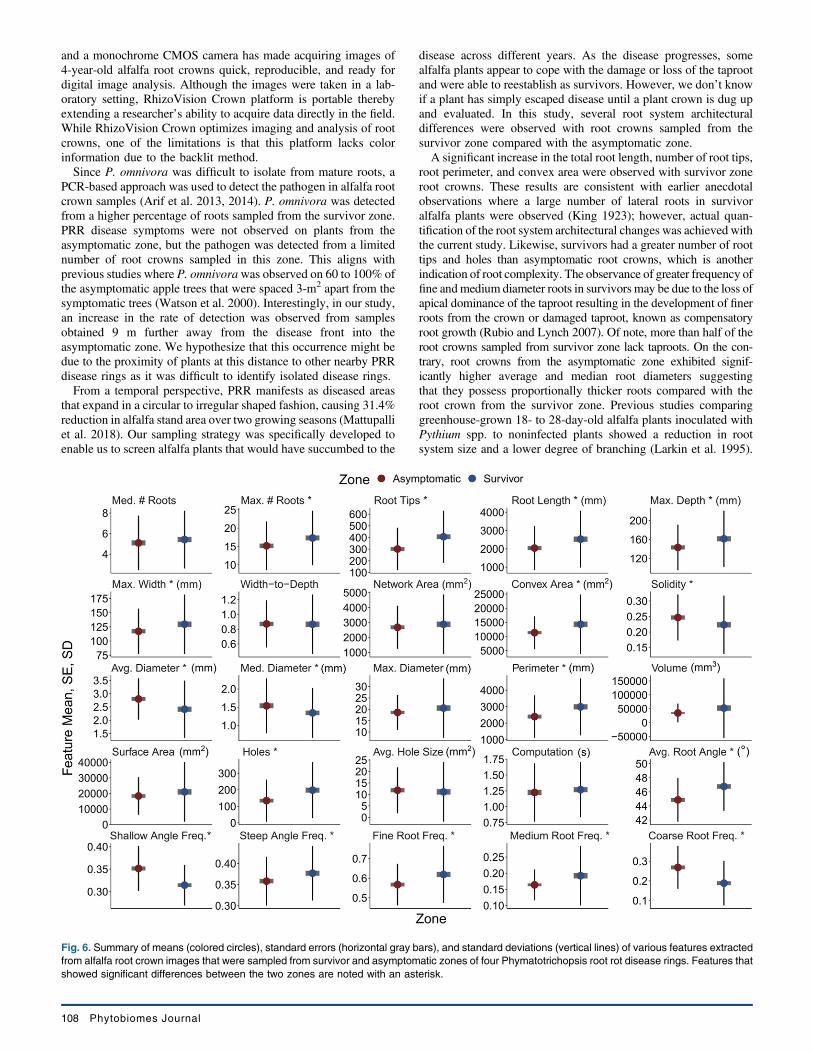
asymptomatic zones are shown in Figure 6. Root crowns sampledfrom the survivor zone (17.3 ± 0.68) had greater numbers of rootscompared with samples from the asymptomatic zone (15.2 ± 0.61).Likewise root crowns from the survivor zone had more total rootlength (2,531.3 ± 142.05 mm), perimeter (2,991.3 ± 157.86 mm),root depth (161.4 ± 5.41 mm), root width (129.7 ± 4.78 mm), androot tips (406.7 ± 20.38) than those from asymptomatic zone(2,043.5 ± 110.33 mm; 2,399.3 ± 117.57 mm; 143.3 ± 4.37 mm;117.2 ± 3.65 mm; 301.9 ± 16.6, respectively). However, there wasno significant difference in root width to depth ratio between rootcrowns from asymptomatic and survivor zones.Network area was not significantly different between root crowns
of asymptomatic and survivor zones (Fig. 6). Convex area wassignificantly greater in root crowns from survivor zone (14,329 ±960.93 mm2) than those from asymptomatic zone (11,353 ±530.41 mm2). Interestingly the solidity feature, which was calcu-lated as ratio of network and convex areas, was significantly greaterfor roots from asymptomatic zone (0.25 ± 0.01) than the rootcrowns sampled from survivor zone (0.22 ± 0.01).There was no difference between maximum diameter of root
crowns from survivor and asymptomatic zones, but the average andmedian root diameter of asymptomatic zone root crowns wassignificantly different (2.8 ± 0.07 mm; 1.54 ± 0.07 mm) fromsurvivor zone roots (2.4 ± 0.1 mm; 1.34 ± 0.06 mm). Total rootvolume, lower root area and surface area were not different betweenthe two zone root crowns. Roots from the asymptomatic zone hadsignificantly higher frequency of coarse roots (0.27 ± 0.01), while
TABLE 1Description of various features extracted from digital images in the study
Features extracteda Description
Median and maximum number of roots The number of roots was counted by performing horizontal line scans from left to rightin each row through the segmented image. In each of the line scans, we check ifthere is a pixel value transition from the previous pixel value to the current pixel valueon its right side.
Number of root tips Tips were defined as the end-point pixels of the skeletonized image.
Total root length Computed by calculating Euclidean distances along the skeletonized image.
Depth, maximum width, and width-to-depth ratio Depth and maximum width of the root in the segmented image. The ratio of maximumwidth to depth of the image was noted as width-to-depth ratio.
Network area, convex area, and solidity Network area is based on the total number of pixels in the segmented image. Theconvex hull of a geometric shape is minimal sized convex polygon that can containthe shape. The ratio of network area and the convex area was noted as the solidity.
Perimeter Perimeter is the count of total number of pixels in the perimeter image (outlining theroots).
Average, median, and maximum diameter For each pixel on the skeletonized image, the distance to the nearest nonroot pixel wascomputed and noted as the radius at that pixel. The list of radii from all the medialaxis pixels was obtained to determine the average and maximum diameter.
Volume and surface area Using the radii determined earlier, the sum of all cross-sectional areas across all themedial axis pixels were noted as volume and the sum of the perimeter across all themedial axis pixels were noted as surface area.
Lower root area The lower root area was the area of the segmented image pixels that were locatedbelow the location of the medial axis pixel that had the maximum radius.
Holes Holes were the disconnected background components and indicative of root branchingand complexity. They can be counted by inverting the segmented image. Theaverage hole size (area) is also calculated.
Fine diameter frequency, medium diameterfrequency, coarse diameter frequency
From the skeletal image, the medial axis pixels were grouped into fine or coarse rootsbased on the radius values at the pixels.
Root angle orientation frequencies Calculated by observing the slope of 20-pixel window around each pixel of theskeletonized image and binning as steep, medium, or shallow.
Computational time The time taken to extract traits for every plant root image.
a Figure 3B shows a representative root crown image from which various root features were extracted.
106 Phytobiomes Journal

higher frequency of fine (0.62 ± 0.01) and medium roots (0.19 ±0.01) roots were observed with roots from the survivor zone. Theaverage root angle was significantly steeper in the survivor zonethan asymptomatic (46.8 ± 0.32 and 44.9 ± 0.28, respectively). Rootcrowns sampled from the asymptomatic zone had significantlyhigher frequency of shallow angled roots (0.35 ± 0.005) comparedwith the survivor zone (0.31 ± 0.004). On the contrary, survivorzone root crowns had higher medium angle (0.31 ± 0.004) and steepangle (0.38 ± 0.006) frequency of roots than asymptomatic zoneroot crowns. The RhizoVision Analyzer software also calculatedthe number of disconnected background components in the image,described as holes, which is an indication of root structure com-plexity. Data suggested that root crowns from the survivor zone hadsignificantly more holes (198.1 ± 15.49) than the roots from theasymptomatic zone (134.7 ± 11.63), but the average hole size didnot differ. Finally, computation time required to extract all thefeatures in the study was not different for the images obtained fromeither asymptomatic (1.22 ± 0.04 s) or survivor (1.27 ± 0.04 s) zones.Pearson’s correlation coefficient was applied to find relationships
between various features extracted from alfalfa root crown imagesand the results are shown in the form of a heatmap (Fig. 7A). Astrong positive correlation was seen between total root length andthe number of holes indicating root crowns with more complexroots systems have longer total root length. As expected, a negativecorrelation was observed between solidity and convex area.In order to further investigate the correlation structure of these
data, principal component analysis was used (Fig. 7B). The first andsecond components explained 37.6 and 17.8% of the multivariatevariation of the root crowns. The first principal component wasloaded most strongly by total root length, network area, number ofroot tips, holes, and numbers of roots, which are indicators of rootsystem size. The second component was loaded most strongly bydiameters and solidity, which can be viewed as indicators of rootsystem exploration efficiency. Root crowns of asymptomatic andsurvivor zones separated more strongly along the second compo-nent, but overall separation was not substantial.Linear discriminant analysis used the multivariate image feature
data to maximize the separation of the survivor and asymptomaticzone classes (Fig. 8). Given two classes, one linear discriminantfunction was returned that provided an overall classification ac-curacy as either survivor or asymptomatic for individual rootcrowns of 75.6%. Because the data were standardized, the loadingsof individual features in the discriminant function can be interpreted
as the relative influence of any given trait on this classificationability. Total root length had the greatest absolute loading with avalue of _8.5, other notable influencing features being surface area(4.0), perimeter (3.5), number of holes (2.3), number of root tips(1.8), and median diameter (1.1), with other features having loadingsof one or less. Negative values indicate that the feature contributedto the asymptomatic class, while positive values indicate the featurecontributed to the survivor class.
DISCUSSION
Several root phenotypes such as crown roots, differential pro-duction of hypocotyl-borne roots and root cortical aerenchyma havebeen identified in beans and maize that enhance greater acquisitionof water and nutrients like nitrogen and phosphorus from soil whilereducing the metabolic costs involved in soil exploration (Lynch2015). However, such studies focused on annual crops and abioticcomponents of the phytobiome. Perennial crops like alfalfa alsohave strategies such as a taproot that can penetrate over 6 m deepinto soil enabling the plant to acquire water and nutrients fromdeeper soil layers, which makes it one of the most drought-tolerantcrops (Undersander et al. 2011). Yet, several root rot pathogens suchas Phytophthora medicaginis and Phoma sclerotioides threaten thehost reliance on the single taproot for long-term survivability(Samac et al. 2015).This study focused on evaluating roots from 4-year-old alfalfa
plants grown in a stand naturally infested with the causal agent ofPRR. PRR disease symptoms were initially noticed at the end offirst growing year, but over time numerous disease foci emerged inthe stand, each year growing larger in size (Mattupalli et al. 2018).One interesting observation was the presence of surviving plantswithin the disease foci. In order to comprehensively documentalfalfa root system architectural changes to PRR, we utilized theRhizoVision Crown platform and the RhizoVision Analyzersoftware. This approach demonstrated the ability to take digitalimages from mature root crowns and extract many root featuresenabling easy quantification of root system architecture.Previous root studies analyzed images of young seedling roots or
annual plant roots that were obtained by scanning in a flatbedscanner followed by using algorithms such as winRHIZO orROOTEDGE (Arias et al. 2013; Himmelbauer et al. 2004). One ofthe key differences between a flatbed scanner and the RhizoVisionCrown platform is that the latter can preserve the integrity of matureroot crowns, which are more complex and often three dimensional.Scanning on a flatbed scanner would be problematic due to theinability of a mature root crown to lay flat on a planar surface.However, the RhizoVision Crown platform with an LED backlight
Fig. 4. Taproot status of alfalfa root crowns sampled from aPhymatotrichopsis root rot-infested field near Burneyville, OK. S, D, and Arepresent samples from symptomatic, disease front, and asymptomaticzones, respectively. Roots were visually examined for their integrity,based on the presence of healthy taproot, necrotic taproot, or missingtaproot. Letters followed by numbers show the distance (in meters) fromdisease front.
Fig. 5. PCR detection of Phymatotrichopsis omnivora from alfalfa rootcrowns sampled from a Phymatotrichopsis root rot-infested field nearBurneyville, OK. S, D, and A represent samples from symptomatic,disease front, and asymptomatic zones, respectively. Letters followed bynumbers show the distance (in meters) from disease front.
Vol. 3, No. 2, 2019 107
and a monochrome CMOS camera has made acquiring images of4-year-old alfalfa root crowns quick, reproducible, and ready fordigital image analysis. Although the images were taken in a lab-oratory setting, RhizoVision Crown platform is portable therebyextending a researcher’s ability to acquire data directly in the field.While RhizoVision Crown optimizes imaging and analysis of rootcrowns, one of the limitations is that this platform lacks colorinformation due to the backlit method.Since P. omnivora was difficult to isolate from mature roots, a
PCR-based approach was used to detect the pathogen in alfalfa rootcrown samples (Arif et al. 2013, 2014). P. omnivora was detectedfrom a higher percentage of roots sampled from the survivor zone.PRR disease symptoms were not observed on plants from theasymptomatic zone, but the pathogen was detected from a limitednumber of root crowns sampled in this zone. This aligns withprevious studies where P. omnivorawas observed on 60 to 100% ofthe asymptomatic apple trees that were spaced 3-m2 apart from thesymptomatic trees (Watson et al. 2000). Interestingly, in our study,an increase in the rate of detection was observed from samplesobtained 9 m further away from the disease front into theasymptomatic zone. We hypothesize that this occurrence might bedue to the proximity of plants at this distance to other nearby PRRdisease rings as it was difficult to identify isolated disease rings.From a temporal perspective, PRR manifests as diseased areas
that expand in a circular to irregular shaped fashion, causing 31.4%reduction in alfalfa stand area over two growing seasons (Mattupalliet al. 2018). Our sampling strategy was specifically developed toenable us to screen alfalfa plants that would have succumbed to the
disease across different years. As the disease progresses, somealfalfa plants appear to cope with the damage or loss of the taprootand were able to reestablish as survivors. However, we don’t knowif a plant has simply escaped disease until a plant crown is dug upand evaluated. In this study, several root system architecturaldifferences were observed with root crowns sampled from thesurvivor zone compared with the asymptomatic zone.A significant increase in the total root length, number of root tips,
root perimeter, and convex area were observed with survivor zoneroot crowns. These results are consistent with earlier anecdotalobservations where a large number of lateral roots in survivoralfalfa plants were observed (King 1923); however, actual quan-tification of the root system architectural changes was achieved withthe current study. Likewise, survivors had a greater number of roottips and holes than asymptomatic root crowns, which is anotherindication of root complexity. The observance of greater frequency offine andmedium diameter roots in survivors may be due to the loss ofapical dominance of the taproot resulting in the development of finerroots from the crown or damaged taproot, known as compensatoryroot growth (Rubio and Lynch 2007). Of note, more than half of theroot crowns sampled from survivor zone lack taproots. On the con-trary, root crowns from the asymptomatic zone exhibited signif-icantly higher average and median root diameters suggestingthat they possess proportionally thicker roots compared with theroot crown from the survivor zone. Previous studies comparinggreenhouse-grown 18- to 28-day-old alfalfa plants inoculated withPythium spp. to noninfected plants showed a reduction in rootsystem size and a lower degree of branching (Larkin et al. 1995).
Fig. 6. Summary of means (colored circles), standard errors (horizontal gray bars), and standard deviations (vertical lines) of various features extractedfrom alfalfa root crown images that were sampled from survivor and asymptomatic zones of four Phymatotrichopsis root rot disease rings. Features thatshowed significant differences between the two zones are noted with an asterisk.
108 Phytobiomes Journal

However, such studies focused on recently inoculated plants unlikethe current study, which involved 4-year-old field-grown alfalfaplants that have sustained PRR disease stress. Another plausibilityfor the development of a complex root system is the accessibility ofmore soil resources to the survivors owing to sparse stand in thesurvivor zone from loss of plants due to PRR. Similar changes in
root features such as increased lateral root number and greatertaproot diameter have been observed with greater spacing betweenalfalfa plants (Lamb et al. 2000). Although survivors had a complexroot system compared with asymptomatic plants, this did not affectthe computation time required to extract the suite of features fromthe digital images.
Fig. 8. Linear discriminant analysis indicating differences between multivariate features extracted from alfalfa root crown images obtained fromasymptomatic and survivor zones of four Phymatotrichopsis root rot disease rings. Representative images highlighting these differences are shown withextracted features overlaid on the original segmented root crown for asymptomatic (left) and survivor (right) plants.
Fig. 7. Relationship between various root features extracted using RhizoVision Analyzer software. A, Pearson’s correlation heatmap and B, principalcomponent analysis of root crown features.
Vol. 3, No. 2, 2019 109
Roots by the nature of being in the ground are difficult to studywithout using destructive sampling techniques. We utilized theRhizoVision Crown phenotyping platform and showed that alfalfaplants surviving from PRR disease stress have root systems ac-climated to damage caused by the disease. Despite the loss ordamage to the taproot, survivors overcame this stress by developingadditional crown and lateral roots contributing to a root system thatwas more complex than the asymptomatic roots, indicating aninteresting interaction between soil microbes and root system ar-chitecture. Future research will need to address whether the ac-climation of the diseased plant by increasing carbon allocation tocrown and lateral roots is truly adaptive, and if so, whether theability to acclimate by compensatory root growth could be abreeding target for increasing disease resistance. The methods usedin this study could be applied to roots of crop plants undergoingother biotic and abiotic stresses or symbiotic interactions associatedwith the phytobiome.The concept of low-input agriculture and the spatiotemporal
dissimilarities in the distribution of water and nutrient sources in thesoil have led to the identification of traits that have reduced met-abolic cost for soil exploration (Lynch 2015; Lynch et al. 2014).The alfalfa plants from the survivor zone showing alternate rootsystem architecture under PRR disease stress presents a uniqueopportunity to further explore if this mode of survival has improvedresource efficiency that would compensate for the loss of taprootand additional costs incurred by the plant while generating lateraland crown roots. Currently very limited options are available tomanage PRR disease with no known PRR resistant alfalfa varieties.Looking forward, it remains an open question whether the strategyof developing a different root system architecture put forth bysurvivors is a heritable trait that could be used in resistance breed-ing and if these plants will be as persistent under other biotic andabiotic stress conditions.Data availability. Root crown images and R statistical analysis
code generated from this study are available on Zenodo (York et al.2018; https://doi.org/10.5281/zenodo.2172832).
LITERATURE CITED
Arias, M. M. D., Leandro, L. F., and Munkvold, G. P. 2013. Aggressiveness ofFusarium species and impact of root infection on growth and yield ofsoybeans. Phytopathology 103:822-832.
Arif, M., Dobhal, S., Garrido, P. A., Orquera, G. K., Espındola, A. S., Young,C. A., Ochoa-Corona, F. M., Marek, S. M., and Garzon, C. D. 2014. Highlysensitive end-point PCR and SYBR green qPCR detection ofPhymatotrichopsis omnivora, causal fungus of cotton root rot. Plant Dis. 98:1205-1212.
Arif, M., Fletcher, J., Marek, S. M.,Melcher, U., and Ochoa-Corona, F. M. 2013.Development of a rapid, sensitive and field deployable Razor ExBioDetection System and qPCR assay for detection of Phymatotrichopsisomnivora using multiple gene targets. Appl. Environ. Microbiol. 79:2312-2320.
Bucksch, A., Burridge, J., York, L. M., Das, A., Nord, E., Weitz, J. S., andLynch, J. P. 2014. Image-based high-throughput field phenotyping of croproots. Plant Physiol. 166:470-486.
Hancock, J. G. 1991. Seedling and rootlet diseases of forage alfalfa caused byPythium irregulare. Plant Dis. 75:691-694.
Harrington, J. T., Mexal, J. G., and Fisher, J. T. 1994. Volume displacementprovides a quick and accurate way to quantify new root production. TreePlanters’ Notes 45:121-124.
Himmelbauer, M. L., Loiskandl, W., and Kastanek, F. 2004. Estimating length,average diameter and surface area of roots using two different image analysessystems. Plant Soil 260:111-120.
King, C. J. 1923. Habits of the cotton root rot fungus. J. Agric. Res. 26:405-418.Lamb, J. F. S., Johnson, L. D., Barnes, D. K., and Marquez-Ortiz, J. J. 2000. A
method to characterize root morphology traits in alfalfa. Can. J. Plant Sci. 80:97-104.
Larkin, R. P., English, J. T., and Mihail, J. D. 1995. Effects of infection byPythium spp. on root systemmorphology of alfalfa seedlings. Phytopathology85:430-435.
Luc, J. E., Crow, W. T., Stimac, J. L., Sartain, J. B., and Giblin-Davis, R. M.2006. Influence of Belonolaimus longicaudatus on nitrate leaching in turf.J. Nematol. 38:461-465.
Lynch, J. 1995. Root architecture and plant productivity. Plant Physiol. 109:7-13.
Lynch, J. P. 2015. Root phenes that reduce the metabolic costs of soilexploration: Opportunities for 21st century agriculture. Plant Cell Environ.38:1775-1784.
Lynch, J. P., Chimungu, J. G., and Brown, K. M. 2014. Root anatomical phenesassociated with water acquisition from drying soil: Targets for cropimprovement. J. Exp. Bot. 65:6155-6166.
Ma, J., Jaraba, J., Kirkpatrick, T. L., and Rothrock, C. S. 2014. Effects ofMeloidogyne incognita and Thielaviopsis basicola on cotton growth and rootmorphology. Phytopathology 104:507-512.
Mattupalli, C., Komp, M. R., and Young, C. A. 2017. Integrating geospatialtechnologies and unmanned aircraft systems into the grower’s diseasemanagement toolbox. APS Features. doi.org/10.1094/APSFeature-2017-7
Mattupalli, C., Moffet, C., Shah, K., and Young, C. 2018. Supervisedclassification of RGB aerial imagery to evaluate the impact of a root rotdisease. Remote Sens. 10:917.
Ortiz-Ribbing, L. M., and Eastburn, D. M. 2004. Soybean root systems andsudden death syndrome severity: Taproot and lateral root infection. Plant Dis.88:1011-1016.
Paez-Garcia, A., Motes, C., Scheible, W.-R., Chen, R., Blancaflor, E., andMonteros, M. 2015. Root traits and phenotyping strategies for plantimprovement. Plants 4:334-355.
Pang, W., Hafez, S. L., and Sundararaj, P. 2009. Pathogenicity of Meloidogynehapla on onion. Nematropica 39:225-233.
R Core Team. 2018. R: A language and environment for statistical computing.R Foundation for Statistical Computing, Vienna, Austria. https://www.R-project.org
Rubio, G., and Lynch, J. P. 2007. Compensation among root classes inPhaseolus vulgaris L. Plant Soil 290:307-321.
Saengwilai, P., Tian, X., and Lynch, J. P. 2014. Low crown root numberenhances nitrogen acquisition from low nitrogen soils in maize (Zea mays L.).Plant Physiol. 166:581-589.
Samac, D. A., Rhodes, L. H., and Lamp, W. O. 2015. Compendium of AlfalfaDiseases and Pests. American Phytopathological Society, St. Paul,Minnesota.
Seethepalli, A., Guo, H., Liu, X., Griffiths, M., Almtarfi, H., Li, Z., Liu, S., Zare,A., Fritschi, F., Blancaflor, E., Ma, X., and York, L. M. 2019. RhizoVisionCrown: An integrated hardware and software platform for root crownphenotyping. bioRxiv 569707. https://doi.org/10.1101/569707
Seethepalli, A., and York, L. 2019a. RhizoVision Analyzer: Software for high-throughput measurements from images of crop root crowns (Version 1.0).Zenodo. http://doi.org/10.5281/zenodo.2585892
Seethepalli, A., and York, L. 2019b. RhizoVision Imager: Software to controlmachine vision cameras for plant phenotyping (Version 1.0). Zenodo. http://doi.org/10.5281/zenodo.2585882
Streets, R. B., and Bloss, H. E. 1973. Phymatotrichum Root Rot, VolumeMonograph No. 8. American Phytopathological Society, St. Paul, MN.
Trachsel, S., Kaeppler, S. M., Brown, K. M., and Lynch, J. P. 2011.Shovelomics: High throughput phenotyping of maize (Zea mays L.) rootarchitecture in the field. Plant Soil 341:75-87.
Trachsel, S., Kaeppler, S. M., Brown, K. M., and Lynch, J. P. 2013. Maize rootgrowth angles become steeper under low N conditions. Field Crops Res. 140:18-31.
Undersander, D. J., Cosgrove, D. R., Cullen, E. M., Grau, C. R., Rice, M. E.,Renz, M. J., Sheaffer, C. C., Shewmaker, G. E., and Sulc, M. R. 2011. AlfalfaManagement Guide. American Society of Agronomy, Crop Science Societyof America, Soil Science Society of America, Madison, WI.
Uppalapati, S. R., Young, C. A., Marek, S. M., and Mysore, K. S. 2010.Phymatotrichum (cotton) root rot caused by Phymatotrichopsis omnivora:Retrospects and prospects. Mol. Plant Pathol. 11:325-334.
Venables, W. N., and Ripley, B. D. 2002. Modern applied statistics with S, 4thEd. Springer-Verlag, New York. 10.1007/978-0-387-21706-2
Watson, W. T., Kenerley, C. M., and Appel, D. N. 2000. Visual and infraredassessment of root colonization of apple trees by Phymatotrichopsisomnivora. Plant Dis. 84:539-543.
Wickham, H. 2016. ggplot2: Elegant Graphics for Data Analysis. Springer, NewYork.
110 Phytobiomes Journal

Wickham, H., Francois, R., Henry, L., andMuller, K. 2018. dplyr: A grammar ofdata manipulation. R package version 0.7.6. https://CRAN.R-project.org/package=dplyr
York, L. M. 2018. Phenotyping crop root crowns: General guidance and specificprotocols for maize, wheat, and soybean. Pages 23-32 in: Root Development:Methods and Protocols. D. Ristova and E. Barbez, eds. Humana Press, NewYork. 10.1007/978-1-4939-7747-5_2
York, L. M., Young, C. A., Mattupalli, C., and Seethepalli, A. 2018. Images andstatistical analysis of alfalfa root crowns from inside and outside disease rings
caused by cotton root rot (Version 1.0.0) [Data set]. Zenodo. https://doi.org/10.5281/zenodo.2172832
York, L. M., and Lynch, J. P. 2015. Intensive field phenotyping of maize (Zeamays L.) root crowns identifies phenes and phene integration associated withplant growth and nitrogen acquisition. J. Exp. Bot. 66:5493-5505.
Young, C. A., Uppalapati, S. R., Mysore, K. S., and Marek, S. M. 2015.Phymatotrichopsis root rot. Pages 44-46 in: Compendium of Alfalfa Diseasesand Pests. D. A. Samac, L. H. Rhodes, and W. O. Lamp, eds. AmericanPhytopathological Society, St. Paul, MN.
Vol. 3, No. 2, 2019 111