digital pathology 101 · digital pathology 101 workshop this workshop will provide participants...
TRANSCRIPT

Digital Pathology
101

@dpatweet #PV14
Digital Pathology 101 Workshop
This workshop will provide participants with a
broad introductory overview of the field of digital
pathology (DP), including:
Applications, benefits and limitations of DP
DP technology and financial considerations (primarily whole-slide imaging)
Interoperability, regulatory, workflow
Hands-on session focused on basic digital slide viewing using a variety of digital slides

@dpatweet #PV14
Speakers
Lewis Hassell, MD
Associated Professor of Pathology,
University of Oklahoma
William DeSalvo, BS,
HTL(ASCP)
System Production Manager,
Sonora Quest Laboratories
Elizabeth Chlipala,
BS, HTL(ASCP)QIHC Partner, Premier Laboratory, LLC

@dpatweet #PV14
Applications & Benefits of WSI
Lewis A. Hassell, MD
Associate Professor of Pathology
University of Oklahoma

@dpatweet #PV14
So does DP scratch an itch we have?

@dpatweet #PV14
DATA MANAGEMENT CHALLENGES
DEMOGRAPHIC CHALLENGES Aging pathologists community Geographic shortages Increase in sub-specialty expertise Increase in biopsies (aging population,
less invasive surgical procedures) increase in tests, driven by new CDx increase in patient advocacy
Pathologist’s primary unmet need is improving the quality and efficiency of pathology services
LOGISTICAL CHALLENGES

@dpatweet #PV14
Digital Pathology – Definition & Historical Context
Digital Pathology is an environment for the management and interpretation of pathology
information that is enabled by the digitization of a glass slide
Today, WSI is considered the primary means of DP image capture; other approaches include: Camera on a microscope (single field-of-view)
Robotic microscopy (remote control of microscope stage, objective lens turret and focus; single field-of-view)
Whole slide imaging (capture/assemble multiple fields-of-view to create an image corresponding to an entire glass slide)

@dpatweet #PV14
Overview of a Digital Pathology system
Remote Viewing Image Analysis Data Management
Hardware & Software Capabilities
Image Capture
Image Management
Image Analysis
Image Viewing
Image Storage
Interface to LIS, EHR …
All applications of digital pathology utilize some (all) of the primary three functions of a DP systems; which are enabled by specific hardware and software capabilities.
Successful implementation requires user training, validation, IT integration, etc…
Primary Functionality

@dpatweet #PV14
So what’s it really good for?
Will it end up as a Door-stop at DCPA?

@dpatweet #PV14
Applications-
Undergrad Pathology Education
• Upwards of 60% of US medical schools use Digital Slides exclusively
• Adoption in Dental Schools, Veterinary Schools and other fields is also significant
• Student response– increased time spent studying slides, lowers barriers to access and results in better or no change in performance on examination
• Could it still be made more effective and engaging? YES!

@dpatweet #PV14

@dpatweet #PV14
Resident Education
• ACGME Competencies
• Atlas for case review
• Remote Conferencing
• Skill building, core and advanced
• Standardization
• Progress assessments
• Board Review
• User Interface Critical!

@dpatweet #PV14
• Digital slides are now almost ubiquitous in
pathology meetings covering anatomical
pathology
• Transform the ability to create enduring
educational materials
• Engage interaction beyond the meeting
room- On-going virtual meeting
• Are they used as much, more than, or less
than glass slides from prior slide seminars?
Postgraduate Education

@dpatweet #PV14

@dpatweet #PV14
Proficiency Testing and Qualifying Exams
• CAP Proficiency Testing DigitalScope™
• American Board of Pathology AP exams:
– Mexican Qualifying exam:
– European exam still uses glass
• Competency assessments, pre-employment, or
with new modalities, e.g. WSI FS or Validating
IHC interpretation

@dpatweet #PV14
Self-Directed Learning
• Board Review http://pathinfo.wikia.com/wiki/Pathology_Links
• Slide Sharing services- “Just in Time” learning and consultation
• Journals employing WSI
• Textbooks and on-line atlases https://digitalpathologyassociation.org/whole-slide-imaging-repository

@dpatweet #PV14

@dpatweet #PV14
Optimized Archival Management
• Loss avoidance (referral of unique slides)
• Maintenance for teaching and patient care
after consultation
• More rapid retrieval for comparison with
current materials (enhanced patient care)
• Simultaneous consultations
• Patient-retained records (24andMyslides?)
• Reduce tissue consumption (recuts)

@dpatweet #PV14
Image Analysis
@dpatweet #PV14
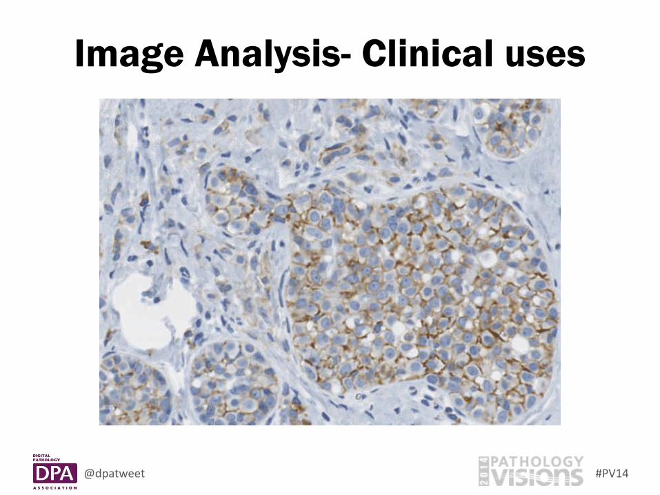
Image Analysis- Clinical uses

@dpatweet #PV14
Advanced Image Analysis
Webster et al. Investigation into diagnostic agreement using automated computer-assisted histopathology pattern recognition image analysis, J Pathol Inform 2012; 3:18.

@dpatweet #PV14
Advanced Image Analysis
• Computer-Assisted Diagnosis
• Morphologic feature based searches
• Spectral Partition and automated
quantitation, reconstructions, manipulation
• Automated ROI selections and harvest by
laser-capture
• More, and more to come

@dpatweet #PV14
Consultation
• “Local”– colleague within the same practice,
but at a different site
-Real time QA
-Specialist practices
-Intra-op
• Remote consultants– Dr. Gno Itall
• Consensus Panels
• Study or Trial enrollment

@dpatweet #PV14
International
Consultation Service
Internet/Cloud/Server
Referring pathologist
Slide Scanner and Server
Consultant Pathologist A
Consultant Pathologist B

@dpatweet #PV14
Consultation-
Integration of Multiple Locations

@dpatweet #PV14
“Consultation”
Technical Services
• Specialized, slide-based service performed
at off-site lab, then slide is digitized and
access opened to provider
• Professional, interpretive services performed
locally

@dpatweet #PV14 ER Protein Controls over time
Quality Control

@dpatweet #PV14
Quality Assurance
• Optimize current QA
reviews; shorten TAT
• JIT consultation
• Optimize section and
stain quality
• Lower barriers to
review of prior slides
• Tighten kappa value
on problematic areas

@dpatweet #PV14
Business Uses
• Archival storage for faster retrieval for case
comparison, reduced clerical costs
• Expedited consultation at lower cost
• Marketing “value-add” for clients and patients
• Archiving unique or limited availability materials
(e.g. medical-legal materials, in-coming
consults)
Business Uses

@dpatweet #PV14
Limitations of WSI-
A short list of problems to be solved
• Large files take added time to load and
manipulate over slower networks,
• Z-stack and high magnification challenges
make use in cytology and hematology less
efficient and somewhat problematic
• Retention of WSI over time will require
abundant storage space
• Point-of-care microscopy

@dpatweet #PV14
Other Limitations
Polarized light
Condenser adjustment to enhance refraction
@dpatweet #PV14
Benefits

@dpatweet #PV14
Benefits
• Students– learn anywhere, anytime, and
anything
• Quality of diagnostic opinion
• Speed of consultation opinion
• Value-adds – what you couldn’t do w/glass
-Patient and MD education
-Advanced image analysis
-Archiving and transport

@dpatweet #PV14
Digital Pathology will enable pathologists to play an increasingly important role in the future of patient care
Slide
scanning
Tumor
identification
Pathologist Micro-dissection
Tumor
tissue
Laser Capture

@dpatweet #PV14
DP is a vibrant market that continues to attract new entrants, including large companies

@dpatweet #PV14
Adoption of digital pathology is well under way for specific uses; installed base > 1,500 systems
Application
EducationTeaching and self-education
Proficiency testing
ResearchBiomarker discovery (image analysis)
General research
Clinical
Consultations (IOCs, formal/informal)
Quality Assurance
Tumor boards
Archival & Retrieval (prior cases, risk management)
Decision Support
IHC Quantification – FDA clearance
Primary diagnosis – FDA approval (full adoption)
Most common clinical applications are tumor boards, consultations, archival/retrieval and IHC quantification
60% are using DP for clinical use, 80% for education & research
Most clinical users have adopted DP for 3 specific uses; strong demand to add more
85% surgical pathology, 15% cytology/ hematology
@dpatweet #PV14
Engraftment vs. Rejection
• Host organism– climate and culture
• Population dynamics
• External defenses– Regulation
• Validation protocols– dosage
• Needs basis- What is the context?
-Clinical
-Educational
-Quality
-Efficiency

@dpatweet #PV14
Straub E T REVIEW OF EDUCATIONAL RESEARCH
2009;79:625-649 Copyright © by American Educational Research Association

@dpatweet #PV14
Concerns-Based
Adoption Model
• Change is a process, not an event.
• Change is accomplished by individuals.
• Change is a highly personal experience.
• Change involves developmental growth.
• Change is best understood in operational
terms.
• The focus of facilitation should be on
individuals, innovations, and context

@dpatweet #PV14
Technology and Financial
Considerations
William DeSalvo, BS, HTL(ASCP)
System Production Manager,
Sonora Quest Laboratories

@dpatweet #PV14
Technologies of Digital Pathology
• Scanners
• IT Infrastructure
• Viewers
• Image Management Software

@dpatweet #PV14
Whole Slide Scanners - Overview
Creates whole slide images from slides.
Key characteristics:
- Speed
- Image Quality
- Slide Loading Mechanisms
- Scan Types
- Operator Time Required
No device is best for all use cases or
purposes

@dpatweet #PV14
Scan Time = Time/Slide (ex: 60 seconds)
Throughput = Slides/Time (ex: 30 slides/hr)
1 / Scan Time ≠ Throughput !!!
Include load / unload time, selection of scan areas, re-
scan rate, slide movement time, and parallel operations.
Whole Slide Scanners Considerations - Speed
Auto assembly plant example…
~24 hours to build each car ~50 cars built per hour
@dpatweet #PV14
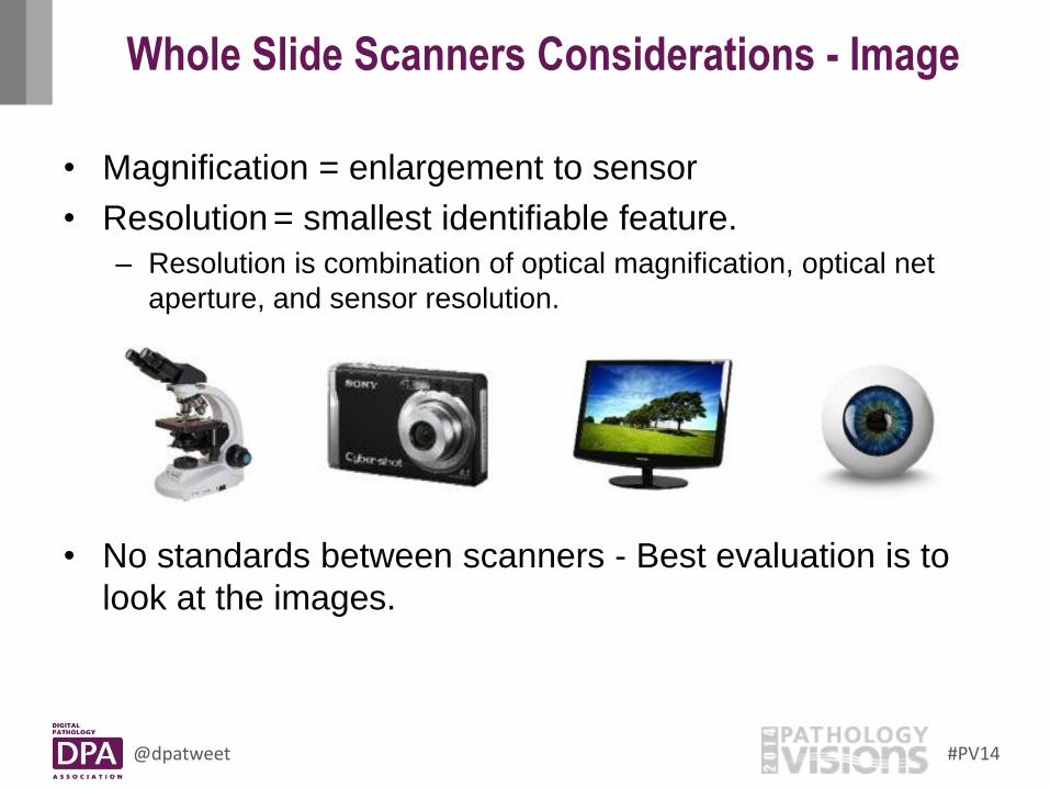
• Magnification = enlargement to sensor
• Resolution = smallest identifiable feature.
– Resolution is combination of optical magnification, optical net
aperture, and sensor resolution.
• No standards between scanners - Best evaluation is to
look at the images.
Whole Slide Scanners Considerations - Image

@dpatweet #PV14
Whole Slide Scanners - Magnification & Resolution
With a microscope, your eyeball is the sensor.
With a digital pathology,
your eyeball views a projection on a screen.
Computers can enlarge
view of an image to any
size.
Image resolution
determines crispness of
image as it is enlarged.

@dpatweet #PV14
Whole Slide Scanners - Magnification & Resolution
Both images captured with 20X magnification objective.
10 microns / pixel on sensor
0.05 microns / pixel optical resolution
5.5 micron / pixel on sensor
0.0275 microns / pixel optical resolution
Apparent magnification at full resolution is
1.7X larger.

@dpatweet #PV14
• Total Capacity = maximum unattended operation
• Cartridge Size = # of slides per load unit
• Continuous = ability to load w/o interruption
• Override = ability to immediately scan STAT
• Best mechanism varies by use case
Considerations include staffing schedules, turnaround
time variations, batch sizes, etc.
Whole Slide Scanners Considerations –
Loading Mechanisms
1 jumbo versus 5 regional jets
@dpatweet #PV14
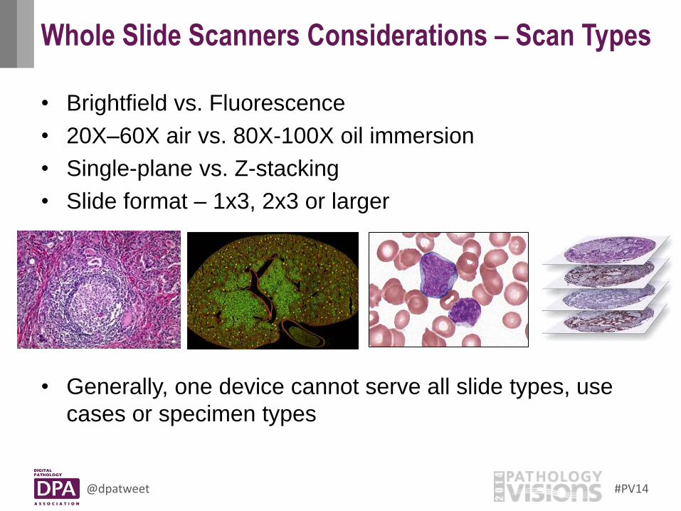
• Brightfield vs. Fluorescence
• 20X–60X air vs. 80X-100X oil immersion
• Single-plane vs. Z-stacking
• Slide format – 1x3, 2x3 or larger
• Generally, one device cannot serve all slide types, use
cases or specimen types
Whole Slide Scanners Considerations – Scan Types

@dpatweet #PV14
Whole Slide Scanners Overview
Provide performance, reliability, and support.
Key components: – Network
– Storage
– Systems Management
– Integration
Determine scope early to identify stakeholders
to include in planning:
- Departmental Solution vs. Enterprise Solution

@dpatweet #PV14
• Latency = time for roundtrip (ms)
• Bandwidth = amount of data/time (GB/sec)
• Requirements differ for storing images (moving the
whole image file) versus viewing images (streaming on
demand).
IT Infrastructure Considerations - Network
Between point A and B… How much time for 1 car to travel? How many cars per hour travel?

@dpatweet #PV14
• File Size = size per image
– File sizes will vary by lab based on mix of image compression
format, scan resolutions, and average tissue per slide.
• Volume = images/year
• Retention = time to keep images
• Incorporate requirements such as backups, availability,
and legal requirements/mitigation.
IT Infrastructure Considerations - Storage

@dpatweet #PV14
• Data = eliminating redundant entry
• Workflow = experience working across apps
• Storage = use common enterprise platform
• Imaging = sharing between diagnosticians
– Common Systems: APLIS, Barcoding, PACS
NOTE: Check minimum versions and platform for compatibility
– Common Standards: HL7, IHE, DICOM
• Identify how the integration will facilitate the user
workflow before diving into specifications.
IT Infrastructure Considerations - Integration
@dpatweet #PV14
• Backup = protection against data loss
• Availability = required uptime level
• Disaster Recovery = continuity after catastrophe
• Higher levels of reliability incur additional costs that
must be weighed against impact to business.
IT Infrastructure Considerations - Reliability

@dpatweet #PV14
• Meet local privacy laws
• Fulfill organizations policies
• Most organizations have defined requirements or audit
questionnaires for all new IT solutions.
IT Infrastructure Considerations – Security & Privacy
@dpatweet #PV14
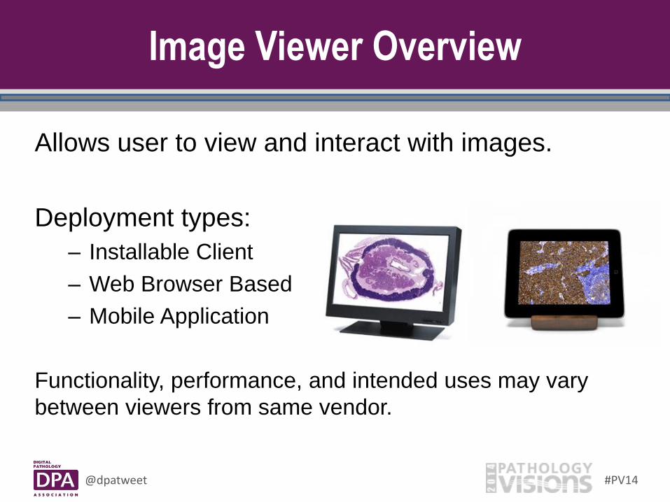
Image Viewer Overview
Allows user to view and interact with images.
Deployment types:
– Installable Client
– Web Browser Based
– Mobile Application
Functionality, performance, and intended uses may vary
between viewers from same vendor.
Image Viewer Overview

@dpatweet #PV14
Image Management Software Overview
Image Management Software - Overview
Organize images to support the use case.
Providers:
Digital Pathology Scanner Manufacturers
Digital Pathology Software-only Providers
Lab Information Systems (LIS)
In-house Developed Solutions
Evaluate solutions against your use case(s).

@dpatweet #PV14
Financial Considerations

@dpatweet #PV14
Total Cost = Primary Costs + Other Costs
Primary Costs
Scanning instruments – range of choices
Software licenses – varies based on use
Implementation and training – varies by complexity and integration
Other Costs
Maintenance & Support HW & SW -- break fix, routine service, and upgrades
Post-Implementation Training Critical to continue to benefit from the technology post-implementation
Storage / IT Highly dependent on storage strategy and business service levels
Labor Scanning, image Q&A, ongoing IT monitoring and support
Costs associated with implementing a digital pathology systems

@dpatweet #PV14
A faster scanner is not necessarily more cost-effective e.g., 2x faster and 3x more expensive is not more cost-effective Scanning prep and re-scan rates have labor costs
Factors to include in cost/slide include Labor costs to load/unload slides Labor costs to set scanning parameters Labor costs to Q/A digital slides Reliability / up-time (a system that’s “down” is not adding value) Ease of use (a system that’s easier to use requires less user time) Storage (a more highly compressed image file requires less
storage) Viewing performance (a “sluggish” viewer requires more time)
Consider cost/slide as a potential metric for assessing a DP system

@dpatweet #PV14
A variety of purchasing models exist to acquire solutions:
Outright purchase high upfront payment with ongoing maintenance
Lease fixed monthly payments for period of time
Usage-based ex: pay per slide or case
Considerations to balance:
Short-term expenses versus total long-term costs
Availability of budget initially versus ongoing
Predictability of use
Acquisition Models
@dpatweet #PV14
Some applications have strong ROIs for pathology department -- reduce travel time, reduce admin time, reimbursements
digital IHC frozen sections archive and retrieval
Others have a strong ROI for the institution but not for the pathology
department -- improved quality, clinician/patient satisfaction, competitiveness
tumor boards consultations quality assurance
ROI for using DP clinically requires a “big picture” perspective beyond
the benefits only to the pathology department
Return-on-Investment (ROI)

@dpatweet #PV14
Regulatory & Compliance and Workflow
Considerations
Elizabeth Chlipala, BS,HTL(ASCP)QIHC
Partner, Premier Laboratory, LLC
@dpatweet #PV14
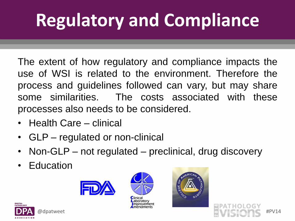
Regulatory and Compliance
The extent of how regulatory and compliance impacts the
use of WSI is related to the environment. Therefore the
process and guidelines followed can vary, but may share
some similarities. The costs associated with these
processes also needs to be considered.
• Health Care – clinical
• GLP – regulated or non-clinical
• Non-GLP – not regulated – preclinical, drug discovery
• Education

@dpatweet #PV14
Follow FDA Guidelines and Industry Standards:
Federal Register - 21 CFR part 58, 21 CFR part 11
• Validation of equipment – IQ, OQ
IQ - Installation Qualification
OQ – Operational Qualification
• Validation of process – PQ or UAT
Process Qualification
User Acceptance Testing
The extent, how and what is validated will be dependent upon Quality Management
• Dependent on the complexity and criticality of the instrument or process
• Governed by your SOP’s
Regulated - GLP

@dpatweet #PV14
1. DPA White Paper and STP Position Paper – Validation of Digital Pathology Systems in the Regulated Nonclinical Environment
http://digitalpathologyassociation.org/_data/files/DPA_White_Paper_Final_-_2011-11-17.pdf
http://tpx.sagepub.com/content/41/1/115.full.pdf+html
2. Society of Toxicologic Pathology Position Paper on Pathology Image Data: Compliance with 21 CFR Parts 58 and 11
http://tpx.sagepub.com/content/35/3/450.full
3. Federal Register – 21 CFR part 58 and 21 CFR part 11
4. ISPE Baseline Pharmaceutical Engineering Guide for Commissioning and Qualification, Volume 5, First Edition/March 2001
GLP – Resources

@dpatweet #PV14
Validate for specific application – the entire process or the outcome of that process is validated
• Secondary consultations
• Frozen sections
• Image Analysis - ER/PR, Her2, Her2 FISH
– 510K cleared kits, algorithms – still must validate in house
– validate staining, validate algorithm
• May be moving towards IQ/OQ/PQ
Health Care – Clinical

@dpatweet #PV14
• Develop a team of individuals who will work on this process, needs to include all areas of the lab that are impacted by the whole slide scanning process and digital read or image analysis
• Determine a plan of action
– Define the process - work flow documents, forms, etc.
– Define the individuals involved in each step of the process
• Write a validation plan or protocol
• Review that plan or protocol
• Execute that plan or protocol
• Compile results and write a report
– In research we write a technical report
67
Follow the CAP guidelines for
validation in clinical settings

@dpatweet #PV14
1. Archives of Pathology and Laboratory Medicine. Validating Whole Slide Imaging for Diagnostic Purposes in Pathology.
http://www.archivesofpathology.org/doi/pdf/10.5858/arpa.2013-0093-CP
2. DPA White Paper.
https://digitalpathologyassociation.org/_data/files/DPA-Healthcare-White-Paper--FINAL_v1.0.pdf
3. CAP/ASCO Guidelines for ER/PR and Her2 http://www.archivesofpathology.org/doi/pdf/10.5858/arpa.2013-0953-SA
4. Archive of Pathology and Laboratory Medicine. Validation of Whole Slide Imaging for Primary Diagnosis in Surgical Pathology http://www.archivesofpathology.org/doi/pdf/10.5858/arpa.2011-0678-OA
Health Care - Resources
@dpatweet #PV14
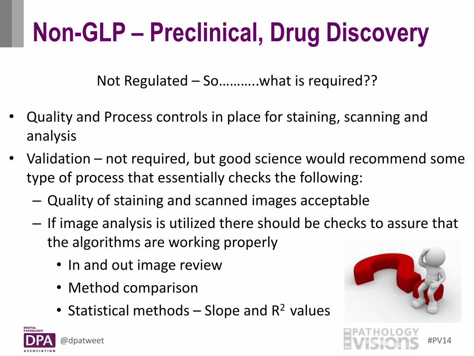
Not Regulated – So………..what is required??
• Quality and Process controls in place for staining, scanning and analysis
• Validation – not required, but good science would recommend some type of process that essentially checks the following:
– Quality of staining and scanned images acceptable
– If image analysis is utilized there should be checks to assure that the algorithms are working properly
• In and out image review
• Method comparison
• Statistical methods – Slope and R2 values
Non-GLP – Preclinical, Drug Discovery

@dpatweet #PV14
Not Regulated
• Student interaction positive
• Management and access to images
Education
@dpatweet #PV14
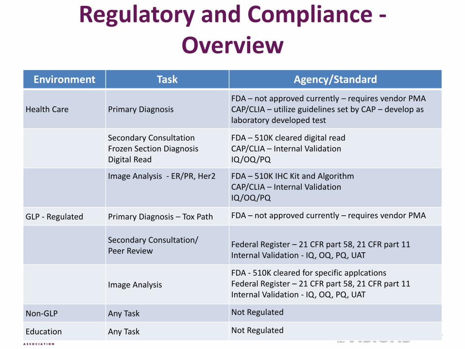
Regulatory and Compliance - Overview
Environment Task Agency/Standard
Health Care Primary Diagnosis FDA – not approved currently – requires vendor PMA CAP/CLIA – utilize guidelines set by CAP – develop as laboratory developed test
Secondary Consultation Frozen Section Diagnosis Digital Read
FDA – 510K cleared digital read CAP/CLIA – Internal Validation IQ/OQ/PQ
Image Analysis - ER/PR, Her2 FDA – 510K IHC Kit and Algorithm CAP/CLIA – Internal Validation IQ/OQ/PQ
GLP - Regulated Primary Diagnosis – Tox Path FDA – not approved currently – requires vendor PMA
Secondary Consultation/ Peer Review
Federal Register – 21 CFR part 58, 21 CFR part 11 Internal Validation - IQ, OQ, PQ, UAT
Image Analysis
FDA - 510K cleared for specific applcations Federal Register – 21 CFR part 58, 21 CFR part 11 Internal Validation - IQ, OQ, PQ, UAT
Non-GLP Any Task Not Regulated
Education Any Task Not Regulated
@dpatweet #PV14
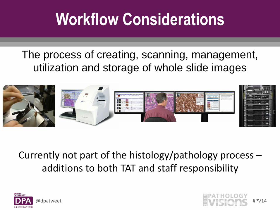
Workflow Considerations
The process of creating, scanning, management,
utilization and storage of whole slide images
Currently not part of the histology/pathology process – additions to both TAT and staff responsibility
@dpatweet #PV14
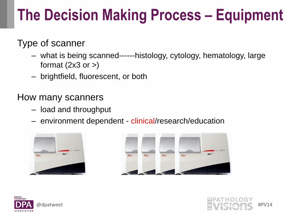
Type of scanner
– what is being scanned------histology, cytology, hematology, large
format (2x3 or >)
– brightfield, fluorescent, or both
How many scanners
– load and throughput
– environment dependent - clinical/research/education
The Decision Making Process – Equipment

@dpatweet #PV14
Software
– Image management and viewing
– Image Analysis
– Reporting
– Interoperability - LIS
– Bundle or different providers
Storage/Archive
– Internal/External – Cloud
– Back-up
Software and Storage/Archive

@dpatweet #PV14
Location of scanner
What will be scanned
Scan in batches or a tray at a time
What magnification – 20x, 40x, 100x – specimen dependent
Who will be responsible for scanning
Who will be responsible for image/data management
Who will have access to images
How do you notify that images are available
How and who will be trained
Is validation required
Do I need system integration (LIS)
Is there any data migration
Image life cycle management
Additional decisions to be made

@dpatweet #PV14
SLIDE PREPARATION – Process Controls
Develop embedding and sectioning criteria for all sample
types
• embedding orientation – skin, gut, etc.
• section thickness
• tissue placement/orientation on slide, number of sections
per slide
• overall section quality – define what is acceptable
• IHC for image analysis will have different quality criteria
than for subjective scoring
• QC in place – prior to scanning and after scanning
Process controls – Slide Preparation

@dpatweet #PV14
Examples

@dpatweet #PV14
Interoperability and LIS
Interoperability eliminates the duplicate entry of
data and provides immediate access to information
across the workflow, providing for:
• Reduction in manual work
• Reduction in case turnaround time
• Reduction in errors

@dpatweet #PV14
Digital Pathology in the AP Workflow
79
Digital Pathology outside clinical workflow QC, teaching, education, tumor boards, etc.
Digital Pathology within the clinical workflow

@dpatweet #PV14
Integrated Imaging Workflow
80
Analytic
Digital
Preparation
Documentation
Communication
Tissue
Preparation Initiation

@dpatweet #PV14
Existing APLIS/DPS Implementations
Metadata in the Barcode
81

@dpatweet #PV14
Existing APLIS/DPS Implementations
Dynamic Metadata Exchange
82

@dpatweet #PV14
Existing APLIS/DPS Implementations
APLIS Integrated Viewing
83

@dpatweet #PV14
Digital Pathology Workflow is Divided Between
WSI System and AP LIS
• Notification of case availability – maybe WSI
• Accessioning into AP LIS – AP LIS
• Distribution of case to pathologist – WSI, APLIS
• Slide review and interpretation – WSI
• Generation of report – AP LIS
• Additional studies if needed – AP LIS
With time, integration between WSI and AP LIS should increase.

@dpatweet #PV14
Routine pathology
• Slides are created in the
laboratory
• Consult slides arrive with
necessary (hopefully)
paperwork
Notification of Case Availability
Digital pathology
• Slides must be scanned –
successfully – to create
digital slides
• Some type of notification
so that the recipient
knows when digital slides
are available or
“received”
• Slides do not arrive

@dpatweet #PV14
Routine pathology
• Lab personnel match slides with
information in accompanying
documents to confirm:
– Patient/case ID and slide
labels match
– All slides are received
– Slides are not damaged
– All necessary accompanying
information is present
• Case is accessioned into LIS
Accessioning into AP LIS – Digital Consults
Digital pathology
• Lab personnel need access to required information (see next slide) in order to accession case
– Electronic requisition?
– Available in slide viewing software?
– e-mail?
• Lab personnel need access to the digital slides in order to confirm:
– Patient ID and slides match
– All expected slides are available
– No obvious scan problems
• Case is accessioned into LIS

@dpatweet #PV14
Routine pathology
• Slides, working draft
report from LIS, and other
paperwork with
necessary information
(e.g. requisition) are
delivered to pathologist
Distribution of Case to Pathologist
Digital pathology
• Pathologist needs
notification that case is
waiting.
– Working draft from LIS
may be delivered to
pathologist
• Pathologist needs to see
other necessary information
– Access to scanned
documents?
– Printout of electronic
requisition?

@dpatweet #PV14
Routine pathology
• Pathologist places slides
on microscope and
navigates slides at own
pace
• Pathologist arrives at
diagnosis based on
review of glass slide
Pathologist Slide Review and Interpretation
Digital pathology
• Pathologist accesses digital
slides in viewer system and
views on monitor
– Needs to know how to
locate digital slides for
each case
• Pace of navigating slides is
determined in part by
system response time for
viewing slides
• May need to defer diagnosis
to glass slide review

@dpatweet #PV14
Routine pathology
• Creation of pathology
report in LIS typically
accomplished through:
– Dictation with
transcription
– Direct entry
– Speech-to-text
Generation of Pathology Report
Digital pathology
• Options for creation of report
in LIS are differ depending
on functionality
• If at remote location,
pathologist needs access to
LIS and/or transcription
services, voice recognition or
a procedure is needed for
documentation and eventual
report
• Should report note that digital
pathology was used in the
case?

@dpatweet #PV14
More pathologist time per case will be required
for digital pathology
• Using digital pathology will be a slower experience for a pathologist than is moving a glass slide around on a microscope.
– Navigation of and viewing of microscopic fields by virtual microscopy
– System and network response times (latency) can cause issues
• Additional time per case could be expected to increase with increasing case complexity (number of slides, amount of tissue per slide).

Hands on
Portion of the
Workshop

@dpatweet #PV14
Logistics
Every course attendee should have a team number (1-2) and a participant number (1-8).
Every team is assigned to a different workstation
Each team will complete two 30-min exercises and every team member will participate in both exercises
Exercise #1 - Basic Viewer Functionality
Exercise #2 – Viewing Different Sample Types
We will wrap up with team discussion and reporting of findings to the entire group
Hands-On Session Guidelines

@dpatweet #PV14
Objective: Demonstrate the following basic viewing capabilities using 20x digital slide images
Panning/zooming
Viewing multiple images side-by-side
Creating annotations and running positive pixel count image analysis
Exercise #1: Basic Viewer Functionality
Participant Digital Slide(s) Action Comments
1 Ex1_H&E20_1 Open H&E slide, examine at low power, pan and zoom Tumor or Tonsil H&E
2 Ex1_IHC20_1 Open IHC slide and view side-by-side with corresponding H&E slide (Ex1_H&E20_1)
Beta-catenin-1 or CD3 E272
3 Ex1_IHC20_2 Open IHC slide and view side-by-side with corresponding H&E slide (Ex1_H&E20_1) and corresponding IHC slide (Ex1_IHC20_1); total 3 slides
Beta catenin EP35 or CD3 rabbit polyclonal
4 Ex1_H&E20_2 Open H&E slide, examine at low power, pan and zoom Goat bone H&E
5 Ex1_SS20 Open SS slide and view side-by-side with corresponding H&E slide (Ex1_H&E20_2)
Goat bone Safranin O
6 Ex1_IHC20_1 Ex1_IHC20_3
Open IHC slide; annotate small region and run positive pixel count image analysis; review results
IHC Stain image analysis

@dpatweet #PV14
Objective: Demonstrate viewing different sample types Comparing 40x slides to 20x slides
Viewing slides with defects
Viewing thick slides
Viewing different sample types (cytology, hematology)
Viewing multiple sections on one slide, measuring distance using “digital ruler”
Exercise #2: Viewing Different Sample Types
Participant Digital Slide(s) Action
1 Ex2_H&E20_1 Ex2_H&E40_1
Open the 20x and 40x H&E slides, view side-by-side and observe differences
2 Ex2_H&E20_2 Open H&E slide and examine areas with artifacts (tissue folds, bubbles, etc.)
3 Ex2_H&E20_3 Open H&E slide and examine image quality when tissue section too thick
4 Ex2_Cyto20_1 Open cytology slides and examine areas of slide that are out of focus
5 Ex2_Hem100_1 Open hematology slide and evaluate 100x resolution
6 Ex2_H&E20_4 Open H&E slide and examine multiple sections of same slide; open slide again and compare side-by-side (at different magnification); measure distance using ruler