digital watermarking : applicability for developing trust
TRANSCRIPT
Digital watermarking : applicability fordeveloping trust in medical imaging
workflows state of the art reviewQasim, AF, Meziane, F and Aspin, R
http://dx.doi.org/10.1016/j.cosrev.2017.11.003
Title Digital watermarking : applicability for developing trust in medical imaging workflows state of the art review
Authors Qasim, AF, Meziane, F and Aspin, R
Publication title Computer Science Review
Publisher Elsevier
Type Article
USIR URL This version is available at: http://usir.salford.ac.uk/id/eprint/44477/
Published Date 2017
USIR is a digital collection of the research output of the University of Salford. Where copyright permits, full text material held in the repository is made freely available online and can be read, downloaded and copied for non-commercial private study or research purposes. Please check the manuscript for any further copyright restrictions.
For more information, including our policy and submission procedure, pleasecontact the Repository Team at: [email protected].

1
Digital Watermarking: Applicability for Developing Trust in Medical
Imaging Workflows State of the Art Review
Asaad F. Qasim1, 2, Farid Meziane1 and Rob Aspin1
1 School of Computing, Science and Engineering, University of Salford, Greater Manchester, M5 4WT, UK
2 Ministry of Higher Education and Scientific Research, Baghdad, Iraq
[email protected], [email protected], [email protected]
Abstract
Medical images can be intentionally or unintentionally manipulated both within the secure
medical system environment and outside, as images are viewed, extracted and transmitted.
Many organisations have invested heavily in Picture Archiving and Communication Systems
(PACS), which are intended to facilitate data security. However, it is common for images, and
records, to be extracted from these for a wide range of accepted practices, such as external
second opinion, transmission to another care provider, patient data request, etc. Therefore,
confirming trust within medical imaging workflows has become essential. Digital
watermarking has been recognised as a promising approach for ensuring the authenticity and
integrity of medical images. Authenticity refers to the ability to identify the information origin
and prove that the data relates to the right patient. Integrity means the capacity to ensure that
the information has not been altered without authorisation.
This paper presents a survey of medical images watermarking and offers an evident scene for
concerned researchers by analysing the robustness and limitations of various existing
approaches. This includes studying the security levels of medical images within PACS system,
clarifying the requirements of medical images watermarking and defining the purposes of
watermarking approaches when applied to medical images.
Keywords: Medical Imaging; Digital Watermarking; Reversible Watermarking; Integrity;
Authentication

2
Contents
Abstract ...................................................................................................................................... 1
Contents ..................................................................................................................................... 2
1. Introduction and Motivation ............................................................................................... 4
1.1. Introduction ................................................................................................................. 4
1.2. Motivation for Medical Image Watermarking ............................................................ 5
1.3. Digital Watermarking Requirements .......................................................................... 6
2. Digital Watermarking ......................................................................................................... 9
2.1. Principal Components of a Watermarking System ..................................................... 9
2.2. Digital Watermarking Classifications ....................................................................... 10
2.3. Digital Watermarking Techniques ............................................................................ 12
2.3.1. Spatial Domain Techniques ............................................................................... 12
2.3.1.1. Least Significant Bit ................................................................................... 12
2.3.1.2. Local Binary Pattern ................................................................................... 12
2.3.1.3. Histogram Modification ............................................................................. 13
2.3.2. Transform Domain Techniques ......................................................................... 13
2.3.2.1. Discrete Cosine Transform ......................................................................... 14
2.3.2.2. Discrete Wavelet Transform ....................................................................... 14
2.3.2.3. Discrete Fourier Transform ........................................................................ 15
3. State of the Art .................................................................................................................. 17
3.1. Schools of Thought in Medical Image Watermarking .............................................. 17
3.1.1. Classical Methods while Minimising the Distortion ......................................... 17
3.1.2. Region of Interest and Region of Non-Interest Watermarking Methods ........... 17
3.1.3. Reversible Watermarking Methods ................................................................... 19
3.1.3.1. Compression Based Technique .................................................................. 20
3.1.3.2. Histogram Modification Based Technique ................................................. 21
3.1.3.3. Quantization Based Technique ................................................................... 22
3.1.3.4. Expansion Based Technique ....................................................................... 23
3.2. Purposes of Medical Image Watermarking ............................................................... 24
3.2.1. Authentication Schemes..................................................................................... 24
3.2.2. EPR Data Hiding Schemes ................................................................................ 29
3.2.3. Authentication and EPR Data Hiding Schemes ................................................. 30
4. Evaluation Benchmarks of Watermarking Algorithms .................................................... 31
4.1. Imperceptibility Assessment of Watermarked Image ............................................... 31

3
4.1.1. Mean Square Error ............................................................................................. 31
4.1.2. Peak Signal to Noise Ratio ................................................................................ 32
4.1.3. Structural Similarity Index ................................................................................. 32
4.2. Robustness Evaluation of Extracted Watermark ....................................................... 32
4.2.1. Correlation Coefficient ...................................................................................... 32
4.2.2. Similarity Measure ............................................................................................. 33
4.2.3. Bit Error Rate ..................................................................................................... 33
4.2.4. Accuracy Ratio (AR) ......................................................................................... 33
5. Discussion and Conclusions ............................................................................................. 36
References ................................................................................................................................ 38

4
1. Introduction and Motivation
Through the exponential development of modern technologies in the areas of communication
and computer networks, the conventional diagnosis has mostly migrated to a technology
enabled e-diagnosis. Most Hospital Information Systems (HIS) and medical imaging systems
generate and store medical images in different modalities such as X-ray, Ultrasound, Magnetic
Resonance Imaging (MRI) and Computerised Tomography (CT). These images are usually
managed within a digital workflow based on the Digital Imaging and Communications in
Medicine (DICOM) standard [1].
1.1. Introduction
In healthcare systems, a hierarchical scheme can be considered as a pyramid with hospitals at
the base and the general Picture Archiving and Communication Systems (PACS) at its top.
Images are taken in a hospital and are immediately saved in the PACS. Within few minutes,
these images are transferred to an upper PACS, which collects data coming from hospitals
belonging to the same division. These files stay in this system for some hours, typically staying
for the night, during which time their integrity is not maintained accurately. Then, these files
are transmitted to the hierarchically higher PACS until they reach the top-PACS. In the top-
PACS, the data are eternally saved and collected in tapes, physical drives or optical supports
with associated hash signature, to become ready for the diagnostic workflow operations.
Furthermore, the data are encrypted utilising the secret key of the PACS manager. This
operation is called consolidation [2].
For security purposes, the authorised archive is managed off-line, while available data are kept
on the top-PACS discs. In most situations, it is difficult to foretell the security issues for each
intermediate system, and the data could be altered intentionally or unintentionally: this is the
first serious case. Moreover; the data are not directly consolidated when reaching the top-PACS
but after approximately 24/36 hours. During this time, PACS professionals, which have access
to both the metadata as well as the image’s pixels due to the structure of DICOM images, are
permitted to edit the files as needed for adjusting potential flaws in patients’ data. This matter
indicates to the second significant case which allows the malicious PACS manipulation to
modify the images before the consolidation process. Hospital’s system can retrieve the images
from the top-PACS when requested by the physicians. In the case of expected claims, such as
regular medical reports, files are pre-fetched in the hospital’s PACS, e.g. through night-time or
transmitted as soon as possible. The separation between the legal archive (secured data) and
5
the available data (used by clinicians) points out the last crucial case of PACS scenario. If the
medical images have been modified in the top-PACS discs, there will be no possibility to
automatically discover the manipulation because the authorised archive is saved off-line and
the data are not quickly accessible. Definitely, it will be possible to discover the alterations that
have been applied to the data in PACS discs, but it might be too late for patients' safety [2].
Furthermore, transmitting medical images between hospitals, located at various locations and
different administrative organisations has become a common practice for many reasons, such
as diagnosis, treatment, distance learning, training purposes, teleconferences between
clinicians and medical consultation between physicians and radiologists [3]. Malicious
alterations on the medical images are feasible for getting counterfeit health insurance demands
by some insurance company or for hiding medical situations for gaining personal advantages
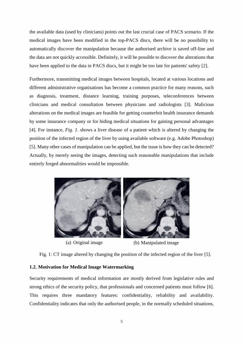
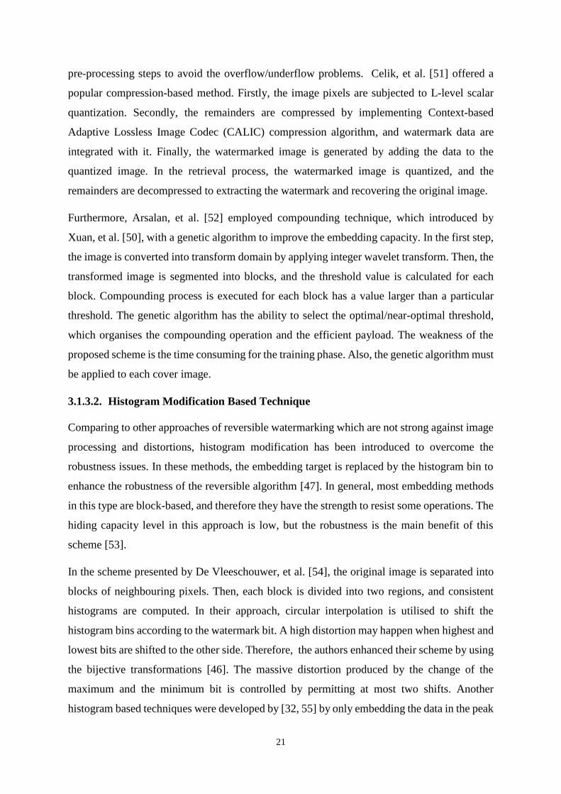
[4]. For instance, Fig. 1. shows a liver disease of a patient which is altered by changing the
position of the infected region of the liver by using available software (e.g. Adobe Photoshop)
[5]. Many other cases of manipulation can be applied, but the issue is how they can be detected?
Actually, by merely seeing the images, detecting such reasonable manipulations that include
entirely forged abnormalities would be impossible.
Fig. 1: CT image altered by changing the position of the infected region of the liver [5].
1.2. Motivation for Medical Image Watermarking
Security requirements of medical information are mostly derived from legislative rules and
strong ethics of the security policy, that professionals and concerned patients must follow [6].
This requires three mandatory features: confidentiality, reliability and availability.
Confidentiality indicates that only the authorised people, in the normally scheduled situations,
(a) Original image (b) Manipulated image
6
have access to the data. Reliability may be decomposed into two aspects: i) Integrity which
verifies that the information has not been changed, and, ii) Authentication which ensures that
the data belongs to the right patient and is delivered from the verified source. Availability
defines the capability of the authorised users to utilise the information system in the normally
scheduled situations of access and practice [7].
Confidentiality of the image data can be accomplished by applying many techniques such as
encryption, access control and firewall. Integrity can be fulfilled by encrypting the images
when sharing them over the network. Authentication needs measures being implemented to
discover whether confidentiality and/or the integrity of the data has been breached [8].
Two techniques are commonly employed to ensure integrity and authenticity within the data;
metadata and digital watermarking [4, 9]. In medical imaging, the metadata refers to the data
stored along with the image [9]. The common approach of metadata inclusion is Part 15 of the
DICOM standard, where the digital signature data is placed in its header [1]. The metadata has
also been employed to offer confidentiality, using the data of DICOM header to encrypt the
images [10]. Existing metadata techniques do not provide a robust link between the medical
image and its metadata. It is, therefore, almost easy to decay the metadata rendering the image
unreliable. This shortcoming can be fixed with digital watermarking [9]. Digital watermarking
is a technique that hides data known as a watermark into the digital object such that the
concealed watermark can then be detected/extracted to make a confirmation about the object
[11]. Image watermarking is one of the earliest techniques to improve integrity and authenticity
of the digital data. In recent times, authentication is one of the main watermarking requirements
in medical applications [12].
1.3. Digital Watermarking Requirements
The essential requirements for designing a general watermarking scheme can be described as
follows [13]:
Fidelity
It is the most important feature in the watermarking system which defines the similarity
between both the original and watermarked images. The watermark should remain invisible to
human perception although the incidence of slight distortions in the host image [14].
7
Robustness
This requirement signifies the ability of the watermarking scheme to resistant to different image
processing attacks. These attacks aim to frustrate the watermark from fulfilling its intended
purpose. The wide class of existing attacks can be categorized into four groups: removal,
geometric, protocol and cryptographic attacks [15, 16]. Watermarking algorithms cannot
survive with all types of attacks. Some of the algorithms are strong against several attacks.
However, they fail to comply with other stronger operations. Furthermore, not all applications
require robust watermark, but in some applications, it is needed to be fragile [17].
Data Payload (Capacity)
This property refers to the number of bits that can be concealed without affecting the image
quality. This factor defines how many bits can be embedded as a watermark so that it can be
efficiently discovered through the extraction process. The embedding capacity depends on the
required application. Several watermarking applications have various capacity requirements
[18].
Security
The capability of resisting the intentional attacks. A watermarking scheme is supposed to be
secure if the unauthorised user cannot extract the watermark without having full information
about the algorithm that has been used to embed the watermark. The security factor is crucial
to the watermarking system, and only the authorised person can extract the watermark [19].
Computational Complexity
This feature is defined as the amount of time required for embedding and extraction processes.
For instance, the real-time application requires both fast and efficient algorithms. On the other
hand, more computational complexity is needed for high-security applications [20].
Perceptibility
This concept indicates the amount of degradation that occurs on a watermarked image when
embedding the data. This feature should be as little as possible in the invisible watermarking
schemes [13].
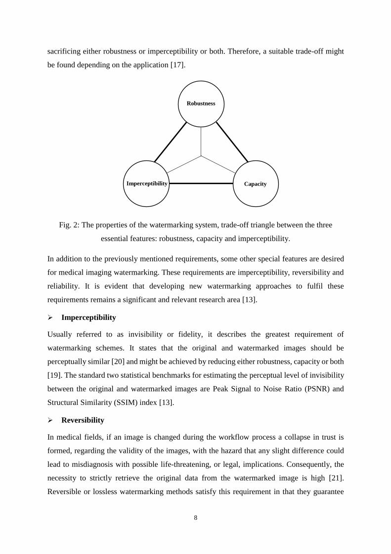
Watermarking capacity is determined by the other two significant features of the watermarking
system, which are imperceptibility and robustness. The relationship between the properties of
the watermarking scheme is shown in Fig. 2. Obviously, a high capacity can be achieved by
8
sacrificing either robustness or imperceptibility or both. Therefore, a suitable trade-off might
be found depending on the application [17].
Fig. 2: The properties of the watermarking system, trade-off triangle between the three
essential features: robustness, capacity and imperceptibility.
In addition to the previously mentioned requirements, some other special features are desired
for medical imaging watermarking. These requirements are imperceptibility, reversibility and
reliability. It is evident that developing new watermarking approaches to fulfil these
requirements remains a significant and relevant research area [13].
Imperceptibility
Usually referred to as invisibility or fidelity, it describes the greatest requirement of
watermarking schemes. It states that the original and watermarked images should be
perceptually similar [20] and might be achieved by reducing either robustness, capacity or both
[19]. The standard two statistical benchmarks for estimating the perceptual level of invisibility
between the original and watermarked images are Peak Signal to Noise Ratio (PSNR) and
Structural Similarity (SSIM) index [13].
Reversibility
In medical fields, if an image is changed during the workflow process a collapse in trust is
formed, regarding the validity of the images, with the hazard that any slight difference could
lead to misdiagnosis with possible life-threatening, or legal, implications. Consequently, the
necessity to strictly retrieve the original data from the watermarked image is high [21].
Reversible or lossless watermarking methods satisfy this requirement in that they guarantee
Robustness
Capacity
Imperceptibility
9
extraction of the watermark along with exactly reconstructing the unmodified original image
[22]. However, a watermarked image is not distortion-free, especially in reversible techniques,
but the modified image is employed as a cover for carrying out the watermark, not for
diagnostic purposes and the recovered image is used for diagnosis, intervention planning, etc.
[23].
Reliability
This may be decomposed into two parts [24]:
Integrity: The capability of proving that the data has not been changed without
authorisation.
Authentication: The ability to identify information origin and confirming that the data
refers to the right patient.
This paper offers a review of digital watermarking schemes and compares some recent works
in watermarking medical imaging to define some research issues for the future. The rest of the
paper is organised as follows. Section 2 illustrates the basic principles of digital watermarking.
Section 3 reviews the recently published approaches in the field of medical imaging
watermarking and highlights the limitations of some of these algorithms. In section 4,
evaluation benchmarks of watermarking algorithms were demonstrated. Finally, discussion
and conclusions drawn from this research will be stated in sections 2.
2. Digital Watermarking
Digital watermarking is the hiding of information (the watermark) within the digital data, such
that the embedded watermark can be identified or extracted later to produce a confirmation of
the validity of the data [11].
2.1. Principal Components of a Watermarking System
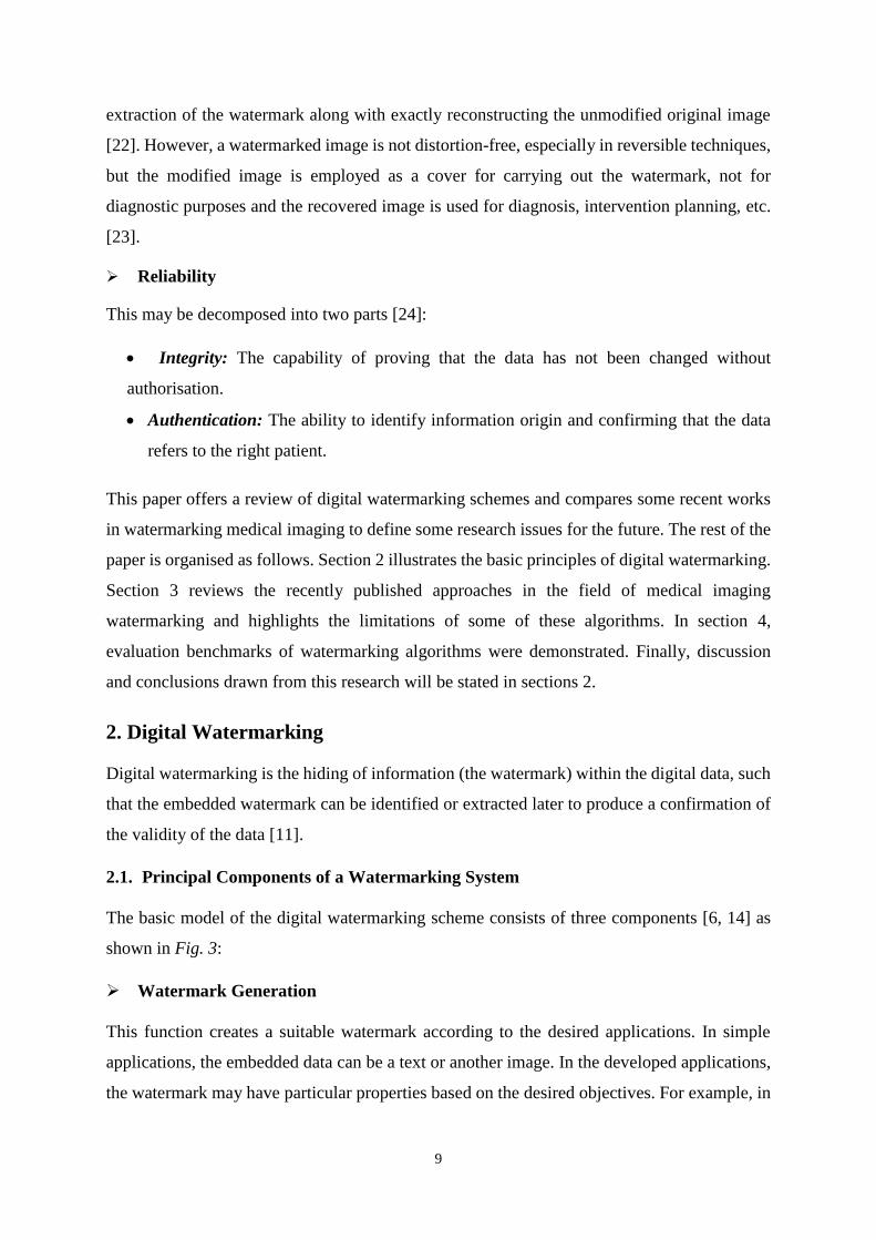
The basic model of the digital watermarking scheme consists of three components [6, 14] as
shown in Fig. 3:
Watermark Generation
This function creates a suitable watermark according to the desired applications. In simple
applications, the embedded data can be a text or another image. In the developed applications,
the watermark may have particular properties based on the desired objectives. For example, in
10
medical applications, the watermark may need the patient information or image features to
confirm the integrity and authenticity of the watermarked data.
Watermark Hiding
The hiding process is done at the source end. In this step, the watermark is inserted into the
original data by applying a certain algorithm and a secret key to generate the watermarked data.
Watermark Extracting
The extraction process is done by reversing the implemented hiding algorithm and use the
secret key and/or the original data to detect/extract the embedded watermark.
Generation
Algorithm
Message
Original data Watermark
Embedding
Algorithm
Secret key
Original dataWatermarked
data
Watermark
Extraction
Algorithm
Secret key
Watermark
Original data
(a) Watermark generation
(c) Watermark extraction(b) Watermark embedding
Fig. 3: Main components of watermarking schemes.
2.2. Digital Watermarking Classifications
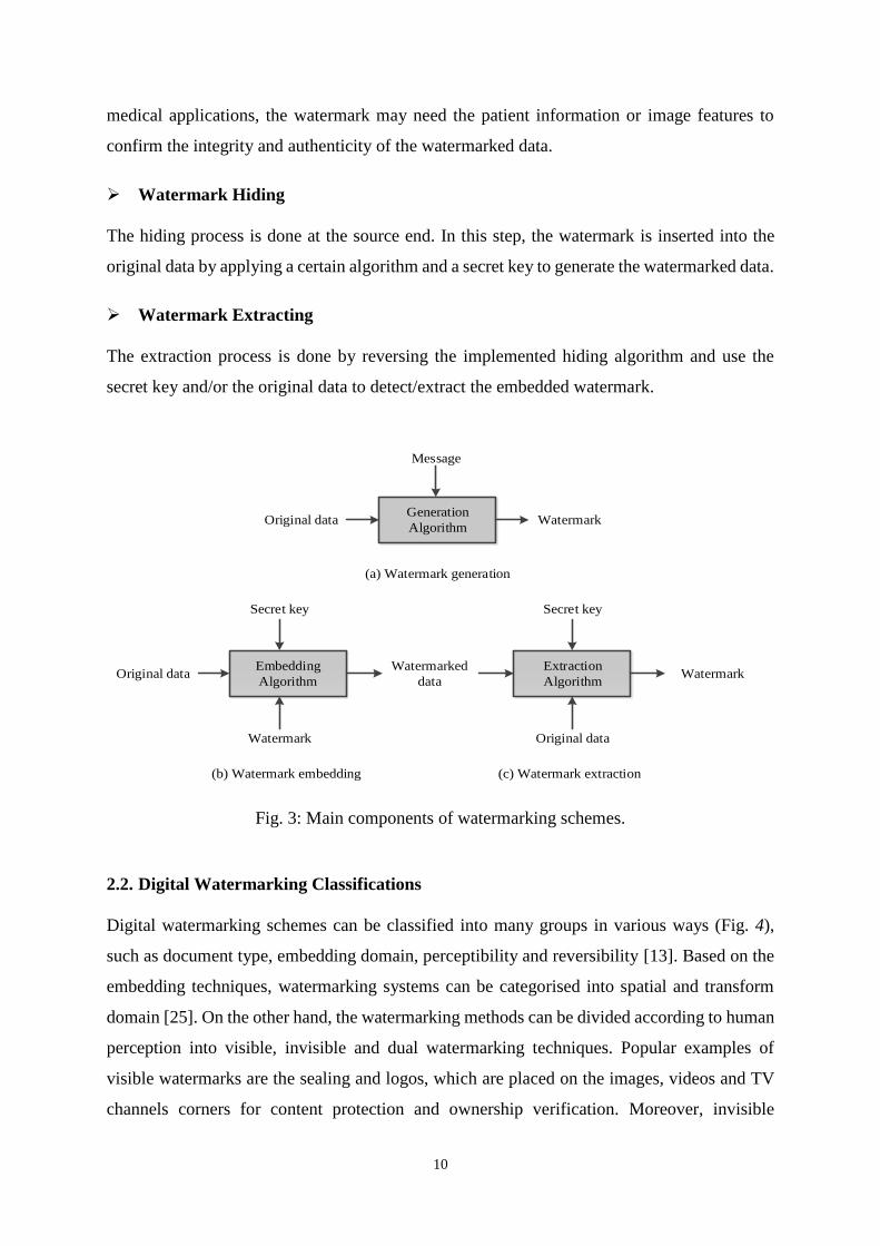
Digital watermarking schemes can be classified into many groups in various ways (Fig. 4),
such as document type, embedding domain, perceptibility and reversibility [13]. Based on the
embedding techniques, watermarking systems can be categorised into spatial and transform
domain [25]. On the other hand, the watermarking methods can be divided according to human
perception into visible, invisible and dual watermarking techniques. Popular examples of
visible watermarks are the sealing and logos, which are placed on the images, videos and TV
channels corners for content protection and ownership verification. Moreover, invisible
11
watermarks are hidden in such a way that they cannot be seen, but they can be removed by
utilising the exact algorithm. Invisible watermarking schemes are suitable for many purposes
like authentication, integrity control and ownership verification of digital files. In some
application, visible and invisible watermarks can be applied together. This technique is called
the dual watermarking, and in this situation, the invisible watermark is assumed as a backup
for the visible one [26].
Digital watermarking
Type of document Hiding domain Human perception Reversibility
Reversible
Non-reversible
Invisible
Visible
Dual
Spatial
Transform
Text
Image
Audio
video
HybridSemi-fragileFragileRobust
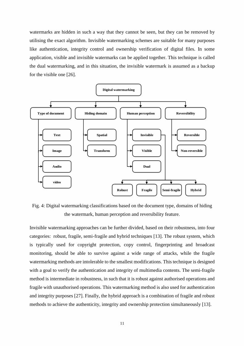
Fig. 4: Digital watermarking classifications based on the document type, domains of hiding
the watermark, human perception and reversibility feature.
Invisible watermarking approaches can be further divided, based on their robustness, into four
categories: robust, fragile, semi-fragile and hybrid techniques [13]. The robust system, which
is typically used for copyright protection, copy control, fingerprinting and broadcast
monitoring, should be able to survive against a wide range of attacks, while the fragile
watermarking methods are intolerable to the smallest modifications. This technique is designed
with a goal to verify the authentication and integrity of multimedia contents. The semi-fragile
method is intermediate in robustness, in such that it is robust against authorised operations and
fragile with unauthorised operations. This watermarking method is also used for authentication
and integrity purposes [27]. Finally, the hybrid approach is a combination of fragile and robust
methods to achieve the authenticity, integrity and ownership protection simultaneously [13].
12
In addition to the previous classifications, the reversible watermarking also called invertible or
lossless watermarking is another significant feature of watermarking techniques. Compared to
the traditional watermarking systems, reversible algorithms can restore both the embedded
watermark and the original data exactly. This feature is a crucial requirement for many fields
such as medical, military and law-enforcement applications [22].
2.3. Digital Watermarking Techniques
Current watermark embedding techniques can be divided into two main groups. The following
are a brief explanation of the properties of each group.
2.3.1. Spatial Domain Techniques
In these methods, the watermark is inserted into the cover image by directly modifying the
pixel values of the original image. These algorithms are simple, fast and offer high embedding
capacity [25]. Also, a small watermark can be hidden several times. This advantage provides
additional robustness against any attack because of the possibility of removing all watermarks
is very low. Spatial domain techniques may have some benefits, but their main drawback is
that they cannot survive against many operations like adding noise and lossy compression
methods. Moreover, when discovering the utilised watermarking method, the hidden
watermark can easily be altered by an unauthorised user [28].
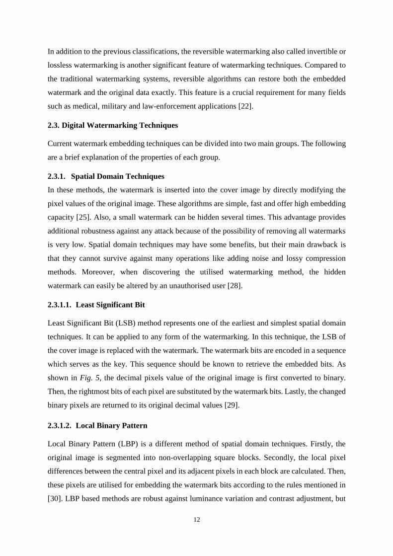
2.3.1.1. Least Significant Bit
Least Significant Bit (LSB) method represents one of the earliest and simplest spatial domain
techniques. It can be applied to any form of the watermarking. In this technique, the LSB of
the cover image is replaced with the watermark. The watermark bits are encoded in a sequence
which serves as the key. This sequence should be known to retrieve the embedded bits. As
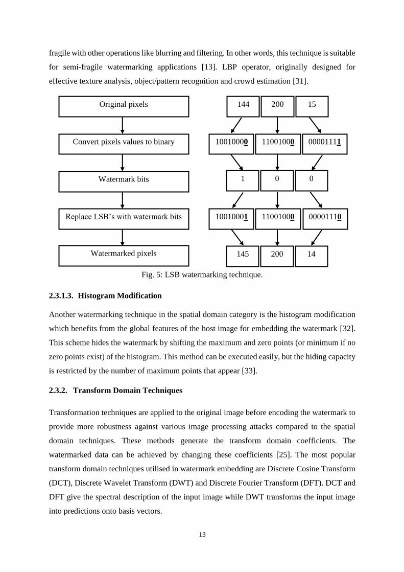
shown in Fig. 5, the decimal pixels value of the original image is first converted to binary.
Then, the rightmost bits of each pixel are substituted by the watermark bits. Lastly, the changed
binary pixels are returned to its original decimal values [29].
2.3.1.2. Local Binary Pattern
Local Binary Pattern (LBP) is a different method of spatial domain techniques. Firstly, the
original image is segmented into non-overlapping square blocks. Secondly, the local pixel
differences between the central pixel and its adjacent pixels in each block are calculated. Then,
these pixels are utilised for embedding the watermark bits according to the rules mentioned in
[30]. LBP based methods are robust against luminance variation and contrast adjustment, but
13
fragile with other operations like blurring and filtering. In other words, this technique is suitable
for semi-fragile watermarking applications [13]. LBP operator, originally designed for
effective texture analysis, object/pattern recognition and crowd estimation [31].
Fig. 5: LSB watermarking technique.
2.3.1.3. Histogram Modification
Another watermarking technique in the spatial domain category is the histogram modification
which benefits from the global features of the host image for embedding the watermark [32].
This scheme hides the watermark by shifting the maximum and zero points (or minimum if no
zero points exist) of the histogram. This method can be executed easily, but the hiding capacity
is restricted by the number of maximum points that appear [33].
2.3.2. Transform Domain Techniques
Transformation techniques are applied to the original image before encoding the watermark to
provide more robustness against various image processing attacks compared to the spatial
domain techniques. These methods generate the transform domain coefficients. The
watermarked data can be achieved by changing these coefficients [25]. The most popular
transform domain techniques utilised in watermark embedding are Discrete Cosine Transform
(DCT), Discrete Wavelet Transform (DWT) and Discrete Fourier Transform (DFT). DCT and
DFT give the spectral description of the input image while DWT transforms the input image
into predictions onto basis vectors.
Original pixels
Convert pixels values to binary
Watermark bits
Replace LSB’s with watermark bits
Watermarked pixels
144 200 15
10010000 11001000 00001111
1 0 0
10010001 11001000 00001110
145 200 14
14
2.3.2.1. Discrete Cosine Transform
DCT is one of the greatest attractive methods implemented to transform the data from the
spatial domain to transform domain. It is a linear transform, which maps an n-dimensional
vector to a set of n-coefficients. DCT is robust to JPEG compression because JPEG standard
is based on DCT technique. However, DCT lacks resistance to strong geometric attacks like
scaling, cropping, translation, rotation, etc. [18].
By applying this technique, the image will be segmented into three frequency groups: low (FL),
middle (FM) and high (FH). Most of the energy is focused in the low-frequency region, while
high-frequency part contains the least amount of energy. The mathematical equations of
forward and inverse transform of 2D-DCT are shown in Eq. 1 and Eq. 2, respectively [34].
𝐶(𝑢, 𝑣) =2
√𝑚𝑛𝛼(𝑢)𝛼(𝑣) ∑ ∑ 𝑓(𝑥, 𝑦)
𝑛−1
𝑦=0
𝑚−1
𝑥=0
∗ 𝑐𝑜𝑠(2𝑥 + 1)𝑢𝜋
2𝑚∗ 𝑐𝑜𝑠
(2𝑦 + 1)𝑣𝜋
2𝑛 (1)
𝑓(𝑥, 𝑦) =2
√𝑚𝑛∑ ∑ 𝛼(𝑢)𝛼(𝑣)𝑓(𝑥, 𝑦)
𝑛−1
𝑣=0
𝑚−1
𝑢=0
∗ 𝑐𝑜𝑠(2𝑥 + 1)𝑢𝜋
2𝑚∗ 𝑐𝑜𝑠
(2𝑦 + 1)𝑣𝜋
2𝑛 (2)
Where m and n define the block size, f(x, y) represents the spatial domain pixel value, C (u, v)
is the DC coefficient and 𝛼(𝑢), 𝛼(𝑣) can be calculated in Eq. 3:
𝛼(𝑢), 𝛼(𝑣) = {1
√2⁄ 𝑖𝑓 𝑢, 𝑣 = 0
1 𝑒𝑙𝑠𝑒 (3)
The DC coefficients are used for hiding the watermark to prevent changing the significant part
of the image because they contain middle sub-band coefficients of the DCT. All other
coefficients are titled the AC coefficients [13].
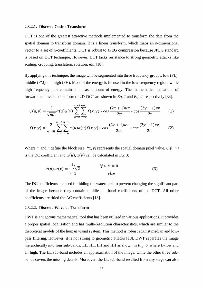
2.3.2.2. Discrete Wavelet Transform
DWT is a vigorous mathematical tool that has been utilised in various applications. It provides
a proper spatial localisation and has multi-resolution characteristics, which are similar to the
theoretical models of the human visual system. This method is robust against median and low-
pass filtering. However, it is not strong to geometric attacks [18]. DWT separates the image
hierarchically into four sub-bands: LL, HL, LH and HH as shown in Fig. 6, where L=low and
H=high. The LL sub-band includes an approximation of the image, while the other three sub-
bands covers the missing details. Moreover, the LL sub-band resulted from any stage can also
15
be decomposed continuously to gain another level until reaching the required number of levels
based on the application [35].
Fig. 6: Four levels of DWT decomposition which divide the input image into four sub-bands
in each level (LL, HL, LH and HH).
In digital watermarking systems, lower decomposition levels of the image, which contain a
lower amount of energy, are more suitable to modifications. This energy is calculated by Eq. 4
[13]:
𝐸𝑘 =1
𝑁𝑘𝑀𝑘∑ ∑ |𝐼𝑘(𝑖, 𝑗)|
𝑗𝑖 (4)
Where k is the level of the decomposition, Nk and Mk are the dimensions of the sub-band, and
Ik indicates the coefficients of the corresponding sub-band.
2.3.2.3. Discrete Fourier Transform
DFT denotes the most popular technique to convert the images from the spatial domain to
transform domain [36]. It offers more robustness against geometric attacks. DFT decomposes
an image in sine and cosine form. Therefore, watermark embedding can be implemented in two
ways: direct hiding and the template based hiding [37]. Consider f (x,y) an image of size M×N,
with x = 0,1,2,…,M−1, and y = 0,1,2,…,N−1. The forward discrete Fourier transform and its
inverse transform are shown in Eq. 5 and Eq. 6, respectively [13]:
𝐹(𝑢, 𝑣) = ∑ ∑ 𝑓(𝑥, 𝑦)𝑒−𝑗2𝜋(𝑢𝑥𝑁
+𝑣𝑦𝑀
)
𝑀−1
𝑦=0
𝑁−1
𝑥=0
(5)
= 𝑅(𝑢, 𝑣) + 𝑗𝐼(𝑢, 𝑣)
Original
Image
DWT
16
𝑓(𝑥, 𝑦) =1
𝑁𝑀∑ ∑ 𝐹(𝑢, 𝑣)𝑒𝑗2𝜋(
𝑢𝑥𝑁
+𝑣𝑦𝑀
)
𝑀−1
𝑣=0
𝑁−1
𝑢=0
(6)
Where: F(u,v) is the DFT coefficient, u=0,1,2,…,M−1, and v=0,1,2,…,N−1, R(u,v) and I(u,v)
are the real and imaginary parts of DFT, respectively.
The polar of the DFT [13] can also be explained by Eq. 7:
𝐹(𝑢, 𝑣) = |𝐹(𝑢, 𝑣)|𝑒𝑗∅(𝑢,𝑣) (7)
Where |F(u,v)| and ϕ(u,v) represent amplitude and phase components respectively, which can
be calculated by Eq. 8 and Eq. 9:
|𝐹(𝑢, 𝑣)| = [𝑅2(𝑢, 𝑣) + 𝐼2(𝑢, 𝑣)]1
2⁄ (8)
∅(𝑢, 𝑣) = 𝑡𝑎𝑛−1 [𝐼(𝑢, 𝑣)
𝑅(𝑢, 𝑣)] (9)
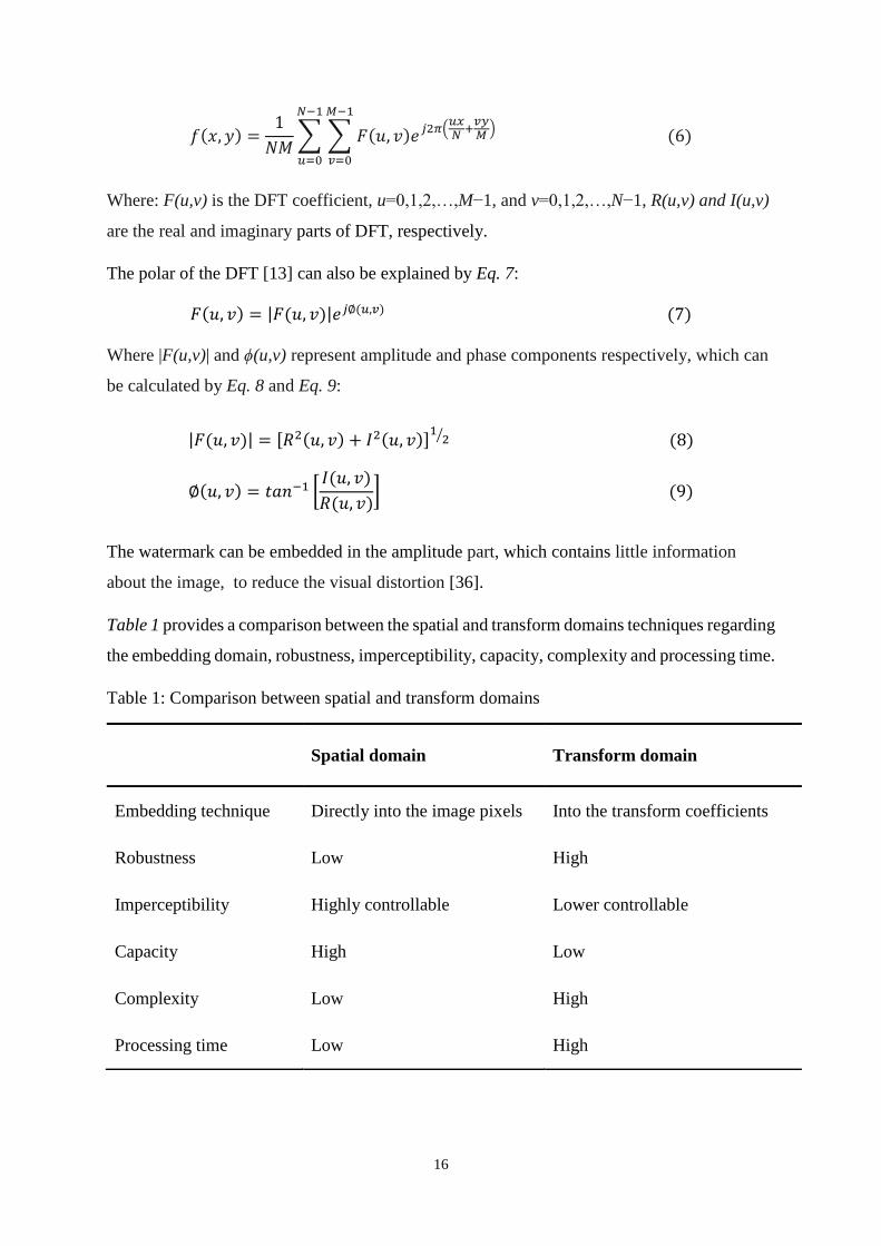
The watermark can be embedded in the amplitude part, which contains little information
about the image, to reduce the visual distortion [36].
Table 1 provides a comparison between the spatial and transform domains techniques regarding
the embedding domain, robustness, imperceptibility, capacity, complexity and processing time.
Table 1: Comparison between spatial and transform domains
Spatial domain Transform domain
Embedding technique Directly into the image pixels Into the transform coefficients
Robustness Low High
Imperceptibility Highly controllable Lower controllable
Capacity High Low
Complexity Low High
Processing time Low High
17
3. State of the Art
In this section, a list of significant published work in the area of medical images watermarking
will be reviewed. This survey aims to highlight the advantages and limitations of recently
published techniques concerning the medical images integrity and authenticity and Electronic
Patient Record (EPR) data hiding. It is also aimed to provide a path for future researchers to
address the limitations of existing watermarking techniques.
3.1. Schools of Thought in Medical Image Watermarking
There are three kinds of medical images watermarking approaches; classical methods, a Region
of Interest (ROI) and Region of Non Interest (RONI) watermarking approaches and reversible
watermarking techniques. Whatever algorithm is used, the computational complexity should
not cause a delay in the clinician’s time [38]. The following subsections discuss the existing
digital watermarking methods applied to medical images. A comparison of these techniques
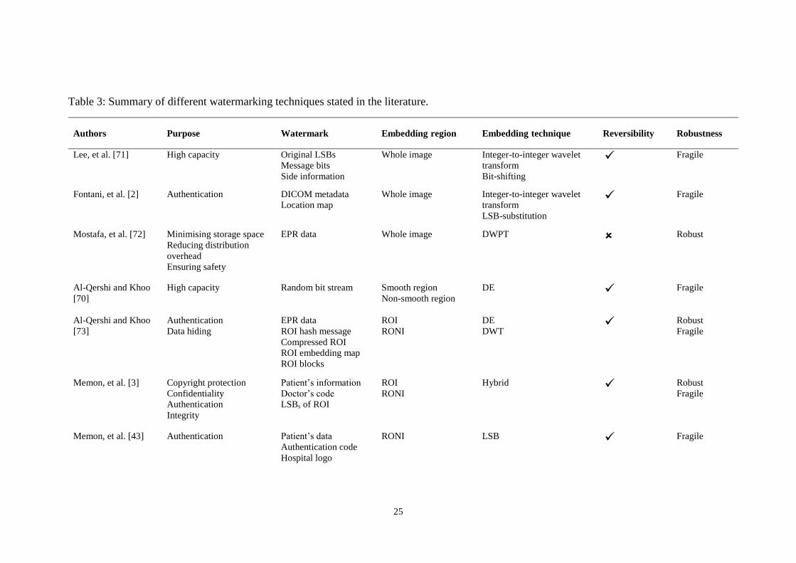
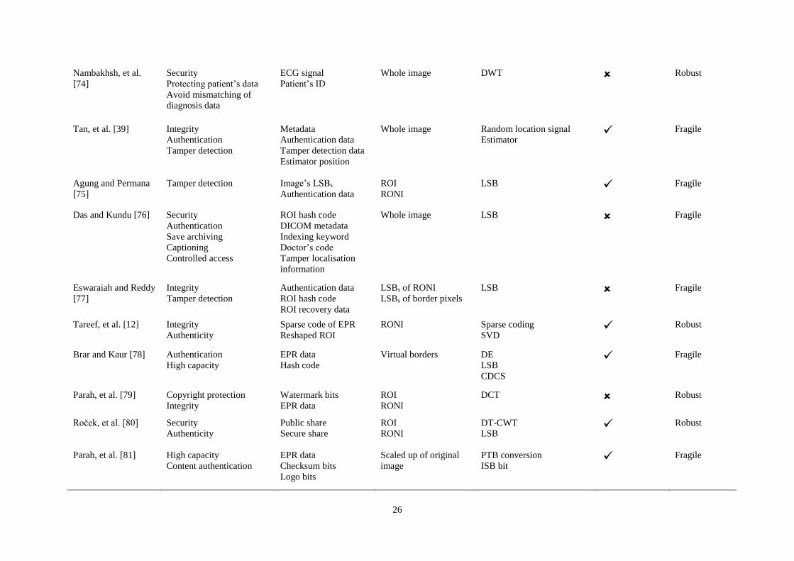
regarding the robustness, capacity, imperceptibility and objective is also illustrated in Table 3.
3.1.1. Classical Methods while Minimising the Distortion
In conventional watermarking methods, the watermark is embedded in the whole cover image
by replacing some details like LSBs or losing some details when using lossy image
compression methods [38]. When implementing a digital watermarking scheme for a medical
image, the images must not be perceptually changed because no radiologist will agree to use
the degraded image for taking a decision, no matter how small the alteration is. Hence, the
watermarking algorithm must be reversible [2]. The irreversible watermarking approaches
remain subjected to an admission by radiologists while the original images stay usually
preferred for investigation purposes [39].
3.1.2. Region of Interest and Region of Non-Interest Watermarking Methods
Coatrieux, et al. [40] assumed that medical images can be divided into two regions ROI and
RONI. ROI section includes the informative region which is used for diagnostic purposes and
must be stored without any distortion. However, RONI usually represents the black background
of the image, but occasionally it can contain grey level parts of slight interest [41]. In ROI
watermarking, spatial or transform domain techniques is utilised for hiding the watermark. The
encoded watermark may be robust or fragile based on the purpose and the application in hand.

18
Table 2: A comparison of existing schools of medical image watermarking
Hiding
School
Hiding
Technique Robustness Imperceptibility Capacity Reversibility Objective
Classical
methods
Spatial
domain
Fragile High High Integrity
Authentication
Transform
domain
Robust Low Low Ownership
protection
ROI & RONI
methods
Spatial
domain
Fragile High Dependent Integrity
Authentication
Transform
domain
Robust Low Dependent Ownership
protection
Reversible
methods
Compression
based
Fragile High High Integrity
Authentication
Histogram
based
Robust
Semi-fragile
Low Low Ownership
protection
Quantization
based
Fragile High High Integrity
Authentication
Expansion
based
Fragile High High Integrity
Authentication
19
These watermarks are implemented in a particular way without impacting the visual image
quality [42, 43].
Using ROI sections for embedding the watermark may deform the pixels in those regions which
may consequently cause the wrong diagnosis. On the other hand, RONI watermarking
approaches embed watermarks in areas that unimportant in medical diagnosis, but they have
several drawbacks such as they can be only implemented if RONI exists, the amount of
information to be embedded depend on the RONI area size and ROI may not be protected
against malicious attacks.
3.1.3. Reversible Watermarking Methods
The embedding of the secret message as a watermark, no matter how trivial the modification
is, can cause degradation to the host image quality. In some applications, such as military,
medical, legal and archival applications, where the authentication requirements are often
essential, there are typically strict restraints on data reliability that prevent any deformation in
the watermarking operation. For example, modifying a patient’s medical image could affect
the patient’s life by causing errors in diagnosis and treatment. As a result, reversible
watermarking techniques have been developed which can stop this shortcoming by applying a
technique that can recover both the embedded watermark and the original image. Reversible
watermarking techniques can be utilised for image authentication. Reversible watermarks for
authentication applications offer a comprehensive framework, the authentication feature
maintains the integrity of the image, while the advantage of reversibility protects the quality
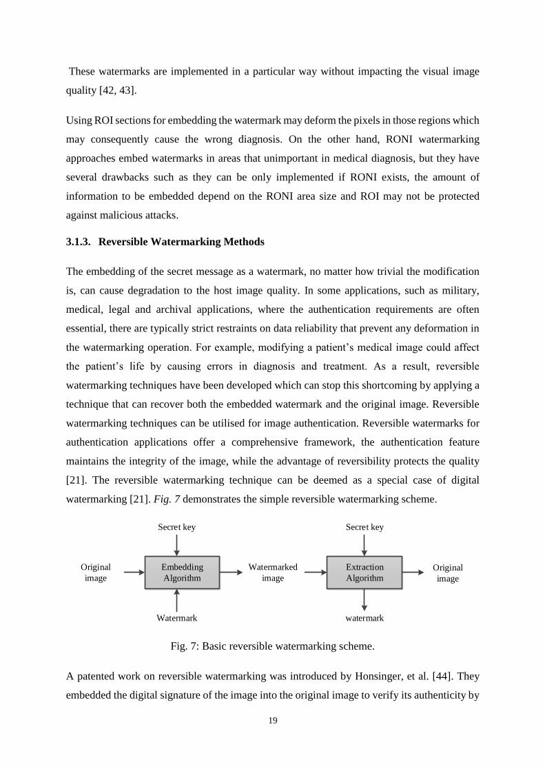
[21]. The reversible watermarking technique can be deemed as a special case of digital
watermarking [21]. Fig. 7 demonstrates the simple reversible watermarking scheme.
Embedding
Algorithm
Secret key
Original
image
Watermarked
image
Watermark
Extraction
Algorithm
Secret key
Original
image
watermark
Fig. 7: Basic reversible watermarking scheme.
A patented work on reversible watermarking was introduced by Honsinger, et al. [44]. They
embedded the digital signature of the image into the original image to verify its authenticity by
20
implementing a spatial additive watermark method joint with modulo additions 256. Macq [45]
suggested an expansion to the patchwork algorithm to design a robust reversible watermarking
technique. Both approaches proposed by Honsinger, et al. [44] and Macq [45] suffer from salt
and pepper noise and delays in retrieving the watermark because of the use of modulo additions
256. A different method was designed by De Vleeschouwer, et al. [46] through utilising circular
interpretation of bijective transformations of the histograms to reduce the salt and pepper noise
found in the previous approaches. Some metrics evaluated the algorithm, but they did not show
the results that compare payload capacity to image distortion.
Feng, et al. [47] categorised reversible watermarking approaches into three groups: Data
Compression (DC), Difference Expansion (DE) and Histogram Bin Shifting (HBS). Some
challenges met the authors in this area are also summarised. Pan, et al. [48] classified the
reversible watermarking systems into additive and substitution methods based on hiding
technique. The comparison is presented in the experiential study of particular reversible
schemes on medical images. Caldelli, et al. [49] proposed a different survey by categorising
the reversible watermarking methods into three types: robust, fragile and semi-fragile
techniques. They also classified the reversible watermarking according to the hiding domain
into spatial and transform techniques. Recently, reversible watermarking approaches based on
DE concept have been suggested in many types of research, and they typically exceed the other
kinds of techniques methods in that they offer high embedding capacity and low computational
complexity compared to the other methods [21].
The following subsections present a review of reversible watermarking methods by classifying
them into four groups: compression based, histogram modification based, quantization based
and expansion based techniques.
3.1.3.1. Compression Based Technique
In the case of reversible watermarking, additional information is required to be encoded along
with the watermark to recover the original unmodified image. As a result, the length of the
watermark is much more than the traditional methods. A simple technique to improve the
capacity will be by compressing a part of the host image [21, 47].
Several reversible watermarking approaches are stated in the literature and are being
implemented. Xuan, et al. [50] developed a high-capacity and distortion-free reversible
technique utilising integer wavelet transform and compounding method. The proposed system
embeds the watermark in the high-frequency coefficients by histogram shifting and applying
21
pre-processing steps to avoid the overflow/underflow problems. Celik, et al. [51] offered a
popular compression-based method. Firstly, the image pixels are subjected to L-level scalar
quantization. Secondly, the remainders are compressed by implementing Context-based
Adaptive Lossless Image Codec (CALIC) compression algorithm, and watermark data are
integrated with it. Finally, the watermarked image is generated by adding the data to the
quantized image. In the retrieval process, the watermarked image is quantized, and the
remainders are decompressed to extracting the watermark and recovering the original image.
Furthermore, Arsalan, et al. [52] employed compounding technique, which introduced by
Xuan, et al. [50], with a genetic algorithm to improve the embedding capacity. In the first step,
the image is converted into transform domain by applying integer wavelet transform. Then, the
transformed image is segmented into blocks, and the threshold value is calculated for each
block. Compounding process is executed for each block has a value larger than a particular
threshold. The genetic algorithm has the ability to select the optimal/near-optimal threshold,
which organises the compounding operation and the efficient payload. The weakness of the
proposed scheme is the time consuming for the training phase. Also, the genetic algorithm must
be applied to each cover image.
3.1.3.2. Histogram Modification Based Technique
Comparing to other approaches of reversible watermarking which are not strong against image
processing and distortions, histogram modification has been introduced to overcome the
robustness issues. In these methods, the embedding target is replaced by the histogram bin to
enhance the robustness of the reversible algorithm [47]. In general, most embedding methods
in this type are block-based, and therefore they have the strength to resist some operations. The
hiding capacity level in this approach is low, but the robustness is the main benefit of this
scheme [53].
In the scheme presented by De Vleeschouwer, et al. [54], the original image is separated into
blocks of neighbouring pixels. Then, each block is divided into two regions, and consistent
histograms are computed. In their approach, circular interpolation is utilised to shift the
histogram bins according to the watermark bit. A high distortion may happen when highest and
lowest bits are shifted to the other side. Therefore, the authors enhanced their scheme by using
the bijective transformations [46]. The massive distortion produced by the change of the
maximum and the minimum bit is controlled by permitting at most two shifts. Another
histogram based techniques were developed by [32, 55] by only embedding the data in the peak
22
bin pixels. However, these approaches require additional overhead to retrieve the watermark
and reconstruct the original image, but they provided a reasonable watermarked image quality.
In order to raise the hiding capacity, Lin, et al. [56] offered a multilevel reversible method
using the histogram of difference image for hiding the data. The difference image is produced
by utilising the difference between two neighbouring pixels. The image is partitioned into non-
overlapping (4x4) blocks, and then a variance matrix of size (3x4) is created for every block.
For data hiding, histogram shifting is applied to each difference block. Although this approach
has a high capacity due to implementing a multi-level embedding method, it suffers from the
massive amount of side-information like saving the peak value for all blocks. Tsai, et al. [57]
proposed a high capacity scheme by employing a residue image. This remainder indicates a
difference between an original pixel and every other pixel in the non-overlapping block rather
than the difference between neighbouring pixels. Though, since they need a highest and zero
points for each block for obtaining the reversibility feature, that information should be involved
to message bits and therefore reducing the hiding capacity of the scheme.
Khan and Malik [58] reported a new high capacity reversible watermarking method which
exploits the idea of down sampling for improving the implementation. Down sampling offers
two sub-sampled forms, the reference and the data hiding to produce area for embedding by
utilising histogram shifting. Moreover, to obtain a blind scheme, the location map was
compressed and embedded in the watermarked image. The proposed system provided an
excellent imperceptibility versus capacity trade-off and can detect tamper attacks.
3.1.3.3. Quantization Based Technique
Though quantization based watermarking approaches are, in general, robust, reversible
quantization based watermarking methods are typically fragile in nature [21]. In Cheung and
Wu [59] system, a Sequential Quantization Strategy (SQS) has been suggested to make the
variation of a pixel value dependent on the other pixels. Therefore, a balance between security
enhancements can be achieved for authenticity and integrity verification. The combination
between proposed SQS and the reversible watermarking mechanism has increased the
opportunity of detecting the illegal modifications. Saberian, et al. [60] introduced a Weighted
Quantization Method (WQM) which is able to be executed in both spatial and transform
domains. Comparing to other schemes, the deformation produced by this approach is not
capacity dependent.
23
In general, the classical Quantization Index Modulation (QIM) watermarking methods cannot
recover the original image due to the irreversible alterations that produced in the watermarked
image because of using the quantization algorithm. Nevertheless, Ko, et al. [61] proposed a
reversible watermarking method for medical image applications using the QIM-based
technique. The capacity of the developed system was improved by taking the benefits of the
suggested nest structure. Ko, et al. [62] outperformed the proposed nested approach by
developing a reversible method employing QIM with Fractional Discrete Cosine Transform
(FDCT) to reconstruct the original image correctly.
3.1.3.4. Expansion Based Technique
The concept of DE was first introduced in 2003 [63]. It offered a new way to the reversible
watermarking techniques. The proposed scheme embeds one bit of watermark data into the
LSB of the difference value of two pixels. The selected pairs can either be any two
neighbouring (horizontal or vertical) pixels or any two pixels selected in a pre-defined pattern.
The weakness of this approach is the reduction of the hiding capacity because of the unexpected
but required location map. Alattar [64] extended the previous scheme by hiding two bits into
the differences of a triple of pixels. The proposed algorithm used spatial and spectral triplets
of pixels to conceal a pair of bits to raise the embedding capacity. A spatial triplet denotes any
three pixels chosen from the corresponding spectral, or colour part. On the other hand, the
spectral triplet can be any three pixels values picked from various spectral components.
Soon after the DE has been suggested, Alattar [65] developed a novel reversible watermarking
using DE of quads of colour images. This method embeds three bits in the DE of a group of
four pixels. The simplest method of selecting the quads is to suppose every 2x2 adjacent pixels
are a quad. The maximum hiding capacity of the proposed system is considered to be 0.75 bpp.
However, in practice, the capacity is estimated to be lower because some quads may not be
usable due to overflow/underflow problems. For example, the difference may be (more than
255 or less than 0) for 8-bits depth grayscale images. Alattar [66] generalised the previous
algorithms by implementing DE of vectors, instead of pairs, triplets and quads, to improve the
embedding capacity of colour images. The proposed system hides several bits in the difference
of each vector of connected pixels.
A significant development of the DE technique introduced by Thodi and Rodriguez [67], which
is called Prediction Error (PE) expansion. In this method, PE is used instead of DE of two
adjacent pixels since the error is slighter than the difference between pixels’ value. The
24
embedding process is done by expanding the PE values. To prevent overflow/underflow issues,
only expandable pixels are chosen for embedding process. A compressed location map of the
selected embedded locations is also combined with the watermark bits. Thodi and Rodríguez
[68] enhanced their previous approach by combining PE and histogram shifting instead of
location map. Histogram shifting method requires an overflow/underflow map, which requires
comparatively less space than location map. This approach reduced the deformation at low
hiding amounts and moderated the capacity control issue that is caused by the location map.
According to Khan, et al. [21], the simple difference between histogram shifting and location
map approaches is the degradation produced in the hiding process. In the case of using location
map, only watermarked pixels are changed, and then the deformation only happens in these
pixels. While, in histogram shifting technique, pixels that are not employed for watermarking
are also suffering from deformation due to the using of shifting operation.
Most watermarking systems based on DE techniques are pixel-wise or block based where the
damage of data does not impact the next one. However, destroying the location map causes a
mismatching to all later pixels. So, these schemes are also fragile under attacks, and they are
suitable for authenticity and integrity applications. Also, not all pixels can be used for carrying
the watermark bits because of the overflow/underflow problems. Therefore, a threshold to
avoid these problems is needed [47].
3.2. Purposes of Medical Image Watermarking
Navas and Sasikumar [69] have divided medical images watermarking methods into two
groups: authentication and integrity control, and embedding the EPR data. Al-Qershi and Khoo
[70] classified medical images watermarking schemes based on the purposes of the application
into three classes: authentication (containing tamper detection and restoration), EPR hiding and
systems that merge both authentication and EPR hiding to verify the information source and
detect the manipulations. In the following subsections, each group will be explained and
discussed. A summary of these approaches is also illustrated in Table 3.
3.2.1. Authentication Schemes
There are several approaches for preserving the authentication of the medical images. One
method is based on hiding the EPR data to prove that the information relates to the right patient.
25
Table 3: Summary of different watermarking techniques stated in the literature.
Authors Purpose Watermark Embedding region Embedding technique Reversibility Robustness
Lee, et al. [71] High capacity Original LSBs
Message bits
Side information
Whole image Integer-to-integer wavelet
transform
Bit-shifting
Fragile
Fontani, et al. [2] Authentication DICOM metadata
Location map
Whole image Integer-to-integer wavelet
transform
LSB-substitution
Fragile
Mostafa, et al. [72] Minimising storage space
Reducing distribution
overhead
Ensuring safety
EPR data Whole image DWPT Robust
Al-Qershi and Khoo
[70]
High capacity Random bit stream Smooth region
Non-smooth region
DE Fragile
Al-Qershi and Khoo
[73]
Authentication
Data hiding
EPR data
ROI hash message
Compressed ROI
ROI embedding map
ROI blocks
ROI
RONI
DE
DWT Robust
Fragile
Memon, et al. [3] Copyright protection
Confidentiality
Authentication
Integrity
Patient’s information
Doctor’s code
LSBs of ROI
ROI
RONI
Hybrid Robust
Fragile
Memon, et al. [43] Authentication
Patient’s data
Authentication code
Hospital logo
RONI LSB Fragile
26
Nambakhsh, et al.
[74]
Security
Protecting patient’s data
Avoid mismatching of
diagnosis data
ECG signal
Patient’s ID
Whole image DWT Robust
Tan, et al. [39] Integrity
Authentication
Tamper detection
Metadata
Authentication data
Tamper detection data
Estimator position
Whole image Random location signal
Estimator Fragile
Agung and Permana
[75]
Tamper detection Image’s LSBs
Authentication data
ROI
RONI
LSB Fragile
Das and Kundu [76] Security
Authentication
Save archiving
Captioning
Controlled access
ROI hash code
DICOM metadata
Indexing keyword
Doctor’s code
Tamper localisation
information
Whole image LSB Fragile
Eswaraiah and Reddy
[77]
Integrity
Tamper detection
Authentication data
ROI hash code
ROI recovery data
LSBs of RONI
LSBs of border pixels
LSB Fragile
Tareef, et al. [12] Integrity
Authenticity
Sparse code of EPR
Reshaped ROI
RONI Sparse coding
SVD Robust
Brar and Kaur [78] Authentication
High capacity
EPR data
Hash code
Virtual borders DE
LSB
CDCS
Fragile
Parah, et al. [79] Copyright protection
Integrity
Watermark bits
EPR data
ROI
RONI
DCT Robust
Roček, et al. [80] Security
Authenticity
Public share
Secure share
ROI
RONI
DT-CWT
LSB Robust
Parah, et al. [81] High capacity
Content authentication
EPR data
Checksum bits
Logo bits
Scaled up of original
image
PTB conversion
ISB bit Fragile
27
A second approach can be achieved by inserting the unique identifiers (UIDs) provided by the
header of DICOM images which is accompanied by the raw image data. The watermark allows
validating the header raw data combination and the source image recovery. Alternative
methods involve hiding the full DICOM header but due to some of the metadata are updated
each time the image is distributed; only patient information related to the image must be used.
Another technique connects the header with the raw image data by hiding the digital signature
of the header. Although this technique reduces the length of the embedded message, the header
should be attached to the image when transmitted. On the other hand, integrity control usually
implemented by hiding a Digital Signature (DS) or a Message Authentication Code (MAC) of
the whole image or some specific features. At the extraction process, the integrity of the image
can be verified by comparing between the recomputed DS/MAC and the hidden one [38]. The
priority order of authentication and integrity watermarking systems is imperceptibility,
robustness and capacity [69].
Several watermarking approaches were proposed to maintain the authenticity and integrity of
medical images. Blind reversible watermarking systems established on integer-to-integer
wavelet transform technique were introduced by [2, 71]. In these systems, the image is
segmented into blocks, and the watermark is inserted into each block by LSB-substitution or
bit-shifting technique. The original image can be precisely retrieved at the extraction side since
the required information for realising the reversibility such as location map of changeable and
unchangeable LSB is also embedded in the image.
Memon, et al. [43] introduced a blind fragile watermarking to ensure the content authentication
of CT images. The watermark information, which consists of patient data, hospital logo and
authentication code, is embedded in RONI to preserve ROI information and confirm the
integrity control. Automatic segmentation technique has been applied instead of drawing a
square [82] or ellipse [83] for splitting the ROI and RONI.
Tan, et al. [39] proposed a dual layer reversible watermarking approach to confirm the integrity
and authenticity of DICOM images. Firstly, the images were decomposed into 2x2 non-
overlapping blocks. Then, one pixel from each block is selected as an estimator, and the other
pixels are used for concealing three bits (one bit each). In the first layer, metadata,
authentication information and position of the estimator is embedded. In the second layer,
tamper detection information is embedded. For tamper localisation, Cyclic Redundancy
Check (CRC-16) is calculated and hidden in the same block. The embedding capacity reached

28
is 0.75bpp. However, this scheme can detect tampered areas it cannot recover the altered
region.
Agung and Permana [75] modified Liew, et al. [84] and Zain and Fauzi [85] approaches by
presenting a reversible watermarking technique for detecting the tamper and retrieving the
original medical images. The modification based on compressing the original LSBs applying
RLE compression technique before encoding it into the RONI section. Firstly, the medical
image is separated into ROI and RONI regions. Then, tamper detection and recovery data
embedded in ROI, while RONI used to insert the whole LSBs of the image instead of just LSBs
of ROI which proposed by Liew, et al. [84] to guarantee the reversibility of the watermarking
method.
Das and Kundu [76] developed a blind, fragile and ROI reversible watermarking scheme. The
proposed system joins lossless compression and encryption method to hide DICOM metadata,
image hash and tamper localisation information into the medical image. Secure Hash
Algorithm (SHA-256) was adopted to calculate the hash of the ROI part. This hash is utilised
as a message summary to prove the medical image integrity.
Eswaraiah and Reddy [77] proposed a fragile and block based watermarking method for
validating the integrity of ROI, identifying the manipulated blocks in ROI and recovering the
original ROI region. In this technique, the medical image is segmented into three zones; ROI,
RONI and the border region. Then, the hash code of ROI is computed using the SHA-256
method and is hidden in the border pixels. Authentication and recovery information of ROI are
inserted into RONI.
Al-Haj [86] suggested an algorithm based on symmetric and asymmetric encryption to ensure
confidentiality, integrity and authenticity of the header data, as well as the pixel data of
transmitted DICOM images. The pixel data is totally encrypted to realise the confidentiality
while integrity and authenticity are guaranteed using digital signatures. A new approach was
projected by Roček, et al. [80] by combining the features of reversible, zero and RONI
watermarking methods. The basic idea is that the image is segmented into two parts ROI and
RONI. The technique merges the zero-watermarking principle in ROI with the high capacity
of reversible watermarking in RONI.
29
3.2.2. EPR Data Hiding Schemes
In order to avoid the detachment between image and patients data as well as to decrease the
required storage space, the EPR such as patient name, ID, age, sex, demographic information
and diagnosis result can be embedded into the patient image [24]. Hence, the capacity
represents a significant requirement. So, the priority order of EPR data embedding is
imperceptibility, capacity and robustness [69].
Several watermarking approaches were reported for the aim of EPR data hiding. Mostafa, et
al. [72] presented a blind watermarking method for medical image organisation. The proposed
system embeds the EPR in the image to minimise the required storage space, reduce
distribution overhead and to ensure the safety of the shared data. The EPR is inserted as a
watermark into the Discrete Wavelet Packet Transform (DWPT) of the host image. To improve
the robustness of the embedding technique, EPR data is coded by applying Bose-Chaudhuri-
Hocquenghem (BCH) error correcting code. The drawback of this approach is the low capacity
which embeds only one bit in each 4x4 block of pixels. Also, the error correction code
decreases the real hiding capacity to be lower than a single bit per 4x4 block.
Nambakhsh, et al. [74] used Electrocardiograph (ECG) signal and patient’s ID as dual
watermarks to protect patient’s data and avoid mismatching diagnosis information. These
watermarks are inserted into the grayscale image. The image is decomposed into seven sub-
bands implementing dual level DWT. The watermarks are hidden into the two-dimensional
wavelet sub-bands using a texture feature extraction process. The evaluation demonstrates that
the watermark is robust against several operations. A watermarked image with high quality
was achieved for JPEG compressed image up to the quality factor of 85%. Furthermore, the
quality of the image tends to degeneration if the size of ECG signal rises. Also, tamper detection
which is crucial for medical image authentication is not combined with the proposed scheme.
To increase the embedding capacity for medical images, Al-Qershi and Khoo [73] developed
two reversible data hiding approaches based on DE method. The first approach combined Tian
[63] technique with Chiang, et al. [87] scheme, and the second method combined Alattar [64]
technique with Chiang, et al. [87] scheme. One of the special features of medical images, in
comparison to nonmedical images, is the large smooth areas. The proposed scheme divided the
image into smooth and non-smooth regions instead of ROI and RONI. For the smooth area, a
high hiding capacity technique is utilised. However, DE method is applied to the non-smooth
regions.
30
Parah, et al. [79] presented two different blind methods based on transform domain. The
medical images were segmented into ROI and RONI. The digital watermark and EPR data
were concealed in both regions in the first technique. In the second algorithm, RONI was
utilised to embed the digital watermark and EPR. DCT transform was used to hide the
watermark information.
3.2.3. Authentication and EPR Data Hiding Schemes
Al-Qershi and Khoo [70] presented a mixture watermarking system to verify ROI
authentication, tamper detection and retrieving the tampered region. The DICOM image was
segmented into ROI and RONI sections. Patient information and ROI hash message are hidden
into ROI part using DE technique. However, tamper detection and retrieval data which contains
the location map, the average ROI blocks and a compressed ROI, are inserted into RONI region
by implementing a robust scheme based on DWT method. The limitation of this approach is
the manual segmentation of ROI. Also, hiding the EPR data, which includes vital and
confidential information, in the ROI part by using a fragile watermarking method may not
protect it against attacks.
Memon, et al. [3] reported a hybrid method which hides multiple watermarks for ensuring the
confidentiality and integrity of medical images. The robust watermark is applied to hide patient
data, doctor authentication code and LSBs of ROI into the RONI part to achieve copyright
protection, while data integrity was obtained by embedding a fragile watermark into the ROI
region. In this scheme, the location map is generated instead of histogram shifting to avoid
overflow/underflow. The proposed system allows the simultaneous storing and transmitting the
encrypted EPR data which can be removed at the destination without needing of the original
image.
Tareef, et al. [12] proposed a recovery algorithm to confirm the integrity and authenticity of
the medical images. The developed technique can be utilised for many purposes like EPR data
hiding, authentication of the ROI and retrieving the manipulated area. The sparse coding of the
EPR data and the reshaped ROI is hidden in the transform domain of the RONI. In the first part
of the sparse coding, the patient information was saved along with the image, while the second
part was used for verifying the authentication. The hidden sparse coded ROI can be extracted
to reconstruct the altered image.
31
To decrease the storage and communication cost, an efficient reversible watermarking system
was presented by Brar and Kaur [78] based on DE technique. The Message Digest 5 (MD5)
Algorithm was used to calculate the image hash to provide authentication. EPR data was
encoded by utilising Class Dependent Coding Scheme (CDCS) to increase the hiding capacity.
The watermarking process is executed utilising pixel difference of virtual borders. Parah, et al.
[81] proposed a high capacity reversible watermarking system for medical applications. Pixel
to Block (PTB) conversion method was applied to the cover image to guarantee the
reversibility. The watermark, which consists of EPR, block checksum and logo bits, was
embedded in the patient’s image using Intermediate Significant Bit (ISB) substitution for
ensuring content authentication at receiver.
4. Evaluation Benchmarks of Watermarking Algorithms
In the digital watermarking scheme, it is necessary to preserve the quality of the images. So,
for evaluating both the watermarked image and the watermark itself, two sets of metrics are
required; the first set is to measure the quality of the images, while the second set is to evaluate
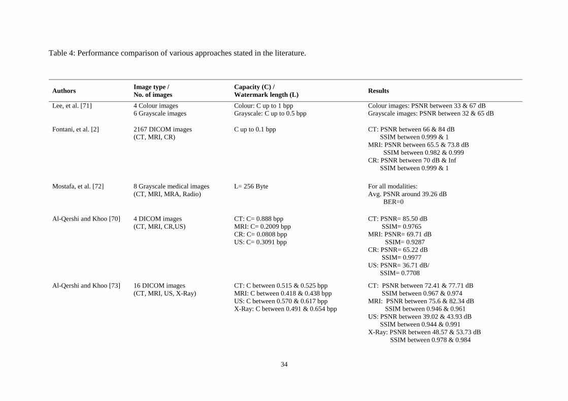
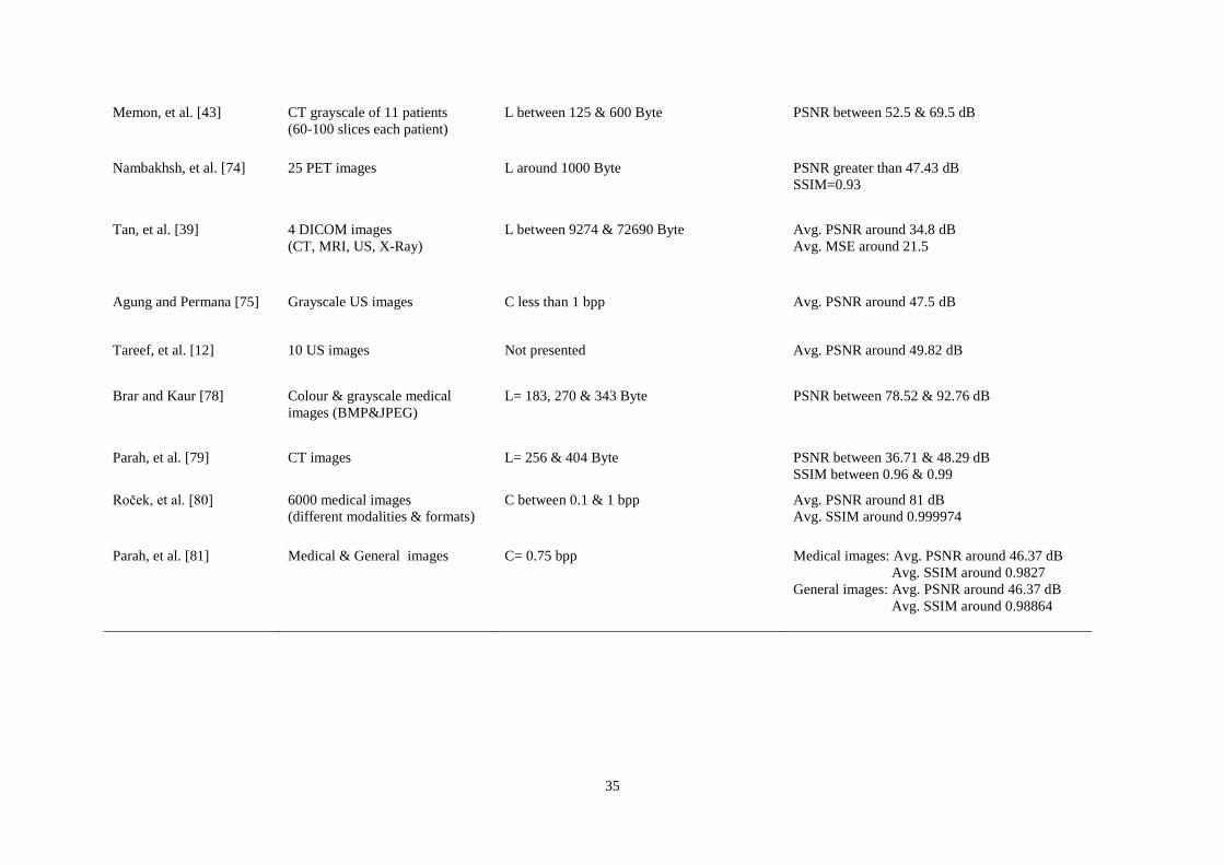
the accuracy of the extracted watermark. Performance comparison of the different approaches
that have been discussed in this research regarding these benchmarks is also demonstrated in
Table 3
4.1. Imperceptibility Assessment of Watermarked Image
There are several metrics used to estimate the distortion of watermarked images. Mean Square
Error (MSE), PSNR and SSIM index are the most popular metrics utilised for this purpose.
The PSNR and MSE metrics measure the error sensitivity variations between the unmodified
and modified images while the SSIM metric draws more concern to the structures of these
images [88]. In all of the following equations, N×M is the images dimension, and I, Iw
represent the original and the watermarked images, respectively.
4.1.1. Mean Square Error
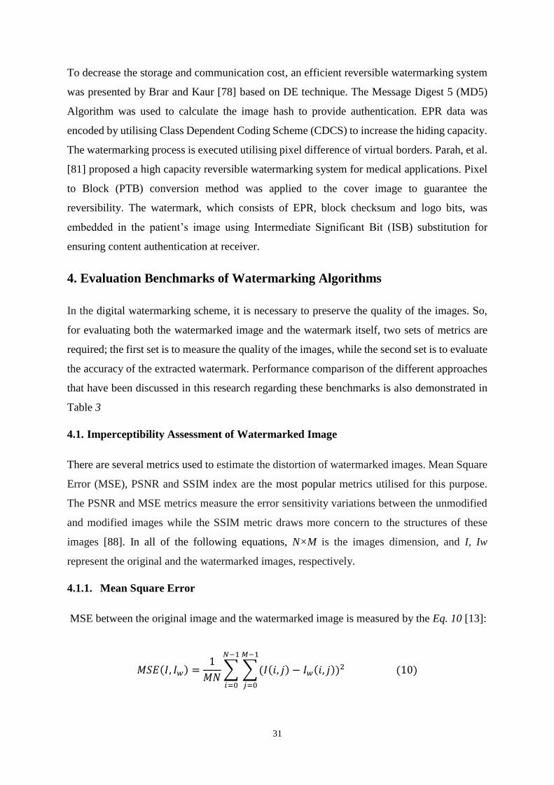
MSE between the original image and the watermarked image is measured by the Eq. 10 [13]:
𝑀𝑆𝐸(𝐼, 𝐼𝑤) =1
𝑀𝑁∑ ∑ (𝐼(𝑖, 𝑗) − 𝐼𝑤(𝑖, 𝑗))2
𝑀−1
𝑗=0
𝑁−1
𝑖=0
(10)
32
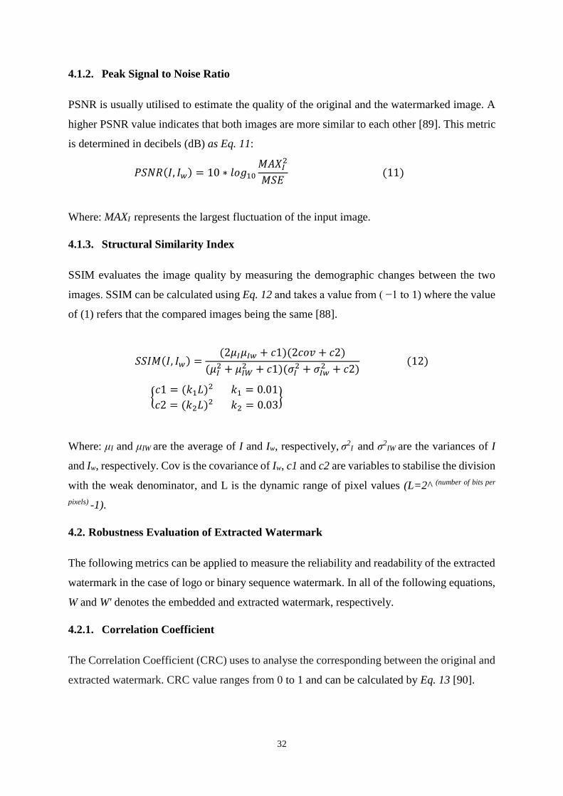
4.1.2. Peak Signal to Noise Ratio
PSNR is usually utilised to estimate the quality of the original and the watermarked image. A
higher PSNR value indicates that both images are more similar to each other [89]. This metric
is determined in decibels (dB) as Eq. 11:
𝑃𝑆𝑁𝑅(𝐼, 𝐼𝑤) = 10 ∗ 𝑙𝑜𝑔10
𝑀𝐴𝑋𝐼2
𝑀𝑆𝐸 (11)
Where: MAXI represents the largest fluctuation of the input image.
4.1.3. Structural Similarity Index
SSIM evaluates the image quality by measuring the demographic changes between the two
images. SSIM can be calculated using Eq. 12 and takes a value from ( −1 to 1) where the value
of (1) refers that the compared images being the same [88].
𝑆𝑆𝐼𝑀(𝐼, 𝐼𝑤) =(2𝜇𝐼𝜇𝐼𝑤 + 𝑐1)(2𝑐𝑜𝑣 + 𝑐2)
(𝜇𝐼2 + 𝜇𝐼𝑊
2 + 𝑐1)(𝜎𝐼2 + 𝜎𝐼𝑤
2 + 𝑐2) (12)
{𝑐1 = (𝑘1𝐿)2 𝑘1 = 0.01
𝑐2 = (𝑘2𝐿)2 𝑘2 = 0.03}
Where: μI and μIW are the average of I and Iw, respectively, σ2
I and σ2IW are the variances of I
and Iw, respectively. Cov is the covariance of Iw, c1 and c2 are variables to stabilise the division
with the weak denominator, and L is the dynamic range of pixel values (L=2^ (number of bits per
pixels) -1).
4.2. Robustness Evaluation of Extracted Watermark
The following metrics can be applied to measure the reliability and readability of the extracted
watermark in the case of logo or binary sequence watermark. In all of the following equations,
W and W' denotes the embedded and extracted watermark, respectively.
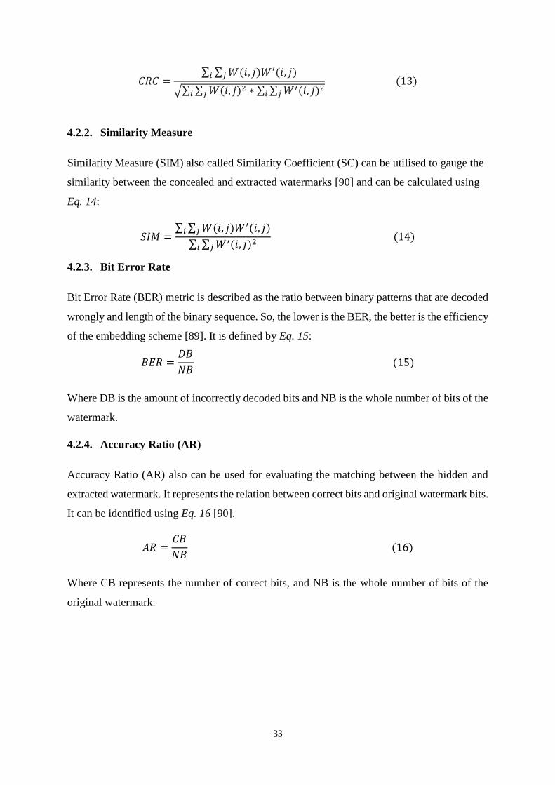
4.2.1. Correlation Coefficient
The Correlation Coefficient (CRC) uses to analyse the corresponding between the original and
extracted watermark. CRC value ranges from 0 to 1 and can be calculated by Eq. 13 [90].
33
𝐶𝑅𝐶 =∑ ∑ 𝑊(𝑖, 𝑗)𝑊′(𝑖, 𝑗)𝑗𝑖
√∑ ∑ 𝑊(𝑖, 𝑗)2𝑗𝑖 ∗ ∑ ∑ 𝑊′(𝑖, 𝑗)2
𝑗𝑖
(13)
4.2.2. Similarity Measure
Similarity Measure (SIM) also called Similarity Coefficient (SC) can be utilised to gauge the
similarity between the concealed and extracted watermarks [90] and can be calculated using
Eq. 14:
𝑆𝐼𝑀 =∑ ∑ 𝑊(𝑖, 𝑗)𝑊′(𝑖, 𝑗)𝑗𝑖
∑ ∑ 𝑊′(𝑖, 𝑗)2𝑗𝑖
(14)
4.2.3. Bit Error Rate
Bit Error Rate (BER) metric is described as the ratio between binary patterns that are decoded
wrongly and length of the binary sequence. So, the lower is the BER, the better is the efficiency
of the embedding scheme [89]. It is defined by Eq. 15:
𝐵𝐸𝑅 =𝐷𝐵
𝑁𝐵 (15)
Where DB is the amount of incorrectly decoded bits and NB is the whole number of bits of the
watermark.
4.2.4. Accuracy Ratio (AR)
Accuracy Ratio (AR) also can be used for evaluating the matching between the hidden and
extracted watermark. It represents the relation between correct bits and original watermark bits.
It can be identified using Eq. 16 [90].
𝐴𝑅 =𝐶𝐵
𝑁𝐵 (16)
Where CB represents the number of correct bits, and NB is the whole number of bits of the
original watermark.
34
Table 4: Performance comparison of various approaches stated in the literature.
Authors Image type /
No. of images
Capacity (C) /
Watermark length (L) Results
Lee, et al. [71] 4 Colour images
6 Grayscale images
Colour: C up to 1 bpp
Grayscale: C up to 0.5 bpp
Colour images: PSNR between 33 & 67 dB
Grayscale images: PSNR between 32 & 65 dB
Fontani, et al. [2] 2167 DICOM images
(CT, MRI, CR)
C up to 0.1 bpp CT: PSNR between 66 & 84 dB
SSIM between 0.999 & 1
MRI: PSNR between 65.5 & 73.8 dB
SSIM between 0.982 & 0.999
CR: PSNR between 70 dB & Inf
SSIM between 0.999 & 1
Mostafa, et al. [72] 8 Grayscale medical images
(CT, MRI, MRA, Radio)
L= 256 Byte For all modalities:
Avg. PSNR around 39.26 dB
BER=0
Al-Qershi and Khoo [70] 4 DICOM images
(CT, MRI, CR,US)
CT: C= 0.888 bpp
MRI: C= 0.2009 bpp
CR: C= 0.0808 bpp
US: C= 0.3091 bpp
CT: PSNR= 85.50 dB
SSIM= 0.9765
MRI: PSNR= 69.71 dB
SSIM= 0.9287
CR: PSNR= 65.22 dB
SSIM= 0.9977
US: PSNR= 36.71 dB/
SSIM= 0.7708
Al-Qershi and Khoo [73] 16 DICOM images
(CT, MRI, US, X-Ray)
CT: C between 0.515 & 0.525 bpp
MRI: C between 0.418 & 0.438 bpp
US: C between 0.570 & 0.617 bpp
X-Ray: C between 0.491 & 0.654 bpp
CT: PSNR between 72.41 & 77.71 dB
SSIM between 0.967 & 0.974
MRI: PSNR between 75.6 & 82.34 dB
SSIM between 0.946 & 0.961
US: PSNR between 39.02 & 43.93 dB
SSIM between 0.944 & 0.991
X-Ray: PSNR between 48.57 & 53.73 dB
SSIM between 0.978 & 0.984
35
Memon, et al. [43] CT grayscale of 11 patients
(60-100 slices each patient)
L between 125 & 600 Byte PSNR between 52.5 & 69.5 dB
Nambakhsh, et al. [74] 25 PET images L around 1000 Byte PSNR greater than 47.43 dB
SSIM=0.93
Tan, et al. [39] 4 DICOM images
(CT, MRI, US, X-Ray)
L between 9274 & 72690 Byte Avg. PSNR around 34.8 dB
Avg. MSE around 21.5
Agung and Permana [75] Grayscale US images C less than 1 bpp Avg. PSNR around 47.5 dB
Tareef, et al. [12] 10 US images Not presented Avg. PSNR around 49.82 dB
Brar and Kaur [78] Colour & grayscale medical
images (BMP&JPEG)
L= 183, 270 & 343 Byte PSNR between 78.52 & 92.76 dB
Parah, et al. [79] CT images L= 256 & 404 Byte PSNR between 36.71 & 48.29 dB
SSIM between 0.96 & 0.99
Roček, et al. [80] 6000 medical images
(different modalities & formats)
C between 0.1 & 1 bpp Avg. PSNR around 81 dB
Avg. SSIM around 0.999974
Parah, et al. [81] Medical & General images C= 0.75 bpp Medical images: Avg. PSNR around 46.37 dB
Avg. SSIM around 0.9827
General images: Avg. PSNR around 46.37 dB
Avg. SSIM around 0.98864
36
5. Discussion and Conclusions
The necessity of protecting medical images and other patients’ data is not only for
confidentiality purposes but also to prevent manipulations that might happen by authorised and
unauthorised users while using these images. Therefore, there is a need to use a technique for
ensuring trust in digital medical workflows. Digital watermarking has been recognised as a
favourable approach for ensuring data integrity and authenticity in medical environments. In
this paper, we have presented a comprehensive review of medical image watermarking
schemes and discussed various issues related to each approach.
Many techniques have been proposed in the literature for watermarking the medical images
utilising both spatial and transform domains. These techniques hide the watermark in the whole
image or in the images' ROI and RONI by implementing reversible and irreversible methods.
In comparison to the transform domain techniques, which are suitable for ownership
verification applications, techniques based on the spatial domain are less complex and provide
higher capacity and visual quality. However, the spatial domain methods are fragile and cannot
survive against many operations making them appropriate for integrity and authentication
applications. Also, it is noticed that transform domain schemes based on DCT and DFT are
rarely used for medical image watermarking and the majority of the studies prefer the
techniques based on DWT due to it is offering an accurate matching of the human visual
system.
RONI watermarking systems embed watermarks in regions that are insignificant in medical
diagnosis, but they have several drawbacks such as they can be only applied if a RONI exists,
the size of the watermark depends on the RONI size and also the ROI may not be protected
against malicious attacks. So, applying these methods depends on the image characteristics.
Medical requirements are extremely strict with the quality of medical images and do not allow
non-clinical based modification in any way. The irreversible watermarking methods remain
subject to acceptance by the radiologists while the original images stay typically favoured for
diagnosis purposes. So, the watermarking algorithms applied to medical images should have
the ability to retrieve the original non-modified image. Reversible watermarking assures
recovering the original image precisely after extracting the embedded watermark successfully.
Consequently, the hiding capacity and the number of potential methods that can be performed
on medical images have been restricted significantly because of this feature.
37
In order to apply watermarking to medical information systems, it is fundamental to choose an
appropriate and reliable approach. The performance of all watermarking schemes, which have
been reviewed in this paper, had been assessed only in terms of perceptibility by utilising
physical metrics. PSNR and SSIM benchmarks are the most widely used physical image quality
metrics, but they do not take into account all characteristics that are clinically relevant in getting
an accurate medical diagnosis [91]. Therefore, this study recommends relating these metrics to
the visual/clinical assessment approaches to improve their validity and applicability. This needs
expert images' readers to visually evaluate the differences between the original and
watermarked images through an objective question set. Also, it is recommended that further
work is required for testing the watermarking system regarding image quality in a fully
operational PACS where the medical images are archived and retrieved.
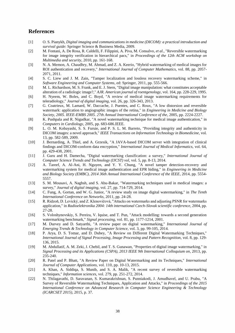
38
References
[1] O. S. Pianykh, Digital imaging and communications in medicine (DICOM): a practical introduction and
survival guide: Springer Science & Business Media, 2009.
[2] M. Fontani, A. De Rosa, R. Caldelli, F. Filippini, A. Piva, M. Consalvo, et al., "Reversible watermarking
for image integrity verification in hierarchical pacs," in Proceedings of the 12th ACM workshop on
Multimedia and security, 2010, pp. 161-168.
[3] N. A. Memon, A. Chaudhry, M. Ahmad, and Z. A. Keerio, "Hybrid watermarking of medical images for
ROI authentication and recovery," International Journal of Computer Mathematics, vol. 88, pp. 2057-
2071, 2011.
[4] S. C. Liew and J. M. Zain, "Tamper localization and lossless recovery watermarking scheme," in
Software Engineering and Computer Systems, ed: Springer, 2011, pp. 555-566.
[5] M. L. Richardson, M. S. Frank, and E. J. Stern, "Digital image manipulation: what constitutes acceptable
alteration of a radiologic image?," AJR. American journal of roentgenology, vol. 164, pp. 228-229, 1995.
[6] H. Nyeem, W. Boles, and C. Boyd, "A review of medical image watermarking requirements for
teleradiology," Journal of digital imaging, vol. 26, pp. 326-343, 2013.
[7] G. Coatrieux, M. Lamard, W. Daccache, J. Puentes, and C. Roux, "A low distorsion and reversible
watermark: application to angiographic images of the retina," in Engineering in Medicine and Biology
Society, 2005. IEEE-EMBS 2005. 27th Annual International Conference of the, 2005, pp. 2224-2227.
[8] K. Pushpala and R. Nigudkar, "A novel watermarking technique for medical image authentication," in
Computers in Cardiology, 2005, pp. 683-686.IEEE.
[9] L. O. M. Kobayashi, S. S. Furuie, and P. S. L. M. Barreto, "Providing integrity and authenticity in
DICOM images: a novel approach," IEEE Transactions on Information Technology in Biomedicine, vol.
13, pp. 582-589, 2009.
[10] J. Bernarding, A. Thiel, and A. Grzesik, "A JAVA-based DICOM server with integration of clinical
findings and DICOM-conform data encryption," International Journal of Medical Informatics, vol. 64,
pp. 429-438, 2001.
[11] J. Guru and H. Damecha, "Digital watermarking classification: a survey," International Journal of
Computer Science Trends and Technology (IJCST) vol, vol. 5, pp. 8-13, 2014.
[12] A. Tareef, A. Al-Ani, H. Nguyen, and Y. Y. Chung, "A novel tamper detection-recovery and
watermarking system for medical image authentication and EPR hiding," in Engineering in Medicine
and Biology Society (EMBC), 2014 36th Annual International Conference of the IEEE, 2014, pp. 5554-
5557.
[13] S. M. Mousavi, A. Naghsh, and S. Abu-Bakar, "Watermarking techniques used in medical images: a
survey," Journal of digital imaging, vol. 27, pp. 714-729, 2014.
[14] C. Fung, A. Gortan, and W. G. Junior, "A review study on image digital watermarking," in The Tenth
International Conference on Networks, 2011, pp. 24-28.
[15] R. Ridzoň, D. Levický, and Z. Klenovičová, "Attacks on watermarks and adjusting PSNR for watermarks
application," in Radioelektronika 2004: 14th international Czech-Slovak scientific conference, 2004, pp.
27-28.
[16] S. Voloshynovskiy, S. Pereira, V. Iquise, and T. Pun, "Attack modelling: towards a second generation
watermarking benchmark," Signal processing, vol. 81, pp. 1177-1214, 2001.
[17] M. Durvey and D. Satyarthi, "A review paper on digital watermarking," International Journal of
Emerging Trends & Technology in Computer Science, vol. 3, pp. 99-105, 2014.
[18] P. Arya, D. S. Tomar, and D. Dubey, "A Review on Different Digital Watermarking Techniques,"
International Journal of Signal Processing, Image Processing and Pattern Recognition, vol. 8, pp. 129-
136, 2015.
[19] M. Abdullatif, A. M. Zeki, J. Chebil, and T. S. Gunawan, "Properties of digital image watermarking," in
Signal Processing and its Applications (CSPA), 2013 IEEE 9th International Colloquium on, 2013, pp.
235-240.
[20] R. Patel and P. Bhatt, "A Review Paper on Digital Watermarking and its Techniques," International
Journal of Computer Applications, vol. 110, pp. 10-13, 2015.
[21] A. Khan, A. Siddiqa, S. Munib, and S. A. Malik, "A recent survey of reversible watermarking
techniques," Information sciences, vol. 279, pp. 251-272, 2014.
[22] N. Thilagavathi, D. Saravanan, S. Kumarakrishnan, S. Punniakodi, J. Amudhavel, and U. Prabu, "A
Survey of Reversible Watermarking Techniques, Application and Attacks," in Proceedings of the 2015
International Conference on Advanced Research in Computer Science Engineering & Technology
(ICARCSET 2015), 2015, p. 37.
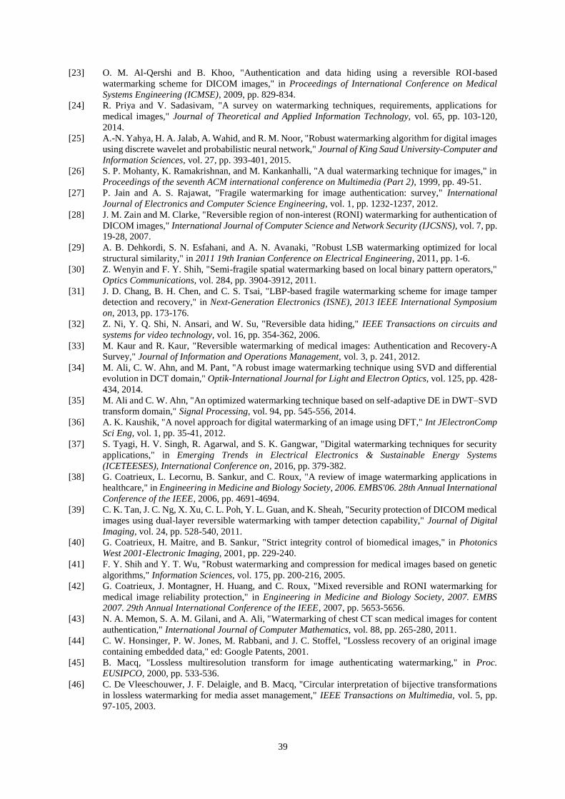
39
[23] O. M. Al-Qershi and B. Khoo, "Authentication and data hiding using a reversible ROI-based
watermarking scheme for DICOM images," in Proceedings of International Conference on Medical
Systems Engineering (ICMSE), 2009, pp. 829-834.
[24] R. Priya and V. Sadasivam, "A survey on watermarking techniques, requirements, applications for
medical images," Journal of Theoretical and Applied Information Technology, vol. 65, pp. 103-120,
2014.
[25] A.-N. Yahya, H. A. Jalab, A. Wahid, and R. M. Noor, "Robust watermarking algorithm for digital images
using discrete wavelet and probabilistic neural network," Journal of King Saud University-Computer and
Information Sciences, vol. 27, pp. 393-401, 2015.
[26] S. P. Mohanty, K. Ramakrishnan, and M. Kankanhalli, "A dual watermarking technique for images," in
Proceedings of the seventh ACM international conference on Multimedia (Part 2), 1999, pp. 49-51.
[27] P. Jain and A. S. Rajawat, "Fragile watermarking for image authentication: survey," International
Journal of Electronics and Computer Science Engineering, vol. 1, pp. 1232-1237, 2012.
[28] J. M. Zain and M. Clarke, "Reversible region of non-interest (RONI) watermarking for authentication of
DICOM images," International Journal of Computer Science and Network Security (IJCSNS), vol. 7, pp.
19-28, 2007.
[29] A. B. Dehkordi, S. N. Esfahani, and A. N. Avanaki, "Robust LSB watermarking optimized for local
structural similarity," in 2011 19th Iranian Conference on Electrical Engineering, 2011, pp. 1-6.
[30] Z. Wenyin and F. Y. Shih, "Semi-fragile spatial watermarking based on local binary pattern operators,"
Optics Communications, vol. 284, pp. 3904-3912, 2011.
[31] J. D. Chang, B. H. Chen, and C. S. Tsai, "LBP-based fragile watermarking scheme for image tamper
detection and recovery," in Next-Generation Electronics (ISNE), 2013 IEEE International Symposium
on, 2013, pp. 173-176.
[32] Z. Ni, Y. Q. Shi, N. Ansari, and W. Su, "Reversible data hiding," IEEE Transactions on circuits and
systems for video technology, vol. 16, pp. 354-362, 2006.
[33] M. Kaur and R. Kaur, "Reversible watermarking of medical images: Authentication and Recovery-A
Survey," Journal of Information and Operations Management, vol. 3, p. 241, 2012.
[34] M. Ali, C. W. Ahn, and M. Pant, "A robust image watermarking technique using SVD and differential
evolution in DCT domain," Optik-International Journal for Light and Electron Optics, vol. 125, pp. 428-
434, 2014.
[35] M. Ali and C. W. Ahn, "An optimized watermarking technique based on self-adaptive DE in DWT–SVD
transform domain," Signal Processing, vol. 94, pp. 545-556, 2014.
[36] A. K. Kaushik, "A novel approach for digital watermarking of an image using DFT," Int JElectronComp
Sci Eng, vol. 1, pp. 35-41, 2012.
[37] S. Tyagi, H. V. Singh, R. Agarwal, and S. K. Gangwar, "Digital watermarking techniques for security
applications," in Emerging Trends in Electrical Electronics & Sustainable Energy Systems
(ICETEESES), International Conference on, 2016, pp. 379-382.
[38] G. Coatrieux, L. Lecornu, B. Sankur, and C. Roux, "A review of image watermarking applications in
healthcare," in Engineering in Medicine and Biology Society, 2006. EMBS'06. 28th Annual International
Conference of the IEEE, 2006, pp. 4691-4694.
[39] C. K. Tan, J. C. Ng, X. Xu, C. L. Poh, Y. L. Guan, and K. Sheah, "Security protection of DICOM medical
images using dual-layer reversible watermarking with tamper detection capability," Journal of Digital
Imaging, vol. 24, pp. 528-540, 2011.
[40] G. Coatrieux, H. Maitre, and B. Sankur, "Strict integrity control of biomedical images," in Photonics
West 2001-Electronic Imaging, 2001, pp. 229-240.
[41] F. Y. Shih and Y. T. Wu, "Robust watermarking and compression for medical images based on genetic
algorithms," Information Sciences, vol. 175, pp. 200-216, 2005.
[42] G. Coatrieux, J. Montagner, H. Huang, and C. Roux, "Mixed reversible and RONI watermarking for
medical image reliability protection," in Engineering in Medicine and Biology Society, 2007. EMBS
2007. 29th Annual International Conference of the IEEE, 2007, pp. 5653-5656.
[43] N. A. Memon, S. A. M. Gilani, and A. Ali, "Watermarking of chest CT scan medical images for content
authentication," International Journal of Computer Mathematics, vol. 88, pp. 265-280, 2011.
[44] C. W. Honsinger, P. W. Jones, M. Rabbani, and J. C. Stoffel, "Lossless recovery of an original image
containing embedded data," ed: Google Patents, 2001.
[45] B. Macq, "Lossless multiresolution transform for image authenticating watermarking," in Proc.
EUSIPCO, 2000, pp. 533-536.
[46] C. De Vleeschouwer, J. F. Delaigle, and B. Macq, "Circular interpretation of bijective transformations
in lossless watermarking for media asset management," IEEE Transactions on Multimedia, vol. 5, pp.
97-105, 2003.
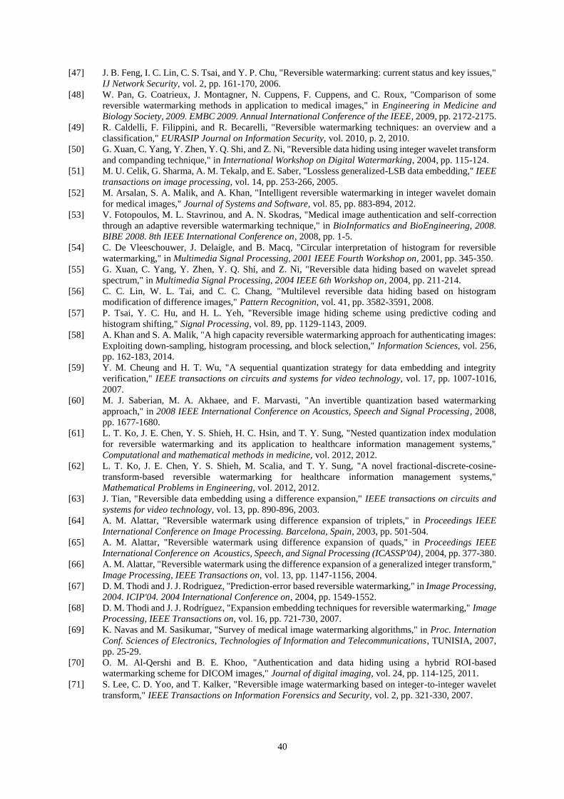
40
[47] J. B. Feng, I. C. Lin, C. S. Tsai, and Y. P. Chu, "Reversible watermarking: current status and key issues,"
IJ Network Security, vol. 2, pp. 161-170, 2006.
[48] W. Pan, G. Coatrieux, J. Montagner, N. Cuppens, F. Cuppens, and C. Roux, "Comparison of some
reversible watermarking methods in application to medical images," in Engineering in Medicine and
Biology Society, 2009. EMBC 2009. Annual International Conference of the IEEE, 2009, pp. 2172-2175.
[49] R. Caldelli, F. Filippini, and R. Becarelli, "Reversible watermarking techniques: an overview and a
classification," EURASIP Journal on Information Security, vol. 2010, p. 2, 2010.
[50] G. Xuan, C. Yang, Y. Zhen, Y. Q. Shi, and Z. Ni, "Reversible data hiding using integer wavelet transform
and companding technique," in International Workshop on Digital Watermarking, 2004, pp. 115-124.
[51] M. U. Celik, G. Sharma, A. M. Tekalp, and E. Saber, "Lossless generalized-LSB data embedding," IEEE
transactions on image processing, vol. 14, pp. 253-266, 2005.
[52] M. Arsalan, S. A. Malik, and A. Khan, "Intelligent reversible watermarking in integer wavelet domain
for medical images," Journal of Systems and Software, vol. 85, pp. 883-894, 2012.
[53] V. Fotopoulos, M. L. Stavrinou, and A. N. Skodras, "Medical image authentication and self-correction
through an adaptive reversible watermarking technique," in BioInformatics and BioEngineering, 2008.
BIBE 2008. 8th IEEE International Conference on, 2008, pp. 1-5.
[54] C. De Vleeschouwer, J. Delaigle, and B. Macq, "Circular interpretation of histogram for reversible
watermarking," in Multimedia Signal Processing, 2001 IEEE Fourth Workshop on, 2001, pp. 345-350.
[55] G. Xuan, C. Yang, Y. Zhen, Y. Q. Shi, and Z. Ni, "Reversible data hiding based on wavelet spread
spectrum," in Multimedia Signal Processing, 2004 IEEE 6th Workshop on, 2004, pp. 211-214.
[56] C. C. Lin, W. L. Tai, and C. C. Chang, "Multilevel reversible data hiding based on histogram
modification of difference images," Pattern Recognition, vol. 41, pp. 3582-3591, 2008.
[57] P. Tsai, Y. C. Hu, and H. L. Yeh, "Reversible image hiding scheme using predictive coding and
histogram shifting," Signal Processing, vol. 89, pp. 1129-1143, 2009.
[58] A. Khan and S. A. Malik, "A high capacity reversible watermarking approach for authenticating images:
Exploiting down-sampling, histogram processing, and block selection," Information Sciences, vol. 256,
pp. 162-183, 2014.
[59] Y. M. Cheung and H. T. Wu, "A sequential quantization strategy for data embedding and integrity
verification," IEEE transactions on circuits and systems for video technology, vol. 17, pp. 1007-1016,
2007.
[60] M. J. Saberian, M. A. Akhaee, and F. Marvasti, "An invertible quantization based watermarking
approach," in 2008 IEEE International Conference on Acoustics, Speech and Signal Processing, 2008,
pp. 1677-1680.
[61] L. T. Ko, J. E. Chen, Y. S. Shieh, H. C. Hsin, and T. Y. Sung, "Nested quantization index modulation
for reversible watermarking and its application to healthcare information management systems,"
Computational and mathematical methods in medicine, vol. 2012, 2012.
[62] L. T. Ko, J. E. Chen, Y. S. Shieh, M. Scalia, and T. Y. Sung, "A novel fractional-discrete-cosine-
transform-based reversible watermarking for healthcare information management systems,"
Mathematical Problems in Engineering, vol. 2012, 2012.
[63] J. Tian, "Reversible data embedding using a difference expansion," IEEE transactions on circuits and
systems for video technology, vol. 13, pp. 890-896, 2003.
[64] A. M. Alattar, "Reversible watermark using difference expansion of triplets," in Proceedings IEEE
International Conference on Image Processing. Barcelona, Spain, 2003, pp. 501-504.
[65] A. M. Alattar, "Reversible watermark using difference expansion of quads," in Proceedings IEEE
International Conference on Acoustics, Speech, and Signal Processing (ICASSP'04), 2004, pp. 377-380.
[66] A. M. Alattar, "Reversible watermark using the difference expansion of a generalized integer transform,"
Image Processing, IEEE Transactions on, vol. 13, pp. 1147-1156, 2004.
[67] D. M. Thodi and J. J. Rodriguez, "Prediction-error based reversible watermarking," in Image Processing,
2004. ICIP'04. 2004 International Conference on, 2004, pp. 1549-1552.
[68] D. M. Thodi and J. J. Rodríguez, "Expansion embedding techniques for reversible watermarking," Image
Processing, IEEE Transactions on, vol. 16, pp. 721-730, 2007.
[69] K. Navas and M. Sasikumar, "Survey of medical image watermarking algorithms," in Proc. Internation
Conf. Sciences of Electronics, Technologies of Information and Telecommunications, TUNISIA, 2007,
pp. 25-29.
[70] O. M. Al-Qershi and B. E. Khoo, "Authentication and data hiding using a hybrid ROI-based
watermarking scheme for DICOM images," Journal of digital imaging, vol. 24, pp. 114-125, 2011.
[71] S. Lee, C. D. Yoo, and T. Kalker, "Reversible image watermarking based on integer-to-integer wavelet
transform," IEEE Transactions on Information Forensics and Security, vol. 2, pp. 321-330, 2007.
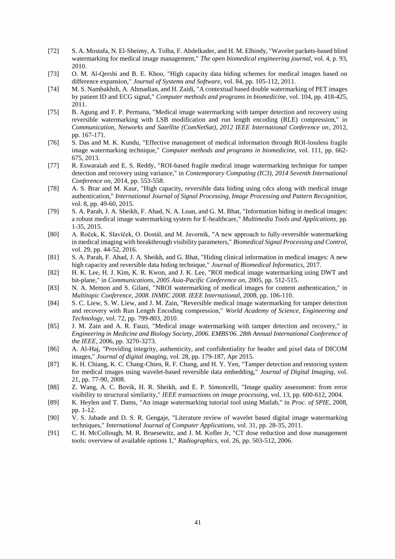
41
[72] S. A. Mostafa, N. El-Sheimy, A. Tolba, F. Abdelkader, and H. M. Elhindy, "Wavelet packets-based blind
watermarking for medical image management," The open biomedical engineering journal, vol. 4, p. 93,
2010.
[73] O. M. Al-Qershi and B. E. Khoo, "High capacity data hiding schemes for medical images based on
difference expansion," Journal of Systems and Software, vol. 84, pp. 105-112, 2011.
[74] M. S. Nambakhsh, A. Ahmadian, and H. Zaidi, "A contextual based double watermarking of PET images
by patient ID and ECG signal," Computer methods and programs in biomedicine, vol. 104, pp. 418-425,
2011.
[75] B. Agung and F. P. Permana, "Medical image watermarking with tamper detection and recovery using
reversible watermarking with LSB modification and run length encoding (RLE) compression," in
Communication, Networks and Satellite (ComNetSat), 2012 IEEE International Conference on, 2012,
pp. 167-171.
[76] S. Das and M. K. Kundu, "Effective management of medical information through ROI-lossless fragile
image watermarking technique," Computer methods and programs in biomedicine, vol. 111, pp. 662-
675, 2013.
[77] R. Eswaraiah and E. S. Reddy, "ROI-based fragile medical image watermarking technique for tamper
detection and recovery using variance," in Contemporary Computing (IC3), 2014 Seventh International
Conference on, 2014, pp. 553-558.
[78] A. S. Brar and M. Kaur, "High capacity, reversible data hiding using cdcs along with medical image
authentication," International Journal of Signal Processing, Image Processing and Pattern Recognition,
vol. 8, pp. 49-60, 2015.
[79] S. A. Parah, J. A. Sheikh, F. Ahad, N. A. Loan, and G. M. Bhat, "Information hiding in medical images:
a robust medical image watermarking system for E-healthcare," Multimedia Tools and Applications, pp.
1-35, 2015.
[80] A. Roček, K. Slavíček, O. Dostál, and M. Javorník, "A new approach to fully-reversible watermarking
in medical imaging with breakthrough visibility parameters," Biomedical Signal Processing and Control,
vol. 29, pp. 44-52, 2016.
[81] S. A. Parah, F. Ahad, J. A. Sheikh, and G. Bhat, "Hiding clinical information in medical images: A new
high capacity and reversible data hiding technique," Journal of Biomedical Informatics, 2017.
[82] H. K. Lee, H. J. Kim, K. R. Kwon, and J. K. Lee, "ROI medical image watermarking using DWT and
bit-plane," in Communications, 2005 Asia-Pacific Conference on, 2005, pp. 512-515.
[83] N. A. Memon and S. Gilani, "NROI watermarking of medical images for content authentication," in
Multitopic Conference, 2008. INMIC 2008. IEEE International, 2008, pp. 106-110.
[84] S. C. Liew, S. W. Liew, and J. M. Zain, "Reversible medical image watermarking for tamper detection
and recovery with Run Length Encoding compression," World Academy of Science, Engineering and
Technology, vol. 72, pp. 799-803, 2010.
[85] J. M. Zain and A. R. Fauzi, "Medical image watermarking with tamper detection and recovery," in
Engineering in Medicine and Biology Society, 2006. EMBS'06. 28th Annual International Conference of
the IEEE, 2006, pp. 3270-3273.
[86] A. Al-Haj, "Providing integrity, authenticity, and confidentiality for header and pixel data of DICOM
images," Journal of digital imaging, vol. 28, pp. 179-187, Apr 2015.
[87] K. H. Chiang, K. C. Chang-Chien, R. F. Chang, and H. Y. Yen, "Tamper detection and restoring system
for medical images using wavelet-based reversible data embedding," Journal of Digital Imaging, vol.
21, pp. 77-90, 2008.
[88] Z. Wang, A. C. Bovik, H. R. Sheikh, and E. P. Simoncelli, "Image quality assessment: from error
visibility to structural similarity," IEEE transactions on image processing, vol. 13, pp. 600-612, 2004.
[89] K. Heylen and T. Dams, "An image watermarking tutorial tool using Matlab," in Proc. of SPIE, 2008,
pp. 1-12.
[90] V. S. Jabade and D. S. R. Gengaje, "Literature review of wavelet based digital image watermarking
techniques," International Journal of Computer Applications, vol. 31, pp. 28-35, 2011.
[91] C. H. McCollough, M. R. Bruesewitz, and J. M. Kofler Jr, "CT dose reduction and dose management
tools: overview of available options 1," Radiographics, vol. 26, pp. 503-512, 2006.