disposition and metabolism...
TRANSCRIPT

DMD # 35386
1
DISPOSITION AND METABOLISM OF
[14C]SB-649868 AN OREXIN 1 and 2 RECEPTOR
ANTAGONIST IN HUMANS
Authors:
Cecilia Renzulli, Mike Nash, Mark Wright, Steven Thomas, Stefano Zamuner, Mario
Pellegatti, Paolo Bettica and Gary Boyle.
Department of Drug Metabolism and Pharmacokinetics (CR, MN, MW, ST, MP, GB),
Clinical Pharmacology Modeling and Simulation (SZ) and Discovery Medicine (PB),
GlaxoSmithKline Research & Development, Verona, Italy and Ware, United Kingdom
DMD Fast Forward. Published on November 2, 2010 as doi:10.1124/dmd.110.035386
Copyright 2010 by the American Society for Pharmacology and Experimental Therapeutics.
This article has not been copyedited and formatted. The final version may differ from this version.DMD Fast Forward. Published on November 2, 2010 as DOI: 10.1124/dmd.110.035386
at ASPE
T Journals on February 23, 2020
dmd.aspetjournals.org
Dow
nloaded from
DMD # 35386
2
RUNNING TITLE
a) Disposition and metabolism of [14C]SB-649868 in humans
b) Gary Boyle, DMPK, GlaxoSmithKline, Ware (UK), tel 0044 1920 882465, fax 0044
1920 884374, [email protected]
Number of text pages: 25
Number of Tables: 4
Number of Figures: 6
Number of References: 16
Number of words in Abstract: 231 words
Number of words in Introduction: 258 words
Number of words in Discussion: 1529 words
d) Abbreviations used are:
AUC = area under the plasma concentration-time curve
BQL = below quantification limit
Cmax = maximal plasma concentration
CL = systemic plasma clearance
CI = confidence interval
F= Bioavailability
HPLC = high performance liquid chromatography
HPLC-MS/MS = high performance liquid chromatography tandem mass spectrometry
QC = quality control
Q-TOF = quadrupole/ time-of-flight
NMR = nuclear magnetic resonance spectroscopy
SB-649868 = N-[[(2S)-1-[[5-(4-fluorophenyl)-2-methyl-4-thiazolyl]carbonyl]-2-
piperidinyl]methyl]-4-benzofurancarboxamide
This article has not been copyedited and formatted. The final version may differ from this version.DMD Fast Forward. Published on November 2, 2010 as DOI: 10.1124/dmd.110.035386
at ASPE
T Journals on February 23, 2020
dmd.aspetjournals.org
Dow
nloaded from

DMD # 35386
3
tmax = time to reach Cmax
t1/2= half life
WMA= World Medical Association
This article has not been copyedited and formatted. The final version may differ from this version.DMD Fast Forward. Published on November 2, 2010 as DOI: 10.1124/dmd.110.035386
at ASPE
T Journals on February 23, 2020
dmd.aspetjournals.org
Dow
nloaded from

DMD # 35386
4
ABSTRACT
SB-649868 is a novel orexin 1 and 2 receptor antagonist under development for
insomnia treatment. The disposition of [14C]SB-649868 was determined in eight
healthy male subjects using an open label study design after a single oral dose of 30
mg. Blood, urine and feces were collected at frequent intervals after dosing and
samples were analyzed by High Performance Liquid Chromatography/Mass
Spectrometry coupled with off-line radiodetection for metabolite profiling and
characterization. Nuclear magnetic resonance spectroscopy was also employed to
further characterize certain metabolites. Elimination of drug-related material was
almost complete over a 9 day period, occurring principally via the feces (79%),
whereas urinary excretion accounted only for 12% of total radioactivity. Mean
apparent half life (t1/2) of plasma radioactivity was notably longer (39.3 h), with
respect to that of unchanged SB-649868 (4.8 h), suggesting the presence of more
slowly cleared metabolites. SB-649868 and an unusual hemiaminal metabolite, M98
(GSK2329163), resulting from oxidation of the benzofuran ring and subsequent re-
arrangement, were the principal circulating components in plasma extracts. Two
additional minor metabolites were also observed, a benzofuran ring opened
carboxylic acid M25 (GSK2329158) and an amine metabolite (M8). SB-649868 was
extensively metabolized and only negligible amounts were excreted unchanged. The
principal route of metabolism was via oxidation of the benzofuran ring with the
resultant M25 being the principal metabolite in excreta, representing at least 12% of
the administered dose across urine and feces.
This article has not been copyedited and formatted. The final version may differ from this version.DMD Fast Forward. Published on November 2, 2010 as DOI: 10.1124/dmd.110.035386
at ASPE
T Journals on February 23, 2020
dmd.aspetjournals.org
Dow
nloaded from

DMD # 35386
5
INTRODUCTION
SB-649868, N-[[(2S)-1-[[5-(4-fluorophenyl)-2-methyl-4-thiazolyl]carbonyl]-2-
piperidinyl]methyl]-4-benzofurancarboxamide, is a potent and selective orally active
orexin 1 and orexin 2 (OX1/OX2) receptor antagonist under development for
insomnia treatment. A large body of evidence supports a role for orexin in the control
of arousal and sleep/wake regulation [Lin et al., 1999; Chemilli et al., 1999; Kilduff
and Peyron, 2000].
Animal studies have shown that SB-649868 has a hypnotic activity similar to that of
zolpidem, but that it can be differentiated from benzodiazepine-like agents acting at
the GABAA receptor complex, in terms of hypnotic profile and side effect potential
(Gerrard et al., manuscript in preparation).
Single oral doses up to a maximum dose of 80 mg and repeat doses up to a
maximum dose of 30 mg have been administered to healthy volunteers (Bettica et
al., submitted). After single dose, SB-649868 Cmax and AUC increased
approximately proportionally over the 10-60 mg dose range with terminal phase half-
life in the 2.2-7.4 h range. SB-649868 steady-state exposure was dose- and time-
dependent and generally slightly higher than following single-dose administration.
The excretion, metabolism and pharmacokinetics of SB-649868 in rat, dog and
mouse have been studied (unpublished data). Elimination of SB-649868 in rat and
mouse was largely by metabolism, with metabolites secreted primarily in the bile
and/or feces. Elimination of SB-649898 in the Beagle dog appeared to be largely as
unchanged parent compound via the feces. The elimination half-life was short in rat
and dog (t1/2<1h).
The purpose of this work was to characterize the disposition and metabolism of [14C]
SB-649868 after a single oral administration to healthy male volunteers.
This article has not been copyedited and formatted. The final version may differ from this version.DMD Fast Forward. Published on November 2, 2010 as DOI: 10.1124/dmd.110.035386
at ASPE
T Journals on February 23, 2020
dmd.aspetjournals.org
Dow
nloaded from

DMD # 35386
6
MATERIALS AND METHODS
Chemicals. [14C]SB-649868, SB-649868, [2H4]SB-649868 (deuterated in the
monofluoro ring) and two synthetic metabolites (provided as a semi-crude reaction
mixture), M25 (coded as GSK2329158) and M98 (coded as GSK2329163), were all
supplied by Synthetic Chemistry, GlaxoSmithKline Research and Development,
Stevenage (UK). These materials were used as chromatographic, mass
spectrometric and/or nuclear magnetic resonance spectroscopy standards during the
study. Commercially obtained chemicals and solvents were of high-performance
liquid chromatography (HPLC) or analytical grade. Liquid scintillation cocktails were
obtained from PerkinElmer LAS (UK) Limited (Beaconsfield, Bucks, UK).
SB-649868 Formulation. [14C]SB-649868 (2.37 µCi /mg, radiochemical purity
99.8%) was provided as powder in the bottle, to be reconstituted into a solution prior
to dosing via a hard gelatine capsule (00 size). [14C]SB-649868 (604 mg, 1.43 mCi)
was weighed into a new glass vial. Propylene glycol monocaprylate (14.2 g) was
added to the vial and the vial sonicated for 80 min and stability of the dose
subsequently confirmed. A portion of the solution (0.737 g, equivalent to 30 mg of
[14C]SB-649868) was weighed into pre-weighed capsules.
Study Design and Subjects. The clinical study (GSK study number OXS109139)
was performed at Charles River Laboratories Clinical Services (Edinburgh, UK) in
accordance with Good Clinical Practice and the principles of the WMA of the
Declaration of Tokyo (2004).The protocol was reviewed and approved, as
appropriate, by the Edinburgh Independent Ethics Committee for Medical Research
and the Medicine and Healthcare products Regulatory Agency (MHRA). The
proposed radioactive dose was approved by the Administration of Radioactive
This article has not been copyedited and formatted. The final version may differ from this version.DMD Fast Forward. Published on November 2, 2010 as DOI: 10.1124/dmd.110.035386
at ASPE
T Journals on February 23, 2020
dmd.aspetjournals.org
Dow
nloaded from

DMD # 35386
7
Substances Advisory Committee (ARSAC). Written consent was obtained from all
subjects before any protocol-specific procedures.
The study was an open-label, single session study. Eight healthy male subjects
(Caucasian), between 30 and 55 years of age, body weight >50 Kg and body mass
index between 18.5 and 29.9 kg/m2 were selected from a panel of volunteers. They
were determined healthy by a responsible physician based on medical evaluation.
Subjects had normal levels of FSH, LH and testosterone at screening and with no
history of drug or alcohol abuse, and were on no other medication at the time of the
study, with no prescribed medication within 14 days of the study commencing.
Study Procedure. All subjects received a single oral administration of 30 mg
(approximately 70 µCi) of [14 C]SB-649868 in a capsule dose swallowed with 160 ml
of water. After oral administration, blood samples (7 ml) for total radioactivity and
parent drug analysis were collected in potassium EDTA tubes at pre-dose, 0.25, 0.5,
0.75, 1, 2, 3, 4, 6, 9,12, 18, 24, 32, 48, 72 and 96 h post-dose; additional samples (30
ml) were collected from each subject at 3, 12, 24, and 48 h post-dose for metabolite
analysis and an additional 20 ml were collected from one single subject as pre-dose
sample. Blood samples were mixed, immediately chilled on crushed ice, and
centrifuged for 10 min at 1500g at approximately 4°C to obtain plasma. Total
radioactivity was measured using triplicate aliquots of plasma (0.25 ml); the
remaining plasma was stored at -80°C before the assay for SB-649868 or metabolite
profiling by HPLC.
Urine samples were collected pre-dose and between 0 to 6 h, 6 to 12 h and 12 to 24
h after drug administration and then at 24 h intervals until 216 h post-dose.
The urine samples collected at each time point from each subject were combined
and the pH and weight recorded. A single sub-sample (50 ml) was removed from the
This article has not been copyedited and formatted. The final version may differ from this version.DMD Fast Forward. Published on November 2, 2010 as DOI: 10.1124/dmd.110.035386
at ASPE
T Journals on February 23, 2020
dmd.aspetjournals.org
Dow
nloaded from
DMD # 35386
8
bulk sample for metabolite profiling and identification and stored at -20°C. Total
radioactivity was measured using triplicate aliquots (ca. 1 ml) of urine.
Feces samples were collected quantitatively pre-dose and at 24 h intervals until 216
h post-dose. Feces from each collection interval were mixed, weighed and
homogenized 1:1 with water and the total homogenate weight recorded. Triplicate
aliquots (0.3 g) of feces homogenates were used for radioactivity determination; a
sub-sample (30 g) was stored at -20°C for metabolite profiling and identification.
Assay of Total Radioactivity. Triplicate aliquots of urine (1 ml) and plasma (0.25 ml)
were mixed with Aquasafe 500 Plus scintillation fluid (10 ml, Zinsser Analytic,
Maidenhead, UK) for Liquid Scintillation Counting (LSC), together with blank and
spiked samples. Plasma samples had an additional 1 ml of water added. Triplicate
aliquots of feces homogenates (0.3 g) were weighed into Combustocones containing
Combustopads for oxidation using a Packard Tri-Carb 307 automatic sample oxidizer
(PerkinElmer LAS (UK) Ltd.). The 14 CO2 generated was collected by absorption in
CarboSorb (8 ml) to which Permafluor was added. Radioactivity was quantified using
a liquid scintillation counter (PerkinElmer LAS (UK) Ltd.) with automatic quench
correction using an external standard method (Botta et al., 1985). Prior to calculation
of individual results a background count rate was determined and subtracted from
each sample count rate.
SB-649868 Quantification. Plasma concentrations of SB-649868 were quantified
using a validated analytical method based on protein precipitation with acetonitrile,
followed by HPLC tandem mass spectrometry (HPLC-MS/MS) analysis. SB-649868
was extracted from 50 μl of human plasma sample by protein precipitation using
acetonitrile (200 μl) containing [2H4]SB-649868 (at a concentration of 0.5 μg/ml) as
an internal standard. After vortex mixing, the deproteinized samples were centrifuged
This article has not been copyedited and formatted. The final version may differ from this version.DMD Fast Forward. Published on November 2, 2010 as DOI: 10.1124/dmd.110.035386
at ASPE
T Journals on February 23, 2020
dmd.aspetjournals.org
Dow
nloaded from

DMD # 35386
9
for 15 min at approximately 3000g. The supernatant was analyzed by HPLC-MS/MS
using a TurboIonSpray™ interface and multiple reaction monitoring. The
chromatography made use of a 50 x 2.1 mm i.d. Hypersil- Keystone BetaMax Neutral
C18 (5 μ) column (column temperature set at 40°C) and an isocratic elution at a flow
rate of 0.8 ml/min. The isocratic mobile phase composition was 0.1% formic
acid:acetonitrile, 50:50 (v/v). The mass spectrometer used was a IONICs EP10 triple
quadrupole (Applied Biosystems, Concord, Ontario, Canada) operated in the
positive-ion mode. The temperature of the probe was maintained at 480°C with a
curtain gas setting of 6 and collision gas setting of 4.
The concentration of SB-649868 in plasma samples was back calculated from
calibration plots of the analyte peak area ratio to that of the internal standard versus
the nominal concentration, constructed with a duplicate set of calibration standards
prepared at known concentration of SB-649868 in human plasma. A weighted 1/x2
linear regression was applied in each case over the range 5 to 5000 ng/ml.
Quality control (QC) samples, prepared at three different analyte concentrations
and stored with study samples, were analyzed with each batch of samples against
separately prepared calibration standards. QC samples and calibration standards
were prepared using independently prepared stock solutions of SB-649868 reference
materials. For the analysis to be acceptable, no more than one third of the QC results
were to deviate from the nominal concentration by more than 15%, and at least 50%
of the results from each QC concentration were to be within 15% of nominal.
Quantification and Profiling of Metabolites in Plasma, Urine and Feces.
Plasma samples from each time point for each volunteer were analyzed separately.
Representative samples of urine and feces were pooled by total weight ratio for each
volunteer, in order to obtain a pool containing 90% or greater of the radioactivity
This article has not been copyedited and formatted. The final version may differ from this version.DMD Fast Forward. Published on November 2, 2010 as DOI: 10.1124/dmd.110.035386
at ASPE
T Journals on February 23, 2020
dmd.aspetjournals.org
Dow
nloaded from

DMD # 35386
10
excreted via that route. Urine was generally pooled over a 0 – 72 h collection period
for each volunteer, whereas for feces, typical pooling schedule incorporated 24 – 72
h collection, however in two of the volunteers, feces collected up to the 120 h
collection was incorporated in the pool.
After thawing, aliquots of plasma samples were taken for LSC prior to solvent
extraction. Samples were extracted by vortex-mixing aliquots (1 to 4 ml) with five
volumes (5 to 20 ml) of methanol. The extracts were then centrifuged at 3,300g for
10 min at 4°C. The supernatant was removed and the process repeated two
additional times. The supernatants were combined, evaporated to dryness and
reconstituted in methanol:water, 50:50 (v/v, 0.5 ml) prior to radio-HPLC analysis.
The residual pellet was digested in 1 M sodium hydroxide and the total digest was
split into further aliquots, each of which was subsequently diluted with scintillation
fluid (15 ml) before being analyzed by LSC to determine actual recovery in the
extract.
Fecal homogenates (approximately 2 g) were extracted by mixing with 10 ml of
methanol. After centrifugation, the supernatant was removed and the process was
repeated two additional times. The residual pellets were dried in a drying cabinet
before being oxidised to determine the extent of any unextracted radioactivity in the
pellets. An aliquot (500 µl) of the initial methanol extract (which contained the
majority of the radioactivity) was diluted with 500 µl of distilled water and the entire
sample subjected to radio-HPLC or HPLC-MSn analysis.
No pre-treatment of any pooled urine samples was undertaken prior to radio-HPLC or
HPLC-MSn analysis.
All spiked control and blank control samples were subjected to the same methods of
pre-treatment as detailed for the test samples.
This article has not been copyedited and formatted. The final version may differ from this version.DMD Fast Forward. Published on November 2, 2010 as DOI: 10.1124/dmd.110.035386
at ASPE
T Journals on February 23, 2020
dmd.aspetjournals.org
Dow
nloaded from

DMD # 35386
11
HPLC method: The chromatographic instrument for radio-HPLC analysis consisted
of a Jasco Intelligent binary pump PU1580, autosampler and column oven (30oC)
(Jasco Model AS 1555 and Jasco model CO160, respectively) using a Waters
Symmetry Shield RP8, 25 cm x 4.6 mm, 5 μm with a Phenomenex C8 security guard
column (Phenomenex Inc, Macclesfield, Cheshire, UK). The chromatographic
instrument for HPLC-MSn analysis consisted of a HP 1100 gradient pump,
autosampler (CTC Analysis LC PAL) using a Waters Symmetry Shield RP8, 25 cm x
4.6 mm, 5 μm with a Phenomenex C8 security guard column.
The mobile phase consisted of 45 mM ammonium acetate pH 3.8 (with formic acid)
(solvent A) and acetonitrile (solvent B) at a flow rate of 1 ml/min. A complex gradient
system was utilised which included three phases of linear increases each followed by
an isocratic period. Initial conditions were set at 5% B. The first phase included a two
step linear increase; initially to 25% B after 5 min and then up to 36% B after 20 min,
followed by an isocratic period until 24 min. The second phase included a further two
step linear increase; initially to 42% B after 30 min and then up to 55% B after 35
min, followed by a second isocratic period until 40 min. The third phase of the
gradient was a linear increase to 95% B by 45 min, with these conditions being
maintained for a further 5 min.
HPLC column recoveries were determined on selected samples by collecting
the total HPLC column eluate for the appropriate run and assaying the radioactivity to
assess recovery of injected radioactivity. Full recoveries of radioactivity were
obtained from the HPLC eluate collected.
Radio-HPLC data were captured off-line (Bruin et al., 2006) with chromatographic
fractions collected using a Gilson 222XL fraction collector into 96 deep-well microtitre
LUMAPLATES™ containing yttrium silicate solid scintillant (Perkin Elmer LAS (UK)
This article has not been copyedited and formatted. The final version may differ from this version.DMD Fast Forward. Published on November 2, 2010 as DOI: 10.1124/dmd.110.035386
at ASPE
T Journals on February 23, 2020
dmd.aspetjournals.org
Dow
nloaded from

DMD # 35386
12
Ltd). Radioactivity determination was performed by scintillation counting using a
Packard Topcount NXT counter (Perkin Elmer LAS (UK) Ltd).
Metabolites of interest were isolated by preparative HPLC (Agilent HP 1100 pumps)
with a Symmetry shield RP8, 25 cm x 4.6 mm, 5 µm. The mobile phase consisted of
45 mM ammonium acetate in D2O (solvent A) and acetonitrile (solvent B) at a flow
rate of 1 ml/min. The fractions were submitted directly for NMR analysis.
Structural Identification of Metabolites. Structural characterization was performed
on selected samples by HPLC-MSn using triple quadrupole Quattro Micro (Waters
MS Technologies, Manchester, UK.). Electrospray ionization, in the positive ion
mode, was used. The HPLC flow was split (1:3) between mass spectrometer and
waste or fraction collector.
Metabolites were identified based on charged molecular ions and their collision-
induced dissociation fragmentation (Oliveira and Watson, 2000). Authentic
standards, when available, were used to compare chromatographic retention times
and fragmentation patterns. Supporting data from preclinical studies were also used
in the assignment of metabolite structures. For many metabolites, confirmation of the
structure has been obtained by 1H- NMR experiments using a Bruker Avance 600
spectrometer (Bruker, Rheinstetten, Germany) equipped with an inverse 5-mm TCI
Cryo-Probe (Bruker) (1H/13C/15N) operating at 600.40 MHz under the control of
Topspin 2.1. 1H-NMR spectra were acquired using a standard NOESYPRESAT pulse
sequence with spoil gradients for solvent suppression with time-shared double
presaturation of the water and acetonitrile frequencies. In these experiments,
typically 128 transients were acquired into 48 K data points over a spectral width of
12019 Hz (20 ppm) with an interscan delay of 3 s giving a pulse repetition time of 5 s.
Acquisition time was extended up to 1k transients for some metabolites to improve
This article has not been copyedited and formatted. The final version may differ from this version.DMD Fast Forward. Published on November 2, 2010 as DOI: 10.1124/dmd.110.035386
at ASPE
T Journals on February 23, 2020
dmd.aspetjournals.org
Dow
nloaded from

DMD # 35386
13
signal to noise. Fully characterized metabolites were designated by the letter M
followed by a number; where a synthetic standard was available, a GSK code
number was assigned.
Pharmacokinetic analysis. Actual blood collection time and the actual dose
administered were used for all pharmacokinetic calculations. Standard
noncompartmental methods were used to derive pharmacokinetic parameters using
WinNonlin (version 5.01 Pharsight corporation, Mountain View, CA). Maximum
plasma concentration (Cmax) and time of Cmax (tmax) were taken directly from the
pharmacokinetic concentration-time data. Where possible, the terminal plasma
elimination rate constant (λz) was estimated from log-linear regression analysis of the
terminal phase of the plasma concentration time profile. The numbers of points
included in the terminal phase was determined by visual inspection of the semi-log
plots of the plasma concentration time profiles. The associated t1/2 was calculated as
ln2/ λz. Values of AUC0-t and AUC 0-inf were calculated by a combination of linear and
logarithmic trapezoidal methods. The linear trapezoidal method was used for all
incremental trapezoids arising from increasing concentrations and the logarithmic
trapezoidal method was used for those arising from decreasing concentrations.
Values of CL/F were calculated as Dose/AUC0-inf.
This article has not been copyedited and formatted. The final version may differ from this version.DMD Fast Forward. Published on November 2, 2010 as DOI: 10.1124/dmd.110.035386
at ASPE
T Journals on February 23, 2020
dmd.aspetjournals.org
Dow
nloaded from

DMD # 35386
14
RESULTS
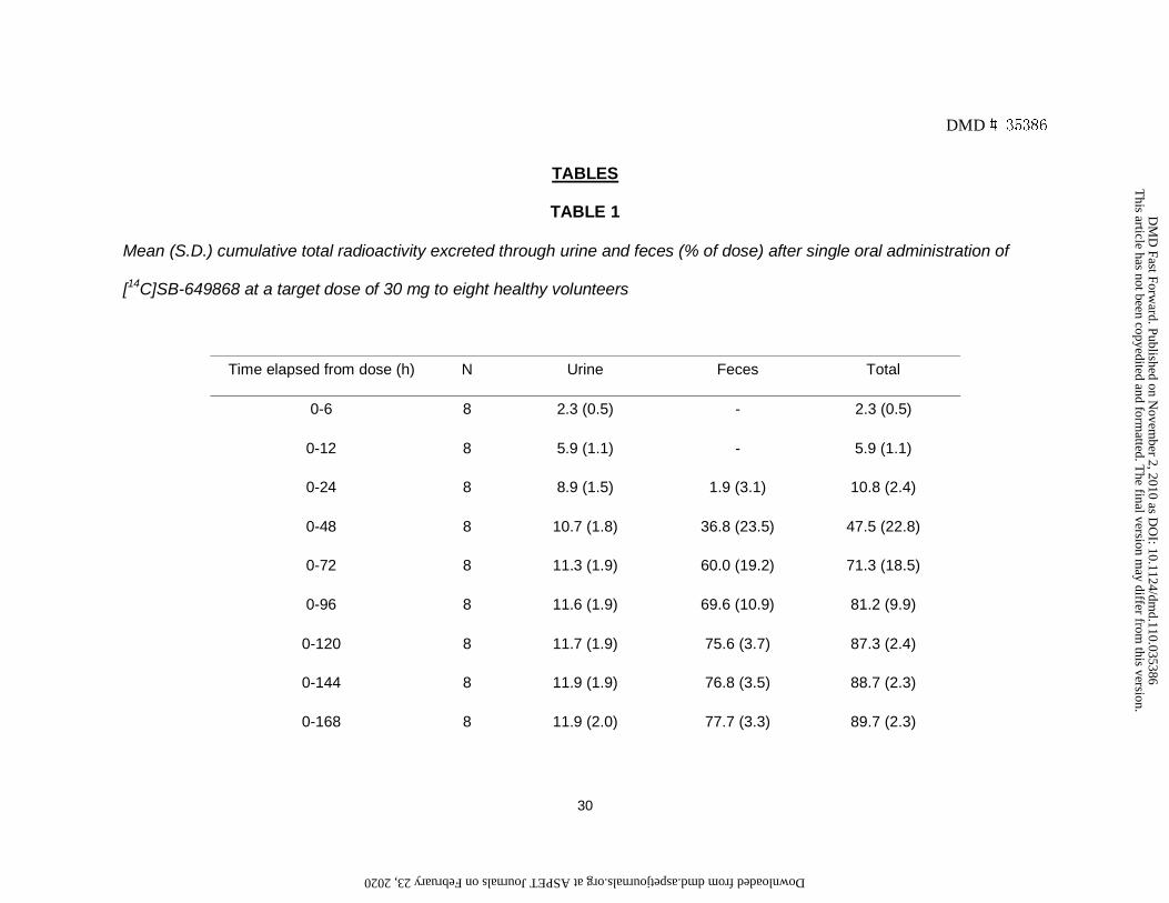
Mass Balance of Total Radioactivity. The actual oral dose of [14C]SB-649868
administered ranged from 29.6 to 30.2 mg (69.8 to 71.9 μCi, respectively). The
cumulative excretion of total radioactivity following single oral administration of
[14C]SB-649868 to healthy male subjects at a target dose of 30 mg was 91.1%,
details of which are shown in Table 1.
Following oral administration, total radioactivity was eliminated primarily in the feces,
accounting for a mean of 79.1% of the administered dose by 216 h post-dose. Total
radioactivity recovered in urine accounted for a mean of 12.0% of the administered
dose by the end of collection period. A mean recovery of 81.2% was reached by 96 h
post-dose, with the remaining radioactivity recovered between urine and feces up to
216 h post-dose.
Pharmacokinetics. A summary of the plasma pharmacokinetic parameters for SB-
649868 and total radioactivity following oral administration is presented in Table 2.
Median plasma concentrations of SB-649868 and total radioactivity following oral
administration are shown in Fig. 2.
In plasma, concentrations of total drug-related material (radioactivity) were
measurable in all subjects between 0.5 and 96 h post-dose (last blood sampling
time). Mean plasma levels of total radioactivity reached a peak of 1.66 μg equiv /ml
by 4 h post-dose, declining thereafter in a biphasic manner with an inflection point
about 18 to 24 h, and were still clearly detectable (0.108 μg equiv/ml) in the final
plasma sample taken at 96 h post-dose. The mean apparent t1/2 of total plasma
radioactivity was 39.3 h.
SB-649868 was measurable in plasma from all subjects between 0.5 and 24 h post-
dose, with mean Cmax (1.2 μg/ml) occurring at 4 h post-dose. Concentrations of SB-
This article has not been copyedited and formatted. The final version may differ from this version.DMD Fast Forward. Published on November 2, 2010 as DOI: 10.1124/dmd.110.035386
at ASPE
T Journals on February 23, 2020
dmd.aspetjournals.org
Dow
nloaded from

DMD # 35386
15
649868 declined thereafter with a t1/2 ranging between 4 and 6 h, and by 96 h post-
dose, SB-649868 concentrations were below the limit of quantification in all subjects.
Metabolic profiles. Plasma. The mean recovery of radioactive material following
extraction of human plasma samples was initially high but decreased at later time-
points, from ca. 97% at 3 h to 34% at 48 h post-dose. Fig. 3 shows three
representative radiochromatogram profiles at 3 h, 12 h and 24 h post-dose.
Quantification of drug-related material in plasma extracts after oral administration at
selected time-points is summarized in Table 3.
At each of the four time-points examined, metabolite profiles were qualitatively similar
with unchanged SB-649868 being the principal component at 3 h (68%) and 12 h
(33%) and decreasing to 10% of plasma radioactivity at 24 h post-dose. Another
notable radiolabeled component observed at each time point was a hemiaminal
metabolite M98 (GSK2329163), which accounted for between 6% and 19% of
plasma radioactivity across the four time-points examined and was the predominant
component in plasma extracts at 24 h and 48 h post-dose. A benzofuran ring opened
carboxylic acid M25 (GSK2329158) and an amine metabolite (M8) were also
detected at each of the time-points, representing up to 7% and 3% of plasma
radioactivity, respectively. A ratio of the relative exposures of each metabolite can be
determined using the percentage of total radioactivity for each component at each of
the four time-points (to calculate an approximate AUC0-48h). This shows M98, M25
and M8 accounting for around 11%, 2% and 1% of the total radioactivity, with parent
compound approximating to 26% of the total. Peak assignments accounted for
between 18% and 75% of the plasma radioactivity across the four time-points
examined. Proposed structures and supporting spectral data are shown in Table 4.
This article has not been copyedited and formatted. The final version may differ from this version.DMD Fast Forward. Published on November 2, 2010 as DOI: 10.1124/dmd.110.035386
at ASPE
T Journals on February 23, 2020
dmd.aspetjournals.org
Dow
nloaded from

DMD # 35386
16
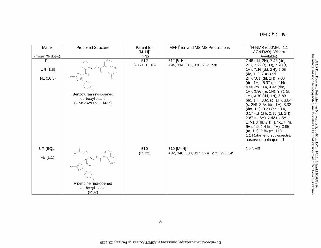
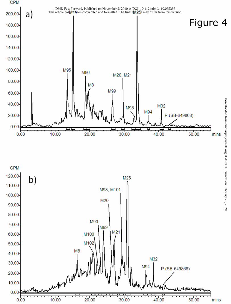
Urine. Fig. 4a shows a representative radiochromatogram of urine. The major
radiolabeled metabolites detected in human urine following oral administration were
GSK2329158 (M25), its glucuronide conjugate (M41) and a benzofuran dihydrodiol or
ring opened carboxylic acid glucuronide (M95), which accounted for ca. 13%, 14%
and 11% of urinary radioactivity, respectively (each less than 2% of the administered
dose, respectively). Other metabolites detected included M8 and a hydroxylated
benzofuran component (M86), each representing 5 - 6% of urinary radioactivity,
respectively (<1% of the administered dose, respectively). All other identified urinary
radiometabolites accounted for 0.3% or less of the dose, and SB-649868 was not
quantifiable. Proposed structures and supporting spectral data are shown in Table 4.
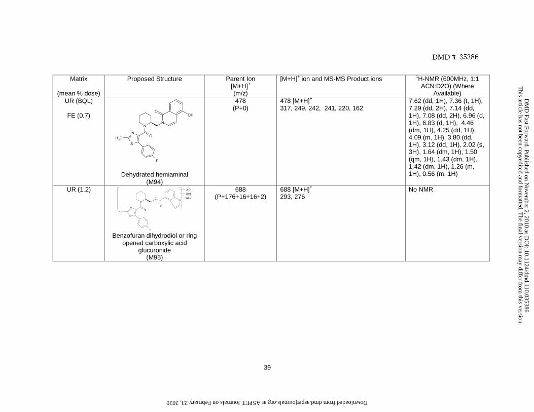
Feces. Radio-HPLC analysis of fecal extracts after oral administration of [14C]SB-
649868 showed many radiometabolites. A representative radiochromatogram of fecal
extracts is shown in Fig. 4b.
Mean recovery of radioactive material in feces following solvent extraction was 67%.
Radio-HPLC analysis of human fecal extracts revealed a complex pattern of
radiolabeled peaks with GSK2329158 (M25) being the major radiolabeled component
in fecal extracts (mean of ca. 14% of the fecal radioactivity and 10% of the
administered dose). Other noteworthy metabolites included benzofuran dihydrodiols
(M20/M21) which together accounted for 9% of fecal radioactivity (6% of the dose)
and oxygenated benzofuran dihydrodiol or ring opened carboxylic acid metabolites
(M99, M100, M102), which in total represented 10% of the fecal radioactivity (7% of
the administered dose). The hemiaminal, M98 (GSK2329163), which co-eluted with a
hydrated metabolite (M101), together represented ~4% of the fecal radioactivity (~3%
of the administered dose). All other identified fecal radiometabolites accounted for
This article has not been copyedited and formatted. The final version may differ from this version.DMD Fast Forward. Published on November 2, 2010 as DOI: 10.1124/dmd.110.035386
at ASPE
T Journals on February 23, 2020
dmd.aspetjournals.org
Dow
nloaded from

DMD # 35386
17
2% or less of the dose, with SB-649868 present in negligible amounts. Proposed
structures and supporting spectral data are shown in Table 4.
This article has not been copyedited and formatted. The final version may differ from this version.DMD Fast Forward. Published on November 2, 2010 as DOI: 10.1124/dmd.110.035386
at ASPE
T Journals on February 23, 2020
dmd.aspetjournals.org
Dow
nloaded from

DMD # 35386
18
DISCUSSION
Following a single oral dose of [14C]SB-649868 at 30 mg (approximately 70 μCi) to
eight male healthy subjects, the compound was safe and well tolerated with the most
frequent adverse event being somnolence, that was expected and is considered
related to the pharmacology of SB-649868.
Total radioactivity was eliminated primarily in feces (approximately 79%), with urinary
excretion of total radioactivity accounting for 12% of the dose after the 9 day
collection period. Mean recovery of total radioactivity was > 91% over a 9-day period
and can be considered complete (Roffey et al., 2007), with 81% excreted within the
first 4 days, with fecal elimination largely occurring between 24 and 72 h. Post 96 h,
the remaining radioactivity was subsequently eliminated more slowly and was still
detectable in fecal samples at the end of the continuous collection period (9 days),
although clearance of SB-649868 from plasma was almost complete within 24 h of
dosing.
The mean t1/2 of SB-649868 was short (4 to 6 h), whilst apparent half life of total
radioactivity was much longer (about 40 h), suggesting the presence of metabolites
that are either formed at later time-points or are much more slowly cleared from
plasma (or both), as further evidenced by the ratio of systemic exposure (AUC and
Cmax) of SB-649868 plasma concentrations to total radioactivity of approximately 22%
and 72%, respectively.
SB-649868 represented the principal component in extracts of human plasma up to
12 h after dose administration. An unusual hemiaminal metabolite, M98
(GSK2329163), was the only other drug-related material observed in plasma extracts
at >10% drug-related material, and was more notable relative to parent compound at
later time-points. Two additional minor metabolites were also observed in human
This article has not been copyedited and formatted. The final version may differ from this version.DMD Fast Forward. Published on November 2, 2010 as DOI: 10.1124/dmd.110.035386
at ASPE
T Journals on February 23, 2020
dmd.aspetjournals.org
Dow
nloaded from

DMD # 35386
19
plasma extracts: a ring-opened carboxylic acid M25 (GSK2329158) and an amine
(M8) resulting from cleavage of the molecule. The relative exposures of each
metabolite (AUC0-48h using percentage of total radioactivity values from plasma
profiles) showed M98, M25 and M8 accounting for approximately 11%, 2% and 1% of
the total radioactivity. The characterization of M98 structure in human plasma posed
a significant challenge.
M98 freely lost water, and was first detected as its dehydrated isoquinolinone
product (M94). Nonetheless, on careful handling, sufficient intact material could be
preserved and compared against synthetic material by NMR (Fig. 5). M98 showed
loss of benzofuran resonances and the addition of an aliphatic three spin system
(ABX). Long range proton-carbon correlations ruled out a plausible lactol structure,
unequivocally proving M98 to be the hemiaminal. Mechanism of formation is
tentatively suggested as nucleophilic attack of the amide moiety on an aldehyde
intermediate (Bayer and Maier, 2004). Based on the structure of metabolites a
putative simplified metabolic scheme is shown in Fig. 6. The first step in the
formation of both M98 and M25 was inferred to be the oxidation of the benzofuran
moiety via the formation of a potentially reactive epoxide intermediate (Guengerich,
2002; Kalgutkar et al., 2005). Oxidative ring opening of furan containing moieties
through formation of an epoxide intermediate, mediated by CYP450 has been
previously reported (Dalvie et al., 2002), supporting the hypothesis of the mechanism
of formation of M25. In addition, the formation of M25 can be postulated both via
M86, the hydroxylated benzofuran metabolite, and via an aldehyde and the action of
aldehyde dehydrogenase (Kobayashi et al., 1987). Moreover, in vitro data confirmed
that oxidative metabolism of SB-649868 was occurring largely via the benzofuran
moiety. Experiments conducted utilizing Bactosomes (derived from E.coli) containing
This article has not been copyedited and formatted. The final version may differ from this version.DMD Fast Forward. Published on November 2, 2010 as DOI: 10.1124/dmd.110.035386
at ASPE
T Journals on February 23, 2020
dmd.aspetjournals.org
Dow
nloaded from

DMD # 35386
20
individually overexpressed human CYP450 enzymes (1A2, 2C8, 2C9, 2C19, 2D6
and 3A4) and incubations with human liver microsomes, in the presence and
absence of selective inhibitors (azamulin was used as a selective inhibitor of
CYP3A4), confirmed metabolism was primarily due to CYP3A4. The predominant
metabolite of SB-649868 in these incubations was identified as M86, along with M25.
Following initial oxidation activating the furan ring, competing processes are possible,
intermolecular hydrolysis to the acid (M25) versus intramolecular condensation to the
hemiaminal, M98. Other notable routes of metabolism of SB-649868 in vitro were
oxidation to form benzofuran dihydrodiol isomers (M20 and M21) all of these
biotransformations appearing to be catalyzed primarily by CYP3A4. Interestingly, in-
vitro formation of M86 could be attenuated by the addition of glutathione, with
subsequent preferential formation of the glutathione adduct being observed. The
formation of M98 was not, however, observed with either human liver microsomes or
recombinant enzymes. Subsequent work with rat and mouse Aroclor 1254-induced
liver S9 did show the formation of M98, along with other drug-related components,
which suggests that cytosolic enzymes may contribute to the formation of M98.
All the human metabolites observed in plasma were also seen in preclinical
species. Both GSK2329163 (M98) and GSK2329158 (M25) were observed in plasma
from preclinical species, after single and repeat dose administration, at levels lower
(rat and mouse) or equivalent (dog) to human, with SB-649868 the major observed
component in plasma in all preclinical species evaluated. Subsequent analysis of
both metabolites (using a validated bioanalytical method) following single and repeat
dosing of SB-649868 (once daily) to humans showed similar accumulation of
GSK2329163 (M98) to that seen with parent compound (approximately 1.5-fold,
Bettica et al), whereas GSK2329158 (M25) showed around 2- to 3-fold accumulation.
This article has not been copyedited and formatted. The final version may differ from this version.DMD Fast Forward. Published on November 2, 2010 as DOI: 10.1124/dmd.110.035386
at ASPE
T Journals on February 23, 2020
dmd.aspetjournals.org
Dow
nloaded from

DMD # 35386
21
The result is GSK2329163 has systemic exposures around 10% of parent
compound, whilst GSK2329158 has comparable exposure to that of SB-649868 upon
repeat dose (unpublished data). Both metabolites were pharmacologically tested, in-
vitro, versus OX1 and OX2 receptors and both have lower affinities against each
receptor, and therefore are not anticipated to augment the pharmacological activity of
SB-649868.
SB-649868 was extensively metabolized in human and parent compound was
negligible in both urine and feces. As expected from in-vitro data, metabolism of
SB-649868 in humans occurred predominantly via oxidations of the benzofuran
moiety, some of which are likely to proceed via the formation of a potentially reactive
epoxide intermediate. The principal drug-related component observed in excreta was
M25 (GSK2329158), which accounted for a mean of 12% of the administered dose.
M86 was observed but only as a minor urinary component, and there was an
absence of glutathione or glycylcysteine conjugates. This suggests that either
formation of M25 predominates in-vivo or M86 (or its precursor) is rapidly degrading
or is further metabolized, or potentially binds to protein. Although M98 was notable in
plasma, it was a more minor route of elimination (~3% of dose), indicating formation
may be limited with its presence in plasma suggestive of a low volume of distribution.
Nonetheless, M98 together with benzofuran dihydrodiols (M20/M21), oxygenated
benzofuran dihydrodiol or ring opened carboxylic acid metabolites (M99, M100,
M102), and a glucuronide of an oxygenated benzofuran dihydrodiol or ring opened
carboxylic acid (M41), accounted for a mean of ca. 20% of the administered dose,
and further underline oxidation of the benzofuran as the predominant route of
metabolism in humans. This is consistent with preclinical species, despite the
This article has not been copyedited and formatted. The final version may differ from this version.DMD Fast Forward. Published on November 2, 2010 as DOI: 10.1124/dmd.110.035386
at ASPE
T Journals on February 23, 2020
dmd.aspetjournals.org
Dow
nloaded from

DMD # 35386
22
absence of any glutathione or glycylcysteine conjugates observed preclinically, most
notably within the bile of rats.
Low recovery of radioactive material from feces (and later plasma samples), together
with protracted elimination of total drug-related material, would also be consistent
with metabolic activation to a reactive species and subsequent binding to
endogenous protein material (Zhang et al., 2003). Although no attempt was made to
establish whether the binding to plasma proteins was covalent in nature, there is
evidence indicating covalent binding of drug-related material to microsomal protein
(based on SDS-Page electrophoresis) following in-vitro incubations (unpublished
data). The persistence of low levels of drug-related material in numerous tissues in
rats beyond 10 days post-dosing of [14C]SB-649868 is also consistent with this
inference. The formation of reactive metabolites has been linked to certain toxicities,
e.g hepatotoxicity, which may involve modification of proteins and/or direct cell
damage, either of which could induce an immune-mediated response, although
proper elucidation of this relationship remains unclear (Uetrecht, 2008). Furthermore,
the proposed low therapeutic dose of SB-649868 (20 mg) reduces the risk of toxicity
associated with the presence of a reactive pathway, since the daily dose appears to
be a key underlying factor in drug toxicity (Lammert et al., 2008; Kalgutkar and
Didiuk, 2009).
Following oral administration of [14C]SB-649868 to humans a mean of ca. 37% of the
administered dose was assigned structures, which represented between 59% and
65% of the radioactivity in either pooled (urine) or extracted (feces) samples. The
remaining unaccounted radioactivity can be ascribed to several areas where losses
were incurred:- a balance excretion recovery of ~91% of the administered dose (at
216 h); low recovery during fecal extractions (33% of fecal radioactivity was
This article has not been copyedited and formatted. The final version may differ from this version.DMD Fast Forward. Published on November 2, 2010 as DOI: 10.1124/dmd.110.035386
at ASPE
T Journals on February 23, 2020
dmd.aspetjournals.org
Dow
nloaded from

DMD # 35386
23
unrecovered); numerous components that individually represented 1% or less of the
dose or could not be distinguished above background radioactivity; samples
containing insufficient radioactivity to warrant pooling and quantitative analysis
(typically represented up to 7% of the dose).
In conclusion, following oral administration of [14C]SB-649868 to humans,
drug-related material is cleared almost exclusively via metabolism, predominantly via
oxidation of the benzofuran moiety, with metabolites excreted primarily in feces, the
most notable of which was GSK2329158 (M25). One major circulating metabolite
was identified as GSK2329163 (M98) which accounted for >10% of circulating drug-
related material, although this metabolite was a minor route of elimination. Despite
the complex metabolism of SB-649868 a good understanding of clearance routes in
humans was obtained.
This article has not been copyedited and formatted. The final version may differ from this version.DMD Fast Forward. Published on November 2, 2010 as DOI: 10.1124/dmd.110.035386
at ASPE
T Journals on February 23, 2020
dmd.aspetjournals.org
Dow
nloaded from
DMD # 35386
24
ACKNOWLEDGEMENTS
We thank Peter Szeto for synthesizing SB-649868 and its metabolites, GSK2329158
and GSK2329163, and for his collaboration in the characterization of GSK2329163
structure. We also thank Glynn Williams for the synthesis of [14C]SB-649868, Lindsay
McGregor, Christopher Irvine and Janet Dickson from Charles River, Richard Snell
for HPLC/MS quantification of SB-649868, Steve Plested for the radiometabolite
profiling, and the entire SB-649868 Clinical Pharmacology team for the design,
conduct, and analysis of the clinical portions of the study. We also thank Gordon
Dear and Andy Ayrton for their helpful discussion and Maxine Taylor for provision of
the human enzymology data.
This article has not been copyedited and formatted. The final version may differ from this version.DMD Fast Forward. Published on November 2, 2010 as DOI: 10.1124/dmd.110.035386
at ASPE
T Journals on February 23, 2020
dmd.aspetjournals.org
Dow
nloaded from
DMD # 35386
25
AUTHORSHIP CONTRIBUTION
Participated in research design: Zamuner, Bettica
Conducted experiments: Nash, Thomas, Wright
Performed data analysis: Nash, Thomas, Wright, Zamuner
Wrote or contributed to the writing of the manuscript: Renzulli, Nash, Wright,
Thomas, Zamuner, Pellegatti, Bettica, Boyle
Other: Bettica (Medical monitor)
This article has not been copyedited and formatted. The final version may differ from this version.DMD Fast Forward. Published on November 2, 2010 as DOI: 10.1124/dmd.110.035386
at ASPE
T Journals on February 23, 2020
dmd.aspetjournals.org
Dow
nloaded from

DMD # 35386
26
REFERENCES
Bayer A and Maier ME (2004) Synthesis of enamides from aldehydes and amides.
Tetrahedron 60: 6665-6677
Botta L, Gerber H-U and Schmid K (1985) Measurement of radioactivity in biological
experiments, in Drug Fate and Metabolism, Methods and Techniques (Garrett ER
and Hirtz JL eds) Vol. 5, pp 99–134, Dekker, New York.
Bruin GJ, Waldmeier F, Boernsen KO, Pfaar U, Gross G and Zollinger M (2006) A
microplate solid scintillation counter as a radioactivity detector for high performance
liquid chromatography in drug metabolism: Validation and applications. J.
Chromatogr. A 1133:184-194.
Chemelli RM, Willie JT, Sinton CM, Elmquist JK, Scammell T, Lee C, Richardson JA,
Williams SC, Xiong Y, Kisanuki Y, Fitch TE, Nakazato M, Hammer RE, Saper CB and
Yanagisawa M (1999) Narcolepsy in orexin knockout mice: molecular genetics of
sleep regulation. Cell 98:437-51.
Dalvie DK, Kalgutkar AS, Khojasteh-Bakht SC, Obach RS and O'Donnell JP (2002)
Biotransformation reactions of five-membered aromatic heterocyclic rings. Chem Res
Toxicol 15: 269-99.
This article has not been copyedited and formatted. The final version may differ from this version.DMD Fast Forward. Published on November 2, 2010 as DOI: 10.1124/dmd.110.035386
at ASPE
T Journals on February 23, 2020
dmd.aspetjournals.org
Dow
nloaded from

DMD # 35386
27
Guengerich FP (2003) Cytochrome P450 oxidations in the generation of reactive
electrophiles: epoxidation and related reactions. Arch Biochem Biophys 409: 59-71
Kalgutkar AS, Gardiner I, Obach RS, Shaffer CL, Callegari E, Henne KR, Mutlib AE,
Dalvie DK, Lee JS, Nakai Y, O’Donnell JP, Boer J and Harriman SP (2005) A
comprehensive listing of bioactivation pathways of organic functional groups. Curr
Drug Metab 6: 161-225.
Kalgutkar AS and Didiuk MT (2009) Structural alerts, reactive metabolites, and
protein covalent binding: how reliable are these attributes as predictors of drug
toxicity? Chemistry and Biodiversity 6: 2115-37.
Kilduff TS and Peyron C (2000) The hypocretin/orexin ligand-receptor system:
implications for sleep and sleep disorders. Trends Neurosci 23, 359–365.
Kobayashi T, Sugihara J and Harigaya S. (1987) Mechanism of Metabolic Cleavage
of a Furan Ring. Drug Metab Dispos 15: 877-881
Lammert C, Einarsson S, Saha C, Niklasson A, Bjornsson E and Chalasani N (2008)
Relationship between daily dose of oral medications and idiosyncratic drug-induced
liver injury: search for signals. Hepatology 47:2003-9.
Lin L, Faraco J, Li R, Kadotani H, Rogers W, Lin X, Qiu X, de Jong PJ, Nishino S and
Mignot E (1999) The sleep disorder canine narcolepsy is caused by a mutation in the
hypocretin (orexin) receptor 2 gene. Cell 98: 365-376.
This article has not been copyedited and formatted. The final version may differ from this version.DMD Fast Forward. Published on November 2, 2010 as DOI: 10.1124/dmd.110.035386
at ASPE
T Journals on February 23, 2020
dmd.aspetjournals.org
Dow
nloaded from
DMD # 35386
28
Oliveira EJ and Watson DG (2000) Liquid chromatography-mass spectrometry in the
study of the metabolism of drugs and other xenobiotics. Biomed Chromatogr 14:351-
372.
Roffey SJ, Obach RS, Gedge JI and Smith DA (2007) What is the objective of the
mass balance study? A retrospective analysis of data in animal and human excretion
studies employing radiolabeled drugs. Drug Metab Rev 39: 17-43.
Uetrecht J (2008) Idiosyncratic drug reactions: past, present, and future. Chem Res
Toxicol 21: 84-92.
Zhang D, Krishna R, Wang L, Zeng J, Mitroka J, Dai R, Narasimhan N, Reeves RA
Srinivas NR and Klunk LJ (2005) Metabolism, pharmacokinetics, and protein
covalent binding of radiolabeled maxipost (BMS-204352) in humans. Drug Metab
Dispos 33: 83-93
This article has not been copyedited and formatted. The final version may differ from this version.DMD Fast Forward. Published on November 2, 2010 as DOI: 10.1124/dmd.110.035386
at ASPE
T Journals on February 23, 2020
dmd.aspetjournals.org
Dow
nloaded from

DMD # 35386
29
LEGENDS FOR FIGURES
Figure 1:
Structure of [14C]SB-649868
Figure 2:
Median SB-649868 concentrations and total radioactivity levels (semi-logarithmic
scale) in plasma after oral administration of [14C]SB-649868 at the target dose of 30
mg.
Figure 3:
Representative radiochromatograms of human plasma at various time-points after
oral administration of [14C]SB-649868 at the target dose of 30 mg.
Figure 4:
Representative radiochromatograms of human (a) urine and (b) feces after oral
administration of [14C]SB-649868 at the target dose of 30 mg
Figure 5:
Assigned 1H NMR of hemiaminal metabolite (M98, GSK2329163)
Figure 6:
Putative simplified metabolic scheme for SB-649868 in humans
This article has not been copyedited and formatted. The final version may differ from this version.DMD Fast Forward. Published on November 2, 2010 as DOI: 10.1124/dmd.110.035386
at ASPE
T Journals on February 23, 2020
dmd.aspetjournals.org
Dow
nloaded from
DMD # 35386
30
TABLES
TABLE 1
Mean (S.D.) cumulative total radioactivity excreted through urine and feces (% of dose) after single oral administration of
[14C]SB-649868 at a target dose of 30 mg to eight healthy volunteers
Time elapsed from dose (h) N Urine Feces Total
0-6 8 2.3 (0.5) - 2.3 (0.5)
0-12 8 5.9 (1.1) - 5.9 (1.1)
0-24 8 8.9 (1.5) 1.9 (3.1) 10.8 (2.4)
0-48 8 10.7 (1.8) 36.8 (23.5) 47.5 (22.8)
0-72 8 11.3 (1.9) 60.0 (19.2) 71.3 (18.5)
0-96 8 11.6 (1.9) 69.6 (10.9) 81.2 (9.9)
0-120 8 11.7 (1.9) 75.6 (3.7) 87.3 (2.4)
0-144 8 11.9 (1.9) 76.8 (3.5) 88.7 (2.3)
0-168 8 11.9 (2.0) 77.7 (3.3) 89.7 (2.3)
This article has not been copyedited and form
atted. The final version m
ay differ from this version.
DM
D Fast Forw
ard. Published on Novem
ber 2, 2010 as DO
I: 10.1124/dmd.110.035386
at ASPET Journals on February 23, 2020 dmd.aspetjournals.org Downloaded from

DMD # 35386
31
0-192 8 12.0 (2.0) 78.5 (3.2) 90.5 (2.2)
0-216 8 12.0 (2.0) 79.1 (3.0) 91.1 (2.2)
This article has not been copyedited and form
atted. The final version m
ay differ from this version.
DM
D Fast Forw
ard. Published on Novem
ber 2, 2010 as DO
I: 10.1124/dmd.110.035386
at ASPET Journals on February 23, 2020 dmd.aspetjournals.org Downloaded from

DMD # 35386
32
TABLE 2
Geometric Mean (95% CI) SB-649868 and total radioactivity plasma pharmacokinetic parameters after single oral
administration of [14C]SB-649868 at a target dose of 30 mg
Parameter
SB-649868 Total Radioactivity
N 8 8
AUC(0-∞) (μg.h/mL) a 8.30
(5.90, 11.7)
37.3
(31.0,44.8)
AUC(0-t) (μg.h/mL) a 8.20
(5.80, 11.5)
31.0
(25.0, 38.4)
Cmax (μg/mL) a 1.20
(1.00, 1.50)
1.66
(1.40,2.00)
tmax (h) b 4.00
(3.00-4.00)
4.00
(3.00,6.00)
t1/2 (h) 4.80
(4.10-5.80)
39.3
(33.4,46.3)
CL/F (mL/h) 3610
(2570,5070) -
This article has not been copyedited and form
atted. The final version m
ay differ from this version.
DM
D Fast Forw
ard. Published on Novem
ber 2, 2010 as DO
I: 10.1124/dmd.110.035386
at ASPET Journals on February 23, 2020 dmd.aspetjournals.org Downloaded from

DMD # 35386
33
a Concentration units for total radioactivity are μg-eq/mL.
b Median
Values in parentheses represent the range
This article has not been copyedited and form
atted. The final version m
ay differ from this version.
DM
D Fast Forw
ard. Published on Novem
ber 2, 2010 as DO
I: 10.1124/dmd.110.035386
at ASPET Journals on February 23, 2020 dmd.aspetjournals.org Downloaded from

DMD # 35386
34
TABLE 3
Mean (n=8) percentage of radioactivity of SB-649868 and its metabolites in human
plasma after single oral administration of [14C]SB-649868 at a target dose of 30 mg
Radioactive
Component
Mean % of plasma radioactivity a
Oral administration
3 h 12 h 24 h 48 h
SB-649868 67.7 33.2 9.8 BQL
M8 1.3 1.0 1.9 3.0
GSK2329163 (M98) 6.0 16.8 19.3 15.1
GSK2329158 (M25) BQL 6.7 4.2 BQL
a Observed metabolite radioactivity was determined by 96-well fraction collection with
scintillation counting for 5 minutes after a chromatographic separation was performed by
HPLC. Where the sample preparation step (centrifugation, extraction or reconstitution)
resulted in some loss of radioactive material, if the recovery was <85%, the chromatogram
data have been multiplied by the percentage recovered to calculate % sample radioactivity
and % dose or µg equivalent figures. Mean (n=7 for 3 and 12 h; n=8 for other time points)
percentage of radioactivity per time point does not equal 100% since only distinct radioactive
peaks were assigned values and a few minor metabolites were not reported.
BQL = below quantification limit, set to 1% plasma radioactivity
This article has not been copyedited and formatted. The final version may differ from this version.DMD Fast Forward. Published on November 2, 2010 as DOI: 10.1124/dmd.110.035386
at ASPE
T Journals on February 23, 2020
dmd.aspetjournals.org
Dow
nloaded from

DMD # 35386
35
TABLE 4
Relevant metabolites of SB-649868 in human plasma, urine and feces after single oral administration of [14C]SB-649868 at a
target dose of 30 mg
Matrix
(mean % dose)
Proposed Structure Parent Ion [M+H]+ (m/z)
[M+H]+ ion and MS-MS Product ions 1H-NMR (600MHz, 1:1 ACN:D2O) (Where
Available) PL
UR (BQL)
FE (BQL) S
N
N
OCH3
F
NH
O
O
SB-649868
478 (P)
478, [M+H]+ 460, 317, 316, 242, 241, 220, 145, 98
7.83 (d, 1H), 7.68 (dd, 1H), 7.56 (dd, 1H), 7.37 (dd, 2H), 7.37 (t, 1H), 7.11 (d, 1H), 7.04 (dd, 2H), 5.00 (m, 1H), 3.71 (dd, 1H), 3.31 (dm, 1H), 3.25 (dd, 1H), 2.97 (tt, 1H), 2.17 (s, 3H), 1.72 (dm, 1H), 1.61 (dm, 1H), 1.55 (tm, 1H), 1.47 m 2H), 0.83, m 1H) Signals doubled due to rotamers, major sub-spectrum quoted.
PL
UR (0.6)
FE (1.5) S
N
N
OCH3
F
NH2
Amine (M8)
334 (P-144)
334 [M+H]+ 317, 316, 220, 98
7.39 (dd, 2H), 6.98 (t, 2H), 2.70 (s, 3H) Other resonances obscured
This article has not been copyedited and form
atted. The final version m
ay differ from this version.
DM
D Fast Forw
ard. Published on Novem
ber 2, 2010 as DO
I: 10.1124/dmd.110.035386
at ASPET Journals on February 23, 2020 dmd.aspetjournals.org Downloaded from

DMD # 35386
36
Matrix
(mean % dose)
Proposed Structure Parent Ion [M+H]+ (m/z)
[M+H]+ ion and MS-MS Product ions 1H-NMR (600MHz, 1:1 ACN:D2O) (Where
Available) UR (0.3)
FE (6.2)
S
N
N
OCH3
F
NH
O
O
OH
OH
preferred structure
Benzofuran dihydrodiol
(M20, M21)
512 (P+34)
512 [M+H]+ 478, 317, 257, 239, 222
No NMR
This article has not been copyedited and form
atted. The final version m
ay differ from this version.
DM
D Fast Forw
ard. Published on Novem
ber 2, 2010 as DO
I: 10.1124/dmd.110.035386
at ASPET Journals on February 23, 2020 dmd.aspetjournals.org Downloaded from
DMD # 35386
37
Matrix
(mean % dose)
Proposed Structure Parent Ion [M+H]+ (m/z)
[M+H]+ ion and MS-MS Product ions 1H-NMR (600MHz, 1:1 ACN:D2O) (Where
Available) PL
UR (1.5)
FE (10.3)
S
N
N
OCH3
F
NH
O
OH
O
OH
Benzofuran ring-opened
carboxylic acid (GSK2329158 - M25)
512 (P+2+16+16)
512 [M+H]+ 494, 334, 317, 316, 257, 220
7.46 (dd, 2H), 7.42 (dd, 2H), 7.22 (t, 1H), 7.20 (t, 1H), 7.16 (dd, 2H), 7.05 (dd, 1H), 7.01 (dd, 2H),7.01 (dd, 1H), 7.00 (dd, 1H), 6.97 (dd, 1H), 4.98 (m, 1H), 4.44 (dm, 1H), 3.86 (m, 1H), 3.71 (d, 1H), 3.70 (dd, 1H), 3.69 (dd, 1H), 3.65 (d, 1H), 3.64 (s, 2H), 3.54 (dd, 1H), 3.32 (dm, 1H), 3.23 (dd, 1H), 3.17 (td, 1H), 2.95 (td, 1H), 2.67 (s, 3H), 2.42 (s, 3H), 1.7-1.8 (m, 2H), 1.4-1.7 (m, 6H), 1.2-1.4 (m, 2H), 0.95 (m, 1H), 0.86 (m, 1H) 1:1 Rotameric sub-spectra observed, both quoted.
UR (BQL)
FE (1.1)
S
N
NH
OCH3
F
NH
O
O
OH
O
Piperidine ring-opened
carboxylic acid (M32)
510 (P+32)
510 [M+H]+
492, 348, 330, 317, 274, 273, 220,145
No NMR
This article has not been copyedited and form
atted. The final version m
ay differ from this version.
DM
D Fast Forw
ard. Published on Novem
ber 2, 2010 as DO
I: 10.1124/dmd.110.035386
at ASPET Journals on February 23, 2020 dmd.aspetjournals.org Downloaded from

DMD # 35386
38
Matrix
(mean % dose)
Proposed Structure Parent Ion [M+H]+ (m/z)
[M+H]+ ion and MS-MS Product ions 1H-NMR (600MHz, 1:1 ACN:D2O) (Where
Available) UR (1.6)
S
N
N
OCH3
F
NH
O
OH
OH
OGluc
Benzofuran ring opened
carboxylic acid glucuronide (M41)
688 (P+176+16+16+2)
688 [M+H]+
670, 626, 512, 468, 450, 355, 275
No NMR
UR (0.6)
OH
S
N
N
OCH3
F
NH
O
O
Benzofuran hydroxy
(M86)
494 (P+16)
688 [M+H]+
476, 317, 316, 258, 257, 220, 98 7.58 (dd, 2H), 7.37 (dd, 2H), 6.56 (s, 1H), 4.98 (m, 1H) Other resonances obscured
13C: Benzofuran 3-carbon101.2ppm
FE (0.7)
S
N
N
OCH3
F
NH
O
O
O
Oxygenated
(M90)
494 (P+16)
494 [M+H]+
333, 220 No NMR
This article has not been copyedited and form
atted. The final version m
ay differ from this version.
DM
D Fast Forw
ard. Published on Novem
ber 2, 2010 as DO
I: 10.1124/dmd.110.035386
at ASPET Journals on February 23, 2020 dmd.aspetjournals.org Downloaded from
DMD # 35386
39
Matrix
(mean % dose)
Proposed Structure Parent Ion [M+H]+ (m/z)
[M+H]+ ion and MS-MS Product ions 1H-NMR (600MHz, 1:1 ACN:D2O) (Where
Available) UR (BQL)
FE (0.7)
N
OOH
S
N
N
OCH3
F
Dehydrated hemiaminal (M94)
478 (P+0)
478 [M+H]+
317, 249, 242, 241, 220, 162 7.62 (dd, 1H), 7.36 (t, 1H), 7.29 (dd, 2H), 7.14 (dd, 1H), 7.08 (dd, 2H), 6.96 (d, 1H), 6.83 (d, 1H), 4.46 (dm, 1H), 4.25 (dd, 1H), 4.09 (m, 1H), 3.80 (dd, 1H), 3.12 (dd, 1H), 2.02 (s, 3H), 1.64 (dm, 1H), 1.50 (qm, 1H), 1.43 (dm, 1H), 1.42 (dm, 1H), 1.26 (m, 1H), 0.56 (m, 1H)
UR (1.2)
S
N
N
OCH3
F
NH
O
O
2[O]
Gluc
2[H]
Benzofuran dihydrodiol or ring
opened carboxylic acid glucuronide
(M95)
688 (P+176+16+16+2)
688 [M+H]+
293, 276
No NMR
This article has not been copyedited and form
atted. The final version m
ay differ from this version.
DM
D Fast Forw
ard. Published on Novem
ber 2, 2010 as DO
I: 10.1124/dmd.110.035386
at ASPET Journals on February 23, 2020 dmd.aspetjournals.org Downloaded from

DMD # 35386
40
Matrix
(mean % dose)
Proposed Structure Parent Ion [M+H]+ (m/z)
[M+H]+ ion and MS-MS Product ions 1H-NMR (600MHz, 1:1 ACN:D2O) (Where
Available) PL
UR (0.2)
FE (2.61)
N
OOH
S
N
N
OCH3
F
OH
Hemiaminal
(GSK2329163 – M98)
496 (P+18)
496 [M+H] +
MS-MS of [M-H2O+H]+ ion :478 317, 285, 249, 241, 220
7.40 (dd, 2H), 7.32 (dd, 1H), 7.20 (t, 1H), 7.19 (dd, 2H), 7.05 (dd, 1H), 4.93 (dd, 1H), 4.43 (dm, 1H), 3.95 (m, 1H), 3.57 (dd, 1H), 3.37 (dd, 1H), 3.22 (dd, 1H), 3.10 (dd, 1H), 2.92 (td, 1H), 2.02 (s, 3H), 1.69 (dm, 1H), 1.43 (qm, 1H), 1.34 (dm, 1H), 1.28 (dm, 1H), 1.18 (m, 1H), 0.25(m, 1H)
UR (0.3)
FE (3.6)
S
N
N
OCH3
F
NH
O
O
O
2[O]
2[H]
Oxygenated benzofuran
dihydrodiol or ring opened carboxylic acid
(M99)
528 (P+16+34)
528 [M+H] + 510, 492, 333, 332, 299, 257, 236
No NMR
This article has not been copyedited and form
atted. The final version m
ay differ from this version.
DM
D Fast Forw
ard. Published on Novem
ber 2, 2010 as DO
I: 10.1124/dmd.110.035386
at ASPET Journals on February 23, 2020 dmd.aspetjournals.org Downloaded from

DMD # 35386
41
Matrix
(mean % dose)
Proposed Structure Parent Ion [M+H]+ (m/z)
[M+H]+ ion and MS-MS Product ions 1H-NMR (600MHz, 1:1 ACN:D2O) (Where
Available) FE (1.3)
S
N
N
OCH3
F
NH
O
O
O 2[O]
2[H]
Oxygenated benzofuran
dihydrodiol or ring opened carboxylic acid
(M100)
528 (P+16+34)
528 [M+H] + 333, 332 MS-MS of [M-H2O+H]+ 528 333, 332, 317, 315, 220
No NMR
FE (2.61)
S
N
N
OCH3
F
NH
O
O
H2O
Hydrated (M101)
496 (P+18)
496 [M+H] + MS-MS of [M-H2O+H]+ 478 317, 241, 220
No NMR
FE (2.4)
S
N
N
OCH3
F
NH
O
O
O 2[O]
2[H]
Oxygenated benzofuran
dihydrodiol or ring opened carboxylic acid
(M102)
528 (P+16+34)
528 [M+H] + MS-MS of [M-H2O+H]+ 510
333, 249, 241, 220
No NMR
PL = plasma, UR = urine and FE = feces BQL – Below limit of quantification 1- M98 and M101 co-elute during HPLC analysis.
This article has not been copyedited and form
atted. The final version m
ay differ from this version.
DM
D Fast Forw
ard. Published on Novem
ber 2, 2010 as DO
I: 10.1124/dmd.110.035386
at ASPET Journals on February 23, 2020 dmd.aspetjournals.org Downloaded from

This article has not been copyedited and formatted. The final version may differ from this version.DMD Fast Forward. Published on November 2, 2010 as DOI: 10.1124/dmd.110.035386
at ASPE
T Journals on February 23, 2020
dmd.aspetjournals.org
Dow
nloaded from

This article has not been copyedited and formatted. The final version may differ from this version.DMD Fast Forward. Published on November 2, 2010 as DOI: 10.1124/dmd.110.035386
at ASPE
T Journals on February 23, 2020
dmd.aspetjournals.org
Dow
nloaded from

This article has not been copyedited and formatted. The final version may differ from this version.DMD Fast Forward. Published on November 2, 2010 as DOI: 10.1124/dmd.110.035386
at ASPE
T Journals on February 23, 2020
dmd.aspetjournals.org
Dow
nloaded from
This article has not been copyedited and formatted. The final version may differ from this version.DMD Fast Forward. Published on November 2, 2010 as DOI: 10.1124/dmd.110.035386
at ASPE
T Journals on February 23, 2020
dmd.aspetjournals.org
Dow
nloaded from

This article has not been copyedited and formatted. The final version may differ from this version.DMD Fast Forward. Published on November 2, 2010 as DOI: 10.1124/dmd.110.035386
at ASPE
T Journals on February 23, 2020
dmd.aspetjournals.org
Dow
nloaded from

This article has not been copyedited and formatted. The final version may differ from this version.DMD Fast Forward. Published on November 2, 2010 as DOI: 10.1124/dmd.110.035386
at ASPE
T Journals on February 23, 2020
dmd.aspetjournals.org
Dow
nloaded from