disruption of ple2, the gene for the e2 subunit of the plastid pyruvate dehydrogenase complex, in...
TRANSCRIPT

Plant Molecular Biology 52: 865–872, 2003.© 2003 Kluwer Academic Publishers. Printed in the Netherlands.
865
Disruption of plE2, the gene for the E2 subunit of the plastid pyruvatedehydrogenase complex, in Arabidopsis causes an early embryo lethalphenotype
Ming Lin, Robert Behal and David J. OliverDepartment of Botany, Iowa State University, 353 Bessey Hall, Ames, IA 50011, USA (∗author for correspondence;e-mail [email protected])
Received 26 February 2003; received 1 May 2003
Key words: Arabidopsis mutations, dihydrolipoyl acetyltransferase, E2 subunit, embryogenesis, lipid synthesis,pyruvate dehydrogenase complex, T-DNA knockout
Abstract
The pyruvate dehydrogenase multi-enzyme complex is the main source of acetyl-CoA formation in the plastids ofplants and is composed of multiple copies of four different subunits, E1α, E1β, E2, and E3. A T-DNA insertioninto the gene for the plastidic E2 (dihydrolipoyl acetyltransferase) subunit, plE2, of the complex in Arabidopsisdestroys the expression of that gene. The resulting mutation has no apparent phenotype in the heterozygous state,but the homozygous mutation is lethal. Haploid sperm and eggs that contain only the disrupted plE2 gene functionnormally resulting in the formation of an embryo that is homozygous for the mutation. This embryo only developsto an early stage before the development arrests resulting in an early embryo-lethal phenotype. While the mutationcould not be complemented with the cDNA for the plE2 gene under control of the 35S, the AtSERK1, or thenapin promoter, it could be complemented using the endogenous plE2 promoter to drive expression of the plE2cDNA. This verifies the essential nature of the plastidic pyruvate dehydrogenase complex and its role in embryoformation.
Abbreviations: AtSERK1, Arabidopsis somatic embryogenesis receptor kinase 1 gene; E1, E2, E3, subunits ofpyruvate dehydrogenase complex; PDC, pyruvate dehydrogenase complex; plE2, plastidic E2 subunit
Introduction
Plants contain two pyruvate dehydrogenase complexes(PDC) (Mooney et al., 2002). The mitochondrial formof the enzyme catalyzes a key respiratory reaction pro-viding acetyl CoA for the Krebs cycle and NADHfor oxidative phosphorylation. The plastid form of theenzyme produces the same products for consumptionin that organelle. Biochemical and molecular studieshave demonstrated that this plastid PDC (plPDC) isthe source of acetyl CoA for fatty acid biosynthesis.While some early work had suggested that acetyl CoAsynthetase in the plastid could be a major source of theprecursor for fatty acids in plastids, gene expressionstudies, antisense plants with low acetyl CoA syn-
thetase activity, and biochemical flux studies suggestthat this reaction is not important in lipid formation inleaves and seeds (Bao et al., 1998, Behal et al., 2002;Kang and Rawsthorne 1994, 1996, Ke et al., 2001).
Both the mitochondrial and plastid forms of PDChave the same component protein structure (Mooneyet al., 2002). They contain an E1 component (pyru-vate decarboxylase) composed of α and β subunits,an E2 component (dihydrolipoyl acetyltransferase),and an E3 component (dihydrolipoamide dehydro-genase). This multienzyme complex is very large,several megadaltons, and is probably composed of apentagonal dodecahedron core of E2 proteins deco-rated on the surface with E1α2β2 heterotetramers andE3 homodimers. In addition, the mitochondrial com-

866
Figure 1. Diagram of the plE2 genomic DNA including the intron and exon structure, the insertion site for the T-DNA, and the location andcharacterization of the PCR primers used during this study.
plex, but not that from the plastid, also contains a PDCprotein kinase and phosphatase that control PDC ac-tivity by reversible phosphorylation of the E1 protein(Thelen et al., 1998).
All of the subunits for both PDCs are encodedby nuclear genes with independent genes encodingthe equivalent subunits for each organelle. The plas-tid PDC of Arabidopsis is encoded by one gene forE1α (At1g01090) (Johnston et al., 1997), two genes(A1g30120 and A2g34590) for E1β that are 90% iden-tical (Johnston et al., 1997, 2000; Behal and Oliver,1999) and two genes for E3 that are about 85% iden-tical (Lutziger and Oliver, 2000; Drea et al., 2001).One plastid plE2 protein (At3g25860) has been char-acterized in detail (Mooney et al., 1999). A secondpotential plE2 protein (At1g34430) exists in the data-bases and is more closely related to the plastidic thanthe mitochondrial form of the enzyme. Mooney et al.(2002) suggest that this may be a second plE2 gene ormay represent the plastidic E3-binding protein.
During the course of our studies into the pro-duction of plastidic acetyl-CoA we have isolated aknockout mutant for the plE2 subunit of plastidicPDC (At3g25860) and have characterized that mu-tant. The plE2 knockout mutation had no obviousphenotype in the heterozygous state but was lethal
when homozygous resulting in an early embryo-lethalphenotype.
Materials and methods
Plant lines and growth conditions
Arabidopsis ecotype WS was used in all the experi-ments. Plants were grown on soil at 22 ◦C under con-tinuous light. Agrobacterium strain GV3101 was usedfor transformation. Wild-type and heterozygous plE2knockout mutant plants were transformed with the flo-ral dip method (Clough and Bent, 1998). Seeds ofthe knockout and doubly transformed plants were ger-minated on half-strength MS solid medium plus 2%sucrose, 100 mg/l kanamycin or/and 50 mg/l glufosi-nate. Alternatively, seeds or seedlings were placed insoil and then small plants were selected for herbicideresistance by spraying 0.5% Liberty (glufosinate).
Identification of plE2 mutant
The plE2 T-DNA knockout mutant was identified inassociation with the Arabidopsis Knockout Facility atthe University of Wisconsin following their standardprocedures (http//:www.biotech.wisc.edu/arabidopsis/default.htm). The plE2 T-DNA knockout mutant was

867
Figure 2. Plasmids used to create transgenic plants in order tocomplement the plE2 knockout mutant.
Figure 3. PCR demonstration that all of the plants in the finalT-DNA pool were heterozygous for the plE2 knockout. Lane 1, noDNA; lane 2, wild-type WS DNA; lanes 3–8, DNA from individualplants in the pool. Plant 7 is 50-102 which was used throughout therest of the study. The primers for A were plE2NT1 and plE2CT1,the primers for B were plE2NT1 and XR2.
screened from seed pool SCH123-50 from ARBC(Arabidopsis Biological Resource Center, Ohio StateUniversity, Columbus, OH). The primers used in thisstudy are detailed in Figure 1. The plE2 gene-specificprimers, plE2T1and plE2T2, and the T-DNA bordersequence primers, JL-202 and XR-2, were used toidentify the T-DNA insertion lines. The binding site ofplE2T1 and plE2T2 are at about 400 bp upstream ofstart codon and 500 bp downstream of stop codon ofthe plE2 gene, respectively. In order to verify the pres-ence of the T-DNA insertion within the plE2 gene, theamplified products were separated on 0.8% agarosegel, blotted onto nylon membrane, and probed with a2 kb plE2 genomic DNA fragment amplified by PCRwith primers plE2T1 and plE2T2 and labeled with 23Pby means of Invitrogen’s Rad Prime DNA LabelingSystem (Carlsbad, CA). The blots were pre-hybridizedin 0.3 M sodium phosphate pH 7.2 plus 7% SDS at65 ◦C for 30 min. Hybridizations proceeded under thesame conditions overnight. Once the PCR productswere confirmed by hybridization, the DNA bands werepurified from the gel and sequenced. One line that washeterozygous for the plE2 knockout (line 50–102) wasstudied in detail.
Complementation of plE2 knockout mutant
A series of vectors were constructed to express theplE2 cDNA with different promoters in an attempt
Figure 4. Southern blot using the plE2 genomic DNA as a probe todemonstrate that the progeny from 50-102 are heterozygous for theplE2 knockout. Lane 1, wild-type WS DNA; lanes 2–6 are progenyresulting from selfing of 50-102. The first group were digested withBamHI, the second with EcoRI, and the third group with HindIII.
to rescue the homozygous plE2 knockout mutant bycomplementation (Figure 2). The promoters used werethe constitutive 35S promoter from plasmid pBE2s,the promoter from the gene for napin, a seed storageprotein from Brassica napa from plasmid pBNapPE2for expression during cotyledon formation (Hoglundet al., 1992), a 1.1 kb 5′-UTR fragment of an earlyembryo gene, the Arabidopsis somatic embryogenesisreceptor kinase 1 (AtSERK1) gene (Hecht et al., 2001)that was amplified with primers AtSERK1Pf-1 andAtSERK1Pr-1, and the endogenous promoter of theplE2 gene from Arabidopsis. The promoter of the plE2gene was a 0.9 kb PCR product of the upstream regionof the plE2 gene that was amplified and confirmed bysequencing. Each of these promoters was cloned into abinary vector (pBE2s, pBNapPE2, pBAtserk1PE2 andpBE2PE2) containing the cDNA for plE2 to form atranscriptional fusion. The vectors contained the bargene for glufosinate resistance.
Genetic analysis
The plE2 heterozygous mutant, 50-102, and F1 seedsfrom a reciprocal cross between WS and 50-102 plantswere germinated on half-strength MS medium plus100 mg/l kanamycin. After one week, the seedlingswere sorted and the kanamycin-resistant seedlingswere transplanted onto soil. Mature siliques from bothself-pollinated 50–102 and the F1 plants of the WS× 50–102 and 50–102 × WS crosses were dissectedunder a stereo microscope. Seeds were sorted as vi-able (green and plump) or non-viable (brown andshriveled).

868
Light microscopy
Siliques were fixed in Histochoice Tissue Fixative(Sigma H-2904, Sigma Chemical) for more than 1 hand then cleared in Hoyer’s solution (Patton et al.,1998). Samples were visualized under a Zeiss Axio-plan microscope (Carl Zeiss, Thornwood, NY) witha Nomarski (differential interference contrast) device.Images from both stereo and Nomarski microscopeswere captured with a video camera (AxioCam HRC)connected to a PC computer with AxioVision 3.06.1.Adobe Photoshop software was used to enhance thepictures.
RT-PCR
Total RNA samples was isolated from plant tissuewith TRIzol (Invitrogen) and cDNA was reverse tran-scribed with 2 µg total RNA and oligo (dT)20 asprimer following the instructions for Invitrogen’s Su-perScript II kit. Gene-specific primers were used forPCR with the cDNAs as templates.
Results
Following the procedures of the Arabidopsis Knock-out Facility, we were able to obtain an Arabidopsisline with a T-DNA insertion into the plE2 gene. Fromthe final seed pool six individual plants with T-DNAintegrated in plE2 ORF were identified by PCR. Us-ing the plE2NT1 and plE2CT2 primers that primeboth ends of the coding region, it was possible to am-plify a 2 kb fragment from both the wild-type and allthe T-DNA knockout lines suggesting that both con-tained a wild-type copy of the plE2 gene and that theplants containing the plE2 knockout were heterozy-gous (Figure 3). One of the original lines, 50-102,was selfed and five kanamycin-resistant progeny weresubjected to Southern analysis with the plE2 genomicDNA as probe (Figure 3). All of these lines, in ad-dition to showing the original wild-type restrictionfragments (there are one EcoRI and HindIII and twoBamHI fragments in wild-type WS) presented newfragments that result from the T-DNA insertion intothe plE2 gene. This confirms that all of the surviv-ing kanamycin-resistant lines were heterozygous forthe knockout. This line was followed by PCR forthree more generations and no homozygous knockoutlines were identified. This strongly suggests that thehomozygous plE2 knockout mutation is lethal.
PCR analysis of the heterozygous line showed thatthe T-DNA was integrated into the second exon of theplE2 gene about 1 kb downstream from the start codonand 1 kb upstream from the stop codon (Figure 1).This was confirmed by sequencing the PCR fragmentsadjacent to both ends of the T-DNA insert (data notpresented).
Quantitative and visual examination of the het-erozygous plE2 knockouts revealed no detectable phe-notype (data not shown). There were, however, brownand shriveled non-viable seeds in the siliques that re-sulted from selfing the heterozygous mutant plE2 linesthat were absent from the wild-type WS siliques (Fig-ure 5). We postulated that these aborted seeds arehomozygous for the E2 knockout mutation and testedthis idea genetically. In order to test this postulate, 29siliques from the five progenies of plE2 mutant, 50–102, that were characterized by Southern blot as beingheterozygous for the plE2 knockout (Figure 4) weredissected under a stereo microscope and the seeds col-lected were characterized as green and viable or brownand aborted. The ratio of viable seeds to non-viableseeds was 480:169. Chi-square analysis confirmed the3:1 (viable/aborted) segregation ratio supporting thatthese brown shriveled seeds were the homozygous T-DNA knockout mutants. This idea was further testedby examining the number of seeds that germinated onkamanycin-containing plates. Under these conditions,where plants that were homozygous for the wild-typeallele would not survive, 561 seeds were heterozygousand germinated and grew while 280 were homozygouswild-type and germinated but then died. This is consis-tent with the hypothesis that the homozygous knock-outs seeds are missing from the population harvestedand that the resulting seeds are two-thirds heterozy-otes and one-third homozygous wild-type. Finally,we made reciprocal crosses between the heterozygousmutant line and WS wild-type plants, selected proge-nies on kanamycin media to obtained the heterozygousF1 plants and then, after confirming their F1 geno-type by PCR, selfed these F1 plants and plated theirseeds on kanamycin plates. Again, one-third of theseedlings were kanamycin-sensitive. When 386 seedsfrom 7 siliques of 50-102× WS and 394 seeds from8 siliques of WS × 50-102 were analyzed, the ratioof viable/aborted seeds was 3:1 for both groups. Thisfurther supports that the homozygous plE2 knock-outs were lethal. Furthermore, the segregation of theF2 progenies of both reciprocal crosses showed thesame 2:1 kanamycin-resistant/kanamycin-sensitive ra-tio (532:263 from selfing WS × 50-102 and 160:88

869
Figure 5. Micrographs of siliques and seeds from heterozygous plE2 knockout mutant 50–102. A. Young silique showing normal green andaborted seeds. B. Older silique showing normal green and brown shriveled seeds. C. Individual green seed showing heart-shaped embryo. D.Shriveled seed showing lack of embryo development.
from selfing 50-102 × WS). This suggested that boththe megaspore and the microspore haploid reproduc-tive cells survived at the same efficiency as wild-typegerm cells even though they were lacking the plE2gene.
When these aborted seeds were examined micro-scopically by means of Nomarski optics, the seedswere largely empty (Figure 5). There was a smallgroup of cells at the base of the seeds that appeared tobe a small cluster of embryo cells. Based on the sizeand shape of this cluster they seemed to suggest thatdevelopment arrested at an early embryo stage. Thismutation, therefore, would fall into the group of earlyembryo-lethal mutants (Meinke, 1985).
In order to verify that the embryo-lethal pheno-type was the result of the homozygous knockout of theplE2 gene, we attempted to complement the heterozy-gous knockout and then to obtain homozygous plE2knockouts by selfing these double transgenic plants.We were not sure when during development the lossof plE2 expression caused the embryo-lethal pheno-type. As a result, we made four different constructswith four different promoters driving expression ofthe Arabidopsis plE2 cDNA under the assumption thatone of these promoters would express at a time thatwould allow plPDC accumulation before the lethal-ity was irreversible. The promoters we used werethe 35S promoter from cauliflower mosaic virus forhigh-level nearly constitutive expression, the napinpromoter from the gene for the major storage pro-tein in Brassica embryos (Hoglund et al., 1992), theAtSERK1 promoter from the gene for SERK1 that is
expressed early in embryogenesis (Hecht et al., 2001),and the promoter from the endogenous ArabidopsisplE2 gene.
In no case were we able to obtain homozygousplE2 knockout mutants in the T2 progeny from plantstransformed with the 35S promoter, the napin pro-moter, or the AtSERK1 promoter. In each case wescreened over 40 glufosinate and kanamycin-resistantT2 plants for each construct and none of the plantslacked a wild-type copy of the plE2 allele. If the com-plementation had been successful, we would expectthat one-third of these progeny would be homozygousfor the knockout.
The construct with the endogenous plE2 promoterto control expression of the plE2 cDNA, however, wassuccessful. Of the 40 T2 lines analyzed by PCR, 8lacked the wild-type plE2 allele (illustrated in Fig-ure 6). The primer set plE2NT1 and plE2CT1 occurwithin the open reading frame of the of plE2 gene andwill give a signal in both the wild-type allele and thecDNA (although they are different size). Both bandsare present in the heterozygous lines (lanes 5 where thelarger band from the genomic sequence signal is weak)but only the lower cDNA-derived band is present in theother progeny (lanes 6–13 in Figure 6A). Primer setplE2NT1 and plE2GIR2 will amplify only the wild-type allele and it is found in the wild-type WS, theparental heterozygous plants (lane 2) and one of theprogeny (lane 5) but it is absent from the other proge-nies with homozygous knockout plE2 (Figure 6B).The final primer set, plE2NT1 and XR2 (which primesthe left border of the insert T-DNA), will only amplify
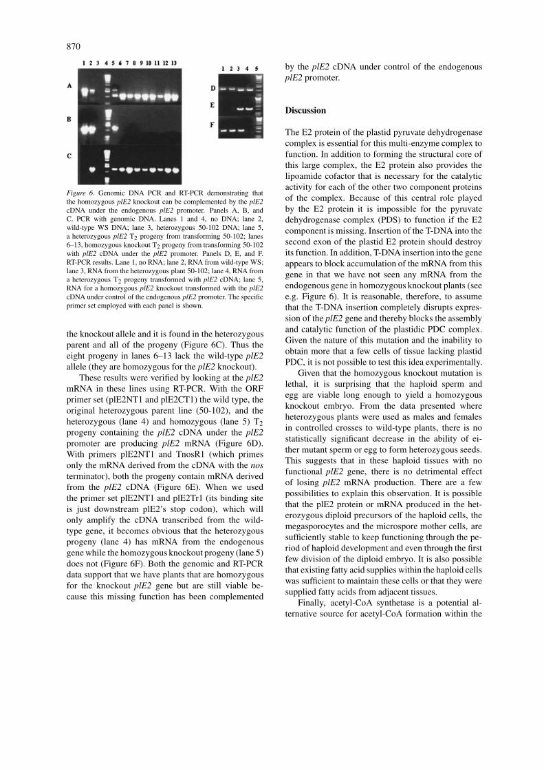
870
Figure 6. Genomic DNA PCR and RT-PCR demonstrating thatthe homozygous plE2 knockout can be complemented by the plE2cDNA under the endogenous plE2 promoter. Panels A, B, andC. PCR with genomic DNA. Lanes 1 and 4, no DNA; lane 2,wild-type WS DNA; lane 3, heterozygous 50-102 DNA; lane 5,a heterozygous plE2 T2 progeny from transforming 50-102; lanes6–13, homozygous knockout T2 progeny from transforming 50-102with plE2 cDNA under the plE2 promoter. Panels D, E, and F.RT-PCR results. Lane 1, no RNA; lane 2, RNA from wild-type WS;lane 3, RNA from the heterozygous plant 50-102; lane 4, RNA froma heterozygous T2 progeny transformed with plE2 cDNA; lane 5,RNA for a homozygous plE2 knockout transformed with the plE2cDNA under control of the endogenous plE2 promoter. The specificprimer set employed with each panel is shown.
the knockout allele and it is found in the heterozygousparent and all of the progeny (Figure 6C). Thus theeight progeny in lanes 6–13 lack the wild-type plE2allele (they are homozygous for the plE2 knockout).
These results were verified by looking at the plE2mRNA in these lines using RT-PCR. With the ORFprimer set (plE2NT1 and plE2CT1) the wild type, theoriginal heterozygous parent line (50-102), and theheterozygous (lane 4) and homozygous (lane 5) T2progeny containing the plE2 cDNA under the plE2promoter are producing plE2 mRNA (Figure 6D).With primers plE2NT1 and TnosR1 (which primesonly the mRNA derived from the cDNA with the nosterminator), both the progeny contain mRNA derivedfrom the plE2 cDNA (Figure 6E). When we usedthe primer set plE2NT1 and plE2Tr1 (its binding siteis just downstream plE2’s stop codon), which willonly amplify the cDNA transcribed from the wild-type gene, it becomes obvious that the heterozygousprogeny (lane 4) has mRNA from the endogenousgene while the homozygous knockout progeny (lane 5)does not (Figure 6F). Both the genomic and RT-PCRdata support that we have plants that are homozygousfor the knockout plE2 gene but are still viable be-cause this missing function has been complemented
by the plE2 cDNA under control of the endogenousplE2 promoter.
Discussion
The E2 protein of the plastid pyruvate dehydrogenasecomplex is essential for this multi-enzyme complex tofunction. In addition to forming the structural core ofthis large complex, the E2 protein also provides thelipoamide cofactor that is necessary for the catalyticactivity for each of the other two component proteinsof the complex. Because of this central role playedby the E2 protein it is impossible for the pyruvatedehydrogenase complex (PDS) to function if the E2component is missing. Insertion of the T-DNA into thesecond exon of the plastid E2 protein should destroyits function. In addition, T-DNA insertion into the geneappears to block accumulation of the mRNA from thisgene in that we have not seen any mRNA from theendogenous gene in homozygous knockout plants (seee.g. Figure 6). It is reasonable, therefore, to assumethat the T-DNA insertion completely disrupts expres-sion of the plE2 gene and thereby blocks the assemblyand catalytic function of the plastidic PDC complex.Given the nature of this mutation and the inability toobtain more that a few cells of tissue lacking plastidPDC, it is not possible to test this idea experimentally.
Given that the homozygous knockout mutation islethal, it is surprising that the haploid sperm andegg are viable long enough to yield a homozygousknockout embryo. From the data presented whereheterozygous plants were used as males and femalesin controlled crosses to wild-type plants, there is nostatistically significant decrease in the ability of ei-ther mutant sperm or egg to form heterozygous seeds.This suggests that in these haploid tissues with nofunctional plE2 gene, there is no detrimental effectof losing plE2 mRNA production. There are a fewpossibilities to explain this observation. It is possiblethat the plE2 protein or mRNA produced in the het-erozygous diploid precursors of the haploid cells, themegasporocytes and the microspore mother cells, aresufficiently stable to keep functioning through the pe-riod of haploid development and even through the firstfew division of the diploid embryo. It is also possiblethat existing fatty acid supplies within the haploid cellswas sufficient to maintain these cells or that they weresupplied fatty acids from adjacent tissues.
Finally, acetyl-CoA synthetase is a potential al-ternative source for acetyl-CoA formation within the
871
plastid (Behal et al. 2002, Ke et al., 2000). It is possi-ble that this enzyme is capable of supplying the neededacetyl-CoA for pollen, egg, and early embryo cellsdevelopment. We have noted that while acetyl-CoAsynthetase mRNA is low in most tissues, it does appearto accumulate to higher levels in flowers, suggestingthat this protein might have a function in those tis-sues. Should the presence of acetyl-CoA synthetasein pollen and eggs be able to provide acetyl-CoA fortheir development, it is not surprising that it cannotplay a similar role in embryo development. Earlier re-sults from this group clearly demonstrated that whilethe cDNA for the subunits of PDC increased in embryotissue, particularly during the period of fatty acid accu-mulation in Arabidopsis seeds, the level of acetyl-CoAsynthetase mRNA, which was low in early embryos,soon decreased to nearly undetectable levels (Ke et al.,2000). As much as it is possible to correlate enzymeactivities with mRNA levels, this would suggest thatthere was insufficient acetyl-CoA synthetase to main-tain embryo growth. In our plE2 knockout mutants wesee that the embryos are not dividing during this devel-opmental period where PDC mRNA accumulates to itshighest level.
The observation that the plE2 knockout is an earlyembryo-lethal is important in determining the func-tion of second potential plE2 protein or plE3-bindingprotein (At1g34430). If At1g34430 does represent asecond plE2 protein it is surprising that it fails tocomplement the knockout of the known plE2 gene.The At1g34430 gene is expressed during early embryodevelopment in flowers (data not presented). Finalconclusions on the role of this protein await furthercomplementation studies. If At1g34430 does repre-sent an plE3 binding protein as postulated by Mooneyet al. (2001), this will be the first such protein iden-tified in plants as this activity appears to be missingfrom plant mitochondria.
While the plE2 cDNA was able to complementthe missing plE2 genomic DNA, it only did so whenunder control of the endogenous plE2 promoter. Itis interesting that the 35S, napin, and AtSERK1 pro-moters all failed to provide plE2 mRNA at this earlydevelopmental period where the protein is needed tomake the necessary PDC. Although the 35S promoteris often described as constitutive, it has been shown incotton with 35S promoter-GFP expression constructsthat this promoter is not expressed well in cottonembryos (Sunilkumar et al., 2002). Similarly, napinexpression occurs fairly late in embryo developmentin canola with its accumulation noted 14–20 days af-
ter flowering (Hoglund et al., 1992). Our work withnapin-GUS reporter lines suggest that GUS accumu-lates several days after flowering in Arabidopsis (datanot presented). Work with northern analysis togetherwith promoter-GUS reporter constructs suggested At-SERK1 is expressed early in embryo development inArabidopsis (Hecht et al., 2001) Its expression is,however, apparently too late or not in the cells whichneed the expression plE2 gene to rescue the plE2knockout mutant. It is possible that plE2 promoter isfunctioning in the haploid germ cells and that PDCactivity is necessary at that point to produce embryosthat are viable after more than a few divisions.
Over 500 mutations that result in altered em-bryogenesis have been described (see Patton et al.,1998; Meinke, 1995). Interestingly, three of theseembryo-defective mutations result from disruption inthe ability of the plant to synthesize biotin (Schnei-der et al., 1989; Shellhammer and Meinke, 1990;Patton et al., 1996; Patton et al., 1998). The acetyl-CoA produced by plastidic PDC is next converted tomalonyl-CoA by a specific plastid form of acetyl-CoAcarboxylase (Ke et al. 1997; Sun et al., 1997). Thisis a biotin-containing enzyme and it is very likelythat disruption of biotin biosynthesis functionally dis-rupts expression of acetyl-CoA carboxylase. Thus, itis quite reasonable that disrupting these two sequentialmetabolic reactions, plastidic pyruvate dehydrogenaseand acetyl-CoA carboxylase, should result in verysimilar phenotypes.
References
Bao, X., Pollard, M. and Ohlrogge, J. 1998. The biosynthesis of eru-cic acid in developing embryos of Brassica rapa. Plant Physiol.118: 183–190.
Behal, R.H. and Oliver, D.J. 1999. A second gene encoding the plas-tidic pyruvate dehydrogenase β-subunit in Arabidopsis thaliana.Plant Physiol. 121: 312– 313.
Behal, R.H., Lin, M., Back, S. and Oliver, D.J. 2002. Role of acetyl-coenzyme A synthetase in leaves of Arabidopsis thaliana. Arch.Biochem. Biophys. 402: 259–267.
Clough, S.J. and Bent, A.F. 1998. Floral dip: a simplifiedmethod for Agrobacterium-mediated transformation of Ara-bidopsis thaliana. Plant J. 16: 735–743.
Drea, S.C., Mould, R.M., Hibberd, J.M., Gray, J.C. and Kavanagh,T.A. 2001. Tissue-specific and developmental-specific expres-sion of an Arabidopsis thaliana gene encoding the lipoamide de-hydrogenase component of the plastid pyruvate dehydrogenasecomplex. Plant Mol. Biol. 46: 705–715.
Hecht, V., Vielle-Calzada, J.P., Hartog, M.V., Schmidt, E.D.,Boutilier, K., Grossniklaus, U. and de Vries, S.C. 2001. The Ara-bidopsis SOMATIC EMBRYOGENESIS RECEPTOR KINASE 1gene is expressed in developing ovules and embryos and en-

872
hances embryogenic competence in culture. Plant Physiol. 127:803–816.
Hoglund, A.-S., Rodin, J., Larsson, E. and Rask, L. 1992. Distrib-ution of napin and cruciferin in developing rape seed embryos.Plant Physiol. 98: 509–515.
Johnston, M.L., Leuthy, M.H., Miernyk, J.A. and Randall,D.D. 1997. Cloning and molecular analyses of the Arabidop-sis thaliana plastid pyruvate dehydrogenase subunits. Biochim.Biophys. Acta 1321: 200–206.
Johnston, M.L., Meirnyk, J.A. and Randall, D.D. 2000. Import,processing , and assembly of the α- and β-subunits of chloroplastpyruvate dehydrogenase. Planta 211: 72–76.
Kang, F. and Rawsthorne, S. 1994. Starch and fatty acid synthesisin plastids from developing embryos of oilseed rape. Plant J. 6:795–805.
Kang, F. and Rawsthorne, S. 1996. Metabolism of glucose-6-phosphate and utilization of multiple metabolites for fatty acidsynthesis by plastids from developing oilseed rape embryos.Planta 199: 321–327.
Ke, J., Choi, J.-K., Smith, M., Horner, H.T., Nikolau, B.J. andWurtele, E.S. 1997. Structure of the CAC1 gene and in situcharacterization of its expression: the Arabidopsis thaliana genecoding for the biotin-containing subunit of the plastidic acetyl-coenzyme A carboxylase. Plant Physiol. 113: 357–365.
Ke, J., Behal, R.H., Back, S.L., Nikolau, B.J., Wurtele, E.S.and Oliver, D.J. 2000. The role of pyruvate dehydrogenaseand acetyl-coenzyme A synthetase in fatty acid synthesis indeveloping Arabidopsis seeds. Plant Physiol. 123: 497–508.
Lutziger, I. and Oliver, D.J. 2000. Molecular proof of a uniquelipoamide dehydrogenase in plastids: analysis of plastidiclipoamide dehydrogenase from Arabidopsis thaliana. FEBS Lett.484: 12–16.
Meinke, D.W. 1985. Embryo-lethal mutants of Arabidopsisthaliana: analysis of mutants with a wide range of lethal phases.Theor. Appl. Genet. 69: 543–552.
Meinke, D.W. 1995. Molecular genetics of plant embryogenesis.Annu. Rev. Plant Physiol. Plant Mol. Biol. 46: 369–394.
Mooney, B.P., Miernyk, J.A. and Randall, D.D. 1999. Cloningand characterization of the dihydrolipoamide S-acetyltransferasesubunit of the plastid pyruvate dehydrogenase complex (E2)from Arabidopsis. Plant Physiol. 120: 443–452.
Mooney, B.P., Miernyk, J.A. and Randall, D.D. 2002. The complexfate of α-ketoacids. Annu. Rev. Plant Biol. 53: 357–375.
Patton, D.A., Schetter, A.L., Franzmann, L.H., Nelson, K., Ward,E.R. and Meinke, D.W. 1998. An embryo-defective mutant ofArabidopsis disrupted in the final step of biotin synthesis. PlantPhysiol. 116: 935–946.
Schneider, T., Dinkins, R., Robinson, K., Shellhammer, J. andMeinke, D.W. 1989. An embryo-lethal mutant of Arabidopsisthaliana is a biotin auxotroph. Dev. Biol. 131: 161–167.
Shellhammer, J. and Meinke, D. 1990. Arrested embryos from thebio1 auxotroph of Arabidopsis thaliana contain reduced levels ofbiotin. Plant Physiol. 93: 1162–1167.
Sun, J., Ke, J., Johnson, J.L., Nikolau, B.J. and Wurtele, E.S. 1997.Biochemical and molecular biological characterization of CAC2,the Arabidopsis thaliana gene coding for the biotin carboxylasesubunit of the plastidic acetyl-coenzyme A carboxylase. PlantPhysiol. 115: 1371–1383.
Sunilkumar, G., Mohr, L., Lopata-Finch, E., Emani, C. andRathore, K.S. 2002. Developmental and tissue-specific expres-sion of CaMV 35S promoter in cotton as revealed by GFP. PlantMol. Biol. 50: 463–474.
Thelan, J.J., Muszynski M.G., Miernyk J.A. and Randall, D.D.1998. Molecular analysis of two pyruvate dehydrogenase kinasesfrom maize. J. Biol. Chem. 273: 26618–26623.