do gastrointestinal taste receptors contribute to ......although it has been well established that...
TRANSCRIPT

G. J. Golden, A. M. Hussey and B. A. Kimballbehavior?
Do gastrointestinal taste receptors contribute to associative learning and foraging
doi: 10.2527/jas.2012-5089 originally published online July 24, 20122012, 90:4297-4307.J ANIM SCI
http://www.journalofanimalscience.org/content/90/12/4297the World Wide Web at:
The online version of this article, along with updated information and services, is located on
www.asas.org
at National Animal Disease Ctr on January 16, 2013www.journalofanimalscience.orgDownloaded from

4297
© 2012 American Society of Animal Science. All rights reserved. J. Anim. Sci. 2012.90:4297–4307 doi:10.2527/jas2012-5089
Key words: conditioned taste aversion, foraging behavior, gastric taste, herbivory, refl ux
ABSTRACT: Foraging behavior is an expression of learning, context, and experience arising from integra-tion of sensory information obtained during feeding with postingestive consequences of food ingestion. Although it has been well established that gustatory and olfactory systems of the mouth and nose provide sensory information to the consumer (in the form of fl a-vor), sweet and bitter taste receptors have recently been identifi ed in the intestinal tract of humans and rodents. It remains possible that sensory information gener-ated in the gut could contribute to the learning process. Thus, a series of experiments was conducted to deter-mine if classical associative learning occurs when the conditional stimulus circumvents oronasal presentation via direct delivery to the gut or peritoneal cavity. Mice receiving an intragastric infusion of 5 mM sodium sac-charin immediately followed by LiCl administration demonstrated a signifi cant decrease in preference for 5 mM saccharin in 4 consecutive 23h, 2-bottle preference tests versus water (P = 0.0053). Saccharin was highly preferred in mice receiving intragastric (IG) saccha-rin only or interperitoneal (i.p.) injection of LiCl only.
This reduced preference indicated that mice “tasted” saccharin infused into the gut. However, efforts to rep-licate with a reduced infusion volume failed to result in decreased preference. To understand if there were alter-native pathways for oral detection of infused saccharin, mice received intragastric infusions (5.4 mM) and i.p. injections (10.8 mM) of sodium fl uorescein. Fluores-cence was observed from the tongues and esophagi of mice infused with volumes of 0.5 mL or more or injected with volumes of 0.25 mL or greater. Interperi-toneal injections of 5 mM saccharin in mice resulted in reduced preference for 5 mM saccharin presented orally in 2-bottle preference tests (P = 0.0287). Oral delivery of a 500-fold less concentration of saccharin (0.01 mM) during conditioning resulted in a similar preference expression as shown in the initial IG experiment. These results demonstrate that although compounds may be tasted in the mouth absent of oral contact, associative learning is attenuated. Therefore, intestinal taste recep-tors are unlikely to participate directly in learning and recognition of foods during foraging events.
Do gastrointestinal taste receptors contribute to associative learning and foraging behavior?1
G. J. Golden,*2 A. M. Hussey,* and B. A. Kimball*†
*Monell Chemical Senses Center, Philadelphia, PA 19104; and †USDA-APHIS-WS-National Wildlife Research Center, Philadelphia, PA 19104
INTRODUCTION
Taste, smell and somatosensation are essential che-mosensory processes foraging animals use to identify
benefi cial nutrients, non-edible miscellany, and poten-tially deleterious toxins. Ingested foods are mechani-cally and chemically digested in the stomach or rumen, liberating individual compounds that may not have been detected orally. From the perspective of mammalian diet selection, detection of these compounds by taste recep-tors in the intestinal tract could provide valuable sensory information.
Although studies with herbivores are more prevalent (Provenza et al., 1992; Hobbs, 1996; Moore and Foley, 2005; Manier and Hobbs, 2006), mammalian diet selec-tion is strongly infl uenced by integration of cue and con-sequence across many foraging guilds (Forbes, 1998; Brodie, 1999; Baker et al., 2007; Webb et al., 2008). To
1The authors are grateful to Anthony Sclafani for allowing GJG to visit his laboratory and study his intragastric catheter surgery technique. We are also most grateful to Michael Tordoff for com-menting on an earlier version of this manuscript. Special thanks to Karen Yee and Linda Wysocki for advice with histology and light microscopy and to Eleanora Robinson, Danielle DeNofa, and Caroline Robiolle for technical assistance. This work was supported by the NIH training grant NIDCD 5T32DC000014-30 (GJG) and USDA CA-09-7442-0585.
2Corresponding author: [email protected] January 4, 2012.Accepted June 23, 2012.
at National Animal Disease Ctr on January 16, 2013www.journalofanimalscience.orgDownloaded from

Golden et al.4298
represent mammals in general, experiments with a mouse model were designed to determine if novel compounds presented in the intestine could provide information use-ful for decisions regarding diet selection in mammals. To separate taste per se from multiple post-ingestive feed-backs, the current study used a non-nutritive sweet taste stimulus (i.e., saccharin) in a well-established method-ology for ingestive behavior and associative learning; namely, conditioned taste aversion (CTA; Garcia et al., 1955). Evidence of CTA learning resulting from saccharin infused into the stomach (intragastric; IG) paired with the toxic effects of LiCl would indicate that taste receptors in the gut convey gustatory information to the brain.
Results of our initial experiment suggested that, in-deed, a lithium-induced aversion to IG saccharin was evident in mice upon oral presentation of a saccharin so-lution. Regurgitation in a mouse model seemed unlikely, although artifi cial refl ux produced by the experimental delivery system could not be ruled out. When replicating
these fi ndings, we modifi ed the IG delivery procedures and evaluated an alternative pathway for gustatory infor-mation to reach the brain (i.e., taste receptors in the oral cavity via circulating blood). These experiments similarly relied on CTA to evaluate preferences for a novel taste cue presented IG, intraperitoneally (i.p.), or orally in as-sociation with i.p. LiCl to test the hypothesis that chemo-sensory input from gastrointestinal (GI) taste receptors modifi es the taste response (Table 1).
METHODS
All experimental protocols were approved by the Monell Chemical Senses Center Institutional Animal Care and Use Committee.
Subjects
Male outbred CD-1 mice (Charles River Labora-
Table 1. Experimental schedule1
Experiment Treatment identifi er n
Conditioning Preference testing
d 1, 3, 52,3
d 8 to 11 d 15 to 18Conditioning stimulusUnconditional
stimulus4
1 IG 5 1.0 mL of 5 mM Saccharin5 None 5 mM Saccharin NoneIG+LiCl 5 1.0 mL of 5 mM Saccharin5 LiCl vs.LiCl 8 None LiCl Water
2 10IG 9 0.5 mL of 10 mM Saccharin3 None 5 mM Saccharin 10 mM Saccharin10IG+LiCl 9 0.5 mL of 10 mM Saccharin3 LiCl vs. vs.10LiCl 9 None LiCl Water Water
3 Dye 3 0.25, 0.5, 0.75, 1.0, 1.25, or 1.50 mL of 5.4 mM None NA NAControl 2 Fluorescein6 None
None4 i.p. Dye 5 0.125, 0.25, 0.375, 0.5, 0.625, or 0.75 mL 10.8 mM None NA NA
Control 1 Fluorescein7 NoneNone
5 IP 8 1.0 mL of 5 mM Saccharin None 5 mM Saccharin NoneIP+LiCl 8 1.0 mL of 5 mM Saccharin LiCl vs.IPLiCl 8 None LiCl Water
IPSaccon8 8 1.0 mL of 5 mM Saccharin LiCl6 5Oral+LiCl 8 25 mL of 5 mM Saccharin LiCl 5 mM Saccharin None
0.01Oral+LiCl 8 25 mL of 0.01mM Saccharin LiCl vs.0.01Oralsaccon8 8 25 mL of 0.01mM Saccharin LiCl7 Water0.01Oralexp 8 25 mL of 0.01mM Saccharin NoneOral 8 None None
1IG = intragastric; LiCl = lithium chloride; IP = interperitoneal (i.p.); Sac = saccharin; Con = concurrent delivery; Oral = oral presentation of conditioning stimulus; Exp = experienced (subjects familiarized with saccharin); NA = not applicable (no preference testing).
2d 2, 4, 6, and 7 were rest days.3Fluorescein infusion or injection was conducted on d 1 only. Excised tissues examined by light microscopy.NA = not applicable4Lithium chloride (LiCl) was delivered by intraperitoneal (i.p.) injection 30 min after delivery of conditioning stimulus.5The conditional stimulus was delivered directly to the stomach by intragastric infusion by a syringe pump.6Delivery rates varied according to fi nal volume (i.e., 0.25, 0.5, 0.75, 1.0, 1.25, and 1.5 mL); 3 mice per volume.7Injected volumes were 0.125, 0.25, 0.375, 0.5, 0.625, and 0.75 mL; 5 mice per volume.8LiCl was mixed directly with the saccharin solution in Exp.5 and given immediately before solution presentation in Exp. 6.
at National Animal Disease Ctr on January 16, 2013www.journalofanimalscience.orgDownloaded from

Taste in the Gut 4299
tories, Wilmington, MA) were used in all experiments. Subjects were housed in individual cages (28 cm × 12.5 cm × 12 cm) in a temperature-controlled room at 23°C on a 12-h light (12-h dark cycle) and had free access to the Teklad Rodent Diet 8604 (Harlan, Madison, WI).
Intragastric Catheter Surgery
Mice were deeply anesthetized with an i.p. injection of a ketamine hydrochloride (42.8 mg/kg), xylazine hy-drochloride (8.6 mg/kg) and acepromazine (1.5 mg/kg) mixture (10 mL/kg) and anesthesia was maintained with 1% isofl urane. The abdomen of each mouse was shaved from the sternum to approximately 5 cm caudal of the sternum and the shaved area was cleaned with alternate gauze swabs of 70% alcohol and iodine disinfectant (Betadine, Purdue Pharma L.P., Stamford, CT). An inci-sion along the midline was made with scissors (~1.5 cm below sternum). On both sides of the incision, the sur-rounding skin was separated from the underlying mus-cle layer using a needle holder. A shorter incision was made in the abdominal muscles to display the abdominal cavity. The stomach was removed from the abdominal cavity and laid on a sterile cotton swab. A purse suture was made in the fundus of the stomach using 6-0 silk suture. The heat-fl ared end of a micro-renathane cath-eter was inserted into the stomach via a small puncture into the center of the area enclosed by the purse suture, which was pulled closed and tied off. The stomach was replaced in the abdominal cavity and a small hole was made in the abdominal muscle ~1 cm from right side of incision with #7 curved forceps. The catheter was pulled through the opening and the abdominal muscle opening closed with Surgi-Lock liquid suture (Meridian Animal Health, Omaha, NE). The catheter was routed under the skin to the back of the neck and the stomach incision was closed with 5-0 silk suture. A back mount (Plastics One, Roanoke, VA) was attached to the muscle layer on each side of the mount using 5-0 silk suture. The cath-eter was connected to side port of the back mount and the neck incision was tightly closed with 5-0 suture fol-lowed by 18 mm wound clips. All incisions were treated with triple antibiotic (Neosporin, Johnson & Johnson, New Brunswick, NJ). Mice were prophylactically treat-ed with an antibiotic (2.0 mg/kg Gentamicin intramus-cularly) and given postoperative treatment (1.0 mg/kg Buprenorphine subcutenously) for pain.
Several days before, and for several days immedi-ately after surgery, mice were fed a mixture of choco-late Ensure (Abbott Laboratories, Abbott Park, IL) and ground Teklad Rodent Diet 8604 chow (Harlan) to fa-cilitate digestion and BW maintenance. Mice were given 5 to 12 d to recover from surgery during which food and water was available ad libitum.
Apparatus
Mice infused IG were conditioned in groups of 4 (i.e., 2 IG infusion subjects and 2 controls). Conditioning was conducted in a 22.5 cm × 24 cm × 20 cm polycarbonate cage. For IG infusions, polyethylene tubing connected a multi-syringe infusion pump (Harvard Apparatus, Hollis-ton, MA) to the input port of a 22-gauge swivel (Instech Laboratories, Inc. Plymouth Meeting, PA) clamped to a ring stand. The output port of the swivel was attached to the input port of the back mount of each mouse with polyethylene tubing surrounded by a stainless-steel spring (PlasticsOne) for protection. Control mice were placed in an identical polycarbonate cage placed next to the infu-sion pump to permit access to environmental (e.g., odor or auditory) cues occurring during infusion. Fecal matter was removed promptly during infusion sessions and cag-es were cleaned with 70% isopropyl alcohol and allowed to dry between each set of mice.
Exp. 1
At the beginning of the experiment, mice were 8 wk old and had a mean BW of 39.5 ± 0.6 g. Mice implanted with IG catheters were acclimated to the infusion appa-ratus with 5-min infusions of 0.5 mL of water for 3 con-secutive d followed by 2 d of rest. Food was removed from all groups at 1600 h on the day before condition-ing days and returned at 1600 h after conditioning. Mice were randomly assigned to 3 treatment groups.
Conditioning occurred between 0930 h and 1130 h. Mice in the IG+LiCl (n = 5) and IG (n = 5) groups were infused with 0.1 mL/min of 5 mM sodium saccharin (Sigma Aldrich, St.Louis, MO) over 10 min for a total of 1 mL of sodium saccharin infused. Thirty minutes after intragastric infusion of 5 mM sodium saccharin, mice in the IG+LiCl treatment received ~1.4 mL i.p. injection of 150 mM LiCl (230 mg/kg). The IG treatment group re-ceived an IG infusion of a taste stimulus alone. The LiCl treatment group (n = 8) was placed in a polycarbonate cage identical to the infusion cage and 30 min later was injected with ~1.4 mL of 150 mM LiCl 230 mg/kg. Mice in the LiCl treatment group were not implanted with IG catheters. Mice were conditioned on 3 d with a day of rest in between each conditioning day (Table 1).
After 2 d of rest, mice were given 4 consecutive 23-h, 2-bottle preference tests (5 mM saccharin vs. water) in their home cages. The position of the saccharin was counterbalanced among all groups and switched daily. Solutions were offered in 30-mL syringes that were modifi ed to accept a standard stainless steel sipper tube (Allentown Caging Equipment Co., Allentown, NJ). The syringes were mounted on the front of the cage with the drinking spouts penetrating so that the tips were ~ 4.2
at National Animal Disease Ctr on January 16, 2013www.journalofanimalscience.orgDownloaded from

Golden et al.4300
cm apart and ~ 4.6 cm above the cage fl oor.
Exp. 2
In light of research suggesting that intraluminal pressure reaches maximal accommodation without dis-tension in the mouse stomach at approximately 0.5 mL (Dixit et al., 2006), Exp. 1 was repeated with a smaller infusion volume (decreased to 0.5 mL from 1.0 mL). At the beginning of the experiment, mice (n = 32) were 8-wk old and had a mean BW of 33.2 ± 0.3 g. To ac-commodate the change in volume, the concentration of the sodium saccharin solution was increased to 10 mM (from 5 mM in Exp. 1) to maintain identical sac-charin doses between the 2 experiments. Mice in the 10IG+LiCl (n = 9) and 10IG (n = 9) groups were in-fused with 10 mM sodium saccharin (50 μL/min for 10 min) for a total of 0.5 mL. Mice in the 10LiCl group (n = 9) were exposed to identical environmental conditions, but did not receive surgery. The 2-choice preference test procedure was identical to Exp. 1, with the addition of 10 mM saccharin versus water in a second, separate se-ries of 4 consecutive 23-h, 2-bottle preference tests.
Exp. 3
The previous experiments raised the possibility that large volumes of solutions infused into the stomach might stimulate oral taste by esophageal refl ux. To test this directly, we infused various volumes of the fl uores-cent dye, sodium fl uorescein (5.4 mM; prepared in 0.01 M PBS) into the stomach and looked for its appearance in the oral cavity.
Twenty mice from Exp. 2 were used. Food was re-moved from all groups at 1600 h on the day prior the day of infusion. Two mice received neither surgical treatment nor fl uorescein infusion to act as a control for autofl uorescence. For the remaining 18 mice, 5.4 mM sodium fl uorescein (Sigma Aldrich) was infused so as to deliver over a 10-min period these volumes: 0.25, 0.5, 0.75, 1.0, 1.25, and 1.50 mL. Three mice were infused at each volume. After infusion, mice were returned to their home cages for 90 min and were then euthanized by CO2 asphyxiation.
Although the mouse was still intact, an observer blind to the experimental treatments recorded the pres-ence of fl uorescence in the oral cavity detected with a hand-held ultraviolet. The anterior tongue (i.e., rostral of the intramolar eminence), esophagus, and heart were harvested and stored separately in 0.01 M PBS for 24 h. Esophagi and hearts were halved to expose the interior tissue and all tissues were mounted on glass slides for examination under light microscopy (4× magnifi cation) using a fl uorescein fi lter set. Images were captured using
a Nikon digital camera (DXM1200C) attached to a Nikon Eclipse 80i microscope (Nikon Inc., Melville, NY). The exposure times of the camera of the microscope were set for the brightest fl uorescence (e.g., tongue 1/30 s, esoph-agus 1/25 s, and heart 1/12 s) and all images of specifi c tissues were taken at those exposure times.
Exp. 4
To determine if fl uorescein was being transported throughout the body via the circulatory system, fl uo-rescein was injected directly into the peritoneal cavity. At the beginning of the experiment, mice (n = 31) were 9-wk old and had a mean BW of 39.6 ± 0.4 g. Food was removed from all groups at 1600 h on the day before the day of injection. To compensate for the small size of the peritoneal cavity, the fl uorescein concentration was doubled (10.8 mM) and these volumes were delivered by i.p. injection: 0.0, 0.125, 0.25, 0.375, 0.5, 0.625, and 0.75 mL. With the exception of a single control mouse (no injection), 5 mice were infused at each volume. After i.p. injection, mice were returned to their home cages for 90 min and were then euthanized by CO2 asphyxiation. Tissues were evaluated for fl uorescence as in Exp. 3.
Exp. 5
To determine if the circulatory system provided a route to oral detection of saccharin, Exp. 1 was repeated except that saccharin was delivered by i.p. injection and training occurred in the home cages of the mice. In ad-dition, a 300 mM LiCl concentration was employed to reduce the injection volume because saccharin was also being delivered i.p. in this experiment. At the beginning of the experiment, mice (n = 32) were 8-wk old and had a mean BW of 33.1 ± 0.3 g. Food was removed from all groups at 1600 h on the day before the day of injection. The IP+LiCl group (n = 8) received a 1 mL i.p. injection of 5 mM saccharin followed 30 min later by a second i.p. injection of 300 mM LiCl (230 mg/kg; 0.55 mL for a 30 g mouse). The IP treatment group (n = 8) received an i.p. injection of 5 mM saccharin alone. The IPLiCl treatment group (n = 8) was injected with 300 mM LiCl (230 mg/kg; 0.55 mL for a 30-g mouse). A fourth treat-ment was added to Exp. 5. The IPSaccon group (n = 8; concurrent delivery of saccharin and LiCl) received a 1 mL i.p. injection of a 5 mM saccharin and LiCl (230 mg/kg) solution such that delivery of the taste stimulus and unconditional stimulus were simultaneous.
Exp. 6
In the fi nal experiment, we verifi ed the aversive re-sponse to oral presentation of 5 mM saccharin paired
at National Animal Disease Ctr on January 16, 2013www.journalofanimalscience.orgDownloaded from

Taste in the Gut 4301
with lithium toxicosis and evaluated the aversive re-sponse to a 500-fold smaller concentration of saccharin (0.01 mM). The experiment was performed to offer a potential explanation for the results of Exp. 1 based on the results of Exp. 2 to 5. If saccharin infused into the gut was reaching the oral cavity, one might expect that it would be dilute in comparison with the concentration originally infused (Exp. 1) or injected (Exp. 5). Experi-ment 1 was repeated except there was no food restric-tion, saccharin was presented orally during condition-ing, and conditioning occurred in the home cage of each mouse. At the beginning of the experiment, mice (n = 40) were 8-wk old and had a mean BW of 29.6 ± 0.2 g. Water was removed from all groups at 1600 h on the day before the days of conditioning to encourage the mice to drink during the 30-min saccharin exposure. Mice in the 0.01Oral+LiCl (n = 8) and 5Oral+LiCl (n = 8) groups were presented orally with 25 mL of 0.01 mM or 5 mM sodium saccharin, respectively, for 30 min followed by i.p. injection of 150 mM LiCl. Mice in the 0.01Oralsac-con (n = 8; concurrent delivery of saccharin and LiCl) group were given an i.p. injection of 150 mM LiCl fol-lowed immediately by an oral presentation of 25 mL of 0.01 mM sodium saccharin for 30 min. Mice in the Oral (n = 8) and 0.01Oralexp (n = 8; experienced with sac-charin) groups were presented orally with 25 mL of dis-tilled water or 0.01 mM sodium saccharin, respectively, for 30 min, but did not receive any LiCl treatment during conditioning. Preference testing was identical to Exp. 1.
Data Analyses
Intakes of 2-bottle test taste solutions were mea-sured (to the nearest 0.1 g) daily. Intakes were not cor-rected according to BW. Data for each experiment were analyses separately and examination of residuals indi-cated that use of ANOVA was valid in each case. For Exp. 1, 2, and 5, preference scores were calculated as the ratio of saccharin solution to total fl uid (saccharin + wa-ter) intake. Scores were analyzed by repeated measures ANOVA using the MIXED procedure (SAS Inst. Inc., Cary, NC). Treatment group and the interaction were fi xed effects. Mice were considered random effects. Multiple comparisons of least square means were made using the false discovery rate controlling procedure (Benjamini and Hochberg, 1995). Residuals were tested for assumptions (location, normality, and independence) using the UNIVARIATE procedure of SAS.
To draw comparisons across multiple experiments, control group means from appropriate experiments were employed as hypothetical means. “Difference scores” were calculated as 5-mM preference scores of mice receiving saccharin paired with lithium (i.e., IG+LiCl, IP+LiCl, 5Oral+LiCl, and 0.01Oral+LiCl) in Exp. 1,
5, and 6 minus the mean preference score of the cor-responding unconditioned treatment (i.e., IG, IP, and Oral). Thus, difference scores represented the change in 5-mM saccharin preference relative to the unconditioned response. Scores were analyzed by 1-way ANOVA using the MIXED procedure in SAS. Multiple comparisons of least square means were made using the false discovery rate controlling procedure. Comparisons of the initial BW for mice in each group were analyzed by mixed-model ANOVA with treatment as a fi xed effect and sub-ject a random effect.
RESULTS
Exp. 1
There was no difference in initial BW of the 3 groups (P > 0.1). Preference scores were impacted by treatment (P = 0.0053) and position (P = 0.0029; position refers to the right or left position of the saccharin tube as placed in the test cage). No other effects were signifi cant (P > 0.05). In 23 h, 2-bottle tests of 5 mM saccharin vs. water, the IG+LiCl group had a reduced preference for saccharin in comparison with both the IG (P = 0.0061) and LiCl (P = 0.0024) groups, which expressed a strong preference for the saccharin in comparison with water (Figure 1). This level of decreased preference was main-tained throughout the 4 d of 2-bottle tests; thus, there was no evidence of extinction. The reduced preference for saccharin displayed by the IG+LiCl group suggested that mice “tasted” saccharin infused into the gut and as-sociated this taste with lithium toxicosis.
Exp. 2
There was no difference in initial BW for all groups (P > 0.1). Preference for 5 mM saccharin in comparison with water did not differ among the treatment groups (P = 0.134; Figure 2a); or for 10 mM (P = 0.369; Figure 2b). These results suggest that mice could not “taste” the re-duced volume of saccharin infused directly into the gut.
Exp. 3
The results obtained by observing fl uorescence on the tongues of intact mice were almost identical to the results seen under light microscopy, so we present only the latter here. Auto-fl uorescence, a natural condition in tissue samples, was observed in all tissue samples (in-cluding tissue never exposed to fl uorescein), but a strong and unmistakable fl uorescent signal was observed from the tongues and esophagi of mice infused with a vol-umes of 0.5 mL of fl uorescein or more. However, no fl uorescence was observed from the heart tissues (Fig-
at National Animal Disease Ctr on January 16, 2013www.journalofanimalscience.orgDownloaded from

Golden et al.4302
ure 3). These results suggest that solutions of 0.5-mL volume or more infused into the mouse stomach may leak through the esophageal sphincter and enter the oral cavity by traveling through the esophagus.
Exp. 4
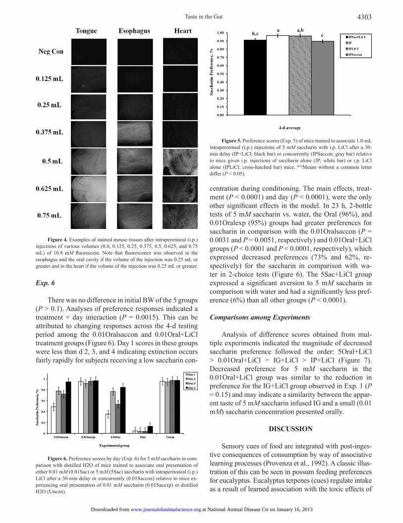
The results obtained by observing fl uorescence on the tongues of intact mice were almost identical to the results seen under light microscopy, so again we present only the latter here. Contrary to the results of Exp. 3, fl uorescence
was detected with a hand-held ultraviolet light in shallow tissue extremities (i.e., toes and ears). Auto-fl uorescence was observed in all tissue samples (including tissue never exposed to fl uorescein) but a strong and unmistakable fl uorescent signal was observed from the tongues and esophagi of mice injected with volumes of 0.25 mL of fl uorescein (equivalent to IG infusions of 0.5 mL) or more (Figure 4). These results suggest that solutions of 0.5-mL volume or more injected into the mouse peritoneal cavity travel to the oral cavity through the blood.
Exp. 5
There was no difference in initial BW of the 3 groups (P > 0.1). Preference scores were impacted by treatment (P = 0.0287) and day (P = 0.0012). No other effects were observed (P > 0.05). In 23 h, 2-bottle tests of 5 mM saccharin vs. water, the IP+LiCl (91%) and IP-Saccon (89%) groups had reduced preferences for sac-charin in comparison with the IP (P = 0.0465 and P = 0.0151, respectively) group, which expressed a strong preference (96%) for the saccharin in comparison with water (Figure 5). The IPSaccon (89%) group had a re-duced preference for saccharin in comparison with the IPLiCl (P = 0.0198) group, which also expressed a strong preference (96%) for the saccharin in comparison with water (Figure 5). This level of decreased preference was maintained throughout the 4 d of 2-bottle tests; thus, there was no evidence of extinction. Although the mag-nitude of the decrease in saccharin was small, reduced preference for saccharin in the IP+LiCl and IPSaccon treatments demonstrate that mice “tasted” saccharin in-jected into the peritoneal cavity and associated this taste with lithium toxicosis.
Figure 3. Examples of stained mouse tissue after 10-min intragastric (IG) infusion of various volumes (0.0, 0.25, 0.5, 0.75, 1.0, 1.25, or 1.5 mL) of 5.4 mM fl uorescein. Note that fl uorescence was observed in the esophagus and the oral cavity if the volume of the infusion was 0.5 mL or greater. No fl uorescence was observed in heart tissues.
Figure 1. Preference scores (Exp. 1) of mice trained to associate 1.0 mL of intragastric (IG) infusions of 5 mM saccharin with intraperotineal (i.p.) lithium chloride (IG+LiCl; black bar) relative to mice given IG sac-charin alone (IG; white bar) or i.p. LiCl alone (LiCl; cross-hatched bar) mice. a,bMeans without a common letter differ (P < 0.05).
Figure 2. Preference scores of mice in Exp. 2. Mice were trained to associate 0.5 mL of intragastric (IG) infusions of saccharin with intraper-otineal (i.p.) lithium chloride (IG+LiCl; black bar) relative to mice given IG saccharin alone (IG; white bar) or i.p. LiCl alone (LiCl; cross-hatched bar). Top panel: tests using 5 mM saccharin; bottom panel: tests using 10 mM sac-charin. There were no differences in preferences among the groups (P > 0.05).
at National Animal Disease Ctr on January 16, 2013www.journalofanimalscience.orgDownloaded from
Taste in the Gut 4303
Exp. 6
There was no difference in initial BW of the 5 groups (P > 0.1). Analyses of preference responses indicated a treatment × day interaction (P = 0.0015). This can be attributed to changing responses across the 4-d testing period among the 0.01Oralsaccon and 0.01Oral+LiCl treatment groups (Figure 6). Day 1 scores in these groups were less than d 2, 3, and 4 indicating extinction occurs fairly rapidly for subjects receiving a low saccharin con-
centration during conditioning. The main effects, treat-ment (P < 0.0001) and day (P < 0.0001), were the only other signifi cant effects in the model. In 23 h, 2-bottle tests of 5 mM saccharin vs. water, the Oral (96%), and 0.01Oralexp (95%) groups had greater preferences for saccharin in comparison with the 0.01Oralsaccon (P = 0.0031 and P = 0.0051, respectively) and 0.01Oral+LiCl groups (P < 0.0001 and P < 0.0001, respectively), which expressed decreased preferences (73% and 62%, re-spectively) for the saccharin in comparison with wa-ter in 2-choice tests (Figure 6). The 5Sac+LiCl group expressed a signifi cant aversion to 5 mM saccharin in comparison with water and had a signifi cantly less pref-erence (6%) than all other groups (P < 0.0001).
Comparisons among Experiments
Analysis of difference scores obtained from mul-tiple experiments indicated the magnitude of decreased saccharin preference followed the order: 5Oral+LiCl > 0.01Oral+LiCl = IG+LiCl > IP+LiCl (Figure 7). Decreased preference for 5 mM saccharin in the 0.01Oral+LiCl group was similar to the reduction in preference for the IG+LiCl group observed in Exp. 1 (P = 0.15) and may indicate a similarity between the appar-ent taste of 5 mM saccharin infused IG and a small (0.01 mM) saccharin concentration presented orally.
DISCUSSION
Sensory cues of food are integrated with post-inges-tive consequences of consumption by way of associative learning processes (Provenza et al., 1992). A classic illus-tration of this can be seen in possum feeding preferences for eucalyptus. Eucalyptus terpenes (cues) regulate intake as a result of learned association with the toxic effects of
Figure 4. Examples of stained mouse tissues after intraperotineal (i.p.) injections of various volumes (0.0, 0.125, 0.25, 0.375, 0.5, 0.625, and 0.75 mL) of 10.8 mM fl uorescein. Note that fl uorescence was observed in the esophagus and the oral cavity if the volume of the injection was 0.25 mL or greater and in the heart if the volume of the injection was 0.25 mL or greater.
Figure 5. Preference scores (Exp. 5) of mice trained to associate 1.0-mL intraperotineal (i.p.) injections of 5 mM saccharin with i.p. LiCl after a 30-min delay (IP+LiCl; black bar) or concurrently (IPSaccon; gray bar) relative to mice given i.p. injections of saccharin alone (IP; white bar) or i.p. LiCl alone (IPLiCl; cross-hatched bar) mice. a-cMeans without a common letter differ (P < 0.05).
Figure 6. Preference scores by day (Exp. 6) for 5 mM saccharin in com-parison with distilled H2O of mice trained to associate oral presentation of either 0.01 mM (0.01Sac) or 5 mM (5Sac) saccharin with intraperitoneal (i.p.) LiCl after a 30-min delay or concurrently (0.01Saccon) relative to mice ex-periencing oral presentation of 0.01 mM saccharin (0.01Sacexp) or distilled H2O (Uncon).
at National Animal Disease Ctr on January 16, 2013www.journalofanimalscience.orgDownloaded from

Golden et al.4304
diformlyphloroglucinol compounds (consequences) also present in eucalyptus leaves (Lawler et al., 1999). In fact, this example clearly demonstrates that compounds giv-ing rise to consequences of forage consumption are rarely the same as those serving as cues (Provenza and Balph, 1990). Preferences are similarly formed when sensory cues are associated with benefi cial consequences of in-gestion (e.g., nutrients). Importantly, this interplay of cues and consequences (i.e., palatability) has implications far beyond diet selection. Landscape heterogeneity (Manier and Hobbs, 2006), ecosystem function (Hobbs, 1996), and herbivore population dynamics (Moore and Foley, 2005; Wang et al., 2006), among other landscape-level processes, are infl uenced by foraging behaviors arising from detection and ingestion of phytochemicals.
Mammals, from small rodents to large herbivores, are equipped with anatomical and biochemical attributes permitting detection, use, and detoxifi cation of forage. In-tegration of gustatory and visceral information is made possible by the confl uence of neurons in the solitary tract of the nucleus, allowing for learned preferences and aver-sions (Provenza, 1995a). Such affective processes, long characterized in rodents (Swank et al., 1996; Thiele et al., 1996; Houpt et al., 1997), are also well recognized in large herbivores (Provenza, 1995b). Thus, information regard-ing integration of sensory and post-ingestive information obtained in model rodents is relevant to other mammals and their interactions with their foraging environments.
Role of Intestinal Taste Receptors
For obvious reasons, olfactory and taste receptors
present in the nose and mouth have been considered pri-mary participants in sensory evaluation of forage items. Until recently, it was commonly thought that G-coupled protein taste receptors were restricted to the mouth in mammals. However, it is now clear that they also ex-ist in the GI mucosa of humans and rodents (Furness et al., 1999; Rozengurt, 2006; Sternini et al., 2008). In particular, GI G-coupled protein T1R sweet and T2R bitter taste receptors and parts of their second messenger pathways have been identifi ed (Dyer et al., 2005; Wu et al., 2005; Rozengurt, 2006; Margolskee et al., 2007; Rozengurt and Sternini, 2007; Sternini, 2007; Hass et al., 2010) and activation of these pathways in GI cells has been demonstrated (Rozengurt and Sternini, 2007). Information transmitted from taste receptors in the in-testinal tract appears to initiate neural activation in the amygdala, hypothalamus, nucleus of the solitary tract and other brain regions related to gustatory processes (Hao et al., 2008, 2009).
Although researchers have been investigating the potential roles of these intestinal taste receptors, the rea-son for their presence in the intestinal tract remains un-certain. Sweet and bitter compounds acting on GI bitter taste receptors modify taste response (Tracy et al., 2004; Glendinning et al., 2008) and GI motility (Glendinning et al., 2008). Recent work suggests that gustatory infor-mation is transmitted to the brain regarding taste quali-ties associated with a conditioned taste aversion (Tracy et al., 2004; Tracy and Davidson, 2006). These experi-ments employed nutrients (i.e., maltodextrin and corn oil) with post-ingestive effects of their own as condi-tioned stimuli. It is not known which taste receptors are activated by polycose and corn oil, although it has been shown that polycose does not act on the sweet receptor (Treesukosol et al., 2009; Zukerman et al., 2009). Thus, it is unclear if secondary post-ingestive effects of these nutrients were associated with the toxic effects of the primary unconditioned stimulus or if these compounds served as conditional stimuli via chemical signals origi-nating in the gut. Although saccharin activates intestinal sweet taste receptors (Margolskee et al., 2007), it is not known to have nutrient-like conditioning effects, partic-ularly at the concentrations employed here. It is impor-tant that the test compound used have no post-ingestive consequences of its own so that later preference testing is not infl uenced by these effects. Put another way, it has not been established whether chemosensory input from GI taste receptors alone is suffi cient to modify the taste response to a substance infused directly into the gut (i.e., stomach or small intestine). Uncertainty regarding the role of taste receptors in the gastrointestinal tract sug-gests that no functions should be considered implausible until adequately tested. This information is critical to understanding palatability and understanding herbivore
Figure 7. Preference score reductions of conditioned mice in relation to unconditioned mice (control groups) from each conditioned taste aver-sion experiment. Mice were trained to associate oral presentations of 5 mM saccharin with intraperitoneal (i.p.) LiCl after a 30-min delay (5Oral+LiCl; white bar; vs. Oral), 0.01 mM saccharin with i.p. LiCl after a 30-min delay (0.01Oral+LiCl; cross-hatched bar; vs. Oral), 1.0-mL IG infusions of 5 mM saccharin with i.p. LiCl (IG+LiCl; gray bar; vs. IG), or 1.0-mL i.p. injections of 5 mM saccharin with i.p. LiCl after a 30-min delay (IP+LiCl; black bar; vs. IP). a-cMeans without a common letter differ (P < 0.05) in preference compared with controls among the groups.
at National Animal Disease Ctr on January 16, 2013www.journalofanimalscience.orgDownloaded from

Taste in the Gut 4305
responses to their phytochemical environments.
Gastric Taste Aversion
Mice were used in this study because intragastric receptors have been well characterized in this model and the testing apparatus is best described in its use with rodents (Sclafani, 2004). When intragastric infusion of 5 mM saccharin was paired with i.p. injection of LiCl, mice expressed reduced preference for 5 mM saccharin in 2-bottle tests. When viewed in comparison with the results of Exp. 2, the results of Exp. 1 do not lend weight to the hypothesis that saccharin can be tasted by recep-tors in the GI tract. However, based on the results of this experiment alone, it could be interpreted as evidence that a taste compound with little or no post-ingestive consequence can be detected by gastrointestinal taste re-ceptors and processed by the brain in a manner similar to taste receptor feedback from the oral cavity. Because delays in processing of sensory cues can be detrimental to the learning process, such a detection system would be expected to operate on the same temporal scale as the oronasal receptor systems. Furthermore, rapid recogni-tion of the sensory cues would be required for cessation of feeding on toxic foods at future encounters. Thus, mammals could benefi t from concurrent sensory input directly from the intestinal tract when assessing diets.
Many mammals, particularly laboratory rodent spe-cies, are likely incapable of emesis (Andrews and Horn, 2006). Although gastric distension by itself did not ap-pear to serve as an unconditioned stimulus, suffi cient back pressure could have resulted in refl ux into the oral cavity. Concerned with unintentional delivery of the taste stimulus to the oral cavity via refl ux, the volume infused into the gut was halved for Exp. 2. Mice did not express an aversion to saccharin using the reduced infusion vol-ume. Two plausible mechanisms may explain these re-sults. First, a critical volume was exceeded, above which experimentally-induced refl ux forced the taste solution into the oral cavity via the esophagus. In the second mechanism, increased osmolality of the greater tastant concentration promoted rapid adsorption and delivery to the oral cavity via circulating blood. Exp. 3 to 6 explored these 2 potential mechanisms for compounds to reach the oral cavity. In humans, saccharin is tasted on the tongue shortly after entering the blood stream (Fishberg et al., 1933). In rats, an aversion to the taste of saccharin has been conditioned after intravenous injections of sac-charin being paired with exposure to gamma radiation (Bradley and Mistretta, 1971) suggesting that rats can “taste” saccharin after intravenous injection. However, gamma radiation paired with i.p. injections of saccharin did not result in reduced preferences in a different study (Scarborough and McLaurin, 1961). Importantly, the be-
havioral data indicate that the “taste” of 5 mM saccha-rin presented intragastrically is not the same as an oral presentation, once it reaches the oral cavity. Preference scores from IG+LiCl mice in Exp. 1 only show indiffer-ence (~43%) in their preference for oral 5 mM saccharin, whereas 5Oral+LiCl mice show a strong aversion (6%) to oral 5 mM saccharin.
Refl ux as a Pathway to the Oral Cavity
We evaluated gastric refl ux as a potential pathway for an infused taste stimulus to reach the oral cavity by infusing mice with 5.4 mM fl uorescein and examining various body tissues under light microscopy. Fluores-cence was detected on the interior surface of the esoph-agus and the anterior tongue, but not the heart, with infusion volumes greater than 0.25 mL. In Exp. 3, no fl uorescence was observed from heart tissues, suggest-ing that a tastant infused into the stomach of a mouse is unlikely to reach the oral cavity by transport through the blood. These results suggested that stomach disten-sion caused by infusion volumes of 0.5 mL or greater may force fl uids through the esophageal sphincter, the esophagus itself, and into the oral cavity. However, these results do not fundamentally establish that dye present on these tissues was a result of refl ux. In fact, injection of 10.8 mM fl uorescein in the peritoneal cavity did result in observed fl uorescence in heart tissues. Importantly, fl uorescence was detected in esophageal tissue of mice injected with 0.5 mL of fl uorescein in both Exp. 3 and 4, but this volume of saccharin did not evoke a behavioral response in Exp. 2. The major difference between Exp. 3 and 4 was that an unmistakable fl uorescent signal was observed from the heart tissues of mice injected i.p. with volumes of 0.5 mL or more. It is not clear why injection with 0.25 mL fl uorescein resulted in a fl uorescent signal from heart tissue but tongues and esophagi were stained regardless of the method of delivery. It is possible that tongue and esophageal tissue are more sensitive to the dye. The dichotomy of the results in Exp. 3 and 4 sug-gest that there are other potential pathways from the gut to the oral cavity. Earlier studies provided evidence that this pathway could involve circulating blood (Fishberg et al., 1933; Bradley and Mistretta, 1971).
Blood as a Pathway to the Oral Cavity
Experiment 5 was designed to evaluate circulating blood as a pathway for oral taste sensation. We paired i.p. injections of 5 mM saccharin with both delayed (30 min) and simultaneous presentations of LiCl. A small but statistically signifi cant decrease in saccharin preference was demonstrated when i.p. saccharin was paired with either simultaneous or delayed exposure to LiCl. There
at National Animal Disease Ctr on January 16, 2013www.journalofanimalscience.orgDownloaded from

Golden et al.4306
was no decrease in saccharin preference for IPLiCl con-trol group despite being injected with a hypertonic solu-tion of LiCl. A previous study pairing i.p. saccharin with X-ray radiation in rats demonstrated a more dramatic reduction in saccharin intake, but these differences were not statistically signifi cant (Scarborough and McLaurin, 1961). The timing of saccharin adsorption and illness onset was shown to be a critical aspect of i.p. saccharin aversions. In an earlier study, a delay of approximately 120 min between initial i.p. delivery of 2% saccharin and LiCl injection was needed to produce a strong aver-sion (Bellingham and Lloyd, 1987). Thus, delivery of the conditional stimulus to oral taste receptors via circu-lating blood is a relatively slow process in the context of food consumption.
Although intragastric infusion of 5 mM saccharin paired with i.p. injection of LiCl reduced preference for 5 mM saccharin during expression testing, this re-duced preference was not as pronounced as the aversion produced by oral presentation of 5 mM saccharin. This difference may represent conditioned stimulus/uncondi-tioned stimulus delay, where information regarding sac-charin taste was not immediately processed, or a con-centration effect, where intragastric 5 mM saccharin was interpreted as being less in concentration than oral 5 mM saccharin. The magnitude of reduced saccharin prefer-ence in Exp. 1 (IG) relative to preference reduction in Exp. 5 (i.p.) suggests a concentration effect. Both the 0.01 Oral+LiCl and IG+LiCl groups expressed a simi-lar decrease in preference for 5 mM saccharin, whereas only a minor reduction in preference was observed in the IP+LiCl group. However, it is important to note that the decreased preference for 5 mM saccharin expressed in IG+LiCl mice remained relatively stable over the 4 d of 2-bottle testing, whereas the decreased preference among 0.01 Oral+LiCl mice moved rapidly toward ex-tinction. This difference could arise from differing quali-tative taste properties in the mouth of 0.01 mM saccharin delivered directly and 5 mM saccharin arriving indirect-ly. Considering the effects observed from i.p. saccharin presentation, experimentally-induced gastric refl ux rep-resents the most likely route for rapid presentation of saccharin in the oral cavity at a reduced concentration. Furthermore, the resulting aversion was attenuated as compared with oral presentation of the tastant.
Conclusion
It is imperative that mammals maximize intake of primary plant metabolites and minimize toxin ingestion when selecting among natural forages. To accomplish this, they rely on associative and cognitive processes to recog-nize and respond behaviorally to the phytochemicals they encounter (Provenza et al., 1992). These results demon-
strate that taste stimuli liberated in the GI tract may result in recognition via oral sensory activation. When that route to oral taste receptors is adsorption and delivery via circu-lating blood, the signifi cant delay will attenuate formation of an aversion. Similarly, when that route is regurgitation (among species capable of emesis) or artifi cially-induced refl ux (as in our experimental model), dilution of tastant concentration will also attenuate the aversion, albeit to a lesser extent. Although taste cues liberated in the gut may ultimately be detected by taste receptors residing in the oral cavity, impediments to formation of necessary prefer-ences and aversions to forage items render this alternative mechanism inadequate for learning. Ultimately, the cur-rent study does not support the hypothesis that “intestinal taste” contributes to palatability and foraging behavior.
LITERATURE CITEDAndrews, P. L., and C. C. Horn. 2006. Signals for nausea and emesis:
Implications for models of upper gastrointestinal diseases. Au-ton. Neurosci. 125:100–115.
Baker, S. E., P. J. Johnson, D. Slater, R. W. Watkins, and D. W. Mac-donald. 2007. Learned food aversion with and without an odour cue for protecting untreated baits from wild mammal foraging. Appl. Anim. Behav. Sci. 102:410–428.
Bellingham, W. P., and D. Lloyd. 1987. Injected fl avor as a cs in the conditioned aversion preparation. Anim. Learn. Behav. 15:62–68.
Benjamini, Y., and Y. Hochberg. 1995. Controlling the false discov-ery rate: A practical and powerful approach to multiple testing. J. Royal Stat. Soc. B. 57:289–300.
Bradley, R. M., and C. M. Mistretta. 1971. Intravascular taste in rats as demonstrated by conditioned aversion to sodium saccharin. J. Comp. Physiol. Psychol. 75:186–189.
Brodie, E. D. 1999. Predator-prey arms races. BioScience 49:557–568.
Dixit, D., N. Zarate, L. W. Liu, D. R. Boreham, and J. D. Huizinga. 2006. Interstitial cells of cajal and adaptive relaxation in the mouse stomach. Am. J. Physiol. Gastrointest. Liver Physiol. 291:G1129–G1136.
Dyer, J., K. S. Salmon, L. Zibrik, and S. P. Shirazi-Beechey. 2005. Expression of sweet taste receptors of the t1r family in the in-testinal tract and enteroendocrine cells. Biochem. Soc. Trans. 33:302–305.
Fishberg, A. M., W. M. Hitzig, and F. H. King. 1933. Measurement of the circulation time with saccharin. Proc. Soc. Exp. Biol. Med. 30:651–652.
Forbes, J. M. 1998. Dietary awareness. Appl. Anim. Behav. Sci. 57:287–297.
Furness, J. B., W. A. Kunze, and N. Clerc. 1999. Nutrient tasting and signaling mechanisms in the gut. Ii. The intestine as a sen-sory organ: Neural, endocrine, and immune responses. Am. J. Physiol. 277:G922–G928.
Garcia, J., D. J. Kimeldorf, and R. A. Koelling. 1955. Conditioned aversion to saccharin resulting from exposure to gamma radia-tion. Science 122:157–158.
Glendinning, J. I., Y. M. Yiin, K. Ackroff, and A. Sclafani. 2008. Intragastric infusion of denatonium conditions fl avor aver-sions and delays gastric emptying in rodents. Physiol. Behav. 93:757–765.
Hao, S., M., Dulake, E. Espero, C. Termini, H. E. Raybould, and L. Rinaman. 2009. Central fos expression and conditioned fl avor
at National Animal Disease Ctr on January 16, 2013www.journalofanimalscience.orgDownloaded from

Taste in the Gut 4307
avoidance in rats following intragastric administration of bit-ter taste receptor ligands. Am. J. Physiol. Regul. Integr. Comp. Physiol. 296:R528–R536.
Hao, S., C. Sternini, and H. E. Raybould. 2008. Role of cck1 and y2 receptors in activation of hindbrain neurons induced by in-tragastric administration of bitter taste receptor ligands. Am. J. Physiol. Regul. Integr. Comp. Physiol. 294:R33–R38.
Hass, N., K. Schwarzenbacher, and H. Breer. 2010. T1r3 is expressed in brush cells and ghrelin-producing cells of murine stomach. Cell Tissue Res. 339:493–504.
Hobbs, N. T. 1996. Modifi cation of ecosystems by ungulates. J. Wildl. Manag. 60:695–713.
Houpt, T. A., R. Berlin, and G. P. Smith. 1997. Subdiaphragmatic va-gotomy does not attenuate c-fos induction in the nucleus of the solitary tract after conditioned taste aversion expression. Brain Res. 747:85–91.
Lawler, I. R., J. Stapley, W. J. Foley, and B. M. Eschler. 1999. Eco-logical example of conditioned fl avor aversion in plant-herbi-vore interactions: Effect of terpenes of eucalyptus leaves on feeding by common ringtail and brushtail possums. J. Chem. Ecol. 25:401–415.
Manier, D. J., and N. T. Hobbs. 2006. Large herbivores infl uence the composition and diversity of shrub-steppe communities in the rocky mountains, USA. Oecologia 146:641–651.
Margolskee, R. F., J. Dyer, Z. Kokrashvili, K. S. H. Salmon, E. Il-egems, K. Daly, E. L. Maillet, Y. Ninomiya, B. Mosinger, and S. P. Shirazi-Beechey. 2007. T1r3 and gustducin in gut sense sugars to regulate expression of na+-glucose cotransporter 1. Proc. Natl. Acad. Sci. U.S.A. 104:15075–15080.
Moore, B. D., and W. J. Foley. 2005. Tree use by koalas in a chemi-cally complex landscape. Nature 435:488–490.
Provenza, F. D. 1995a. Tracking variable environments - there is more than one kind of memory. J. Chem. Ecol. 21:911–923.
Provenza, F. D. 1995b. Postingestive feedback as an elementary de-terminant of food preference and intake in ruminants. J. Range Manage. 48:2–17.
Provenza, F. D., and D. F. Balph. 1990. Applicability of fi ve diet selection models to various foraging challenges ruminants en-counter. Pages 423–459 in Behavioural mechanisms of food selection. R. N. Hughes, ed. Springer-Verlag, Heidelberg, Ger-many.
Provenza, F. D., J. A. Pfi ster, and C. D. Cheney. 1992. Mechanisms of learning in diet selection with reference to phytotoxicosis in herbivores. J. Range Manage. 45:36–45.
Rozengurt, E. 2006. Taste receptors in the gastrointestinal tract. I. Bitter taste receptors and alpha-gustducin in the mammalian gut. Am. J. Physiol. Gastrointest. Liver Physiol. 291:G171–G177.
Rozengurt, E., and C. Sternini. 2007. Taste receptor signaling in the mammalian gut. Curr. Opin. Pharmacol. 7:557–562.
Scarborough, B. B., and W. McLaurin. 1961. The effect of intraperi-toneal injection on aversive behavior conditioning with x-irra-diation. Radiat. Res. 15:829–835.
Sclafani, A. 2004. Oral and postoral determinants of food reward. Physiol Behav 81:773–779.
Sternini, C. 2007. Taste receptors in the gastrointestinal tract. Iv. Functional implications of bitter taste receptors in gastrointesti-nal chemosensing. Am. J. Physiol. Gastrointest. Liver Physiol. 292:G457–G461.
Sternini, C., L. Anselmi, and E. Rozengurt. 2008. Enteroendocrine cells: A site of ‘taste’ in gastrointestinal chemosensing. Curr. Opin. Endocrinol. Diabetes Obes. 15:73–78.
Swank, M. W., A. E. Ellis, and B. N. Cochran. 1996. C-fos antisense blocks acquisition and extinction of conditioned taste aversion in mice. Neuroreport 7:1866–1870.
Thiele, T. E., M. F. Roitman, and I. L. Bernstein. 1996. C-fos induc-tion in rat brainstem in response to ethanol- and lithium chlo-ride-induced conditioned taste aversions. Alcohol Clin. Exp. Res. 20:1023–1028.
Tracy, A. L., and T. L. Davidson. 2006. Comparison of nutritive and nonnutritive stimuli in intestinal and oral conditioned taste aver-sion paradigms. Behav. Neurosci. 120:1268–1278.
Tracy, A. L., R. J. Phillips, M. M. Chi, T. L. Powley, and T. L. David-son. 2004. The gastrointestinal tract “tastes” nutrients: Evidence from the intestinal taste aversion paradigm. Am. J. Physiol. Regul. Integr. Comp. Physiol. 287:R1086–R1100.
Treesukosol, Y., G. D. Blonde, and A. C. Spector. 2009. T1r2 and t1r3 subunits are individually unnecessary for normal affective licking responses to polycose: Implications for saccharide taste receptors in mice. Am. J. Physiol. Regul. Integr. Comp. Physiol. 296:R855–R865.
Wang, G. M., N. T. Hobbs, R. B. Boone, A. W. Illius, I. J. Gordon, J. E. Gross, and K. L. Hamlin. 2006. Spatial and temporal vari-ability modify density dependence in populations of large her-bivores. Ecology 87:95–102.
Webb, J. K., D. Pearson, and R. Shine. 2008. A native dasyurid pred-ator (common planigale, planigale maculata) rapidly learns to avoid a toxic invader. Austral. Ecol. 33:821–829.
Wu, S. V., M. C. Chen, and E. Rozengurt. 2005. Genomic organiza-tion, expression, and function of bitter taste receptors (t2r) in mouse and rat. Physiol. Genomics 22:139–149.
Zukerman, S., J. I. Glendinning, R. F. Margolskee, and A. Sclafani. 2009. T1r3 taste receptor is critical for sucrose but not polycose taste. Am. J. Physiol. Regul. Integr. Comp. Physiol. 296:R866–R876.
at National Animal Disease Ctr on January 16, 2013www.journalofanimalscience.orgDownloaded from

Referenceshttp://www.journalofanimalscience.org/content/90/12/4297#BIBLThis article cites 40 articles, 12 of which you can access for free at:
at National Animal Disease Ctr on January 16, 2013www.journalofanimalscience.orgDownloaded from