document s1. figures s1–s6
TRANSCRIPT

Developmental Cell, Volume 30
Supplemental Information
Notch and Hippo Converge on Cdx2
to Specify the Trophectoderm Lineage
in the Mouse Blastocyst
Teresa Rayon, Sergio Menchero, Andres Nieto, Panagiotis Xenopoulos, Miguel Crespo,
Katie Cockburn, Susana Cañon, Hiroshi Sasaki, Anna-Katerina Hadjantonakis, Jose
Luis de la Pompa, Janet Rossant, and Miguel Manzanares


Role of Notch in trophectoderm specification Rayon et al
SUPPLEMENTAL FIGURE LEGENDS
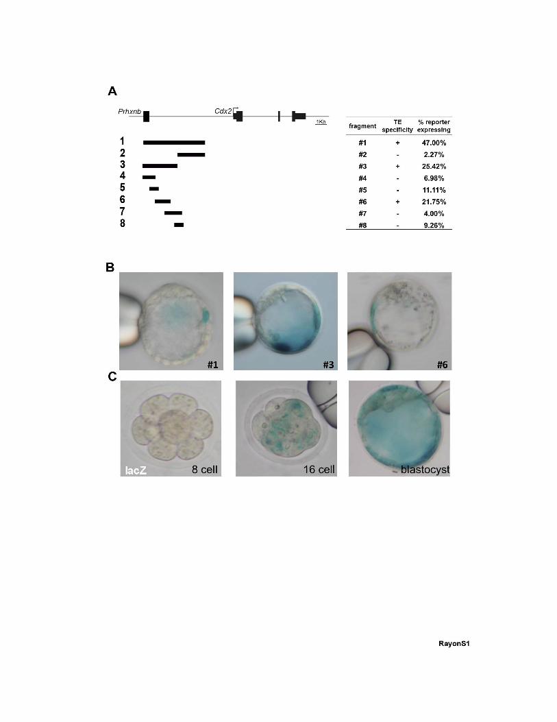
Figure S1, related to Figure 1: Identification of the Cdx2 TEE. (A) Diagram of the Cdx2
locus showing the fragments tested in transient transgenic embryos, the specific activity of
the fragments in the TE, and the percentage of embryos for each construct showing any sign
of reporter activity. (B) Representative transient transgenic embryos showing lacZ reporter
activity driven by those constructs showing specific activity in the TE. (C) Temporal dynamics
of a stable lacZ-TEE line for construct #3 at the 8-cell, 16-cell and blastocyst stages.
Figure S2, related to Figure 2: The Cdx2 TEE is not an auto-regulatory element. (A-B)
Activity of the TEE, detected by immunohistochemistry with anti-mRFP antibody (red), and
immunodetection of CDX2 (green) in (A) wild type (wt) and (B) Cdx2 mutant blastocysts.
Nuclei were stained with DAPI (blue). (C) Average cell number in wild type and Cdx2 mutant
blastocysts. (D) Percentage of TEE-positive cells per embryo in wild type (n=149, 4 embryos)
and Cdx2 mutant blastocysts (n=145, 3 embryos). (E) Average cell number in wild type
blastocyst, Tead4 heterozygotes and Tead4 mutant blastocysts. Differences in cell number
among the genotypes are not significant. (F) Percentage of TEE-positive cells per embryo in
wild type blastocysts (n=218, 3 embryos), Tead4 heterozygotes (n=464, 8 embryos) and
Tead4 mutant homozygotes (n=78, 2 embryos). Data are means ± s.d.
Figure S3, related to Figure 3: Evidence for a role of the Notch signaling pathway in
the TE. (A) Sequence of the 1.3 kb TEE, highlighting putative binding sites for RBPJ (blue)
and TEAD (green). (B) Immunodetection of N1ICD with the signal enhanced by using a
tyramide amplification kit (red). Nuclei were stained with DAPI (blue). Merged image is
shown in the right panel. Scale bars, 10 μm.

Role of Notch in trophectoderm specification Rayon et al
Figure S4, related to Figure 4: The RBPJ and TEAD binding sites of the TEE are
functional. (A) Effect of the TEAD–YAP inhibitor Verteporfin (VP) on TEE activity (red) and
endogenous CDX2 (green). (B) TEETEADmut activity in transient transgenic embryos treated
with DMSO, VP or RO. (C) TEERBPJmut activity in transient transgenic embryos treated with
DMSO, RO or VP. Nuclei were stained with DAPI. Scale bars, 10 μm.
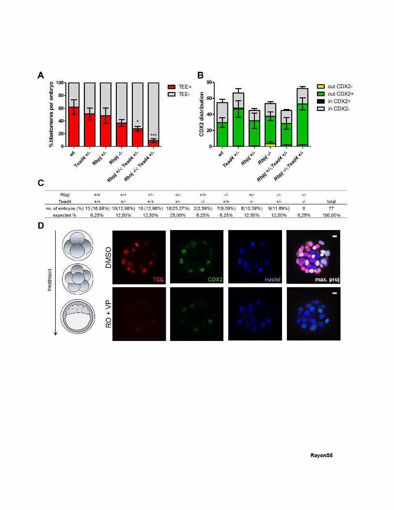
Figure S5, related to Figure 5: Phenotype of embryos with different Rbpj;Tead4 allelic
combinations. (A) Percentage of TEE-positive cells per embryo in wild type blastocysts
(n=162, 3 embryos) and in Tead4+/- (n=200, 3 embryos), Rbpj+/- (n=187, 5 embryos), Rbpj-/-
(n=115, 3 embryos), Rbpj+/-;Tead4+/- (n=238, 5 embryos) and Rbpj-/-;Tead4+/- allelic
combinations (n=176, 2 embryos). *p<0.05, ***p<0.001 by Bonferroni post test. (B) Average
number of inside and outside cells positive or negative for CDX2 in wild type (n=164, 3
embryos), Tead4+/- (n=200, 3 embryos), Rbpj+/- (n=179, 4 embryos), Rbpj-/- (n=214, 4
embryos), Rbpj+/-; Tead4+/- (n=179, 4 embryos) and Rbpj-/-;Tead4+/- allelic combinations
(n=217, 3 embryos) quantified in Figure 4F. Data are means ± s.d. (C) Distribution (%) of
embryos for the different allelic combinations of Rbpj and Tead4, compared with the
expected distribution. Dead embryos inside the zona, which could include double
homozygotes, were observed but could not be genotyped due to DNA degradation (Table
S1). (D) TEE activity (red) and CDX2 immunodetection (green) in embryos from the mRFP
line treated with DMSO or RO+VP from 4 cells until blastocyst stage (left panel). Maximal
projections of merged images are shown in the right panels. Nuclei were stained with DAPI.
Scale bars, 10 μm.
Role of Notch in trophectoderm specification Rayon et al
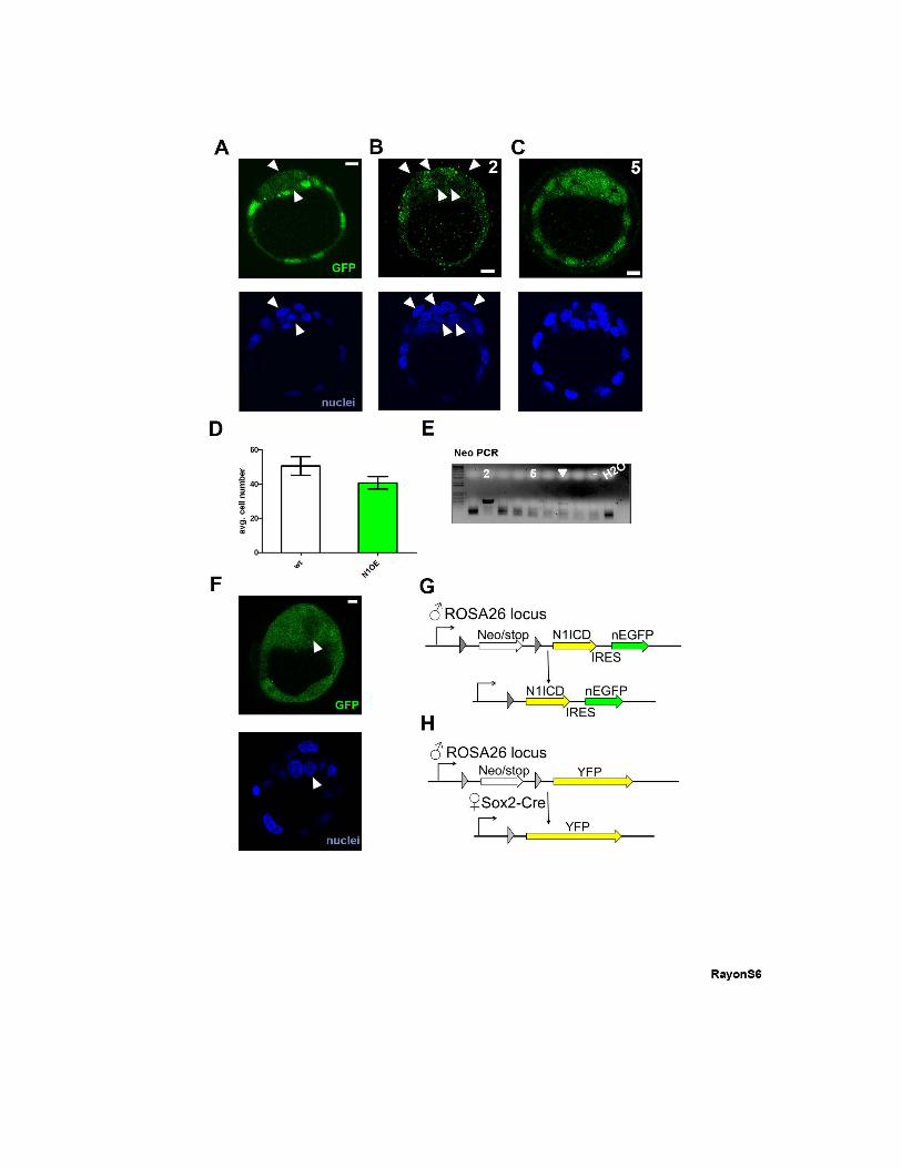
Figure S6, related to Figure 6: Mosaicism of maternal Sox2-Cre activity in the
blastocyst. (A) Mosaicism of reporter expression (green) from the R26-stop-N1ICD-ires-
EGFP line when recombined by maternal Sox2-Cre. (B) Example of a blastocyst with a very
high proportion of non-recombined cells. (C) Example of a blastocyst with recombination
occurring in all cells. (D) Average cell number in wild type (wt) and N1ICD-overexpressing
(N1OE) blastocysts. Data are means ± s.d. (E) Detection of mosaic recombination in
blastocysts by PCR of Neo. The varying degree of detection of the non-recombined allele in
different embryos is shown, ranging from non-recombined (embryo 2, shown in panel B),
mosaic recombination (white arrowhead), to full recombination of the Neo allele (embryo 5,
shown in panel C). Negative control (-) and H2O samples are indicated. (F) Reporter
mosaicism (cytoplasmic, green) from the R26-stop-YFP line when recombined by maternal
Sox2-Cre. (G-H) Breeding strategy for the ♂R26-stop-N1ICD-ires-EGFP X ♀Sox2-Cre cross
(G) and for the ♂R26-stop-YFP X ♀Sox2-Cre cross. (H). White arrowheads in (A-C, F) mark
non-recombined cells. Nuclei in (A-C and F) were stained with DAPI. Scale bars, 10 μm.