dog mummy 2014.pdf
TRANSCRIPT
Otranto et al. Parasites & Vectors 2014, 7:2http://www.parasitesandvectors.com/content/7/1/2
RESEARCH Open Access
The enigma of the dog mummy from AncientEgypt and the origin of ‘Rhipicephalus sanguineus’Domenico Otranto1*, Jean-Bernard Huchet2, Alessio Giannelli1, Cecile Callou2 and Filipe Dantas-Torres1,3
Abstract
Background: Ticks belonging to the Rhipicephalus sanguineus group are amongst the most important vectors ofpathogenic microorganisms to dogs and humans. However, the taxonomy of this species group is still the subjectof debate, especially because there is no type specimen or reliable morphological description for Rhipicephalussanguineus sensu stricto. Recently, a comprehensive morphological and genetic study on representative tickspecimens from Europe, Africa, Americas, and Oceania, revealed the existence of at least four morphologically andgenetically distinct species under the name ‘R. sanguineus’ infesting dogs from different countries.
Methods: Herein, we examined morphologically tick specimens retrieved on a dog mummy from Ancient Egypt(ca. 1st century – 4th century A.D.). The dog mummy and associated ticks were found during an archaeologicalexpedition conducted in El Deir.
Results: Scanning electron micrographs allowed us to assess their identity as belonging to the R. sanguineus group.In addition on the basis of the scutal punctation pattern, spiracular plates, width of dorsal tail of spiracular platesrelative to the adjacent festoon, female genital aperture, male adanal plates and accessory shields, these ticks weretentatively identified as Rhipicephalus sp. II (=temperate species).
Conclusions: It can be concluded that R. sanguineus group ticks have infested dogs living in the Mediterraneanregion since ancient times. This finding represents the oldest record of ticks on any animal species and adds a newpiece in the complex puzzle regarding tick parasitism on dogs and humans and their role as vectors of pathogens.
Keywords: Tick, Rhipicephalus sanguineus, Dog mummy, Archeoparasitology, Origins
BackgroundAmong domestic animals, dogs have always beenalongside humans while hunting, migrating aroundthe globe, and even while exploring the moon. In-deed, for their devotion to their owners and friendlybehaviour, dogs have represented the most commonpet for humankind throughout their history. Mean-while, as good friends, dogs share many things withhumans, including zoonotic endo- and ecto-parasites[1,2]. Amongst the arthropod parasites of both dogsand humans, the brown dog tick Rhipicephalus san-guineus (Latreille, 1806) is an efficient vector of a di-verse group of pathogens [3]. However, in spite ofbeing well-studied, the taxonomy of R. sanguineus islargely debated among scientists, because there is no
* Correspondence: [email protected] of Veterinary Medicine, University of Bari, Valenzano, Bari, ItalyFull list of author information is available at the end of the article
© 2014 Otranto et al.; licensee BioMed CentraCommons Attribution License (http://creativecreproduction in any medium, provided the orwaiver (http://creativecommons.org/publicdomstated.
type specimen or reliable morphological descriptionfor this species [4]. Therefore, while R. sanguineuscould be regarded as a nomen nudum, this tick isstill listed as a valid taxon [5].During the last century, R. sanguineus was placed
in synonymy with many species [6,7], whereasothers, morphologically similar, were described andranked within the so-called R. sanguineus group,whose definition and number of species is also argu-able [7-9]. Several authors have endeavoured tostudy this group of ticks using both morphologicaland molecular tools [4,10-16]. Recently, a compre-hensive study was undertaken on representative tickspecimens belonging to the R. sanguineus groupfrom 17 countries in Europe, Africa, Americas, andOceania [4]. Morphological and molecular analysesrevealed the existence of at least four integrated op-erational taxonomic units (i.e., R. sanguineus sensu lato,
l Ltd. This is an Open Access article distributed under the terms of the Creativeommons.org/licenses/by/2.0), which permits unrestricted use, distribution, andiginal work is properly cited. The Creative Commons Public Domain Dedicationain/zero/1.0/) applies to the data made available in this article, unless otherwise
Otranto et al. Parasites & Vectors 2014, 7:2 Page 2 of 6http://www.parasitesandvectors.com/content/7/1/2
Rhipicephalus sp. I, Rhipicephalus sp. II, and Rhipicepha-lus sp. III) under the name ‘R. sanguineus’ [4]. None-theless, in the absence of a consensus on the identity ofR. sanguineus sensu stricto, the taxonomical status andactual distribution of these species remains enigmatic.As for any investigational research, parasitologists need
to look for any clue (e.g., morphological, molecular, bio-logical, ecological evidence) to address their questions orhypotheses. Of great interest is to understand when andhow parasites developed in animal and human populations.Under these circumstances, archeoparasitology not only in-vestigates the causes of the death of the hosts infected byparasites [17], but also how they moved from one area toanother, along with animals and humans during historicalmigrations [18]. Oddly enough, studies in the field of arche-oparasitology have been mainly focussed on protozoa andhelminths in coprolites, intestinal contents or latrine de-posits [19-23]. In contrast, despite the tough chitinous exo-skeleton of arthropods, a relatively low number ofarchaeoparasitological surveys are available for ectopara-sites [24-29], probably because of their location on thehost coat, therefore more exposed to the outdoor en-vironment. In addition, archeoparasitological studies onpets are limited to the retrieval of lice from cats [30] anddogs [28,31]. To the best of our knowledge, the only pos-sible iconographic illustration of ticks from Ancient Egyptis constituted by a tomb painting from ancient Thebes(Dra Abu el-Naga, Western Thebes, ca. 1473-1458 B.C.),which displays a hyaena-like animal with excrescenceswithin the ear that were supposed to be ticks [32].The recent finding of well-mummified dogs from An-
cient Egypt in an archaeological expedition conducted inEl Deir led to the retrieval of some specimens of ixodidticks and louse flies on a young dog [33]. Considering thecurrent debate on the taxonomy of this tick species, we de-cided to carefully re-examine these ticks morphologically,using scanning electron micrographs to assess whetherthey fit with any of the species illustrated in ref. 4.
MethodsThe dog mummy was found in a tomb surrounding aRoman fortress in El Deir [33]. This archaeological site,located 30 km northeast of the town of Kharga (KhargaOasis, Egypt) at the bottom of Gebel Umm el Ghanayim,has been excavated since 1988 [34-37] in an area includ-ing an important agricultural inhabited region, whichexisted at least from the Ptolemaic period (332–30 B.C.).The necropolises are dated back to a period from the 4th
century B.C. to the 5th century A.D. Although this an-cient village has not been precisely located, a number oftombs were spread over this area, and, most of themwere spoiled over the centuries.Some anatomical investigations were conducted on
many rests of dogs [38], but the mummy of a young dog
from tomb P5 was the only well preserved specimen.This mummy displayed a massive infestation by ectopar-asites with 61 ticks found firmly attached to the animalcoat, mostly in the right ear. Additionally, a single speci-men of the louse fly Hippobosca longipennis, as well asmore than two hundred puparia of sarcosaprophagousflies of the families Sarcophagidae and Calliphoridae,were also identified (for more details, see ref. 33).Of the 61 ticks collected, only five specimens (i.e., three
males and two females) were better preserved and there-fore herein morphologically studied. Ticks were examinedat El Deir archaeological site, using a portable scan-ning electron microscope (SEM) (Nikon JCM-5000, JEOLNeoScope at 10 kV), without metallization (metal coat-ing). Micrographs were taken from the five specimensand several characters (see below) were examined and/ormeasured.All images were carefully evaluated and taxonomically
relevant characters for the differentiation of ticks belong-ing to the R. sanguineus group [4,8,39] assessed. In par-ticular, the following characters were studied: idiosoma,dorsal scutum, angles of basis capituli, female poroseareas, female genital opening, spiracular plates, lateral andpostmediam grooves, cervical pits, cervical fields, internaland external cervical margins, marginal lines, male adanalplates, accessory plates, and male caudal process. Mea-surements were not taken due to the low number of speci-mens available and their preservation status. Moreover, notick specimen or parts of it were available for molecularanalysis.
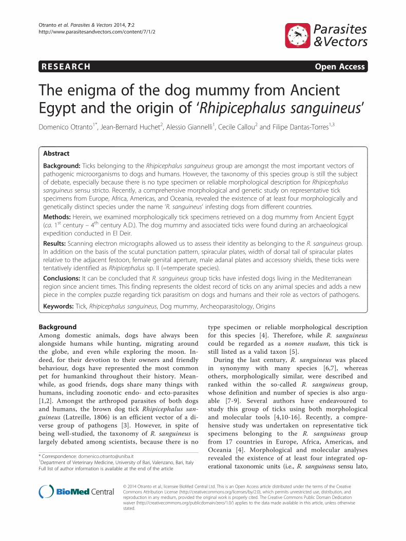
ResultsAll ticks were unengorged except for one female, and theywere identified as belonging to the genus Rhipicephalus,based on the following general characters: eyes present,anal groove posterior to anus, basis capituli hexagonalin shape, palpi short, coxae I deeply cleft, spiracular platescomma-shaped, and male adanal plates and accessoryshields present.Males presented the following characters: small puncta-
tions scattered over the posterior portion of dorsal scutum;larger punctations on the scapular region; marginal groovedeep and marked by medium-size punctations (Figure 1A);posteromedian groove distinctly elongated; lateral groovescircular in shape; spiracular plates elongated, and with anarrow dorsal tail (less than half of the adjacent festoon)(Figure 1B); adanal plates large at basis (not sickle-shaped);accessory shields sharply pointed (Figure 1C); caudalprocess present; posteromedian spur on coxa I longer thanthe posterolateral spur; angles of basis capituli in anteriorthird of its length (Figure 1D).Females presented the following characters: dorsal scutum
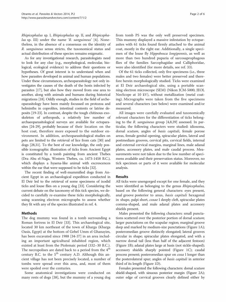
shield-shaped, with sinuous posterior margin (Figure 2A);outer edge of cervical grooves clearly defined either by
Figure 1 Rhipicephalus sanguineus group male. Dorsal scutum with small punctations scattered over the posterior portion and largerpunctations on the scapular region (A); spiracular plate with narrow dorsal tail (B); adanal plate large at base and accessory shield sharply pointed(C); basis capituli hexagonal), dorsal view (D).
Otranto et al. Parasites & Vectors 2014, 7:2 Page 3 of 6http://www.parasitesandvectors.com/content/7/1/2
slope or punctations; dorsal tail of spiracular plate narrow(Figure 2B); genital aperture broadly U-shaped (Figure 2C);posteromedian spur on coxa I longer than the posterolat-eral spur; angles of basis capituli at about mid-length; por-ose areas small, rounded and well separated (Figure 2D).Based on these key characters, all ticks were identified
as belonging to the R. sanguineus group. Furthermore,on the basis of the scutal punctation pattern, spiracularplates, width of dorsal tail of spiracular plates relative tothe adjacent festoon, female genital aperture, male ada-nal plates and accessory shields, these ticks were identi-fied as R. sp. II (=temperate species) (Figures 1 and 2).
DiscussionThe present study, based on a detailed morphologicalexamination of ticks found on a dog mummy fromAncient Egypt, confirms that they belong to the genusRhipicephalus. Nonetheless, the actual specific identityof these ticks is difficult to assess, mainly because ofthe limited number of specimens available and because oftheir conservation status. However, ticks herein examinedwere not Rhipicephalus turanicus or R. sp. III, due to thedifferences in scutal punctation pattern, spiracular platesand/or female genital aperture [4]. Indeed, these ticks re-sembled those of R. sanguineus sensu lato, and some
specific morphological characters (e.g., female genitalaperture, male adanal plates and accessory shields, scutalpunctation pattern and spiracular plates of both malesand females) allowed us to tentatively identify those ticksas R. sp. II (=temperate species or southern lineage) [4].The high intensity of tick infestation found on this
young mummified dog, along with the absence of anyapparent trauma [33], may suggest that the cause of hisdeath could be related to this massive ectoparasite infest-ation and/or to the infection by tick-borne pathogens.Indeed, R. sanguineus group ticks have been implicated asa vector of a wide range of pathogenic microorganisms todogs (e.g., Babesia vogeli, Ehrlichia canis, Hepatozooncanis and Rickettsia conorii) some of which have zoonoticpotential [3,40]. In spite of their long co-evolution withtheir hosts, tick-borne pathogens may cause severe diseaseand, eventually, the death of infected animals [41]. Thedog’s death could also be attributed to a more virulentpathogen strain circulating in Ancient Egypt. For instance,although Anaplasma platys is often considered as a lesspathogenic organism in dogs [42], virulent strains havealso been associated to severe clinical disease in Israel[43]. Tick-borne pathogens commonly cause more severedisease in young individuals at their first exposure [44].Furthermore, the sequential or simultaneous infection
Figure 2 Rhipicephalus sanguineus group female. Dorsal scutum shield-shaped, with sinuous posterior margin (A); spiracular plate with narrowdorsal tail (B); genital aperture broadly U-shaped (C); basis capituli hexagonal, dorsal view (D).
Otranto et al. Parasites & Vectors 2014, 7:2 Page 4 of 6http://www.parasitesandvectors.com/content/7/1/2
with more than one tick-borne pathogen may also resultin the exacerbation of clinical signs and potentiation ofhaematological abnormalities [45]. This was recently dem-onstrated in young dogs coinfected with B. vogeli andA. platys, in which more severe clinical and haematologicalalterations were eventually recorded than in dogs with B.vogeli only [46]. Another possible explanation for the deathof the young mummified dog could be a fatal paralytic syn-drome associated with massive tick infestation. Indeed, arecent study reported neurological signs in 14 young dogsheavily infested by R. sanguineus group ticks, ten of whichdied from this condition and presented neurological signsof different degrees [47].Although it was not possible to carry out any further
parasitological investigation on other dog mummies due totheir poor condition, the high number of ticks found on thestudied specimen suggests that other animals were alsoinfested. This might have been the cause of an epidemic oftick-borne disease leading to the death of many young ani-mals. Unfortunately, the unavailability of material of ticksfor molecular processing and detection of pathogens doesnot allow us to bring these hypotheses from the realm ofspeculations to reality. In the same way, the possibility thatthe dog died due to a viral infectious disease (commoncause of sudden death in puppies) cannot be ruled out.
The so-called temperate species (=R. sp. II) is wide-spread in Mediterranean countries, such as France,Portugal, Spain and Italy [4]. Accordingly, our findingmight indicate that this tick species has been present fora long time in Egypt. However, the discovery of a tick-infested dog mummy in a tomb surrounding a Romanfortress raises interesting questions on the origin of thisdog and his ticks. During the Roman Empire and itscolonization, which started about 270 B.C., the Mediter-ranean area was a theatre for relevant historical eventsand a hub of different cultures as well as the final destin-ation for several populations. The intense waves of mi-gration occurring before, during and after the RomanEmpire could have contributed to the dissemination ofdog ticks throughout the Mediterranean region. Indeed,the Roman Empire expanded for more than 400 yearsthrough Eurasia (from 275 B.C. to 117 A.D.) and, at itsgreatest extent, it colonized all the countries touchingthe Mediterranean sea as far as Germany and Britain(north), Turkey, Lebanon, Iran and Arabia (east) untilthe split in Eastern and Western sections (395 A.D.). Onthe other hand, because Rhipicephalus is typically anAfrican tick genus, the most probable hypothesis is thatR. sanguineus group ticks were introduced into Europeat a certain point of time, most probably with people
Otranto et al. Parasites & Vectors 2014, 7:2 Page 5 of 6http://www.parasitesandvectors.com/content/7/1/2
from North Africa, soon after the collapse of the RomanEmpire.
ConclusionThe history of tick species threatening dog and hu-man health often crosses with those of the hosts theyparasitize as a part of the everyday existence of indi-vidual animals everywhere and in every time. Whetherthe retrieval of ticks on a mummified young dog, whichsuccumbed around 2,500 years ago due to an obscureillness, can contribute to a better understanding of theR. sanguineus group or not is uncertain. Certainly, it addsa new piece in the complex puzzle regarding tick parasit-ism on dogs and their role as vectors of pathogens to dogsand humans.
Competing interestsThe authors declare that they have no competing interests.
Authors’ contributionsDO and FD-T conceived the research and wrote the first draft. JBH and CCexamined the dog mummy, collected the tick specimens and performedthe SEM photos. FD-T, DO, and AG did the morphological study. All authorsread and approved the final version of the manuscript, contributed withinterpretation and revision of the manuscript.
AcknowledgmentsAuthors thank Bayer Animal Health for supporting the publications costs.Jean-Bernard Huchet and Cécile Callou are indebted to Françoise Dunand andRoger Lichtenberg (University of Strasbourg, France), heads of the Frencharcheological mission at El Deir site and to Michel Lemoine (UMR 7209”Archéozoologie, Archéobotanique, MNHN, France) for his precious helpregarding the SEM photographs of the archaeological samples. This study wasconducted under the frame of the EurNegVec COST Action TD1303.
Author details1Department of Veterinary Medicine, University of Bari, Valenzano, Bari, Italy.2UMR 7209 du CNRS, Archéozoologie, Archéobotanique, Muséum Nationald’Histoire Naturelle, Paris, France. 3Department of Immunology, AggeuMagalhães Research Centre, Oswaldo Cruz Foundation, Recife, Pernambuco50670420, Brazil.
Received: 6 December 2013 Accepted: 21 December 2013Published: 20 January 2014
References1. Dantas-Torres F, Otranto D: Dogs, cats, parasites, and humans in Brazil:
opening the black box. Parasit Vectors, in press.2. Otranto D, Dantas-Torres F, Brianti E, Traversa D, Petrić D, Genchi C, Capelli G:
Vector-borne helminths of dogs and humans in Europe. Parasit Vectors 2013,6:16.
3. Dantas-Torres F, Chomel BB, Otranto D: Ticks and tick-borne diseases:a One Health perspective. Trends Parasitol 2012, 28:437–446.
4. Dantas-Torres F, Latrofa MS, Annoscia G, Giannelli A, Parisi A, Otranto D:Morphological and genetic diversity of Rhipicephalus sanguineus sensulato from the New and Old Worlds. Parasit Vectors 2013, 6:213.
5. Guglielmone AA, Robbins RG, Apanaskevich DA, Petney TN, Estrada-Peña A,Horak IG: Comments on controversial tick (Acari: Ixodida) species namesand species described or resurrected from 2003 to 2008. Exp Appl Acarol2009, 48:311–327.
6. Neumann LG: Ixodidae. Berlin: Das Tierreich; 1911.7. Camicas JL, Hervy JP, Adam F, Morel PC: Les tiques du monde. Nomenclature,
stades décrits, hôtes, répartition (Acarida, Ixodida). Paris: Éditions de l’Orstom;1998.
8. Walker JB, Keirans JE, Horak IG: The Genus Rhipicephalus (Acari, Ixoidae).A Guide to the Brown Ticks of the World. Cambridge, UK: Cambridge Univ.Press; 2000.
9. Gray J, Dantas-Torres F, Estrada-Peña A, Levin M: Systematics and ecologyof the brown dog tick, Rhipicephalus sanguineus. Ticks Tick Borne Dis 2013,4:171–180.
10. Oliveira PR, Bechara GH, Denardi SE, Saito KC, Nunes ET, Szabó MP, Mathias MI:Comparison of the external morphology of Rhipicephalus sanguineus(Latreille, 1806) (Acari: Ixodidae) ticks from Brazil and Argentina. Vet Parasitol2005, 129:139–147.
11. Szabó MP, Mangold AJ, João CF, Bechara GH, Guglielmone AA: Biologicaland DNA evidence of two dissimilar populations of the Rhipicephalussanguineus tick group (Acari: Ixodidae) in South America. Vet Parasitol2005, 130:131–140.
12. Burlini L, Teixeira KR, Szabó MP, Famadas KM: Molecular dissimilarities ofRhipicephalus sanguineus (Acari: Ixodidae) in Brazil and its relation withsamples throughout the world: is there a geographical pattern? Exp ApplAcarol 2010, 50:361–374.
13. Moraes-Filho J, Marcili A, Nieri-Bastos FA, Richtzenhain LJ, Labruna MB:Genetic analysis of ticks belonging to the Rhipicephalus sanguineusgroup in Latin America. Acta Trop 2011, 117:51–55.
14. Levin ML, Studer E, Killmaster L, Zemtsova G, Mumcuoglu KY: Crossbreedingbetween different geographical populations of the brown dog tick,Rhipicephalus sanguineus (Acari: Ixodidae). Exp Appl Acarol 2012, 58:51–68.
15. Nava S, Mastropaolo M, Venzal JM, Mangold AJ, Guglielmone AA:Mitochondrial DNA analysis of Rhipicephalus sanguineus sensu lato(Acari: Ixodidae) in the Southern Cone of South America. Vet Parasitol2012, 190:547–555.
16. Latrofa MS, Dantas-Torres F, Annoscia G, Cantacessi C, Otranto D:Comparative analyses of mitochondrial and nuclear genetic markersfor the molecular identification of Rhipicephalus spp. Infect Genet Evol2013, 20C:422–427.
17. Reinhard KJ: Archaeoparasitology in North America. Am J Phys Anthropol1990, 82:145–163.
18. Otranto D, Stevens JR, Brianti E, Dorchies P: Human and livestockmigrations: a history of bot fly biodiversity in the Mediterranean region.Trends Parasitol 2006, 22:209–213.
19. Fry GF: Analysis of fecal material. In The Analysis of Prehistoric Diets. Editedby Gilbert RI Jr, Mielke J. Orlando: Academic Press; 1985:127–154.
20. Reinhard KJ, Confalonieri UE, Herrmann B, Ferreira LF, Araujo AJG: Recoveryof parasite eggs from coprolites and latrines: aspects ofpaleoparasitological technique. Homo 1998, 37:217–239.
21. Gonçalves MLC, Araujon A, Ferreira LF: Human intestinal parasites in thepast: new findings and a review. Mem Inst Oswaldo Cruz 2003, 98:103–118.
22. Bouchet F, Harter S, Le Bailly M: The state of the art of paleoparasitologicalresearch in the old world. Mem Inst Oswaldo Cruz 2003, 98:95–101.
23. Araújo A, Reinhard K, Leles D, Sianto L, Iñiguez A, Fugassa M, Arriaza B,Orellana N, Ferreira LF: Paleoepidemiology of intestinal parasites and licein pre-Columbian America. Rev Chil Hist Nat 2001, 43:303–313.
24. Capasso L, Di Tota G: Lice buried under the ashes of Herculaneum.Lancet 1998, 351:992.
25. Yvinec JH, Ponel P, Beaucournu JC: Premiers apports archéoentomologiquesde l’étude des Puces, Aspects historiques et anthropologiques(Siphonaptera). Bull Soc Ent Fr 2000, 105:419–425.
26. Rick FM, Rocha GC, Dittmar K, CoimbraX CE Jr, Reinhard K, Bouchet F,Ferreira LF, Araujo A: Crab louse infestation in pre-Columbian America.J Parasitol 2002, 88:1266–1267.
27. Guerra RMSNC, Gazeta GS, Amorim M, Duarte AN, Serra-Freire NM: Ecologicalanalysis of acari recovered from coprolites from archaeological site ofnortheast Brazil. Mem Inst Oswaldo Cruz 2003, 98:181–190.
28. Dittmar K, Mamat U, Whiting M, Goldmann T, Reinhard K, Guillen S:Techniques of DNA-studies on Prehispanic Ectoparasites (Pulex sp.,Pulicidae, Siphonaptera) from animal mummies of the Chiribaya culture,Southern Peru. Mem Inst Oswaldo Cruz 2003, 98:53–58.
29. Reinhard KJ, Buikstra J: Louse infestation of the Chiribaya culture, SouthernPeru: variation in prevalence by age and sex. Mem Inst Oswaldo Cruz 2003,98:173–179.
30. Guerra RMSNC, Duarte AN, Oliveira HH, Mello RP, Serra-Freire NM: Thefinding of Felicola felis (Mallophaga: Trichodectidae) and exuviae ofAmblycera in Felidae coprolites from the archaeological site of Furnado Estrago, Pernambuco state, Brazil. Entomol Vect 2001, 8:395–402.
31. Martinson E, Reinhard KJ, Buikstra JE, Dittmar de la Cruz K: Pathoecology ofChiribaya Parasitism. Mem Inst Oswaldo Cruz 2003, 98:195–205.
32. Arthur DR: Ticks in Egypt in 1500 B.C.? Nature 1965, 206:1060–1061.
Otranto et al. Parasites & Vectors 2014, 7:2 Page 6 of 6http://www.parasitesandvectors.com/content/7/1/2
33. Huchet JB, Callou C, Lichtentberg R, Dunand F: The dog mummy, the ticksand the louse fly: archaeological report of a severe ectoparasitosis inAncient Egypt. Int J Paleopathol 2013, 3:165–175.
34. Dunand F, Lichtenberg R: Dix ans d’exploration des nécropoles d’El-Deir(oasis de Kharga). Un premier bilan. Chron Egypte 2008, 83:258–288.
35. Dunand F, Coudert M, Letellier-Willemin F: Decouverte d’une nécropolechrétienne sur le site d’El-Deir (oasis de Kharga). In Etudes coptes X,Douzième journée d’études (Lyon, 19–21 mai 2005). Edited by Cahiers de laBibliothèque copte. Paris: Boud’hors A and Louis C; 2008:137–155.
36. Dunand F, Heim JL, Lichtenberg R, Brones S, Devaux E, Dussarps L,Letellier-Willemin F, Tallet G: El-Deir Nécropoles II. Les Nécropoles Nordet Nord-Est. Paris: Cybèle; 2012.
37. Dunand F, Heim JL, Lichtenberg R, Brones S, Letellier-Willemin F: El-DeirNécropoles I. La Nécropole Sud. Paris: Cybèle; 2010.
38. Callou C, Dunand F, Lichtenberg R: In Archaeological and ArchaeozoologicalStudy of Dogs from El-Deir. Edited by PalArch’s J Archaeol. Egypt/Egyptol.Cairo, Egypt: Proceeding of International meeting of The Bioarchaeology ofAncient Egypt; 2013:10.
39. Filippova NA: Fauna of Russia and Neighbouring Countries. Ixodid Ticks ofSubfamily Amblyomminae. Moscow, Russia: Nauka Publishing House;1997:436.
40. Dantas-Torres F: The brown dog tick, Rhipicephalus sanguineus (Latreille,1806) (Acari: Ixodidae): from taxonomy to control. Vet Parasitol 2008,152:173–185.
41. Otranto D, Dantas-Torres F, Breitschwerdt EB: Managing canine vector-borne diseases of zoonotic concern: part two. Trends Parasitol 2009,25:228–235.
42. Bradfield JF, Vore SJ, Pryor WH Jr: Ehrlichia platys infection in dogs. LabAnim Sci 1996, 46:565–568.
43. Harrus S, Aroch I, Lavy E, Bark H: Clinical manifestations of infectiouscanine cyclic thrombocytopenia. Vet Rec 1997, 141:247–250.
44. Kontos VJ, Koutinas AF: Clinical observations in 15 spontaneous cases ofcanine babesiosis. Canine Practice 1997, 22:30–34.
45. Kordick SK, Breitschwerdt EB, Hegarty BC, Southwick KL, Colitz CM, HancockSI, Bradley JM, Rumbough R, Mcpherson JT, MacCormack JN: Coinfectionwith multiple tick-borne pathogens in a Walker Hound kennel in NorthCarolina. J Clin Microbiol 1999, 37:2631–2638.
46. de Caprariis D, Dantas-Torres F, Capelli G, Mencke N, Stanneck D, BreitschwerdtEB, Otranto D: Evolution of clinical, haematological and biochemical findingsin young dogs naturally infected by vector-borne pathogens. Vet Microbiol2011, 149:206–212.
47. Otranto D, Dantas-Torres F, Tarallo VD, Ramos RA, Stanneck D, Baneth G,de Caprariis D: Apparent tick paralysis by Rhipicephalus sanguineus(Acari: Ixodidae) in dogs. Vet Parasitol 2012, 188:325–329.
doi:10.1186/1756-3305-7-2Cite this article as: Otranto et al.: The enigma of the dog mummy fromAncient Egypt and the origin of ‘Rhipicephalus sanguineus’. Parasites &Vectors 2014 7:2.
Submit your next manuscript to BioMed Centraland take full advantage of:
• Convenient online submission
• Thorough peer review
• No space constraints or color figure charges
• Immediate publication on acceptance
• Inclusion in PubMed, CAS, Scopus and Google Scholar
• Research which is freely available for redistribution
Submit your manuscript at www.biomedcentral.com/submit