doi 978-9934-588-64-8-7 the new
TRANSCRIPT

107
DOI https://doi.org/10.30525/ 978-9934-588-64-8-7
THE NEW MODEL OF DIAGNOSTICS, TREATMENT AND PREVENTION OF PURULENT-SEPTIC COMPLICATIONS
IN PATIENTS WITH BURN INJURY
Zaporozhan S. Y., Savchyn V. S., Ostapiuk L. R., Tuziuk N. V. INTRODUCTION According to WHO, burn injury ranks third place in overall trauma.
The world annually records about 180.000 deaths from this type of trauma, most of which occur in middle- and low-income countries. The proportion of burns ranges from 5.6% to 10%, which is one third of all types of traumatic injuries.
According to the Public Health Center of Ukraine, burns also rank third place among all injuries. 70% of them are home care burns. About a third of them occur in children under 5 years of age. This problem is one of the most difficult in the clinical course. It is characterized by the complexity of treatment, its high cost and possible complications.
The deep economic and social crisis in our country has led to the destruction of the system of surgical care for victims of burns. This is reflected in the time of admission of patients to medical institutions, the delay of patients at the stages of evacuation and the violation of the quality of care for patients of this category.
In patients with burn injury, who have chronic diseases, including diabetes, tuberculosis, immunodeficiency, there is a tendency to slow down the healing process. It should be also noted, that in patients with burns there is endogenous intoxication. Despite the introduction into medical practice modern achievements of resuscitation, the latest methods of intensive care, the incidence of multiple organ failure remain quite high. The main factors of mortality among patients with widespread burns are the development of systemic inflammatory process, multiorgan failure and infectious complications1. This leads to increase the cost of treatment of these patients, an increase the number of bed days and the need for long-term rehabilitation. This also increases the level of burden on the medical staff of medical institutions and it is not cost-effective enough.
Necrotizing diseases of the tissues of the head and face, which are also accompanied by defects of the outer coverings and soft tissues and the
1 Kovalenko O.M. Pathogenetic substantiation of programs of surgical treatment of
children with common burns and their influence on the course of wound process. Doctor’s thesis. 2012. Kyiv : O. Bohomolets National Medical University. 298 p.

108
development of deforming scars remain practically unexplored both in the context of diagnosis and the choice of treatment tactics. Some surgical interventions and injuries of the head and face lead to the same relative functional and social consequences, which remain an insufficiently studied problem.
At the same time, significant progress has been made in the field of combustiology and reconstructive surgery over the last decade. The concept of early surgical necrectomies of burn wounds with their primary plasticity is widely spread. Means of prevention and treatment of wound infection, restoration of anatomical structures and non-surgical correction in the postoperative period are developed. Methods of medical and social rehabilitation of patients are being improved.
New opportunities for surgical treatment of patients with thermal lesions are associated with the development of new technological tools in reconstructive surgery. Among them should be noted the replacement of scar arrays with stretched flaps and revascularization of deep anatomical structures with complex tissue complexes.
At the same time, a number of important theoretical and practical issues remain studied insufficiently. The issues of radicalness of early surgical interventions for head and face burns, volume and terms of their performance, means of plastic wound closure, correction of general disorders of homeostasis remain debatable. The role of local and general disorders, that characterize the severity of damage is covered insufficiently. The dynamics of histoimmunological changes of wounds and scars at different stages of their development, as well as the mechanisms of their occurrence depending on their etiology have also not been studied. There is no data in the scientific literature about the dynamics of these disorders and methods of their correction2. To date, there is no way to assess accurately the area of the defect (burn wound, necrotizing lesions) of the outer coverings of the head and face, there is no systematization of such lesions, that would meet clinical needs. A large number of tactical approaches and a variety of treatments for patients with defects of the outer coverings and soft tissues of the head and face necessitate the development of the reasonable system of comprehensive surgical treatment, that would combine the achievements of combustiology, plastic, reconstructive surgery and the achievements of other related specialties3.
Thus, the treatment of patients with burn injuries causes a number of significant medical, economic and psychological problems, that require a constructive approach to their solution. This issue is especially relevant in
2 Savchyn V.S. Features of reparative processes in patients with deep burns of the head
and neck. Archive of Clinical and Experimental Medicine. 2014. № 23(2). P. 149–152. 3 Savchyn V.S. Features of the inflammatory response in burn head and neck injury.
Archive of Clinical and Experimental Medicine. 2014. № 2 (part 2). P. 112–113.

109
the period of the implementation of medical reform in Ukraine, which provides maximum optimization and a rational approach to the treatment of patients. Therefore, the situation is somewhat contradictory regarding to the presence of comorbidities in many patients with burn injuries and the feasibility of rational use of bed days in the hospitals nowadays.
These data indicate the high social and medical significance of improving the quality of treatment of patients with burn injuries with defects of the outer coverings and soft tissues of the head and face and determine the urgency of this problem.
Despite the undoubted achievements in the development of new methodological approaches and the introduction of modern methods of treatment patients with burn injuries, the frequency of septic complications remains high. It should be noted, that the development of systemic inflammation, organ failure and infectious complications are the main causes of mortality. Therefore, the objective requirement of the time is the development of the approach, that involves the introduction and use of new effective methods of diagnosis and treatment of patients with burn injuries.
Modern advances in medicine are closely linked to the successful development of biomedical research, particularly in the field of biological chemistry. Modern diagnostic trends involve the widespread use of biochemical research methods for their diagnosis, choice of treatment tactics and monitoring of the effectiveness of the treatment. Also especially promising is using of physical research methods in medical practice. The method of fluorescence spectroscopy (MFS), which we use in our research, is the most universal method in biological spectroscopy. It has been widely used by us for the diagnosis of patients with sepsis and purulent-septic complications in surgical practice, in women with postpartum purulent-inflammatory diseases4, as well as for the diagnosis of endogenous intoxication in patients with burn injuries.
The aim of the research is to improve the immediate and long-term functional and cosmetic results of surgical treatment of patients, including defects of the outer coverings and soft tissues of the head and face, based on the development of principles of early diagnosis, including using MFS, monitoring and early reconstructive surgery.
1. Current problems of treatment tactics in patients with burn injuries
We have previously studied the possibilities of improving treatment tactics for patients with burn injuries by investigating of the effectiveness of modern methods of local treatment for the development of the cellular phase
4 Ostapiuk L. Diagnostic and therapeutic model of sepsis and purulent-inflammatory
diseases. International Journal of Clinical Medicine. 2019. № 10. P. 577–595. URL: https://doi.org/10.4236/ijcm.2019.1011047.
110
of the inflammatory response in patients with burns of flame and boiling water of first-and second-degree (type A and B) with a total area of 18 to 45% of the body surface, including the head and neck.
The effectiveness of modern methods of local treatment on the development of the cellular phase of the inflammatory response in patients with deep burns of the head and neck was studied. It is established, that using of active tactics of wound healing helps to optimize the course of the wound process. This is confirmed by the presence of inflammatory-regenerative type of cytograms with a significant content of lymphocytes and monocytes, an increase of neutrophilic granulocytes in the state of complete phagocytosis and a significant decrease in the content of detritus and microorganisms. The study of the dynamics of changes in wound healing is the additional component to assess the effectiveness of the applied method of treatment. This makes it possible to investigate the course of the wound process and to establish the optimal time for plastic closure of wound defects.
A comprehensive treatment of defects of the outer coverings and soft tissues of the head and face with different options for early reconstructive and plastic surgery and a system of non-surgical correction in the postoperative period was developed. It can improve significantly the treatment of patients with burns and necrotizing diseases of the head and face. It can also prevent or reduce significantly the number of complications, correct structural and functional disorders and eliminate or limit cosmetic problems. Clinical and laboratory studies allowed to classify burns of fourth degree depending on the principles of treatment into suprafascial – fourth-degree (type A) and subfascial – fourth-degree (type B). Depending on the depth of burns, the following classification of necrectomies was proposed: superficial or sequential, dermo-epidermal, suprafascial and necrectomies-amputations.
Optimization of the treatment tactics for the children with head and neck burns was performed. It was proved, that the use of ointments on a hydrophilic basis with the content of silver sulfadiazine in patients with burns of first-and second-degree led to the rapid epithelialization of wounds. Early surgical excision of necrotic tissues in patients with deep burns of the face and neck, temporary wound closure with lyophilized xenografts with subsequent autodermoplasty, using a full-layer or thick-layer graft on the background of the appointment of broad-spectrum antibiotics helped to prevent the appearance of the infectious diseases. It also allowed to achieve a good cosmetic and functional result of treatment5.
Tactical approaches in 23 patients with electric head burns was developed. They allowed to avoid postoperative mortality and helped to
5 Savchyn V. S. Features of surgical treatment of children with burns of the head and
neck. Clinical Surgery. 2012. № 11. P. 30.
111
provide a significant improvement of the quality of life of the operated patients. The application of the surgical treatment tactics was proposed on the basis of the own classification. It allowed to improve significantly the results of treatment of the patients with burn injuries. At the first stage radical early necrectomy to all patients was done. Appropriate surgical tactics was chosen for each clinical group. It was based on the patient's condition6.
Early surgical excision of necrotic tissues in patients with deep facial burns, temporary closure with the lyophilized xenografts, delayed autodermoplasty with thick-layer or full-layer grafts and complex therapy with broad-spectrum antibiotics prevent the development of infectious complications and provide good cosmetic and functional results. The use of ointment containing silver sulfadiazine in patients with burns of first-third (type A) degree promotes rapid epithelialization of wounds.
Thus, there is a tendency to optimize gradually the treatment of patients with the burn injuries. In particular, the expediency of active surgical tactics for the treatment of patients with burn injury is substantiated. This helps to optimize the course of the wound process. At the same time, the increased risk of the developing of purulent-inflammatory diseases in this category of patients remains a significant problem. It should be noted, that the main causes of mortality in patients with burn injury are purulent-septic complications. They are associated with the conditions for the development of the inflammatory process in the area of the burn wound, prolonged rejection of necrotic scab, decreased natural resistance and immunological reactivity of the patients. Burn necrotic scab is a source of infection and intoxication. So, it should be removed as soon, as possible. According to the most of authors, for the superficial burns this period is 2–3 days, and for deep – 3–5 days7.
Early diagnosis of the purulent-septic conditions is the guarantee of timely and successful treatment of this serious complication. It will help to increase the survival and recovery from this disease. At the same time, modern diagnostic methods, which are widely used in health care facilities, do not allow to do it properly. At present, use of physical research methods in modern medical practice are widespread. They are highly accurate and sensitive. Therefore, their use can improve significantly modern diagnostic algorithms.
6 Classification and treatment of deep burns of the head, caused by electric current /
I.D. Gerych et al. Clinical Surgery. 2009. № 11/12. P. 29–30. ISSN 0023-2130. 7 Kovalenko O.M. Tactics of wound closure in critical burns in children. Pediatric
Surgery. 2010. № 1(26). P. 28–32.
112
2. The experience of use of the method of fluorescence spectroscopy for the diagnosis of patients with purulent-inflammatory diseases
and sepsis in surgical and obstetric and gynecological practice In our research we focused on the use of the MFS for diagnosis in
medical practice, in particular for the diagnosis of endogenous intoxication in patients with burn injuries. It is used successfully in various sectors of the economy, including medicine. To understand the phenomenon of luminescence, it should be noted, that the molecules in the materials may be in certain discrete energy states. When the system is excited by the light of a certain wavelength, the transitions of electrons in them from the lower to higher energy states occur. In this case, the waves will form the absorption spectra of the molecules. And their return transitions from the upper excited states will be accompanied by radiation, which is called luminescence.
Thus, luminescence occurs due to the absorption of the light by the system. It is connected with the transition of molecules from the excited state to the ground state. The radiation, that occurs, is called luminescence. Luminescent analysis is the method of studying of various objects, based on the observation of their luminescence. It can be used to study the luminescent characteristics of biological objects both in normal and in various pathological conditions. Luminescence of tissues and biological fluids, in particular blood serum (BS), urine and synovial fluid can be registered.
According to the afterglow duration, τ luminescence is divided into two types:
− fluorescence if τ < 10-7 seconds, i. e. the extinction of luminescence occurs very quickly (for the eye, instantly);
− phosphorescence if τ > 10-4 seconds (in this case, the extinction occurs relatively slowly and is often clearly visible to the naked eye).
In our research we study the fluorescence of the BS. Fluorescence emission spectra are the dependence of fluorescence intensity from the wavelength of radiation.
It should be noted, that MFS is now used successfully in the world medical practice for conducting up-to-date prospective studies, based on the latest developments of molecular biology. They allow to identify certain genetic mutations in humans and their individual predisposition for the development of certain pathological conditions8.
The results of these studies open the way to the successful development of “personalized medicine”. It means, that it is possible to identify the individual risk of certain diseases for each individual and to propose appropriate
8 Performance and clinical evaluation of a sensitive multiplex assay for the rapid
detection of common NPM1 mutations / M. Hafes et al. Journal of Molecular diagnostics. September 2010. V. 12. № 5. P. 629–635.

113
measures to prevent the possibility of their occurrence. This opens wide opportunities to find effective drugs, so-called “gene therapy”, which offers broad prospects for the treatment of various diseases, including cancer.
The research took place between January, 2001 and March, 2020. It included 4 stages. The first and second stages of the study were performed on the basis of the purulent-septic center of the Lviv’s municipal clinical hospital of emergency medical services. The third stage of the study was performed on the basis of department of gynecology of Vinnytsia Сity Сlinical Hospital. The fourth stage of the research was performed on the clinical base of City Center of Thermal Injury and Plastic Surgery of Lviv’s Communal Clinical Hospital No 8. The luminescent laboratory of the Department of Experimental Physics at the Ivan Franko National University of Lviv was an experimental research center. The studies were supported by using the MDR-2 and MDR-12 optical monochromators. The object of the study were samples of the BS of patients of main and control groups9.
Excitation of biological objects by ultraviolet light (λ=280 nm) makes it possible to observe the glow of proteins of their structure, in particular human serum albumin. It is connected with the presence of amino acids: tryptophan, tyrosine and phenylalanine. The luminescence of most proteins is mainly connected with tryptophan residues.
At the first stage of the study, a series of in vitro studies were performed. It was necessary to form a clinical understanding and correct interpretation of the results of the study using MFS. The aim of the research was to obtain enough understanding at the molecular level about the changes, which are registered using MFS in the study of BS of patients with various diseases and treatments. For this purpose, standard solutions of donor BS with distilled water (DW), 20% albumin solution, sugar broth (SB), centrifuged (CF) and non-centrifuged (NCF) crops of bacterial culture of Staphylococcus aureus were prepared and tested within the MFS.
The fluorescence spectra (FS) of the 20% albumin solution and the donor (control group) are very similar, although the fluorescence band of the donor BS is slightly wider: λmax for 20% albumin solution is 330nm, and for the reference donor – 337 nm. Spectral fluorescence characteristics of 20% albumin solution are very close to similar characteristics of BS. That’s why we used a 20% solution of albumin as a reference in the study of BS of patients in medical practice.
Excitation of samples of BS was performed by the light with a wavelength λex = 280nm. FS of BS look like a λ-type curves with maximum fluorescence in the region of 330.1-335.1nm. The main indicators used for
9 Fluorescence spectroscopy: possibilities of application in medical practice / I. D.
Gerych etc. Lviv : Liga-Press. 2015. 366 p.

114
the analysis in the conducted work are the values of fluorescence intensity IF and position of λmax. Among a number of experiments, that correspond to the proposed series of “modeling of BS changes of diseases in vitro”, studies of solutions of BS of donors with distilled water were very important. 15 standard solutions of BS with different concentration of DW were prepared:1 – BS, 2 – 90% BS, 3 – 80% BS, 4 – 70% BS, 5 – 60% BS, 6 – 50% BS, 7 – 40% BS, 8 – 30% BS, 9 – 20% BS, 10 – 10% BS, 11 – 5% BS, 12 – 2.5% BS, 13 – 1.25% BS, 14 – 0.625% BS, 15 – DW. The results of the study of FS of dilutions of these solutions are depicted in Figure 1.
Figure 1. Effect of dilution with distilled water (DW) on the fluorescence spectra
of donor blood serum (BS) (1 – ВS 2 – 90% ВS, 3 – 80% ВS, 4 – 70% ВS, 5 – 60% ВS, 6 – 50% ВS, 7 – 40% ВS, 8 – 30% ВS, 20%ВS , 10 – 10% ВS, 11 – 5%
ВS, 12 – DW : ІF = 0) During the dilution of BS with DW the position of ІF of the fluorescence
bands λmax does not change. However, a clear dependence of ІF from the concentration ratio of BS and DW in the investigated solutions was revealed. There was a gradual increase in ІF. It reached maximum at a concentration (C) ≈ 2.5% BS in solution. Changes of ІF are connected with the concentration quenching of the fluorescent characteristics of solutions.

115
Thus, reducing of the content of BS in the prepared solutions causes gradual decrease of the number of albumin molecules and, accordingly, the weakening of the concentration quenching of the fluorescence of BS. But the growth of ІF occurs to a certain limit, which corresponds to ≈2.5% of BS in the solution. With a further decrease in the content of BS in the DW there is a sharp decrease in ІF of the BS until its complete attenuation. The case, when ІF is directly proportional to the concentration of BS in solution, corresponds to the condition, when the optical density of the solution D ≤ 0.1.
Spectral-fluorescent characteristics of dilutions of BS in vitro reflect changes of BS, typical for diseases, which are accompanied by hypoproteinemia and hypoalbuminemia. The results of these studies indicate a high sensitivity of MFS, which is manifested in a pronounced change in the FS of the BS at any, even minimal, change of its components. These changes are specific and depend on the concentration of different types of dilute substances in the BS.
The spectral-fluorescent characteristics of BS dilutions by CF and NCF crops of bacterial culture (6-day culture on Staph. aureus) were investigated in order to simulate changes of BS in patients with sepsis in vitro. That’s why we will focus on the detection of spectral-fluorescent signs of pathognomic for sepsis pathological constellation blood serum+bacteria-the phenomenon of bacteremia. In order to verify the correctness of our statements, we decided to create a simulation of changes in BS in sepsis in vitro by diluting BS with NCF and CF of bacteria10.
11 standard solutions of NCF of bacteria were made: 1 – BS, 2 – 90% BS, 3 – 80% BS, 4 – 70% BS, 5 – 60% BS, 6 – 50% BS, 7 – 40% BS, 8 – 30% BS, 9 – 20% BS, 10 – 10% BS, 11 – NCF. The volume ratios of BS and CF were the same, as the proportions of BS and NCF. Excitation of these solutions was performed with the light with a wavelength of 280 nm. The results of the study of FS dilutions of BS with NCF and CF of bacteria are depicted in Figure 2 and Figure 3. The results of the study of FS of solutions of BS with NCF and CF are qualitatively different from the results of FS of dilutions of BS with DW, SB and 20% albumin solution. Dilution of BS with NCF and CF of bacteria leads to decrease of ІF gradually with increasing content of bacterial culture in solution. There is also a long-wave shift of the fluorescence bands (λmax).
It should be noted, that the detected effect of changes of the spectral-fluorescent characteristics of dilutions of BS with NCF and CF of bacteria are connected with the influence of bacteria and products of its metabolism on the fluorescent characteristics of BS.
10 Modeling of changes in blood serum in various diseases and treatments /
O.V. Bulavenko et al. Biomedical and Biosocial Anthropology. 2013. № 20. P. 8–14.

116
Figure 2. Effect of dilution non-
centrifuged (NCF) crops on fluorescence spectra of donor blood
serum (BS) (1 – blood serum (BS), 2 – 90% BS, 3 – 80% BS, 4 – 70% BS, 5 – 60% BS, 6 – 50% BS, 7 – 40% BS, 8 – 30% BS, 9 – 20% BS, 10 – 10% BS,
11 – NCF crops). λex =280 nm
Figure 3. Effect of dilution centrifuged (CF) crops on
fluorescence spectra of donor blood serum (BS) (1 – blood serum (BS) 2 –
90% BS, 3 – 80% BS, 4 – 70% BS, 5 – 60% BS, 6 – 50% BS, 7 – 40% BS, 8 – 30% BS, 9 – 20% BS, 10 –
10% BS, 11 – CF crops). λex =280 nm It should be noted, that the changes of the FS of the dilutions of the BS
with NCF and CF of bacteria have a specific character. This forms the basis for the development of the method of fluorescent spectroscopy for early diagnosis of sepsis by modeling changes of BS in the case of sepsis in vivo.
In the second stage of the research, we conducted a study of FS of BS of 100 patients with purulent-inflammatory diseases, among whom were 15 patients with sepsis. The control group consisted of 40 healthy individuals without chronic diseases.
It should be noted, that albumin of BS has the ability to complex11. In the case of presence of endogenous intoxication, which is typical for patients with purulent-septic complications and sepsis, the conditions for the formation of albumin molecules with altered physicochemical properties appear12.
In the blood of patients with sepsis there are two types of albumin molecules: one is normal, and the other is with altered physicochemical characteristics due
11 Grizunov Y.A. and Dobretsov G.E. Serum albumin in clinical medicine. Moscow :
Geotar, 1998. 440 p. 12 Andreeva O.L. Changes in the binding centers of serum albumin in assessing the
state of the body in various pathologies. Doctor’s thesis. 2003.

117
to the blocking of albumin centers by toxins, which are products of bacterial metabolism. As the amount of complete albumin in the BS decreases, the intensity of the FS decreases and a long-wavelength shift of the fluorescence band occurs. At a certain concentration of “pathological” type of albumin with altered physicochemical characteristics, a pathological “septic” peak is formed. The appearance of the peak is an unfavorable prognostic sign.
For example, we present the results of a study of FS of BS of the person, who was treated in Lviv’s municipal clinical hospital of emergency medical services with the diagnosis of severe sepsis. Due to the well-timed hospitalization and early surgical elimination of the foci of infection, the availability of the septic process was considerably lower. This significantly reflected on the dynamics of changes in the spectral-fluorescent characteristics of its BS (Figure 4). Analyzing the results in Figure 4, one can conclude, that eliminating of the source of the infection on the background of intensive antibiotic therapy, this patient with clinically insignificant course of sepsis during a certain period experienced bacteremia. When sowing a bacterial culture on sugar broth Klebsiella pneumonia was detected (curves 1–3). Three blood samples were taken for FS testing. At the stage of treatment, the decrease in fluorescence band intensity reached maximum (0.16 IF) only at the end of the bacterаemic period (Figure 4, curve 3).
Figure 4. FS of BS in the patient 2 with sepsis: 1 – 03.06, 2 – 05.06, 3 – 06.06, 4 – 07.06, 10.06 and donor of BS. λex 280 nm
Figure 5. FS of BS of patient 3 with sepsis and diabetes: 03.06, 05.06, 06.06 and donor BS. λex =280 nm
At the same time, in this case there was no significant long-wave shift of
the fluorescence bands of the BS. It is possible, that it is connected with the easier course of the septic process due to the timely elimination of purulent-
118
inflammatory focus of infection. Subsequently, with the gradual recovery of the the patient, there was a significant increase in the fluorescence intensity of the BS up to 0.76 IF (Figure 4, curve 5).
The analysis of the case, depicted in Figure 4, allows to draw a conclusion about the positive dynamics of the disease in the “postbacterial” period. At the same time, the study of the spectral-fluorescent characteristics of the BS of this patient, in contrast to conventional methods of clinical and laboratory assessment of his condition, allowed to follow clearly the nature of the disease until recovery.
Quite another scenario of the results of the study of the spectral-fluorescent characteristics of the BS of a patient with sepsis and diabetes is presented in Figure 5.
It should be noted, that patients with diabetes have an increased level of glycolization of albumin of BS. The level of this indicator is 6% in healthy persons and 9% in patients with diabetes.
Albumin, which is overloaded with sugar residues, can not fully perform its functions, including transport and detoxification.
That’s why, the patient's condition during the period of observation was constantly deteriorating, despite surgery and intensive antibiotic therapy. This can be explained by the presence of a number of severe comorbidities and her older age. It should be noted, that negative dynamics of the condition of this patient is reflected in the unfavorable dynamics of the parameters of the spectral-fluorescent characteristics of her BS – a constant decrease in the intensity of the fluorescence bands (Figure 5, curves 1, 2, 3). The patient died as a result of an advanced process of generalization of infection and multiple organ failure.
The above results indicate two different scenarios for the development of sepsis. It should be noted, that the dynamics of changes of the spectral-fluorescent characteristics of the BS of the patients with sepsis objectively reflects the clinical features of this disease, which significantly depends on the quality of diagnosis and correlates with the effectiveness of treatment tactics.
In the study of the spectral-fluorescence characteristics of the blood serum of patients with purulent-septic complications, two plausible trends were observed: the shift of the fluorescence band maxima for patients with preseptic pathology and sepsis to the long-wave region up to Δλ= 40 nm and decrease of the fluorescence intensity up to 70–80% compared to the donor unit. Both indicators had no correlation with the standard laboratory-biochemical parameters of conventional control of these patients, but correlated properly with the integrated clinical criteria for the severity of the patient's condition and the phenomenon of verified bacteremia. The revealed changes of the spectral-fluorescence characteristics of BS in patients with sepsis in most cases were preliminary: they were usually recorded 24–

119
48 hours before the appearance of obvious clinical and laboratory signs of a significant changes in the general somatic status of patients.
Though, the structure of the excitation spectra of the fluorescence of donors and patients with sepsis is generally similar, but in patients with sepsis intensities of the excitation spectra are much lower, than in donors.
A clear dependence of the spectral-fluorescent characteristics of the BS of these patients in accordance with the nature of the disease, demonstrated their clarity and dynamism (when the condition of patients deteriorates, fluorescence intensity in the region λ = 340nm decreases, and when the condition of patients improves – fluorescence intensity increases and FS becomes similar to the FS of donor BS). It was found, that the structure of FS of BS is an effective marker of disease severity, which makes it possible to assess quickly and efficiently the threat of critical purulent-inflammatory diseases at the pre-manifestation stage (the patent of Ukraine №76953)13.
In the third stage of the research, we studied FS of BS of 170 women after the labor with a histologically confirmed diagnosis of postpartum endometritis. The control group consisted of 40 women with uncomplicated postpartum period. Considerable attention in the study was also focused on the finding prognostic factors for the development of postpartum endometritis and its diagnosis in order to prevent the development of obstetric sepsis. At first glance, this study was qualitatively different from the previous stage of the research of patients with purulent-septic complications and sepsis. After all, most patients were young, without severe extragenital pathology. But now the number of women, who plan pregnancy in the late reproductive age, increases. In such patients there are extragenital diseases, often they have comorbid pathology. The frequency of surgical activity in obstetrics is growing, which increases significantly the risk of postpartum purulent-inflammatory diseases. In addition, physiological immunodeficiency is typical for pregnancy, which contributes to the emergence and progression of this complications. The disease is mainly polyetiological without nosological specificity. It was installed a discrepancy between the nonspecific general reaction of the organism and the severity of the pathological process at the local level. Diagnosis of postpartum purulent-septic conditions is difficult, because such characteristic signs of inflammation as fever, leukocytosis, increased erythrocyte sedimentation rate, which we traditionally use for diagnosis of inflammatory
13 Method for early diagnosis of septic complications by the method of fluorescence
spectroscopy / I.D. Gerych, O.V. Bulavenko, L.R. Ostapiuk, A.S. Voloshinovskii and S.V. Myagkota. Pat. №76953 Ukraine А61В 17/00 G01N 33/48, G01N 21/64 ; Applicant and Patentee: Pirogov Vinnytsia National Medical University. № 201207441; stat. 19.06.2012; publ. 25.01.2013, Bull. № 2.
120
process, are uninformative in obstetric practice. Tachycardia and tachypnea can also occur during physiological pregnancy. Therefore, considerable attention should be focused on finding methods for early diagnosis of postpartum purulent-inflammatory diseases, as well as the problem of prognostic assessment of its development.
During the study of FS of BS of women after the labor with postpartum purulent-inflammatory diseases, a decrease in the intensity of the BS was also recorded, but it was not as pronounced, as in patients with a surgical profile.
This is due to the absence of the cases of obstetric sepsis among the patients of the main group. It is connected with the careful monitoring of women during pregnancy, childbirth and in the postpartum period. In rather severe cases, there was also a long-wave shift of the maximum position λmax of FS of BS (the patent of Ukraine № 133472)14.
Studies of BS using MFS were conducted in dynamics during the treatment. The obtained results of spectral-fluorescence characteristics of BS for women after the labor with postpartum purulent-inflammatory diseases are illustrated by the example of two patients. Their FS of BS are presented in Figure 6–7. Quite indicative and interesting are the results of the study of FS of BS of the patient 61 in postpartum period, depicted in Figure 6. This patient had a history of treated mycoplasmosis and extragenital pathology (chronic bronchitis). The pregnancy was marked by the threat of premature birth at 32 weeks of pregnancy. In childbirth there was a rupture of the cervix of the first degree. Bacterial vaginosis was also detected during the analysis of vaginal discharge. Complains (fever up to 38°С and lower abdominal pain) in patients arose on the 23rd day of the postpartum period. Patient 61 was admitted to the gynecology department on the 24th day of the postpartum period. After vacuum aspiration of the walls of the uterine cavity on the 2th of February, 2015 histological examination of the endometrium was done and the diagnose of endometritis was confirmed. It should be noted, that classic endometritis occurs at the third or fifth day, and in the case of erased form - at the eighth or tenth day of the postpartum period. After manual vacuum aspiration during the next two days there was a decrease in the fluorescence intensity of the BS from 0.56 r.u. (curve 61) to 0.53 r.u. (curve 61′) with subsequent normalization as the result of effective antibiotic therapy of the patient (curves 61′′ and 61′′′). Thus, we recorded a positive dynamics of changes of the spectral-fluorescent characteristics of
14 Method of early diagnosis of postpartum purulent-septic complications using the
method of fluorescence spectroscopy / O.V. Bulavenko, L.R. Ostapiuk, V.O. Rud, A.S. Voloshinovskii and T.S. Маlui. Pat. №133472. Ukraine GO1N 33/48 (2006.01) GO1N 21/64 (2006.01); Applicant and Patentee : Pirogov Vinnytsia National Medical University. № u2018 10669; stat. 29.10.2018; publ. 10.04.2019, Bull. № 7.

121
BS of patient 61, which qualitatively reproduces the recovery scenario of the patient with sepsis (curves 1′, 3′, 4′).
Figure 6. Fluorescence spectra of
blood serum in patient with postpartum endometritis in dynamics (61 – 2.02.2015; 61′ – 4.02.2015, 61′′ – 6.02.2015, 61′′′ – 30.04.2015), women
with uncomplicated course of postpartum period (2), patient with sepsis (1′, 3′, 4′) and 20% albumin
solution (a) (λex = 280 nm)
Figure 7. Fluorescence spectra of serum of the woman after childbirth with endometritis in dynamics (62 –
14.02.2015; 62′ – 17.02.2015), a woman with uncomplicated course of postpartum period (2), patient with sepsis (1′, 3′, 4′) and 20% albumin
solution (a). (λex = 280 nm)
Quite revealing are the results of the study of the spectral fluorescence
characteristics of the BS of another patient 62 with endometritis, depicted in Figure 7. She had pleurisy in 2013. She also had urolithiasis and chronic adnexitis. In childbirth, the anhydrous period duration was 6 hours 30 minutes. In the postpartum period, anemia, proteinuria, 3rd-degree purity of the vagina and the expansion of the uterine cavity according to ultrasound examination were revealed. After the manual vacuum aspiration of the walls of the cavity of the uterus of Patient 62, antibacterial and uterotonic therapy the patient’s condition improved. We investigated the FS of the BS on the 15th of July, 2015, and revealed a significant decrease in the fluorescence intensity to 0.35 r.u. and a noticeable long-wave shift of its band (curve 62). In the following experiment, a marked increase of IF of BS of this patient was recorded up to 0.6 r.u. and the shift into the shortwave region (see curve 62') was fixed. This figure shows also the results of the study of the FS of BS of the patient with sepsis in dynamics for comparison by dashed lines. It should be noted, that curve 62 in this figure was shifted to the septic area, indicating the critical condition of this patient. This information allowed us to adjust properly the treatment tactics, which led to the recovery of this patient.
122
3. Modern approach to optimizing the diagnosis of endogenous intoxication in patients with burn injury, using the method
of fluorescence spectroscopy We have already noted, that the main causes of mortality of patients with
burn injuries are multiple organ failure and infectious complications, which led to the development of sepsis. The lethal complications are not caused by the direct burn injury, but by the body’s reactive response to the thermal stimulus. It is based on the implementation of local and then generalized inflammatory response, mediated by the number of proinflammatory cytokines. Endogenous intoxication is a chain of pathochemical, pathophysiological, biochemical and pathomorphological reactions, that have escaped natural control and joined to the vicious circle of mutual potentiation. At the stage of burn toxemia many toxic products accumulate in the BS. Albumin has a significant role in the detoxification process. In our previous research, MFS was tested to diagnose endogenous intoxication in patients with burn injury15.
The clinical base of the research was City Center of Thermal Injury and Plastic Surgery of Lviv’s Communal Clinical Hospital No 8, and the experimental base – the laboratory of luminescence of the Department of Experimental Physics, Ivan Franko National University of Lviv. The term of the research was 2015–2019 years. Three groups of patients were formed.
The main group consisted of 19 patients with burn injury, for whom 97 BS samples were examined within the MFS. The standard treatment algorithm for these patients was supplemented with infusion of albumin solution. Object of study – BS of patients with flame burns and burns with boiling point first-and second-degree (type A and B), including head and neck burns 16. The comparison group consisted of 10 patients, whose BS were not tested using MFS. But therapeutic tactics with donor albumin solution was also used for these patients17. The control group of the research consisted of 40 healthy individuals (donors) without chronic diseases, for whom BS samples were tested within the MFS.
Research methods: clinical, general blood test, biochemical, microbiological, morphological (structure of the areas of lesions, scars), immunological (immunohistochemistry of the skin), cytology of the wound, encephalography, ultrasound, doppler ultrasound, examination of microenvironment, computed tomography, densitometry, rheovasography, MFS.
15 Approbation of the fluorescence spectroscopy method for the diagnosis of
endogenous intoxication with burn injury / V.S. Savchyn et al. 2016. № 6. P. 68–70. 16A new look at the diagnosis of endogenous intoxication in patients with burn injury /
V.S. Savchyn et al. Journal of Hospital Surgery. 2019. № 1. P. 20–24. https://doi.org/10.11603/2414-4533.2019.1.9907.
17 Cherniy V.I. The role and place of albumin in modern infusion transfusion therapy. Emergency Medicine. 2017. № 1 (180). P. 1–11. p-ISSN 2224-0586, e-ISSN 2307-1230.

123
In the framework of this research, we investigated and analyzed the main factors, including clinical data, classification of burns depending on the etiological factor, area and depth of lesion, localization, laboratory examination data (general blood test, general analysis of urine, biochemical blood test, bacterioscopic examination), ultrasonographic examination, MFS.
During our research, we analyzed clinical data, the results of examinations of patients of the main group, group of comparison and of healthy controls. We also studied spectral-fluorescence parameters of the patients of the main group and spectral-fluorescence characteristics of healthy individuals (controls). The main indicators, used for the analysis of FS of BS, were the fluorescence intensity (IF) and the position of the maximum fluorescence band (λmax). All patients of the main group and group of comparison underwent surgical treatment of the affected burn surfaces with subsequent wound closure by lyophilized xenografts in the hospital. The wounds were epithelized partly under dry skin, partly under dry necrosis and applicators. The residual wounds were epithelialized under dry applicators. Patients also received anti-inflammatory treatment, antibiotic therapy, infusion therapy, including using albumin solution and desensitizing therapy. After successful completion of treatment, all patients were discharged from the hospital in satisfactory condition under the supervision of the surgeon at the place of residence. In order to compare the spectral-fluorescence characteristics of BS of patients with burn injury, we shall also present in the relevant figures the results of the spectral-fluorescence characteristics of a patient with sepsis, who recovered after successful treatment.
The results of studies in the dynamics FS of BS and data for the spectral-fluorescence characteristics of the BS of one of the patients with burn injury are depicted on the Figure 8 and Table 8.
He was admitted to the hospital on the 27th of June, 2015 with the area of the burn surface 38%. Staphyloccus aureus 105 та Pseudomonas aeruginosa 106 were verified in this patient on the basis of a microbiological study. He was immediately prescribed appropriate treatment, including antibiotic therapy and infusion therapy with a volume of 2–3 liters daily and a 20% albumin solution (100 ml 8 times in different days). Due to the infusion therapy, the intensity of FS of BS compared with the fluorescence intensity of albumin (IF = 1.00) did not decrease significantly for several days (IF = 0.88), which correlates with the results of in vitro studies. At the same time, no significant shift of the FS of BS into the longwave region was recorded, despite the verification of several pathogens. Obviously, the intake of sufficient albumin allowed to improve significantly the work of detoxification systems of the body, which had a positive effect on the spectral-fluorescence parameters.
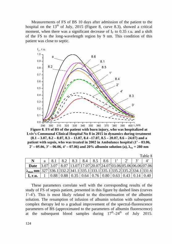
124
Measurements of FS of BS 10 days after admission of the patient to the hospital on the 13th of July, 2015 (Figure 8, curve 8.3), showed a critical moment, when there was a significant decrease of IF to 0.35 r.u. and a shift of the FS to the long-wavelength region by 9 nm. This condition of this patient was close to septic.
Figure 8. FS of BS of the patient with burn injury, who was hospitalized at
Lviv’s Communal Clinical Hospital No 8 in 2015 in dynamics during treatment (8.1 – 3.07, 8.2 – 8.07, 8.3 – 13.07, 8.4 –17.07, 8.5 – 20.07, 8.6 – 24.07) and a
patient with sepsis, who was treated in 2002 in Ambulance hospital (1′ – 03.06, 2′ – 05.06, 3′ – 06.06, 4′ – 07.06) and 20% albumin solution (a), λex = 280 nm
Тable 8 N а 8.1 8.2 8.3 8.4 8.5 8.6 1′ 2′ 3′ 4′
Date 3.07 3.07 8.07 13.07 17.07 20.07 24.07 03.06 05.06 06.06 07.06 λmax nm 327 336.1 332.2 341.1 335.1 333.1 335.1 335.2 335.2 334.1 331.6 I, r.u. 1 0.88 0.88 0.35 0.64 0.76 0.80 0.63 0.43 0.14 0.40
These parameters correlate well with the corresponding results of the
study of FS of sepsis patient, presented in this figure by dashed lines (curves 1′–4′). This is most likely related to the discontinuation of the albumin solution. The resumption of infusion of albumin solution with subsequent complex therapy led to a gradual improvement of the spectral-fluorescence parameters of BS (approximated to the parameters of albumin fluorescence) at the subsequent blood samples during 17th–24th of July 2015.
125
They correlated well with the clinical parameters and laboratory test results of the patient. Therefore, he was discharged from the hospital in satisfactory condition on the 24th of July, 2015.
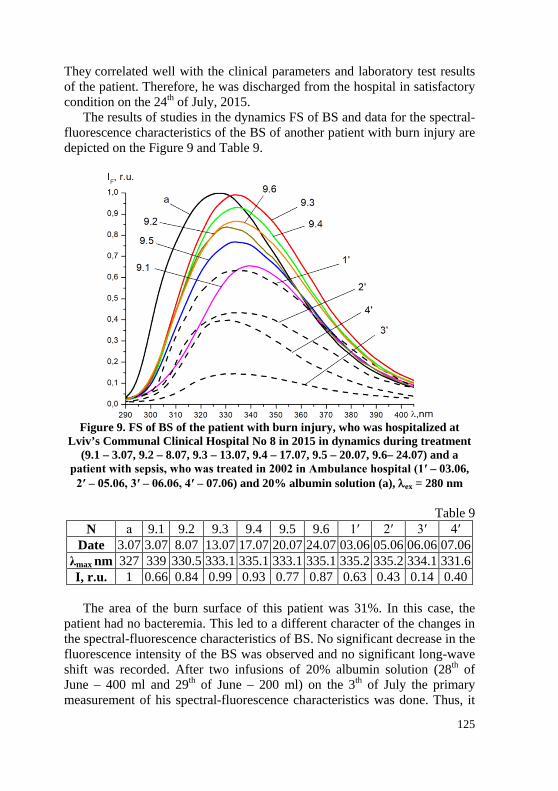
The results of studies in the dynamics FS of BS and data for the spectral-fluorescence characteristics of the BS of another patient with burn injury are depicted on the Figure 9 and Table 9.
Figure 9. FS of BS of the patient with burn injury, who was hospitalized at
Lviv’s Communal Clinical Hospital No 8 in 2015 in dynamics during treatment (9.1 – 3.07, 9.2 – 8.07, 9.3 – 13.07, 9.4 – 17.07, 9.5 – 20.07, 9.6– 24.07) and a
patient with sepsis, who was treated in 2002 in Ambulance hospital (1′ – 03.06, 2′ – 05.06, 3′ – 06.06, 4′ – 07.06) and 20% albumin solution (a), λex = 280 nm
Table 9 N а 9.1 9.2 9.3 9.4 9.5 9.6 1′ 2′ 3′ 4′
Date 3.07 3.07 8.07 13.07 17.07 20.07 24.07 03.06 05.06 06.06 07.06 λmax nm 327 339 330.5 333.1 335.1 333.1 335.1 335.2 335.2 334.1 331.6 I, r.u. 1 0.66 0.84 0.99 0.93 0.77 0.87 0.63 0.43 0.14 0.40
The area of the burn surface of this patient was 31%. In this case, the
patient had no bacteremia. This led to a different character of the changes in the spectral-fluorescence characteristics of BS. No significant decrease in the fluorescence intensity of the BS was observed and no significant long-wave shift was recorded. After two infusions of 20% albumin solution (28th of June – 400 ml and 29th of June – 200 ml) on the 3th of July the primary measurement of his spectral-fluorescence characteristics was done. Thus, it

126
was obtained IF = 0.66 r.u. and shift in the longwave region to 339 nm (Table 9). During the continuation of treatment (massive infusion therapy and 100 ml of 20% albumin solution on the 13th of July), slight changes in the spectral-fluorescence characteristics of BS were recorded. After the cancellation of treatment, the patient's condition gradually stabilized (on the 24th of July IF = 0.87 r.u.) (Table 9) and he was discharged from the hospital on the 24th of July, 2015 in satisfactory condition.
In a slightly different scenario, there was a change in the spectral-fluorescence characteristics of the BS of the next patient with burn injury, with a burn surface area of 40%. The results of the study of FS of this patient are depicted in Figure 10, and in table 10 – data for his spectral-fluorescence characteristics. On the basis of microbiological research, there were verified the presence of two pathogens (Staphyloccus aureus 5 × 10⁶, St. haemolyticus 5 × 10⁶). This patient received infusion, antibacterial therapy, as well as infusion of 20% albumin solution (total volume 1000 ml). For this patient on the 8th of July, 2015 (curve 3.1), a marked decrease in the fluorescence intensity was recorded to 0.53r.u.
Further in dynamics on the 15th of July, 2015 there was an increase in the fluorescence intensity of the patient's BS ( IF = 1.05 r.u., Table 10), which can not be interpreted as absolute hypoproteinemia, which typically causes a decrease in the fluorescence concentration quenching inherent in transient fluorescence.
Figure 10. FS of BS of the patient with burn injury, who was hospitalized at
Lviv’s Communal Clinical Hospital No 8 in 2015 in dynamics during treatment (10.1 – 8.07, 10.3 – 15.07, 10.4 – 17.07, 10.5 – 24.07, 10.6 – 28.07), and a patient
with sepsis, who was treated in 2002 in Ambulance hospital (1′ – 03.06, 2′ – 05.06, 3′ – 06.06, 4′ – 07.06) and 20% albumin solution (a), λex = 280 nm

127
Table 10 N а 10.1 10.3 10.4 10.5 10.6 1′ 2′ 3′ 4′ 10.4
Date 3.07 8.07 15.07 17.07 24.07 28.07 03.06 05.06 06.06 07.06 3.07 λmax nm 327 332 333.1 337.1 333.1 333.1 335.2 335.2 334.1 331.6 327 I, r.u. 1 0.53 1.05 0.88 0.57 0.81 0.63 0.43 0.14 0.40 1
Thereafter, there was a gradual decrease in the fluorescence intensity of the
BS to IF = 0.57 r.u. (24th of July, 2015). In the future, the patient’s condition has stabilized (IF = 0.81r.u.) and he was discharged from the hospital.
Particularly noteworthy are those depicted in Figure 11 the results of the study in the dynamics of FS of the BS of the patient with combined body trauma, concussion, multiple laceration wounds of the frontal parietal region, chest slaughter, lung slaughter, left hemothorax, abdominal wall slaughter, traumatic detachment of the left lower third of left thigh, shock of third grade and sepsis. The results of the spectral-fluorescence characteristics of her BS are presented in Table 11. Based on the microbiological study, the presence of three pathogens was verified for this patient (Ps. aeryginosa 1 × 10⁵, Staphyloccus aureus 1 × 10⁴ та Klebsiella pneumoniae 1 × 10⁴).
Figure 11. FS of BS of the patient with burn injury, who was hospitalized at
Lviv’s Communal Clinical Hospital No 8 in 2015 in dynamics during treatment (11.1 – 17.07, 11.2 – 20.07, 11.3 –24.07,11.4 – 28.07, 11.5 – 31.07, 11.6 – 4.08), and a patient with sepsis, who was treated in 2002 in Ambulance hospital (1′ – 03.06,
2′ – 05.06, 3′ – 06.06, 4′ – 07.06) and 20% albumin solution (a), λex = 280 nm

128
Table 11 N а 11.1 11.2 11.3 11.4 11.5 11.6 1′ 2′ 3′ 4′
Date 3.07 17.07 20.07 24.07 28.07 31.07 4.08 03.06 05.06 06.06 07.06 λmax nm 327 335.1 335.1 333.1 333.1 335.1 335.1 335.2 335.2 334.1 331.6
I, r.u. 1 0.98 0.83 0.76 0.78 0.96 0.98 0.63 0.43 0.14 0.40
It was important for us to study in dynamics the changes of FS of the BS
of this patient and compare them with the corresponding results of patients with burn injury.
The effective treatment was immediately prescribed to this patient. It included antibiotic and infusion therapy with a volume of 2–3 liters every day, including a 20% albumin solution (100 ml and 260 ml during 2 days). Due to the large amount of infusion therapy, the fluorescence intensity of the BS was not reduced (curve 11.1), which correlates with the results of the in vitro study and the results of the BS study of the severe burn patients, described above. Subsequently, the fluorescence intensity decreased to IF=0.76 r.u. (curve 11.3). On the background of further treatment, there was a significant improvement in the patient's condition (IF = 0.98 r.u., curve 11.5) and she was discharged from the hospital. It should be noted, that according to the results of our studies, the condition of this patient was much easier compared to the conditions of previous patients.
Due to the infusion of the significant amount, the fluorescence of the BS was not reduced (curve 11.1), which correlates with the results of the in vitro study and the results of the study of FS of BS of the severe patients with burns (Figure 8-10).
Subsequently, the fluorescence intensity decreased to IF =0.76 r.u. (curve 11.3). The patient received further treatment, her condition improved significantly (IF = 0.98 r.u., curve 11.5) and she was discharged from the hospital. It should be noted, that according to the results of our research, the condition of this patient was much easier compared to the conditions of previous patients.
The results of FS of BS of two more patients with burn injury are depicted on the Figure 12, 13. The corresponding results for the spectral-fluorescence characteristics for their BS are presented in Tables 12, 13. They were hospitalized at Lviv’s Communal Clinical Hospital No 8 in 2017. For the patient with burn area 32% there was a significant volume of infusion therapy (more than 2 liters daily), so the fluorescence intensity was higher than 1 (curves 12.1 and 12.2). This is consistent with the results of the in vitro study. As the volume of infusion therapy decreased, the fluorescence intensity began to decrease (curve 12.3), which was caused
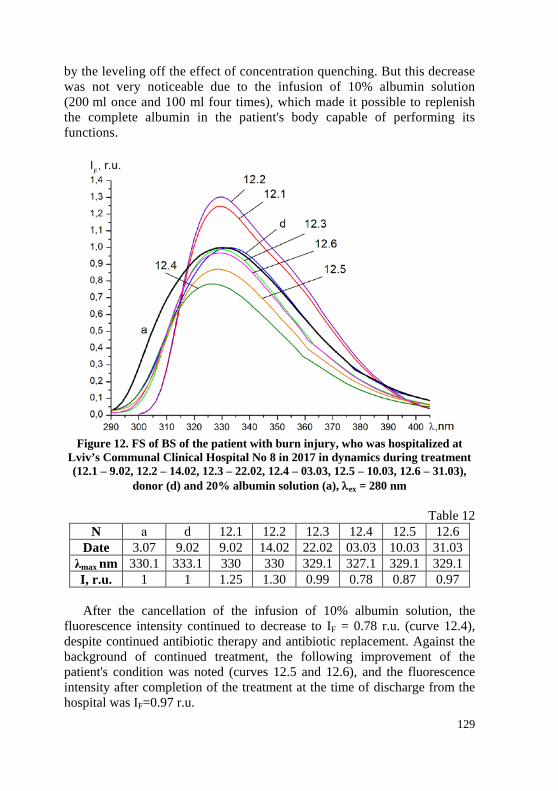
129
by the leveling off the effect of concentration quenching. But this decrease was not very noticeable due to the infusion of 10% albumin solution (200 ml once and 100 ml four times), which made it possible to replenish the complete albumin in the patient's body capable of performing its functions.
Figure 12. FS of BS of the patient with burn injury, who was hospitalized at
Lviv’s Communal Clinical Hospital No 8 in 2017 in dynamics during treatment (12.1 – 9.02, 12.2 – 14.02, 12.3 – 22.02, 12.4 – 03.03, 12.5 – 10.03, 12.6 – 31.03),
donor (d) and 20% albumin solution (a), λex = 280 nm
Table 12 N а d 12.1 12.2 12.3 12.4 12.5 12.6
Date 3.07 9.02 9.02 14.02 22.02 03.03 10.03 31.03 λmax nm 330.1 333.1 330 330 329.1 327.1 329.1 329.1 I, r.u. 1 1 1.25 1.30 0.99 0.78 0.87 0.97 After the cancellation of the infusion of 10% albumin solution, the
fluorescence intensity continued to decrease to IF = 0.78 r.u. (curve 12.4), despite continued antibiotic therapy and antibiotic replacement. Against the background of continued treatment, the following improvement of the patient's condition was noted (curves 12.5 and 12.6), and the fluorescence intensity after completion of the treatment at the time of discharge from the hospital was IF=0.97 r.u.

130
FS of BS of the next patient are depicted on the Figure 13. The area of the burn surface of this patient was 28%. The volume of infusion therapy of this patient did not differ significantly from the corresponding volume of the previous patient, but the fluorescence intensity of his BS was low (curve 12.1 IF= 0.41r.u., curve 12.2 IF = 0.37r.u.). His condition was much more severe, than the condition of the previous patient.
Figure 13. FS of BS of the patient with burn injury, who was hospitalized at
Lviv’s Communal Clinical Hospital No 8 in 2017 in dynamics during treatment (13.1 – 9.02, 13.2 – 14.02, 13.3 – 22.02,13.4 –27.02,13.5 – 03.03,13.6 – 10.03, 13.7 – 31.03), and a patient with sepsis, who was treated in 2002 in Ambulance hospital
(1′ – 03.06) and 20% albumin solution (a), λex = 280 nm
Table 13 N а d 1′ 13.1 13.2 13.3 13.4 13.5 13.6 13.7
Date 06.06 06.06 06.06 9.02 14.02 22.02 27.02 03.03 10.03 31.03 λmax nm 330.1 333.1 333 335.1 339.1 337 334 335.1 331.1 332.0 I, r.u. 1 1 0.16 0.41 0.37 0.46 0.61 0.79 0.89 0.95
During further treatment, including effective antibiotic therapy, as well
as infusion of 10% albumin solution (6th, 10th, 15th, 18th of July, 2017), the fluorescence intensity began to increase from 0.46 r.u. (curve 6.3) up to 0.95 r.u. (curve 6.7). After that, the patient was discharged from the hospital in satisfactory condition.

131
In Figure 14–16 and tables 14–16 are depicted the results of the study of FS of BS of patients with moderate burns. The areas of burn surfaces were 22% (Figure 14), 18% (Figure 15) and 16% (Figure 16), respectively. Changes of the spectral-fluorescent characteristics of the BS of these patients are close to those characteristic of preseptic pathology18.
Figure 14. FS of BS of the patient with burn injury,
who was hospitalized at Lviv’s Communal Clinical Hospital No 8 in 2015 in dynamics during treatment (14.1 – 7.07, 14.2 – 10.07,
14.3 –15.07, 14.4 –17.07, 14.5 –24.07) and 20% albumin solution (a), λex = 280 nm
Table 14
N а 14.1 14.2 14.3 14.4 14.5 Date 7.07 7.07 10.07 15.07 17.07 24.07
λmax nm 327 334.2 336.0 335.1 335.1 333.1 I, r.u. 1 1.12 0.77 0.87 0.94 0.96
18 Prospects for the diagnosis of sepsis and purulent-septic complications: the method
of fluorescence spectroscopy / I.D. Gerych et al. The Journal of the Dental Medicine Academy. 2009. № 9(125). P. 248–256.

132
Figure 15. FS of BS of the patient with burn injury, who was hospitalized at Lviv’s
Communal Clinical Hospital No 8 in 2015 in dynamics during treatment (15.1 – 3.07, 15.2 – 7.07, 15.3 – 10.07) and 20% albumin solution (a), λex = 280 nm
Table 15
N а 15.1 15.2 15.3 Date 3.07. 3.07. 7.07. 10.07.
λmax nm 327 334 335.4 334.5 I, r.u. 1 1.03 0.81 0.90
Figure 16. FS of BS of the patient with burn injury, who was hospitalized at
Lviv’s Communal Clinical Hospital No 8 in 2015 in dynamics during treatment (16.1 – 7.07, 16.2 – 10.07, 16.3 – 15.07, 16.4 – 17.07) and 20% albumin solution
(a), λex = 280 nm

133
Table 16 N а 16.1 16.2 16.3 16.4
Date 7.07 7.07 10.07 15.07 17.07 λmax nm 327 334.1 336.0 335.2 335.1 I, r.u. 1 1.08 0.81 0.87 0.98 It should be noted, that these patients received infusion and antibiotic
therapy. After 3–4 days, these patients had a slight decrease in the fluorescence intensity of the BS without a noticeable shift of λmax in the long-wavelength region. After some correction of treatment tactics against the background of further monitoring of patients, there was a further improvement in their condition. This has been confirmed by the results of a study of the BS of these patients (Figure 14–16, Tables 14–16). Upon completion of the treatment process, they were discharged in satisfactory condition to the supervision of a local surgeon.
The results of the study of FS of BS of the patient with a burn injury of moderate severity are depicted in Figure 17 and table 17. Не was hospitalized at Lviv’s Communal Clinical Hospital No 8 in 2015 with the area of the burn surface 26%.
Figure 17. FS of BS of the patient with burn injury, who was hospitalized at
Lviv’s Communal Clinical Hospital No 8 in 2015 in dynamics during treatment (17.1 – 7 .07, 17.2 – 10.07, 17.3 – 15.07, 17.4. – 17.07) and 20% albumin solution
(a), λex = 280 nm

134
Table 17 N а 17.1 17.2 17.3 17.4
Date 7.07 7.07 10.07 15.07 17.07 λmax nm 327 332.0 335.0 333.1 333.0 I, r.u. 1 0.53 0.76 1.05 0.88 After his admission to the hospital, intensive infusion therapy took place
in a significant volume (1.5 liters daily). At the same time, two days later, a significant decrease in fluorescence intensity was recorded within the MFS (curve 17.1). After correction of treatment tactics (including infusion therapy with 20% albumin solution twice – 200 and 100 ml), the patient's condition significantly improved. This has been also confirmed by the results of the study of FS of BS (curve 17.3). Subsequently, the patient's condition stabilized and on the 20th of July, 2015 he was discharged from the hospital in satisfactory condition to the supervision of a local surgeon.
Thus, we successfully used MFS for patients of the main group to diagnose endogenous intoxication and monitor their condition. The behavior of spectral-fluorescent characteristics of the BS of patients of different severity was under study. Standard treatment regimens was improved. It is noteworthy, that infusions of albumin solution were provided in case the patients felt worse. This ensured a successful treatment process and proper control. We took into account the treatment process under the control of MFS and formed a comparison group of 10 patients, who were treated without supervision within the MFS. But in case of the negative clinical dynamics of the patient's condition, we adjusted the treatment process by including infusions of albumin solution. In all cases, a positive effect was observed due to the changes of treatment tactics. All the patients, 60% of whom were in serious condition, were discharged from the hospital in satisfactory condition after the successful completion of their treatment.
The comparison group consisted of patients with burn injuries of first-and second-degree burns (type A and B), who were hospitalized at the stage of burn shock in the burn department of Lviv’s Communal Clinical Hospital No 8 in 2019–2020. The area of burnt surfaces of patients in the comparison group ranged from 10% to 35%. 60% of the patients in the comparison group were admitted to the hospital in serious condition, and 40% of the patients were in moderate condition. Now we can focus on a few clinical cases. A 38-year-old patient’s occupational injury was treated in the hospital from the 30th of August to the 13th of November, 2019. At the time of admission, the patient's condition was serious. The main diagnosis was second-degree (type A and B) flame burn of 35% of the head, neck, back and both upper limbs, second-degree burn shock. The patient had a fever and endogenous intoxication. The general blood test revealed leukocytosis with the increased

135
number of rod granulocytes and the increased rate of erythrocyte sedimentation. The patient underwent a successful surgical treatment. He received an anti-inflammatory, antibacterial, anticoagulant, antifungal and hormonal therapy, infusions of albumin solution (total amount of albumin solution is 700 ml). The patient also received erythromass (4 times) and native plasma (5 times). The daily infusion volume was more than 3000 ml. Considering the successful experience of using infusions albumin solution for treating patients with burns in the main group, we applied this experience to the comparison group. The patient’s condition was under reliable monitoring. Infusions of albumin solution were provided in the most critical periods of the patient's condition. They made it possible to balance the amount of complete albumin in the BS and improve the patient's condition. After successful completion of the treatment process (75 bed days), the patient was discharged from the hospital in satisfactory condition.
Also notable is the clinical case of another patient, a 46-year-old man, who stayed in the hospital from the 19th of October till the 19th of December, 2019 with a 30%-surface burn injury. The patient's diagnosis was hot steam burn second-degree burns (type A and B) of up to 30% of the torso and both upper limbs. The patient also suffered from a first-degree burn shock.
The patient was hospitalized with acute intense throbbing pain in the affected areas, chills. There was a severing clinical picture of the disease. The patient had endogenous intoxication. He received an appropriate treatment, including surgery (autodermoplasty). He also received anti-inflammatory, antibacterial, infusion therapy with saline and non-saline solutions as well as 20% albumin solution. The treatment process was accompanied by the positive dynamics of changes in the patient's condition. After 61 bed days, he was discharged from the hospital in satisfactory condition.
The condition of the next patient, a 71-year-old woman with a burning injury, which she received at home, was easier to some extent. The main diagnosis was burns of boiling water first-and second-degree burns (type A and B) on 10% of the left lower limb, buttocks and left upper limb. On admission, the patient complained of moderate persistent pain and numbness in the affected area. The general condition was moderate. She also had co-morbidities: hypertension, second functional class, second degree heart failure and atrium fibrillation. There was also a moderate burn shock. The general blood screening revealed leukocytosis, the increased number of rod-shaped leukocytes. The patient underwent surgery (necrectomy and autodermoplasty with a split graft). She received antibacterial, infusional therapy with saline solutions, transfusion of erythrocyte mass (twice). Besides, the patient received the infusion of albumin solution at the most critical moments (twice). After this, positive trends in her condition were observed. They were confirmed by the data of her laboratory tests. It is

136
obvious, that in addition to the treatment of burn injuries, she received therapy for her concomitant cardiovascular pathology. The patient was discharged from the hospital after 67 bed days in satisfactory condition.
Noteworthy is the clinical case of the burn injury of a 51-year-old man, who was hospitalized on the 1th of January, 2020 in serious condition. The diagnosis during hospitalization was second-degree (type A and B) flame burn of 25% of the head, chest, back, both forearms and hands, and first- and second-degree burn shock. The injury was received at home due to the explosion of a blowtorch. The patient underwent 4 surgeries (autodermoplasties and appendectomy). He also received massive infusion therapy with colloidal, saline and non-saline solutions. He received erythromass 5 times (213–319 ml) and blood plasma 4 times (180–260 ml). The patient also received infusions of 20% albumin solution 100 ml twice a day 5 times (total amount is 1000 ml). This contributed greatly to the improvement of his condition. Upon the successful completion of the treatment 76 days later, the patient was discharged home in satisfactory condition to the supervision of a local surgeon.
We should also focus on another case of severe burn injury: a 62-year-old man stayed in hospital from the 17th of August till the 26th of November, 2019. He was hospitalized due to his occupational industrial injury on the 17th of August, 2019. The main diagnosis was 25% second-degree (type A and B) flame burn with damage of both hands and legs as well as a second-degree burn shock. The concomitant diseases were second-degree hypertension, sinus tachycardia, impaired glucose tolerance. The patient underwent a surgical treatment (necrectomy and autodermoplasty). He received anti-inflammatory, antibacterial and infusion therapy with saline, non-saline and colloidal solutions. He received plasma transfusion twice and erythrocyte mass thrice. The sufficient amount of 20% albumin solution was infused. After successful treatment, the patient was discharged after 101 bed days in satisfactory condition to the supervision of a local surgeon.
So, this section illustrates the successful experience of using MFS to diagnose, control and improve the treatment process for patients with burn injuries. The experience and skills, gained by using this method, have contributed greatly to the improvement of treatment tactics for severe patients with burn injuries, whose treatment was carried out without the use of MFS. At the same time, further thorough research is very important to improve the diagnosis and treatment tactics, especially during severe purulent-inflammatory diseases, like sepsis.
CONCLUSIONS For the first time the spectral-fluorescent characteristics of blood serum
for patients with burn injury were obtained, using the method of

137
fluorescence spectroscopy. They proved to be effective markers of the severity of this disease. Their study in the dynamics allows us to monitor their behavior during treatment and manage effectively the process of treatment. This allows us to assess quickly and efficiently the risk of critical purulent-septic complications, adjust treatment and prevent the development of septic conditions in patients.
The positive effect of infusion therapy with albumin solution in the treatment of burn injury was substantiated pathogenetically. Probable scenarios of behavior of spectral-fluorescent characteristics of BS of patients with burn inhury were established. They depended on the severity of the disease. Patients with severe burns can be the model objects for the study of sepsis, including the improved treatment tactics for this disease.
The method of fluorescence spectroscopy makes it possible to carry out diagnostics at the preclinical stage, assess the threat of critical purulent-septic complications quickly and qualitatively and monitor the treatment process.
Changes in the spectral-fluorescence characteristics of blood serum in patients with sepsis in most cases were preliminary: they were usually recorded 24-48 hours before the appearance of obvious clinical and laboratory signs of a significant change in the general somatic status of patients.
In the blood serum of patients with purulent-inflammatory diseases and sepsis there are structural changes in albumin molecules. This leads to decrease of the level of “effective” albumin, capable for performing its basic functions, including detoxification and transport. Therefore, the pathogenetic components of the treatment of these diseases are antibiotic therapy and infusion therapy. The aim of using albumin solutions is to replenish the amount of “effective” albumin in the blood serum in patients with endogenous intoxication.
SUMMARY According to the WHO, burn injury ranks third place in the structure of
general injuries. The main factors of mortality are the development of systemic inflammation, multiorgan failure and infectious complications. Therefore, the objective requirement is to develop the approach, that involves the introduction and the use of new effective methods of diagnosis and treatment of these patients.
Modern advances in medicine are linked closely to the successful development of biomedical research, particularly in the field of biological chemistry. Modern diagnostic trends involve the widespread use of biochemical research methods for their diagnosis, choice of treatment tactics and monitoring the effectiveness of treatment. The use of physical research methods in medical practice, especially the method of fluorescence spectroscopy (MFS), is also particularly promising.
138
The aim of the research is to improve the immediate and long-term functional and cosmetic results of surgical treatment of patients, including defects of the outer coverings and soft tissues of the head and face, based on the development of principles of early diagnosis, including using MFS, monitoring and early reconstructive surgery.
The research took place between January, 2001 and March, 2020. It included 4 stages. The first and second stages of the study were performed on the basis of the purulent-septic center of the Lviv’s municipal clinical hospital of emergency medical services. The third stage of the study was performed on the basis of department of gynecology of Vinnytsia Сity Сlinical Hospital. The fourth stage of the research was performed on the clinical base of City Center of Thermal Injury and Plastic Surgery of Lviv’s Communal Clinical Hospital No 8. The luminescent laboratory of the Department of Experimental Physics at the Ivan Franko National University of Lviv was an experimental research center.
The objects of the study were samples of BS of patients of main and control groups. Excitation of biological objects by ultraviolet light (UV) (λ=280 nm) allows to observe the glow of human serum albumin. If endogenous intoxication is present in the body, albumin molecules with altered physicochemical properties appear. Albumin binding centers are blocked by the products of bacterial metabolism and therefore such albumin (“pathological”) is unable to perform its functions, including transport and detoxification.
Results. The spectral-fluorescent characteristics of BS dilutions with distilled water, 20% albumin solution, sugar broth, centrifuged and non-centrifuged cultures of bacterial culture were studied. In patients with purulent-inflammatory diseases and sepsis in the blood there are two types of albumin molecules: normal and pathological. In the study of FS of BS of patients with purulent-inflammatory diseases there is a decrease in the fluorescence intensity of BS and a long-wave shift of the position of the maximum fluorescence (λmax), the appearance of which is an unfavorable prognostic sign. These changes were recorded 24–48 hours before the appearance of obvious clinical and laboratory signs of infection. In patients with burn injury, depending on the severity of the disease, there were two different scenarios – with a slight decrease of intensity in patients of moderate severity and a marked decrease of the intensity of fluorescence in severe patients.
Conclusions. Within the framework of MFS, diagnostics, monitoring and control of patients’ condition were performed. Correction of treatment tactics was carried out on the basis of pathogenetically substantiated concept of changes in BS albumin in the presence of endogenous intoxication in the body. An important role of albumin solution infusions in the treatment tactics was established.
139
REFERENCES 1. Kovalenko O.M. Pathogenetic substantiation of programs of surgical
treatment of children with common burns and their influence on the course of wound process. Doctor’s thesis. 2012. Kyiv : O. Bohomolets National Medical University. 298 p.
2. Savchyn V.S. Features of reparative processes in patients with deep burns of the head and neck. Archive of Clinical and Experimental Medicine. 2014. № 23(2). P. 149–152.
3. Savchyn V.S. Features of the inflammatory response in burn head and neck injury. Archive of Clinical and Experimental Medicine. 2014. № 2 (part 2). P. 112–113.
4. Ostapiuk L. Diagnostic and therapeutic model of sepsis and purulent-Inflammatory diseases. International Journal of Clinical Medicine. 2019. № 10. P. 577–595. URL: https://doi.org/10.4236/ijcm.2019.1011047.
5. Savchyn V.S. Features of surgical treatment of children with burns of the head and neck. Clinical Surgery. 2012. № 11. P. 30.
6. Classification and treatment of deep burns of the head, caused by electric current / I.D. Gerych et al. Clinical Surgery. 2009. № 11/12. P. 29–30. ISSN 0023-2130.
7. Kovalenko O.M. Tactics of wound closure in critical burns in children. Pediatric Surgery. 2010. № 1 (26). P. 28–32.
8. Performance and clinical evaluation of a sensitive multiplex assay for the rapid detection of common NPM1 mutations / M. Hafes et al. Journal of Molecular diagnostics. September 2010. V. 12. № 5. P. 629–635.
9. Fluorescence spectroscopy: possibilities of application in medical practice / I.D. Gerych et al. Lviv : Liga-Press. 2015. 366 p.
10. Modeling of changes in blood serum in various diseases and treatments / O.V. Bulavenko et al. Biomedical and Biosocial Anthropology. 2013. № 20. P. 8–14.
11. Grizunov Y.A., Dobretsov G.E. Serum albumin in clinical medicine. 1998. Moscow : Geotar. 440 p.
12. Andreeva O.L. Changes in the binding centers of serum albumin in assessing the state of the body in various pathologies. Doctor’s thesis. 2003.
13. Method for early diagnosis of septic complications by the method of flu-orescence spectroscopy / I.D. Gerych, O.V. Bulavenko, L.R. Ostapiuk, A.S. Vo-loshinovskii, S.V. Myagkota. Pat. №76953 Ukraine А61В 17/00 G01N 33/48, G01N 21/64; Applicant and Patentee : Pirogov Vinnytsia National Medical University. № 201207441; stat. 19.06. 2012; publ. 25.01.2013, Bull. № 2.
14. Method of early diagnosis of postpartum purulent-septic complications using the method of fluorescence spectroscopy. Bulavenko O.V., Ostapiuk L.R., Rud V.O., Voloshinovskii A.S. and Маlui T.S. Pat. №133472 Ukraine GO1N 33/48 (2006.01) GO1N 21/64 (2006.01); Applicant and Patentee : Pirogov
140
Vinnytsia National Medical University. – № u2018 10669; stat. 29.10.2018; publ. 10.04.2019, Bull. №7.
15. Approbation of the fluorescence spectroscopy method for the diagnosis of endogenous intoxication with burn injury / V.S. Savchyn, L.R. Ostapiuk, A.S. Voloshynovskyi, T.S. Malyi. Сlinical Surgery. 2016. № 6. P. 68–70.
16. A new look at the diagnosis of endogenous Intoxication in patients with burn injury / V. S. Savchyn et al. Journal of Hospital Surgery. 2019. №1. P. 2–24. https://doi.org/10.11603/2414-4533.2019.1.9907.
17. Cherniy V.I. The role and place of albumin in modern infusion transfusion therapy. Emergency Medicine. 2017. № 1(180). P. 1-11. p-ISSN 2224-0586, e-ISSN 2307-1230.
18. Prospects for the diagnosis of sepsis and purulent-septic complications: the method of fluorescence spectroscopy / I.D. Gerych et al. The Journal of the Dental Medicine Academy. 2009. № 9(125). P. 248–256.
Information about authors:
Zaporozhan S. Y., Doctor of Medical Sciences, Professor,
Professor at the Department of General Surgery Horbachevskyi Ternopil National Medical University
1, Maidan Voli, Ternopil, 46001, Ukraine Savchyn V. S.,
Candidate of Medical Sciences, Head of the Burn Department
Lviv’s Communal Clinical Hospital № 8 23, Navrotsky str., Lviv, 79034, Ukraine
Ostapiuk L. R., Candidate of Medical Sciences,
Obstetrician-gynaecologist Lviv Regional Public Health Centre
45, Lysenka str., Lviv, 79008, Ukraine Tuziuk N. V.,
Postgraduate Horbachevskyi Ternopil National Medical University
1, Freedom Square, Ternopil, 46001, Ukraine Doctor surgeon-combustiologist
Lviv’s Communal Clinical Hospital № 8 23, Navrotsky str., Lviv, 79034, Ukraine