dossier for trabecular metal technology - zimmer biomet€¦ · clinical value dossier for...
TRANSCRIPT

Contact your Zimmer representative or visit us at www.zimmer.com
Clinical Value Dossier for
Trabecular Metal™ Technology

Clinical Value Dossier for Trabecular Metal TechnologyTOC
Table of Contents
List of Tables 1
List of Figures 2
List of Abbreviations 3
Executive Summary 4
1 Burden of Illness 8 1 1 Clinical Characteristics and Presentation 8
1 2 Epidemiology 8
1.2.1 Incidence and Prevalence . . . . . . . . . . . . . . . . . . . . . . . . . . . . . . . . . . . . . . . . . . . . . .8
Global incidences of total joint arthroplasty . . . . . . . . . . . . . . . . . . . . . . . . . . . . . . . . . . . . . . . . .8
1.2.1.1 Hip and Knee . . . . . . . . . . . . . . . . . . . . . . . . . . . . . . . . . . . . . . . . . . . . . . . . . . . . .9
Incidences of total joint arthroplasty among patients age 45-65 . . . . . . . . . . . . . . . . . . . . . . . . . . . . . 10
Total hip arthroplasty procedures outside the US . . . . . . . . . . . . . . . . . . . . . . . . . . . . . . . . . . . . . 11
1.2.1.2 Shoulder . . . . . . . . . . . . . . . . . . . . . . . . . . . . . . . . . . . . . . . . . . . . . . . . . . . . . . 12
The Aging population and total joint arthroplasty . . . . . . . . . . . . . . . . . . . . . . . . . . . . . . . . . . . . . 12
1 3 Clinical Burden 13
Rates of postoperative adverse outcomes . . . . . . . . . . . . . . . . . . . . . . . . . . . . . . . . . . . . . . . . . 13
Causes resulting in the need for revision total joint arthroplasty . . . . . . . . . . . . . . . . . . . . . . . . . . . . . 13
1 4 Humanistic Burden 14
Definitions. . . . . . . . . . . . . . . . . . . . . . . . . . . . . . . . . . . . . . . . . . . . . . . . . . . . . . . . . . . 14
Quality of life considerations leading to joint arthroplasty . . . . . . . . . . . . . . . . . . . . . . . . . . . . . . . . 15
Effects of infection on patient function . . . . . . . . . . . . . . . . . . . . . . . . . . . . . . . . . . . . . . . . . . . 17
Patient function after revision total hip arthroplasty compared to primary hip arthroplasty . . . . . . . . . . . . . . 17
1 5 Economic Burden 18
Economic burden of total joint surgery . . . . . . . . . . . . . . . . . . . . . . . . . . . . . . . . . . . . . . . . . . . 18
Trends involving the economic burden of total joint surgery . . . . . . . . . . . . . . . . . . . . . . . . . . . . . . . 18
2 Conventional Treatments 19 2 1 Description 19
2 2 Conventional Treatment Limitations 19
Cement fixation in total joint surgery . . . . . . . . . . . . . . . . . . . . . . . . . . . . . . . . . . . . . . . . . . . . 19
Traditional porous orthopaedic implants . . . . . . . . . . . . . . . . . . . . . . . . . . . . . . . . . . . . . . . . . . 19

Clinical Value Dossier for Trabecular Metal Technology TOCClinical Value Dossier for Trabecular Metal Technology
Table of Contents (cont )
3 Product Information 20 3 1 Technology Description and Characteristics 20
3 2 Classification and Approval 21
3 3 Device Components and Specifications 21
3 4 Indications 23
3 5 Product Feature Comparison 24
Tantalum use in medical implants . . . . . . . . . . . . . . . . . . . . . . . . . . . . . . . . . . . . . . . . . . . . . . . . . . 24
Trabecular Metal Material properties . . . . . . . . . . . . . . . . . . . . . . . . . . . . . . . . . . . . . . . . . . . . . . . . 24
Trabecular Metal Pore structure . . . . . . . . . . . . . . . . . . . . . . . . . . . . . . . . . . . . . . . . . . . . . . . . . . . 24
The structural, functional, and physiological properties of Trabecular Metal Material . . . . . . . . . . . . . . . . . . . . . 24
4 Clinical value evidence of Trabecular Metal Technology 25 4 1 Clinical Outcomes 25
4.1.1 Use in hip applications . . . . . . . . . . . . . . . . . . . . . . . . . . . . . . . . . . . . . . . . . . . . . . 25
4.1.2 Use in knee applications . . . . . . . . . . . . . . . . . . . . . . . . . . . . . . . . . . . . . . . . . . . . . 26
Trabecular Metal Monoblock implants . . . . . . . . . . . . . . . . . . . . . . . . . . . . . . . . . . . . . . . . . . . 26
Trabecular Metal ON Rod prosthesis . . . . . . . . . . . . . . . . . . . . . . . . . . . . . . . . . . . . . . . . . . . . 29
Trabecular Metal Implants in revision total hip arthroplasty . . . . . . . . . . . . . . . . . . . . . . . . . . . . . . . 30
4.1.3 Removal of Trabecular Metal Components . . . . . . . . . . . . . . . . . . . . . . . . . . . . . . . . . . . . 31
4 2 Functional and Quality-of-Life studies 32
4 3 Clinical Value Summary for Trabecular Metal Technology 34
5 References 35
Clinical Value Dossier for Trabecular Metal TechnologyTOC
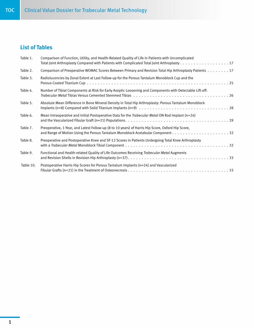
List of Tables
Table 1. Comparison of Function, Utility, and Health-Related Quality of Life in Patients with Uncomplicated Total Joint Arthroplasty Compared with Patients with Complicated Total Joint Arthroplasty . . . . . . . . . . . . . . . . . . 17
Table 2. Comparison of Preoperative WOMAC Scores Between Primary and Revision Total Hip Arthroplasty Patients . . . . . . . . 17
Table 3. Radiolucencies by Zonal Extent at Last Follow-up for the Porous Tantalum Monoblock Cup and the Porous-Coated Titanium Cup . . . . . . . . . . . . . . . . . . . . . . . . . . . . . . . . . . . . . . . . . . . . . . . . . . . . 25
Table 4. Number of Tibial Components at Risk for Early Aseptic Loosening and Components with Detectable Lift-off: Trabecular Metal Tibias Versus Cemented Stemmed Tibias . . . . . . . . . . . . . . . . . . . . . . . . . . . . . . . . . . . 26
Table 5. Absolute Mean Difference in Bone Mineral Density in Total Hip Arthroplasty: Porous Tantalum Monoblock Implants (n=8) Compared with Solid Titanium Implants (n=9) . . . . . . . . . . . . . . . . . . . . . . . . . . . . . . . . . 28
Table 6. Mean Intraoperative and Initial Postoperative Data for the Trabecular Metal ON Rod Implant (n=24) and the Vascularized Fibular Graft (n=21) Populations. . . . . . . . . . . . . . . . . . . . . . . . . . . . . . . . . . . . . . 29
Table 7. Preoperative, 1-Year, and Latest Follow-up (8 to 10 years) of Harris Hip Score, Oxford Hip Score, and Range of Motion Using the Porous Tantalum Monoblock Acetabular Component . . . . . . . . . . . . . . . . . . . . . 32
Table 8. Preoperative and Postoperative Knee and SF-12 Scores in Patients Undergoing Total Knee Arthroplasty with a Trabecular Metal Monoblock Tibial Component . . . . . . . . . . . . . . . . . . . . . . . . . . . . . . . . . . . . . . 32
Table 9. Functional and Health-related Quality of Life Outcomes Receiving Trabecular Metal Augments and Revision Shells in Revision Hip Arthroplasty (n=37) . . . . . . . . . . . . . . . . . . . . . . . . . . . . . . . . . . . . . 33
Table 10. Postoperative Harris Hip Scores for Porous Tantalum Implants (n=24) and Vascularized Fibular Grafts (n=21) in the Treatment of Osteonecrosis . . . . . . . . . . . . . . . . . . . . . . . . . . . . . . . . . . . . . 33
1

Clinical Value Dossier for Trabecular Metal Technology TOC
List of Figures
Figure 1. Number of Primary (1-A) and Revision (1-B) Total Hip and Knee Arthroplasty Procedures in the US: 1990-2002 . . . . . . .9
Figure 2. Number of US Knee and Hip Reconstruction Procedures Estimated Between 2007 and 2013 . . . . . . . . . . . . . . . . .9
Figure 3. Projected Relative Proportion of Patients < 65 Years for Primary and Revision Total Hip and Knee Arthroplasty Procedures Between 2010 and 2030 . . . . . . . . . . . . . . . . . . . . . . . . . . . . . . . . . . . . . . . . . . . . . . . 10
Figure 4. The Projected Number of Primary Total Knee Arthroplasty Procedures in the Kaiser Permanente of Southern California Patient Population . . . . . . . . . . . . . . . . . . . . . . . . . . . . . . . . . . . . . . . . . . . . . 10
Figure 5. Estimated Growth in Hip and Knee Implants In Select European Markets from 2007 to 2013 . . . . . . . . . . . . . . . . 11
Figure 6. Rates of Total Shoulder Arthroplasty per 100,000 People in the US by Age Group . . . . . . . . . . . . . . . . . . . . . . . 12
Figure 7. Preoperative Quality of Well Being and SF-36 Scores for Patients Undergoing Total Hip Arthroplasty and for General Population Norms. . . . . . . . . . . . . . . . . . . . . . . . . . . . . . . . . . . . . . . . . . 15
Figure 8. Preoperative Quality of Well Being and SF-36 Scores for Patients Undergoing Total Knee Arthroplasty and for General Population Norms. . . . . . . . . . . . . . . . . . . . . . . . . . . . . . . . . . . . . . . . . . 16
Figure 9. Projected Number of Joint Replacements in the US from 2005 to 2015. . . . . . . . . . . . . . . . . . . . . . . . . . . . . 18
Figure 10. Trabecular Bone (A) and Trabecular Metal Material (B) . . . . . . . . . . . . . . . . . . . . . . . . . . . . . . . . . . . . . . 20
Figure 11. Reticulated Vitreous Carbon Network Used in the Manufacture of Trabecular Metal Material . . . . . . . . . . . . . . . . . 20
Figure 12. Illustration of the "Bend Before Break" Characteristic of Trabecular Metal Material . . . . . . . . . . . . . . . . . . . . . . 20
Figure 13. Trabecular Metal Tibial Baseplates and Patella Component . . . . . . . . . . . . . . . . . . . . . . . . . . . . . . . . . . . 21
Figure 14. Trabecular Metal Monoblock Acetabular Cups. . . . . . . . . . . . . . . . . . . . . . . . . . . . . . . . . . . . . . . . . . . 21
Figure 15. Trabecular Metal Revision Cup and Polyethylene Liner . . . . . . . . . . . . . . . . . . . . . . . . . . . . . . . . . . . . . . 21
Figure 16. Trabecular Metal Acetabular Augment . . . . . . . . . . . . . . . . . . . . . . . . . . . . . . . . . . . . . . . . . . . . . . . 22
Figure 17. Trabecular Metal Primary Stem with Ceramic Head . . . . . . . . . . . . . . . . . . . . . . . . . . . . . . . . . . . . . . . . 22
Figure 18. Trabecular Metal Tibial Cone . . . . . . . . . . . . . . . . . . . . . . . . . . . . . . . . . . . . . . . . . . . . . . . . . . . . 22
Figure 19. Trabecular Metal Glenoid Component . . . . . . . . . . . . . . . . . . . . . . . . . . . . . . . . . . . . . . . . . . . . . . . 22
Figure 20. Trabecular Metal Avascular Necrosis Rods. . . . . . . . . . . . . . . . . . . . . . . . . . . . . . . . . . . . . . . . . . . . . 23
Figure 21. Trabecular Metal Pore Structure . . . . . . . . . . . . . . . . . . . . . . . . . . . . . . . . . . . . . . . . . . . . . . . . . . 24
Figure 22. SEM Image of Tantalum Metal . . . . . . . . . . . . . . . . . . . . . . . . . . . . . . . . . . . . . . . . . . . . . . . . . . . 24
Figure 23. Rate of Change of Bone Density as a Function of Age Among Women Not Using Antiresorptive Therapy (A) and by Menopausal Status (B) . . . . . . . . . . . . . . . . . . . . . . . . . . . . . . . . . . . . 27
Figure 24. Mean Absolute (A) and Percentage (B) Change in Bone Mineral Density Around the Trabecular Metal Acetabular components (n=9) and Titanium Acetabular Components (n=8) in the Posterosuperior Region . . . . . . . . . . . . . . . . . . . . . . . . . . . . . . . . . . . . . . . . . . . . . . . . . . . 28
Figure 25. Cumulative Survival of the Trabecular Metal Tibial Component . . . . . . . . . . . . . . . . . . . . . . . . . . . . . . . . . 31
2

Clinical Value Dossier for Trabecular Metal TechnologyTOC
List of Abbreviations
AAOS American Academy of Orthopaedic Surgeons
BMD Bone Mineral Density
HRQoL Health-related Quality of Life
KPSC Kaiser Permanente of Southern California
MCS Mental Component Summary
MTPM Maximum Total Point Motion
PCS Physical Component Summary
QWB Quality of Well-being Scale
SF-12 Medical Outcomes Study Short-Form 12-Item Health Survey
SF-36 Medical Outcomes Study Short-Form 36-Item Health Survey
THA Total Hip Rrthroplasty
TJA Total Joint Arthroplasty
TKA Total Knee Arthroplasty
TSA Total Shoulder Arthroplasty
UK United Kingdom
US United States
WOMAC Western Ontario and McMaster Universities Osteoarthritis Index
3

Executive Summary
Executive Summary
Orthopaedic-related disease is one of the leading clinical burdens worldwide and represents one of the top three core service areas for many United States (US) hospitals. A 2009 report from the American Academy of Orthopaedic Surgeons (AAOS) indicated that half of US adults over 65 years of age have some form of arthritis and two-thirds of all arthritis patients are under 65.1 The report also states that more than 90% of patients who require surgical interventions for orthopaedic conditions have osteoarthritis.1 Furthermore, the report suggests that the key factors contributing to the increase in surgically actionable orthopaedic disease in the US include the aging population, increasing presence of patient comorbidities, patient health habits such as diet and smoking, patient bone mineral density, and the growing prevalence of obesity.1 The report also addressed the substantial burden of orthopaedic disease where joint replacement has emerged as the treatment of choice. This report is designed to present the results of clinical studies that are relevant to Trabecular Metal Technology and have been published in peer-reviewed journals and further aims to assist in making more informed decisions when it comes to choosing implants that utilize Trabecular Metal Technology.
Burden of Orthopaedic Disease
The demand for total joint arthroplasty (TJA) is substantial and rapidly growing. The number of total hip (THA) and total knee (TKA) arthroplasty procedures have increased significantly in the US. For example, a National Hospital Discharge Survey and US Census Data analysis from 1990 to 2002 by Kurtz et al. showed that from 1990 to 2002, primary THA procedures grew by 46% while primary TKA procedures tripled.2 A 2009 Market Report by Millennium Research Group predicts steady short-term growth in large joint implant procedures between 2007 and 2013, with knee reconstruction procedures expected to grow by approximately 32% and hip reconstruction procedures by 23% during this period.3
Studies have also shown that the incidence of THA and TKA is increasing substantially in the younger, more active population under 65 years of age. According to an analysis by Kurtz et al., primary THA and TKA procedures for patients younger than 65 are projected to account for more than 50% of THA procedures by 2011 and TKA procedures by 2016.4 Moreover, the analysis shows that, by 2030, patients younger than 65 are projected to account for 52% of primary THA procedures and about 60% of TKA procedures.4
Based on a survey of 43 patients, Shields et al. found that severely impaired patient functional status and subsequent inability to perform routine daily activities were significant quality-of-life considerations leading to joint arthroplasty.5 According to the authors, Medical Outcomes Study Short-Form 36-Item Health Survey (SF-36) scores have consistently reported 40%-50% or greater improvements in physical function, ability to perform activities of daily living, joint stiffness, and bodily pain following primary TJA.5 According to Espinoza-Ervin et al., revision procedures represent a substantial and growing burden in terms of overall clinical outcomes and health care expenditures that can influence the financial performance and stability of an orthopaedic unit.6
According to Bozic et al. and Ducheyne et al., the most common complications leading to revision arthroplasty were infection and aseptic loosening or migration of the implant.7,8 The National Hospital Discharge Survey and US Census Data analysis from 1990 to 2002 by Kurtz et al. showed the rate of revision procedures had grown dramatically, with 60% growth in revision THA and 166% growth in revision TKA.2 Studies by Espinoza-Ervin et al., Lavernia et al., and Bozic and Reis indicate that revision procedures also have high complication rates (about 20% for THA and 7.3% to 9.3% for TKA), substantially longer operating times and lengths of stay, and in the case of THA, increased requirements for bone grafts and patient transfer to extended care facilities.6,9,10 According to the Millennium Research group report of 2009, recent estimates suggest that the number of revision procedures in the US will continue to grow through 2013, with a 69% increase in knee procedures and a 26% increase in hip revisions during the period from 2007 to 2013.3
The economic burden of joint replacement surgeries in the US is projected to increase markedly. According to a study of data from 1997 to 2004 by Kim, total annual hospital costs for primary and revision hip and knee arthroplasty procedures in the US were estimated at $9.1 billion in 2004. Based on this data, if current trends continue, Kim anticipated that hospital charges associated with TJA will exceed $80 billion by 2015.11
4
Executive Summary
Limitations of Conventional Orthopaedic Implants
Implants manufactured from conventional orthopaedic implant materials, including titanium and cobalt chromium alloys, often use cement to help achieve fixation and stability. The relatively high stiffness of some solid metal implants also may transfer low loads to the host bone leading to the potential for stress shielding and bone resorption over time.
Some joint replacement implants have porous bone-interface surfaces designed to allow for biological fixation through bone in-growth into the implant. However, adding porous surfaces to metal implants to help achieve bone in-growth does not address the need for the host bone to be physiologically loaded.
Overview of Trabecular Metal Implant Properties
Elemental tantalum, the core material used in Trabecular Metal Material, has been used to make implantable medical devices for more than 50 years.12 Tantalum is an excellent material for this porous in-growth structure as it is biologically inert, ductile, corrosion resistant, and has high fatigue strength. Trabecular Metal Material is a unique, highly porous biomaterial made from tantalum designed with structural and functional properties similar to those of trabecular bone.12
Trabecular Metal Material has the following key characteristics:
• Porosity and structural composition similar to trabecular bone.12
• Pore sizes averaging greater than 300µm, a size that has shown to support vascularization.13 Trabecular Metal Material has an average pore size of 547µm.12,14,15
• High porosity and low modulus of elasticity supporting biological in-growth.16
• A high coefficient of friction against cancellous bone (0.98 for net shapes) that has been shown to help to support initial stability.17
• In a study by Macheras et al. of 86 hips that had received Trabecular Metal Monoblock Acetabular Implants with a mean follow-up of 7.3 years (7 to 7.5), 25 hips showed immediate postoperative gaps between the bone and the implant. The study found that all of the gaps were closed at 24 weeks with no acetabular migration and the authors reported no revisions.22
• Further described by Macheras et al., all 86 hips showed improvement from preoperative Harris Hip scores, no dislocations or implant-related complications were observed and all patients regained their previous activities. At the last follow-up examinations (7.3 years), the authors reported no radiolucent lines and no areas of osteolysis in any of the 86 hips. Again, the authors reported no acetabular implant had been revised.22
Value Evidence Supporting Trabecular Metal Technology
This section describes the available clinical and economic evidence supporting the value of Trabecular Metal Technology as reported in peer-reviewed, published clinical studies.
Implant Fixation and Stability A 1999 laboratory study by Zhang et al. found that the structure of Trabecular Metal Material provides a high coefficient of friction against cancellous bone which would be expected to lead to higher initial stability against bone as compared to interfaces with natural bone grafts or other traditional porous metals.17 Additional studies by Gruen et al., Macheras et al., Dunbar et al., and Levine et al. involving acetabular components, knee tibial fixation surfaces, and knee augments have found that Trabecular Metal Material may potentially result in improved implant fixation and stability that may lead to reduced migration potential.18-21
Rapid and effective biologic fixation is critical to the reduction of implant migration and loosening, and could potentially affect the long term stability and fixation of the implant. Several factors can lead to implant loosening and revision surgery, including mechanical detachment, bone degeneration surrounding the implant, and infection. The presence of radiolucent lines surrounding the implant is one key indicator of suboptimal attachment of at-risk bone to the implant interface; a strong presence of radiolucent lines was seen as an early indicator of potential total joint failure in a study performed by Ducheyne et al.8
The porous nature of Trabecular Metal Material offers advantages compared with conventional porous implants. It supports biologic in-growth and fixation from the high coefficient of friction and porosity.
Randomized clinical studies, including studies comparing specific Trabecular Metal Technology products with porous-coated titanium found the following:
5
Executive Summary
Stress ShieldingThe relatively low stiffness of Trabecular Metal Monoblock Components may have the potential to result in a more natural physiological transfer of stresses to the peri-acetabular bone, potentially decreasing detrimental stress shielding.23 In a study involving nine porous tantalum and eight titanium acetabular implants, Meneghini et al. found a statistically significant difference in both absolute and percentage change in BMD in the Trabecular Metal Implant group compared with the solid titanium group. BMD values may be used as a clinical indicator measurement for bone strength and fracture risk. The authors noted that “the elasticity [stiffness] of porous tantalum is proposed to create a more physiologic transfer of stresses to the peri-acetabular bone, theoretically decreasing detrimental stress-shielding.”23
Osteonecrosis of the Femoral HeadMultiple studies have evaluated the benefits of using the Trabecular Metal Osteonecrosis (ON) Rod in the treatment of Osteonecrosis. Nadeau et al. noted that Osteonecrosis occurs in 5% to 12% of potential THA patients when blood supply to the femoral head is disrupted.24 A study by Shuler et al. involving 45 patients compared porous tantalum implants with vascularized fibular grafting in the treatment of early Osteonecrosis. According to the authors, early outcomes suggest that porous tantalum implants are a viable option for femoral head salvage.25 Studies by Tsao et al. and Veillette et al. also support the use of Trabecular Metal ON Rod Implants for Osteonecrosis in Steinberg Stage I and Stage II disease.26,27
Tibial Implants
In a study involving 105 primary knees, Helm et al. evaluated uncemented Trabecular Metal Tibial Monoblock Implants.28 At three years, no revisions occurred due to mechanical failure and no radiolucent lines were detected on postoperative radiographs, with only one revision of the tibia reported due to trauma.28
The authors state that reported adverse events for the 105 knees at three years were comparable or superior to reported peri-operative outcomes on knee implants in the overall US population or Medicare population.
• Gruen et al. evaluated the radiographs of 574 patients at 2 to 5 years with porous tantalum Monoblock Acetabular cups and observed no progression of any postoperative gap, no evidence of continuous peri- acetabular interface radiolucencies, no evidence of lysis, and no revisions for loosening in this patient group; while 7 sockets were revised early (6 recurrent dislocations and 1 early trauma-related loosening), and 3 additional revisions were for sepsis.18
• In a series of 151 Trabecular Metal Monoblock Acetabular Implants studied by Macheras et al., no dislocations, implant-related complications, radiolucent lines, or areas of osteolysis were reported at 8 to 10 years.19
• Also in the Macheras study of 151 hips, it was reported that no acetablular component was revised or needed revision at the last follow-up due to aseptic loosening. Mechanical failure of the femoral component occurred in one hip at 8 years postoperatively (<1%) and required only femoral component revision; the acetabular component was stable without evidence of polyethylene wear.19
• A radiostereometric analysis (RSA) showed that, at six to 12 months, none of the Trabecular Metal Implants were deemed to be at risk of aseptic loosening while 19% of the cemented components were at risk for aseptic loosening.20
• Over time, the maximum total point motion (MTPM) scores for Trabecular Metal Components declined, while cemented components showed significantly more occurrences of lift-off; indicating greater potential for implant migration.20
• RSA also showed that the 28 patients who received Trabecular Metal Components experienced reduced long-term migration and fewer occurrences of lift-off compared with the 21 patients who received cemented components.20
Trabecular Metal Technology has also been studied relative to reducing implant migration and loosening in primary TKA. Dunbar et al. studied 70 patients who received either a Trabecular Metal Monoblock Tibial Component or a conventional cemented stemmed tibial component. This study found the following: 20
6

Executive Summary
Removal of Trabecular Metal Implants
The Zimmer Trabecular Metal Explant® Acetabular Cup Removal System has been designed to facilitate the removal of Trabecular Metal Acetabular Cups while providing a high level of bone preservation, even in the case of a well-fixed component. A study by Mitchell et al. involving the removal of 31 well-fixed Trabecular Metal Acetabular Cups using the aforementioned explant system indicated that the cups were removed without difficulty and with minimal further bone loss.29 The authors observe that, “the ease of removal with this system and the lack of any further damage to the host bone illustrate that the Explant Acetabular Cup Removal System is a safe and reliable tool for use in the revision of well-fixed components.”29 Another study involving four revision knees by Klein et al., found that the removal of Trabecular Metal Monoblock Tibial Implants were performed with no intraoperative tibial fractures or complications related to removal.30
Measured Changes in Harris and Oxford Hip Scores
In a study involving 151 Trabecular Metal Monoblock Implants, Macheras et al. found that functional outcomes, including pain, range of motion, and flexion at one year, as measured by the Harris Hip Score and the Oxford Hip Score, improved compared to preoperative scores.19 Shuler et al. found that Trabecular Metal ON Rod Implants have also improved functional scores in the treatment of Osteonecrosis of the femoral head, as measured by the Harris Hip Score. The study showed that, at 39-month follow-up, 19 of 22 (86%) Trabecular Metal ON Rod patients had “good-to-excellent results and three (14%) “were classified as poor outcomes” while only three of 17 fibular graft patients had “good or excellent outcomes, and 14 (78%) were classified as poor.”25
7

SECTION
1
8
Burden of Illness
1 Burden of Illness
1 1 Clinical Characteristics and Presentation 1 2 Epidemiology
A March 2009 AAOSNow report from the American Academy of Orthopaedic Surgeons (AAOS) indicated that approximately one in every two adults aged 65 and over has some form of arthritis.1 According to the report, data from 2004 indicated osteoarthritis and rheumatoid arthritis are increasing in frequency among the aging population of the United States (US) and other industrialized countries, with nearly two-thirds of those afflicted being younger than 65. The report also showed that, although arthritis affects both men and women, it is more common in women.1 In a study of total shoulder arthroplasty (TSA) patients, Jain et al. noted that osteoarthritis commonly presents with a gradual increase in pain, stiffness, and decreased range of motion in the affected joint.34
Joint replacement has become a common treatment in restoring function to severely arthritic joints. The March 2009 AAOSNow report indicated the most frequently replaced joints are the knee and hip, followed by the shoulder.1 The report goes on to say that, “Total hip and primary knee replacements are performed almost exclusively due to osteoarthritis, with two of three persons hospitalized for osteoarthritis in 2004 undergoing a joint replacement procedure. A small proportion of replacements are due to rheumatoid arthritis or another condition.”1 In a literature review by Felson and Zhang of findings about osteoarthritis, as well as in a study by Lawrence et al., it was reported that other factors, such as genetic susceptibility, nutrition, osteoporosis, joint injury, or body weight may be associated with the occurrence and progression of osteoarthritis, especially of the knee, and thus may lead to joint arthroplasty.32,33
1 2 1 Incidence and Prevalence of Joint Replacement
Globally, the incidence of joint replacement is significant and rising.2,3,35,36
The prevalence of primary joint replacement has increased steadily since the 1990s because of expanded numbers of contributing factors, including osteoarthritis, the aging population, and patient comorbidities.2,3,35,36
According to data from NHDS, US Census, and the Millennium Research Group, the incidence of joint arthroplasty worldwide has increased steadily over the past two decades and continues to rise as global populations grow. Joint arthroplasty procedures are performed more often in women than in men, and that in both men and women, the procedure rates increase with age as patients reach their late 70s, after which the rates decline.2,3,35,36
• Joint arthroplasty treats a variety of musuloskelatal conditions that cause severe joint pain, joint stiffness, and limited range of motion in a wide variety of patient groups.31-33
• Osteoarthritis is the underlying condition in the majority of joint arthroplasty cases.1,35
• Globally, the incidence of joint replacement is significant and rising. 2,3,35,36
• The prevalence of primary joint replacement has increased steadily since the 1990s because of expanded numbers of contributing factors, including osteoarthritis, the aging population, and patient comorbidities.2,3,35,36
• The incidence of total knee arthroplasty is increasing significantly in the younger population 45 to 65 years of age.4
• In non-US markets, the incidence of total knee and hip procedures is also growing significantly.31,37-40
• The projected increase in the aging population will contribute to the growing incidence of joint arthroplasty.36

1SECTION
9
Burden of Illness
1 2 1 1 Hip and KneeThe number of primary and revision hip and knee arthroplasties increased steadily in the US from 1990-2002 based on the National Hospital Discharge Survey and US Census Data (Figure 1).2 The same study noted the rate of primary total hip arthroplasty (THA) per 100,000 persons increased by 46%, and the rate of primary total knee arthroplasty (TKA) almost tripled during that time frame. The rate of revision THA increased by 60%, whereas the rate of revision TKA increased by 166%.
According to Kurtz et al., the rates of primary TKA and THA were significantly higher for women than for men (P < 0.05), and significantly higher in the 65-74 and 75-84 age groups compared with the 45-64 age group (P < 0.01).2 However, the rates of primary TKA and THA steadily increased from 1990-2002 in the 45-64 age group.2 Similarly, a study by Jain et al. found that the incidence of TKA in patients aged 50-59 years increased from 6.2 per 10,000 in 1990 to 14.2 per 10,000 in 2000.36
Figure 1 Number of Primary (1-A) and Revision (1-B) Total Hip and Knee Arthroplasty Procedures in the US: 1990-2002
Figure 2 Number of US Knee and Hip Reconstruction Procedures Estimated Between 2007 and 2013
Source: Kurtz et al. (2005)2
Source: Millennium Research Group (2009)3
THA = total hip arthroplasty; TKA = total knee arthroplasty.
1-A500
400
300
200
100
0
Proc
edur
es (x
1,0
00)
Year
1-B
Rev
isio
n Pr
oced
ures
(x 1
,000
)
50
40
30
20
10
0
1990 1991 1992 1993 1994 1995 1996 1997 1998 1999 2000 2001 2002
Year1990 1991 1992 1993 1994 1995 1996 1997 1998 1999 2000 2001 2002
Primary THAPrimary TKA
Revision TKARevision THA
The total number of revision THAs almost doubled, and the total number of revision TKAs tripled from 1990-2002, during which the number and rate of primary TKA were higher than those of THA; however, the rate of revision THA was higher than that for revision TKA.2 A 2009 Market Report by Millennium Research Group predicts steady short-term growth in large joint implant procedures between 2007 and 2013, with knee reconstruction procedures expected to grow by approximately 32%, from 598,000 to 787,000, and hip reconstruction procedures by 23%, from 440,000 to 543,000, during this period (Figure 2).3
1,600,000
1,200,000
400,000
0
Proc
edur
es
Year2007 2008 2009 2010 2011 2012 2013
Knee reconstructionHip reconstruction
800,000

SECTION
1
10
Burden of Illness
The incidence of total knee arthroplasty is significantly increasing in the younger population 45 to 65 years of age.4
A 2009 study of the Nationwide Inpatient Sample by Kurtz et al. found that, while the incidence of THA and TKA has increased in the younger patient population, i.e., younger than 65 years, the relative proportion of the younger population undergoing these procedures also grew from 1993-2006.4 According to the study, in 1993 32% of primary or revision THAs and 25% to 27% of primary or revision TKAs were performed on patients younger than 65. In 2006, the relative proportion of patients younger than 65 receiving primary or revision THA or TKA had increased from 40% to 46%, respectively.4 The demand for primary THA and TKA among patients younger than 65 is projected to account for more than 50% of THA procedures by 2011 and TKA procedures by 2016 (Figure 3). By 2030, the demand for THA and TKA among patients younger than 65 is projected to be 52% of primary THAs and about 60% of primary or revision TKAs (Figure 3)4. The future demand for primary TKA is projected to grow the fastest for the 45-54 age group.4
Figure 3 Projected Relative Proportion of Patients < 65 Years for Primary and Revision Total Hip and Knee Arthroplasty Procedures Between 2010 and 2030
Source: Kurtz et al. (2009)4
THA = total hip arthroplasty; TKA = total knee arthroplasty.
65
55
50
45
40
35
Pati
ent %
< 6
5 ye
ars
of a
ge
Year
60
2010 2015 2020 2025
Primary THA
Primary TKARevision THA
Revision TKA
2030
Using a Kaiser Permanente Southern California (KPSC) database, Khatod et al. predicted that the total TKA patient population will increase by approximately 200% between 2005 and 2020 (Figure 4), which mirrors the general trend in the broader US population reported by Khatod et al.41
Figure 4 The Projected Number of Primary Total Knee Arthroplasty Procedures in the Kaiser Permanente of Southern California Patient Population
Source: Khatod et al. (2008)41
12,000
6,000
4,000
2,000
0
Proj
ecte
d nu
mbe
r of c
ases
Year
8,000
10,000
2005 2006 2007 2008 2009 2010 2011 2012 2013 2014 2015 2016 2017 2019 20202018

1SECTION
11
Burden of Illness
In non-US markets the incidence of total knee and hip procedures is also growing significantly.31,37-40
Mirroring the increase in THA and TKA procedures in the US market, the incidence of THA and TKA in other global markets is also projected to increase. Estimates by Millennium Research Group in four primary European markets, including the United Kingdom (UK), Germany, France, and Italy suggested similar growth (Figure 5).35 Considering demographic changes, Birrell et al. projected that the demand for THA in the UK will increase by 40% by 2021.37 Sibana et al. reported that in the UK, there were 327,557 primary THA and TKA procedures performed between April 1, 2003 and September 30, 2006 according to the National Health Service and the Hospital Episode Statistics database.31 These authors also found the revision rate following primary THA was 0.7% at one year and 1.4% at three years while the revision rate following TKA was 0.4% at one year and 1.4% at three years.31
Figure 5 Estimated Growth in Hip and Knee Implants In Select European Markets from 2007-2013
Source: Millennium Research Group (2009)35
1,500,000
1,000,000
500,000
0
Uni
ts
Year2007 2008 2009 2010 2011 2012 2013
GermanyUKItalyFrance
Wells et al. estimated that THA increased by 25.9% and TKA by 42.8% from 1994 to 1998 in Australia,38 while a review of South Korean national registry data by Kim et al. suggested an almost two-fold increase in TKA from 2002 to 2005 in South Korea.42
Similarly, a review by Lohmander et al. of national hip registers in five Nordic countries (Denmark, Finland, Iceland, Norway, and Sweden) showed that from 1996 to 2000, the average age-adjusted annual incidence per 100,000 primary THAs for osteoarthritis ranged from 60 to 92 in females and from 48 to 75 in males.39 In addition, a discharge register and census bureau review by Ostendorf et al. showed that the incidence of primary THAs increased 68% from 1986 to 1997 in The Netherlands.40
The annual incidence of THAs in The Netherlands is projected to increase by 44% by the year 2020.40 In global markets evaluated, incidence rates are generally higher in females than males, and in both genders the rate increases with age as patients approach their late 70s, after which the rates decline.

SECTION
1
12
Burden of Illness
Figure 6 Rates of Total Shoulder Arthroplasty per 100,000 People in the US by Age Group
Source: Jain et al. (2006)34
1 2 1 2 ShoulderThe March 2009 AAOSNow issue reported that shoulder arthroplasty is the third most frequently reported joint replacement procedure in the US.1 Jain et al. reported a “minor increase in the rates of TSA from 1990-2000 for most age groups” (Figure 6).34 The report indicates that “most of the procedures were performed in patients 65-79 years of age” and, as in THA and TKA, females accounted for the majority of these procedures.34 Also, osteoarthritis was the primary diagnosis for the majority of cases undergoing TSA, whereas fractures of the proximal humerus, rheumatoid arthritis, and aseptic necrosis of the humeral head were the second, third, and fourth most frequent underlying diagnoses, respectively.34
20
18
16
4
6
2
Tota
l sho
ulde
r art
hopl
asty
rate
per 1
00,0
00 p
erso
ns
Year
14
12
10
8
1990 1991 1992 1993 1994 1995 1996 1997 1998 1999 2000
50-59
≥ 80
60-64
65-79
≥ 65
The projected increase in the aging population may contribute to the growing incidence of joint arthroplasty.2,7,36
According to Kurtz et al. and Jain et al. a variety of causes account for the global increase in the incidence of joint replacement procedures, including the growing elderly population and the increase in the prevalence of underlying conditions such as osteoarthritis.2,36
Based on these findings, all developed countries with an aging population are likely to experience an increase in the incidence of total joint arthroplasty (TJA). In addition, Jain et al. suggest that the indications for TJA may be broadening, especially for younger patients, further adding to the increase in procedures.36 Also, Ostendorf et al. suggest that “as patients become better informed and more aware of present treatments, they may be less inclined to accept their disability and prefer treatment at an earlier stage of their disease.”40 According to Kurtz et al., “the explosive growth in both the number and the prevalence of primary total knee arthroplasty for both genders and all age categories can be explained, at least in part, by the increased recognition, by the candidates for surgery and the members of the orthopaedic community, of the effectiveness of the procedure in treating degenerative disease of the knee.”2 Furthermore, the authors observed that “the major increase in the population of obese patients in the United States appears to contribute to the increasing trends in primary joint replacement.”2
1SECTION
13
Burden of Illness
1 3 Clinical Burden
• A variety of postoperative complications are associated with joint arthroplasty, many of which involve significant patient risk and/or additional procedures to address complications.44
• Rates of postoperative adverse outcomes are higher after revision total joint arthroplasty than after primary joint arthroplasty.44
• The most common causes of revision total knee arthroplasty are infection and implant loosening.45
• The most common indications for revision total hip arthroplasty are hip instability and mechanical loosening.46
Rates of postoperative adverse outcomes are higher after revision total joint arthroplasty than after primary joint arthroplasty.44,46
A variety of postoperative complications are associated with joint arthroplasty, many of which involve significant patient risk and/or additional procedures to address complications.44
Because joint replacement surgery is an invasive procedure, complications are possible. Phillips et al. reported on potential complications after THA in the medicare population from July 1, 1995 to June 30, 1996 which included dislocation, pulmonary embolism, and deep infection.44 These authors studied claims data to estimate the incidence rates of these complications among Medicare beneficiaries undergoing primary or revision THA from July 1, 1995, to June 30, 1996. Incidence rates were calculated per 10,000 person-weeks. For patients receiving a primary THA, 3.9% had a dislocation, 0.9% had a pulmonary embolism, and 0.2% had a deep hip infection in the first 26 postoperative weeks.44 In the revision THA group, 14.4% had a dislocation, 0.8% had a pulmonary embolism, and 1.1% had a deep hip infection.44 According to the report, the incidence rates were, “highest immediately after total hip replacement, but they continue to be elevated throughout the first three postoperative months.”44 The authors found that these complications impose a burden on the clinical team and patient, as strategies to treat and manage patient complications are often resource intensive for the hospital.44,46
The most common causes of revision total knee arthroplasty are infection and implant loosening.45
The most common indications for revision total hip arthroplasty are hip instability/dislocation and mechanical loosening, and infection.46
As noted previously (See Section 1.2.1), the rates of revision TJA continue to rise. In the US from 1990-2002, the rate of revision THA increased by 60%, while the rate of revision TKA increased by 166%.2 Bozic et al. analyzed Nationwide Inpatient Sample data from 60,355 revision TKA procedures performed in the US in 2005 and 2006. They found that “the most common causes of revision TKA were infection (25.2%) and implant loosening (16.1%), and the most common type of revision TKA procedure reported was all component revision (35.2%).”45 In another study using the same nationwide inpatient database from the same time period, Bozic et al. analyzed 51,345 revision THA procedures. They reported that the most common type of revision THA procedure performed was “all-component revision (41.4%), followed by femoral component revision (13.2%), acetabular component revision (12.7%), and isolated femoral head and acetabular liner exchange (12.6%).”46 They also found that “the most common causes of revision were instability/dislocation (22.5%), mechanical loosening (19.7%), and infection (14.8%).”7 Revision surgeries impose a burden on the clinical team and patient in the form of patient management and additional hospital stays, respectively.44,46 With the expected growth of revision surgery in the US (See Section 1.2.1.1), this burden may increase.

SECTION
1
14
Burden of Illness
1 4 Humanistic Burden
DefinitionsAn evaluation of patient health-related quality of life (HRQoL) and functional status characterizes the scope and nature of preoperative patient quality-of-life and serves as a means to assess the degree of improvement that patients may experience following primary or revision joint arthroplasty. A variety of generic and disease-specific instruments is used to measure HRQoL and functional status in TJA. Following is a brief description of each of these instruments.
• The Medical Outcomes Study Short-Form 36-Item Health Survey (SF-36) is a self-administered questionnaire that measures each of the following eight health concepts: physical functioning; role limitation (physical); bodily pain; general health; vitality; social functioning; role limitation (emotional); and mental health. In addition, two summary scores may be calculated. The Physical Component Summary (PCS), composed of the physical functioning, role limitation (physical), bodily pain, and general health subscales, provides a measure of the subject’s perception of his/her physical health. The Mental Component Summary (MCS), composed of the vitality, social functioning, role limitation (emotional), and mental health subscales, provides a measure of the subject’s perception of his/her mental health. The PCS and MCS were designed to reduce the SF-36 from an eight-scale profile to two summary measures without a substantial loss of information. One of the strengths of these two summary scores is their value in distinguishing physical outcomes from mental health outcomes.
• The quality-of-life considerations leading to joint arthroplasty are substantial where compromised functional status impairs the patient’s ability to perform routine activities of daily living.5,47,48
• Patients with total joint arthroplasty complications such as infection have reduced quality of life and functioning compared with patients without complications.49
• Patient function after revision total hip arthroplasty is lower than after primary total hip arthroplasty, indicating a potential unmet need for more successful revision surgical techniques and implants.50
• The Medical Outcomes Study Short-Form 12-Item Health Survey (SF-12) reduces the instrument to 12 items but retains all eight domains and the two summary scores.
• The Quality of Well-being Scale (QWB) is an interviewer-administered instrument that was developed as a generic index of population health status. The QWB combines preference-weighted values for symptoms and functioning. The preference weights were obtained by the ratings of 856 people from the general population who rated the desirability of health conditions by placing each on the continuum between death (0.00) and optimum health (1.00). Symptoms are assessed by questions about the presence or absence of different symptoms complexes. Functioning is assessed by a series of questions designed to record functional limitations over the previous three days within the three separate domains of mobility, physical activity, and social activity. The three domain scores are combined into a total score that provides a numerical point-in-time expression of well-being that ranges from zero (0) for death to one (1.0) for asymptomatic optimum functioning.
• The Western Ontario and McMaster Universities Osteoarthritis Index (WOMAC) is a self-administered, disease-specific instrument that probes clinically important and patient-relevant symptoms in the areas of pain, stiffness, and physical function in patients with osteoarthritis of the hip and/or knee. The WOMAC is a valid, reliable, and responsive measure of patient-reported outcomes and is widely used in the evaluation of knee and hip osteoarthritis. The index comprises 24 questions and includes the following three separate subscales:
– The WOMAC Pain subscale comprises five questions regarding the amount of pain experienced due to osteoarthritis in the study joint in the past seven days. This subscale score ranges from 0 to 20, with higher scores indicating more pain.
– The WOMAC Stiffness subscale comprises two questions regarding the amount of stiffness experienced in the study joint in the past seven days. This subscale score ranges from 0 to 8, with higher scores indicating more stiffness. Stiffness is defined as a sensation of decreased ease in which the subject moves the study joint.
– The WOMAC Physical Function subscale comprises 17 questions regarding the degree of difficulty experienced due to arthritis in the study joint in the past seven days. This subscale score ranges from 0 to 68, with higher scores indicating worse physical function. Physical function refers to the subject’s ability to move around and perform the usual activities of daily living.
1SECTION
15
Burden of Illness
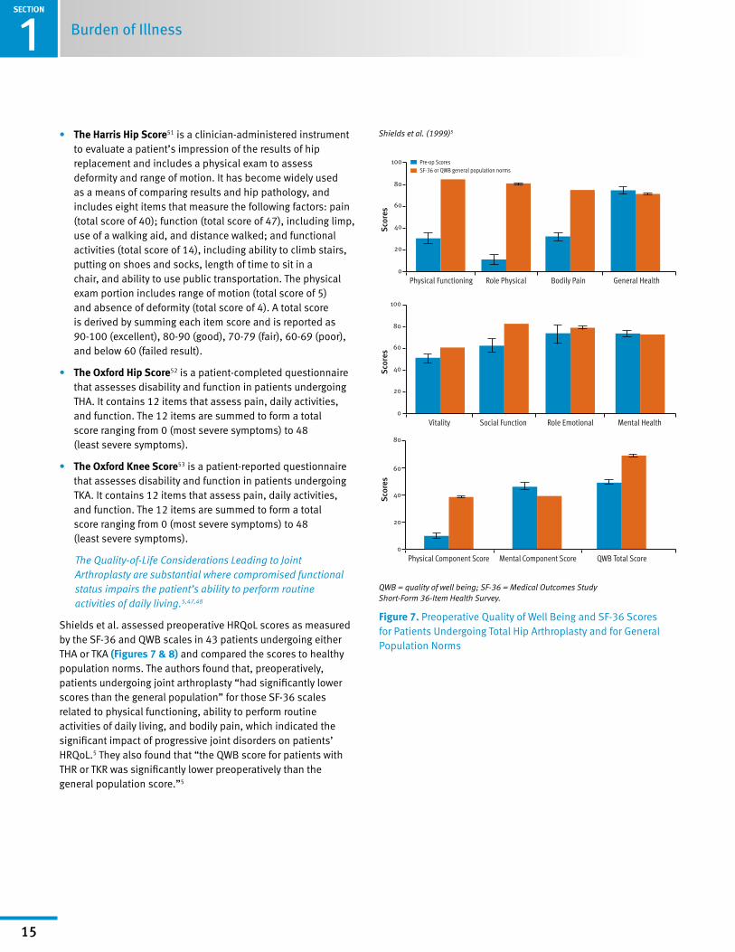
• The Harris Hip Score51 is a clinician-administered instrument to evaluate a patient’s impression of the results of hip replacement and includes a physical exam to assess deformity and range of motion. It has become widely used as a means of comparing results and hip pathology, and includes eight items that measure the following factors: pain (total score of 40); function (total score of 47), including limp, use of a walking aid, and distance walked; and functional activities (total score of 14), including ability to climb stairs, putting on shoes and socks, length of time to sit in a chair, and ability to use public transportation. The physical exam portion includes range of motion (total score of 5) and absence of deformity (total score of 4). A total score is derived by summing each item score and is reported as 90-100 (excellent), 80-90 (good), 70-79 (fair), 60-69 (poor), and below 60 (failed result).
• The Oxford Hip Score52 is a patient-completed questionnaire that assesses disability and function in patients undergoing THA. It contains 12 items that assess pain, daily activities, and function. The 12 items are summed to form a total score ranging from 0 (most severe symptoms) to 48 (least severe symptoms).
• The Oxford Knee Score53 is a patient-reported questionnaire that assesses disability and function in patients undergoing TKA. It contains 12 items that assess pain, daily activities, and function. The 12 items are summed to form a total score ranging from 0 (most severe symptoms) to 48 (least severe symptoms).
The Quality-of-Life Considerations Leading to Joint Arthroplasty are substantial where compromised functional status impairs the patient’s ability to perform routine activities of daily living.5,47,48
Shields et al. assessed preoperative HRQoL scores as measured by the SF-36 and QWB scales in 43 patients undergoing either THA or TKA (Figures 7 & 8) and compared the scores to healthy population norms. The authors found that, preoperatively, patients undergoing joint arthroplasty “had significantly lower scores than the general population” for those SF-36 scales related to physical functioning, ability to perform routine activities of daily living, and bodily pain, which indicated the significant impact of progressive joint disorders on patients’ HRQoL.5 They also found that “the QWB score for patients with THR or TKR was significantly lower preoperatively than the general population score.”5
100
80
60
40
20
0
Sco
res
Physical Functioning Role Physical Bodily Pain General Health
Pre-op ScoresSF-36 or QWB general population norms
100
80
60
40
20
0
Vitality Social Function Role Emotional Mental Health
Sco
res
80
60
40
20
0
Physical Component Score
Sco
res
Mental Component Score QWB Total Score
Figure 7 Preoperative Quality of Well Being and SF-36 Scores for Patients Undergoing Total Hip Arthroplasty and for General Population Norms
Shields et al. (1999)5
QWB = quality of well being; SF-36 = Medical Outcomes Study Short-Form 36-Item Health Survey.

SECTION
1
16
Establish the Tibial Platform
Figure 8 Preoperative Quality of Well Being and SF-36 Scores for Patients Undergoing Total Knee Arthroplasty and for General Population Norms
Shields et al. (1999)5
QWB = quality of well being; SF-36 = Medical Outcomes Study
Short-Form 36-Item Health Survey.
100
80
60
40
20
0
Sco
res
Physical Functioning Role Physical Bodily Pain General Health
Pre-op ScoresSF-36 or QWB general population norms
100
80
60
40
20
0
Vitality Social Function Role Emotional Mental Health
Sco
res
80
60
40
20
0
Physical Component Score
Sco
res
Mental Component Score QWB Total Score
HRQoL has also been intensively studied using disease-specific HRQoL instruments.
Chiu et al. evaluated the physical functioning and HRQoL of 46 patients undergoing THA using the Harris Hip Score and the SF-36. A total score of 70 points or below indicates poor physical functioning. As compared to a relatively low mean score of 45.2 (SD=17.3) among the patients in the study, the mean Harris score was 91.6 (SD=10.7) six months after operation, with an improvement rate of 103%.54 According to the report, “such difference in the total Harris score indicated a statistically significant improvement in overall physical functioning after THR.”54 Patients also experienced significant improvements in all critical subdomains of the Harris Hip Score, including pain (+137%), deformity (+32%), and functional measures such as gait (+112%) and activity (+69%), supporting the beneficial role of TJA in diminishing symptoms and restoring physical function.54
In a qualitative literature review, Ethgen et al. reported that THAs and TKAs “were found to be quite effective in terms of improvement in health-related quality-of-life dimensions, with the occasional exception of the social dimension.”48 They noted improvements in pain management, physical function, and the ability to perform activities of daily living, irrespective of age or gender.48 As would be expected, patients with complicating comorbidities or procedural complications typically report lesser quality-of-life improvements following surgery.

1SECTION
17
Burden of Illness
Patients with total joint arthroplasty complications such as infection have reduced quality of life and functioning compared with patients without complications.49
One of the most common periprocedural complications associated with joint arthroplasty is infection.43 Cahill et al. found that in a comparison of the outcomes of patients with or without infection after TJA, WOMAC scores of pain, stiffness, and function “were all significantly poorer in the complicated group,” and that “the overall health-related quality of life (AQoL utility) was significantly lower for the complicated group.”49 Furthermore, “six of the eight SF-36 scale dimensions were significantly poorer in the complicated group,” particularly with regard to physical function, ability to perform routine activities of daily living, and pain management (Table 1).49
Table 1 Comparison of Function, Utility, and Health-Related Quality of Life in Patients with Uncomplicated Total Joint Arthroplasty Compared with Patients with Complicated Total Joint Arthroplasty
Table 2 Comparison of Preoperative WOMAC Scores Between Primary and Revision Total Hip Arthroplasty Patients
Outcome
Total Joint Arthroplasties
P valueUncomplicated
Mean (SD)Complicated Mean (SD)
WOMAC
Function (0-68) 20 (14) 38 (14) < 0.0001
Pain (0-20) 6 (4) 9 (5) < 0.0001
Stiffness (0-8) 2 (2) 4 (2) < 0.0001
AQoL Utility 0.6 (0.3) 0.4 (0.3) < 0.0001
SF-36
Physical functioning
53 (28) 24 (21) < 0.0001
Role limitation (physical)
53 (44) 24 (35) < 0.0001
Bodily pain 61 (28) 39 (18) < 0.0001
General health 64 (22) 62 (24) 0.595
Vitality 60 (23) 47 (20) 0.006
Social functioning 79 (25) 63 (35) 0.012
Role limitation (emotional)
77 (36) 74 (39) 0.673
Mental health 78 (15) 67 (22) 0.003
AQoL = Assessment of Quality of Life; SD = standard deviation; SF-36 = Medical
Outcomes Study Short-Form 36-Item Health Survey; WOMAC = Western Ontario and
McMaster Universities Osteoarthritis Index.
Source: Patil et al. (2008)50
SD = standard deviation; THA = total hip arthroplasty; WOMAC = Western Ontario
and McMaster Universities Osteoarthritis Index.
Cahill et al. reported that functional and health-related quality-of-life outcomes after infection are devastating for the patient in a study that compared quality of life and functional outcomes in 62 uncomplicated TJR patients and 32 complicated patients. The authors found that, “infection had a great impact on physical functioning and ability to live independently and perform activities of daily living.”49
Patient function after revision total hip arthroplasty is lower than after primary total hip arthroplasty, indicating a potential unmet need for more successful revision surgical techniques and implants.50
Although both primary and revision joint arthroplasty significantly improve HRQoL for patients, Patil et al. found that physical function and quality of life are often comparatively poorer following revision surgery. In a study comparing 143 revision THA patients and 144 primary THA patients, the authors reported that “postoperative functional outcome was significantly better in patients with primary THA,” (Table 2) and that “the magnitude of improvement in quality of life is greater for the patient with primary THA.”50 In addition, quality-of-life gains took longer to realize following revision procedures.50
Primary THA (SD)
Revision THA (SD)
Difference (P)
Function 42.5 (18.8) 48.7 (22.3) –6.2 (0.0126)
Pain 41.9 (18.9) 52.8 (23.6) –10.9 (< 0001)
Stiffness 40.2 (20.9) 45.4 (24.0) –5.3 (0.0503)
Improving clinical outcomes and HRQoL following revision procedures remains a significant area of unmet need in TJA. Reduced HRQoL outcomes associated with revision procedures suggest that the use of implant materials that (a) reduce the likelihood of implant failure following the primary procedure and (b) preserve bone stock to ensure the greatest likelihood of revision success without complication are key considerations for improving patient quality of life following revision procedures.

SECTION
1
18
Burden of Illness
Figure 9 Projected Number of Joint Replacements in the United States from 2005 to 2015
Source: Kim et al. (2008)11
Note that shaded area of each line includes 95% prediction interval; circles and
squares denote observed data points.
The economic burden of joint replacement surgeries is significant; the estimated total annual hospital cost for primary hip and knee replacements in the US in 2004 was $9.1 billion.11
The economic burden of primary and revision joint replacements is projected to increase substantially over the next five years driven by an expanding number of procedures.3,11,55
According to a 2008 study by Kim, “the (economic) burden resulting from hip/knee joint replacement is not only substantial but also increasing at an alarmingly steep rate.”11 Kim reports that, based on Medicare data in the US, “the hospital costs and the actual transaction of reimbursements in the year 2004 were estimated to be $9.1 billion.”11 If current growth trends continue (Figure 9), Kim anticipates that the hospital costs associated with primary and revision hip and knee joint replacement procedures will exceed $80 billion by the year 2015.11
1 5 Economic Burden
• The economic burden of joint replacement surgeries is significant; the estimated total annual hospital cost for primary hip and knee replacements in the US in 2004 was $9.1 billion.11
• The economic burden of primary and revision joint replacements is projected to increase substantially over the next five years driven by an expanding number of procedures.3.11.55
• Revision total joint arthroplasty consumes more health care resources per procedure than primary joint arthroplasty.55
1,500,000
1,000,000
750,000
500,000
250,000
0
Proj
ecte
d nu
mbe
r of
join
t rep
lace
men
ts
Year
1,250,000
20131995 1997 1999 2001 2003 2005 2007 2009 2011 2015
Primary total knee replacementPrimary total hip replacement
The number of total hip and knee replacement procedures is projected to grow steadily by approximately 16% between 2010 and 2013.3 A 2007 report by Kurtz et al. projects that from 2005, the number of TKA and THA revision surgeries in the US is projected to double by 2015 and 2026 respectively, representing a significant economic burden on the US health system.55 Similar trends are also occurring in other industrialized nations, indicating that the cost of total joint replacement will likely make up an increasing share of global health care costs.
As revision TJAs grow substantially in the future, they will consume an increasing share of resources in orthopaedics. Bozic et al. report that in 2003, the mean total hospital cost in the US for revision THA was $31,341 compared with $24,170 for primary THA, reflecting a difference of almost 30%.46 Kim reports that the median US hospital charges in 2004 were approximately $5,000 higher for both revision THA and revision TKA compared with primary implant procedures,11 possibly reflecting the greater complexity and resource requirements associated with total joint revision procedures.

SECTION
19
Conventional Treatments2
2 Conventional Treatments
2 1 Description
Joint replacement is among the most common surgeries performed in the US and is most typically performed when a patient’s existing joint is compromised by arthritis. The joints most likely to be replaced include knees, hips, and shoulders.
Replacement joints are typically manufactured from biocompatible metals and high-density polyethylene components. Some metals are used in their pure form while others are used as part of an alloy. In orthopaedic implants, the most frequently used metals are titanium and cobalt-chromium alloys. The selection of an appropriate implant can vary based on the joint to be replaced, the patient’s specific needs, and the treating physician’s preference.
Implantation involves the surgical removal of the damaged or eroded joint surface, in whole or in part, and the insertion of an appropriate artificial joint surface. The new joint is typically either cemented in place, sometimes with screw augmentation, or press-fit to the remaining healthy supporting bone structure. When press-fit, no cement is used and future biologic in-growth into or onto the implant is sometimes required to achieve secure fixation of the implant.
2 2 Conventional Treatment Limitations
• Description of traditional total joint replacement materials
• Limitations of traditional materials
• Cement fixation in total joint surgery
Conventional orthopaedic implant materials, including bulk titanium and cobalt-chromium alloys, have mechanical properties that are different from the natural bone they are used to replace. Therefore, they are typically dependent on cement or mechanical fixation for their long-term stability.
• Corces and Garcia reported that all metallic alloys have a modulus of elasticity [stiffness] significantly higher than that of bone. They comment that “this mechanical incompatibility causes implants to be structurally stiffer than bones” and theorize that, “alloys with elastic moduli closer to bone may cause less stress shielding.”56
• Trabecular Metal Material has a coefficient of friction of 0.98 for net shape parts against cancellous bone.17 Zhang et al. report that “besides the mechanical characteristics of the graft itself, the interfacial friction between the graft surface and the opposing host bed is often crucial to successful initial load carriage and stability.”17 Bobyn et al. correlates this high friction with greater implant stability.57
Their findings were consistent with the observations of Dunbar, et al. who studied 28 patients who received Trabecular Metal Monoblock Tibias and 21 patients who received a cemented tibial implant. The authors found that despite extensive migration in the postoperative period, none of the 28 Trabecular Metal Monoblock Tibias were at risk of loosening after one year as opposed to the cemented group where four of the 21 tibial components were considered to be at risk.20
Traditional porous implant materials versus Trabecular Metal Technology
Differences in traditional joint replacement materials such as Titanium and Cobalt chrome versus Trabecular Metal Technology:
• Gruen, et al. observed the number and extent of radiolucencies postoperatively to most recent follow-up of 414 THA cases available for 2 year follow-up where Trabecular Metal and porous titanium acetabular cups were used. The authors found that “the radiographic outcomes for the prospective randomized series showed a significantly larger ( P <.0001) number of initial gaps that completely filled in for the porous tantalum (Trabecular Metal) monoblock cup (5 of 5, 100%), as compared with the low frequency of gap resolution observed for the porous-coated titanium cup (3 of 15, 20%). Also of significance (P < .0001) was the large number of new radiolucencies (16 of 40 THAs, 40%) that formed for the porous-coated titanium cup whereas a new radiolucency was found in only 1 of 43 cases using the porous tantalum (Trabecular Metal) monoblock cup.”18 The authors concluded that “our data indicate that direct bone apposition and an absence of new radiolucencies were significantly more probable for the porous tantalum monoblock cup as compared with the porous-coated titanium cup of the same outer geometry.”18
• Macheras, et al. report that a “benefit of this material [Trabecular Metal Material] is the more normal physiological transfer of load to host bone.”22 The authors reference a finite element analysis by Poggie, et al. that “demonstrated that the porous tantalum (Trabecular Metal) monoblock component loaded the acetabular bone similarly to a cemented all-polyethylene component, with load effectively transferred to the superomedial portion of the acetabulum as occurs physiologically.”22,58 The authors also indicated that, “finite element analysis of a titanium-backed acetabular component revealed stress shielding of the superomedial portion of the acetabulum. These findings were attributed to the bone matched elastic modulus of the porous tantalum (3 GPa), compared with titanium alloy (110 GPa).” 22,58

SECTION
20
Product Information 3
3 Product Information
3 1 Technology Description and Characteristics
Effective fixation of orthopaedic implants is based on: 1) Structure of the in-growth material (which determines the mechanical properties of the bone/implant interface); 2) Function of the in-growth material (how the structure performs under load); 3) Physiology of the host bone (the reaction of the host’s biology to the structure and function). Elemental tantalum, the core material used in Trabecular Metal Material, has been used to make implantable medical devices for more than 50 years.12 Bobyn et al. report that it is considered to be one of the most biocompatible elements available.12
Trabecular Metal Material is a porous, flexible, tantalum-based material used in implantable joint replacement and stability devices (Figure 10).
Figure 10 Trabecular Bone (A) and Trabecular Metal Material (B)Figure 12 Illustration of the “Bend Before Break” Characteristic of Trabecular Metal Material
(A)
(B)
Trabecular Metal Material is manufactured using a reticulated vitreous carbon network upon which a proprietary chemical vapor deposition process is used to layer elemental tantalum onto the underlying carbon structure at the atomic level (Figure 11). The strength of the material can be controlled by varying the thickness of the tantalum layers deposited during manufacture.12
The resulting Trabecular Metal Material maintains tantalum’s ductile characteristics while exhibiting a self-supporting, three-dimensional structure with high fatigue strength. In a study reported in 1999, Bobyn et al. found that Trabecular Metal Material “displays high ductility” and that the “struts deform plastically in compression, tension, and torsion without brittle failure” (Figure 12).12
Figure 11 Reticulated Vitreous Carbon Network Used in the Manufacture of Trabecular Metal Material

SECTION
21
Product Information3
3 2 Classification and Approval
Black reported that tantalum has been used in many implantable medical devices, including applications such as cranialmaxillofacial plates, ocular implants, and pacemakers.59
Trabecular Metal Implants, formerly branded as Hedrocel® Implants have been commercially available in the U.S. since 1997.
3 3 Device Components and Specifications
Trabecular Metal Monoblock implant designs are available for the acetabular cup, tibial knee component, and the patella. These Implants are constructed from a self-supporting, three-dimensional structure of Trabecular Metal Material into which a polyethylene liner is compression molded. This molding process integrates the two materials, resulting in a single unit that eliminates backside wear between the polyethylene and the metal and provides no pathway for debris to infiltrate the joint. Monoblock acetabular cups are available in a variety of diameters and in both screw-holed and non-holed designs (Figure 14). Trabecular Metal Tibial and Patellar components combine monoblock construction with the geometries of the Zimmer NexGen® Complete Knee Solution Cruciate Retaining and NexGen Legacy® Knee Posterior Stabilized articular surfaces (Figure 13).
Clinical results for Trabecular Metal Monoblock Hip implants cited in the reference section of this document: 19,22,23,58
Clinical results for Trabecular Metal Monoblock Knee implants cited in the reference section of this document: 18,20,28,58
Figure 14 Trabecular Metal Monoblock Acetabular Cups
Trabecular Metal Modular Implants provide options to meet unique patient needs and physician preferences. Modular acetabular cups are available in a variety of diameters and in non-holed, cluster-holed, and multi-holed designs where the polyethylene liner is snapped into the shell during surgery. Modular tibial components can accommodate cruciate retaining and posterior stabilized polyethylene liners for the Zimmer NexGen Legacy Knee System.
The Trabecular Metal Revision Cup is modular in that the doctor may choose which liner is appropriate for the shell (Figure 15). Unlike the other modular implants, the revision cup liner is cemented into the shell creating a construct that has load sharing characteristics that are similar to the monoblock component as bone cement has a low modulus of elasticity similar to cancellous bone and Trabecular Metal Material.
Clinical results for Trabecular Metal Revision Hip implants cited in the reference section of this document: 67,69,70,71,75
Figure 13 Trabecular Metal Monoblock Tibial Baseplates and Tibial Components
Figure 15 Trabecular Metal Revision Cup and Polyethylene Liner
SECTION
22
Product Information 3
Figure 16 Trabecular Metal Acetabular Augment
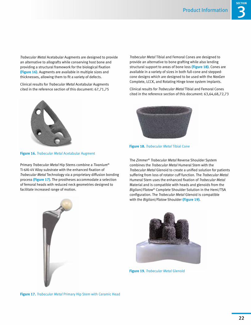
Trabecular Metal Acetabular Augments are designed to provide an alternative to allografts while conserving host bone and providing a structural framework for the biological fixation (Figure 16). Augments are available in multiple sizes and thicknesses, allowing them to fit a variety of defects.
Clinical results for Trabecular Metal Acetabular Augments cited in the reference section of this document: 67,71,75
Primary Trabecular Metal Hip Stems combine a Tivanium® Ti-6Al-4V Alloy substrate with the enhanced fixation of Trabecular Metal Technology via a proprietary diffusion bonding process (Figure 17). The prostheses accommodate a selection of femoral heads with reduced neck geometries designed to facilitate increased range of motion.
Trabecular Metal Tibial and Femoral Cones are designed to provide an alternative to bone grafting while also lending structural support to areas of bone loss (Figure 18). Cones are available in a variety of sizes in both full-cone and stepped-cone designs which are designed to be used with the NexGen Complete, LCCK, and Rotating Hinge knee system implants.
Clinical results for Trabecular Metal Tibial and Femoral Cones cited in the reference section of this document: 63,64,68,72,73
The Zimmer® Trabecular Metal Reverse Shoulder System combines the Trabecular Metal Humeral Stem with the Trabecular Metal Glenoid to create a unified solution for patients suffering from loss of rotator cuff function. The Trabecular Metal Humeral Stem uses the enhanced fixation of Trabecular Metal Material and is compatible with heads and glenoids from the Bigliani/Flatow® Complete Shoulder Solution in the Hemi/TSA configuration. The Trabecular Metal Glenoid is compatible with the Bigliani/Flatow Shoulder (Figure 19).
Figure 17 Trabecular Metal Primary Hip Stem with Ceramic Head
Figure 18 Trabecular Metal Tibial Cone
Figure 19 Trabecular Metal Glenoid

SECTION
23
Product Information3
Trabecular Metal ON Rods are designed for Stage I or II Osteonecrosis of the femoral head (Figure 20).24 Minimally invasive installation of the rod is an alternative to bone grafts. These implantable avascular necrosis rods are available in 10mm diameters and a variety of lengths to suit individual patient needs.
Clinical results for Trabecular Metal ON Rod implants cited in the reference section of this document: 24-27
3 4 Indications
Trabecular Metal Implants are indicated for joint reconstruction and replacement. Trabecular Metal Technology has been incorporated into the following implants.
For hips:
• Monoblock cups
• Modular cups
• Revision cups
• Augments
• Primary hip prosthesis
For knees:
• Monoblock tibias
• Modular tibias
• Patellas
• Tibial and femoral cones
• Revision augments
For shoulders:
• Reverse Shoulder System
• Humeral stems
• Glenoids
For Osteonecrosis intervention:
• Avascular necrosis rods
Figure 20 Trabecular Metal Osteonecrosis Intervention Implant

SECTION
24
3Product Information
Figure 21 Trabecular Metal Material Pore Structure
Figure 22 SEM Image of Tantalum Metal
Tantalum use in medical implants
Tantalum, the underlying material used in Trabecular Metal Material, has been used to make implantable medical devices for more than 50 years.11 A study by Bobyn et al. found that the metal was inert in the human body, was highly corrosion resistant, and was considered to be a highly biocompatible element.12,57
Trabecular Metal Material properties
According to Bobyn et al., the structure of Trabecular Metal Material “consists of a regular array of highly interconnected pores,” giving the material a “structure resembling trabecular bone.”12 Bobyn et al. also reported that in the material studied, “the volume porosity is 75-80%, 2-3 fold greater than with conventional porous materials.”12
Trabecular Metal pore structure
Trabecular Metal Technology uses a manufacturing process that is designed to provide a consistently shaped, interconnected, and appropriately sized pore structure to support vascularization and biologic in-growth (Figure 21). In a study of the porosity of three-dimensional biomaterial scaffolds, Karageorgiou and Kaplan found that pore sizes of at least 300µm “are recommended, due to enhanced new bone formation and the formation of capillaries.”13 Bobyn et al. reported that “the average two-dimensional porosity is 430 ± 270µm” for Trabecular Metal Material, and that “the average full pore diameter of the porous tantalum is 547 ± 52µm.”12 This vascular accommodation provides a framework that supports biologic in-growth.
The structural, functional, and physiological properties of Trabecular Metal Material
The porosity of Trabecular Metal Material and the crystallized structure of chemical vapor deposition tantalum are designed to support the stability of implants (Figure 22). According to laboratory tests conducted and reported by Zhang et al., “the friction coefficient of the porous tantalum material was very high in comparison to natural bone autografts or allografts and to conventional orthopaedic implant coating materials (sintered beads and wire mesh)” 17
3 5 Product Feature Comparison
• Tantalum in medical implants
• The structural, functional, and physiological properties of Trabecular Metal Material

SECTION
25
Clinical Value Evidence of Trabecular Metal Technology4
4 Clinical Value Evidence of Trabecular Metal Technology
This section provides an overview of the evidence that supports the potential value of Trabecular Metal Technology as reported in peer-reviewed, published clinical studies in primary and revision TJA.
4 1 Clinical Outcomes
Table 3 Radiolucencies by Zonal Extent at Last Follow-up for the Porous Tantalum Monoblock Acetabular Cup and the Porous-Coated Titanium Cup
• Trabecular Metal Material and implant fixation
• Trabecular Metal Monoblock acetabular implants
• Trabecular Metal Monoblock knee implants
• Trabecular Metal ON Rod prosthesis
• Trabecular Metal Monoblock implant history
Rapid and effective osseointegration into the porous structure of an implant is critical to the reduction of implant migration in cementless TJA procedures. Excessive migration can lead to loosening; therefore, solid initial fixation helps to ensure overall implant stability and survival. Aseptic loosening of a joint implant occurs when the implant mechanically detaches and/or the bone surrounding the joint begins to break down. This type of loosening is not caused by bacterial infection but results from inadequate fixation.
In a 2002 review of the clinical applications of Trabecular Metal Technology, Christie suggested that “porous tantalum approaches the ideal biomaterial: it is porous, strong, flexible, and biocompatible.”60 He also noted that “for use in components for total hip arthroplasty, porous tantalum provides a substitute for lost bone, immediate bone loading, bone in-growth, and soft-tissue attachment.”60 Trabecular Metal Material also demonstrates the potential to help enable initial structural stability and osseointegration by improving implant fixation and reducing the potential for mechanical loosening.19,20,61,62
4 1 1 Use in Hip ApplicationsGruen et al. conducted a randomized prospective study involving 414 hips replaced with the Trabecular Metal Monoblock Acetabular Cup available for a two-year follow-up to evaluate cup stability and the degree of osseointegration. Forty-three of these hips were compared with 40 hips receiving a porous-coated titanium acetabular component. The study found “a significantly larger number of porous-coated titanium cups with unresolved initial gaps and the appearance of new radiolucencies.” (Table 3).18
Source: Gruen et al. (2005)18
THA = total hip arthroplasty.
Porous-coated Titanium Cup
Trabecular Metal Acetabular Cup
# Gaps / # THA 21/40 (53%) 1/43 (2%)
Zone I 4 1
Zone II 4 0
Zone III 3 0
Zone I and II 0 0
Zone I and III 5 0
Zone II and III 1 0
Zone I-III (incomplete) 4 0
Zone I-III (complete) 0 0
Gruen et al. concluded that “the resolution of the majority postoperative gaps, the absence of continuous and progressive radiolucencies, the absence of osteolysis, and the maintenance of stability are attributed to the frictional stability and osteoconductivity inherent to the porous tantalum biomaterial and the monoblock design.”18 Their findings are significant as Ritter et al. have shown that insufficient postoperative gap closure and a greater number of radiolucencies have been correlated with implant failures due to sub optimal implant fixation and peri-operative complications.63

SECTION
26
4Clinical Value Evidence of Trabecular Metal Technology
Table 4 Number of Tibial Components at Risk for Early Aseptic Loosening and Components with Detectable Lift-off: Trabecular Metal Tibias Versus Cemented Stemmed Tibias
Trabecular Metal Monoblock implants
According to the National Osteoporosis Foundation, the relationship between bone mineral density (BMD) and bone health has been well established.64 Maintenance of appropriate BMD has been directly correlated to the risk for complicating adverse events, such as fracture and implant failure.
Source: Dunbar et al. (2009)20In another study by Macheras et al., 82 patients (86 hips) who received primary Trabecular Metal Monoblock Acetabular Cups in 1998 were followed for 7 to 7.5 years. Immediate postoperative radiographs showed a gap in 30 of the hips, five of which were less than 0.5mm and due to “incomplete reaming at the bottom of the acetabular bone bed.”22 According to the authors, “by 24 weeks all (30) gaps had closed radiologically.”22 Furthermore, at 24 months, the mean total component migration was 0.41mm and the mean Harris Hip Score improved from 52 pre-operatively to 94 in the group of 56 patients without a gap, and from 50 to 93 in the 30 patients with acetabular interface gaps.22 The study also reported that at the last follow-up there had been no dislocations or implant-related complications, all patients had regained their previous activities, no radiolucent lines and no areas of osteolysis were observed, and no revisions had been necessary.22
4 1 2 Use in Knee ApplicationsStudies have also indicated that the Trabecular Metal Monoblock Tibia has demonstrated a reduction of implant migration and loosening in primary total knee replacement.
Trabecular Metal Monoblock Tibia study results:
In a prospective study of 70 patients comparing fixation of an uncemented Trabecular Metal Monoblock Tibial Component (n = 37) with a conventional cemented stemmed tibial component (n = 33), Dunbar et al. found that the proportion of implants at risk for early aseptic loosening was 19% (4 of 21; 95% confidence interval [CI], 0.08 to 0.4) in the cemented group and 0% in the Trabecular Metal Implant group (0 of 28; 95% CI, 0.0 to 0.12) (Table 4).20 Although 12-month data were comparable, at 24 months, the cemented components group had significantly higher maximum total point motion (MTPM) scores compared with the Trabecular Metal Implant group. The authors tied this result to a greater potential for early loosening requiring corrective surgery.20 A subset of the Trabecular Metal Components migrated significantly in the postoperative period but all stabilized by one year, and the proportion considered by the authors to be at risk for early aseptic loosening was zero (95% CI in the group as a whole).20 Nineteen percent of cemented components included in this study were considered to be at risk for early aseptic loosening.20
Trabecular Metal Monoblock Tibial
Component
Cemented Stemmed Tibial
Component P Value
At risk for early loosening
0 of 28 patients 4 of 21 patients 0.028
Lift-off detected 0 of 28 patients 5 of 21 patients 0.01

SECTION
27
Clinical Value Evidence of Trabecular Metal Technology4
Evaluation of BMD can be an important factor in assessing bone health.65 In a 5-year longitudinal study, Berger et al. assessed the average rate of change in BMD as a function of age among Canadians aged 25 to 85 years. BMD of the lumbar spine, femoral neck, and total hip were measured in 4,433 women as a baseline with 3- and 5-year follow-up (Figure 23)65. The authors found that BMD in the femoral neck and total hip were “relatively stable among women younger than 45 not taking antiresorptive agents.”65 Furthermore, they found that “the mean bone loss at both sites also began at age 45 and reached a maximum rate of decline by age 50-54.”65
Source: Berger et al. (2008)65
A
Rate
of c
hang
e pe
r yea
r, g
/cm
2
0.014
0.006
0.002
-0.002
0.010
-0.006
-0.010
-0.01425-29 30-34 35-39 40-44 45-49 50-54 55-59 60-84 65-69 70-74 75-79 80-85
Femoral neck
Total hip
B
Rate
of c
hang
e pe
r yea
r, g
/cm
2
0.014
0.006
0.002
-0.002
0.010
-0.006
-0.010
-0.014
Age at baseline, year
25-29 30-34 35-39 40-44 45-49 50-54 55-59 60-84 65-69 70-74 75-79 80-85
Postmenopause
PremenopauseIn transition
The rate of decline increased again among women older than 70 years.65 The authors reported that bone loss “was greatest among women aged 50-54 who were transitioning from premenopause to postmenopause, with a change from baseline of –6.8% (95% CI, –7.5% to –4.9%) over 5 years.”65 These findings suggest that considering BMD when selecting an implant for certain at-risk patients may be an indicator of potential bone quality.
Figure 23 Rate of Change of Bone Density as a Function of Age Among Women Not Using Antiresorptive Therapy (A) and by Menopausal Status (B)

SECTION
28
4Clinical Value Evidence of Trabecular Metal Technology
In a comparative study of nine Trabecular Metal Monoblock Acetabular Implants and eight conventional solid titanium implants, Meneghini et al. used CT scans to measure the BMD of the peri-acetabular bone around the implant and the equivalent acetabular regions in each patient’s native contralateral hip (Table 5) (Figure 24)23. According to the authors, the results “support the development of decreased BMD around solid-metal osseointegrated cementless acetabular components.”23 At a mean follow-up of 7.7 years (range, 6.1-8.8 years), “5 of the 10 acetabular levels in the porous tantalum group demonstrated an increase in the mean BMD difference over the control hip (range, 10.8-38.4 mg/mL).”23 Furthermore, “8 of 10 acetabular levels in the porous tantalum group demonstrated an increase in the mean percent change in BMD over the control hip and ranged from an increase of 5.3% to 41.0% in BMD,” while the mean percentage change in BMD “decreased in 8 of 10 acetabular levels around the titanium implants.”23 The authors conclude that “data demonstrates stress-shielding continues beyond the short-term around standard solid titanium cementless acetabular components. The data also supports that stress-shielding likely occurs less around a highly porous monoblock metal implant with an elastic modulus similar to bone.”23
Table 5 Absolute Mean Difference in Bone Mineral Density in Total Hip Arthroplasty: Porous Tantalum Monoblock Acetabular Implants (n=8) Compared with Solid Titanium Implants (n=9)
Figure 24 Mean Absolute (A) and Percentage (B) Change in Bone Mineral Density Around the Trabecular Metal Acetabular components (n=9) and Titanium Acetabular Components (n=8) in the Posterosuperior Region
Source: Meneghini et al. (2009)23
BMD = bone mineral density.
BMD Difference (mg/mL) Percentage Change in BMD
Porous Tantalum Titanium P Value
Porous Tantalum Titanium P Value
Posterosuperior
Zone 1 −0.3 −21.2 0.60 6.1 −12.2 0.22
Zone 2 −14.1 −43.6 0.37 −8.8 −44.7 0.12
Zone 3 −3.7 −42.9 0.02 –1.5 −31.1 0.014
Zone 4 10.8 −45.5 0.006 5.3 –31.2 0.017
Zone 5 38.4 −28.6 0.20 26.1 –11.0 0.38
Anterosuperior
Zone 1 16.0 −19.2 0.38 31.7 6.5 0.41
Zone 2 −9.5 −67.4 0.13 12.2 −27.3 0.12
Zone 3 −11.9 −62.9 0.16 28.3 −37.4 0.10
Zone 4 12.9 −27.2 0.19 41.0 −19.0 0.18
Zone 5 29.9 9.0 0.45 30.1 10.0 0.47
Periacetabular levels: posterosuperior
0
-60
-80
-100
Periacetabular levels: posterosuperior
Porous tantalumTitanium
B
Level 1 Level 2 Level 4Level 3 Level 5
-20
-40
100
40
20
80
60
0
-50
-100
-150
Chan
ge in
BM
D (m
g/cc
)
50
100
150
Porous tantalumTitanium
A
Level 1 Level 2 Level 4Level 3 Level 5
Chan
ge in
BM
D (%
)
Source: Meneghini et al. (2009)23
BMD = bone mineral density.
Note: Level I is closest to, and Level 5 farthest from, the implant.
Levels 3 and 4 demonstrate statistically significant improvement
in BMD (< 0.02) in porous tantalum THAs compared with solid
titanium THAs.
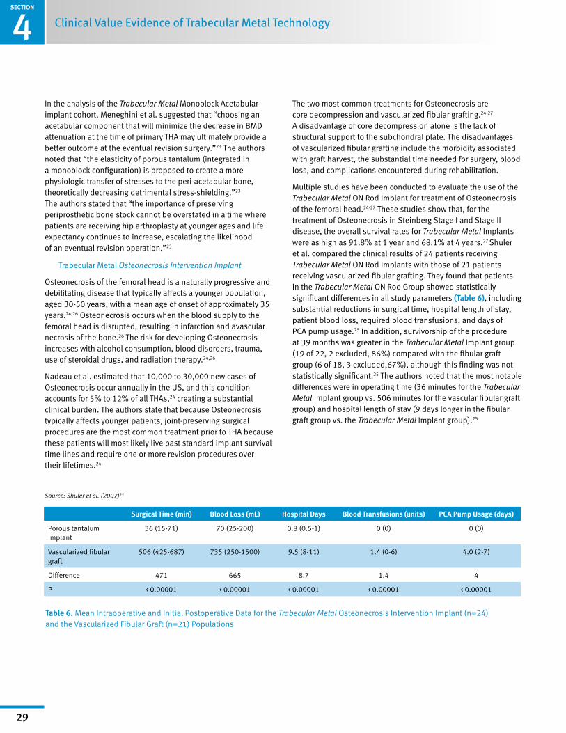
SECTION
29
4 Clinical Value Evidence of Trabecular Metal Technology
In the analysis of the Trabecular Metal Monoblock Acetabular implant cohort, Meneghini et al. suggested that “choosing an acetabular component that will minimize the decrease in BMD attenuation at the time of primary THA may ultimately provide a better outcome at the eventual revision surgery.”23 The authors noted that “the elasticity of porous tantalum (integrated in a monoblock configuration) is proposed to create a more physiologic transfer of stresses to the peri-acetabular bone, theoretically decreasing detrimental stress-shielding.”23 The authors stated that “the importance of preserving periprosthetic bone stock cannot be overstated in a time where patients are receiving hip arthroplasty at younger ages and life expectancy continues to increase, escalating the likelihood of an eventual revision operation.”23
Trabecular Metal Osteonecrosis Intervention Implant
Osteonecrosis of the femoral head is a naturally progressive and debilitating disease that typically affects a younger population, aged 30-50 years, with a mean age of onset of approximately 35 years.24,26 Osteonecrosis occurs when the blood supply to the femoral head is disrupted, resulting in infarction and avascular necrosis of the bone.26 The risk for developing Osteonecrosis increases with alcohol consumption, blood disorders, trauma, use of steroidal drugs, and radiation therapy.24,26
Nadeau et al. estimated that 10,000 to 30,000 new cases of Osteonecrosis occur annually in the US, and this condition accounts for 5% to 12% of all THAs,24 creating a substantial clinical burden. The authors state that because Osteonecrosis typically affects younger patients, joint-preserving surgical procedures are the most common treatment prior to THA because these patients will most likely live past standard implant survival time lines and require one or more revision procedures over their lifetimes.24
The two most common treatments for Osteonecrosis are core decompression and vascularized fibular grafting.24-27
A disadvantage of core decompression alone is the lack of structural support to the subchondral plate. The disadvantages of vascularized fibular grafting include the morbidity associated with graft harvest, the substantial time needed for surgery, blood loss, and complications encountered during rehabilitation.
Multiple studies have been conducted to evaluate the use of the Trabecular Metal ON Rod Implant for treatment of Osteonecrosis of the femoral head.24-27 These studies show that, for the treatment of Osteonecrosis in Steinberg Stage I and Stage II disease, the overall survival rates for Trabecular Metal Implants were as high as 91.8% at 1 year and 68.1% at 4 years.27 Shuler et al. compared the clinical results of 24 patients receiving Trabecular Metal ON Rod Implants with those of 21 patients receiving vascularized fibular grafting. They found that patients in the Trabecular Metal ON Rod Group showed statistically significant differences in all study parameters (Table 6), including substantial reductions in surgical time, hospital length of stay, patient blood loss, required blood transfusions, and days of PCA pump usage.25 In addition, survivorship of the procedure at 39 months was greater in the Trabecular Metal Implant group (19 of 22, 2 excluded, 86%) compared with the fibular graft group (6 of 18, 3 excluded,67%), although this finding was not statistically significant.25 The authors noted that the most notable differences were in operating time (36 minutes for the Trabecular Metal Implant group vs. 506 minutes for the vascular fibular graft group) and hospital length of stay (9 days longer in the fibular graft group vs. the Trabecular Metal Implant group).25
Table 6 Mean Intraoperative and Initial Postoperative Data for the Trabecular Metal Osteonecrosis Intervention Implant (n=24) and the Vascularized Fibular Graft (n=21) Populations
Source: Shuler et al. (2007)25
Surgical Time (min) Blood Loss (mL) Hospital Days Blood Transfusions (units) PCA Pump Usage (days)
Porous tantalum implant
36 (15-71) 70 (25-200) 0.8 (0.5-1) 0 (0) 0 (0)
Vascularized fibular graft
506 (425-687) 735 (250-1500) 9.5 (8-11) 1.4 (0-6) 4.0 (2-7)
Difference 471 665 8.7 1.4 4
P < 0.00001 < 0.00001 < 0.00001 < 0.00001 < 0.00001

SECTION
30
4Clinical Value Evidence of Trabecular Metal Technology
Vascularized fibular grafting is a difficult procedure but, according to Shuler et al., high success rates ‘’are obtainable by a select few with extensive experience.”25 Shuler et al. state that in regards to the use of the Trabecular Metal ON Rod, “Our data support the idea that the implant procedure is less invasive and far more accessible to the general orthopedic community with a smaller learning curve. There is no need for microsurgical expertise or harvesting with the implant either, which eliminates harvest morbidity and complications.”25 They also reported that “preliminary data show the tantalum implant is at least equivalent to the vascularized fibular graft in terms of survival and outcomes.”25
Trabecular Metal Monoblock implants in revision total hip and knee arthroplasty
Revision total hip and knee arthroplasty can be challenging procedures, mainly due to acetabular and tibial or patellar bone stock deficiency.11,61,66,67 A number of studies have shown favorable results using Trabecular Metal Revision Shell that features a cemented polyethylene liner.
Lewallen et al. reported no evidence of migration or loosening along with significant clinical improvements in a cohort of 114 consecutive acetabular revisions using the Trabecular Metal Revision Shell with a cemented polyethylene liner.68 Unger et al. studied 60 consecutive patients that underwent revision THA using the Trabecular Metal Revision Cup with a cemented polyethylene liner. The authors reported that “Independent radiographic assessment of these cases showed excellent gap and defect filling with host bone and no evidence of radiographic deterioration.”69 Weeden et al. in a review of 43 acetabular revisions treated with tantalum acetabular (Trabecular Metal Revision) implants reported that, “based on our short-term results with (Trabecular Metal) acetabular cups and the results of others, we believe tantalum components and augments provide a viable treatment option in the reconstruction of Paprosky type 3 defects.”70
For revision TKA, Meneghini et al. reviewed 15 revision knee cases where porous tantalum (Trabecular Metal) metaphyseal cones were implanted.61 The authors reported in 2008 that, “reconstruction of large osseous defects in the femur and tibia during revision total knee replacement remains a challenging clinical problem.”61 Radnay and Scuderi reported results from 10 cases where Trabecular Metal Tibial cones were used to fill cavitary defects. These authors concluded that, “For the complex reconstruction with severe bone loss, Trabecular Metal augments with stem fixation provide an alternative technique with early clinical and radiographic success.”71 From 1998-2002, Stulberg reported on 43 revision TKA procedures that were performed that required femoral and/or tibial augments. In these cases, Trabecular Metal Augments were used. He concluded that, “Porous augments allow biologic fixation of revision TKR components placed in the presence of segmental bone loss. This fixation may be particularly useful for younger patients requiring revision and for patients with bone stock which is inappropriate for cement fixation.”72
The longer-term use of Trabecular Metal Monoblock Acetabular Implants in THA has been studied by the Macheras et al. prospective study of 151 hip implants with a patient follow-up period of 8 to 10 years.19 In the study, initial postoperative peri-acetabular dome gaps were reported in approximately 16% of hips (n = 25), although progressive gap filling was observed in all 25 hips by 24 weeks.19 Furthermore, revision due to mechanical failure occurred in less than 1% of patients at 10 years.19 The authors reported no radiographic evidence of gross polyethylene wear, backside wear, progressive radiolucencies, osteolytic lesions, acetabular fracture, or acetabular cup migration.19 Functional and range-of-motion outcomes at 1 year and 10 years were also favorable (See Section 4.2).19
At the 10-year follow-up point in the data from Macheras et al., there was no radiographic evidence of gross polyethylene wear, backside wear, progressive radiolucencies, osteolytic lesions, acetabular fracture, or cup migration.19 Additionally, radiographic evaluation showed increased bone density and remodeling and thickening of trabecular bone around the Trabecular Metal Actabular Implant at final follow-up. Functional and range-of-motion outcomes at one year and 10 years were also favorable (See Section 4.2).19

SECTION
31
4 Clinical Value Evidence of Trabecular Metal Technology
Longer term use of Trabecular Metal Technology has also been studied and reported for knee implants. In a prospective UK study, Helm et al. evaluated the survival of a Trabecular Metal Monoblock Tibial Component for TKA in 103 unselected patients. Of the 103 patients (105 knees), 0% were revised due to mechanical failure, and no radiolucent lines were detected on postoperative radiographs.28 The authors stated that, “in this series Trabecular Metal Monoblock Tibial Components have shown no evidence of loosening at a minimum of 3 years' follow-up and the prosthesis as a whole has performed very well clinically.”28
Helm et al. found that the projected cumulative survival of the Trabecular Metal Tibial Component was approximately 98% at five years (Figure 25). Functional and HRQoL outcomes were also reported (See Section 4.2).28
4 1 3 Removal of Trabecular Metal Components
Although the physical and mechanical properties of Trabecular Metal Material promote superior biologic fixation and implant stability, Trabecular Metal Implants can be removed during revision surgery. Preservation of bone stock is important in implant removal.
The Zimmer Trabecular Metal Explant Acetabular Cup Removal System has been designed to facilitate the removal of Trabecular Metal Acetabular Cups while providing a high level of bone preservation, even in the case of a well-fixed component. A study by Mitchell et al. evaluating the use of the Explant System in the removal of well-fixed Trabecular Metal Cups in 31 consecutive patients found that all the cups were removed “rapidly and without complication”, and that the removal time “did not exceed five minutes” for any patient.29 The indications for acetabular revision in these patients included infection, polyethylene wear, locking mechanism failure, and malposition. The mean interval between primary and revision surgery was 6.7 years. The removal of some host bone was still required to implement this procedure; however, the median difference between the size of the component removed and the size of the final reamer used was 4mm, indicating that no more host bone was removed than the thickness of the Explant Blades.29
Figure 25 Cumulative Survival of the Trabecular Metal Tibial Component
Source: Helm et al. (2009)28
1.0
0.5
0.4
0.3Cum
ulat
ive
surv
ival
Years
0.6
0.9
4 5
0.8
0.7
0.2
0.1
0.01 2 3

SECTION
32
4Clinical Value Evidence of Trabecular Metal Technology
Table 7 Preoperative, 1-Year, and Latest Follow-up (8 to 10 years) of Harris Hip Score, Oxford Hip Score, and Range of Motion Using the Porous Tantalum Monoblock (Trabecular Metal) Acetabular Component
Table 8 Preoperative and Postoperative Knee and SF-12 Scores in Patients Undergoing Total Knee Arthroplasty with a Trabecular Metal Monoblock Tibial Component
Source: Macheras et al. (2009)19
Helm et al. (2009)28
IKS = International Knee Society; IQR = interquartile range; PCS = Physical Component Summary; SD = standard deviation; SF-12 = Medical Outcomes Study
Short-Form 12-Item Health Survey
HHS = Harris Hip Score; OHS = Oxford Hip Score; ROM = range of motion;
SD = standard deviation.a Statistically significant change (P < 0.05) when compared with preoperative scores.b Data not given.
4 2 Functional and Quality-of-Life Studies
In a systematic review, Ethgen et al. reported that in all studies, THAs and TKAs “were found to be quite effective in terms of improvement in health-related quality-of-life dimensions, with the occasional exception of the social dimension.”48 These improvements included scores for physical health, such as those for pain and physical function.48 Improvements in mental and social health were less obvious, with results suggesting either modest improvement or no changes at all.48
The greatest improvement in physical health domains typically occurred within the first three to six months after surgery, and studies that reported long-term follow-up showed long-lasting improvements.48 The following studies present results that are specific to Trabecular Metal Technology.
In the long-term study by Macheras et al. (See Section 4.1), functional outcomes after THA with Trabecular Metal Monoblock Acetabular Implants, as measured from preoperative to one-year postoperative by the Harris Hip Score and the Oxford Hip Score, significantly improved and remained constant at the latest follow-up.19 Similarly, preoperative range of motion for flexion and abduction had significantly improved at the latest follow-up (Table 7).
In the Helm et al. study (See Section 4.1), 89 patients undergoing TKA with Trabecular Metal Monoblock Tibial Components had a minimum follow-up of three years (mean, 44 months). Patient functional status and HRQoL measured by the Oxford Knee Score and the SF-12 scores from the patient population are shown below (Table 8).28 Preoperative International Knee Society scores were not presented for this sample; however, International Knee Society scores were normal at follow-up.
Preoperative ± SD
1-year ± SD
Latest Follow-Up ± SD
HHS (total) 44.0 ± 13.8a 95.2 ± 4.8a 97.0 ± 6.2a
HHS (pain) 14.0 ± 6.7a 42.4 ± 2.0a 43.6 ± 1.6a
OHS 43.3 ± 6.5 a 15.2 ± 2.3a 13.9 ± 2.3a
ROM (flexion)
72.6° ± 21.3°a b 106.9° ± 11.1°a
ROM (abduction)
15.0° ± 3.0°a b 44° ± 2.5°a
Oxford Knee Score (SD) n = 89
SF-12 PCS (SD) n = 89
IKS Clinical Score (SD) n = 79
Pain Component of IKS Clinical Score (SD) n = 10
IKS Functional Score (SD) n = 89
Preoperative median (IQR)
46 (40-49) 25.93 (22.80-30.61)
-- -- --
Follow-up median (IQR)
20 (16-29) 39.42 (30.23-49.00)
94 (90-96) 45 (45-50) 90 (73-100)
P value < 0.0001 < 0.0001 -- -- --

SECTION
33
4 Clinical Value Evidence of Trabecular Metal Technology
In a study reported in 2008 by Siegmeth et al., modular Trabecular Metal Augments and Trabecular Metal Revision Shells with a cemented polyethylene liner were used for the reconstruction of acetabular defects in revision hip arthroplasty. Thirty-four consecutive patients were prospectively followed for a minimum of 24 months (mean, 34 months; range, 24-55 months). Functionality and HRQoL were measured with the SF-12, WOMAC, and Oxford Hip Score, and 26 patients had baseline and follow-up scores for the three instruments. The study showed that “all quality-of-life parameters improved,” and functionality was considered “very good or excellent” (Table 9).73 According to the authors, patients reported “a high degree of satisfaction” with their surgeries.73 Of the 34 patients in the study, 32 “required no further surgery for aseptic loosening, while two had revision.”73
The study by Shuler et al. compared results of the Trabecular Metal ON Rod in 24 patients with a historical group of 21 patients receiving vascularized fibular grafting. The Trabecular Metal Implant group had a greater number of postoperative scores characterized as good or excellent (86%, n = 19) compared with the fibular graft group, which had only three (17%) good or excellent outcomes at 39-month follow-up.25 According to the authors, “Statistical analysis of operative data revealed significant differences in favor of the implant (Trabecular Metal ON Rod) population. The operative time, blood loss, PCA usage, and hospitalization were significantly less for the implant group when compared to fibular grafting (Table 10).”25
Table 9 Functional and Health-related Quality of Life Outcomes Receiving Trabecular Metal Augments and Revision Shells in Revision Hip Arthroplasty (n=37)
Table 10 Postoperative Harris Hip Scores for Porous Tantalum Implants (n=24) and Vascularized Fibular Grafts (n=21) in the Treatment of Osteonecrosis
Source: Siegmeth et al. (2009)73
Source: Shuler et al. (2007)25
MCS = Mental Component Summary; NA = not applicable; PCS = Physical
Component Summary; SD = standard deviation; SF-12 = Medical Outcomes Study
Short-Form 12-Item Health Survey; WOMAC = Western Ontario and McMaster
Universities Osteoarthritis Index
Instrument Baseline ± SD Follow-up (2 years) ± SD
WOMAC Function 39.8 ± 20.8 78.3 ± 18.8
WOMAC Stiffness 45.0 ± 17.7 82.6 ± 17.4
WOMAC Pain 42.8 ± 15.9 89.9 ± 15.2
WOMAC Global 41.0 ± 17.8 81.3 ± 15.8
Oxford Hip Score 34.6 ± 18.0 80.3 ± 16.6
SF-12 PCS 30.4 ± 9.5 44.1 ± 8.5
SF-12 MCS 45.0 ± 12.1 53.9 ± 9.8
Satisfaction Scores
Pain NA 95.4 ± 19.4
Function NA 88.5 ± 18.4
Recreational NA 85.6 ± 20.9
Overall NA 95.6 ± 11.5
Total Score NA 92.0 ± 13.3
Outcomes Average Follow-up Failed Poor Fair Good Excellent
Porous tantalum implant 39 (27-59) 3 (14%) 0 (0%) 0 (0%) 3 (14%) 16 (72%)
Vascularized fibular graft 149 (121-180) 7 (39%) 7 (39%) 1 (6%) 2 (11%) 1 (6%)

SECTION
34
4Clinical Value Evidence of Trabecular Metal Technology
Trabecular Metal Technology applications have been the subject of numerous clinical studies since the material was introduced to the orthopaedic market in 1997. This document outlines the findings of these studies which show the clinical benefits that have been seen with Trabecular Metal Material products. Trabecular Metal Material has been engineered to have high porosity, a high frictional coefficient, and a low modulus of elasticity characteristics of this material for each of its applications that have been described.
Doctors, hospital administrators, and payers must work together to make decisions regarding which implant to use for a patient based on a host of factors specific to the patient and the institution. As demographics continue to drive patient demand for orthopaedic procedures, making informed purchasing decisions can become even more important to the success of the institution from both clinical and economic standpoints.
This dossier provides information about independent clinical studies which should aid in making more informed decisions when it comes to choosing implants that utilize Trabecular Metal Technology.
4 3 Clinical Value Summary for Trabecular Metal Technology
• The performance of Trabecular Metal Technology applications have been well documented in the literature.

SECTION
35
References5
5 References
1. Arthritis and related conditions. 2009. Available at: http://www.aaos.org/news/aaosnow/mar09/research6.asp. Accessed August 20, 2009.
2. Kurtz S, Mowat F, Ong K, Chan N, Lau E, Halpern M. Prevalence of primary and revision total hip and knee arthroplasty in the United States from 1990 through 2002. J Bone Joint Surg Am 2005 Jul;87(7):1487-97.
3. Millennium Research Group. US markets for large-joint reconstructive implants. 2009.
4. Kurtz SM, Lau E, Ong K, Zhao K, Kelly M, Bozic KJ. Future young patient demand for primary and revision joint replacement: national projections from 2010 to 2030. Clin Orthop Relat Res 2009 Oct;467(10):2606-12.
5. Shields RK, Enloe LJ, Leo KC. Health related quality of life in patients with total hip or knee replacement. Arch Phys Med Rehabil 1999 May;80(5):572-9.
6. Espinoza-Ervin C, Zhang X, Huo MH. Can we afford revision total hip replacement? Current Orthopaedic Practice 2009;20(1):29-33 10.1097/BCO.0b013e31819583ab.
7. Bozic KJ, Kurtz, SM, Lau E, Ong K, Vail TP, Berry, DJ. The epidemiology of revision total hip arthroplasty in the united states. J Bone Joint Surg Am 2009 Jan;91(1);128-133.
8. Ducheyne P, Kagan A, Lacey JA. Failure of total knee arthroplasty due to loosening and deformation of the tibial component. J Bone Joint Surg Am 1978 Apr;60;384-391.
9. Lavernia C, Lee DJ, Hernandez VH. The increasing financial burden of knee revision surgery in the United States. Clin Orthop Relat Res 2006 May;446:221-6.
10. Bozic KJ, Ries MD. The impact of infection after total hip arthroplasty on hospital and surgeon resource utilization. J Bone Joint Surg Am 2005 Aug;87(8):1746-51.
11. Kim S. Changes in surgical loads and economic burden of hip and knee replacements in the US: 1997-2004. Arthritis Rheum 2008 Apr 15;59(4):481-8.
12. Bobyn JD, Hacking SA, Chan SP, Toh KK, Krygier JJ, Tanzer M. Characterization of a new porous tantalum biomaterial for reconstructive orthopaedics. Annual Meeting of the American Academy of Orthopaedic Surgeons edition. Anaheim, CA; 1999.
13. Karageorgiou V, Kaplan D. Porosity of 3D biomaterial scaffolds and osteogenesis. Biomaterials 2005 Sep;26(27):5474-91.
14. Fitzpatrick D, Ahn P, Brown T, et al. Friction coefficients or pourous tantalum and cancelours and cortical bone. Presented at the Ann Amer Soc Biomechanics, Clemson, SC. 1997.
15. Levine B, Della Valle CJ, Jacobs JJ. Applications of porous tantalum in total hip arthroplasty. J Am Acad Orthop Surg 2006 Nov;14(12):646-55.
16. Klika AK, Murray TG, Darwiche H, Barsoum WK. Options for acetabular fixation surfaces. J Long Term Eff Med Implants 2007;17(3):187-92.
17. Zhang Y, Ahn PB, Fitzpatrick DC, Heiner A, Poggie RA, Brown TD. Interfacial frictional behavior: cancellous bone, cortical bone, and a novel porous tantalum biomaterial. J Musculoskeletal Res 1999;3(4):245-51.
18. Gruen TA, Poggie RA, Lewallen DG, Hanssen AD, Lewis RJ, O'Keefe TJ, et al. Radiographic evaluation of a monoblock acetabular component: a multicenter study with 2- to 5-year results. J Arthroplasty 2005 Apr;20(3):369-78.
19. Macheras G, Kateros K, Kostakos A, Koutsostathis S, Danomaras D, Papagelopoulos PJ. Eight- to ten-year clinical and radiographic outcome of a porous tantalum monoblock acetabular component. J Arthroplasty 2009 Aug;24(5):705-9.
20. Dunbar MJ, Wilson DA, Hennigar AW, Amirault JD, Gross M, Reardon GP. Fixation of a Trabecular Metal knee arthroplasty component. A prospective randomized study. J Bone Joint Surg Am 2009 Jul;91(7):1578-86.

SECTION
36
References 5
21. Levine B, Sporer S, Della Valle CJ, Jacobs JJ, Paprosky W. Porous tantalum in reconstructive surgery of the knee. J Knee Surg 2007;20:185-94.22. Macheras GA, Papagelopoulos PJ, Kateros K, Kostakos AT, Baltas D, Karachalios TS. Radiological evaluation of the metal-bone interface of a porous tantalum monoblock acetabular component J Bone Joint Surg 2006 Mar;88B(3):304-9.
23. Meneghini RM, Ford KS, McCollough CH, Hanssen AD, Lewallen DG. Bone remodeling around porous metal cementless acetabular components. J Arthroplasty 2009 May 25.
24. Nadeau M, Seguin C, Theodoropoulos JS, Harvey EJ. Short term clinical outcome of a porous tantalum implant for the treatment of advanced osteonecrosis of the femoral head. Mcgill J Med 2007 Jan;10(1):4-10.
25. Shuler MS, Rooks MD, Roberson JR. Porous tantalum implant in early osteonecrosis of the hip: preliminary report on operative, survival, and outcomes results. J Arthroplasty 2007 Jan;22(1):26-31.
26. Tsao AK, Roberson JR, Christie MJ, Dore DD, Heck DA, Robertson DD, et al. Biomechanical and clinical evaluations of a porous tantalum implant for the treatment of early-stage osteonecrosis. J Bone Joint Surg Am 2005;87 Suppl 2:22-7.
27. Veillette CJ, Mehdian H, Schemitsch EH, McKee MD. Survivorship analysis and radiographic outcome following tantalum rod insertion for osteonecrosis of the femoral head. J Bone Joint Surg Am 2006 Nov;88 Suppl 3:48-55.
28. Helm AT, Kerin C, Ghalayini SR, McLauchlan GJ. Preliminary results of an uncemented Trabecular Metal tibial component in total knee arthroplasty. J Arthroplasty 2009 Sep;24(6):941-4.
29. Mitchell PA, Masri BA, Garbuz DS, Greidanus NV, Wilson D, Duncan CP. Removal of well-fixed, cementless, acetabular components in revision hip arthroplasty. J Bone Joint Surg Br 2003 Sep;85(7):949-52.
30. Klein GR, Levine HB, Hartzband MA. Removal of a well-fixed Trabecular Metal monoblock tibial component. J Arthroplasty 2008 Jun;23(4):619-22.
31. Sibanda N, Copley LP, Lewsey JD, Borroff M, Gregg P, MacGregor AJ, et al. Revision rates after primary hip and knee replacement in England between 2003 and 2006. PLoS Med 2008 Sep 30;5(9):e179.
32. Felson DT, Zhang Y. An update on the epidemiology of knee and hip osteoarthritis with a view to prevention. Arthritis Rheum 1998 Aug;41(8):1343-55.
33. Lawrence RC, Helmick CG, Arnett FC, Deyo RA, Felson DT, Giannini EH, et al. Estimates of the prevalence of arthritis and selected musculoskeletal disorders in the United States. Arthritis Rheum 1998 May;41(5):778-99.
34. Jain NB, Higgins LD, Guller U, Pietrobon R, Katz JN. Trends in the epidemiology of total shoulder arthroplasty in the United States from 1990-2000. Arthritis Rheum 2006 Aug 15;55(4):591-7.
35. Millennium Research Group. European markets for large joint reconstructive implants. 2009.
36. Jain NB, Higgins LD, Ozumba D, Guller U, Cronin M, Pietrobon R, et al. Trends in epidemiology of knee arthroplasty in the United States, 1990-2000. Arthritis Rheum 2005 Dec;52(12):3928-33.
37. Birrell F, Johnell O, Silman A. Projecting the need for hip replacement over the next three decades: influence of changing demography and threshold for surgery. Ann Rheum Dis 1999 Sep;58(9):569-752.
38. Wells VM, Hearn TC, McCaul KA, Anderton SM, Wigg AE, Graves SE. Changing incidence of primary total hip arthroplasty and total knee arthroplasty for primary osteoarthritis. J Arthroplasty 2002 Apr;17(3):267-73.
39. Lohmander LS, Engesaeter LB, Herberts P, Ingvarsson T, Lucht U, Puolakka TJ. Standardized incidence rates of total hip replacement for primary hip osteoarthritis in the 5 Nordic countries: similarities and differences. Acta Orthop 2006 Oct;77(5):733-40.
40. Ostendorf M, Johnell O, Malchau H, Dhert WJ, Schrijvers AJ, Verbout AJ. The epidemiology of total hip replacement in The Netherlands and Sweden: present status and future needs. Acta Orthop Scand 2002 Jun;73(3):282-6.

SECTION
37
References5
41. Khatod M, Inacio M, Paxton EW, Bini SA, Namba RS, Burchette RJ, et al. Knee replacement: epidemiology, outcomes, and trends in Southern California: 17,080 replacements from 1995 through 2004. Acta Orthop 2008 Dec;79(6):812-9.
42. Kim HA, Kim S, Seo YI, Choi HJ, Seong SC, Song YW, et al. The epidemiology of total knee replacement in South Korea: national registry data. Rheumatology (Oxford) 2008 Jan;47(1):88-91.
43. Kurtz SM, Lau E, Schmier J, Ong KL, Zhao KE, Parvizi J. Infection burden for hip and knee arthroplasty in the United States. Journal Arthroplasty 2008;23(7):984-91.
44. Phillips CB, Barrett JA, Losina E, Mahomed NN, Lingard EA, Guadagnoli E, et al. Incidence rates of dislocation, pulmonary embolism, and deep infection during the first six months after elective total hip replacement. J Bone Joint Surg Am 2003 Jan;85-A(1):20-6.
45. Bozic KJ, Kurtz SM, Lau E, Ong K, Chiu V, Vail TP, et al. The epidemiology of revision total knee arthroplasty in the United States. Clin Orthop Relat Res 2009 Jun 25.
46. Bozic KJ, Katz P, Cisternas M, Ono L, Ries MD, Showstack J. Hospital resource utilization for primary and revision total hip arthroplasty. J Bone Joint Surg Am 2005 Mar;87(3):570-6.
47. Hirvonen J, Blom M, Tuominen U, Seitsalo S, Lehto M, Paavolainen P, et al. Health-related quality of life in patients waiting for major joint replacement. A comparison between patients and population controls. Health Qual Life Outcomes 2006;4:3.
48. Ethgen O, Bruyere O, Richy F, Dardennes C, Reginster JY. Health-related quality of life in total hip and total knee arthroplasty. A qualitative and systematic review of the literature. J Bone Joint Surg Am 2004 May;86-A(5):963-74.
49. Cahill JL, Shadbolt B, Scarvell JM, Smith PN. Quality of life after infection in total joint replacement. J Orthop Surg (Hong Kong) 2008 Apr;16(1):58-65.
50. Patil S, Garbuz DS, Greidanus NV, Masri BA, Duncan CP. Quality of life outcomes in revision vs primary total hip arthroplasty: a prospective cohort study. J Arthroplasty 2008 Jun;23(4):550-3.
51. Harris WH. Traumatic arthritis of the hip after dislocation and acetabular fractures: treatment by mold arthroplasty. J Bone Joint Surg 1969;51A:737.
52. Dawson J, Fitzpatrick R, Murray D, Carr A. Comparison of measures to assess outcomes in total hip replacement surgery. Qual Health Care 1996;5(2):81-8.
53. Dawson J, Fitzpatrick R, Murray D, Carr A. Questionnaire on the perceptions of patients about total knee replacement. J Bone Joint Surg Br 1998 Jan;80(1):63-9.
54. Chiu HC, Chern JY, Shi HY, Chen SH, Chang JK. Physical functioning and health-related quality of life: before and after total hip replacement. Kaohsiung J Med Sci 2000 Jun;16(6):285-92.
55. Kurtz S, Ong K, Lau E, Mowat F, Halpern M. Projections of primary and revision hip and knee arthroplasty in the United States from 2005 to 2030. J Bone Joint Surg Am 2007 Apr;89(4):780-5.
56. Corces A, Garcia M. Metallic Alloys. eMedicine orthopedic surgery. http://emedicine.medscape.com/article/1230554-overview. Updated on Feb 3, 2010.
57. Bobyn JD, Tanzer M, Krygier JJ, Lewallen DG, Hanssen AD, Lewis RJ. Clinical validation of a structural porous tantalum biomaterial for adult reconstruction. Annual Meeting of the American Academy of Orthopaedic Surgeons edition. San Francisco, CA; 2004.
58. Poggie RA, Brown TD, Pedersen DR. Finite element analysis of peri-acetabular stress of cemented, metal-backed, and porous tantalum backed acetabular components. Poster Exhibit, The 45th Annual ORS Meeting, Feb 1-4, 1999, Anaheim, CA.
59. Black J. Biological performance of tantalum. Clin Mater 1994;16(3):167-73.
60. Christie MJ. Clinical applications of Trabecular Metal. Am J Orthop 2002 Apr;31(4):219-20.
61. Meneghini RM, Lewallen DG, Hanssen AD. Use of porous tantalum metaphyseal cones for severe tibial bone loss during revision total knee replacement. J Bone Joint Surg Am 2008 Jan;90(1):78-84.

SECTION
38
References 5
62. Christie MJ, DeBoer DK, Cohen RC, Poggie RA. Distal femoral reconstruction and TKA using a novel porous tantalum structural biomaterial. 1998 Oct 1.
63. Ritter MA, Zhou H, Keating CM, Keating EM, Faris M, Meding JB, et al. Radiological factors influencing femoral and acetabular failure in cemented Charnley total hip arthroplasties. J Bone Joint Surg Br 1999;81(6):982-6.
64. National Osteoporosis Foundation. Clinician’s guide to prevention and treatment of osteoporosis. 2008. Available at: http://www.nof.org/professionals/Clinicians_Guide.htm.
65. Berger C, Langsetmo L, Joseph L, Hanley DA, Davison KS, Josse R, et al. Change in bone mineral density as a function of age in women and men and association with the use of antiresorptive agents. CMAJ 2008 Jun 17;178(13):1660-8.
66. Flecher X, Sporer S, Paprosky W. Management of severe bone loss in acetabular revision using a Trabecular Metal shell. J Arthroplasty 2008 Oct;23(7):949-55.
67. Nasser S, Poggie RA. Revision and salvage patellar arthroplasty using a porous tantalum implant. J Arthroplasty 2004 Aug;19(5):562-72.
68. Lewallen DG, Meneghini M, Mardones RM, Poggie RA, Hanssen AD. Revision hip arthroplasty with porous tantalum augments and acetabular shells. 2006 Mar 22.
69. Unger AS, Lewis RJ, Gruen T. Evaluation of a porous tantalum uncemented acetabular cup in revision total hip arthroplasty: clinical and radiological results of 60 hips. J Arthroplasty 2005 Dec;20(8):1002-9.
70. Weeden SH, Schmidt RH. The use of tantalum porous metal implants for Paprosky 3A and 3B defects. J Arthroplasty 2007 Sep;22(6 Suppl 2):151-5.
71. Radnay CS, Scuderi GR. Management of bone loss: augments, cones, offset stems. Clin Orthop Relat Res 2006 May;446:83-92.
72. Stulberg SD. The use of porous tantalum components in revision TKA, a 5 year follow-up study. 2004 Mar 10.
73. Siegmeth A, Duncan CP, Masri BA, Kim WY, Garbuz DS. Modular tantalum augments for acetabular defects in revision hip arthroplasty. Clin Orthop Relat Res 2009 Jan;467(1):199-205.

Contact your Zimmer representative or visit us at www.zimmer.com
97-7255-098-00 1003-M20 .5ML Printed in USA ©2010 Zimmer, Inc.
+H124977255098001/$100810H10A