1
Chapter 14: Motor System
Chris RordenUniversity of South CarolinaNorman J. Arnold School of Public HealthDepartment of Communication Sciences and DisordersUniversity of South Carolina

2
Cortical Level
Prefrontal Cortex Responsible for manipulating
discrete and skilled voluntary movements through planning and innervation of muscles
Refers to highly conscious planning and sequencing
Site of reasoning, thinking, planning
Primary Sensory (parietal)Primary Motor (frontal)
Premotor (frontal)

3
Sensorimotor Cortex Areas (1)
Premotor Cortex 30% of Motor Fibers Info from thalamus,
cerebellum, basal ganglia Has some skilled patterns
which are well learned Lesion(s) in the inferior
premotor cortex in the left hemisphere is often associated with verbal apraxia

4
Sensorimotor Cortex Areas (2)
Primary Motor Cortex 30% of motor fibers 2% from Betz Cells which are
large to support long axons Corticospinal tract – (superior
2/3) Voluntary Movements of muscles controlled via spinal nerves.
Corticobulbar tract – (inferior 1/3) Facial and Associated Muscles – project to cranial nerve nuclei.

5
Sensorimotor Cortex Areas (2)
Primary Sensory Cortex40% of motor fibers
– Project through motor cortex with modulation of sensory information
– Corticopontine tract (pons)

6
Physiology of Motor Cortex
Highly Organized in Form of Homunculus
Discovered by Penfield and Roberts who used electrical stimulation of cortex on patients in surgery

7
Corticospinal Tract– From upper two thirds of primary motor cortex, premotor cortex and
sensory cortex– Through Corona Radiata to Internal Capsule and Pes Pedunculi in the
Midbrain Corticobulbar Tract
– From lower third of motor cortex and adjacent area to corona radiata through internal capsule, pes pedunculi across midline to lower cranial nuclei
– Crossed: oculomotor, abducens, trigeminal, facial, vagus, glossopharyngeal and hypoglossal
– Uncrossed: trochlear– Some duplication of tracts offers redundancy
Cra
nial
Ner
ves
Spi
nal N
erve
s
8
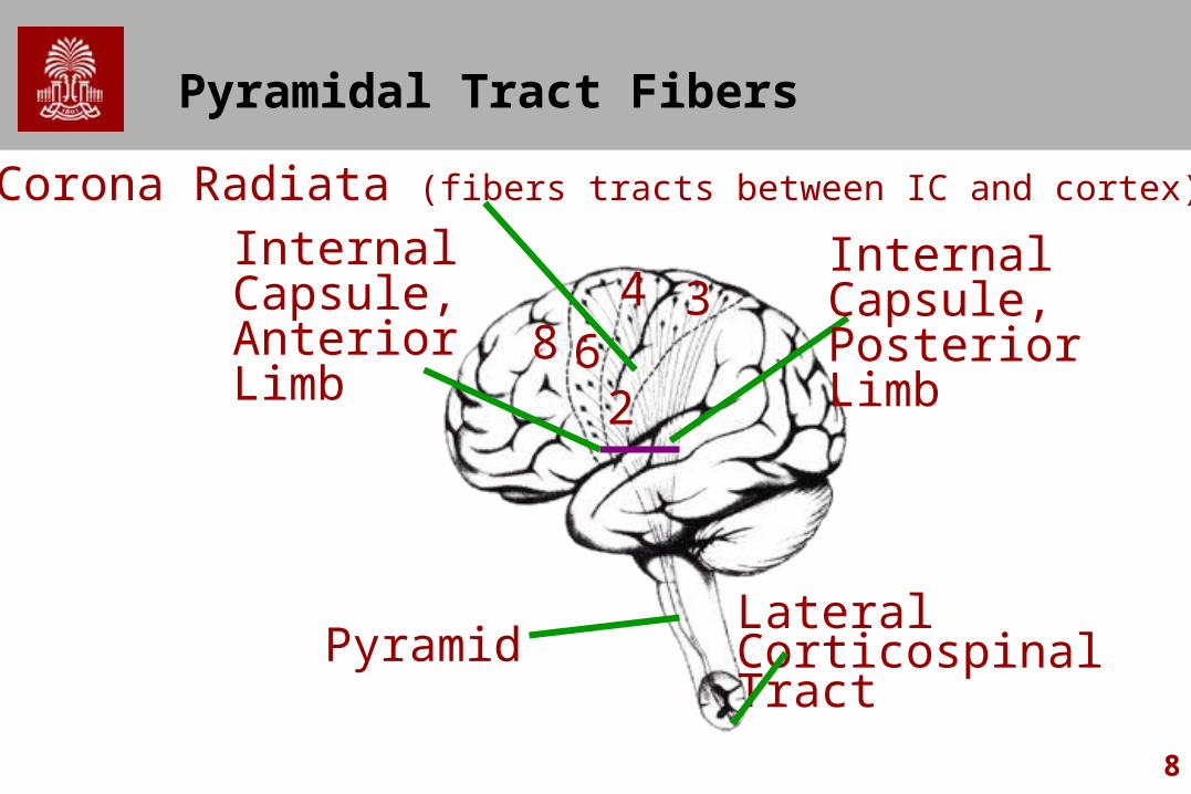
Pyramidal Tract Fibers
62
348
Internal Capsule,Posterior Limb
Lateral Corticospinal Tract
Pyramid
Internal Capsule,Anterior Limb
Corona Radiata (fibers tracts between IC and cortex)

9
Path of upper motor neurons
Lateral: Skeletal Muscle Fingers, Toes, ForearmAnterior: Axial and Girdle muscles

10
Clinical Considerations
Lesions in corticospinal fibers result in spastic hemiplegia
Lesions in corticobulbar fibers result in paralysis of facial, lingual, palatal and laryngeal muscles. More bilateral innervation causes less paralysis

11
Clinical Considerations
Upper Motor Neuron symptoms– Flaccid followed by spastic
hemiplegia– Increased Muscle Tone– + Babinski Sign– Hyperreflexias– Loss of Abdominal Reflexes– Alternating Hemiplegia (Some
Fibers that are crossed and uncrossed)
Normal reflex
Negative Babinski
Abnormal
Positive Babinski

12
Clinical Considerations
Lower motor neuron symptoms– Damage to LMN eliminates the function of the
motor unit– Lesion affecting the LMN causes weakness of
muscles and reduces tendon reflexes– Muscle tone is flaccid– Can be seen in muscular dystrophy and
myasthenia gravis– Absent or greatly reduced Babinski


