
Cell CommunicationCell Communication

Evolution of Cell SignalingEvolution of Cell Signaling• A signal-transduction pathway is a series of A signal-transduction pathway is a series of
steps by which a signal on a cell’s surface is steps by which a signal on a cell’s surface is converted into a specific cellular responseconverted into a specific cellular response
• Signal transduction pathways convert signals on Signal transduction pathways convert signals on a cell’s surface into cellular responsesa cell’s surface into cellular responses
• Pathway similarities suggest that ancestral Pathway similarities suggest that ancestral signaling molecules evolved in prokaryotes and signaling molecules evolved in prokaryotes and have since been adopted by eukaryoteshave since been adopted by eukaryotes

Communication Between Mating Yeast CellsCommunication Between Mating Yeast Cells
factorReceptor
Exchange of mating factors. Each cell type secretes a mating factor that binds to receptors on the other cell type.
Mating. Binding of the factors to receptors induces changes in the cells that lead to their fusion.
New a/ cell. The nucleus of the fused cell includes all the genes from the a and cells.
factorYeast cell,
mating type aYeast cell,
mating type
a/
a
a
1
3
2

Cells CommunicationCells Communication
• Direct contactDirect contact• Paracrine signalingParacrine signaling• Endocrine signalingEndocrine signaling• Synaptic signalingSynaptic signaling
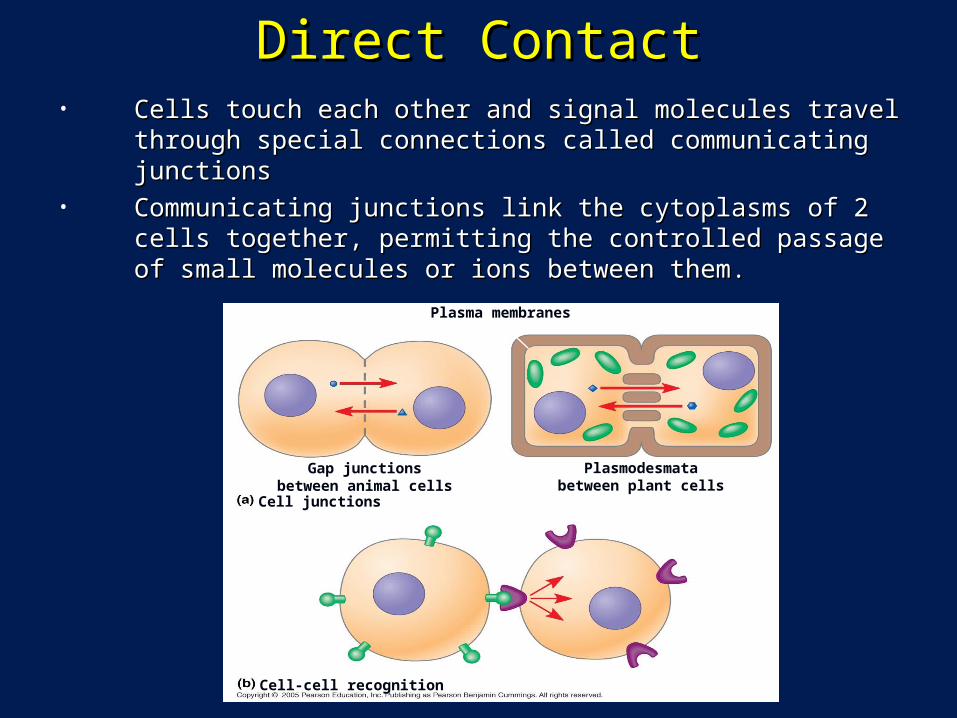
Direct ContactDirect Contact• Cells touch each other and signal molecules travel through Cells touch each other and signal molecules travel through
special connections called communicating junctionsspecial connections called communicating junctions• Communicating junctions link the cytoplasms of 2 cells together, Communicating junctions link the cytoplasms of 2 cells together,
permitting the controlled passage of small molecules or ions permitting the controlled passage of small molecules or ions between them. between them.
Plasma membranes
Gap junctionsbetween animal cells
Cell junctions
Cell-cell recognition
Plasmodesmatabetween plant cells

(a) Paracrine signaling. A secreting cell acts on nearby target cells by discharging molecules of a local regulator (a growth factor, for example) into the extracellular fluid.
(b) Synaptic signaling. A nerve cell releases neurotransmitter molecules into a synapse, stimulating the target cell.
Hormone travelsin bloodstreamto target cells
(c) Hormonal signaling. Specialized endocrine cells secrete hormones into body fluids, often the blood. Hormones may reach virtually all body cells.
Local regulator diffuses through extracellular fluid
Secretingcell
Target cell
Secretoryvesicle
Electrical signalalong nerve celltriggers release ofneurotransmitter
Neurotransmitter diffuses across
synapse
Target cellis stimulated
Local signaling Long-distance signaling
Endocrine cellBloodvessel
Targetcell
Cell Communication In AnimalsCell Communication In Animals• In many other cases, animal cells communicate using local In many other cases, animal cells communicate using local
regulators, messenger molecules that travel only short distancesregulators, messenger molecules that travel only short distances• In long-distance signaling, plants and animals use chemicals called In long-distance signaling, plants and animals use chemicals called
hormoneshormones

Cell SignalingCell Signaling
EXTRACELLULARFLUID
Receptor
Signal molecule
Relay molecules in a signal transduction pathway
Plasma membraneCYTOPLASM
Activationof cellularresponse
Reception Transduction Response1 2 3
• The cells of a organism communicate with each other by releasing The cells of a organism communicate with each other by releasing signal moleculessignal molecules that bind to that bind to receptor proteinsreceptor proteins located either on or inside of located either on or inside of target cellstarget cells..
• Three stages of cell signaling:Three stages of cell signaling:• Reception - each target cell has receptors that detect a specific signal molecule and binds to Reception - each target cell has receptors that detect a specific signal molecule and binds to
itit
• Transduction – binding of the signal molecule changes the receptor protein in some way that Transduction – binding of the signal molecule changes the receptor protein in some way that initiates transduction or conversion of the signal to a form that can bring about a specific initiates transduction or conversion of the signal to a form that can bring about a specific cellular responsecellular response
• Response – transduced signal triggers a specific cellular response, any cell activityResponse – transduced signal triggers a specific cellular response, any cell activity

ReceptionReception• A signal molecule binds to a receptor protein, A signal molecule binds to a receptor protein,
causing it to change shapecausing it to change shape• The binding between signal molecule (ligand) The binding between signal molecule (ligand)
and receptor is highly specificand receptor is highly specific• A conformational change in a receptorA conformational change in a receptor
• Is often the initial transduction of the signalIs often the initial transduction of the signal

ReceptorsReceptors• Intracellular receptors Intracellular receptors
• Some signal molecules that are small or hydrophobic can pass through Some signal molecules that are small or hydrophobic can pass through the plasma membrane and bind to receptors located inside the cell the plasma membrane and bind to receptors located inside the cell
• Intracellular receptors are cytoplasmic or nuclear proteinsIntracellular receptors are cytoplasmic or nuclear proteins
• Cell surface receptors. - Signal molecules that cannot pass through Cell surface receptors. - Signal molecules that cannot pass through the plasma membrane bind to receptors located on the surface of the plasma membrane bind to receptors located on the surface of the membrane the membrane

Intracellular Receptors Intracellular Receptors
• Gene RegulatorsGene Regulators• Signal molecule joins to the receptor, the receptor Signal molecule joins to the receptor, the receptor
changes shape and a DNA binding site is exposed.changes shape and a DNA binding site is exposed.• The DNA binding site joins to a specific segment of The DNA binding site joins to a specific segment of
DNA and activates (or suppresses) a particular geneDNA and activates (or suppresses) a particular gene
• Enzyme ReceptorEnzyme Receptor• These receptors function as enzymes – proteins that These receptors function as enzymes – proteins that
catalyze (speed up) specific chemical reactions. catalyze (speed up) specific chemical reactions. • When a signal molecule joins to the receptor, the When a signal molecule joins to the receptor, the
receptor’s catalytic domain is activated (or receptor’s catalytic domain is activated (or deactivated).deactivated).

Hormone(testosterone)
EXTRACELLULARFLUID
Receptorprotein
Plasmamembrane
Hormone-receptorcomplex
DNA
mRNA
NUCLEUS
CYTOPLASM
New protein
The steroid hormone testosterone passes through the plasma membrane.
1
Testosterone bindsto a receptor proteinin the cytoplasm,activating it.
2
The hormone-receptor complexenters the nucleusand binds to specific genes.
3
The bound proteinstimulates thetranscription ofthe gene into mRNA.
4
The mRNA istranslated into aspecific protein.
5
Steroid hormone interacting with an intracellular receptorSteroid hormone interacting with an intracellular receptor

Surface ReceptorsSurface Receptors• Receptors located on the surface of the membrane, 4 types:Receptors located on the surface of the membrane, 4 types:
• Chemically gated ion channelsChemically gated ion channels• Enzymatic receptorsEnzymatic receptors• G-protein-linked receptorsG-protein-linked receptors• IntegrinsIntegrins

Chemically Gated Ion ChannelsChemically Gated Ion Channels
• An ion channel An ion channel receptor acts as a receptor acts as a gate when the gate when the receptor changes receptor changes shapeshape
• When a signal When a signal molecule binds as molecule binds as a ligand to the a ligand to the receptor, the gate receptor, the gate allows specific allows specific ions, such as Naions, such as Na++ or Caor Ca2+2+, through a , through a channel in the channel in the receptorreceptor
Gate close
Cellularresponse
Gate open
Gate close
Ligand-gatedion channel receptor
Plasma Membrane
Signalmolecule(ligand)
GateClosed Ions

Enzymatic ReceptorsEnzymatic Receptors• Embedded in the plasma membrane, with their catalytic site exposed Embedded in the plasma membrane, with their catalytic site exposed
inside the cell. inside the cell. • Catalytic site activated when the signal molecule joins to the receptor. Catalytic site activated when the signal molecule joins to the receptor. • Function as Function as protein kinasesprotein kinases (enzymes that (enzymes that phosphorylate phosphorylate proteins.)proteins.)

Receptor Tyrosine Kinases
Signalmolecule
Signal-binding site
CYTOPLASM
Tyrosines
Signal molecule Helix in the
Membrane
Tyr
Tyr
Tyr
Tyr
Tyr
TyrTyr
Tyr
Tyr
Tyr
Tyr
Tyr
Tyr
Tyr
Tyr
Tyr
Tyr
Tyr Tyr
Tyr
Tyr
Tyr
Tyr
Tyr
Tyr
Tyr
Tyr
Tyr
Tyr
Tyr
DimerReceptor tyrosinekinase proteins(inactive monomers)
P
P
PP
P
PTyr
Tyr
Tyr
Tyr
Tyr
TyrP
P
P
P
P
PCellularresponse 1
Inactiverelay proteins
Activatedrelay proteins
Cellularresponse 2
Activated tyrosine-kinase regions(unphosphorylateddimer)
Fully activated receptortyrosine-kinase(phosphorylateddimer)
6 ATP 6 ADP

G-protein-linked ReceptorsG-protein-linked Receptors• Signal molecule joins to a receptor, the receptor activates a G Signal molecule joins to a receptor, the receptor activates a G
protein protein • The activated G protein can then activate an ion channel or The activated G protein can then activate an ion channel or
enzyme in the plasma membrane.enzyme in the plasma membrane.
G protein
ActivatedG protein
Enzyme orion channel
Activatedenzyme orion channel
G-protein-linked receptor
Signal

Signal-binding site
G-PROTEIN-LINKED RECEPTORS
G-protein-linkedreceptor
Plasma Membrane
EnzymeG-protein(inactive)CYTOPLASM
Cellular response
Activatedenzyme
Activatedreceptor
Signal molecule Inactiveenzyme
Segment thatinteracts withG proteins
GDP
GDP
GTP
GTP
P i
GDP

Second MessengersSecond Messengers• Some enzymatic receptors Some enzymatic receptors
and most G-protein-linked and most G-protein-linked receptors relay their message receptors relay their message into the cell by activating into the cell by activating other molecules or ions other molecules or ions inside the cell.inside the cell.
• These molecules and ions, These molecules and ions, called called second messengerssecond messengers, , transmit the message within transmit the message within the cell. The 2 most common the cell. The 2 most common second messengers are second messengers are cAMP and Ca++cAMP and Ca++

Signal Transduction PathwaysSignal Transduction Pathways• Transduction usually involves multiple stepsTransduction usually involves multiple steps• Multistep pathwaysMultistep pathways
• Can amplify a signalCan amplify a signal• Provide more opportunities for coordination and regulationProvide more opportunities for coordination and regulation
• The molecules that relay a signal from receptor to The molecules that relay a signal from receptor to response are mostly proteinsresponse are mostly proteins
• The receptor activates another protein, which The receptor activates another protein, which activates another, and so on, until the protein activates another, and so on, until the protein producing the response is activatedproducing the response is activated
• At each step, the signal is transduced into a different At each step, the signal is transduced into a different form, usually a conformational changeform, usually a conformational change

A Phosphorylation CascadeA Phosphorylation Cascade• In many pathways, the signal is transmitted by a cascade of protein In many pathways, the signal is transmitted by a cascade of protein
phosphorylations - phosphatase enzymes remove the phosphatesphosphorylations - phosphatase enzymes remove the phosphates• This phosphorylation and dephosphorylation system acts as a molecular switch, This phosphorylation and dephosphorylation system acts as a molecular switch,
turning activities on and offturning activities on and off
Signal molecule
Activeproteinkinase
1
Activeproteinkinase
2
Activeproteinkinase
3
Inactiveprotein kinase
1
Inactiveprotein kinase
2
Inactiveprotein kinase
3
Inactiveprotein
Activeprotein
Cellularresponse
Receptor
P
P
P
P
P
P
ATPADP
ADP
ADP
ATP
ATP
PP
PP
PP
Activated relaymolecule
A relay moleculeactivates protein kinase 1.
1
Active protein kinase 1transfers a phosphate from ATPto an inactive molecule ofprotein kinase 2, thus activatingthis second kinase.
2
Active protein kinase 2then catalyzes the phos-phorylation (and activation) ofprotein kinase 3.
3
Finally, active proteinkinase 3 phosphorylates aprotein (pink) that brings about the cell’s response tothe signal.
4
Enzymes called proteinphosphatases (PP)catalyze the removal ofthe phosphate groupsfrom the proteins, making them inactiveand available for reuse.
5
i
i
i
Phosphorylation cascade

cAMP Second MessengercAMP Second MessengerG-protein-signaling pathwayG-protein-signaling pathway
1.1. Signal molecule binds to Signal molecule binds to surface receptorsurface receptor
2.2. Surface receptor Surface receptor activates a G proteinactivates a G protein
3.3. G protein activates the G protein activates the membrane-bound membrane-bound enzyme, adenylyl enzyme, adenylyl cyclasecyclase
4.4. Adenylyl cyclase Adenylyl cyclase catalyzes synthesis of catalyzes synthesis of camp, which binds to a camp, which binds to a target proteintarget protein
5.5. Target protein initiates Target protein initiates cellular changecellular change
First messenger(signal moleculesuch as epinephrine)
ATP
GTP
cAMP
Proteinkinase A
Cellular responses
G-protein-linkedreceptor
Adenylylcyclase
G protein
Second messenger

Cyclic AMPCyclic AMP
O–O O
O
N
O
O
O
O
P P P
P
P P
O
O
O
O
O
OH
CH2
NH2 NH2 NH2
N
N
N
N
N
N
N
N
N
N
NO
O
O
ATP
Ch2CH2
O
OH OH
P
O O
H2O
HOAdenylyl cyclase Phoshodiesterase
Pyrophosphate
Cyclic AMP AMPOH OH
O
i
• Cyclic AMP (cAMP) is one of the most widely Cyclic AMP (cAMP) is one of the most widely used second messengersused second messengers
• Adenylyl cyclase, an enzyme in the plasma Adenylyl cyclase, an enzyme in the plasma membrane, converts ATP to cAMP in response membrane, converts ATP to cAMP in response to an extracellular signalto an extracellular signal

Cyclic AMP PathwayCyclic AMP Pathway

Calcium (Ca++) PathwaysCalcium (Ca++) Pathways• Calcium ions (CaCalcium ions (Ca2+2+) act as a second messenger in many pathways) act as a second messenger in many pathways• Calcium is an important second messenger because cells can Calcium is an important second messenger because cells can
regulate its concentrationregulate its concentration
ATP
EXTRACELLULARFLUID
ATPMitochondrion
Ca2+
pump
Plasmamembrane
CYTOSOL
Endoplasmicreticulum (ER)
Ca2+
pump
Ca2+
pump
High [Ca2+]Key
Nucleus
Low [Ca2+]

• A signal transduction pathway may trigger A signal transduction pathway may trigger an increase in calcium in the cytosolan increase in calcium in the cytosol
• Pathways leading to the release of calcium Pathways leading to the release of calcium involve inositol triphosphate (IPinvolve inositol triphosphate (IP33) and ) and
diacylglycerol (DAG) as second diacylglycerol (DAG) as second messengersmessengers
Calcium ions and Inositol Triphosphate (IPCalcium ions and Inositol Triphosphate (IP33))

Ca++ PathwayCa++ Pathway• Signal molecule binds to surface receptorSignal molecule binds to surface receptor• Surface receptor activates a G proteinSurface receptor activates a G protein• G protein activates the membrane-bound G protein activates the membrane-bound
enzyme, phospholipase Cenzyme, phospholipase C• Phospholipase C catalyzes synthesis of Phospholipase C catalyzes synthesis of
inositol triphosphate, which stimulates inositol triphosphate, which stimulates release of Ca++ from ERrelease of Ca++ from ER
• Released Ca++ initiates cellular changeReleased Ca++ initiates cellular change

Calcium and IPCalcium and IP33 in signaling pathways in signaling pathways
2 3
IP3 quickly diffuses throughthe cytosol and binds to an IP3–gated calcium channel in the ERmembrane, causing it to open.
4 The calcium ionsactivate the nextprotein in one or moresignaling pathways.
6 Calcium ions flow out ofthe ER (down their con-centration gradient), raisingthe Ca2+ level in the cytosol.
5
DAG functions asa second messengerin other pathways.
Phospholipase C cleaves aplasma membrane phospholipidcalled PIP2 into DAG and IP3.
EXTRA-CELLULARFLUID
Signal molecule(first messenger)
G protein
G-protein-linkedreceptor
Variousproteinsactivated
Endoplasmicreticulum (ER)
Phospholipase CPIP2
IP3
DAG
Cellularresponses
GTP
Ca2+
(second messenger)
Ca2+
IP3-gatedcalcium channel
A signal molecule bindsto a receptor, leading toactivation of phospholipase C.
1
CYTOSOL

Fine-Tuning of the ResponseFine-Tuning of the Response
• Multistep pathways have two important Multistep pathways have two important benefits:benefits:• Amplifying the signal (and thus the response)Amplifying the signal (and thus the response)• Contributing to the specificity of the responseContributing to the specificity of the response
• Enzyme cascades amplify the cell’s Enzyme cascades amplify the cell’s responseresponse
• At each step, the number of activated At each step, the number of activated products is much greater than in the products is much greater than in the preceding steppreceding step

AmplificationAmplification• Due to the many steps in the cell signaling process, one Due to the many steps in the cell signaling process, one
signal molecule can trigger a “cascade” effectsignal molecule can trigger a “cascade” effect

Cytoplasmic response to a signal: the stimulation of Cytoplasmic response to a signal: the stimulation of glycogen breakdown by epinephrineglycogen breakdown by epinephrine
Glucose-1-phosphate(108 molecules)
Glycogen
Active glycogen phosphorylase (106)
Inactive glycogen phosphorylase
Active phosphorylase kinase (105)
Inactive phosphorylase kinase
Inactive protein kinase A
Active protein kinase A (104)
ATPCyclic AMP (104)
Active adenylyl cyclase (102)
Inactive adenylyl cyclase
Inactive G protein
Active G protein (102 molecules)
Binding of epinephrine to G-protein-linked receptor (1 molecule)
Transduction
Response
Reception

Specificity of Cell SignalingSpecificity of Cell Signaling• Different kinds of cells have Different kinds of cells have
different collections of proteinsdifferent collections of proteins• These differences in proteins These differences in proteins
give each kind of cell give each kind of cell specificity in detecting and specificity in detecting and responding to signalsresponding to signals
• The response of a cell to a The response of a cell to a signal depends on the cell’s signal depends on the cell’s particular collection of proteinsparticular collection of proteins
• Pathway branching and “cross-Pathway branching and “cross-talk” further help the cell talk” further help the cell coordinate incoming signalscoordinate incoming signals
Signalmolecule
Receptor
Relaymolecules
Response 1 Response 2 Response 3
Cell B. Pathway branches,leading to two responses
Cell A. Pathway leads to a single response
Cell C. Cross-talk occursbetween two pathways
Response 4 Response 5
Activationor inhibition
Cell D. Different receptorleads to a different response

Signaling Efficiency: Scaffolding Proteins and Signaling Efficiency: Scaffolding Proteins and Signaling ComplexesSignaling Complexes
• Rather than relying on diffusion of large relay molecules such as proteins, Rather than relying on diffusion of large relay molecules such as proteins, many signal pathways are linked together physically by many signal pathways are linked together physically by scaffolding scaffolding proteins.proteins.
• Scaffolding proteins may themselves be relay proteins to which several Scaffolding proteins may themselves be relay proteins to which several other relay proteins attach.other relay proteins attach.
• This hardwiring enhances the speed, accuracy, and efficiency of signal This hardwiring enhances the speed, accuracy, and efficiency of signal transfer between cells.transfer between cells.
Signalmolecule
Receptor
Scaffoldingprotein
Threedifferentproteinkinases
Plasmamembrane
Figure 11.16


![Lithium-Ion battery SOC estimation...defines the cell’s internal resistance and the cell’s dynamics [14]. Hysteresis is a part of cell behavior and exhibits the dependency of](https://cdn.vdocument.in/doc/165x107/5ebb5ced0c2acc01ef418f19/lithium-ion-battery-soc-estimation-deines-the-cellas-internal-resistance.jpg)


