Download - cerebral cortex st Saher Al_doctor
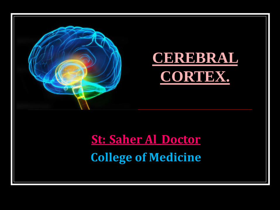
St: Saher Al_Doctor
College of Medicine
CEREBRAL
CORTEX.
OBJECTIVES
Cerebral cortex
External features.
Functional areas.
Lobes.
Connections , functions & applied.
White matter of cerebrum.
Association Fibres.
Commissural
Projection.
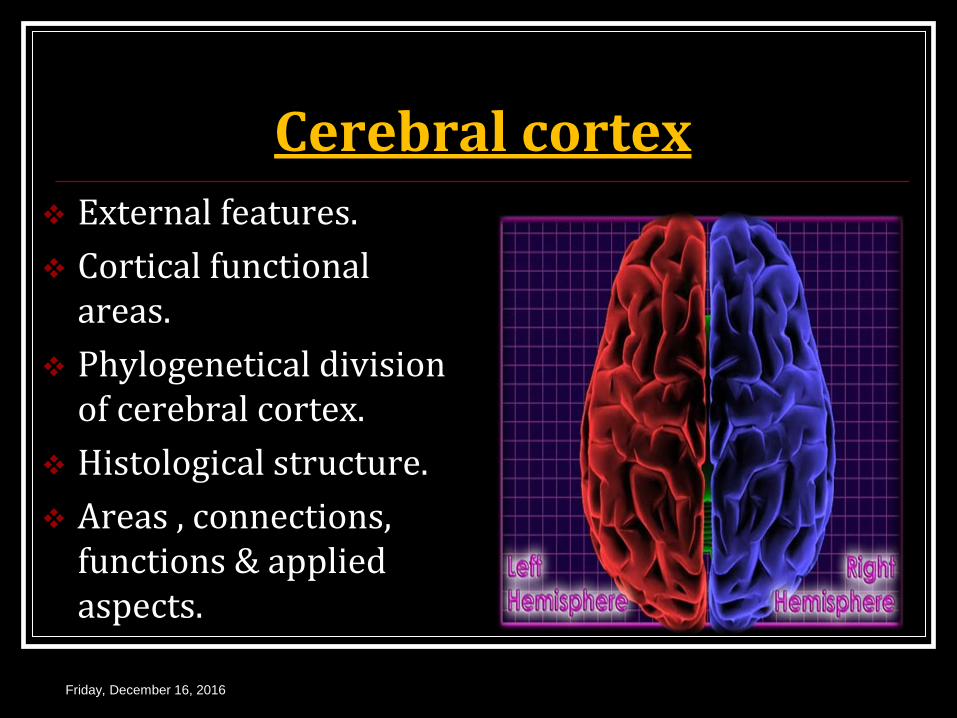
Cerebral cortex
External features.
Cortical functional areas.
Phylogenetical division of cerebral cortex.
Histological structure.
Areas , connections, functions & applied aspects.
Friday, December 16, 2016
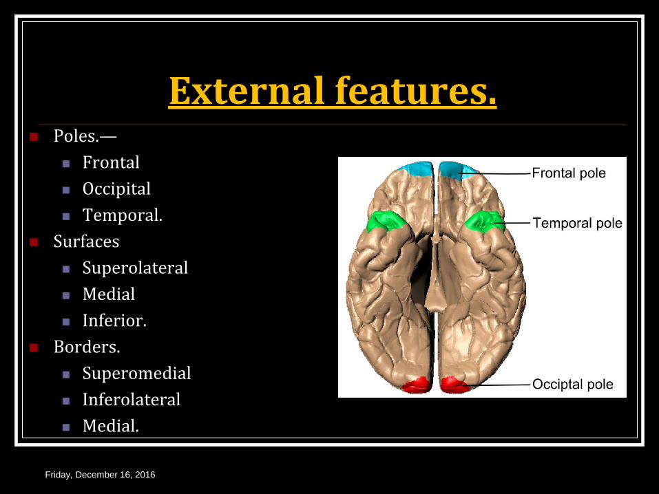
External features. Poles.—
Frontal
Occipital
Temporal.
Surfaces
Superolateral
Medial
Inferior.
Borders.
Superomedial
Inferolateral
Medial.
Friday, December 16, 2016
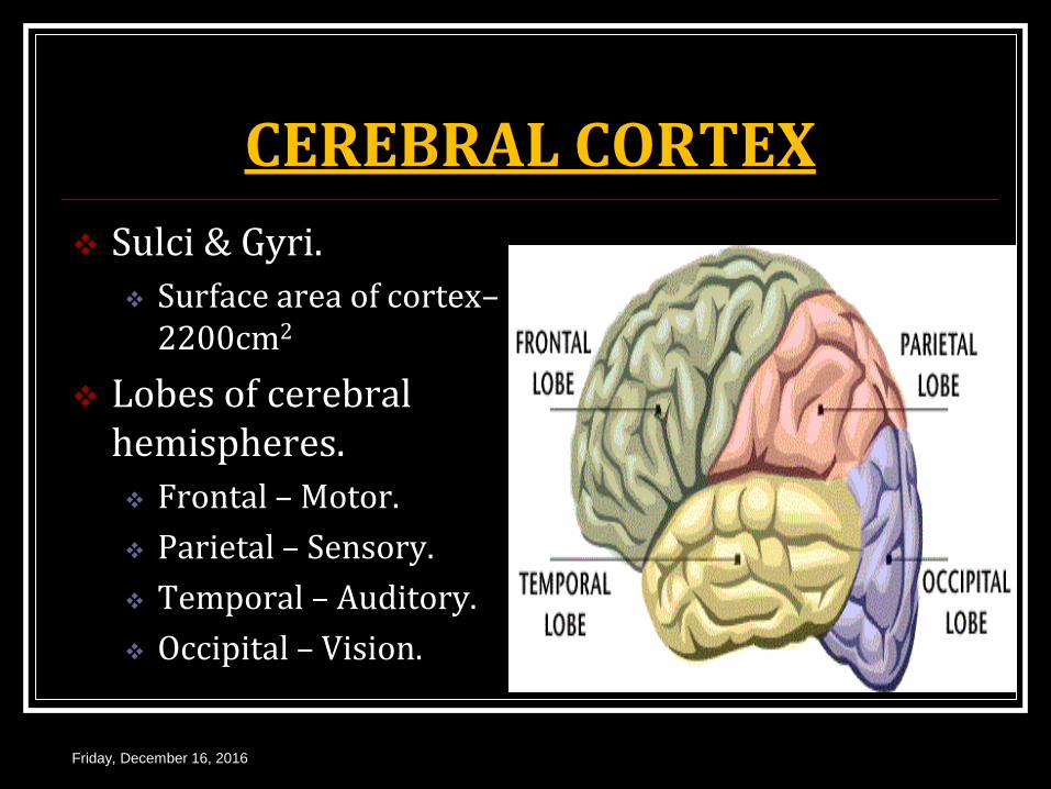
CEREBRAL CORTEX
Sulci & Gyri.
Surface area of cortex– 2200cm2
Lobes of cerebral hemispheres.
Frontal – Motor.
Parietal – Sensory.
Temporal – Auditory.
Occipital – Vision.
Friday, December 16, 2016
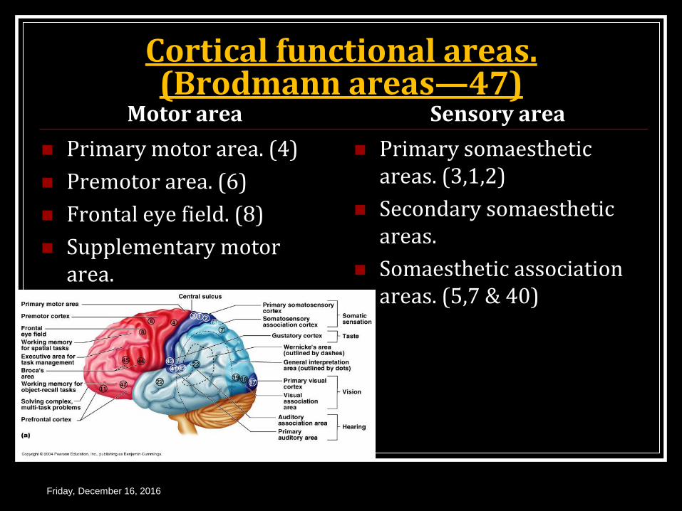
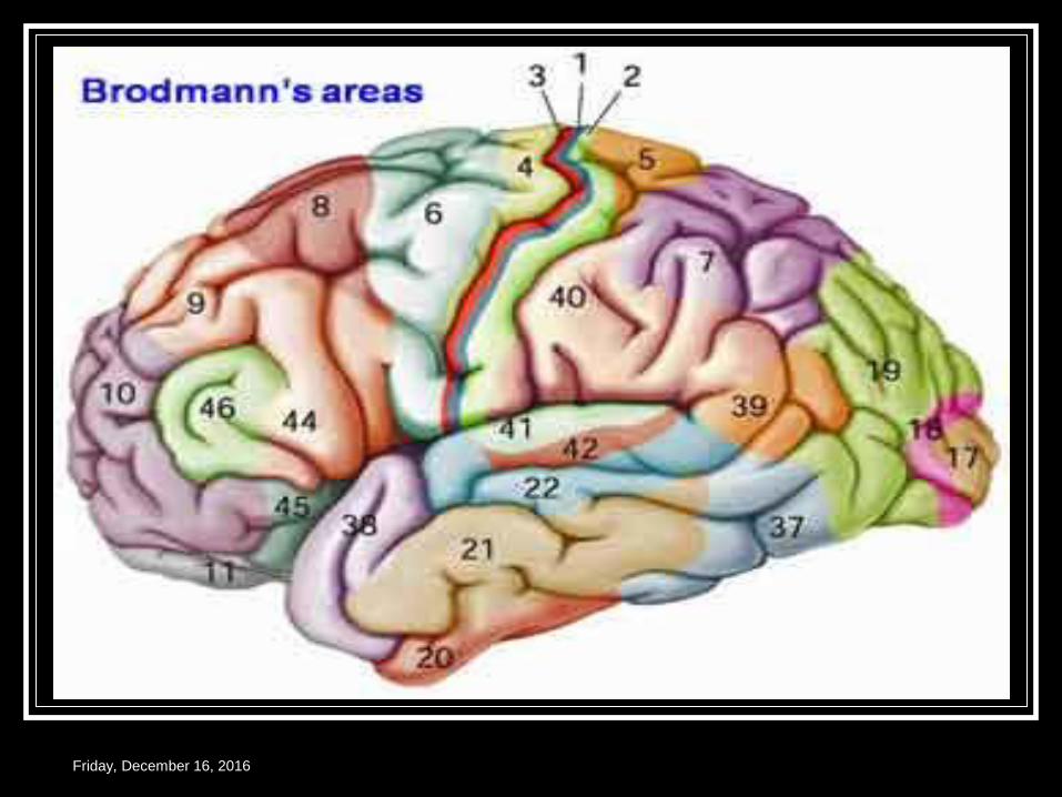
Cortical functional areas. (Brodmann areas—47)
Motor area
Primary motor area. (4)
Premotor area. (6)
Frontal eye field. (8)
Supplementary motor area.
Sensory area
Primary somaesthetic areas. (3,1,2)
Secondary somaesthetic areas.
Somaesthetic association areas. (5,7 & 40)
Friday, December 16, 2016

Cortical functional areas. (Brodmann areas—47)
Auditory area
Primary auditory area. (41)
Auditory association area. (42).
Higher auditory association area. (22)
Visual area.
Primary visual area. Or Visuostriate area.(17)
Visual association area. Or Peristriate area.(18)
Visual association area. Or Perastriate area.(19)
Friday, December 16, 2016
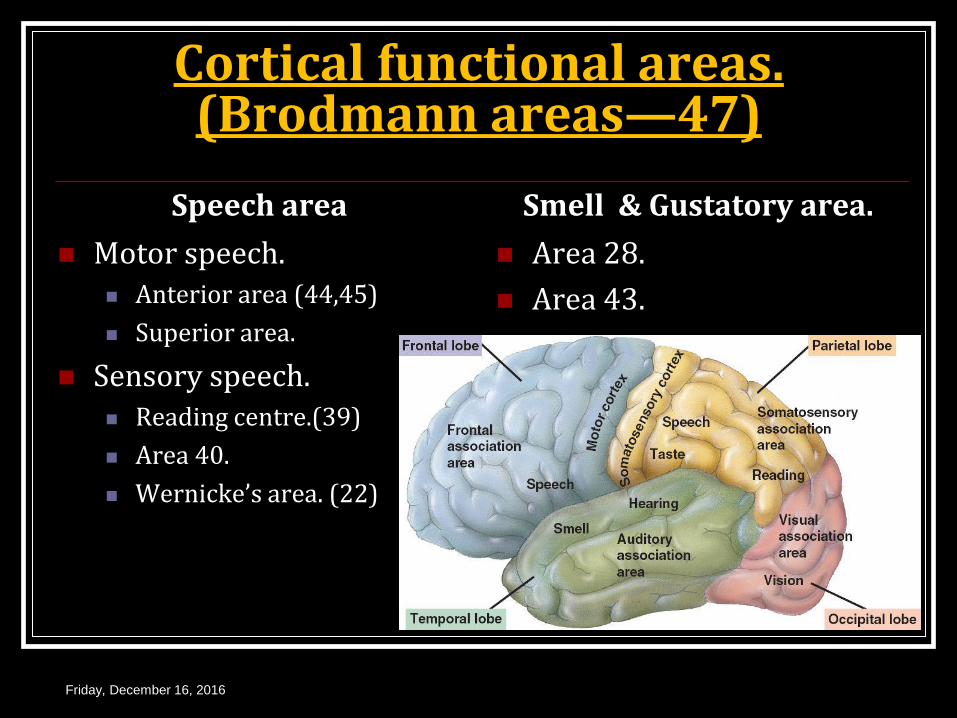
Cortical functional areas. (Brodmann areas—47)
Speech area
Motor speech. Anterior area (44,45)
Superior area.
Sensory speech. Reading centre.(39)
Area 40.
Wernicke’s area. (22)
Smell & Gustatory area.
Area 28.
Area 43.
Friday, December 16, 2016
Friday, December 16, 2016
Friday, December 16, 2016
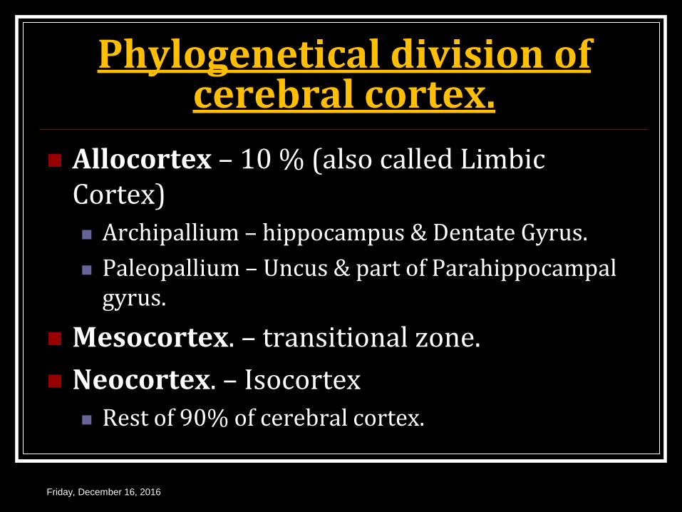
Phylogenetical division of cerebral cortex.
Allocortex – 10 % (also called Limbic Cortex) Archipallium – hippocampus & Dentate Gyrus.
Paleopallium – Uncus & part of Parahippocampal gyrus.
Mesocortex. – transitional zone.
Neocortex. – Isocortex Rest of 90% of cerebral cortex.
Friday, December 16, 2016
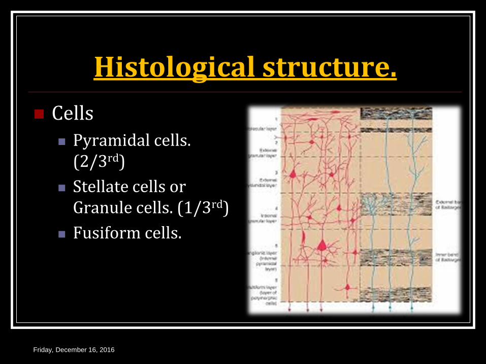
Histological structure.
Cells Pyramidal cells.
(2/3rd)
Stellate cells or Granule cells. (1/3rd)
Fusiform cells.
Friday, December 16, 2016
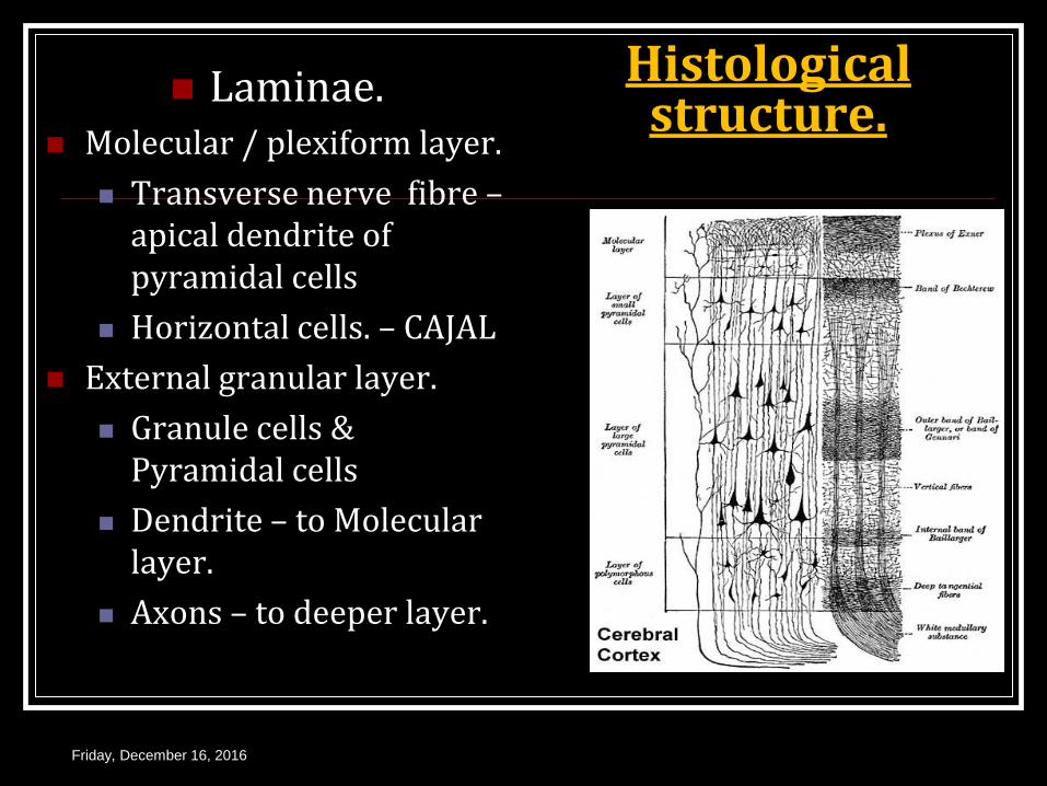
Histological structure.
Laminae. Molecular / plexiform layer.
Transverse nerve fibre – apical dendrite of pyramidal cells
Horizontal cells. – CAJAL
External granular layer.
Granule cells & Pyramidal cells
Dendrite – to Molecular layer.
Axons – to deeper layer.
Friday, December 16, 2016
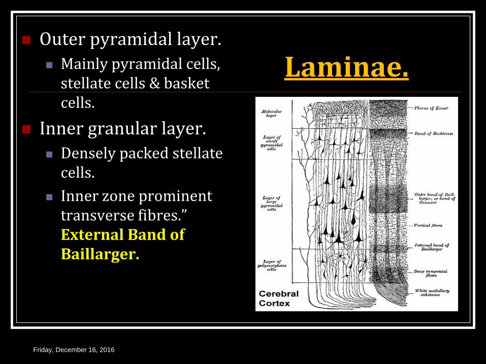
Laminae. Outer pyramidal layer.
Mainly pyramidal cells, stellate cells & basket cells.
Inner granular layer.
Densely packed stellate cells.
Inner zone prominent transverse fibres.” External Band of Baillarger.
Friday, December 16, 2016
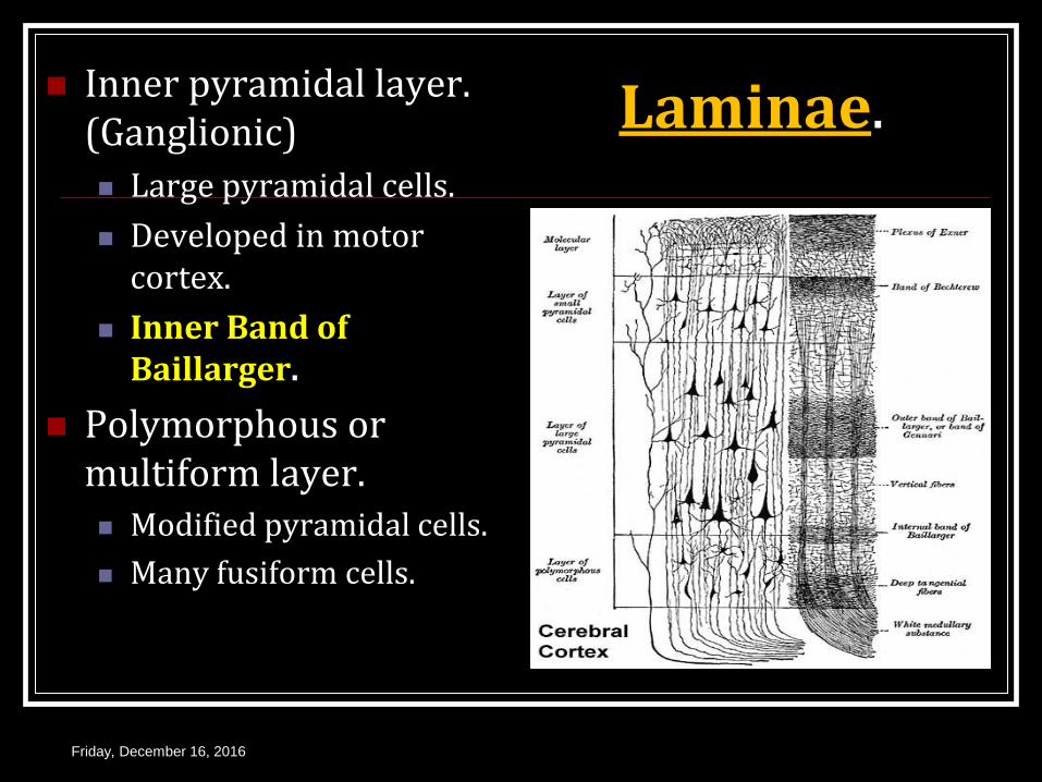
Laminae. Inner pyramidal layer. (Ganglionic)
Large pyramidal cells.
Developed in motor cortex.
Inner Band of Baillarger.
Polymorphous or multiform layer. Modified pyramidal cells.
Many fusiform cells.
Friday, December 16, 2016
Types of Neocortex.
Type I or agranular cortex.
Granule cells & stellate cells absent.
Pyramidal cells – large, Betz cells.
Seen in Motor cortex (4) & Broca’s area. (44)
Type II or frontal cortex.
Triangular granular cells
Seen in frontal lobe.
Friday, December 16, 2016
Types of Neocortex. Type III or Parietal
cortex.
Depth & density of layer II & III increased.
Granule cells – round.
Seen in parietal lobe & junction of parietal, temporal & occipital lobe.
Type IV or Polar Type.
Cortex narrow
All layers reduced depth.
Seen in frontal & occipital pole.
Type V or granular cortex.
Excess granule cells
Seen in sensory cortex & calcarine region.
Friday, December 16, 2016
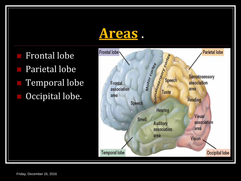
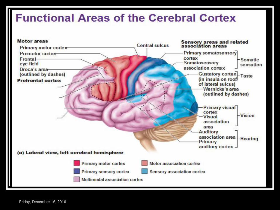
Areas .
Frontal lobe
Parietal lobe
Temporal lobe
Occipital lobe.
Friday, December 16, 2016
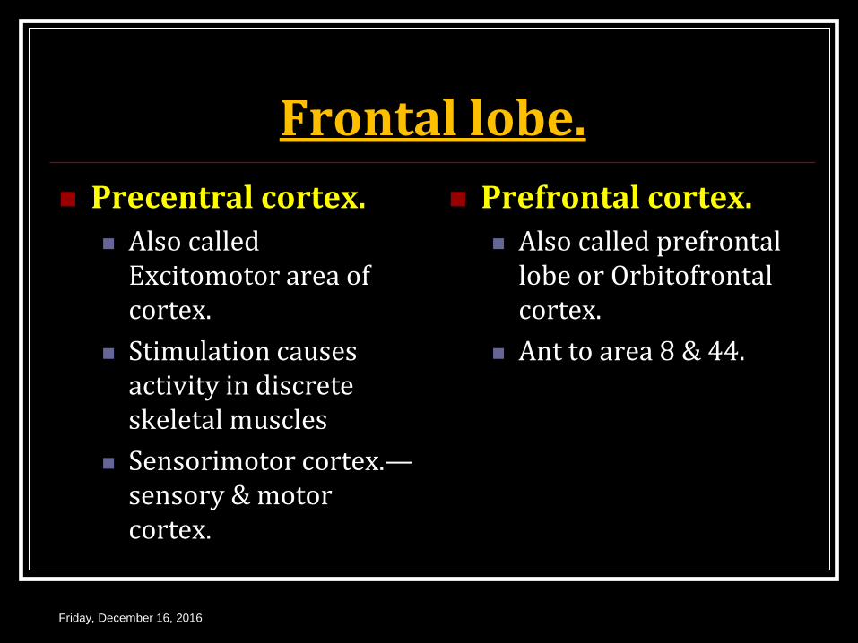
Frontal lobe.
Precentral cortex.
Also called Excitomotor area of cortex.
Stimulation causes activity in discrete skeletal muscles
Sensorimotor cortex.—sensory & motor cortex.
Prefrontal cortex.
Also called prefrontal lobe or Orbitofrontal cortex.
Ant to area 8 & 44.
Friday, December 16, 2016
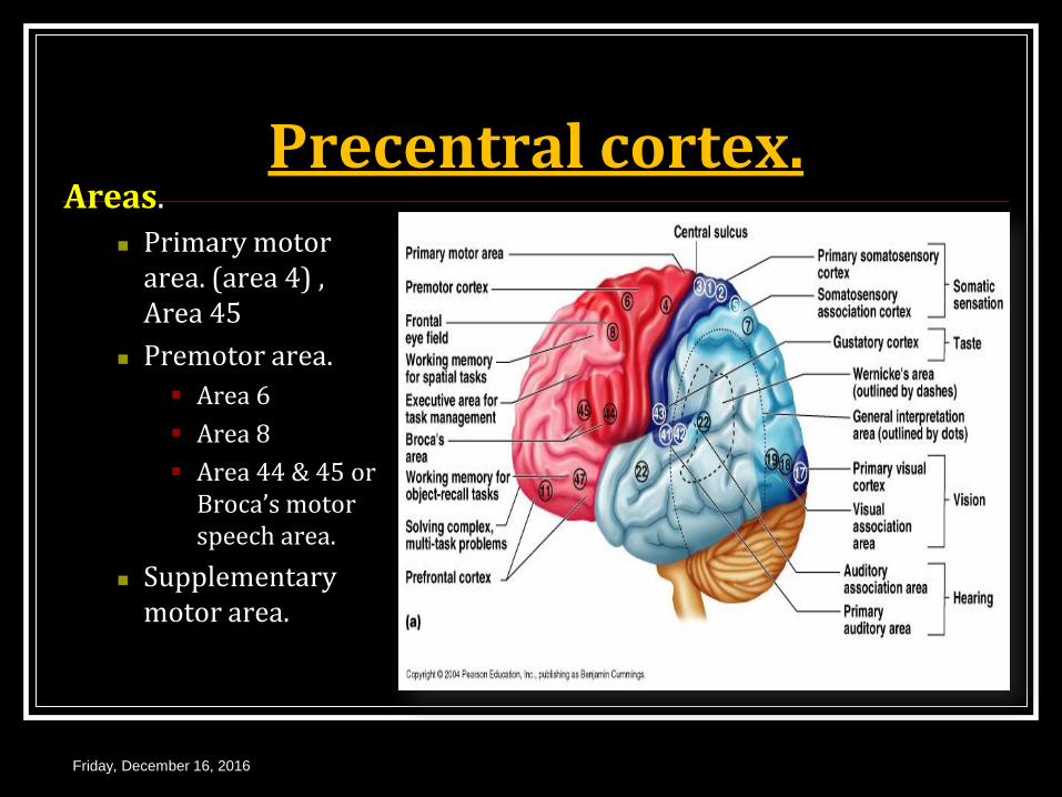
Precentral cortex. Areas.
Primary motor area. (area 4) , Area 45
Premotor area.
Area 6
Area 8
Area 44 & 45 or Broca’s motor speech area.
Supplementary motor area.
Friday, December 16, 2016
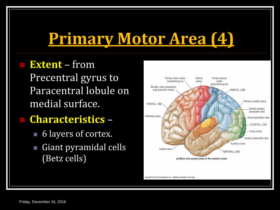
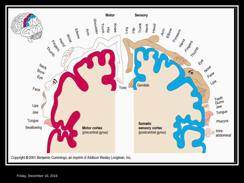
Primary Motor Area (4)
Extent – from Precentral gyrus to Paracentral lobule on medial surface.
Characteristics –
6 layers of cortex.
Giant pyramidal cells (Betz cells)
Friday, December 16, 2016

Topographic Representation.
Contra lateral half of body in inverted order.
Part of body for skilled movements – larger area.
Body represented upside down.
Friday, December 16, 2016
Friday, December 16, 2016
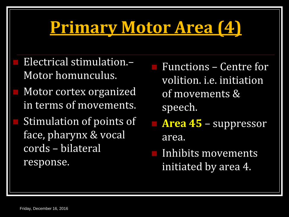
Primary Motor Area (4)
Electrical stimulation.– Motor homunculus.
Motor cortex organized in terms of movements.
Stimulation of points of face, pharynx & vocal cords – bilateral response.
Functions – Centre for volition. i.e. initiation of movements & speech.
Area 45 – suppressor area.
Inhibits movements initiated by area 4.
Friday, December 16, 2016

Sensory Homunculi Motor Homunculi.
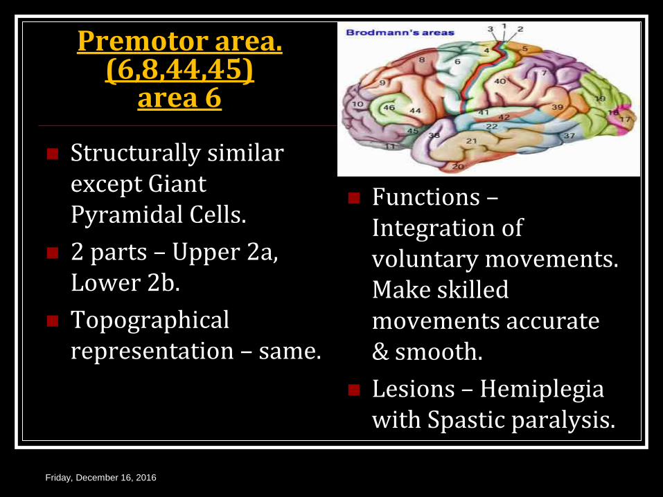
Premotor area. (6,8,44,45)
area 6
Structurally similar except Giant Pyramidal Cells.
2 parts – Upper 2a, Lower 2b.
Topographical representation – same.
Functions – Integration of voluntary movements. Make skilled movements accurate & smooth.
Lesions – Hemiplegia with Spastic paralysis.
Friday, December 16, 2016
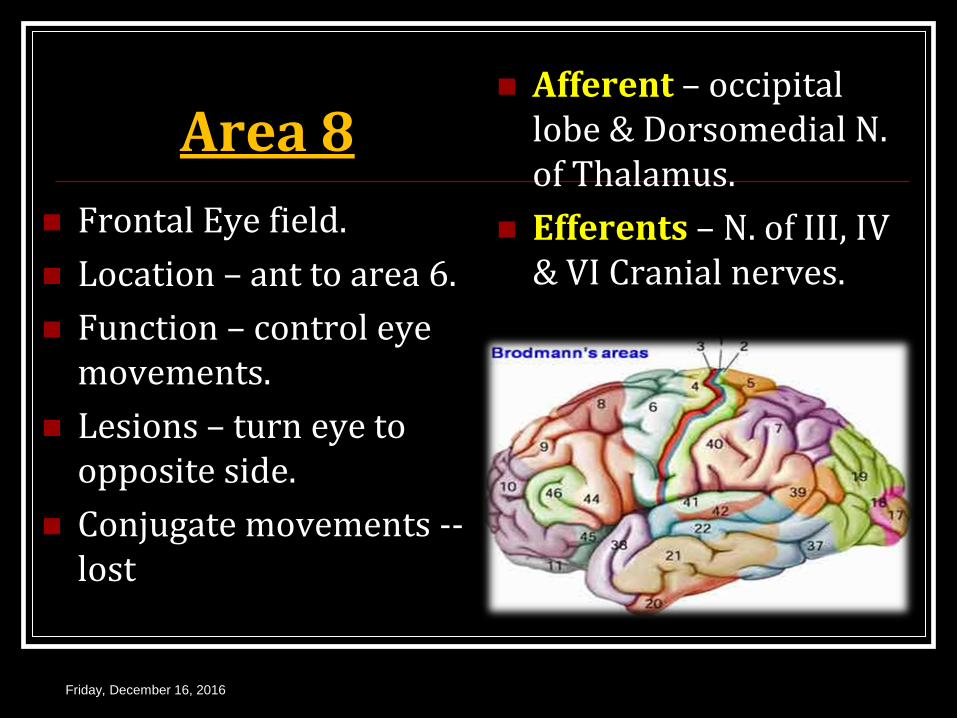
Area 8
Frontal Eye field.
Location – ant to area 6.
Function – control eye movements.
Lesions – turn eye to opposite side.
Conjugate movements -- lost
Afferent – occipital lobe & Dorsomedial N. of Thalamus.
Efferents – N. of III, IV & VI Cranial nerves.
Friday, December 16, 2016
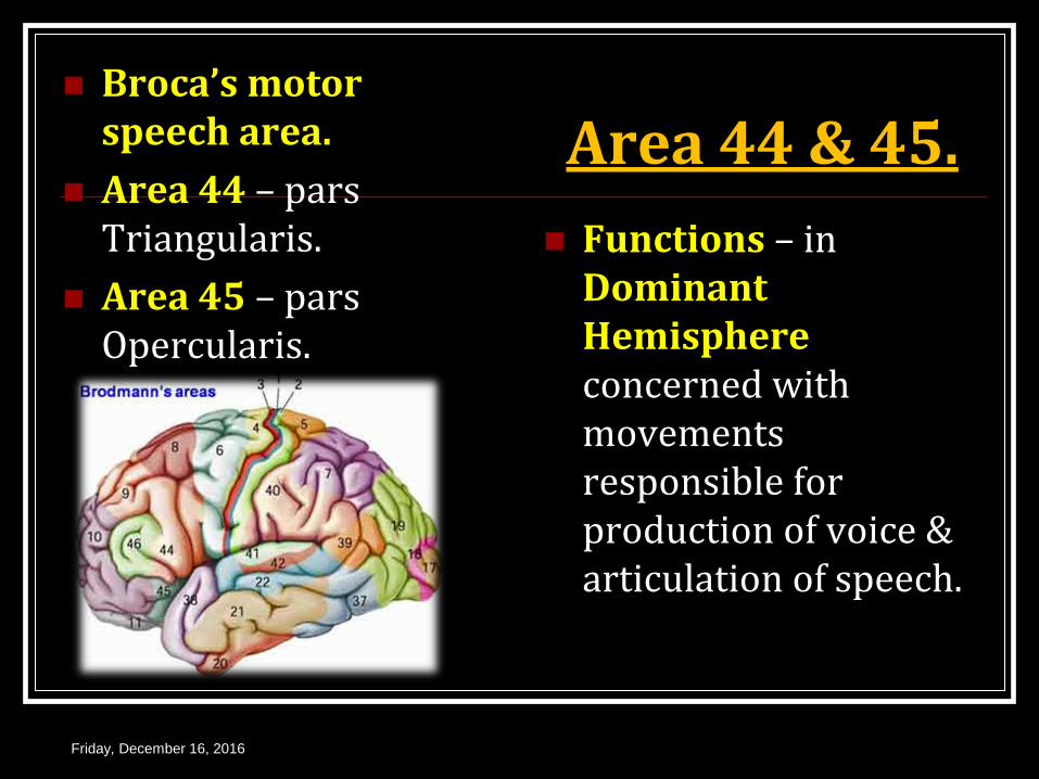
Area 44 & 45. Broca’s motor
speech area.
Area 44 – pars Triangularis.
Area 45 – pars Opercularis.
Functions – in Dominant Hemisphere concerned with movements responsible for production of voice & articulation of speech.
Friday, December 16, 2016
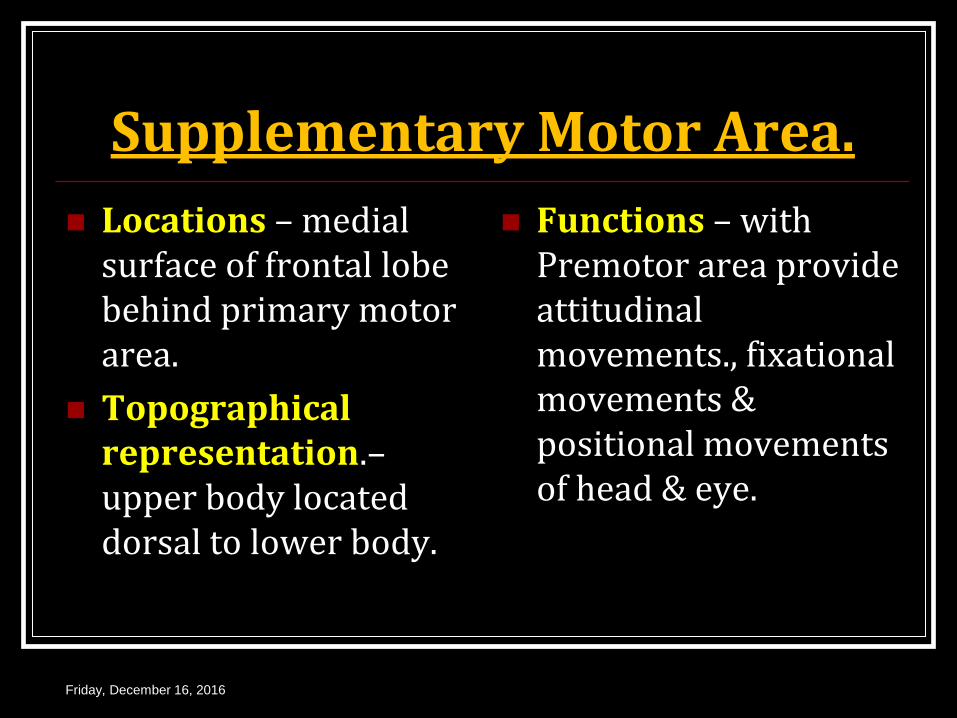
Supplementary Motor Area.
Locations – medial surface of frontal lobe behind primary motor area.
Topographical representation.– upper body located dorsal to lower body.
Functions – with Premotor area provide attitudinal movements., fixational movements & positional movements of head & eye.
Friday, December 16, 2016
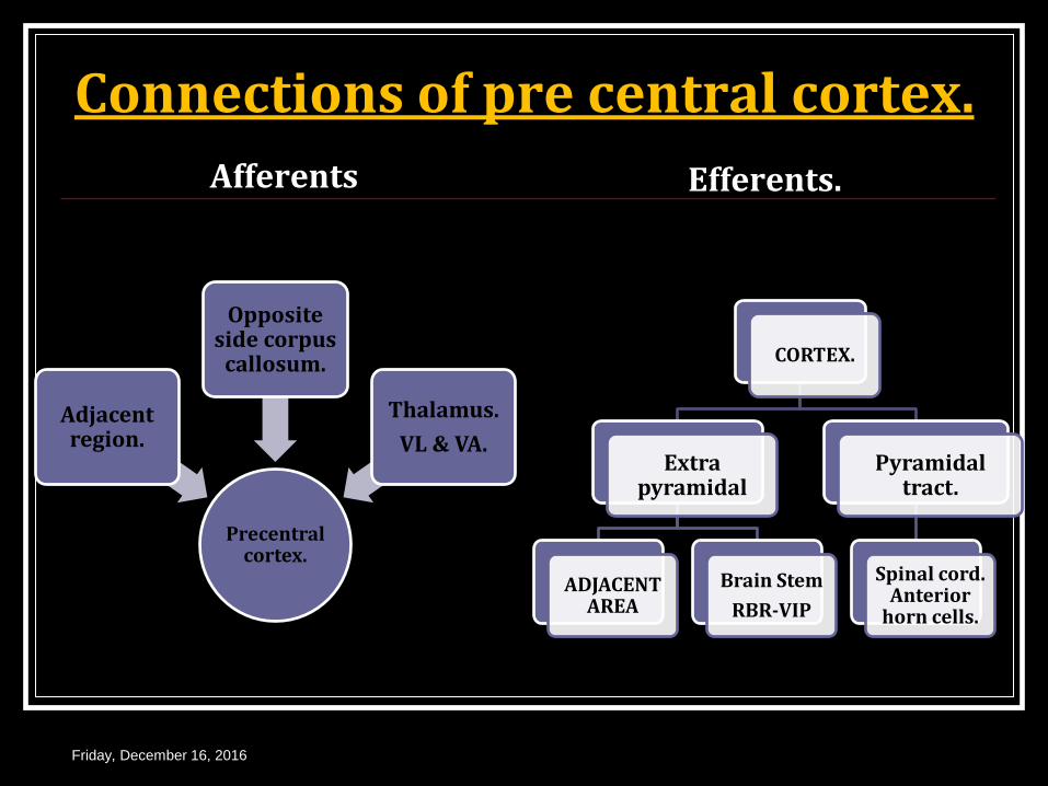
Connections of pre central cortex.
Afferents
Precentral cortex.
Adjacent region.
Opposite side corpus
callosum.
Thalamus.
VL & VA.
Efferents.
Friday, December 16, 2016
CORTEX.
Extra pyramidal
ADJACENT AREA
Brain Stem
RBR-VIP
Pyramidal tract.
Spinal cord. Anterior
horn cells.
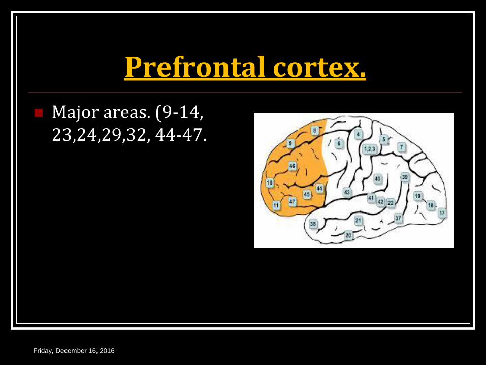
Prefrontal cortex.
Major areas. (9-14, 23,24,29,32, 44-47.
Friday, December 16, 2016
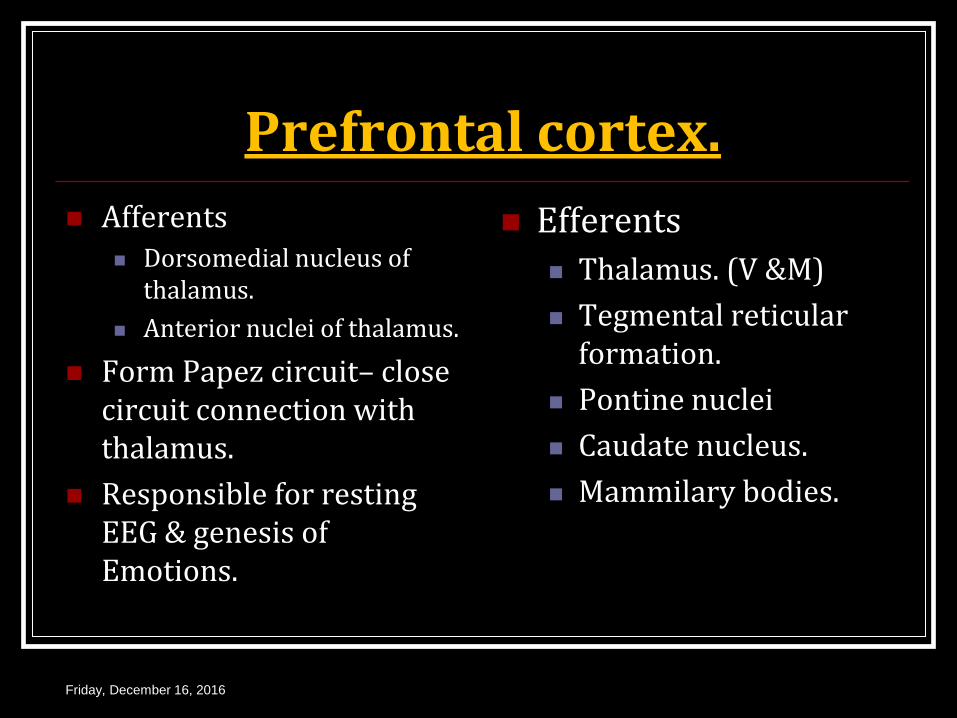
Prefrontal cortex.
Afferents Dorsomedial nucleus of
thalamus.
Anterior nuclei of thalamus.
Form Papez circuit– close circuit connection with thalamus.
Responsible for resting EEG & genesis of Emotions.
Efferents
Thalamus. (V &M)
Tegmental reticular formation.
Pontine nuclei
Caudate nucleus.
Mammilary bodies.
Friday, December 16, 2016
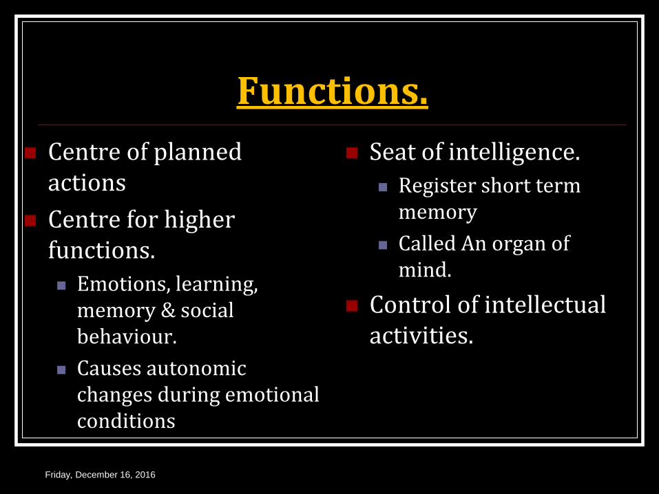
Functions.
Centre of planned actions
Centre for higher functions.
Emotions, learning, memory & social behaviour.
Causes autonomic changes during emotional conditions
Seat of intelligence.
Register short term memory
Called An organ of mind.
Control of intellectual activities.
Friday, December 16, 2016
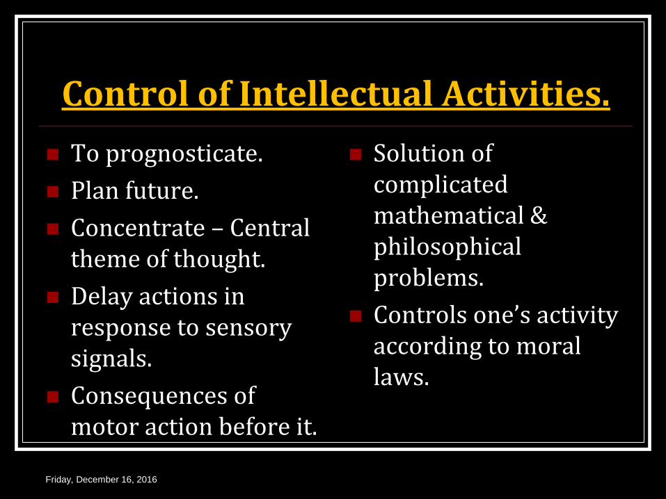
Control of Intellectual Activities.
To prognosticate.
Plan future.
Concentrate – Central theme of thought.
Delay actions in response to sensory signals.
Consequences of motor action before it.
Solution of complicated mathematical & philosophical problems.
Controls one’s activity according to moral laws.
Friday, December 16, 2016
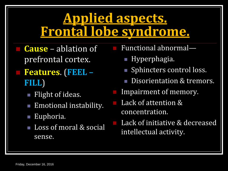
Applied aspects. Frontal lobe syndrome.
Cause – ablation of prefrontal cortex.
Features. (FEEL – FILL)
Flight of ideas.
Emotional instability.
Euphoria.
Loss of moral & social sense.
Functional abnormal—
Hyperphagia.
Sphincters control loss.
Disorientation & tremors.
Impairment of memory.
Lack of attention & concentration.
Lack of initiative & decreased intellectual activity.
Friday, December 16, 2016
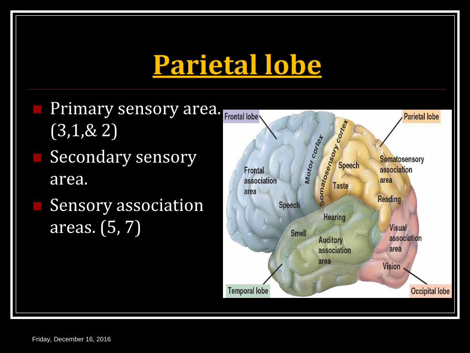
Parietal lobe
Primary sensory area. (3,1,& 2)
Secondary sensory area.
Sensory association areas. (5, 7)
Friday, December 16, 2016
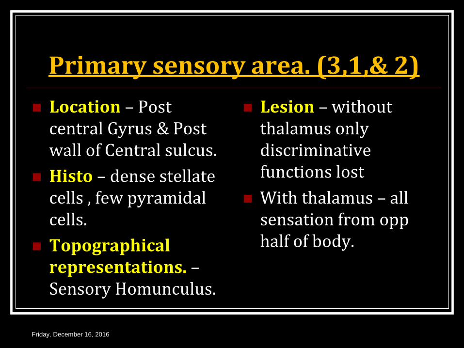
Primary sensory area. (3,1,& 2)
Location – Post central Gyrus & Post wall of Central sulcus.
Histo – dense stellate cells , few pyramidal cells.
Topographical representations. – Sensory Homunculus.
Lesion – without thalamus only discriminative functions lost
With thalamus – all sensation from opp half of body.
Friday, December 16, 2016

Secondary sensory area. Location – post central
Gyrus below 1st somatic sensory area.
Topographical representation – body represented twice in area I & area II.
Lesion – discriminative power lost.
Friday, December 16, 2016
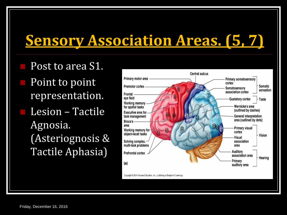
Sensory Association Areas. (5, 7)
Post to area S1.
Point to point representation.
Lesion – Tactile Agnosia. (Asteriognosis & Tactile Aphasia)
Friday, December 16, 2016
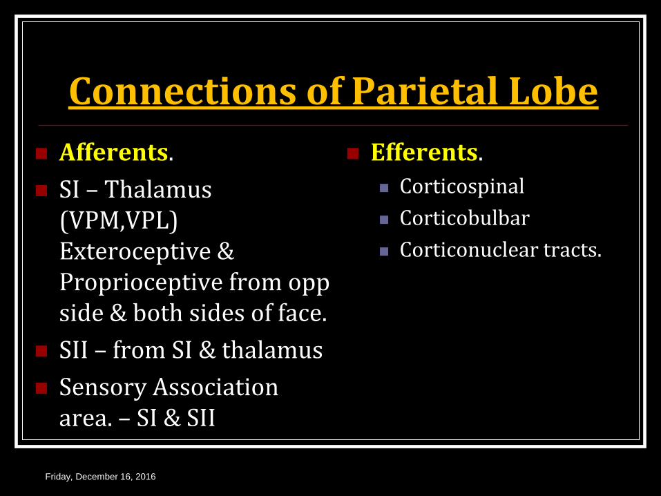
Connections of Parietal Lobe
Afferents.
SI – Thalamus (VPM,VPL) Exteroceptive & Proprioceptive from opp side & both sides of face.
SII – from SI & thalamus
Sensory Association area. – SI & SII
Efferents.
Corticospinal
Corticobulbar
Corticonuclear tracts.
Friday, December 16, 2016
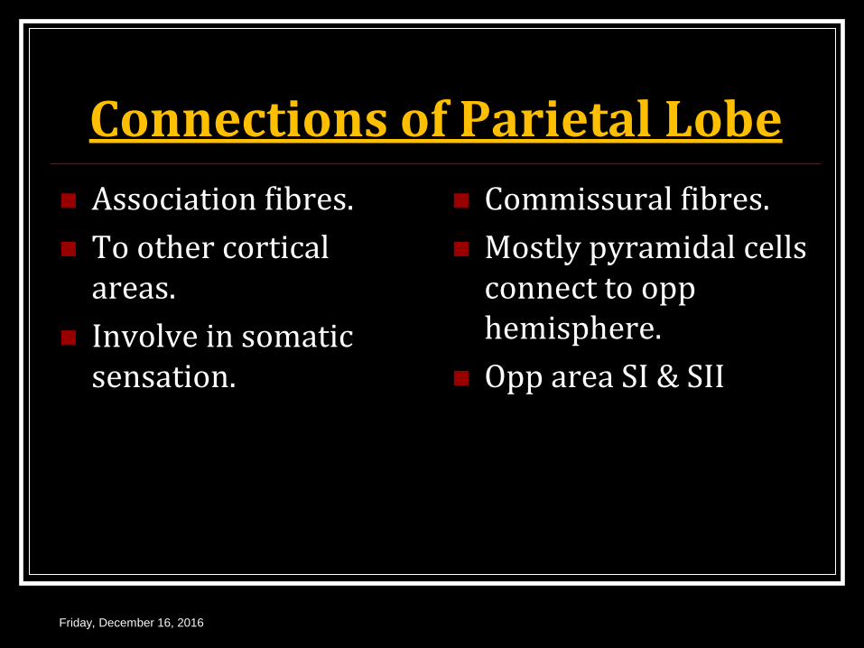
Connections of Parietal Lobe
Association fibres.
To other cortical areas.
Involve in somatic sensation.
Commissural fibres.
Mostly pyramidal cells connect to opp hemisphere.
Opp area SI & SII
Friday, December 16, 2016
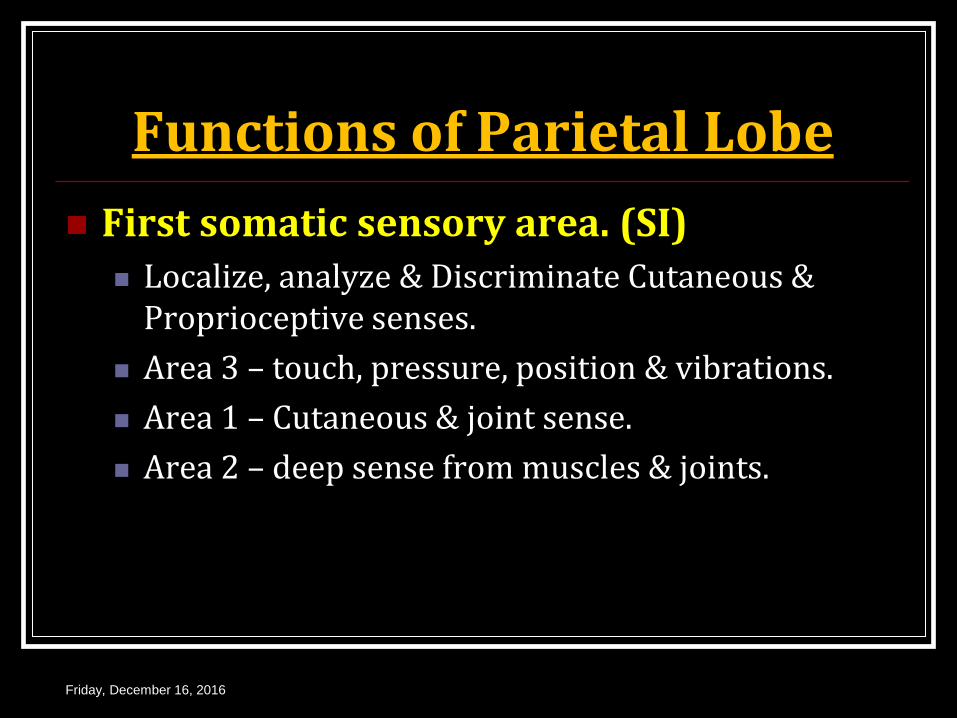
Functions of Parietal Lobe
First somatic sensory area. (SI) Localize, analyze & Discriminate Cutaneous &
Proprioceptive senses.
Area 3 – touch, pressure, position & vibrations.
Area 1 – Cutaneous & joint sense.
Area 2 – deep sense from muscles & joints.
Friday, December 16, 2016
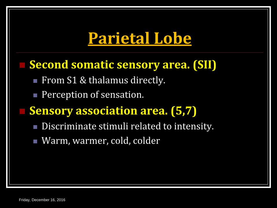
Parietal Lobe
Second somatic sensory area. (SII) From S1 & thalamus directly.
Perception of sensation.
Sensory association area. (5,7) Discriminate stimuli related to intensity.
Warm, warmer, cold, colder
Friday, December 16, 2016
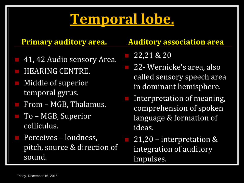
Temporal lobe.
Primary auditory area.
41, 42 Audio sensory Area.
HEARING CENTRE.
Middle of superior temporal gyrus.
From – MGB, Thalamus.
To – MGB, Superior colliculus.
Perceives – loudness, pitch, source & direction of sound.
Auditory association area
22,21 & 20
22- Wernicke's area, also called sensory speech area in dominant hemisphere.
Interpretation of meaning, comprehension of spoken language & formation of ideas.
21,20 – interpretation & integration of auditory impulses.
Friday, December 16, 2016

Applied Aspects
Unilateral removal of temporal lobe
Causes no deafness
As ear bilaterally represented in auditory pathway & project equally to 2 cerebral hemispheres.
Temporal lobe syndrome. (Kluver-Bucy syndrome)
Bilateral removal of temporal lobe with amygdala & Uncus.
Friday, December 16, 2016
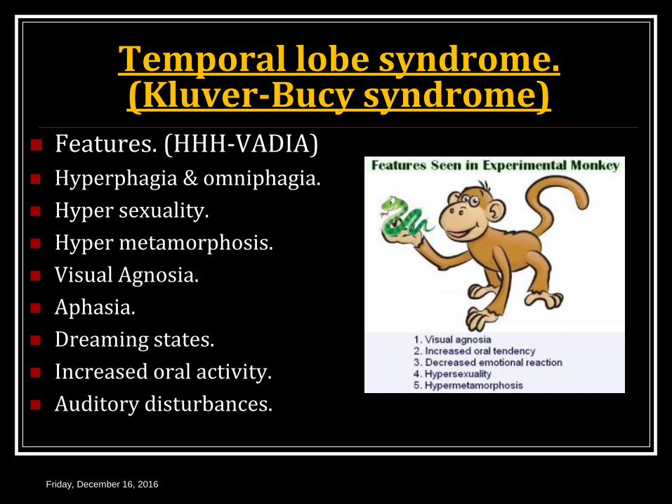
Temporal lobe syndrome. (Kluver-Bucy syndrome)
Features. (HHH-VADIA)
Hyperphagia & omniphagia.
Hyper sexuality.
Hyper metamorphosis.
Visual Agnosia.
Aphasia.
Dreaming states.
Increased oral activity.
Auditory disturbances.
Friday, December 16, 2016
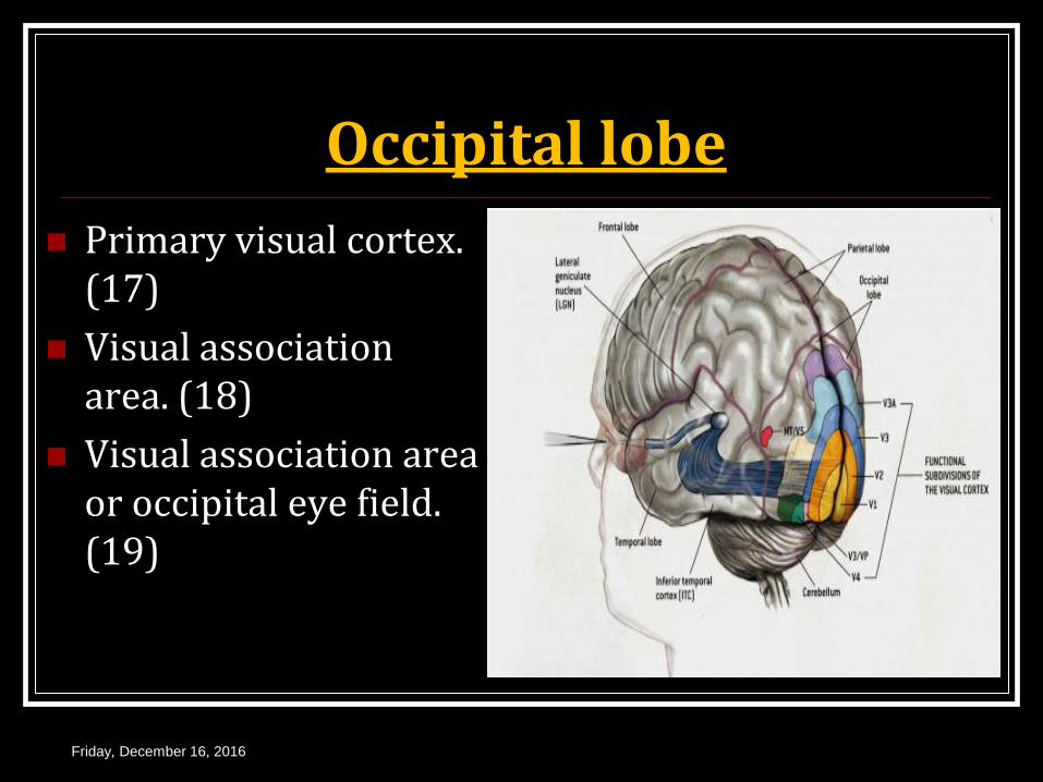
Occipital lobe
Primary visual cortex. (17)
Visual association area. (18)
Visual association area or occipital eye field. (19)
Friday, December 16, 2016
Connections & functions.
Afferents.
LGB via optic radiations.
Efferents.
Frontal eye field for eye movements.
Superior colliculus.
Cortico geniculate projections.
Thalamus.
Functions
Area 17– perception of visual impulses.
Area 18, 19 – interpretation , recognition & identification from memory.
occipital eye field area.– movement of eye ball.
Friday, December 16, 2016

White matter of cerebrum.
Association fibres.
Commissural fibres.
Projection fibres.
Friday, December 16, 2016
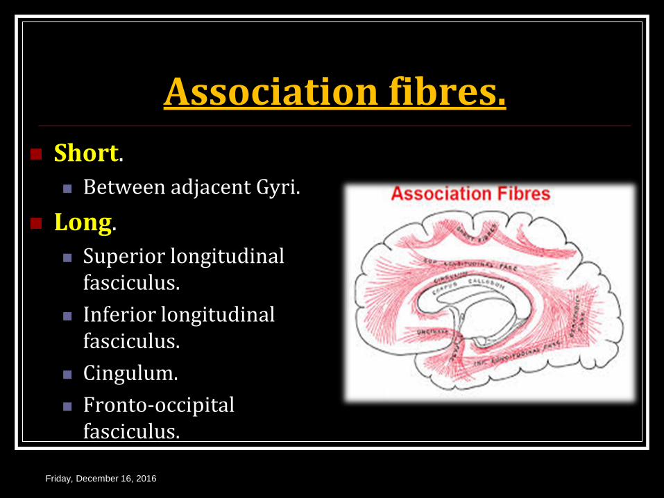
Association fibres.
Short.
Between adjacent Gyri.
Long.
Superior longitudinal fasciculus.
Inferior longitudinal fasciculus.
Cingulum.
Fronto-occipital fasciculus.
Friday, December 16, 2016
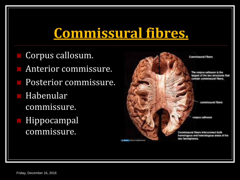
Commissural fibres.
Corpus callosum.
Anterior commissure.
Posterior commissure.
Habenular commissure.
Hippocampal commissure.
Friday, December 16, 2016
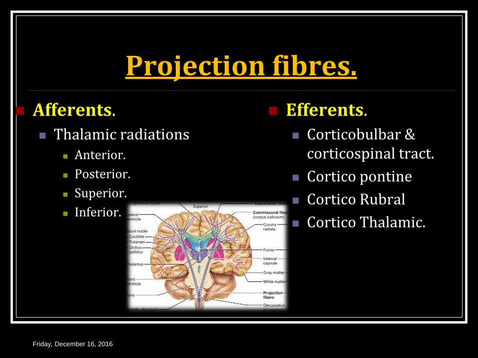
Projection fibres.
Afferents.
Thalamic radiations Anterior.
Posterior.
Superior.
Inferior.
Efferents.
Corticobulbar & corticospinal tract.
Cortico pontine
Cortico Rubral
Cortico Thalamic.
Friday, December 16, 2016

Internal capsule Thick curved band.
Fans out up as corona radiata & down as crus cerebri.
Most common part of Infarction & Hemorrhage.
Most common artery – Striate branch of middle cerebral
artery so called Artery of cerebral hemorrhage of Charcot’s artery.
Friday, December 16, 2016

Thank
You عمو اينشتاين باقي ويتمدد


