1
Ch 26: Reproduction & Development
Keypoints:
Human Chromosomes
Gametogenesis
Fertilization
Fetal development
Maintenance of pregnancy
Parturition
Terminology
� Gonads (testes and ovaries)
– Produce gametes(spermatozoa and ova)
� Gametes are from germ cells
� External genitalia
� Internal genitalia
� Autosomes vs. sex chromosomes
� Meiosis
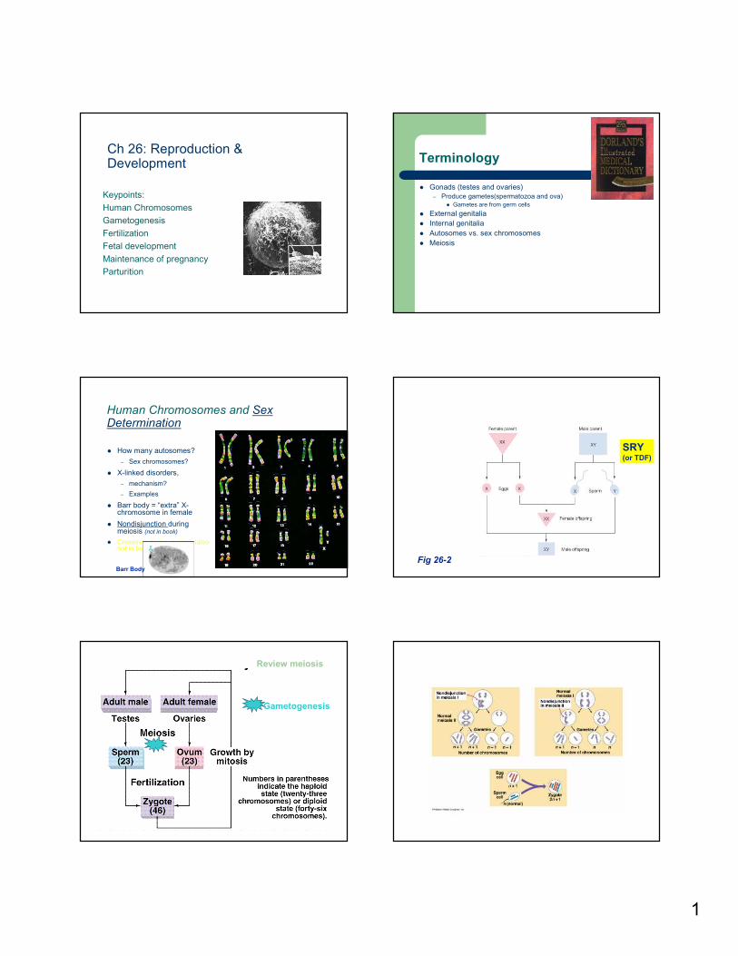
Human Chromosomes and Sex Determination
� How many autosomes?
– Sex chromosomes?
� X-linked disorders,
– mechanism?
– Examples
� Barr body = “extra” X-chromosome in female
� Nondisjunction during meiosis (not in book)
� Crossover during meiosis (also not in book)
Barr Body
Fig 26-2
SRY (or TDF)
Human Life Cycle
Gametogenesis
Review meiosis
2
Nondisjunction
Abnormality Karyotype
� Down Syndrome: Trisomy 21
� Turner Syndrome: X
� Triple-X Syndrome: XXX
� Klinefelter Syndrome: XXY
� Jacob Syndrome: XYY
Turner Syndrome
Relatively normal lives – but no functional
ovaries. 1 in 6,000 birth affected.
Monosomy X (45,X).
Characteristically
broad, "webbed"
neck. Stature
reduced, edema in
ankles and wrists.
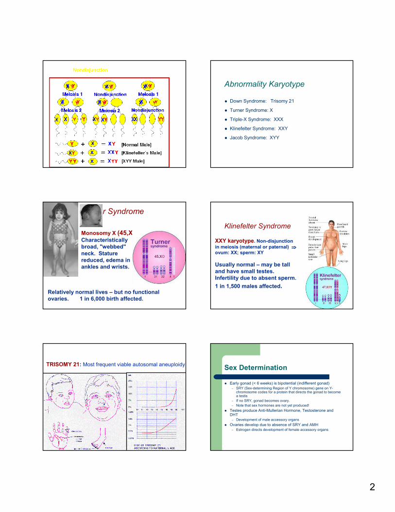
Klinefelter Syndrome
XXY karyotype. Non-disjunction
in meiosis (maternal or paternal) ⇒⇒⇒⇒
ovum: XX; sperm: XY
Usually normal – may be tall
and have small testes.
Infertility due to absent sperm.
1 in 1,500 males affected.
Nondisjunction of Autosomal Chromosomes
TRISOMY 21: Most frequent viable autosomal aneuploidy.Sex Determination
� Early gonad (< 6 weeks) is bipotential (indifferent gonad)
– SRY (Sex-determining Region of Y chromosome) gene on Y-chromosome codes for a protein that directs the gonad to become a testis
– If no SRY, gonad becomes ovary.
– Note that sex hormones are not yet produced!
� Testes produce Anti-Mullerian Hormone, Testosterone and DHT
– Development of male accessory organs
� Ovaries develop due to absence of SRY and AMH
– Estrogen directs development of female accessory organs
3
Intersex
� True hermaphrodite (both male and female gonads): relatively rare and poorly understood
� Pseudohermaphrodite – external genitalia of one sex and internal sex organs of the other sex. Mostly no ambiguity in the sex of the external genitalia ⇒ no question about gender at birth
� Male pseudohermaphroditism due to 5 α-reductase deficiency and ↓ DHT production. Born with female external genitalia
� Androgen Insensitivity Syndrome = XY genotype, but no receptors for androgens. Thus, the phenotype is female. (not in book)
� At puberty, testosterone causes development of male characteristics
Gametogenesis
Starts in utero – resumes at puberty
General principle same for males and females
Male: continuous sperm manufacture. Meiosis produces 4 spermatozoa
Female: born with all possible oocytes. Meiosis produces 1 ovum
Oogenesis: Egg Cell Formation
Oogonia mitosis ceases
before birth
At birth: only primary
oocytes – suspended in
prophase of meiosis I (=
prophase I)ovulation
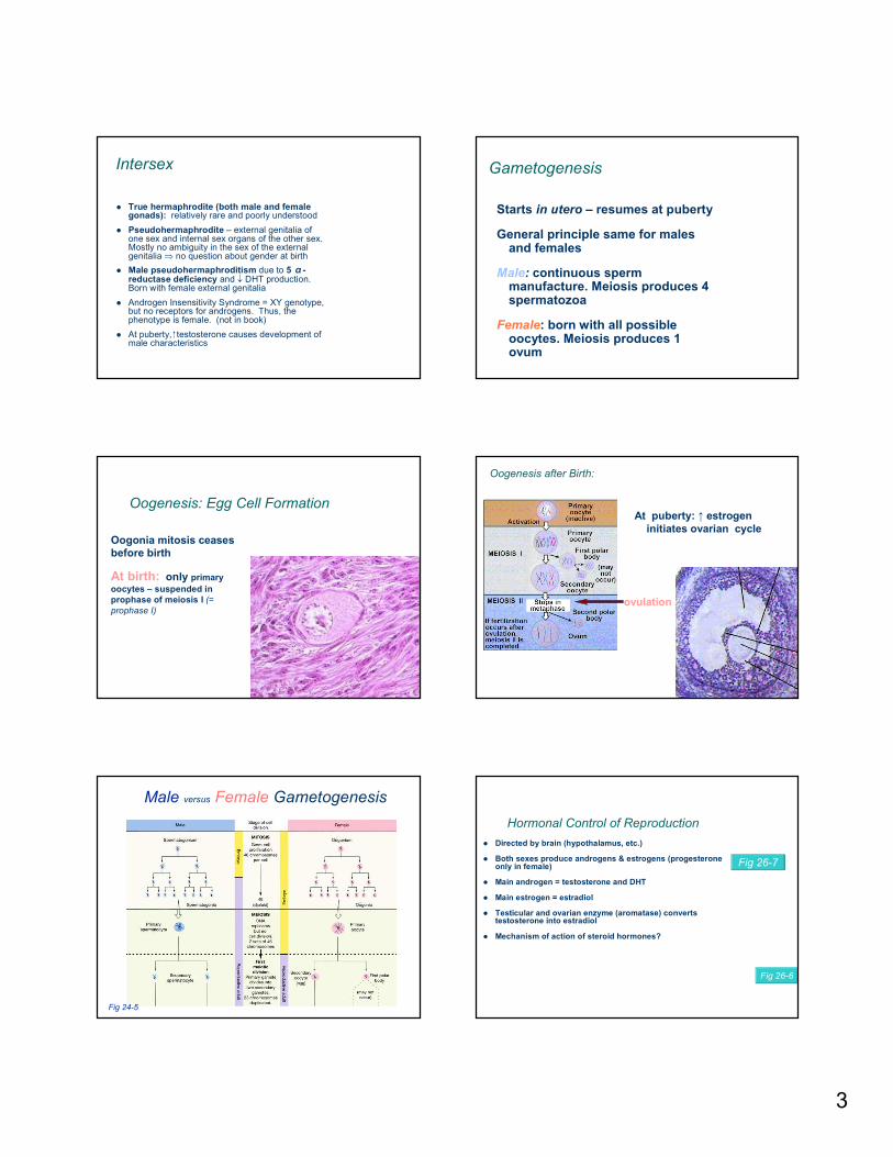
Oogenesis after Birth:
At puberty: ↑ estrogen
initiates ovarian cycle
2nd meiotic division
completed after fertilization
Male versus Female Gametogenesis
Fig 24-5
Hormonal Control of Reproduction
� Directed by brain (hypothalamus, etc.)
� Both sexes produce androgens & estrogens (progesterone only in female)
� Main androgen = testosterone and DHT
� Main estrogen = estradiol
� Testicular and ovarian enzyme (aromatase) converts testosterone into estradiol
� Mechanism of action of steroid hormones?
Fig 26-7
Fig 26-6
4
Interactions Between Hypothalamus, Anterior Pituitary, and Gonads
Short and
long negative
feedback
loops as
typical for
homeostasis
Special: High
levels of
estrogen ⇒⇒⇒⇒ pos.
feedback! ⇒⇒⇒⇒ LH Peak
Biosynthetic Pathway for Steroid Hormones
Theca cellsSynthesize androgens
Granulosa cellsconvert androgens to
estrogen
diffusion
Female Reproduction
Anatomy review:
Ovaries and uterus
Menstrual Cycle lasts ~ 1 month
Ovarian cycle
(changes in follicles)
function: monthly
production of gametes
Uterine cycle
(changes in
endometrial lining)
function: receive
developing embryo
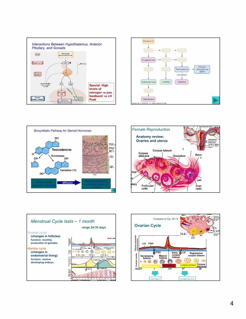
range 24-35 daysOvarian Cycle
estrogen progesterone
Follicular phaseLuteal phaseOvulation
Compare to Fig. 26-13
5
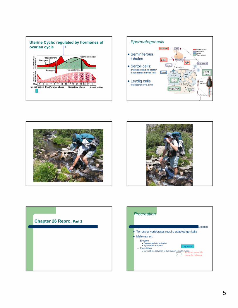
Uterine Cycle: regulated by hormones of
ovarian cycle ?
Beginning of follicular
phase of ovaries
during luteal phase of
ovaries
Spermatogenesis
� Seminiferous
tubules
� Sertoli cells: androgen binding protein
blood testes barrier etc.
� Leydig cells testosterone vs. DHT
Chapter 26 Repro, Part 2
Procreation
� Species specific behaviors ensure reproductive success
� Terrestrial vertebrates require adapted genitalia
� Male sex act:
– Erection� Parasympathetic activation
� Sympathetic inhibition
– Ejaculation� Sympathetic activation of duct system smooth muscle
Fig 26-15
Arterial smooth
muscle relaxes
6
The Erection Reflex
NT: NO
Viagra prolongs the
effect of NO
- Feedback
loop
Birth Control = Contraception
� Voluntary regulation of # of children produced & when
� List methods of contraception from most effective to
least effective
� Infertility
How do birth
control pills
work?
Pregnancy and Parturition p 848
1. Transport of spermatozoa and ova
2. Fertilization
3. Blastogenesis
4. Implantation
5. Maintenance
6. Parturition
7. Lactation
2. Fertilization
Where? /Uterine Tube
~ 100 sperm needed
When? After ovulation
Egg: 12-24 h post ovulation
Sperm: viable for up to 72 h
Then: 3-4 day journey to uterus
Fertilization cont.
� Sperm must penetrate
several layers
� Acrosomal reaction allows
sperm penetration
� 1st sperm reaching egg binds to
sperm-binding receptors on oocyte
membrane & enters
� Cortical reaction prevents
polyspermy
� Resulting structure = ?
7
3. Developing Zygote Implants in Secretory Endometrium
� Dividing zygote moves from distal fallopian
tube to uterine cavity over period of 3-4 days
� Implantation of the blastocyst into the
endometrium~ 7 days after fertilization
Fig 26-18
See your Fig 26-18
4. Implantation
� Protection of
embryo/fetus
� Nutritional support
� Ejection of fetus at
birth
Uterine functions:
Prenatal Genetic Testing
Amniocentesis: Fetus is 14-16 weeks old� Biochemical analysis of fluid searches for disease markers
� Cell culture can take several weeks ⇒⇒⇒⇒ Karyotyping and DNA
testing
Not in textbook
Chorionic Villi Sampling� Placental chorionic villi can be analyzed for genetic
abnormalities
� Can be done at 8 weeks (recommendation: 10 weeks)
– Earlier than amniocentesis
� No cell culture necessary
5. Maintenance of Pregnancy
� Progesterone is generally the hormone that maintains
pregnancy
– Quiescent uterus, no contractions
� hCG secreted by developing placenta (related to LH) ⇒⇒⇒⇒ Prevents
CL from degenerating and stimulates it to continue to produce
progesterone
� Week 7: placenta takes over progesterone production; CL
degenerates
� hCG also important in pregnancy testing (and for male sexual
development)
� hCG used for pregnancy testing

8
6. Labor and Delivery
� Parturition = Birthing process
� At 38 - 40 weeks of gestation
� Initiation poorly – sequence of events well
understood
– (Placental CRH?)
� Relaxin
– From ovaries and placenta
� The positive feedback loop of parturition
– Stopped after cervical stretch is gone
Fig 26-21+ Feedback Loop
7. Lactation
� Development at puberty due to estrogen
� Milk production: prolactin
– Anterior Pituitary
– Prolactin Inhibiting Hormone (PIH)� From hypothalamus
� ↓ during late pregnancy
� hPL: Human Placental Lactogen
– May aid breast development
– May be associated with Gestational Diabetes?
� “Let-down” due to oxytocin
� Colostrum
– Earliest milk after parturition
– Lots of Ab for newborn
Growth and Aging
� Puberty ( GnRH)
– in girls = menarche
� Average 12 y
– In boys, later (harder to pinpoint a time)
� Menopause
– Irregular menstrual periods and cessation
� Andropause
– Similar drop in androgens


