104
Int. J. LifeSc. Bt & Pharm. Res. 2012 Amal Kumar Mondal and Sayantan Tripathi, 2012
COMPARATIVE (QUANTITATIVE ANDQUALITATIVE) STUDIES OF STOMATA
OF SELECTED SIX MEDICINALLYVIABLE SPECIES OF CASSIA L.
Sayantan Tripathi1 and Amal Kumar Mondal1*
Research Paper
The Stomatal diversity (size, shapes, types and orientation) in the foliar epidermis has great
value in plant systematics studies. The present paper deals with comparative study of stomatal
structure of six species of Cassia which are used by rural people of South West Bengal as
medicine. Leaf epidermal studies mainly stomatal studies of the 6 species of Cassia of family
Caesalpiniaceae viz., Cassia alata L., Cassia fistula L., Cassia occidentals L., Cassia siamea
Lamk., Cassia sophera L., Cassia tora L. were made. Leaf clearings and cuticular preparations
were examined with light microscopy. The study is based on the presence and absence of
stomata, types of stomata present in the epidermal surface, stomatal count/cm2, stomatal index
and epidermal cell shape. The size and shapes of stomata are also varied in the tree species
which bear larger size of stomata in respect of other habits and habitats. Three types of stomata
were observed viz. paracytic, anisocytic and anomocytic. Among these 3 types of stomata the
paracytic type of stomata are more common and than other.
Keywords: Epidermis, Medicinal use, Cassia, Paracytic stomata
*Corresponding Author: Amal Kumar Mondal,[email protected]
INTRODUCTIONVarious plants and its parts are used in medicinal
purpose from ancient age. Now a days it is
observed that the African gorillas also use various
plants and its parts to cure their health. The
medicinal plants have been used by Hakims and
in folklore medicines as 80% of the population
ISSN 2250-3137 www.ijlbpr.comVol. 1, No. 3, July 2012
© 2012 IJLBPR. All Rights Reserved
Int. J. LifeSc. Bt & Pharm. Res. 2012
1 Plant Taxonomy, Biosystematics and Molecular Taxonomy Laboratory, Department of Botany & Forestry, Vidyasagar University.
lives in rural areas that mostly depend on Unani
system of medicines (Soomro et al., 1997). The
available literature shows that leaf epidermal
features are important in systematic botany
similar to the use of modern techniques and
chemical composition (Edeoga and Ikem, 2001;
and Mbagwu and Edeoga, 2006). Epidermal
105
Int. J. LifeSc. Bt & Pharm. Res. 2012 Amal Kumar Mondal and Sayantan Tripathi, 2012
structures and stomatal ontogeny of some
Nigerian ferns have been found relevant in their
recognition (Gill and Karatela, 1985).
Many workers such as Edeoga (1991),
Edeoga and Osawe (1996), Mbagwu and Edeoga
(2006), Nwachukwu and Mbagwu (2006)
stressed that epidermal and cuticular traits of
plants epidermal cells, type and arrangement of
stomata, size and shape of trichomes and
number of vascular bundles could serve as vital
tools in solving taxonomic problems in
Angiosperms.
Stomatogenesis has long been studied by
morphologists, physiologists and taxonomist .The
morphology and ontogenies of taxa are important
in intrageneric systematics. Diversity in stomata
types, even on the same surface of an organ,
indicates the weakness in using stomata as a
taxonomic character. (Pant and Kidwai, 1964). In
spite of diversity, the most frequent stomata type
can be used as a taxonomic character (Gopal,
1970).
Caesalpiniaceae is a family of dicotyledonous
plant with about 160 genera and some 2,000
species. The genus Cassia is one of the genuses
of Caesalpiniaceae having about 692 species.
Some of the Cassia species is used as medicine
in Eastern India mainly South West Bengal. We
select Cassia alata L., Cassia fistula L., Cassia
occidentals L., Cassia siamea Lamk., Cassia
sophera L., Cassia tora L. which are frequently
used by the rural and tribal people of south West
Bengal.
Apart from physiognomic characters,
anatomical properties of plant parts are sources
for taxonomic inferences in different groups of
flowering plants (Edeoga et al., 2007; Guimeraes
et al., 2007; Kaplan et al., 2007; Keshavarzi and
Zare, 2006). Despite the immense economic
importance of the legumes and the physiological
importance of the stomatal apertures, reports on
the frequency and the structure of the stomata
are lacking or incomplete for many species.
On the basis of arrangement of epidermal all
neighbouring the guard all, more than 25 main
types of stomata in dicots have been recognized
(Metcalfe and Chalk, 1979). Stace (1980) reported
31 different types of stomata among cotyledonous
plants. But the present study is based on the
paper of Metcalfe and Chalk (1950) who described
the 4 types – i.e. Anisocytic, Anomocytic, Diacytic
and Paracytic.
The aim of present study is to use stomatal
characters as aid in taxonomy of these selected
medicinally useful Cassia species. The study
would help in the identification and authentication
of these medicinal plants on the basis of
stomatogenesis.
MATERIAL AND METHODSPlant Material: The medicinal plant specimens
Cassia alata L., Cassia fistula L., Cassia
occidentals L., Cassia siamea Lamk., Cassia
sophera L. and Cassia tora L. belonging the family
Caesalpiniaccae under order Leguminales were
taken for stomatal study.
Cassia alata L. is popular in traditional
treatment of asthma, leprosy and it is used for
controlling of ringworm (Paria, 2005).
Role of Cassia fistula L. is used as purgative
and also used to treat skin disease. Its leaves
are used to treat skin disease and ulcer. Powder
of Cassia fistula L. root is given to cure menstrual

106
Int. J. LifeSc. Bt & Pharm. Res. 2012 Amal Kumar Mondal and Sayantan Tripathi, 2012
disorder and its pulp of seed is used in high blood
pressure (Paria, 2005).
Cassia ossidentales L. is popularly used as
purgative. It is also used to treat hysteria,
whooping cough and urinary troubles (Paria,
2005).
Cassia sophera L. is an annual erect shrub of
important medicinal properties. The whole plant
extracts and leaves have expectorant properties,
cures cough, asthma and acute bronchitis. They
are specific to eliminate ring worms and also
useful in the treatment of gonorrhoea and syphilis.
The bark is used in the treatment of diabetes; the
roots in elephantiasis, wounds. (Kirtikar and
Basu,1935).
The leaves of Cassia siamea Lamk. are used
in the treatment of diabetes, disturbances in the
bodily functions, lymph node swelling, urine
stones, general deficiency conditions, beri beri ,
classic deficiency of avitaminose by lack of
vitamin B1 (thiamine) in gastrointestinal disorders
- malabsorption - meals taken with polished rice
etc., antihypertensive, insomnia (sleeplessness),
against dysentery and disorders of the large
intestine. (Kirtikar and Basu, 1935).
Cassia tora L. is popularly known for the
treatment of dysentery and eye disease. Leaves
are also used in boils, ring worm, leprosy, bronchitis
and liver complaints. Pest of fresh pods is applied
to cure lentigo. Seed powder is taken to cure
intestinal problems caused by intestinal worms.
Seed pest mixed with boiled rice water is applied
in case of one sided headache (Paria, 2005).
The species identification of the selected
material was determined according to standard
literature. It was done in the months from February
to April. The foliar epidermal peals were taken
from the middle of both surfaces of mature leaves.
Isolation of Epidermis: Epidermis of leaf is
isolated from both fresh and dry plant specimen.
The mature leaves were fixed in FAA solution
(acetic acid: alcohol: formalin: water = 2:5:1:12)
for 24 hours and washed in 70% ethanol. Three
circular disk samples were cut from an area
adjacent to the midrib of each leaf. Disk sample
was boiled in 5% aqueous solution of KOH for
5-10 minute. Epidermal peals were stripped and
stained with 1% in 50% aqueous ethanol,
saffranin and temporary mount in glycerine.
Stomatal frequency counts made and camera
lucida were drawn. The numbers of stomata were
counted in each field (0.001386 cm2). The
stomatal frequency was based on average
obtained from observations of 3 microscopic
fields. Stomatal index (I) was calculated by the
following formula using the no. of stomata(s)
and epidermal cells was present in a unit area
[I=S/(S+E)].
After preparing, slides were observed under
light microscope [(40x)Leica DM 1000] and phase
contrast microscope for detailed analysis and
obtaining better picture as well as measuring the
length and breadth of stomata including guard
cells.
RESULTS AND DISCUSSIONThe results in this investigation were summarised
in tables including stomata count, stomata type,
stomatal index, stomatal complex.
The entire selected species share a number
of common characters, i.e., shape of epidermal
cell, presence of stomata, stomatal index and
stomatal complex which are useful in
distinguishing these species.
Epidermal Cell: The shape of foliar epidermis is
one of the significant taxonomic characters.

107
Int. J. LifeSc. Bt & Pharm. Res. 2012 Amal Kumar Mondal and Sayantan Tripathi, 2012
Taxonomic studies of a number of families are
based on leaf epidermis anatomy (Bhatia, 1984,
Jones, 1986). In this investigation it was found that
the epidermal cell shape varies slightly with growth
habit. The shrub species posses pentagonal to
polygonal shaped epidermal cells. The epidermal
cell shapes of tree species are variously shaped
or undulating to irregular (Table 1).
In these six species only Cassia siamea Lamk.
(Figure 2E, F) bears hypostomatic leaves i.e.
stomata present only at the abaxial surface of
leaves. Here it was found that the shape of
epidermal cells varies in their surfaces. The
adaxial surface of the leaf shows tetragonal to
polygonal shaped epidermal cells but the abaxial
surface i.e. stomata bearing surface shows
undulating to irregular shaped epidermal cells.
Stomata: Three different types of stomata
anomocytic, anisocytic, paracytic were observed
in these selected species. It was found that
abundant stomata were present on abaxial
surface as compared to adaxial surface.
All these selected species bear paracytic
stomata except the adaxial surface of Cassia
alata L. (Figure 2A). The adaxial surface of Cassia
alata L. (Figure 2A) bears anisocytic stomata.
Anisocytic and paracytic stomata were also found
in the abaxial surface of Cassia occidentals L.
(Figure 1B), Cassia sophera L. (Figure 1F) and
Cassia tora L. (Figure 1D). From this investigation
it was found that all the anisocytic stomata
bearing species are shrub in growth habit.
Anomocytic stomata are found only in the abaxial
surface of Cassia siamea Lamk. (Figure 2F). All
the investigated species showed paracytic
stomata, a mixture of stomata in same species
were also observed. Cassia ocidentales L., Cassia
sophera L. and Cassia tora L. are closely related.
They showed paracytic stomata in adaxial surface
and in the abaxial surface they showed paracytic
stomata along with anisocytic type (Table 1).
StomataPresence Stomata Type
S. No. Taxa Habit Surface Leaf Epidermal Cell Shape of Anomo- Aniso- Para- Dia-Stomata cytic cytic cytic cytic
1. Cassia alata L. Shrub Ad. Pentagonal to hexagonal + – + – –
Ab. Pentagonal to hexagonal + – + + –
2. Cassia fistula L. Tree Ad. Variously shaped + – – + –
Ab. Variously shaped + – – + –
3. Cassia occidentals L. Shrub Ad. Pentagonal to polygonal + – – + –
Ab. Pentagonal to polygonal + – + + –
4. Cassia siamea Lamk. Tree Ad. Tetragonal to polygonal – – – – –
Ab. Undulating to irregular + + – + –
5. Cassia sophera L. Shrub Ad Pentagonal to polygonal + – – + –
Ab Undulating to irregular + – + +
6. Cassia tora L. Shrub Ad. Pentagonal to polygonal + – – + –
Ab. Pentagonal to polygonal + – + + –
Table 1: Qualitative Foliar Stomatal Characteristics of the Selected Species of Cassia
108
Int. J. LifeSc. Bt & Pharm. Res. 2012 Amal Kumar Mondal and Sayantan Tripathi, 2012
S. No. Taxa Habit Surface Stomatal Complex. Legth × Width (with guard cell) µm2
1. Cassia alata L. Shrub Ad 23.89(21.28-25.77)×20.27(18.43-22.41)
Ab 18.48(17.46-19.31)×12.36(10.47-14.12)
2. Cassia fistula L. Tree Ad 13.74(13.04-14.65)×11.66(11.32-12.59)
Ab 15.22(12.5-17.5)×10.25(7.75-12.5)
3. Cassia occidentals L Shrub Ad 23.11(22.31-23.81)×16.49(15.67-17.11)
Ab 22.98(21.65-25.48)×17.09(14.28-20.99)
4. Cassia siamea Lamk. Tree Ad –
Ab 23.49(21.34-25.40)×15.32(14.49-16.71)
5. Cassia sophera L. Shrub Ad 24.85(23.61-26.47)×15.24(14.47-16.10)
Ab 22.54(21.24-24.55)×15.29(14.54-16.35)
6. Cassia tora L. Shrub Ad 22.10(19.99-23.16)×13.06(11.84-13.92)
Ab 23.28(23.27-23.29)×15.66(15.17-16.38)
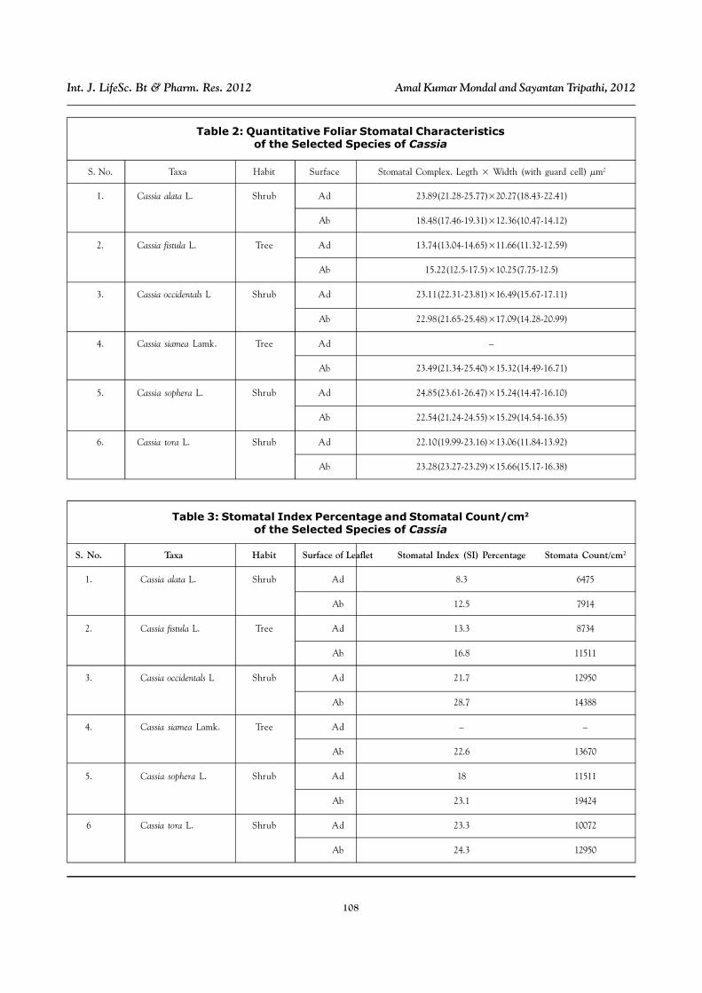
Table 2: Quantitative Foliar Stomatal Characteristicsof the Selected Species of Cassia
S. No. Taxa Habit Surface of Leaflet Stomatal Index (SI) Percentage Stomata Count/cm2
1. Cassia alata L. Shrub Ad 8.3 6475
Ab 12.5 7914
2. Cassia fistula L. Tree Ad 13.3 8734
Ab 16.8 11511
3. Cassia occidentals L Shrub Ad 21.7 12950
Ab 28.7 14388
4. Cassia siamea Lamk. Tree Ad – –
Ab 22.6 13670
5. Cassia sophera L. Shrub Ad 18 11511
Ab 23.1 19424
6 Cassia tora L. Shrub Ad 23.3 10072
Ab 24.3 12950
Table 3: Stomatal Index Percentage and Stomatal Count/cm2
of the Selected Species of Cassia

109
Int. J. LifeSc. Bt & Pharm. Res. 2012 Amal Kumar Mondal and Sayantan Tripathi, 2012
Figure 1: Leaf Epidermis Showing Stomata ofA. Cassia occidentalis (adaxial surface); B. Cassia occidentalis (abaxial surface);
C. Cassia tora (adaxial surface); D. Cassia tora (abaxial surface);E. Cassia sophera (adaxial surface); and F. Cassia sophera (abaxial surface)

110
Int. J. LifeSc. Bt & Pharm. Res. 2012 Amal Kumar Mondal and Sayantan Tripathi, 2012
Figure 2: Leaf Epidermis Showing Stomata ofA. Cassia alata (adaxial surface); B. Cassia alata (abaxial surface);
C. Cassia fistula (adaxial surface); D. Cassia fistula (abaxial surface);E. Cassia siamea (adaxial surface); and F. Cassia siamea (abaxial surface)
111
Int. J. LifeSc. Bt & Pharm. Res. 2012 Amal Kumar Mondal and Sayantan Tripathi, 2012
Quantitative analysis of stomatal complex in
the selected species shows great variation (Table
2). Here it was found that except Cassia fistula L.
Cassia alata L., Cassia occidentals L., Cassia
siamea Lamk., Cassia sophera L., Cassia tora
L. share almost equal size of stomata which
shows close relationship. In these 5 species
Cassia sophera L. and Cassia occidentals L.
share almost equal size of stomata and the ratio
between size of stomata of adaxial and abaxial
surface is almost equal. Smallest stomata were
found in the adaxial surface of Cassia fistula L.
(13.74×11.68 µm2).
Stomatal index is one of the useful tools in order
to distinguish species. It was found that stomatal
index has low value on adaxial surface as
compared to abaxial surface (Table 3). According
to stomatal index Casia alata L. and Cassia fistula
L. are closely related. Cassia occidentals L.,
Cassia siamea Lamk., Cassia sophera L. and
Cassia tora L. showed relationship among them.
Highest value of stomatal index was observed
on abaxial surface of Cassia occidentals L. (28.7),
where lowest value was observed on the adaxial
surface of Cassia alata L. (8.3).
Stomatal count is a significant character to
differentiate various species. Highest stomata
count per unit area was found on the abaxial
surface of Cassia sophera L. (194245/cm2).
Lowest stomata count was found in adaxial
surface of Cassia alata L. (6475/cm2). All the
investigated Cassia species share more or less
equal number of stomata which shows they are
closely related.
CONCLUSIONThe study and investigation mainly focused on
comparative study of quantitative and qualitative
stomatal characters of 6 species of Cassia
belonging to family Caesalpniaceae under the
order Leguminales, used in medicinal purpose in
the South West Bengal region.
From the study it can be concluded that there
is no relationship between the stomata size and
growth habit. Size of stomata and epidermal cell
shape prove that all the investigated species are
closely related. All the investigated species
showed paracytic stomata, a mixture of stomata
in same species were also observed. Cassia
ocidentales L., Cassia sophera L. and Cassia tora
L. are closely related. They showed paracytic
stomata in adaxial surface. In the abaxial surface
they showed both anisocytic and paracytic type
of stomata. Shape of epidermal cells, presence
of stomata, stomatal type, stomatal complex,
stomatal index were investigated. These
parameters are helpful to differentiate the species.
However, the stomatal features may prove to be
a little taxonomic value unless the developments
of different stomata types were studied. A greater
number of information on taxa will be helpful to
understand the taxonomic value of stomata type
and distribution.
ACKNOWLEDGMENTWe are grateful to our honourable Vice Chancellor
of Vidyasagar University, Prof. Ranjan
Chakraborti, and we are also thankful to our
friends (Santanu Dash and Dulal Chandra Bera).
Without their help we cannot complete our work.
REFERENCES1. Ahmad K, Khan M A, Ahamad M, Shaheen
N and Nazir A (2010), “Taxonomic Diversity
in Epidermal cells of some Sub-Tropical
Plant Species”, International journal
112
Int. J. LifeSc. Bt & Pharm. Res. 2012 Amal Kumar Mondal and Sayantan Tripathi, 2012
of Agricultural & Biology, Vol. 12, No. 1,
pp. 115-118.
2. Allaby M (2004), A Dictionary of Plant
Sciences, Oxford University Press.
3. Carpenter SB and Smith N D (1975),
“Stomatal Distribution and Size in Southern
Appalachian Hard Woods”, Canad. J. Bot,
Vol. 53, pp. 1153-1156.
4. Bhatia R C (1984), “Folier epidermal studies
of Heliotropium supinum L. Folier” Geo Bot.
Phytotaxon, Vol. 19, pp. 381-385.
5. Edeoga HO and C I Ikem (2001), “Comparative
Morphology of leaf epidermis in Three
Species of Boerhaaevia L”, J. Econ Tax.
Bot., Vol. 19, pp. 197-205.
6. Edeoga H O, Omosun G, Osuagwu G G E
and Emezue O O (2007), “Microscopic
Anatomy and Histochemistry Of Stem And
Root of Some Mimosa Species”,
(Leguminosae- Mimosoideae)”, Asian
Journal of Plant Sciences, Vol. 6, No. 4,
pp. 688-691.
7. Gill L S, Olabanji G O and Husaini S W H
(1982), “Studies on Structural Variation and
Distribution of Stomata in Some Nigerian
Legumes”, Willdenowia, Vol. 12, pp. 87-94.
8. Gill L S and Y Y Karatela (1985), “Epidermal
Morphology and Stomatal Ontogeny in
Some West African Convolulaceae
species”, Herba Hungarice, Vol. 24,
pp. 11-17.
9. Gopal B V and Mishra A K (1980), “A
New Technique for Plant Epidermal
Studies”, Kenya Science & Tech. (B), Vol. 1,
pp. 99-100.
10. Guimeraes A C, Kuster R M, Amaral A F,
Ferreira J P and Siani A C (2007),
“Histological Study of the Leaf and Stem of
the Amazonian Medicinal Mistletoe
Cladocolea micrantha (Loranthaceae)”,
International Journal of Botany, Vol. 3, No.
2, pp. 218-221.
11. Hameed I, Hussain F and Dastagir G
(2010), “Anatomical Studies of Some
Medicinal Plants of Family Polygonaceae”,
Pak. J. Bot., Vol. 42, No. 5, pp. 2975-2983.
12. Hutchinson J (1964), The Genera of
Flowering Plants, Clarendon Press, Oxford,
Vol. 1.
13. Kaplan A, Hasanoglu A and Ince I A
(2007), “Morphological, Anatomical and
Palynological Properties of Some Turkish
Veronica species (Scrophulariaceae)”,
International Journal of Botany, Vol. 3,
No. 1, pp. 23-32.
14. Keshavarzi M and Zare G (2006),
“Anatomcial Study of Salicornieae Dumort,
(Chenopodiaceae Vent.) Native to Iran”,
International Journal of Botany, Vol. 2,
No. 3, pp. 278-285.
15. Kirtikar K R and Basu B D (1935), Revised
by Blatter E, Caius J F and Mhaskar K S,
Indian Medicinal Plants, Vols. I-IV, 2nd Ed., L
M Basu, Allahabad.
16. Kothari M J, Shah, G L (1975), “Epidermal
Structure and Ontogeny of Stomata in the
Papilionaceae (Tribe Hedysareae)”, Bot.
Gaz., Vol. 136, No. 4, pp. 372-379.
17. Mbagwu F N and H O Edeoga (2006),
“Observations on the Vegetative and Floral

113
Int. J. LifeSc. Bt & Pharm. Res. 2012 Amal Kumar Mondal and Sayantan Tripathi, 2012
Morphology of Some Vigna Species
(Leguminosae-Papilionoideae)” Pakistan
Journal of Biological Sciences, Vol. 9,
No. 9, pp. 1754-1758.
18. Metcalfe, C R and Chalk (1950), “Anatomy
of the Dicotyledons”, Clarendon Press,
Oxford.
19. Mondal A K (2005), “Advanced Plant
Taxonomy), New Central Book Agency,
Kolkata, pp. 254-264.
20. Nwachukwu C U and F N Mbagwu (2006),
“Morphological features in some species of
Indigofera L. (Leguminosae-Papilionoideae)”,
Journal of Fisheries International, Vol. 2-4,
pp. 50-54.
21. Paliwal G S and Bhandari N N (1962),
“Stomatal Development in Some Magno-
liaceae”, Phytomorphology, Vol. 12, pp. 409-
412.
22. Paliwal G S (1966), “Structure and Ontogeny
of Stomata in Some Acanthaceae”, Phyto-
mophology, Vol. 16, pp. 527-532.
23. Paliwal G S (1967), “Permanent Peel
Mounts for Developmental Studies of
Stomata in Leaves”, Curr.Sci., Vol. 36,
p. 191.
24. Pant D D and P F Kidwai (1964), “On the
Diversity in the Development and
Organization of Stomata in Phyla nudiflora
Michx”, Current Sci., Vol. 33, pp. 653-654.
25. Paria N D (2005), Medicinal Plant
Resources of South West Bengal, Saraswaty
Press Limited, pp. 39-42, Kolkata.
26. Prain D (2004), “Bengal Plants”, Vol-I-II,
Reprinted Edition, 1963, Botanical Survey
of India, Kolkata.
27. Raunkiaer C (1937), “Plant Life Forms”,
Clarendon Press, Oxford.
28. Shah G L (1968), “Development of stomata
in some Papilionaceae”, J. Indian Bot. Soc.,
Vol. 47, pp. 305-310.
29. Shah G L (1969), “Development of Stomata
in Some Papilionaceae”, Canad. J. Indian
Bot., Vol. 47, pp. 387-393.
30. Stace C A (1984), “The Taxonomic
Importance of the Leaf Surface”, in
Heywood V H and Moore D M (Eds.), Current
Concepts in Plant Taxonomy, Academic
Press, pp. 67-94, London.
31. Soomro R R, A Qureshi, M T Mahmood, M
A Khan and G A Makka (1997),
“Ethobotanical Uses of Adhatoda vesica
in Chest Diseases”, Hamdard Medicus,
Vol. 38, No. 1, pp. 24-29.
32. Stresburger E (1866), “Ein Beitrag zur
Entwicklungs geschichte der Spaltoff
nungen”, Jahrb. Wiss. Bot., Vol. 5, pp. 297-
342.
33. Wilkinson H P (1979), “The Plant Surface
(Mainly Leaf)”, in Metcalfe C R and Chalk L
(Eds.), Anatomy of the Dicotyledons, 2nd
Edition, Clarendon Press, Vol. 1, pp. 97-165,
Oxford.
34. Vesque I (1889), “De I’ emploides caracte’res
anatomiques dans la classification des
vegetaux”, Bull Soc. Bot. FR., Vol. 36,
pp. 41-77.


