DEVELOPMENT OF COGNITIVE FLEXIBILITY IN LATE ADOLESCENCE:
INVESTIGATING BEHAVIORAL PERFORMANCE AND NEURAL ACTIVATION IN
A TASK-SWITCHING PARADIGM
by
Sarah K. Lazzaro
Submitted to the Faculty of
University of Pittsburgh in partial fulfillment
of the requirements for the degree of
Bachelor of Philosophy in Neuroscience and Psychology
University of Pittsburgh
2018
ii
UNIVERSITY OF PITTSBURGH
University Honors College
This thesis was presented
by
Sarah K. Lazzaro
It was defended on
April 16th, 2018
and approved by
Jamie Hanson, PhD, Assistant Professor, University of Pittsburgh Department of Psychology
Melissa Libertus, PhD, Assistant Professor, University of Pittsburgh Department of Psychology
Jennifer Roth, PhD, Associate Professor, Carlow University Department of Psychology
Thesis Director: Beatriz Luna, PhD, Professor, University of Pittsburgh Department of
Psychiatry

iii
Copyright © by Sarah K. Lazzaro
2018
iv
Successful cognitive control relies on both the ability to instantiate higher-order cognitive
functions and the ability to flexibly switch between them in service of changing task demands,
i.e. cognitive flexibility. While a wealth of important work on the development of cognitive
control in adolescence has focused on the development of executive functions, there has been a
relative lack of work on the development of cognitive flexibility. Here we address this limitation
by investigating the development of cognitive flexibility using a task-switching paradigm in a
large sample of adolescents and young adults (ages 14-32, n = 82). For a subset of subjects that
had usable fMRI data (n=56), we assessed task-switching performance and analyzed fMRI data
collected in-scanner while they performed the task-switching paradigm. We observed that
successful task-switching was associated with widespread activation of frontoparietal and visual
processing brain areas. A component of this larger task-switching system, the left inferior
parietal cortex, showed age-related reductions in neural activation specifically during task-
switching into trials that taxed inhibitory control. These neural findings occurred in parallel with
age-related improvements in successful task-switching performance in the same context. This
pattern of results suggests that task-switching into the most cognitively demanding contexts
follows a protracted development that extends through adolescence and young adulthood.
Further, the age-related reduction in parietal cortex activation suggests that adolescents have
greater reliance on the frontoparietal system, which has been implicated in transient aspects of
cognitive control, to achieve adult-like performance. Taken together, our results suggest that a
key aspect of cognitive maturation in adulthood is the ability to flexibly switch between
cognitive tasks with limited cost to performance and a decreasing reliance on frontoparietal
regions across adolescence.
DEVELOPMENT OF COGNITIVE FLEXIBILITY IN LATE
ADOLESCENCE: INVESTIGATING BEHAVIORAL PERFORMANCE AND
NEURAL ACTIVATION IN A TASK-SWITCHING PARADIGM
Sarah K. Lazzaro
University of Pittsburgh, 2018
v
TABLE OF CONTENTS
PREFACE ................................................................................................................................. VIII
1.0 INTRODUCTION ........................................................................................................ 1
1.1 COGNITIVE CONTROL AND ADOLESCENCE .......................................... 1
1.2 COGNITIVE FLEXIBILITY ............................................................................. 2
2.0 METHODS ................................................................................................................... 5
2.1 SAMPLE ............................................................................................................... 5
2.2 TASK DESIGN .................................................................................................... 6
2.3 ANALYSIS ........................................................................................................... 8
2.3.1 Behavioral......................................................................................................... 8
2.3.2 Functional MRI................................................................................................ 9
2.3.2.1 Data Acquisition .................................................................................... 9
2.3.2.2 Statistical Analysis .............................................................................. 10
3.0 RESULTS ................................................................................................................... 12
3.1 BEHAVIORAL .................................................................................................. 12
3.1.1 Task Performance.......................................................................................... 12
3.1.2 Development of Task Performance .............................................................. 13
3.1.3 In-Scanner Performance ............................................................................... 15
3.2 BRAIN ACTIVITY ........................................................................................... 15
3.2.1 Task Effects .................................................................................................... 15
3.2.2 Developmental Effects ................................................................................... 16
4.0 DISCUSSION ............................................................................................................. 18
4.1 TASK-SWITCHING ......................................................................................... 18
4.2 DEVELOPMENT .............................................................................................. 19
BIBLIOGRAPHY ....................................................................................................................... 23

vi
LIST OF TABLES
Table 1. Participant Demographics. ................................................................................................ 6
Table 2. Task Performance Linear Models for out of-scanner participant visit. .......................... 13

vii
LIST OF FIGURES
Figure 1. Modified multi-source interference task. ........................................................................ 7
Figure 2. Comparative performance for out-of-scanner visit split by task type and switch type. 12
Figure 3. Switch cost plotted as a function of age for Interference and Congruent task types. .... 14
Figure 4. Brain Activation main effects for Task Type (Interference – Congruent contrast). ..... 15
Figure 5. Brain Activation main effects for Switch Type (Switch – Non-Switch contrast). ........ 16
Figure 6. Neural activation for TaskType x SwitchType interaction with associated HRF. ........ 16
Figure 7. Age by Switch Type neural activation patterns for frontal cortex ROIs. ...................... 17
Figure 8. Age by Switch Type interaction for Interference task type only................................... 17

viii
PREFACE
I would like to thank my thesis director, Dr. Beatriz Luna, for the opportunity to research,
write, and defend this thesis with her guidance. Special thanks to Dr. Bart Larsen for his
dedicated tutelage and mentorship during the research and writing of this thesis, and without
whom this project would not have been possible. There are not enough words in the world to
express my gratitude. Additional thanks to all committee members, whose time and advice is
much appreciated, and to everyone in the Laboratory of Neurocognitive Development for their
support during my work here. Lastly, my sincerest appreciation for Kristie Budihardjo, Jennifer
Crook, Renata Mitchell, and Alina Quach for their invaluable encouragement and faith in this
thesis and in me throughout our shared time in undergraduate studies.
1
1.0 INTRODUCTION
1.1 COGNITIVE CONTROL AND ADOLESCENCE
Cognitive control is the ability to coordinate internal goals and external behavior in the face of
complex and potentially distracting environments. Successful cognitive control relies on the
ability to consistently instantiate higher-order cognitive functions, such as working memory,
response selection and inhibition, task-set switching, and performance monitoring, as well as the
ability to flexibly engage and disengage these processes according to task demands (Badre 2011;
Lenartowicz et al. 2010; Sabb et al. 2008). As such, cognitive control may be broken down into
two facets: 1) executive function, i.e. the ability to successfully engage higher-order cognitive
processes, and 2) cognitive flexibility, the ability to successfully switch between these executive
functions (Luna et al. 2015).
The ability to successfully use cognitive control has been linked to the function of
distributed set of brain systems, including the frontoparietal, dorsal attention, and cingulo-
opercular networks. The frontoparietal system, consisting of areas of prefrontal and posterior
parietal cortices, has been functionally linked to transient aspects of cognitive control, such as
rule-updating, task-switching, and performance monitoring, and is thus thought to be central to
adjusting control in response to task feedback or changing task demands (Cole et al., 2013;
Dosenbach et al., 2007; Larsen, Verstynen, Yeh, & Luna, 2017). This is in contrast to the dorsal

2
attention and cingulo-opercular systems (consisting of the intraparietal sulcus and frontal eye
field, and the medial frontal cortex, insula, and frontal operculum respectively) which are
thought to be involved in more sustained aspects of cognitive control, such as goal-directed
sustained attention and across-trial rule-set maintenance (Fox et al. 2006; Vossel, Geng, & Fink,
2014).
Cognitive control continues to develop throughout childhood, adolescence, and early
adulthood. Importantly, children and adolescents can execute cognitive control at a given
instance, but are less consistently successful over time when performance is compared to that of
adults (Luna et al. 2015). Adolescence, in particular, is marked by a reduction in the variability
of cognitive control performance and increasing consistency in successful execution with age; in
this way, adolescence may be conceptualized as a period of refinement of cognitive control,
which manifests as decreasing error rates in tasks with high demands on cognitive function
(Luna, Padmanabhan, & O'Hearn, 2010). Understanding the transition into adult-like cognitive
control is particularly important because adolescence is the age of emergence of a variety of
psychopathology including schizophrenia, mood disorders, substance abuse disorders that are
characterized, at least in part, by impairments in cognitive control (Giedd, Keshavan, & Paus,
2009; Luna & Sweeney, 2004). Thus, understanding the normative development of cognitive
control may provide insight into the development of abnormalities in cognitive control.
1.2 COGNITIVE FLEXIBILITY
Prior work on the development of cognitive control in adolescence has been primarily
focused on development of executive functions, and less is known about the development of
3
cognitive flexibility. Studies have shown that executive functions such as inhibitory control
(Luna & Sweeney, 2004; Thomas, 2013; Williams et al. 1999), working memory (Crone et al.
2006; Luciana et al. 2005), and performance monitoring (Rubia et al., 2006; Wiersema, van der
Meere, & Roeyers, 2007) continue to show improvement through late adolescence both in terms
of correct response rates and response times. Studies involving the development of cognitive
flexibility are scarce, but work in adult task-switching and cognitive flexibility has found
evidence for the differential recruitment of frontoparietal regions associated with transient
cognitive control (Braver, Reynolds, & Donaldson, 2003). Some prior work in this area has
confirmed that adolescents lack the consistency that adults demonstrate in task-switching
conditions and exhibit higher variability in successful performance in task-switching (Cepeda,
Kramer, & Gonzalez de Sather, 2001; Reimers & Maylor, 2005); however, cognitive flexibility
is still largely unexplored in a developmental context.
To add to the body of knowledge on the development of cognitive flexibility in
adolescence, here we investigate the neural underpinnings of cognitive flexibility and their
development during adolescence. Specifically, we employ a task-switching paradigm that
engages flexible switching between executive functions, including working memory and
inhibitory control, in service of changing task demands, and administer it to a developmental
sample of adolescents and young-adults while they undergo fMRI. We hypothesize that
cognitive flexibility (the ability to successfully switch between executive functions) will improve
with age, with adults showing greater rates of correct responses than adolescents during task
switching. Further, considering the role of the frontoparietal system in transient aspects of
cognitive control, such as task switching and rule-updating, we hypothesize that these age-related

4
improvements in cognitive flexibility will be related to the development of components of the
frontoparietal system over the course of adolescence.
5
2.0 METHODS
2.1 SAMPLE
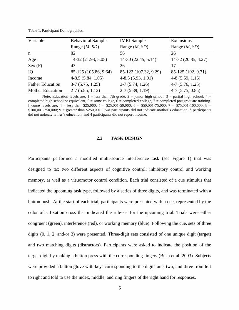
Eighty-two adolescents and young adults participated in this study (ages 14-32; M = 21.93, SD =
5.05; 43 Female). Six of these participants did not complete an MR visit. Subjects were excluded
from all analyses if they had high omission rate (omission > 20%; n = 4) due to concerns with
compliance with experiment instructions. Exclusions from MR analysis included participants
with less than 25 correct trials in the pure condition (n = 4) and those with incomplete MR data
(n = 12); the final sample for MR analyses was comprised of 56 subjects. Participant
demographics are reported in Table 1. Study inclusion criteria included childhood (age 10) IQ
scores greater than 80, no self-reported history of psychiatric or neurological disorder, no current
use of psychiatric medication, no history of head injury with loss of consciousness, and no MRI
contraindications, including pregnancy, claustrophobia, and non-removable metal in the body.
The Institutional Review Board at the University of Pittsburgh approved this study.
6
Table 1. Participant Demographics.
Variable Behavioral Sample
Range (M, SD)
fMRI Sample
Range (M, SD)
Exclusions
Range (M, SD)
n 82 56 26
Age 14-32 (21.93, 5.05) 14-30 (22.45, 5.14) 14-32 (20.35, 4.27)
Sex (F) 43 26 17
IQ 85-125 (105.86, 9.64) 85-122 (107.32, 9.29) 85-125 (102, 9.71)
Income 4-8.5 (5.84, 1.05) 4-8.5 (5.93, 1.01) 4-8 (5.59, 1.16)
Father Education 3-7 (5.75, 1.25) 3-7 (5.74, 1.26) 4-7 (5.76, 1.25)
Mother Education 2-7 (5.85, 1.12) 2-7 (5.89, 1.19) 4-7 (5.75, 0.85)
Note: Education levels are: 1 = less than 7th grade, 2 = junior high school, 3 = partial high school, 4 =
completed high school or equivalent, 5 = some college, 6 = completed college, 7 = completed postgraduate training.
Income levels are: 4 = less than $25,000; 5 = $25,001-50,000; 6 = $50,001-75,000; 7 = $75,001-100,000; 8 =
$100,001-250,000; 9 = greater than $250,001. Two participants did not indicate mother’s education, 8 participants
did not indicate father’s education, and 4 participants did not report income.
2.2 TASK DESIGN
Participants performed a modified multi-source interference task (see Figure 1) that was
designed to tax two different aspects of cognitive control: inhibitory control and working
memory, as well as a visuomotor control condition. Each trial consisted of a cue stimulus that
indicated the upcoming task type, followed by a series of three digits, and was terminated with a
button push. At the start of each trial, participants were presented with a cue, represented by the
color of a fixation cross that indicated the rule-set for the upcoming trial. Trials were either
congruent (green), interference (red), or working memory (blue). Following the cue, sets of three
digits (0, 1, 2, and/or 3) were presented. Three-digit sets consisted of one unique digit (target)
and two matching digits (distractors). Participants were asked to indicate the position of the
target digit by making a button press with the corresponding fingers (Bush et al. 2003). Subjects
were provided a button glove with keys corresponding to the digits one, two, and three from left
to right and told to use the index, middle, and ring fingers of the right hand for responses.

7
Figure 1. Modified multi-source interference task.
Each trial starts with a colored fixation cross that acts as a cue (green represents visuomotor “congruent”,
red represents inhibitory control “interference”, and blue represents working memory “n-back”). “XXX”
trials are n-back probes. The above example is from a “mixed” block in which subjects are required to
switch between conditions. In “pure” blocks subjects see consecutive trials of one trial type only.
Visuomotor response trials (Congruent) featured the target number in a corresponding
position to the physical location of the response digit, while inhibitory control trials
(Interference) introduced a mismatch in target position and location of correct button (Bush et al.
2003). The working memory (N-back) trial type functioned as a two-back n-back task (Kirchner,
1958; Owen, McMillan, Laird, & Bullmore, 2005). Participants had to maintain the sequence of
target identities in working memory and were occasionally presented with probe trials (XXX) to
signal the participant to recall the correct response from two trials back in the sequence. Three
“pure” blocks comprised of 35 sequential trials of one type were included in order to estimate the
hemodynamic response for each trial type (Ollinger, Shulman, & Corbetta, 2001). Six “mixed”
8
blocks of 60 trials each interleaved all three trial types at random to provide a task-switching
condition. Within switch blocks, 11 trials were switches, and switch trials could occur a
minimum of two trials after the previous switch (2-8 trials, M = 4.87, SD = 1.89). The order of
the blocks was counterbalanced across participants. Each trial consisted of a fixation cues lasting
0.5 s, followed by the three-digit stimulus presentation that remained on screen until a response
was made or the trial timed out (1.0, 1.3, and 1.5 s for Congruent, Interference, and N-back
respectively). Each trial was followed by a variable inter-trial interval lasted an average of 1.77
seconds and followed an exponential distribution. Participants performed this task once during a
lab visit outside the MR scanner, then a second time inside the scanner while acquiring fMRI.
Considering the different setup of N-back blocks and the resulting direct measure of performance
for working memory switch trials, for the purpose of this study, we limited trial types to
Interference and Congruent.
2.3 ANALYSIS
2.3.1 Behavioral
For statistical analyses, Task Type was defined as rule-set for the current trial (Congruent,
Interference, Working Memory). Switch Type was defined as either “pure” (trial from the pure
block), “switch” (a switch trial from the mix block), or non-switch (a non-switch trial from the
mix block). The final categorical variable, Switch From, is the trial type of the immediately
preceding trial. Reaction time was calculated as the mean reaction time of correct trials of that
task condition, and accuracy as the total number of correct trials divided by the total number of
9
trials. Switch Cost was calculated as the accuracy of non-switch trials subtracted from the switch
trial accuracy.
Primary behavioral analysis was performed with linear mixed and mixed-effects models
to examine main effects and interactions between task conditions and age measures and were
implemented in MATLAB R2016a (The Mathworks, Inc.; fitlm and fitlme) using default
settings. Trials with omission errors were excluded from the analysis. All statistical analyses
carried out on behavioral data were also performed on behavioral data collected in-scanner.
2.3.2 Functional MRI
2.3.2.1 Data Acquisition
Stimuli were presented onto a screen behind the scanner using EPrime software (Psychology
Software Tools, Inc., Pittsburgh, PA) and were visible to the subject through a mirror mounted to
the head coil. Reaction time was recorded as the interval in milliseconds from the three digit
stimulus to response (button press on MRI safe button box). Trials were marked correct if the
button response (Pointer Finger, Middle Finger, or Ring Finger, right hand glove) correctly
indicated the identity of the target number. If no response was given after a specified period
specific to the trial type (1000, 1300, and 1500ms for Congruence, Interference, and N-back
respectively), the trial was “timed-out” and marked as an omission error.
Imaging data were collected using a 3.0-T Siemens Magnetom TIM Trio (Erlangen,
Germany) at the Magnetic Resonance Research Center at the University of Pittsburgh. Structural
images were collected using a magnetization prepared rapid acquisition gradient-echo (MP-rage)
pulse sequence with 192 slices (1 mm slice thickness; 1 mm isotropic voxels). Functional data
were collected using an echo-planar imaging (EPI) sequence with the following parameters: TR
10
= 1.0s (4x multiband acceleration), TE = 30 ms, Flip Angle = 55, and 96 x 96 acquisition matrix
with a field of view of 220 mm. Sixty slices were collected in the axial plane with an isotropic
voxel size of 2.3 mm.
Standard techniques were used to preprocess the functional data and used the same
pipeline as previous work from our group (Paulsen et al. 2015). This included wavelet de-
spiking, slice timing correction, motion correction (Jenkinson et al. 2002), brain extraction, non-
linear registration of functional data to a standardized anatomical brain (MNI-152 template),
spatial smoothing with FWHM of 4.25 mm (SUSAN; Smith & Brady, 1997), high pass filtering
at 0.008 Hz, and rescaling to a 10,000-unit global median.
2.3.2.2 Statistical Analysis
All imaging analysis was performed with Analysis and Visualization of Functional Neuroimages
(AFNI, Bethesda, MD) software (Cox, 1996). To estimate the average hemodynamic response
function (HRF) for the task events, trial time courses for correct trials were modeled using TENT
basis functions spanning 25s with 26 time steps. Six rigid-body head motion parameters and their
derivatives, as well as run-wise 0 through 3rd order polynomials, were used as nuisance
regressors. The current and preceding TR were censored if the Euclidean norm head motion
distance surpassed 0.9mm. This choice of censoring threshold was guided by work examining
motion outliers in task-based fMRI (Siegel et al., 2014). Notably in this sample, motion was not
significantly correlated with age (r = -0.126, p = 0.32).
Based on visual inspection of the HRF, we limited between-subject analyses to peak trial
evoked response window by averaging the hemodynamic response between TRs 5 and 7 (5-7
seconds) after the onset of the trial to account for hemodynamic lag. In order to detect potential
subtle differences in the shape of the HRF as a function of condition, omnibus group effects were
11
examined on correct trial time courses and entered into a voxel-wise multivariate model
(3dMVM; Chen et al. 2015). To accurately compare differences in activation at peak response to
trial onset, models using either TR (18 time points) or peak (mean of activation for TR 5-8, at
estimated peak of response) were run. Peak or TR (if applicable), Task Type (Congruence and
Interference), and Switch Type (Switch, Non-Switch, and Pure) were entered as within-subject
effects and age measures (Age at MR scan) were entered as between-subject effects. As age was
best modeled as an inverse function in behavioral data analyses, age was also modeled as an
inverse function in fMRI analyses. Post-hoc GLTs were added to test for specific conditions and
comparisons, as well as timepoints (if applicable). An interaction term between condition and
trial type was used to identify voxels whose correct, trial-wise HRF significantly varied as a
function of age or inverse age. Voxelwise testing was masked to only include voxels with a 50%
or greater probability of being grey matter in the MNI-152 template and at least 98% EPI
coverage in all subjects across all runs. Results were corrected for multiple comparisons using
family-wise error correction based on cluster size within the voxelwise space as defined above.
Cluster size thresholds were determined through a Monte Carlo simulation using AFNI's
3dClustSim program with mean spatial autocorrelation parameters estimated from residuals from
TENT deconvolution. This analysis specified that 8 or more contiguous (faces touching) voxels
with a single voxel threshold of p = 0.005 (q < 0.05) for age at scan were required to achieve
corrected, cluster-level alphas of less than 0.05. Timecourses for conditions were extracted and
HRFs plotted for visualization.
12
3.0 RESULTS
3.1 BEHAVIORAL
3.1.1 Task Performance
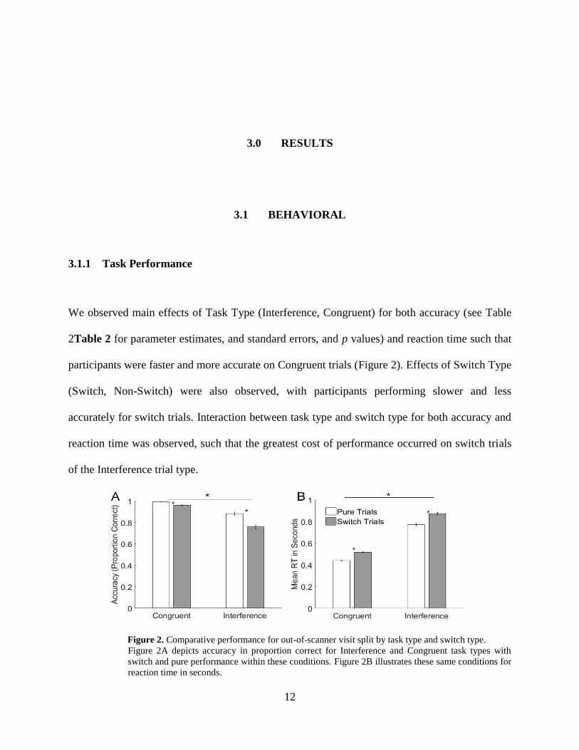
We observed main effects of Task Type (Interference, Congruent) for both accuracy (see Table
2Table 2 for parameter estimates, and standard errors, and p values) and reaction time such that
participants were faster and more accurate on Congruent trials (Figure 2). Effects of Switch Type
(Switch, Non-Switch) were also observed, with participants performing slower and less
accurately for switch trials. Interaction between task type and switch type for both accuracy and
reaction time was observed, such that the greatest cost of performance occurred on switch trials
of the Interference trial type.
Figure 2. Comparative performance for out-of-scanner visit split by task type and switch type.
Figure 2A depicts accuracy in proportion correct for Interference and Congruent task types with
switch and pure performance within these conditions. Figure 2B illustrates these same conditions for
reaction time in seconds.

13
Table 2. Task Performance Linear Models for out of-scanner participant visit.
Linear Model df Coefficient SE p
Accuracy ~ TaskType
778 -0.152 0.007 < 10-73****
Accuracy ~ SwitchType
777 -0.0744 0.0125 < 10-8****
Accuracy ~ TaskType*SwitchType 774
TaskType:Interference -0.108 0.0156 < 10-11****
SwitchType:Non-Switch -0.0075 0.0135 0.578
SwitchType:Switch -0.0312 0.0135 0.0211*
TaskType*SwitchType:Non-Switch -0.0249 0.0191 0.194
TaskType*SwitchType:Switch
-0.0863 0.0191 < 10-5****
RT ~ TaskType
778 0.329 0.00475 < 10-75****
RT ~ SwitchType
777 0.0879 0.0184 < 10-7****
RT ~ TaskType*SwitchType 774
TaskType:Interference 0.329 0.00858 < 10-180****
SwitchType:Non-Switch 0.0425 0.00743 < 10-7****
SwitchType:Switch 0.0746 0.00743 < 10-21****
TaskType*SwitchType:Non-Switch -0.0258 0.0105 0.0143*
TaskType*SwitchType:Switch 0.026 0.0105 0.0111*
Note: Note: *p < 0.05, **p < 0.01, ****p < 0.0001
3.1.2 Development of Task Performance
We observed a significant age by Task Type interaction for both accuracy (Parameter Estimate =
0.0033, SE = 0.0015, p = 0.024*) and reaction time (Parameter Estimate = -0.0019, SE =
0.00094, p = 0.043*), an effect that was driven by age-related improvement in reaction time and
accuracy in interference trials. Age by Switch Type interactions were not significant for either
accuracy (Parameter Estimate = 0.004, SE = 0.0025, p = 0.109) or reaction time (Parameter
14
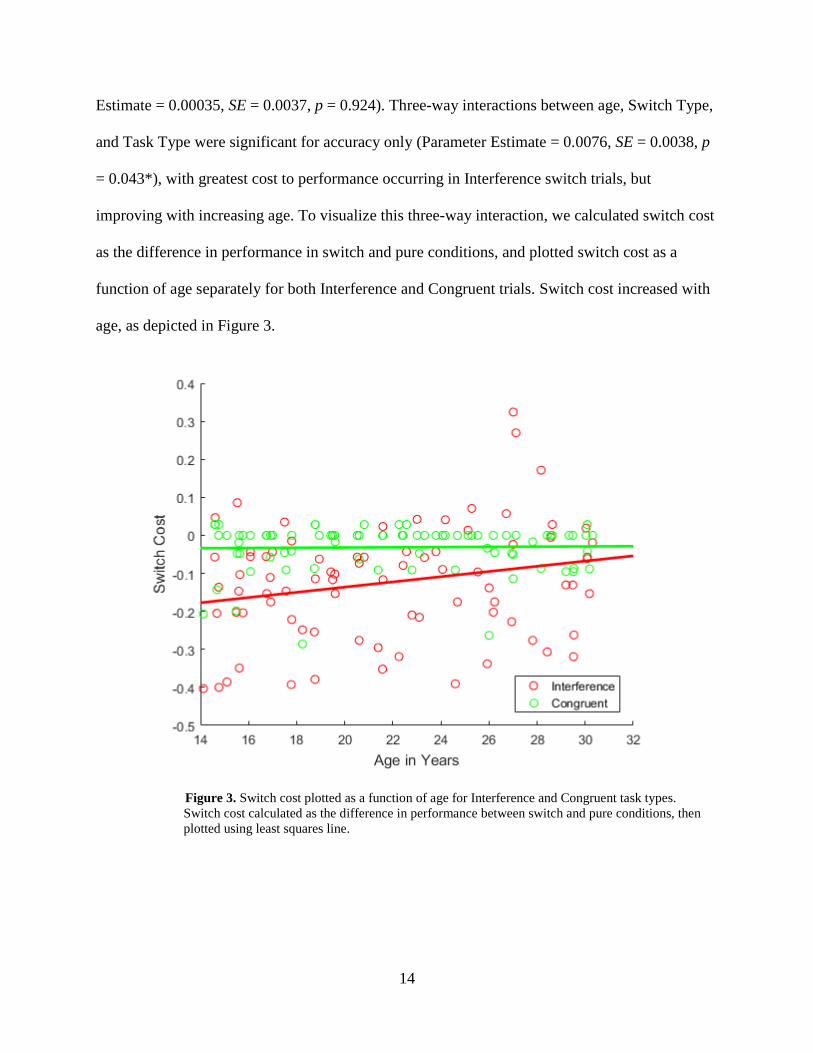
Estimate = 0.00035, SE = 0.0037, p = 0.924). Three-way interactions between age, Switch Type,
and Task Type were significant for accuracy only (Parameter Estimate = 0.0076, SE = 0.0038, p
= 0.043*), with greatest cost to performance occurring in Interference switch trials, but
improving with increasing age. To visualize this three-way interaction, we calculated switch cost
as the difference in performance in switch and pure conditions, and plotted switch cost as a
function of age separately for both Interference and Congruent trials. Switch cost increased with
age, as depicted in Figure 3.
Figure 3. Switch cost plotted as a function of age for Interference and Congruent task types.
Switch cost calculated as the difference in performance between switch and pure conditions, then
plotted using least squares line.

15
3.1.3 In-Scanner Performance
In-scanner performance was similar to out-of-scanner measures in directionality of effects. Task
performance followed the same pattern of results as the behavioral visit. Developmental effects
were no longer significant for reaction time by age and task type (Parameter Estimate =
-0.00018, SE = 0.0033, p = 0.96). In-scanner performance did not show a three-way interaction
between age, task type, and switch type for accuracy (Parameter Estimate = 0.0025, SE = 0.0025,
p = 0.3).
3.2 BRAIN ACTIVITY
3.2.1 Task Effects
We observed main effects of task type (Interference, Congruent) in frontal and parietal cortices,
such that participants showed greater activation in Interference trials (Figure 4). Main effects of
switch type were also observed, with participants showing greater occipital cortex activation in
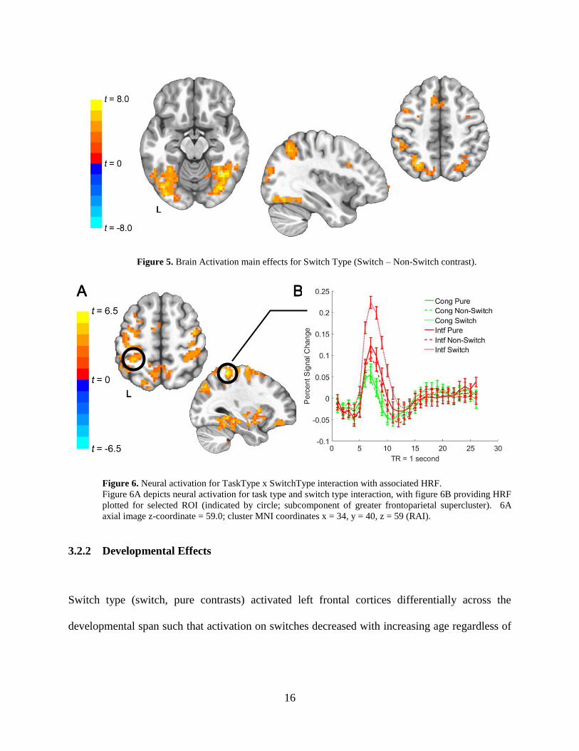
switch conditions compared to non-switch and pure trials (Figure 5). Interaction between task
type and switch type was observed, such that the greatest activation patterns occurred in fronto-
parietal cortex for Interference Switch conditions, as shown in Figure 6.
Figure 4. Brain Activation main effects for Task Type (Interference – Congruent contrast).
16
Figure 5. Brain Activation main effects for Switch Type (Switch – Non-Switch contrast).
Figure 6. Neural activation for TaskType x SwitchType interaction with associated HRF.
Figure 6A depicts neural activation for task type and switch type interaction, with figure 6B providing HRF
plotted for selected ROI (indicated by circle; subcomponent of greater frontoparietal supercluster). 6A
axial image z-coordinate = 59.0; cluster MNI coordinates x = 34, y = 40, z = 59 (RAI).
3.2.2 Developmental Effects
Switch type (switch, pure contrasts) activated left frontal cortices differentially across the
developmental span such that activation on switches decreased with increasing age regardless of

17
the task type condition (Figure 7). No significant task type by age interactions were observed in
the fMRI data.
Figure 7. Age by Switch Type neural activation patterns for frontal cortex ROIs.
7A axial image z-coordinate = 19.0; cluster MNI coordinates x = 53, y = -26, z = 20 (RAI). 7B axial image
z-coordinate = 55.0; cluster MNI coordinates x = 23, y = -32, z = 56 (RAI).
We did not observe a three-way interaction between age, Task Type, and Switch type;
follow up posthoc analyses revealed an age by Switch Type interaction for Interference trials
only in left inferior parietal cortex. The ROI appeared to be more active in Interference switch
conditions for younger participants, and decreased in relative signal change across the
developmental span (Figure 8).
Figure 8. Age by Switch Type interaction for Interference task type only.
Figure 8A depicts neural activation for inferior parietal ROI, with 8B showing age effects for switch,
nonswitch in interference condition. Axial image z-coordinate = 49.0, sagittal image z-coordinate = 47.0.
Cluster MNI coordinates x = 47, y = 38, z = 50 (RAI).
18
4.0 DISCUSSION
Adolescence may be conceptualized as a period of refinement of cognitive control, which we
hypothesize may be driven by continued development of cognitive flexibility. Prior work in
cognitive control has studied the development of executive functioning without much focus on
the element of flexibility, which we investigate here using a task-switching paradigm. This study
design investigates the neural underpinnings of cognitive flexibility by engaging higher-order
cognitive processes and changing task demands in order to assess flexibility. Of particular
interest in our brain analyses were regions implicated with cognitive attentional networks and
linked to multi-source interference tasks, such as the frontoparietal attention system, the cingulo-
opercular network, and the dorsal attention stream.
4.1 TASK-SWITCHING
Strong primary effects of task-switching were evident in behavioral and neural measures.
Performance, as measured by accuracy and reaction time, was significantly affected by more
cognitively demanding conditions such as inhibitory control task types and switch trials meant to
tax executive function when compared to less complex visuomotor tasks. Common attentional
systems were activated by cognitively demanding conditions, with task-switching leading to
greater activation of visual and parietal cortices than congruent tasks and non-switch trials, thus
19
supporting prior work that indicates continued development of aspects of the frontoparietal
system as critical for transient aspects of cognitive control and performance monitoring (Bush &
Shin, 2006; Rubia et al., 2006). Interestingly, we also observed robust activation of occipital
regions following the visual processing stream, indicating the importance of the visual cue for
rule-set information in switch conditions (as opposed to non-switch or pure trials, where the cue
information has little value). Differential activation for switch conditions highlights both the
task-monitoring and top-down components of attention as well as the incorporation of context-
specific cues in successful performance on cognitively demanding trials.
4.2 DEVELOPMENT
Simple task-switching (i.e. between less cognitively demanding tasks) behavior appears adult-
like in our youngest subjects, but more complex switches come with greater costs for accuracy
and reaction time in younger subjects. Complex task-switching that engages the highest level of
cognitive resources appears to show protracted development: we observed a decreasing switch
cost from adolescence to adulthood, with adults showing greater accuracy over adolescents while
switching into the inhibitory control task. This finding replicates prior studies in development of
cognitive control in adolescence, where decreasing switch costs across the developmental span
were also observed (Reimers & Maylor, 2005). The results suggest that a key aspect of cognitive
maturation in adulthood is the ability to flexibly switch between cognitive tasks with limited cost
to performance or speed. These findings were paired with an observation that switch cost for less
cognitively demanding conditions showed no developmental effects, suggesting that this ability
is already mature by adolescence, a finding supported by developmental research in cognitive
20
control (Crone & Dahl, 2012; Luna et al., 2010; Magar, Phillips, & Hosie, 2010). Future work
seeking to investigate the development of these cognitive processes would require a sample with
younger adolescents, and may indeed find age-related changes in less cognitively demanding
conditions as well as more robust developmental effects aligning with our findings.
No effect of task type (inhibitory control vs. visuomotor processing) was observed across
the developmental span, but switch trials showed greater recruitment of both frontoparietal and
ventral stream brain networks when compared to pure and non-switch trials. This effect was such
that adolescents showed greater activation in these regions than adults, implying that adolescents
must have greater activation in this region to successfully switch between conditions and achieve
adult-like performance. Though to a lesser degree than adolescents, adults continued to recruit
these areas on correct trials, indicating that the transient frontoparietal task-attentional and cue-
context visual ventral stream networks are critical for task success regardless of developmental
stage.
Effects of age and task-switching on performance were mirrored by the recruitment of
brain regions associated with transient aspects of cognitive control. Specifically, recruitment of
the left inferior parietal cortex during switches into interference trials was greater for adolescents
than adults and decreased with age across the developmental span, suggesting that these trial
conditions require a more effortful cognitive process for adolescents; reliance on this region may
decrease as synaptic pruning occurs or as complementary brain systems become sufficient to
successfully meet task demands. Developmentally-sensitive activation observed in the inferior
parietal lobule is congruent with findings from other work in adolescent task-switching, that
found task-switching recruited parietal cortex and that recruitment decreased with increasing age
(Rubia et al., 2006). Interestingly, these changes may be occurring in parallel to the recruitment

21
of brain regions involved in more sustained elements of cognitive control as adolescents develop,
such as more inferior aspects of parietal cortex (Velanova, Wheeler, & Luna, 2009), though we
did not observe age-related changes in brain regions thought to be associated with aspects of
cognitive control.
Many conditions, particularly those with low cognitive demand, showed no
developmental effects across our sample, suggesting that these functions have reached
developmental maturity prior to the age-range included in our sample (i.e. <14 years old). It is
important to emphasize that our sample focuses on late adolescence and early adulthood,
increasing the likelihood that certain systems have finished development by the ages represented
in the sample. The lack of developmental maturity in task-switching to a visuomotor task
highlights and lends specificity to our finding that task switches to the inhibitory control task
showed the greatest age-related improvement, with both switch cost to performance and neural
activation of the left inferior parietal lobule decreasing across the developmental span. In adults
this area still shows activation, suggesting that recruitment of the region is essential for
successful task switching but that switches into conditions with high cognitive demand have the
most protracted developmental timecourse.
This study suffers from certain limitations which stem from the task-switching paradigm
and particularly involve the less cognitively demanding task conditions. Participants successfully
completed non-switch trials at high accuracy rates regardless of developmental stage, resulting in
ceiling effects for statistical conclusions. In a broader context, the paradigm chosen taxes a
narrow range of cognitive functions related to task-switching and cognitive flexibility. Tasks
were chosen such that visuomotor response components were common to all task types, with the
primary difference between trials lying in cognitive manipulation required for success.
22
Therefore, this aspect of task-switching tested a narrow range of cognitive functions and did not
engage broader range of contexts that may impact cognitive flexibility, such as reward or
emotional motivation, which may have different timescales of development during the
adolescent period. Future work could address this using task paradigms that tax these processes,
incorporating facets of reward or emotional processing into task-switching and determining if
switches within these contexts follow different developmental trajectories and rely on additional
brain areas for success. Considering prior work on development of reward systems in
adolescence and evidence that adolescents can more reliably perform inhibitory control tasks at
adult-like levels when offered reward incentives (Geier & Luna, 2012; Padmanabhan et al.,
2011), it is possible that task-switching incorporating a reward component may differentially
impact cognitive flexibility for adolescents and adults such that adolescents are more consistently
able to instantiate difficult task-switches and exhibit more adult-like behavior when in a
potentially-rewarding context.
This study has found that cognitive flexibility continues to improve with age, particularly
when task demands are high. These age-related improvements in cognitive flexibility occur in
parallel with age-related changes in brain areas known to be involved in attentional processing
and cognitive control. Successful engagement of cognitive flexibility in high-demand task-
switching contexts continues to develop even over a late adolescent period. These components of
cognitive flexibility showing protracted development may be critical to the refinement of
cognitive control abilities that occurs before adulthood, and therefore integral to the
understanding of emerging psychopathology in this period.
23
BIBLIOGRAPHY
Badre, D. (2011). Defining an ontology of cognitive control requires attention to component
interactions. Topics in Cognitive Science, 3(2), 217–221. https://doi.org/10.1111/j.1756-
8765.2011.01141.x
Braver, T. S., Reynolds, J. R., & Donaldson, D. I. (2003). Neural mechanisms of transient and
sustained cognitive control during task switching. Neuron, 39(4), 713–726.
https://doi.org/10.1016/S0896-6273(03)00466-5
Bush, G., & Shin, L. M. (2006). The Multi-Source Interference Task: An fMRI task that reliably
activates the cingulo-frontal-parietal cognitive/attention network. Nature Protocols, 1(1),
308–313. https://doi.org/10.1038/nprot.2006.48
Bush, G., Shin, L. M., Holmes, J., Rosen, B. R., & Vogt, B. A. (2003). The multi-source
interference task: Validation study with fMRI in individual subjects. Molecular Psychiatry,
8(1), 60–70. https://doi.org/10.1038/sj.mp.4001217
Cepeda, N. J., Kramer, A. F., & Gonzalez de Sather, J. C. (2001). Changes in executive control
across the life span: examination of task-switching performance. Developmental
Psychology, 37(5), 715–730. https://doi.org/10.1037/0012-1649.37.5.715
Chen, G., Saad, Z. S., Adleman, N. E., Leibenluft, E., & Cox, R. W. (2015). Detecting the subtle
shape differences in hemodynamic responses at the group level. Frontiers in Neuroscience,
9(OCT), 1–18. https://doi.org/10.3389/fnins.2015.00375
Cole, M. W., Reynolds, J. R., Power, J. D., Repovs, G., Anticevic, A., & Braver, T. S. (2013).
Multi-task connectivity reveals flexible hubs for adaptive task control. Nature
Neuroscience, 16(9), 1348–1355. https://doi.org/10.1038/nn.3470.Multi-task
Cox, R. W. (1996). AFNI: Software for analysis and visualization of functional magnetic
resonance neuroimages. Computers and Biomedical Research, 29(3), 162–173.
https://doi.org/10.1006/cbmr.1996.0014
Crone, E. A., & Dahl, R. E. (2012). Understanding adolescence as a period of social-affective
engagement and goal flexibility. Nature Reviews Neuroscience, 13(9), 636–650.
https://doi.org/10.1038/nrn3313
Crone, E. A., Wendelken, C., Donohue, S., van Leijenhorst, L., & Bunge, S. A. (2006).
24
Neurocognitive development of the ability to manipulate information in working memory.
Proceedings of the National Academy of Sciences of the United States of America, 103(24),
9315–9320. https://doi.org/10.1073/pnas.0510088103
Dosenbach, N. U. F., Fair, D. A., Miezin, F. M., Cohen, A. L., Wenger, K. K., Dosenbach, R. A.
T., … Petersen, S. E. (2007). Distinct brain networks for adaptive and stable task control in
humans. Proceedings of the National Academy of Sciences, 104(26), 11073–11078.
https://doi.org/10.1073/pnas.0704320104
Fox, M. D., Corbetta, M., Snyder, A. Z., Vincent, J. L., & Raichle, M. E. (2006). Spontaneous
neuronal activity distinguishes human dorsal and ventral attention systems. Proceedings of
the National Academy of Sciences, 103(26), 10046–10051.
Geier, C. F., & Luna, B. (2012). Developmental differences in the effects of incentives on
response inhibition. Child Development, 83(4), 1262–1274. https://doi.org/10.1111/j.1467-
8624.2012.01771.x.Developmental
Giedd, J. N., Keshavan, M., & Paus, T. (2009). Why do many psychiatric disorders emerge
during adolescence? Nature Reviews Neuroscience, 9(12), 947–957.
https://doi.org/10.1038/nrn2513
Jenkinson, M., Bannister, P., Brady, M., & Smith, S. (2002). Improved optimization for the
robust and accurate linear registration and motion correction of brain images. NeuroImage,
17(2), 825–841. https://doi.org/10.1016/S1053-8119(02)91132-8
Kirchner, W. K. (1958). Age differences in short-term retention of rapidly changing information.
Journal of Experimental Psychology, 55(4), 352–358.
Larsen, B., Verstynen, T. D., Yeh, F.-C., & Luna, B. (2017). Developmental Changes in the
Integration of Affective and Cognitive Corticostriatal Pathways are Associated with
Reward-Driven Behavior. Cerebral Cortex, 1–12. https://doi.org/10.1093/cercor/bhx162
Lenartowicz, A., Kalar, D. J., Congdon, E., & Poldrack, R. A. (2010). Towards an Ontology of
Cognitive Control. Topics in Cognitive Science, 2(4), 678–692.
https://doi.org/10.1111/j.1756-8765.2010.01100.x
Luciana, M., Conklin, H. M., Hooper, C. J., & Yarger, R. S. (2005). The Development of
Nonverbal Working Memory and Executive Control Processes in Adolescents. Child
Development, 76(3), 697–712.
Luna, B., Marek, S., Larsen, B., Tervo-Clemmens, B., & Chahal, R. (2015). An Integrative
Model of the Maturation of Cognitive Control. Annual Review of Neuroscience, 38, 151–
170. https://doi.org/10.1146/annurev-neuro-071714-034054
Luna, B., Padmanabhan, A., & O’Hearn, K. (2010). What has fMRI told us about the
Development of Cognitive Control through Adolescence? Brain and Cognition, 72(1), 101–
113. https://doi.org/10.1016/j.bandc.2009.08.005

25
Luna, B., & Sweeney, J. A. (2004). The emergence of collaborative brain function: fMRI studies
of the development of response inhibition. Annals of the New York Academy of Sciences,
1021, 296–309. https://doi.org/10.1196/annals.1308.035
Magar, E. C. E., Phillips, L. H., & Hosie, J. A. (2010). Brief report: Cognitive-regulation across
the adolescent years. Journal of Adolescence, 33(5), 779–781.
https://doi.org/10.1016/j.adolescence.2009.10.002
Ollinger, J. M., Shulman, G. L., & Corbetta, M. (2001). Separating processes within a trial in
event-related functional MRI. NeuroImage, 13(1), 210–217.
https://doi.org/10.1006/nimg.2000.0710
Owen, A. M., McMillan, K. M., Laird, A. R., & Bullmore, E. (2005). N-back working memory
paradigm: A meta-analysis of normative functional neuroimaging studies. Human Brain
Mapping, 25(1), 46–59. https://doi.org/10.1002/hbm.20131
Padmanabhan, A., Geier, C. F., Ordaz, S. J., Teslovich, T., & Luna, B. (2011). Developmental
changes in brain function underlying the influence of reward processing on inhibitory
control. Developmental Cognitive Neuroscience, 1(4), 517–529.
https://doi.org/10.1016/j.dcn.2011.06.004
Paulsen, D. J., Hallquist, M. N., Geier, C. F., & Luna, B. (2015). Effects of incentives, age, and
behavior on brain activation during inhibitory control: A longitudinal fMRI study.
Developmental Cognitive Neuroscience, 11, 105–115.
https://doi.org/10.1016/j.dcn.2014.09.003
Reimers, S., & Maylor, E. A. (2005). Task switching across the life Span: Effects of age on
general and specific switch costs. Developmental Psychology, 41(4), 661–671.
https://doi.org/10.1037/0012-1649.41.4.661
Rubia, K., Smith, A. B., Woolley, J., Nosarti, C., Heyman, I., Taylor, E., & Brammer, M. (2006).
Progressive increase of frontostriatal brain activation from childhood to adulthood during
event-related tasks of cognitive control. Human Brain Mapping, 27(12), 973–993.
https://doi.org/10.1002/hbm.20237
Sabb, FW; Bearden, C E; Glahn, D C; Parker, D S; Freimer, N; Bilder, R. N. (2008). A
collaborative knowledge base for cognitive phenomics. Molecular Psychiatry, 31(4), 350–
360. https://doi.org/10.1109/TMI.2012.2196707.Separate
Siegel, J. S., Power, J. D., Dubis, J. W., Vogel, A. C., Church, J. A., Schlaggar, B. L., &
Petersen, S. E. (2014). Statistical improvements in functional magnetic resonance imaging
analyses produced by censoring high‐motion data points. Human Brain Mapping, 35(5),
1981–1996.
Smith, S. M., & Brady, J. M. (1997). SUSAN—A New Approach to Low Level Image
Processing. International Journal of Computer Vision, 23(1), 45–78.
https://doi.org/10.1023/A:1007963824710
26
Thomas, J. E. C.-G. and K. M. (2013). Inhibitory Control During Emotional Distraction Across
Adolescence and Early Adulthood. Child Development, 84(6), 1954–1966.
https://doi.org/10.1109/TMI.2012.2196707.Separate
Velanova, K., Wheeler, M. E., & Luna, B. (2009). The Maturation of Task Set-Related
Activation Supports Late Developmental Improvements in Inhibitory Control. Journal of
Neuroscience, 29(40), 12558–12567. https://doi.org/10.1523/JNEUROSCI.1579-09.2009
Vossel, S., Geng, J. J., & Fink, G. R. (2014). Dorsal and ventral attention systems: Distinct
neural circuits but collaborative roles. Neuroscientist, 20(2), 150–159.
https://doi.org/10.1177/1073858413494269
Wiersema, J. R., van der Meere, J. J., & Roeyers, H. (2007). Developmental changes in error
monitoring: An event-related potential study. Neuropsychologia, 45(8), 1649–1657.
https://doi.org/10.1016/j.neuropsychologia.2007.01.004
Williams, B. R., Ponesse, J. S., Schachar, R. J., Logan, G. D., & Tannock, R. (1999).
Development of inhibitory control across the life span. Developmental Psychology, 35(1),
205–213. https://doi.org/10.1037/0012-1649.35.1.205


