Enzyme-Catalyzed Modification of
Poly(ethersulfone) Membranes
Norhan Nady Ibrahim Mohamed Kotb
Thesis committee
Thesis supervisor
Prof. dr. ir. R.M. Boom
Professor of Food Process Engineering, Wageningen University
Prof. dr. J.T. Zuilhof
Professor of Organic Chemistry, Wageningen University
Thesis co-supervisor
Dr. ir. C.G.P.H. Schroën
Associate professor, Food Process Engineering Group, Wageningen University
Dr. M.C.R. Franssen
Associate professor, Laboratory of Organic Chemistry, Wageningen University
Other members
Prof. dr. T. Abee
Wageningen University
Prof. dr. W.J.H. van Berkel
Wageningen University
Dr. ir. H.D.W. Roesink
Pentair CPT Water, Enschede, the Netherlands
Prof. dr. dipl.-ing. M. Wessling
RWTH, Aachen, Germany
This research was conducted under the auspices of the Graduate School of VLAG
Enzyme-Catalyzed Modification of
Poly(ethersulfone) Membranes
Norhan Nady Ibrahim Mohamed Kotb
Thesis
submitted in fulfillment of the requirements for the degree of doctor
at Wageningen University
by the authority of the Rector Magnificus
Prof. dr. M.J. Kropff,
in the presence of the
Thesis Committee appointed by the Academic Board
to be defended in public
on Wednesday 21 March 2012
at 1.30 p.m. in the Aula.

Norhan Nady Ibrahim Mohamed Kotb
Enzyme-Catalyzed Modification of Poly(ethersulfone) Membranes.
172 pages.
Thesis, Wageningen University, Wageningen, NL (2012)
With references, with summaries in English, Dutch, and Arabic.
ISBN: 978-94-6173-145-6

Contents
Table of Contents
Item Content Page
Chapter One
General Introduction
1
Chapter Two
Modification Methods for Poly(arylsulfone) Membranes:
A Mini-Review Focusing on Surface Modification
23
Chapter Three
Mild and Highly Flexible Enzyme-Catalyzed Modification
of Poly(ethersulfone) Membranes
45
Chapter Four
Laccase-Catalyzed Modification of PES Membranes with
4-Hydroxybenzoic Acid and Gallic Acid
69
Chapter Five
Enzyme-Catalyzed Modification of PES Surfaces:
Reduction in Adsorption of BSA, Dextrin and Tannin
97
Chapter Six
Listeria monocytogenes Repellence by Enzyme-Catalyzed
Modified PES Surfaces
121
Chapter Seven
General Discussion and Outlook
137
English Summary
148
Nederlandse Samenvatting
151
الملخص العربي
157
Publication List
159
Completed Training Activities
161
Acknowledgements
163
About the Author
165


Chapter One
General Introduction
2
General Introduction
In this chapter, two essential parts of this thesis will be introduced. First, a short description
of membrane processes will be given, including some drawbacks that can be hurdles in their
application, such as protein adsorption and microorganism adhesion. To influence these
effects, membrane modification was investigated. In contrast to (grafting) methods known
from literature, the method presented in this thesis uses the enzyme laccase, which allows
better control over the modification process and enables an essentially mild method.
Therefore, the second section of this introductory chapter is devoted to laccases. The chapter
is concluded with the description of the rationale behind the research, and an overview of the
content of the various chapters.
Chapter One…General Introduction
3
1. Membrane Separation Processes
Membrane-based processes have become an important unit operation for a wide range of
industries and are used for separation, fractionation, concentration, and/or purification of
(molecular) mixtures. Some illustrative examples can be found in the manufacturing of dairy
products, water treatment to remove bacteria or salt (desalination), and dialysis to clear the
blood of people suffering from kidney disease. In addition, novel applications have been
published, e.g. membrane contactors and membrane (bio) reactors and micro reactors. The
main advantages of membrane-based technology are the possibility of ambient temperature
operation (therewith reducing damage to temperature sensitive components), relatively low
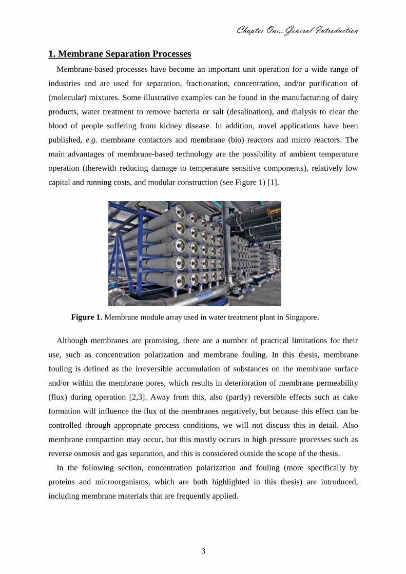
capital and running costs, and modular construction (see Figure 1) [1].
Figure 1. Membrane module array used in water treatment plant in Singapore.
Although membranes are promising, there are a number of practical limitations for their
use, such as concentration polarization and membrane fouling. In this thesis, membrane
fouling is defined as the irreversible accumulation of substances on the membrane surface
and/or within the membrane pores, which results in deterioration of membrane permeability
(flux) during operation [2,3]. Away from this, also (partly) reversible effects such as cake
formation will influence the flux of the membranes negatively, but because this effect can be
controlled through appropriate process conditions, we will not discuss this in detail. Also
membrane compaction may occur, but this mostly occurs in high pressure processes such as
reverse osmosis and gas separation, and this is considered outside the scope of the thesis.
In the following section, concentration polarization and fouling (more specifically by
proteins and microorganisms, which are both highlighted in this thesis) are introduced,
including membrane materials that are frequently applied.
4
1.1. Concentration Polarization
Rejection of certain components by the membranes leads to accumulation of components
that cannot pass the pores, resulting in an increase in concentration close to the membrane.
This then gives a driving force for transport from the component in the (laminar) layer next to
the membrane surface to the bulk liquid. The balance between the component carried toward
the membrane by the applied transmembrane pressure, and back transport due to the
concentration difference is called concentration polarization. Concentration polarization can
be controlled by decreasing the operating pressure, or increasing the cross-flow velocity of the
feed solution. If components present in the concentration polarization layer attach to the
membrane, this can be seen as the initial step of fouling [4,5]. Various components are known
to show high affinity to surfaces, such as proteins, polysaccharides, but also small
components - such as minerals (that cause scaling) and ‘large’ microorganisms (that are able
to form biofilms) - are known to have a detrimental effect on membrane performance.
1.2. Membrane Fouling
As defined earlier, membrane fouling is the irreversible deposition of components on and
into the membrane (see difference between fouled and un-fouled membrane in Figure 2). In
literature, it is stated that fouling depends on several factors, such as the membrane material
which determines the physicochemical interactions between the membrane and the substances
in the feed solution [6,7] (see membrane material section). Obviously, the properties of the
substance are of importance, because they determine to a large degree whether a component
can attach to the membrane surface.
Various components have been described to cause fouling, such as colloidal particles [8,9],
minerals that cause scaling [10,11], antifoam [12,13], proteins [14,15], and microorganisms
[16,17]. Also the location of fouling can be very different, ranging from surface deposition to
in-depth fouling. As a result, various ‘solutions’ have been proposed to influence the
interaction between these foulants and membranes, ranging from adjustment of the system
hydrodynamics [18-20], to surface modification [21,22], and downright regular cleaning
[23,24]. However, none of these methods is able to truly prevent fouling. Especially in-depth
fouling is very hard to remove because the foulant will also partially block the local flow that
is needed to remove and carry away the foulant. Perhaps even more relevantly, once a foulant
is attached to the membrane, it works as an initiator for attachment of more foulants. For
example, protein adsorption can be an initial step for attachment and growth of
microorganisms (i.e., biofilm formation).

Chapter One…General Introduction
5
Figure 2. Fouled (right) and un-fouled (left) spiral wound membranes that are opened up for
inspection; the left membrane is light in color inside the unwrapped module (not to be confused
with the module holder which is yellow/grey in color), where the right membrane is dark brown
all over.
In this thesis, the main focus will be on influencing/preventing protein adsorption, and
some of the modified surfaces that showed promising results were used to investigate
interaction with polysaccharides, polyphenols, and microorganisms (see next sections).
1.2.1. Protein Fouling
Proteins have both hydrophilic and hydrophobic regions, the ratio being different for
different proteins. It is often postulated that increased membrane hydrophilicity is the main
tool to mitigate protein fouling, the main reason being that the hydrophilic surface prefers
water in its neighborhood and this reduces adsorption of proteins [25]. However, not only the
surface hydrophilicity plays a role in protein repellence, but also charge on the protein and
structural changes in the protein [26,27]. Besides, the membrane surface charge and structure
have a significant impact on prevention of protein adsorption and attachment of other foulants
[25,28,29]. In order to influence membrane structure, polymers and monomers have been
grafted to membranes [30] or to the polymer from which the membrane is prepared [31].
Alternatively adsorption of block co-polymers has been proposed [32]. Grafting is mostly
initiated with a glow discharge apparatus or by UV irradiation [33,34]. In some cases this
leads to additional charge on the surface (monomer grafting) or to addition of polymer chains
onto the surface, and these chains may act as a steric hindrance for proteins that are close to
the membrane surface. In this thesis we succeed in attaching (variously sized) polymer chains
to the membranes by enzymatic grafting, and created an effective barrier for foulants.

6
1.2.2. Biofouling
In general, biofouling is defined as the attachment and/or growth of cells e.g.
microorganisms and algae on surfaces. Biofouling occurs on all kinds of surfaces once
adequate circumstances such as the presence of nutrients for cell adhesion and growth are
available. Biofouling is known to cause serious problems in/on medical devices, on ship hulls,
in/on pipelines, receivers, etc. [35], and it is also an important factor in membrane processes
[35-37].
In membrane biofouling three main steps can be distinguished [16,38]: (1) adsorption of
macromolecules e.g. proteins, (2) primary adhesion by fast-adhering cells, and (3)
colonization and growth with development of a biofilm, which ultimately leads to irreversible
blocking of the membrane. In a next step, cells may be expelled locally, and colonize on a
different part of the membrane. Biofouling is a complex process that is reported to be affected
by many factors, including the characteristics of the micro-organisms, membrane surface
properties (i.e., membrane material, charge, roughness, shape, etc.) and environmental factors
such as pH, ionic strength, etc. [17,39].
Generally, two strategies are used to control biofouling (to some extent) in membrane
processes [40]; (i) optimization of operating conditions, including pretreatment of feed and
cleaning procedures, and (ii) membrane modification [37,41,42], e.g. through grafting,
coating, etc. [37,40,43]. In general, it is believed that more hydrophilicity, negative charge,
and smooth surfaces reduce the initial adhesion of microorganisms [35,40,44].
1.3. Membrane Materials
Membranes can be made from various materials, ranging from inorganic materials to (bio-)
polymers; examples are shown in Figure 3. From the material of choice, it should be possible
to form the membrane structure in a controlled way, and this limits the options for membrane
production considerably. Chemically and thermally stable membranes are understandably
targeted by membrane manufacturers, even if these membranes are not intrinsically the most
resistant ones against fouling. Mostly this is mediated by an after-treatment that functionalizes
the membrane further, as is also done in this thesis, starting from poly(ethersulfone).
Popular polymeric membrane materials may take biopolymers as a starting point such as
cellulose acetate (CA), cellulose nitrate, regenerated cellulose, or synthetic polymers e.g.,
polypropylene (PP), polyethylene (PE), polyamide (PA), polytetrafluoroethylene (PTFE),
polyvinylidene fluoride (PVDF), polysulfone (PSf), poly(ethersulfone) (PES), polyimides
(PI), polyetherimids (PEI), polyacrylonitrile (PAN), etc. Inorganic membranes can be made of

Chapter One…General Introduction
7
ceramics, metal, glass or carbon, and recently even silicon wafers have been used in so-called
microsieves. Also hybrid membranes, which consist of polymeric and inorganic materials
together, have been presented [21,22,28,45-47].
Figure 3. Membrane materials: (A) Inorganic ceramic and (B) polymeric.
Membranes can have many different properties. Here we briefly discuss the development of
cellulose acetate membranes. Early cellulose acetate (38-40% acetyl content) membranes
were dense (symmetric) films. Later, cellulose acetate membranes were prepared with
asymmetric structures by Loeb and Sourirajan, and these were used in the first industrial
application on desalination [2,48]. The incorporated swelling agent, e.g. Mg(ClO4)2 and
formamide in an acetate-acetone solution, gelled the membrane in a low-temperature bath.
Subsequently, the membrane was treated in water at 60-90 ºC, which forms a rejection layer
on a more open carrier membrane [48]. The effects of preparation procedure, chemical
composition, and annealing temperature on the properties and performance of cellulosic
membranes have been widely studied [48,49], and it is clear that the preparation conditions
are of great influence on the resulting membrane. Currently, cellulose acetate membranes are
readily used in reversed osmosis, nano- and diafiltration. The main advantages of cellulosic
membranes are their low price and the fact that they are hydrophilic, which makes them less
prone to fouling. On the other hand, the mechanical and thermal stability of the polymer are
not that great, and the membrane is susceptible to (bio) fouling, which makes use of other
artificial materials attractive [3,50].
Nowadays, synthetic polymers are often used in membrane preparation, because of their
resistance to chemical agents and/or heat [2,3,21]. For example, polyamide can withstand
higher temperature and larger pH variations (4-11) than cellulose acetate, but is more prone to
damage by chlorine and oxidation, and is more expensive. Among the most popular
membrane materials are poly(arylsulfone)s, such as PSf and PES, which can be processed
A B

8
well and allow preparation of membranes in different shapes. The relatively high glass
transition temperature of the material allows steam sterilization, which - in combination with
good chemical stability - makes it an interesting material for membrane preparation, in spite
of the relatively high price. The hydrophobic character of the material is partly mediated by
blending with polyvinylpyrrolidone (PVP), which also allows for a wider variation in pore
size [21,22,28]. Unfortunately, poly(arylsulfone) membranes still show a high binding affinity
for different molecules such as proteins and microorganisms, which causes severe fouling of
membranes during operation. To diminish such fouling, various methods have been proposed
to alter the surface properties of poly(arylsulfone) membranes, and to reduce adsorption of
different foulants [21,22]. An overview of the various methods used for modification of
poly(arylsulfone) membranes is given in chapter two of this thesis. In general, the various
suggested methods are effective to some extent, but are typically environmentally adverse.
Clearly, there is still a lot of room for improvement, also regarding the reproducibility of the
modification methods.
In this thesis, poly(ethersulfone) (PES) membranes will be modified using the enzyme
laccase as initiator for free radical formation, which leads to covalently attached
(poly)phenolic components of various sizes and shapes, as described in the various chapters.
The enzymes that catalyze the modification of such so-called ‘chemically inert’ membranes
under mild conditions are introduced in the next section.
2. Laccases
Enzymes differ from ordinary chemical catalysts in several aspects such as mild reaction
conditions, and high reaction specificity [51]. Six classes of enzymes are distinguished:
oxidoreductases, transferases, hydrolases, lyases, isomerases, and ligases. Oxidoreductases
are enzymes that catalyze the transfer of one or more electrons from one molecule (the
reductant, or electron donor) to another molecule (the oxidant, or electron acceptor). In case
of oxidases, they catalyze oxidation reactions involving oxygen as electron acceptor, thereby
reducing oxygen to water or hydrogen peroxide. Catechol oxidases, tyrosinases, and blue
copper oxidases - including laccases, ascorbate oxidases, and ceruloplasmin - are examples of
different types of oxidases [52]. Several reviews on laccases have been published in recent
years, providing excellent summaries of the enzyme kinetics and applications [51,53,54].
Here we summarize the most important points, emphasizing current and future applications of
laccases.
Chapter One…General Introduction
9
Laccases [EC 1.10.3.2] are glycoproteins with a molecular mass ranging from about 50 to
100 kDa [54]. They can be roughly divided into two major groups which show clear
differences (see further in this Chapter), i.e. laccases from higher plants and those from fungi
[53,55]. The first laccase was described by Yoshida at the end of the 19th
century as a
component of the resin ducts of the Chinese or Japanese lacquer tree Rhus vernicifera [56],
(recently renamed to Toxicodendron vernicifluum), and this makes it one of the oldest
enzymes ever described. The name laccase refers to this lacquer tree. Further, laccases have
been identified in trees, various vegetables and fruits [53-56], fungi [51,54,57], and bacteria
[58-63]. Besides, proteins with features typical of laccase have been identified in insects [64]
and prokaryotes [54]. Although known for a long time, laccases only attracted significant
attention after studies on enzymatic degradation of wood by white-rot fungi [53,55,58].
2.1. The Catalytic Center and Reaction Mechanism of Laccase
Laccases catalyze the oxidation of a broad range of substrates such as phenolic compounds,
and aromatic or aliphatic diamines to the corresponding radical cation, which rapidly lose a
proton to give a radical. The redox process takes place with the assistance of a cluster of four
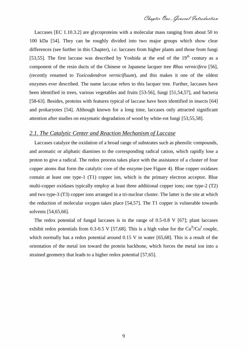
copper atoms that form the catalytic core of the enzyme (see Figure 4). Blue copper oxidases
contain at least one type-1 (T1) copper ion, which is the primary electron acceptor. Blue
multi-copper oxidases typically employ at least three additional copper ions; one type-2 (T2)
and two type-3 (T3) copper ions arranged in a tri-nuclear cluster. The latter is the site at which
the reduction of molecular oxygen takes place [54,57]. The T1 copper is vulnerable towards
solvents [54,65,66].
The redox potential of fungal laccases is in the range of 0.5-0.8 V [67]; plant laccases
exhibit redox potentials from 0.3-0.5 V [57,68]. This is a high value for the CuII/Cu
I couple,
which normally has a redox potential around 0.15 V in water [65,68]. This is a result of the
orientation of the metal ion toward the protein backbone, which forces the metal ion into a
strained geometry that leads to a higher redox potential [57,65].

10
Figure 4. Model of the catalytic center of laccase from Trametes versicolor consisting of a
cluster of four copper atoms [57].
The first step in the catalytic cycle is oxidation of four reducing substrates by the copper
(Cu2+
to Cu+) at the T1 site followed by transfer of the electrons from T1 to the T2/T3 tri-
nuclear site, resulting in the conversion of the fully oxidized form of the enzyme to a fully
reduced state, see Figure 5. The second step is the reduction of dioxygen that takes place via
the formation of a bound oxygen intermediate, namely peroxide di-anion that is protonated
and splits into the oxy radical and a molecule of water. In the final step, all four copper
centers are oxidized again and a second water molecule is released. The intramolecular
electron transfer from T1 to the tri-nuclear copper site is rate limiting in the overall reaction,
not the electron transfer from the substrate to the T1 copper [69-71].
Oxidations by laccase can be performed directly, i.e. the enzyme interacts with the
administered substrate itself, or indirectly, in which the enzyme oxidizes a chemical mediator
which acts as an intermediate substrate, see Figure 6. The oxidized radical forms of these
chemical mediators are able to interact with bulky substrates or compounds having a high
oxidation potential [57,72].

Chapter One…General Introduction
11
Figure 5. Catalytic cycle of laccase [58,71]
Figure 6. Schematic representation of laccase-catalyzed redox cycles for substrate oxidation in
(a) the absence and in (b) the presence of chemical mediators [57].
Although not used in this thesis, for completeness we would like to mention that more than
100 mediator compounds have been described for laccase (see Figure 7 for examples). Here
we only mention 2,2´-azinobis-(3-ethylbenzothiazoline-6-sulfonic acid) ABTS, which was the
first component found. It mediates laccase catalyzed oxidation of nonphenolic compounds,
such as veratryl alcohol to the corresponding aldehyde [73,74]. Various laccases readily
oxidize ABTS to the cation radical ABTS+·
which is intensely green-blue colored (ε420 =
36000 M-1
cm-1
) and is often used in activity assays. The redox potentials of ABTS+·
and

12
ABTS2+
were evaluated as 0.680 V and 1.09 V, respectively [75]. In general, synthetic
mediators are toxic, expensive and mostly inactivate laccase at concentrations above 1 mM;
however, also natural mediators such as p-coumaric acid, 4-hydroxybenzoic acid, and
syringaldehyde [76] have been identified [77] and these components are expected to lead to
less negative side-effects.
Figure 7. Examples of laccase mediators. (a) benzoic acids, R1, R2, and R3 is OH or COOH or
OMe; (b) methyl ester of 4-hydroxy-3,5-dimethoxybenzoic acid (syringic acid); (c) N-
hydroxyacetanilide; (d) 3-hydroxyanthranilic acid; (e) N-hydroxybenzotriazole; (f) violuric acid;
(g) (2,2,6,6-tetramethylpiperidin-1-yl)oxyl (TEMPO); (h) 2,2'-azino-bis-(3-ethylbenzothiazoline-
6-sulphonic acid) (ABTS).
2.2. Applications
As mentioned, laccases produce free radicals from suitable substrates using the oxygen
from air as an oxidant and producing water as the only by-product. The ensuing secondary
reactions are responsible for the versatility of laccases, and this in combination with its
thermal stability, its lack of substrate inhibition and high rates of oxidation (10-100 fold
higher than lignin peroxidase or manganese peroxidase) make laccase an ideal candidate for
the development of enzymatic oxidation processes [58,78].
Laccases have been applied in a variety of processes, such as the clean-up of herbicides,
pesticides [79-82], certain explosives and polycyclic aromatic hydrocarbons in soil [82,83].
Chapter One…General Introduction
13
Furthermore, they have been used in paper processing [84-86] and as cleaning agent for water
purification systems [87-89]. Besides, laccases have been applied in organic synthesis for the
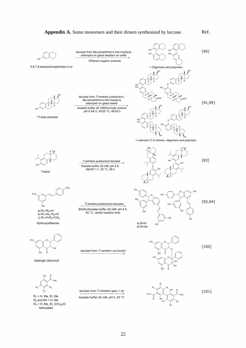
oxidation of alkenes to aldehydes and ketones [90], and for the dimerization of steroids [91],
phenols [92,93], hydroxystilbenes [94], and cyclic alcohols [95]. The most striking examples
of laccase applications are summarized in the next section: delignification and pulp bleaching,
organic synthesis, bioremediation, and biofuel cells with special attention for surface
modification.
2.2.1. Delignification, Organic Synthesis, Bioremediation, and Biofuel Cells
A lot of effort has been put into exploring fungi for technical lignin removal in the pulping
process and for bio-bleaching [96]. Two of the most important and best examined lignin
degrading microorganisms are the white-rot fungi Phanerochaete chrysosporium and
Trametes versicolor [51,73]. Laccase is abundant in theses fungi, while it is absent in brown-
rot fungi [97]. Compared to conventional ozone delignification, pre-treatment of wood pulp
with laccase is milder and cleaner, and less damaging to cellulose [96,98]. Further, it was
found that this enzyme can be used for cross-linking and functionalization of lignocellulose
compounds for e.g. paper-boards [99].
In organic synthesis, laccase has been used in the oxidative dimerization of phenolic
derivatives such as tetrahydronaphthol [95], 17β-estradiol [91,95], totarol [93],
hydroxystilbenes [92,94], flavonols [100], salicylic esters [101], and recently the
flavonolignan silybin [102], which is widely used in human therapy of liver dysfunctions. In
addition, laccase was used in the production of aminoquinones at high specificity through
amination of p-hydroquinones without formation of hydroquinonoids [103]. Moreover, larger
molecules have been produced; polymerization with laccase is considered a green synthesis
route with great flexibility [104]. Just to name some examples, laccase shows remarkable
activity and stability under acidic conditions in the synthesis of polyaniline [105]. Likewise,
laccase was used in the synthesis of a poly(allylamine) catechin conjugate, which is a good
antioxidant [106]. The interested reader can see some of these monomers and dimers in
appendix A.
In bioremediation processes, laccase is used to protect the environment from damage
caused by industrial effluents, through e.g. direct oxidation of phenol derivatives. The
polymeric polyphenolic derivatives that result from the laccase-catalyzed coupling are usually
insoluble and can be separated easily by filtration or sedimentation [107]. Laccase has been
used for direct dechlorination of chlorophenolic compounds [108,109] and for detoxification

14
through conjunction with natural phenols (e.g., syringic acid) [110]. Similarly, poly(catechol)
was formed by laccase [89], which can be removed from wastewater streams in the form of a
precipitate. This polymer can be used further for selective separation processes and in
biosensor applications.
Interestingly, laccase has been successfully used in the presence or absence of a chemical
mediator in a dihydrogen/dioxygen biofuel cell to overcome slow reduction of dioxygen to
water [111,112].
2.2.2. Surface Modification
The laccase from Trametes hirsuta was used to coat flax fibers and fabrics with
hydroquinone and various methoxyphenols to obtain antibacterial surfaces; the combination
of ferulic acid and hydroquinone resulted in a coating with antibacterial effect against Bacillus
subtilis and Staphylococcus aureus [113]. Also wool was treated with laccase to incorporate
water insoluble lauryl gallate to provide antioxidant, antibacterial and water repellent
properties to the textile material [114]. Besides, cellulosic fibers can be colored by grafting
natural flavonoids, which can be carried out without bleaching, resulting in a more
environmentally friendly process [115,116]. Alternatively textile can also be de-colored by
the enzyme [77].
Figure 8. Attachment of phenolic amines to lignin moieties of wood, which can be used for
further functionalization [119].
Laccase can also be used in combination with other modification processes, to create
functional groups on ‘inert’ polymers. Nonwoven polypropylene fabrics were pretreated by

Chapter One…General Introduction
15
argon plasma in the presence of different methacrylate monomers, in order to activate the
inert synthetic polymers, followed by laccase-catalyzed grafting of guaiacol sulfonic acid onto
the modified surface [117]. In a similar way, cellulose fibers were chemically functionalized
with amine groups, that were subsequently coated with enzymatically-synthesized
poly(catechol) in the presence of Trametes villosa laccase [118]. Phenolic amines were
coupled to lignin moieties of wood using the Trametes hirsuta laccase [119]. The amine
group works as anchor group for further grafting of antifungal molecules via chemical or
enzymatic reaction or simply by adsorption, see Figure 8.
Laccases can be used in many different applications, their main feature being that they
catalyze oxidative reactions of functionalized aromatic compounds in a mild and
environmentally friendly way. Laccases produce radicals that can couple to other aromatic
compounds, provided that there are one or more electron-donating groups on the aromatic
ring. Considering the structure of poly(ethersulfone) (PES, see Figure 9), it was hypothesized
at the start of the work reported in this thesis that this important membrane material would be
reactive towards radicals produced by laccase. In this thesis the laccase-catalyzed
modification of PES is investigated mostly in relation to protein repellence of membranes, but
also in relation to biofouling. The specific aim of the research and a short preview on the
contents of the various chapters is given in the next section.
Figure 9. Molecular structure of poly(ethersulfone) (PES).
3. Aim and Outline of the Thesis
From the previous sections it is clear that if a membrane modification method would
become available that prevents (protein) adsorption and allows tight control over the
(functionality of the) modification layer, this would be of great interest for many different
applications. Laccase-catalyzed reactions are known to be versatile and environmentally
friendly, and poly(ethersulfone) is a potential reaction partner for the enzyme-generated
radicals. In this thesis, these two aspects are brought together: laccase-catalyzed modification
of poly(ethersulfone) membranes is investigated in great detail, ranging from a detailed
description of the attachment of the polymer and its growth over time, to the membrane
performance.

16
An overview of modification methods for poly(arylsulfone) [i.e., Polysulfone (PSf) and
Poly(ethersulfone) (PES)] membranes is presented in Chapter two with special reference to
surface modification. Modification methods are compared on various aspects, such as flux
after modification, simplicity, reproducibility, environmental aspects, and cost effectiveness.
The enzyme-catalyzed modification method is highlighted as an environmentally benign
alternative for other modification methods.
The principle of enzyme-catalyzed modification of PES membranes is proven in Chapter
three. Various phenolic acids (enzyme substrates) are investigated under very mild conditions
(room temperature, water, nearly neutral pH) using laccase from Trametes versicolor. The
produced layers are extensively analyzed, both from a chemical and a membrane performance
point of view.
In Chapter four, the performance of laccase-catalyzed modified poly(ethersulfone)
membranes modified with 4-hydroxybenzoic acid and gallic acid as substrates are evaluated
based on e.g. grafting yield, flux, and reduction of protein adsorption. Also the effect of the
enzyme-catalyzed modification on membrane bulk properties is discussed.
Chapter five gives details on adsorption of BSA, dextrin, and tannin on modified model
PES surfaces. Reflectometry is used to follow the adsorbed amount as function of time, and
gives information on the adsorption rate, and on the amounts of reversibly and irreversibly
adsorbed foulant on the various surfaces. Conclusions are drawn on the importance of various
aspects of the modification layer on adsorption.
The effect of modification on suppression of biofouling on model PES surfaces is presented
in Chapter six. Listeria monocytogenes bacteria are used to evaluate both attachment and
biofilm growth under static and dynamic conditions.
A general discussion and an overview of all results is presented in Chapter seven, which is
finalized with an outlook for application on industrial scale and future developments.
Chapter One…General Introduction
17
References
1. Strathmann, H.; Giorno, L.; Drioli, E., An Introduction to Membrane Science and Technology,
Consiglio Nazionale Delle Ricerche, Rome (2006).
2. Ronald, W.R., Handbook of Separation Processes Technology, John Wiley & Sons, New York
(1995).
3. Coulson, J.M.; Richardson, F., Particle Technology and Separation Processes, Vol.2, 5th edition,
Butterworth-Heinemann, New York (2002).
4. Chen, J.C.; Li, Q.; Elimelech, M., In Situ Monitoring Techniques for Concentration Polarization
and Fouling Phenomena in Membrane Filtration, Adv. Colloid Interface Sci. 107 (2004) 83-108.
5. Meier, J., Mechanical Influence of PAC Particles on Membrane Processes, J. Membr. Sci. 360
(2010) 404-409.
6. Nakatsuka, S.; Ase, T.; Miyano, T., High Flux Ultrafiltration for Drinking Water Production,
Water Sci. Technol. 1 (2001) 177-184.
7. Hilal, N.; Ogunbiyi, O.O.; Miles, N.J.; Nigmatullin, R., Methods Employed for Control of Fouling
in MF and UF Membranes: A Comprehensive Review, Sep. Sci. Technol. 40 (2005) 1957-2005.
8. Yiantsios, S.G.; Karabelas, A.J., The Effect of Colloid Stability on Membrane Fouling,
Desalination 118 (1998) 143-152.
9. Zhu, X.; Elimelech, M., Colloidal Fouling of Reverse Osmosis Membranes: Measurements and
Fouling Mechanisms, Environ. Sci. Technol. 31 (1997) 3654-3662.
10. Bonné, P.A.C.; Hofman, J.A.M.H.; van der Hoek, J.P., Scaling Control of RO Membranes and
Direct Treatment of Surface Water, Desalination 132 (2000) 109-119.
11. Sahachaiyunta, P.; Koo, T.; Sheikholeslami, R., Effect of Several Inorganic Species on Silica
Fouling in RO Membranes, Desalination 144 (2002) 373-378.
12. Liew, M.K.H.; Fane, A.G.; Rogers, P.L., Fouling of Microfiltration Membranes by Broth-Free
Antifoam Agents, Biotechnol. Bioeng. 56 (1997) 89-98.
13. Yamagiwa, K.; Kobayashi, H.; Onodera, M.; Ohkawa, A., Antifoam Fouling and its Reduction by
Surfactant Precoat Treatment of Polysulfone Ultrafiltration, Biotechnol. Tech. 8 (1994) 267-270.
14. Hanemaaijer, J.H.; Robbertsen, T.; van den Boomgaard, Th.; Gunnink, J.W., Fouling of
Ultrafiltration Membranes. The Role of Protein Adsorption and Salt Precipitation, J. Membr. Sci.
40 (1989) 199-217.
15. Palacio, L.; Ho, C.-C.; Prádanos, P.; Hernández, A.; Zydney, A.L., Fouling with Protein Mixtures
in Microfiltration: BSA-Lysozyme and BSA-Pepsin, J. Membr. Sci. 22 (2003) 41-51.
16. Flemming, H.-C.; Schaule, G., Biofouling on Membranes – A Microbiological Approach,
Desalination 70 (1988) 95-119.
17. Ridgway, H.; Ishida, K.; Rodriguez, G.; Safarik, J.; Knoell, T.; Bold, R., Biofouling of
Membranes: Membrane Preparation, Characterization, and Analysis of Bacterial Adhesion,
Methods Enzymol. 310 (1999) 463-494.
18. Mohammadi, T.; Moghadam, M.K.; Madaeni, S.S., Hydrodynamic Factors Affecting Flux and
Fouling during Reverse Osmosis of Seawater, Desalination 151 (2002) 239-245.
19. Lueptow, R.M.; Lee, S., Control of Scale Formation in Reverse Osmosis by Membrane Rotation,
Desalination 155 (2003) 131-139.
20. Frappart, M.; Massé, A.; Jaffrin, M.Y.; Pruvost, J.; Jaouen, P., Influence of Hydrodynamics in
Tangential and Dynamic Ultrafiltration Systems for Microalgae Separation, Desalination 265
(2011) 279-283.
21. Ulbricht, M., Advanced Functional Polymer Membranes, Polymer 47 (2006) 2217-2262.
22. Rana, D.; Matsuura, T., Surface Modifications for Antifouling Membranes, Chem. Rev. 110
(2010) 2448-2471.
23. Chai, X.; Kobayashi, T.; Fujii, N., Ultrasound-Associated Cleaning of Polymeric Membranes for
Water Treatment, Sep. Purif. Technol. 15 (1999) 139-146.
24. Bereschenko, L.A.; Prummel, H.; Euverink, G.J.W.; Stams, A.J.M., Effect of Conventional
Chemical Treatment on the Microbial Population in a Biofouling Layer of Reverse Osmosis
Systems, Water Res. 45 (2011) 405-416.
25. Lundström, I., Surface Physics and Biological Phenomena, Phys. Scr. T4 (1983) 5-13.
26. Haynes, C.A.; Norde, W., Structures and Stabilities of Adsorbed Proteins, J. Colloid Interface Sci.
169 (1995) 313-328.
27. Haynes, C.A.; Norde, W., Globular Protein at Solid/Liquid Interface, Colloids Surf. B 2 (1994)
517-566.

18
28. Ulbricht, U., Advanced Functional Polymer Membranes, Polymer 47 (2006) 2217-2262.
29. Minko, S., Grafting on Solid Surfaces: “Grafting to” and “Grafting from” Methods, in: Manfred,
S., (Ed.), Polymer Surfaces and Interfaces: Characterization, Modification and Applications, E-
Publishing, Springerlink.com, Berlin/Heidelberg (2008) 215-234.
30. Taniguchi, M.; Kilduff, J.E.; Belfort, G., Low Fouling Synthetic Membranes by UV-Assisted Graft
Polymerization: Monomer Selection to Mitigate Fouling by Natural Organic Matter, J. Membr.
Sci. 222 (2003) 59-70.
31. Vigo, F.; Uliana, C.; Dondero, G., Ultrafiltration Membrane Obtained by Poly(acrylonitrile)
Grafted onto Poly(vinylchloride), Desalination 70 (1988) 277-292. 32. Schroën, C.G.P.H.; Cohen Stuart, M.A.; van der Voort Maarschalk, K.; van der Padt, A.; van’t
Riet, K., Influence of Preadsorbed Block Copolymers on Protein Adsorption: Surface Properties,
Layer Thickness, and Surface Coverage, Langmuir 11(1995) 3068-3074
33. Vrlinic, T.; Vesel, A.; Cvelbar, V.; Krajnc, M.; Mozetic, M., Rapid Surface Functionalization of
Poly(ethersulphone) Foils Using a Highly Reactive Oxygen-Plasma Treatment, Surf. Interface
Anal. 39 (2007) 476-481.
34. Taniguchi, M.; Belfort, G., Low Protein Fouling Synthetic Membranes by UV-Assisted Surface
Grafting Modification: Varying Monomer Type, J. Membr. Sci. 231 (2004) 147-157.
35. Kang, S.; Hoek, E.M.V.; Choi, H.; Shin, H., Effect of Membrane Surface Properties During the
Fast Evaluation of Cell Attachment, Sep. Sci. Technol. 41 (2006) 1475-1487.
36. Flemming, H.-C., Reverse Osmosis Membrane Biofouling, Exp. Therm. Fluid Sci. 14 (1997) 382-
391.
37. Banerjee, I; Pangule, R.C.; Kane, R.S., Antifouling Coatings: Recent Developments in the Design
of Surfaces that Prevent Fouling by Protein, Bacteria, and Marine Organisms, Adv. Mater. 23
(2011) 690-718. 38. Ridgway, H.F.; Kelly, A.; Justice, C.; Olson, B.H., Microbial Fouling of Reverse-Osmosis
Membranes Used in Advanced Wastewater Treatment Technology: Chemical, Bacteriological, and
Ultrastructural Analyses, Appl. Enviro. Microbiol. 45 (1983) 1066-1084.
39. Roosjen, A.; Norde, W.; Van der Mei, H.C.; Busscher, H.J., The Use of Positively Charged or Low
Surface Free Energy Coatings versus Polymer Brushes in Controlling Biofilm Formation, Progr.
Colloid Polym. Sci. 132 (2006) 138-144.
40. Mansouri, J.; Harrisson, S.; Chen, V., Strategies for Controlling Biofouling in Membrane Filtration
Systems: Challenges and Opportunities, J. Mater. Chem. 20 (2010) 4567-4586.
41. Hilal, N.; Al-Khatib, L.; Atkin, B.P.; Kochkodan, V.; Potapchenko, N., Photochemical
Modification of Membrane Surfaces for (Bio) Fouling Reduction: A Nano-Scale Study Using
AFM, Desalination 158 (2003) 65-72.
42. Hyun, J.; Jang, H.; Kim, K.; Na, K.; Tak, T., Restriction of Biofouling in Membrane Filtration
Using a Brush-Like Polymer Containing Oligoethylene Glycol Side Chains, J. Membr. Sci. 282
(220) 52-59. 43. Olsen, S.M.; Pedersen, T.; Laursen, M.H.; Kiil, S.; Dam-Johansen, K., Enzyme-Based Antifouling
Coatings: A Review, Biofouling, 23 (2007) 369-383.
44. Kouwonou, Y.; Malaisamy, R.; Jones, K.L., Modification of PES Membrane: Reduction of
Biofouling and Improved Flux Recovery, Separ. Sci. Technol. 43 (2008) 4099-4112.
45. Porter, M.C., Handbook of Industrial Membrane Technology, Noyes Publications, USA (1990).
46. Kislik, V.S., Liquid Membranes - Principles and Applications in Chemical Separations and
Wastewater Treatment, Elsevier (2010).
47. Zulfikar, M.A.; Mohammad, A.W.; Hilal, N., Preparation and Characterization of Novel Porous
PMMA-SiO2 Hybrid Membranes, Desalination 192 (2006) 262-270.
48. Loeb, S., The Loeb-Sourirajan Membrane: How It Come About, ACS Symposium Series 153:
Synthetic Membranes, Turbak A.F. (Ed), Washington (1981) 1-9.
49. Ghatge, N.D.; Sabne, M.B.; Gujar, K.B.; Mahajan, S.S., Modified Cellulose Acetate Membranes
for Desalination, J. Appl. Polym. Sci. 29 (1984) 1743-1748.
50. Raymond, D.L., Water Quality and Treatment: Handbook of Community Water Supplies, 5th
edition, McGraw-Hill, Inc., New York (1980).
51. Call, H.P.; Mücke, I., History, Overview and Applications of Mediated Lignolytic Systems,
Especially Laccase-Mediator-Systems (Lignozym®-Process), J. Biotechnol. 53 (1997) 163-202.
52. Held, M.; Schmid, A.; van Beilen, J.B.; Witholt, B., Biocatalysis. Biological Systems for the
Production of Chemicals, Pure Appl. Chem. 72 (2000) 1337-1343.
Chapter One…General Introduction
19
53. Mayer, A.M.; Staples, R.C., Laccase: New Functions for an Old Enzyme, Phytochemistry 60
(2002) 551-565.
54. Claus, H., Laccases: Structure, Reactions, Distribution, Micron 35 (2004) 93-96.
55. Witayakran, S.; Ragauskas, A.J., Synthetic Applications of Laccase in Green Chemistry, Adv.
Synth. Catal. 351 (2009) 1187-1209.
56. Hüttermann, A.; Mai, C.; Kharazipour, A., Modification of Lignin for the Production of New
Compounded Materials, Appl. Microbiol. Biotechnol. 55 (2001) 387-394.
57. Riva, S., Laccases: Blue Enzymes for Green Chemistry, Trends Biotechnol. 24 (2006) 219-226.
58. Baldrian, P., Fungal Laccases Occurrence and Properties, FEMS Microbiol. Rev. 30 (2006) 215-
242.
59. Diamantidis, G.; Effosse, A.; Potier, P.; Bally, R., Purification and Characterization of the First
Bacterial Laccase in the Rhizospheric Bacterium Azospirillum lipoferum, Soil Biol. Biochem. 32
(2000) 919-927.
60. Martins, L.O.; Soares, C.M.; Pereira, M.M.; Teixeira, M.; Costa, T.; Jones, G.H.; Henriques, A.O.,
Molecular and Biochemical Characterization of a Highly Stable Bacterial Laccase that Occurs as a
Structural Component of the Bacillus subtilis Endospore Coat, J. Biol. Chem. 277 (2002) 18849-
18859.
61. Suzuki, T.; Endo, K.; Ito, M.; Tsujibo, H.; Miyamoto, K.; Inamori, Y., A Thermostable Laccase
from Streptomyces lavendulae REN-7: Purification, Characterization, Nucleotide Sequence, and
Expression, Biosci. Biotechnol. Biochem. 67 (2003) 2167-2175.
62. Arias, M.E.; Arenas, M.; Rodríguez, J.; Soliveri, J.; Ball, A.S.; Hernández, M., Kraft Pulp
Biobleaching and Mediated Oxidation of a Nonphenolic Substrate by Laccase from Streptomyces cyaneus CECT 3335, Appl. Environ. Microbiol. 69 (2003) 1953-1958.
63. Jimenez-Juarez, N.; Roman-Miranda, R.; Baeza, A.; Sánchez-Amat, A.; Vazquez-Duhalt, R.;
Valderram, B., Alkali and Halide-Resistant Catalysis by the Multipotent Oxidase from
Marinomonas mediterranea, J. Bacteriol. 117 (2005) 73-86.
64. Kramer, K.J.; Kanost, M.R.; Hopkins, T.L.; Jiang, H.; Zhu, Y.C.; Xu, R.; Kerwind, J.L.; Turecek,
F., Oxidative Conjugation of Catechols with Proteins in Insect Skeletal Systems, Tetrahedron 57
(2001) 385-392.
65. Piontek, K.; Antorini, M.; Choinowski, T., Crystal Structure of a Laccase from the Fungus
Trametes versicolor at 1.90 Å Resolution Containing a Full Complement of Coppers, J. Biol.
Chem. 277 (2002) 37663-37669.
66. Rodakiewicz-Nowak, J., Phenols Oxidizing Enzymes in Water-Restricted Media, Top. Catal.
11/12 (2000) 419-434.
67. Kumar, S.V.S.; Phale, P.S.; Durani, S.; Wangikar, P.P., Combined Sequence and Structure
Analysis of the Fungal Laccase Family, Biotechnol. Bioeng. 83 (2003) 386-394.
68. Sheldon, R.A.; Arends, I.W.C.E., Organocatalytic Oxidations Mediated by Nitroxyl Radicals, Adv.
Synth. Catal. 346 (2004) 1051-1071.
69. Huang, H.W.; Zoppellaro, G.; Sakurai, T., Spectroscopic and Kinetic Studies on the Oxygen-
Centered Radical Formed During the Four-Electron Reduction Process of Dioxygen by Rhus vernicifera Laccase, J. Biol. Chem. 274 (1999) 32718-32724.
70. Zoppellaro, G.; Huang, H.W.; Sakurai, T., Kinetic Studies on the Reaction of the Fully Reduced
Laccase with Dioxygen, Inorg. React. Mech. 2 (1999) 79-84.
71. Wong, D.W.S., Structure and Action Mechanism of Ligninolytic Enzymes, Appl. Biochem.
Biotechnol. 157 (2009) 174-209.
72. Cantarella, G.; Galli, C.; Gentili, P., Free Radical versus Electron-Transfer Routes of Oxidation of
Hydrocarbons by Laccase/Mediator Systems Catalytic or Stoichiometric Procedures, J. Mol. Catal.
B: Enzym. 22 (2003) 135-144.
73. Archibald, F.S.; Bourbonnais, R.; Jurasek, L.; Paice, M.G.; Reid, I.D., Kraft Pulp Bleaching and
Delignification by Trametes versicolor, J. Biotechnol. 53 (1997) 215-236.
74. Bourbonnais, R.; Paice, M.G., Oxidation of Non-Phenolic Substrates: An Expanded Role for
Laccase in Lignin Biodegradation, FEBS Lett. 267 (1990) 99-102.
75. Scott, S.L.; Chen, W.J.; Bakac, A.; Espenson, J.H., Spectroscopic Parameters, Electrode Potentials,
Acid Ionization Constants, and Electron Exchange Rates of the 2,2′-Azinobis(3-
ethylbenzothiazolineine-6-sulfonate) Radicals and Ions, J. Phys. Chem. 97 (1993) 6710-6714.

20
76. Camarero, S.; Ibarra, D.; Martínez, M.J.; Martínez, A.T., Lignin-Derived Compounds as Efficient
Laccase Mediators for Decolorization of Different Types of Recalcitrant Dyes, Appl. Environ.
Microbiol. 71 (2005) 1774-1784.
77. Bulter, T.; Alcalde, M.; Sieber, V.; Meinhold, P.; Schlachtbauer, C.; Arnold, F.H., Functional
Expression of a Fungal Laccase in Saccharomyces cerevisiae by Directed Evolution, Appl.
Environ. Microbiol. 69 (2003) 987-995.
78. Arends, I.W.C.E.; Li, Y.X.; Ausan, R.; Sheldon, R.A., Comparison of TEMPO and its Derivatives
as Mediators in Laccase Catalyzed Oxidation of Alcohols, Tetrahedron 62 (2006) 6659-6665.
79. Bollag, J.M.; Shuttleworth, K.L.; Anderson, D.H., Laccase-Mediated Detoxification of Phenolic
Compounds, Appl. Environ. Microbiol. 54 (1988) 3086-3091.
80. Majcherczyk, A.; Johannes, C.; Hüttermann, A., Oxidation of Polycyclic Aromatic Hydrocarbons
(PAH) by Laccase of Trametes versicolor, Enzyme Microb. Technol. 22 (1998) 335-341.
81. Collins, P.J.; Kotterman, M.J.J.; Field, J.A.; Dobson, A.D.W., Oxidation of Anthracene and
Benzo[a]pyrene by Laccases from Trametes versicolor, Appl. Environ. Microbiol. 62 (1996) 4563-
4567.
82. Gianfreda, L.; Rao, M.A., Potential of Extra Cellular Enzymes in Remediation of Polluted Soils: A
Review, Enzyme Microb. Technol. 35 (2004) 339-354.
83. Durán, N.; Esposito, E., Potential Applications of Oxidative Enzymes and Phenoloxidase-Like
Compounds in Wastewater and Soil Treatment: A Review, Appl. Catal., B 28 (2000) 83-99.
84. Bourbonnis, R.; Paice, M.G.; Freiermuth, B.; Bodie, E.; Borneman, S., Reactivities of Various
Mediators and Laccases with Kraft Pulp and Lignin Model Compounds, Appl. Environ. Microbiol.
63 (1997) 4627-4632.
85. Camarero, S.; García, O.; Vidal, T.; Colom, J.; del Río, J.C.; Gutiérrez, A.; Gras, J.M.; Monje, R.;
Marja J.; Martínez, M.J.; Martínez, A.T., Efficient Bleaching of Non-Wood High-Quality Paper
Pulp Using Laccase-Mediator System, Enzyme Microb. Technol. 35 (2004) 113-120.
86. Gamelas, J.A.F.; Tavares, A.P.F.; Evtuguin, D.V.; Xavier, A.M.B., Oxygen Bleaching of Kraft
Pulp with Polyoxometalates and Laccase Applying a Novel Multi-Stage Process, J. Mol. Catal. B:
Enzym. 33 (2005) 57-64.
87. D’Annibale, A.; Stazi, S.R.; Vinciguerra, V.; Mattia, E.D.; Sermanni, G.G., Characterization of
Immobilized Laccase from Lentinula edodes and its Use in Olive-Mill Wastewater Treatment,
Process Biochem. 34 (1999) 697-706.
88. Kim, Y.J.; Nicell, J.A., Impact of Reaction Conditions on the Laccase-Catalyzed Conversion of
Bisphenol A, Bioresour. Technol. 97 (2006) 1431-1442.
89. Aktaş, N.; Tanyolaç, A., Reaction Conditions for Laccase Catalyzed Polymerization of Catechol,
Bioresour. Technol. 87 (2003) 209-214.
90. Niku-Paavola, M.L.; Viikari, L., Enzymatic Oxidation of Alkenes, J. Mol. Catal. B: Enzym. 10
(2000) 435-444.
91. Nicotra, S.; Intra, A.; Ottolina, G.; Riva, S.; Danieli, B., Laccase-Mediated Oxidation of the
Steroid Hormone 17β-estradiol in Organic Solvents, Tetrahedron: Asymmetry 15 (2004) 2927-
2931.
92. Nicotra, S.; Cramarossa, M.R.; Mucci, A.; Pagnoni, U.M.; Riva, S.; Forti, L.; Biotransformation of
Resveratrol: Synthesis of Trans-Dehydrodimers Catalyzed by Laccases from Myceliophtora
thermophyla and from Trametes pubescens, Tetrahedron 60 (2004) 595-600.
93. Ncanana, S.; Baratto, L.; Roncaglia, L.; Riva, S.; Burton, S.G., Laccase-Mediated Oxidation of
Totarol, Adv. Synth. Catal. 349 (2007) 1507-1513.
94. Ponzoni, C.; Beneventi, E.; Cramarossa, M.R.; Raimondi, S.; Trevisi, G.; Pagnoni, U.M.; Riva, S.;
Forti, L., Laccase-Catalyzed Dimerization of Hydroxystilbenes, Adv. Synth. Catal. 349 (2007)
1497-1506.
95. Intra, A.; Nicotra, S.; Riva, S.; Danieli, B., Significant and Unexpected Solvent Influence on the
Selectivity of Laccase-Catalyzed Coupling of Tetrahydro-2-naphthol Derivatives, Adv. Synth.
Catal. 347 (2005) 973-977.
96. Rodríguez-Couto, S.; Toca-Herrera, J.L., Industrial and Biotechnological Applications of
Laccases: A Review, Biotechnol. Adv.24 (2006) 500-513.
97. Singh Arora, D.; Kumar Sharma, R., Ligninolytic Fungal Laccases and their Biotechnological
Applications, Appl Biochem. Biotechnol. 160 (2010) 1760-1788
98. Gübitz, G.M.; Cavaco-Paulo, A, New Substrates for Reliable Enzymes: Enzymatic Modification of
Polymers, Curr. Opin. Biotechnol. 14 (2003) 577-582.

Chapter One…General Introduction
21
99. Mikolasch, A.; Schauer, F., Fungal Laccases as Tools for the Synthesis of New Hybrid Molecules
and Biomaterials, Appl. Microbiol. Biotechnol. 82 (2009) 605-624.
100. Ghidouche, S.; Es-Safi, N.E.; Ducrot, P.H., Mechanistic Study on the Enzymatic Oxidation of
Flavonols, Tetrahedron Lett. 49 (2008) 619-623.
101. Ciecholewski, S.; Hammer, E.; Manda, K.; Bose, G.; Nguyen, V.T.H., Langer, P.; Schauer, F.,
Laccase-Catalyzed Carbon-Carbon Bond Formation: Oxidative Dimerization of Salicylic Esters by
Air in Aqueous Solution, Tetrahedron 61 (2005) 4615-4619.
102. Gažák, R.; Sedmera, P.; Marzorati, M.; Riva, S.; Křen, V., Laccase-Mediated Dimerization of the
Flavonolignan Silybin, J. Mol. Catal. B: Enzym. 50 (2008) 87-92.
103. Niedermeyer, T.H.J., Mikolasch, A.; Lalk, M., Nuclear Amination Catalyzed by Fungal Laccases:
Reaction Products of p-Hydroquinones and Primary Aromatic Amines, J. Org. Chem. 70 (2005)
2002-2008.
104. Kobayashi, S.; Uyama, H.; Kimura, S., Enzymatic Polymerization, Chem. Rev. 101 (2001) 3793-
3818.
105. Karamyshev, A.V.; Shleev, S.V.; Koroleva, O.V.; Yaropolov, A.I.; Sakharov, I.Yu., Laccase-
Catalyzed Synthesis of Conducting Polyaniline, Enzyme Microb. Technol. 33 (2003) 556-564.
106. Gogoi, P.; Hazarika, S.; Dutta, N.N.; Rao, P.G., Laccase Catalyzed Conjugation of Catechin with
Poly(allylamine): Solvent Effect, Chem. Eng. J. 155 (2009) 810-815.
107. Torres, E.; Bustos-Jaimes, I.; Borgne, S.L., Potential Use of Oxidative Enzymes for the
Detoxification of Organic Pollutants, Appl. Catal. B 46 (2003) 1-15.
108. Roy-Arcand, L.; Archibald, F.S., Direct Dechlorination of Chlorophenolic Compounds by
Laccases from Trametes (Coriolus) versicolor, Enzyme Microb. Technol. 13 (1991) 194-203.
109. Kadhim, H.; Graham, C.; Barratt, P.; Evans, C.S.; Rastall, R.A., Removal of Phenolic Compounds
in Water Using Coriolus versicolor Grown on Wheat Bran, Enzyme Microb. Technol. 24 (1999)
303-307.
110. Bollag, J.M.; Shuttleworth, K.L.; Anderson, D.H., Laccase-Mediated Detoxification of Phenolic
Compounds, Appl. Environ. Microbiol. 54 (1988) 3086-3091.
111. Palmore, G.T.R., Kim, H.H., Electro-Enzymatic Reduction of Dioxygen to Water in the Cathode
Compartment of a Biofuel Cell, J. Electroanal. Chem. 464 (1999) 110-117.
112. Barton, S.C.; Pickard, M.; Vazquez-Duhalt, R.; Heller, A., Electroreduction of O2 to Water at 0.6
V (SHE) at pH 7 on the ‘Wired’ Pleurotus ostreatus Laccase Cathode, Biosens. Bioelectron. 17
(2002) 1071-1074.
113. Schroeder, M.; Aichernig, N.; Guebitz, G.M.; Kokol, V., Enzymatic Coating of Lignocellulosic
Surfaces with Polyphenols, Biotechnol. J. 2 (2007) 334-341.
114. Hossain, Kh.M.G.; González, M.D.; Lozano, G.R.; Tzanov, T., Multifunctional Modification of
Wool Using an Enzymatic Process in Aqueous-Organic Media, J. Biotechnol. 141 (2009) 58-63.
115. Kim, S.; Moldes, D.; Cavaco-Paulo, A., Laccases for Enzymatic Coloration of Unbleached Cotton,
Enzyme Microb. Technol. 40 (2007) 1788-1793.
116. Kim, S.; López, C.; Güebitz, G.; Cavaco-Paulo, A., Biological Coloration of Flax Fabrics with
Flavonoids Using Laccase from Trametes hirsuta, Eng. Life Sci. 8 (2008) 324-330.
117. Schroeder, M.; Fatarella, E.; Kovač, J.; Guebitz, G. M.; Kokol, V., Laccase-Induced Grafting on
Plasma-Pretreated Polypropylene, Biomacromolecules 9 (2008) 2735-2741.
118. Kim, S.Y.; Zille, A.; Murkovic, M.; Güebitz, G.; Cavaco-Paulo, A., Enzymatic Polymerization on
the Surface of Functionalized Cellulose Fibers, Enzyme Microb. Technol. 40 (2007) 1782-1787.
119. Kudanga, T.; Prasetyo, E.N.; Sipilä, J.; Nousiainen, P.; Widsten, P.; Kandelbauer, A.; Nyanhongo,
G.S.; Guebitz, G., Laccase-Mediated Wood Surface Functionalization, Eng. Life Sci. 8 (2008)
297-302.
22
Appendix A. Some monomers and their dimers synthesized by laccase.
Ref.
[95]
[91,95]
[93]
[92,94]
[100]
[101]

Chapter Two
Mini-Review on
Poly(arylsulfone) Surface Modification
This chapter has been published as:
Modification Methods for Poly(arylsulfone) Membranes: A Mini-Review Focusing on
Surface Modification, Norhan Nady, Maurice Franssen, Han Zuilhof, Mohamed Mohy Eldin,
Remko Boom, and Karin Schroën, Desalination 275 (2011) 1-9.
24
Modification Methods for Poly(arylsulfone) Membranes:
A Mini-Review Focusing on Surface Modification
Abstract
Surface modification of membranes is thought to be equally important to the membrane
industry as membrane material and process development; surface functionalization has
already become a key technology, the major aims being performance improvement (flux and
selectivity) by reduction of unwanted protein fouling (often considered the first step for
biofouling).
Poly(arylsulfone) [i.e., Polysulfone (PSf) and Poly(ethersulfone) (PES)] membranes have
been widely used for separation and purification purposes. However, in many cases,
nonspecific (protein) adsorption takes place on the membrane surface and in the membrane
pores due to the inherent hydrophobic characteristics of poly(arylsulfone). Therefore several
(surface) modification techniques for poly(arylsulfone) membranes have been developed.
Given the importance of modification methods for these membranes and their operation, we
decided to dedicate this mini-review solely to this topic.
The modification methods can be divided into the following main groups: (1) coating, (2)
blending, (3) composite, (4) chemical, (5) grafting, or (6) a combination of methods. With all
these methods, interesting results were obtained concerning reduction of protein adsorption
(see respective sections), although the quantification of improved performance is not
straightforward. In the conclusions section, all techniques are compared on various aspects
such as flux after modification, simplicity, reproducibility, environmental aspects, and cost
effectiveness.

Chapter Two…Mini-Review on Poly(arylsulfone) Surface Modification
25
1. Introduction
Polymers are attractive materials for various applications, such as membrane filtration,
coatings, composites, microelectronic devices, thin-film technology, biomaterials, and so on.
The performance of polymeric materials in many applications relies largely upon the
combination of bulk (e.g. mechanical) properties in combination with the properties of their
surfaces. However, polymers very often do not possess the surface properties needed for these
applications. Vice versa, those polymers that have good surface properties frequently do not
possess the mechanical properties that are critical for their successful application. Due to this
dilemma, (surface) modification of polymers without changing the bulk properties has been a
topical aim in research for many years, mostly, because surface modification provides a
potentially easier route than e.g. polymer blending to obtain new polymer properties. The
field is still receiving extensive attention as new applications of polymeric materials emerge
rapidly, especially in the fields of biotechnology, bioengineering, and nanotechnology [1,2].
For membrane separation, fouling is a serious problem that can be decreased (or even
prevented) using surface modification. Membrane fouling is the accumulation of substances
on the membrane surface and/or within the membrane pores, which results in deterioration of
membrane performance. The interaction between membrane surfaces and solution
components plays an important role in the extent of membrane fouling. In ultrafiltration of
e.g. protein-containing liquids, fouling occurs due to protein adsorption, denaturation, and
aggregation at the membrane solution interface. The importance of hydrophilicity for the
prevention of protein adsorption has been shown [3], and has been explained to depend on the
fact that the hydrophilic surface attracts so much water that adsorption of proteins is reduced
[4] and in some cases, it is even claimed that it is prevented. However, not only the surface
hydrophilicity plays a central role for protein repellence but also surface structure has
significant impact on membrane anti-fouling performance. In this respect, e.g. both steric
hindrance and the osmotic effect of hydrated (grafted) polymer branches contribute to
resistance against membrane fouling [4-7]. Thus, membrane researchers and manufacturers
have, for example, tried to graft different kinds of hydrophilic polymers (with different
functional groups) to membranes, or tried to blend polymers to increase hydrophilicity.
Besides, sometimes a change in charge density is achieved, which may be beneficial [1].
In membrane manufacturing, surface functionalization of preformed membranes has
already become a key technology. The aims of surface modification of a membrane are
largely two fold: 1) minimization of undesired interactions (adsorption or adhesion, or in
26
more general terms membrane fouling) that reduce the performance as described previously;
2) improvement of the selectivity or even the formation of entirely novel separation functions
[5]. This can be achieved via the introduction of additional (tailored) interactions (affinity,
responsiveness, or catalytic properties). Novel membranes with a high selectivity, e.g. for
isomers, enantiomers or special biomolecules are in high demand. Consequently, particular
attention should be paid to truly molecule-selective separations, i.e. advanced nanofiltration
and ultrafiltration membranes. In addition, a membrane selectivity that can be switched by an
external stimulus or can adapt to the environment/process conditions would be an important
feature. Such novel developments may seem futuristic, but it is clear that if such advanced or
novel selective membranes were available, they would immediately find applications in many
fields such as analytics, screening, membrane reactors, or bio-artificial membrane systems [2].
Many factors need to be considered in the overall process of membrane modification, such
as uniformity, reproducibility, stability, process control, and reasonable cost, together with
precise control over functional groups, which is a big challenge [1]. Among the surface
modification techniques developed to date, surface grafting has emerged as a simple, useful,
and versatile approach to improve surface properties of polymers for many applications.
Grafting has several advantages: (1) the ability to modify the polymer surface to have distinct
properties through the choice of different monomers, (2) the controllable introduction of graft
chains with a high density and exact localization to the surface, without affecting the bulk
properties, and (3) the long-term chemical stability, which is assured by covalent attachment
of graft chains [1,6]. The latter factor contrasts with physically coated polymer chains that can
in principle be removed rather easily.
In this chapter, we will limit ourselves to poly(arylsulfone) [more specifically, polysulfone
(PSf) and poly(ethersulfone) (PES)], which are very popular membrane materials due to their
high performance low cost profile, and for which a great number of modification methods
have been published. We will discuss various examples of either ‘grafting-to’ polymerization
(coupling polymers to surfaces), or ‘grafting-from’ polymerization (monomers are
polymerized using an initiation reaction on the surface) [5-7], together with other methods
that are used for membrane modification. We will give illustrative examples on how the
membrane (performance) is improved, although it should be noted that frequently more than
one membrane parameter is influenced, which not all may be advantageous. We will mainly
focus on reduction of protein adsorption of poly(arylsulfone) membranes, provided that the
flux is not influenced dramatically by the modification layer. In the discussion section, the

Chapter Two…Mini-Review on Poly(arylsulfone) Surface Modification
27
methods are compared and rated on their applicability for modification of poly(arylsulfone)
membranes.
2. Membrane Material
Nowadays, poly(ethersulfone) (PES, see Figure 1) is the most popular material for
ultrafiltration and microfiltration membrane manufacture. This material provides robust
membranes due to its structural and chemical stability. Further, high flux and reasonable cost
compared to other membrane materials, add to the popularity of this polymer. Unfortunately,
PES is a hydrophobic material, with a relatively low surface energy and high water contact
angle, and membranes made from such material are more vulnerable to adsorptive fouling.
Figure 1. Molecular structures of PES (left) and PSf (right).
In order to capitalize on the usefulness of PES membranes in filtration operations, many
studies have investigated (surface) modification of this material to make it polar and less
hydrophobic. Excellent results have been achieved by using surface modification techniques
such as photo-induced grafting to improve PES membrane wettability. Also, blending the PES
with a hydrophilic polymer to get new material with more hydrophilic surface properties has
been reported.
This chapter provides a comprehensive overview on potential (surface) modification
techniques for PES membranes and polysulfone membranes (PSf, see Figure 1), which are
very comparable in structure [i.e., poly(arylsulfone) membranes]. Several modification
methods for commercially available poly(arylsulfone) membranes have been developed.
These methods can be divided into six main groups: (1) coating, (2) blending, (3) composite,
(4) chemical, and (5) grafting. In addition, (6) combined methods are discussed. The methods
are first discussed individually; in the section thereafter they are compared.
3. (Surface) Modification Methods
3.1. Coating (Thin Film Composites)
Coating is a method wherein the coating material(s) forms a thin layer that non-covalently
adheres to the substrate. Coating methods can be divided into five techniques: coating of a

28
hydrophilic thin layer by physical adsorption [8-10], possibly followed by curing with heat
[11,12], coating with a monolayer using Langmuir-Blodgett or analogous techniques [13],
deposition from a glow discharge plasma [14], and casting or extrusion of two polymer
solutions by simultaneous spinning using e.g. a triple orifice spinneret. In the latter technique,
using different solvents for each polymer solution facilitates adhesion between the upper
coating layer and the base polymer [15,16].
Here we give some examples of coated membranes to illustrate the versatility of the
technique. Charged membranes were prepared by coating PES ultrafiltration membranes with
sulphonated poly(2,6-dimethyl-1,4-phenylene oxide) [17]. PSf membranes were dipped in
methyl methacrylate-based comb polymers with short oligoethylene glycol side chains that
provide the membrane with long-term, bio-repellant surfaces; cell-lysate flux recovery
increased from 47% for unmodified PSf membranes to 94% for the coated membrane after a
five-cycle filtration-washing process [18]. It is claimed that this is caused by the hydrophilic
polyethylene oxide (PEO) groups on the surface. The effect of TiO2 nanoparticle insertion
into the PSf membrane to increase its hydrophilicity was tested by dipping a neat PSf
membrane surface into a 1% TiO2 aqueous suspension and pressurizing it at 400 kPa. The
TiO2-deposited membrane showed a higher fouling mitigation effect compared to a TiO2-
entrapped membrane (i.e., TiO2 nanoparticles mixed with PES, see composite section). The
initial flux loss due to fouling by adsorption in the beginning of filtration decreased from 60
to 15% relative to the original fluxes (22% in case of TiO2-entrapped membrane). This could
be attributed to the higher number of TiO2 nanoparticles (as deduced from SEM images that
were used to distinguish between deposited and entrapped particles) deposited on the
membrane surface through coating; the degree of fouling mitigation is linked to the surface
area of exposed TiO2 nanoparticles [19]. In a quite different example for preparing a
nonporous membrane suitable for gas separation (i.e., separation depends on different
solubility and diffusivity of different gases in the polymer of the separation layer), 6FDA-
durene-1,3-phenylenediamine (50:50) copolyimide (see Figure 2) was prepared and was used
to form the outer, asymmetric separating layer of fluoropolyimide/polyethersulfone dual-layer
hollow fiber membranes [20]. In this system, the actual separation layer was deposited on the
PES support by using a newly designed dual-layer spinneret that allowed depositing a very
uniform thin (10 µm) separating layer by co-extrusion and dry-jet wet-spinning phase
inversion. This thin film showed a high O2/N2 selectivity value (4.6). This new design could
be valuable in laminating a thin layer of new polymers or composite on poly(arysulfone)
supports, to be used in separation of fluids.
Chapter Two…Mini-Review on Poly(arylsulfone) Surface Modification
29
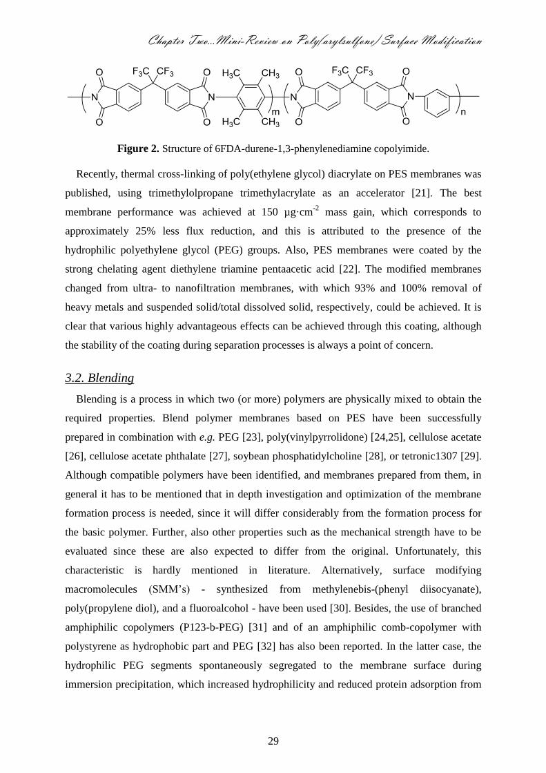
Figure 2. Structure of 6FDA-durene-1,3-phenylenediamine copolyimide.
Recently, thermal cross-linking of poly(ethylene glycol) diacrylate on PES membranes was
published, using trimethylolpropane trimethylacrylate as an accelerator [21]. The best
membrane performance was achieved at 150 µg·cm-2
mass gain, which corresponds to
approximately 25% less flux reduction, and this is attributed to the presence of the
hydrophilic polyethylene glycol (PEG) groups. Also, PES membranes were coated by the
strong chelating agent diethylene triamine pentaacetic acid [22]. The modified membranes
changed from ultra- to nanofiltration membranes, with which 93% and 100% removal of
heavy metals and suspended solid/total dissolved solid, respectively, could be achieved. It is
clear that various highly advantageous effects can be achieved through this coating, although
the stability of the coating during separation processes is always a point of concern.
3.2. Blending
Blending is a process in which two (or more) polymers are physically mixed to obtain the
required properties. Blend polymer membranes based on PES have been successfully
prepared in combination with e.g. PEG [23], poly(vinylpyrrolidone) [24,25], cellulose acetate
[26], cellulose acetate phthalate [27], soybean phosphatidylcholine [28], or tetronic1307 [29].
Although compatible polymers have been identified, and membranes prepared from them, in
general it has to be mentioned that in depth investigation and optimization of the membrane
formation process is needed, since it will differ considerably from the formation process for
the basic polymer. Further, also other properties such as the mechanical strength have to be
evaluated since these are also expected to differ from the original. Unfortunately, this
characteristic is hardly mentioned in literature. Alternatively, surface modifying
macromolecules (SMM’s) - synthesized from methylenebis-(phenyl diisocyanate),
poly(propylene diol), and a fluoroalcohol - have been used [30]. Besides, the use of branched
amphiphilic copolymers (P123-b-PEG) [31] and of an amphiphilic comb-copolymer with
polystyrene as hydrophobic part and PEG [32] has also been reported. In the latter case, the
hydrophilic PEG segments spontaneously segregated to the membrane surface during
immersion precipitation, which increased hydrophilicity and reduced protein adsorption from

30
6.8 to 0.5 µg·cm-2
, whereas only a slight change in permeation properties was observed.
Comparable results were found for PSf-based blended membranes with amphiphilic
copolymers having PSf backbones and PEG side chains, (PSf-g-PEG) [33]. These membranes
exhibited good mechanical characteristics, and remarkably reduced protein adsorption (about
72% reduction in protein adsorption with 10 wt% PSf-g-PEG blending).
Recently, amphiphilic copolymers such as phosphorylcholine copolymer [34] (i.e.,
synthesized copolymer composed of 2-methacryloyloxyethylphosphorylcholine (MPC) and n-
butyl methacrylate (BMA)) were investigated. Blending of this MPC-BMA copolymer with
PES membranes reduced the contact angle from 71° to 39°, and bovine serum albumin (BSA)
adsorption from 65 to 10.6 µg·cm-2
, which the authors attributed to increased hydrophilicity.
Although nice results were obtained with blending of amphiphilic copolymers with PES, only
few of theses amphiphilic copolymers such as tetronic [29] have been synthesized on
commercial scale.
3.3. Composite
A composite is a material made from two or more materials with different physical or
chemical properties which remain separate and distinct on a macroscopic level within the
finished structure. N,O-carboxymethyl chitosan/poly(ethersulfone) (CM-CS/PES) [35]
composite membranes were prepared by immersing PES microfiltration membranes into CM-
CS solutions and cross-linking with glutaraldehyde. Streaming potential measurements
indicate that CM-CS/PES composite membranes possess a weak positive charge at low pH
and a rather strong negative charge at high pH [36]. Therefore, the negative electrostatic
repulsion interactions between membrane and protein molecules at pH 6-8 (i.e., above BSA
isoelectric point) are stronger than the positive electrostatic repulsion interactions at pH 3-4
(i.e., below BSA isoelectric point). Under acidic conditions, relatively high adsorption levels
occur, also caused by denaturation and aggregation of the protein below its isoelectric point.
This gives the CM-CS/PES composite membranes a dual functionality; they resist protein
fouling at high pH, and separate proteins by adsorption at low pH, which can subsequently be
recovered by increasing the pH.
Sulfonated poly(ether-ethersulfone)-poly(ethersulfone) (SPEES-PES) and sulfonated
poly(ether-ethersulfone)-poly(ethersulfone)/Poly (vinyl chloride) (SPEES-PES/PVC) [37]
composites were used to measure glucose and hydrogen peroxide permselectivity in
amperometric biosensors. Also, highly charged cation-permeable composite membranes were
prepared from blends of sulfonated PES with sulfonated poly(ether-ether-ketone) [38].

Chapter Two…Mini-Review on Poly(arylsulfone) Surface Modification
31
TiO2 nanoparticles were added to the polymer solution and then TiO2-entrapped PSf
membranes were prepared by phase inversion [19]. These membranes showed less flux
decline (38%) compared to neat PSf membranes (85%). Also, PES-TiO2 composite
membranes (4% wt) showed better flux behavior (29% higher) compared to PES membranes
[39]. On the other hand, the included TiO2 nanoparticles resulted in improvements in
mechanical properties of PES membrane by increasing the breaking strength from 3.2 to 4.1
MPa while decreasing the elongation ratio from 16 to 12%. Fouling mitigation increased with
nanoparticle content, but it reached a limit above which fouling mitigation was not improved.
The TiO2 nanoparticle acts mainly on hydrophobic substances, suggesting a possible use as a
new anti-fouling component in composite membranes [40]. Using Al2O3 instead of TiO2 and
at much lower concentration [10 times lower] resulted in reduction in cake formation from
82% to 18%. The flux loss during operation was diminished by over 10% [41].
Similar to the case of blending of amphiphilic copolymers - the range of components that
are suitable for composite formation and are ready available and have been synthesized on
large scale is limited.
3.4. Chemical
For chemical modification, the membrane material is treated with modifying agents to
introduce various functional groups on the membrane surface. For example,
[−CH2CH2CH2SO3−] [42,43] groups have been coupled onto the surfaces of PSf hollow fibers
using the reaction of PSf, propane sultone, and Friedel-Crafts catalysts. The resulting
membranes were claimed to show excellent anti-adsorption behavior. Also, a surface reaction
of PSf hollow fibers with propylene oxide and a Friedel-Crafts catalyst was carried out, and a
hydrophilic surface without charged segments (“hydroxyl” type; −CH(CH3)CH2OH) [44,45]
was obtained. The membranes were tested by ultrafiltration of a mix of BSA and γ-globulin. It
was found that BSA is concentrated in the retentate and γ-globulin is concentrated in the
permeate when a modified membrane with −CH(CH3)CH2OH segments is used, while the
unmodified membranes cannot separate the proteins. The ultrafiltration of the mixture at pH 9
(BSA and γ-globulin have the same net negative charge) suggested that the separation
mechanism is not due to a sieving effect or to charge repulsion but resulted from the balance
of hydrophilic and hydrophobic segments on the surface of the modified membranes.
In addition, sulfonation, chloromethylation, aminomethylation, and lithiation reactions
were applied to PSf membranes [46-48]. The main challenge for modification by chemical
treatment of commercial membranes is that the modification agent may partly block the pores

32
of the membranes. Even if the modified membranes are less prone to fouling, the total flux
after modification is generally smaller than before modification. In some cases, chemical
modification during membrane formation is preferred, since it seems to compromise the flux
less [49].
3.5. Grafting
Grafting is a method wherein monomers are covalently bonded onto the membrane. Some
examples of monomers used for PES modification are shown in Figure 3. The techniques to
initiate grafting are: (i) chemical, (ii) photochemical and/or via high-energy radiation, (iii) the
use of a plasma, and (iv) enzymatic. The choice for a specific grafting technique depends on
the chemical structure of the membrane and the desired characteristics after surface
modification.
3.5.1. Chemical Initiation Technique
In chemical grafting, free radicals are produced that are transferred to the substrate to
initiate polymerization and form graft co-polymers. A few studies showed that redox
initiation-grafting could be successfully applied to PES ultrafiltration membranes [50,51]. For
these, peroxydisulfate and metabisulfite oxidizing agents have been used to initiate free-
radical polymerization grafting of methacrylic acid, polyethyleneglycol-methacrylate, and
sulfopropylmethacrylate in aqueous solution at ambient temperature. In general, this
technique is simple and cheap, leading to membranes that are claimed to be less sensitive to
fouling due to the presence of the hydrophilic grafted monomers, but it is a harsh treatment.
3.5.2. Photochemical and Radiation Initiation Techniques
When a chromophore on a macromolecule absorbs light, the molecule is brought in an
exited state, and one or more chemical bonds may dissociate into radicals that can act as
initiators for the grafting progress. Radicals that are generated in this manner on the
membrane surface can react with the monomer to form the grafted co-polymer. If the
absorption of light does not lead to the formation of free radical sites, this can be promoted by
addition of photosensitizers that form radicals, which in turn abstract hydrogen atoms from
the base polymer surface and produce the radical sites required for grafting [11].

Chapter Two…Mini-Review on Poly(arylsulfone) Surface Modification
33
Figure 3. Chemical structure of some monomers used to modify PES membranes.
The irradiation of macromolecules can cause homolytic fission and thus forms free radicals
on the membrane. UV irradiation and UV-assisted graft polymerization are techniques that
can selectively alter membrane surface properties without affecting the bulk polymer. UV-
assisted graft polymerization modifies the membrane surface by grafting polymer chains onto
the surface and in the pores. UV irradiation can cross-link polymer chains and cleave polymer
bonds, therewith forming functional groups such as hydroxyls, carbonyls, or carboxylic acids

34
on the surface. Initial attempts to carry out the graft modification of poly(arylsulfone)
membranes were conducted in the presence of benzophenone as a photosensitizer [52].
However, it was soon discovered that all poly(arylsulfone) membranes are intrinsically
photosensitive and generate free radicals upon irradiation with 254 nm UV light [53-55]. The
UV irradiation should be carefully used because it leads to severe degradation of the pore
structure with loss of membrane function, which needs to be partially compensated by grafted
polymer. This technique was used to graft several hydrophilic monomers (N-vinyl-2-
pyrrolidinone, N-vinylcaprolactam, and N-vinylformamide) onto 10 kDa PES ultrafiltration
membranes, and their fouling during BSA filtration was compared with that of an unmodified
membrane. Membranes modified with N-vinyl-2-pyrrolidinone (25% increase in
hydrophilicity) exhibited the best combination of low fouling (50% decrease in BSA fouling)
and high flux, although membrane permeability was significantly decreased because the
grafted polymer chains blocked the membrane pores (over 25% reduction in flux due to
modification) [56].
UV-assisted graft polymerization of the same monomer (N-vinyl-2-pyrrolidinone) onto 50
kDa PES membranes created highly wettable PES membranes with high fouling resistance
compared to the base membrane [57-59]. Two methods were used: dip modification
(irradiation after dipping in monomer solution) and immersion modification (irradiation in
monomer solution). The irreversible flux decrease due to adsorptive fouling, i.e. the
permanent flux drop after water cleaning with respect to the initial buffer flux, was reduced
significantly, from 42% for the base membrane to 9% for the modified membrane. The
immersion technique created the best membranes for applications in which high protein
retention is required, while the dip-modified membranes performed best for applications in
which high protein transmission is preferred. When comparing the dip and immersion method
it becomes clear that the dip method seems more suitable for industrial applications. It
requires less monomer, it can more easily be adapted to a continuous process, and is easier to
control.
UV-assisted graft polymerization was used for three hydrophilic monomers, N-vinyl-2-
pyrrolidinone, 2-acrylamidoglycolic acid monohydrate, and 2-acrylamido-2-methyl-1-
propanesulfonic acid, on PES and PSf membranes of 50 kDa. The ultrafiltration membranes
were modified using the dip method with 300 nm wavelength lamps [60]. Four conditions
were found to give superior filtration performance: high monomer concentrations (5 wt%),
low irradiation energy (<65 mJ·cm-2
for PES and <130 mJ.cm-2
for PSf), low degree of
grafting (i.e., DG < 0.53), and intermediate wettability (contact angle between 35o
- 42o).
Chapter Two…Mini-Review on Poly(arylsulfone) Surface Modification
35
Most probably, under these conditions, the grafted polymer chains are placed in such a way
that they extend from the surface, therewith preventing protein penetration. Interestingly, it
was found that PES is much more sensitive to UV-assisted graft polymerization than PSf, and
thus, requires far less energy to attain a desired degree of grafting. Surface modification of
PES ultrafiltration membranes via simultaneous photografting polymerization has been
successfully done to prepare low-fouling UF membranes (≥ 300 nm wavelength lamps). The
hydrophilic monomers that were used were poly(ethylene glycol) methacrylate [61], N-2-
vinyl pyrrolidinone, 2-hydroxyethyl methacrylate, acrylic acid, 2-acrylamidoglycolic acid, 3-
sulfopropyl methacrylate, 2-acrylamido-2-methyl-1-propanesulfonic acid [62,63], quaternary
2-dimethylamino-ethylmethacrylate [64]. Moreover, polymers such as poly(vinyl alcohol),
polyethylene glycol, and chitosan [65] have been photografted onto PES membranes.
Membrane permeability increased by 50% for UV dip-modified membranes in the presence of
a low concentration (10 mM) of chain transfer agent (2-mercaptoethanol) and by 20-200%
(with severe reduction in membrane rejection) when high (50 mM) 2-mercaptoethanol
concentrations were used [66].
Radicals can also be formed by electromagnetic radiation of a shorter wavelength (i.e.,
gamma irradiation) [38,67]. Free radical grafting initiated by radiation proceeds in three
different ways, which are pre-irradiation, peroxidation, and mutual irradiation. In pre-
irradiation grafting, the membrane backbone is first irradiated in vacuum or in the presence of
an inert gas to form free radicals. Then, the irradiated membrane is treated with the monomer,
in liquid or vapor state. In peroxidation grafting, the membrane is subjected to radiation in the
presence of air or oxygen to form hydroperoxides or diperoxides. The stable peroxy products
are then treated with the monomer at higher temperature, where the peroxides undergo
decomposition to radicals and initiate grafting. In mutual irradiation, the membrane and the
monomers are irradiated simultaneously, to form free radicals that are subsequently grafted to
the surface. Radiation grafting can also proceed through an ionic mode [68].
Resistance of poly(arylsulphone) resins to γ-irradiation in vacuum and air at various
temperatures has been studied [67,69]; both cross-linking and chain scission occur when PSf
and PES were subjected to γ-irradiation at 30 °C. Cross-linking is predominant for irradiation
under vacuum, whereas for irradiation in air scission predominates over cross-linking.
Asymmetric PES ultrafiltration membranes have been modified with acrylic acid or
acrylamide [70], and at certain combinations of experimental parameters, water flux, and
solute retention were improved compared with the untreated membrane.

36
3.5.3. Plasma Initiation Technique
Given enough energy, any gas can be excited into the plasma state, which is a mixture of
ions, electrons, excited species, and free radicals. Plasma surface treatment usually refers to a
plasma reaction that either results in modification of the molecular structure of the surface, or
atomic substitution. Plasma treatment is a useful tool in the modification of surface properties.
Currently, more and more attention is being given to its applications in membrane separation
science. The accelerated electrons from the plasma have sufficient energy to induce cleavage
of the chemical bonds in the membrane structure and to form macromolecule radicals, which
subsequently initiate graft co-polymerization [71]. Plasma treatment can be done by either
regular plasma treatment [72,73],
or plasma graft co-polymerization (PGC) [74]. Low
temperature plasma techniques, which are very surface selective, have been used to modify
various types of membranes, specifically to reduce protein-surface attractive interaction. For
example, simple inert gas [75,76], nitrogen [73], or oxygen [72] plasmas have been used to
increase the surface hydrophilicity of membranes [14,77], and ammonia plasmas have
successfully yielded functionalized PSf membranes [78]. Also a water plasma treatment that
renders asymmetric PSf membranes permanently hydrophilic has been reported [79], and this
technique was also successfully applied to PES membranes and, to a lesser extent,
polyethylene membranes [77,80]. Further, Ar-plasma treatment followed by graft
copolymerization with acrylamide in the vapor phase was used to make PES membranes
highly hydrophilic [74]. The grafting yield for polyacrylamide on the membrane surfaces
increased nearly linearly with the Ar-plasma pretreatment time, with grafting yields (GY)
higher than 100 µg·cm-2
. The membranes obtained a permanent hydrophilicity (almost no
change in contact angle after one year), and BSA adsorption was reduced to less than half that
of the control membrane (306 to 148 µg·cm-2
). Although, plasma treatment (without grafting)
is often claimed to increase hydrophilicity (attributed to structural rearrangement of polymer
chains with decrease in surface energy [76]), and therewith protein repellence, mostly this
effect tends to be temporary, which could imply that the treatment has to be repeated.
3.5.4. Enzymatic Initiation Technique
The enzymatic grafting method is quite new. The principle involved is that an enzyme
initiates the chemical/electrochemical grafting reaction [71]. This method employs enzymes
to convert the substrate (monomer, oligomer or polymer chains) into a reactive free radical(s),
which undergoes subsequent non-enzymatic reaction with the membrane [81-86]. There are
several potential advantages for the use of enzymes in membrane modification. With respect

Chapter Two…Mini-Review on Poly(arylsulfone) Surface Modification
37
to health and safety, enzymes offer the potential of eliminating the need for (and hazards
associated with) reactive reagents (and solvents). A potential environmental benefit for using
enzymes is that their selectivity may be exploited to eliminate the need for wasteful protection
and deprotection steps. Finally, enzyme specificity may offer the potential for precisely
modifying macromolecular structure to better control polymer function [83]. Some polymers
that were successfully modified with enzymes are mentioned here briefly.
Enzymatic grafting of chitosan resins and films using tyrosinase and chlorogenic acid [83],
4-hydroxybenzoic acid, 3,4-dihydroxybenzoic acid, 3,4-dihydroxyphenylacetic acid, and
hydrocaffeic acid phenol derivatives [84,85] has been reported. The main target for chitosan
enzyme-catalyzed grafting is to alter the surface and rheological properties, under basic
conditions, and cationic dye-adsorption properties. Reactions were conducted under
heterogeneous conditions using chitosan films, and under homogeneous conditions using
aqueous methanolic mixtures capable of dissolving both substrates and chitosan [84].
Tyrosinase was shown to convert the substrate into a reactive o-quinone, which undergoes a
subsequent non-enzymatic reaction with chitosan. Tyrosinase has a broad substrate range for
phenols such as poly(4-hydroxystyrene), 4-tert-butylcatechol, p-hydroxyphenoxyacetic acid
and p-cresol [81,82]. In addition to reacting with a range of monomeric phenols, tyrosinase is
known to react with oligomeric and polymeric substrates [86], therewith indicating the
versatility that enzyme-catalyzed modification could yield.
Very recently, we published an enzyme based method for grafting of PES membranes [87].
This modification method uses laccase from Trametes versicolor to create free radicals, and
graft phenolic acid monomers (e.g., gallic acid or 4-hydroxybenzoic acid) to the membrane.
The modified membranes have high fluxes and in some cases excellent protein repellent
properties, but a clear relation between the grafted layer and its effectiveness against protein
adsorption has not been established yet. What is clear, is that this modification method is very
mild and environmentally benign compared to the more traditional methods; it can be carried
out at room temperature, and uses only oxygen and water, and no toxic chemicals.
3.6. Combined Methods
Recently, combined techniques were presented for PES membranes [88,89], in which the
membrane was blended with a copolymer of acrylonitrile and acrylic acid, and subsequently
grafted with bovine serum albumin [88]. Sulfonic acid groups were generated on the PES
membrane surfaces by chemical sulfonation, followed by dipping the membranes into a TiO2
solution [89,90]. This modification (8 % wt. TiO2 deposited) reduces the loss of flux due to
38
fouling from 80 to 65%. Another combined modification was carried out by blending PES
with polyimide and treatment with diethanolamine to introduce –OH groups on the membrane
surface, followed by dipping in TiO2 colloidal solution, and irradiation with UV light [91].
Combined modification sequences lead to lower fouling of the membrane. The results as such
are interesting, although the complexity of the technique could prove to be a major hurdle.
4. Overview of Modification Methods and Conclusions
An overall comparison between the different surface modification methods is presented in
Table 1, which is our interpretation of the presented results in literature. To the criteria that
have been mentioned thus far has now been added ‘cost effectiveness’, a factor that depends
on the costs of chemicals and equipment used. Note that it is not always straightforward to
interpret and compare results, because many parameters may be influenced simultaneously by
one modification method; here we only attempt to give a general impression.
All the surface modification methods mentioned earlier allow modification without
affecting the bulk properties too much when appropriate conditions are selected; mostly the
flux is similar to the base membrane or slightly lower (as indicated by the yellow circles in
Table 1).
It is well known that modification by creating a chemical bond (i.e., covalent bonding) is
more stable than physical adhesion (e.g., coating). Complete and seemingly permanent
hydrophilic modification of poly(arylsulfone) membranes is achieved by blending and
photoinduced grafting, although it should be mentioned that protein adsorption is reduced at
the produced hydrophilic surfaces, but never completely prevented. Chemical treatment
usually employs harsh treatment; often it may lead to undesirable surface changes and
contamination, and may not be the best choice in environmental terms. Plasma treatment is
probably one of the most versatile poly(arylsulfone) membrane surface treatment techniques.
However, its high costs and technical complexity remain drawbacks for large-scale use.
Enzyme-catalyzed grafting of poly(ethersulfone) membranes, which has just been reported, is
in this respect a method that could be an environmentally benign alternative for other
poly(arylsulfone) modification methods, but is in need of further development. Combination
of two or three modification techniques is complex in terms of cost effectiveness and
environmental drawbacks, but could lead to multi-functional membranes that are of great
interest for ‘membranes of the future’. Such membranes may need more functions than ‘only’
providing a selective barrier with high performance (flux and stability). It is expected that
membrane properties can be tuned for specific applications through the discussed methods,

Chapter Two…Mini-Review on Poly(arylsulfone) Surface Modification
39
although they still need to be developed further in such a way that they allow even better and
more environmentally friendly control over modification. To be complete, it should be noted
that all mentioned methods influence membrane smoothness/roughness. However, since its
effect on protein repellence and flux is still heavily debated in literature as illustrated in [92],
we will not consider it further.
Table 1. Advantages and disadvantages of modification methods.
No. Modification method Flux after
modification
Simplicity/
Versatility
Reproducibility Environmental
aspects
Cost
effectiveness
1 Coating
2 Blending
3 Composite
4 Chemical
5 Grafting initiated by:
Chemical
Photochemical
Radiation
Plasma
Enzymatic
6 Combined methods
Excellent, High, Low
40
References
1. Gopal, R.; Zuwei, M.; Kaur, S.; Ramakrishna, S., Surface Modification and Application of
Functionalized Polymer Nanofibers, in: Mansoori, G.A.; George, Th.F.; Assoufid, L.; Zhang, G.
(Eds.), Molecular Building Blocks for Nanotechnology: From Diamondiods to Nanoscale
Materials and Applications, E-Publishing, Springerlink.com, Berlin/Heidelberg (2007) 72-91.
2. Kato, K.; Uchida, E.; Kang, E.T.; Uyama, Y.; Ikada, Y., Polymer Surface with Graft Chains, Prog.
Polym. Sci. 28 (2003) 209-259.
3. Golander, C.G.; Kiss, E., Protein Adsorption on Functionalized and ESCA-Characterized Polymer
Films Studied by Ellipsometry, J. Colloid Interface Sci. 121 (1988) 240-253.
4. Lundström, I., Surface Physics and Biological Phenomena, Phys. Scr. T4 (1983) 5-13.
5. Ulbricht, U., Advanced Functional Polymer Membranes, Polymer 47 (2006) 2217-2262.
6. Minko, S., Grafting on Solid Surfaces: “Grafting to” and “Grafting from” Methods, in: Manfred, S.
(Ed.), Polymer Surfaces and Interfaces: Characterization, Modification and Applications, E-
Publishing, Springerlink. com, Berlin/Heidelberg (2008) 215-234.
7. Schroën, C.G.P.H.; Stuart, M.A.C.; Maarschalk, K.V.; van der Padt, A.; van ‘t Riet, K., Influence
of Preadsorbed Block Copolymers on Protein Adsorption: Surface Properties, Layer Thickness,
and Surface Coverage, Langmuir 11 (1995) 3068-3074.
8. Kim, K.J.; Fane, A.G.; Fell, C.J.D., The Performance of Ultrafiltration Membranes Pretreated by
Polymer, Desalination 70 (1988) 229-249.
9. Jönsson, A.S.; Jönsson, B., The Influence of Nonionic and Ionic Surfactants on Hydrophobic and
Hydrophilic Ultrafiltration Membranes, J. Membr. Sci. 56 (1991) 49-76.
10. Nyström, M., Fouling of Unmodified and Modifies Polysulfone Ultrafiltration Membranes by
Ovalbumin, J. Membr. Sci. 44 (1989) 183-196.
11. Hvid, K.B.; Nielsen, P.S.; Stengaard, F.F., Preparation and Characterization of a New
Ultrafiltration Membrane, J. Membr. Sci. 53 (1990) 189-202.
12. Stengaard, F.F., Characteristics and Performance of New Types of Ultrafiltration Membranes with
Chemically Modified Surfaces, Desalination 70 (1988) 207-224.
13. Kim, K.J.; Fane, A.G.; Fell, C.J.D., The Effect of Langmuir-Blodgett Layer Pretreatment on the
Performance of Ultrafiltration Membranes, J. Membr. Sci. 43 (1989) 187-204.
14. Ulbricht, M.; Belfort, G., Surface Modification of Ultrafiltration Membranes by Low Temperature
Plasma.1.Treatment of Polyacrylonitrile, J. Appl. Polym. Sci. 56 (1995) 325-343.
15. Duarte, L.T.; Habert, A.C.; Barges, C.P., Preparation and Morphological Characterization of
Polyurethane/Polyethersulfone Composite Membranes, Desalination 145 (2002) 53-59.
16. Duarte, L.T.; Pereira, C.C.; Habert, A.C.; Borges, C.P., Polyurethane/Polyethersulphone
Composite Hollow Fibers Produced by Simultaneous Spinning of Two Polymer Solutions, J.
Membr. Sci. 311 (2008) 12-22.
17. Hamza, A.; Chowdhury, G.; Matsuura, T.; Sourirajan, S., Sulphonated Poly (2,6-
dimethylphenylene oxide)-Polyethersulphone Composite Membranes: Effects of Composition of
Solvent System, used for Preparing Casting Solution, on Membrane-Surface Structure and Reverse
Osmosis Performance, J. Membr. Sci. 129 (1997) 55-64.
18. Hyun, J.; Jang, H.; Kim, K.; Na, K.; Tak, T., Restriction of Biofouling in Membrane Filtration
using a Brush-Like Polymer Containing Oligoethylene Glycol Side Chains, J. Membr. Sci. 282
(2006) 52-59.
19. Bae, T.H.; Tak, T.M., Effect of TiO2 Nanoparticles on Fouling Mitigation of Ultrafiltration
Membranes for Activated Sludge Filtration, J. Membr. Sci. 249 (2005) 1-8.
20. Li, D.F.; Chung, T.H.; Wang, R.; Liu, Y.Y., Fabrication of Fluoropolyimide /Polyethersulfone
(PES) Dual-Layer Asymmetric Hollow Fiber Membranes for Gas Separation, J. Membr. Sci. 198
(2002) 211-223.
21. Mu, L.J.; Zhao, W.Z., Hydrophilic Modification of Polyethersulfone Porous Membranes via A
Thermal-Induced Surface Crosslinking Approach, Appl. Surf. Sci. 255 (2009) 7273-7278.
22. Boricha, A.G.; Murthy, Z.V.P., Preparation, Characterization and Performance of Nanofiltration
membranes for the Treatment of Electroplating Industry Effluent, Sep. Purif. Technol. 65 (2009)
282-289.
23. Idrisa, A.; Zaina, N.M.; Noordin, M.Y., Synthesis, Characterization and Performance of
Asymmetric Polyethersulfone (PES) Ultrafiltration Membranes with Polyethylene Glycol of
Different Molecular Weights as Additives, Desalination 207 (2007) 324-339.

Chapter Two…Mini-Review on Poly(arylsulfone) Surface Modification
41
24. Marchese, J.; Ponce, M.; Ochoa, N.A.; Prádanos, P.; Palacio, L.; Hernández, A., Fouling Behavior
of Polyethersulfone UF Membranes Made with Different PVP, J. Membr. Sci. 211 (2003) 1-11.
25. Lu, S.; Zhuang, L.; Lu, J., Homogeneous Blend Membrane Made from Poly(ethersulfone) and
Poly(vinylpyrrolidone) and its Application to Water Electrolysis, J. Membr. Sci. 300 (2007) 205-
210.
26. Mahendran, R.; Malaisamy, R.; Mohan, D.R., Cellulose Acetate and Polyethersulfone Blend
Ultrafiltration Membranes. Part I: Preparation and Characterizations, Polym. Adv. Technol. 15
(2004) 149-157.
27. Rahimpour, A.; Madaeni, S.S., Polyethersulfone (PES)/Cellulose Acetate Phthalate (CAP) Blend
Ultrafiltration Membranes: Preparation, Morphology, Performance and Antifouling Properties, J.
Membr. Sci. 305 (2007) 299-312.
28. Wang, Y.Q.; Wang, T.; Su, Y.L.; Peng, F.B.; Wu, H.; Jiang, Z.Y., Protein Adsorption-Resistance
and Permeation Property of Poly(ethersulfone) and Soybean Phosphatidylcholine Blend
Ultrafiltration Membranes, J. Membr. Sci. 270 (2006) 108-114.
29. Rahman, N.A.; Maruyama, T.; Matsuyama, H., Performance of Polyethersulfone /Tetronic1307
Hollow Fiber Membrane for Drinking Water Production, J. Appl. Sci. Environ. Sanit. 3 (2008) 1-7.
30. Zhang, L.; Chowdhury, G.; Feng, C.; Matsuura, T.; Narbaitz, R., Effect of Surface Modifying
Macromolecules and Membrane Morphology on Fouling of Polyethersulfone Ultrafiltration
Membrane, J. Appl. Polym. Sci. 88 (2002) 3132-3138.
31. Wang, Y.Q.; Su, Y.L.; Sun, Q.; Ma, X.L. Jiang, Z.Y., Generation of Anti Biofouling Ultrafiltration
Membrane Surface by Blending Novel Branched Amphiphilic Polymers with Polyethersulfone, J.
Membr. Sci. 286 (2006) 228-236.
32. Ma, X.; Su, Y.; Sun, Q.; Wang, Y.; Jiang, Z., Preparation of Protein-Adsorption-Resistant
Poly(ethersulfone) Ultrafiltration Membranes through Surface Segregation of Amphiphilic Comb
Co-polymer, J. Membr. Sci. 292 (2007) 116-124.
33. Park, J.Y.; Acar, M.H.; Akthakul, A.; Kuhlman, W.; Mayes, A.M., Polysulfone-Graft-
Poly(ethylene glycol) Graft Copolymers for Surface Modification of Polysulfone Membranes,
Biomaterials, 27 (2006) 856-865.
34. Su, Y.; Li, C.; Zhao, W.; Shi, Q.; Wang, H.; Jiang, Z.; Zhu, S., Modification of Poly(ethersulfone)
Ultrafiltration Membranes with Phosphorylcholine Copolymer Can Remarkably Improve the
Antifouling and Permeation Properties, J. Membr. Sci. 322 (2008) 171-177.
35. Zhao, Z.P.; Wang, W.; Wang, S.C., Formation, Charged Characteristic and BSA Adsorption
Behavior of Carboxymethyl Chitosan/PES Composite MF Membrane, J. Membr. Sci. 217 (2003)
151-158.
36. Zhao, Z.; Wang, Z.; Ye, N.; Wang, S., A Novel N,O-Carboxymethyl Amphoteric
Chitosan/Poly(ethersulfone) Composite MF Membrane and its Charged Characteristics,
Desalination 144 (2002) 35-39.
37. Benmakroha, Y.; Christie, I.; Desai, M.; Vadgama, P., Poly(vinyl chloride), Polysulfone and
Sulfonated Polyether ether sulfone Composite Membranes for Glucose and Hydrogen Peroxide
Perm-Selectivity in Amperometric Biosensors, Analyst 121 (1996) 521-526.
38. Gohil, G.S.; Nagarale, R.K.; Binsu, V.V.; Shahi, V.K., Preparation and Characterization of
Monovalent Cation Selective Sulfonated Poly(ether-ether ketone) and Poly(ethersulfone)
Composite Membranes, J. Colloid Interface Sci. 298 (2006) 845-853.
39. Li, J.F.; Xu, Z.L.; Yang, H.; Yu, L.Y.; Liu, M., Effect of TiO2 Nanparticles on the Surface
Morphology and Performance of Microporous PES Membrane, Appl. Surf. Sci. 255 (2009) 4725-
4732.
40. Luo, M.L.; Zhao, J.Q.; Tang, W.; Pu, C.H., Hydrophilic Modification of Poly(ethersulfone)
Ultrafiltration Membrane Surface by Self-Assembly of TiO2 Nanoparticles, Appl. Surf. Sci. 249
(2005) 76-84.
41. Maximous, N.; Nakhla, G.; Wan, W.; Wong, K., Preparation, Characterization and Performance of
Al2O3/PES Membrane for Wastewater Filtration, J. Membr. Sci. 341 (2009) 67-75.
42. Higuchi, A.; Iwata, N.; Tsubaki, M.; Nakagawa, T., Surface-Modified Polysulfone Hollow Fibers,
J. Appl. Polym. Sci. 36 (1988) 1753-1767.
43. Higuchi, A.; Iwata, N.; Nakagawa, T., Surface-Modified Polysulfone Hollow Fibers II. Fibers
Having CH2CH2CH2SO3 Segments and Immersed in HCl Solution, J. Appl. Polym. Sci. 40 (1990)
709-717.

42
44. Higuchi, A.; Nakagawa, T., Surface Modified Polysulfone Hollow Fibers. III. Fibers Having a
Hydroxide Group, J. Appl. Polym. Sci. 41 (1990) 1973-1979.
45. Higuchi, A.; Mishima, S.; Nakagawa, T., Separation of Proteins by Surface Modified Polysulfone
Membranes, J. Membr. Sci. 57 (1991) 175-185.
46. Guiver, M.D.; Apsimon, J.W., The Modification of Polysulfone by Metalation, Polym. Sci., Part
C: Polym. Lett. 26 (1988) 123-127.
47. Michael, D.; Guiver, M.D.; Kutowy, O.; Apsimon, J.W., Functional Group Polysulphones by
Bromination-Metalation, Polymer 30 (1989) 1137-1142.
48. Breitbach, L.; Hinke, E.; Staude, E., Heterogeneous Functionalizing of Polysulfone Membranes,
Die Angew. Makromol. Chem. 184 (1991) 183-196.
49. Anke Nabe, A; Staude, E.; Belfort, G., Surface Modification of Polysulfone Ultrafiltration
Membranes and Fouling by BSA solutions, J. Membr. Sci. 133 (1997) 57-72.
50. Belfer, S.; Fainchtain, R.; Purinson, Y.; Kedem, O., Surface Characterization by FTIR-ATR
Spectroscopy of Polyethersulfone Membranes-Unmodified, Modified and Protein Fouled J.
Membr. Sci. 172 (2000) 113-124.
51. Reddy, A.V.R.; Trivedi, J.J.; Devmurari, C.V.; Mohan, D.J.; Singh, P.; Rao, A.P.; Joshi, S.V.;
Ghosh, P.K., Fouling Resistant Membranes in Desalination and Water Recovery, Desalination 183
(2005) 301-306.
52. Ulbricht, M.; Riedel, M.; Marx, U., Novel Photochemical Surface Functionalization of Polysulfone
Ultrafiltration Membranes for Covalent Immobilization of Biomolecules, J. Membr. Sci. 120
(1996) 239-259.
53. Nyström, M.; Järvinen, P., Modification of Polysulfone Ultrafiltration Membranes with UV
Irradiation and Hydrophilicity Increasing Agents, J. Membr. Sci. 60 (1991) 275-296.
54. Yamagishi, H.; Crivello, J.V.; Belfort, G., Development of a Novel Photochemical Technique for
Modifying Poly(arylsulfone) Ultrafiltration Membranes, J. Membr. Sci. 105 (1995) 237-247.
55. Yamagishi, H.; Crivello, J.V.; Belfort, G., Evaluation of Photochemically Modified
Poly(arylsulfone) Ultrafiltration Membranes, J. Membr. Sci. 105 (1995) 249-259.
56. Pieracci, J.; Crivello, J.V.; Belfort, G., Photochemical Modification of 10 kDa Polyethersulfone
Ultrafiltration Membranes for Reduction of Biofouling, J. Membr. Sci. 156 (1999) 223-240
57. Pieracci, J.; Wood, D.W.; Crivello, J.V.; Belfort, G., UV-Assisted Graft Polymerization of N-
vinyl-2-pyrrolidinone onto Poly(ethersulfone) Ultrafiltration Membranes: Comparison of Dip
versus Immersion Modification Techniques, Chem. Mater. 12 (2000) 2123-2133.
58. Kilduft, J.E.; Mattaraj, S.; Pieracci, J.; Belfort, G., Photochemical Modification of
Poly(ethersulfone) and Sulfonated Poly(sulfone) Nanofiltration Membranes for Control of Fouling
by Natural Organic Matter, Desalination 132 (2000) 133-142.
59. Taniguchi, M.; Pieracci, J.; Samsonoff, W.A.; Belfort, G., UV-Assisted Graft Polymerization of
Synthetic Membranes: Mechanistic Studies, Chem. Mater. 15 (2003) 3805-3812.
60. Kaeselev, B.; Pieracci, J.; Belfort, G.; Photoinduced Grafting of Ultrafiltration Membranes:
Comparison of Poly(ethersulfone) and Poly(sulfone), J. Membr. Sci. 194 (2001) 245-261.
61. Susanto, H.; Balakrishnan, M.; Ulbricht, M., Via Surface Functionalization by Photograft Co-
Polymerization to Low-Fouling Polyethersulfone-Based Ultrafiltration Membranes, J. Membr. Sci.
288 (2007) 157-167.
62. Taniguchi, M.; Belfort, G., Low Protein Fouling Synthetic Membranes by UV-Assisted Surface
Grafting Modification: Varying Monomer Type, J. Membr. Sci. 231 (2004) 147-157.
63. Taniguchi, M.; Kilduff, J.E.; Belfort, G., Low Fouling Synthetic Membranes by UV-Assisted Graft
Polymerization: Monomer Selection to Mitigate Fouling by Matural Organic Matter, J. Membr.
Sci. 222 (2003) 59-70.
64. Hilal, N.; Al-Khatib, L.; Atkin, B.P.; Kochkodan, V.; Potapchenko, N., Photochemical
Modification of Membrane Surfaces for (Bio) Fouling Reduction: A Nanoscale Study Using AFM,
Desalination 158 (2003) 65-72.
65. Liu, S.X.; Kim, J.-T.; Kim, S., Effect of Polymer Surface Modification on Polymer-Protein
Interaction via Hydrophilic Polymer Grafting, J. Food Sci. 73 (2008) 143-150.
66. Pieracci, J.; Crivello, J.V.; Belfort, G., Increasing Membrane Permeability of UV-Modified
Poly(ethersulfone) Ultrafiltration Membranes, J. Membr. Sci. 202 (2002) 1-16.
67. Brown, J.R., Effects of Gamma Radiation on Two Aromatic Polysulfones, J. Appl. Polym. Sci. 19
(1975) 405-417.

Chapter Two…Mini-Review on Poly(arylsulfone) Surface Modification
43
68. Bhattacharya, A; Misra, B.N., Grafting: A Versatile Means to Modify Polymers Techniques,
Factors and Applications, Prog. Polym. Sci. 29 (2004) 767-814.
69. Brown, J.R.; Effects of Gamma Radiation on Two Aromatic Polysulfones. II. A Comparison of
Irradiation at Various Temperatures in Air-Vacuum Environments, J. Appl. Polym. Sci. 23 (1979)
2763-2775.
70. Keszler, B.; Kovács, G.; Tóth, A.; Bertóti, I.; Hegyi, M., Modified Polyethersulfone Membranes, J.
Membr. Sci. 62 (1991) 201-210.
71. Tsujii, Y.; Ohno, K.; Yamamoto, S.; Goto, A.; Fukuda, T., Structure and Properties of High-
Density Polymer Brushes Prepared by Surface-Initiated Living Radical Polymerization, Adv.
Polm. Sci. 197 (2006) 1-45.
72. Vrlinic, T.; Vesel, A.; Cvelbar, V.; Krajnc, M.; Mozetic, M.; Rapid Surface Functionalization of
Poly(ethersulphone) Foils Using a Highly Reactive Oxygen-Plasma Treatment, Surf. Interface
Anal. 39 (2007) 476-481.
73. Kull, K.R.; Steen, M.L.; Fisher, E.R., Surface Modification with Nitrogen-Containing Plasmas to
Produce Hydrophilic, Low-Fouling Membranes, J. Membr. Sci. 246 (2005) 203-215.
74. Wavhal, D.S.; Fisher, E.R., Membrane Surface Modification by Plasma-Induced Polymerization of
Acrylamide for Improved Surface Properties and Reduced Protein Fouling, Langmuir 19 (2003)
79-85.
75. Gancarz,I; Poźniak, G.; Bryjak, M., Modification of Polysulfone membranes 1. CO2 Plasma
Treatment, Eur. Polym. J. 35 (1999) 1419-1428.
76. Gröning, P.; Coen, M.C.; Küttel, O.M.; Schlapbach, L., Influence of Gas Pressure on the Plasma
Polyethersulphone, Appl. Surf. Sci. 103 (1996) 79-89.
77. Wavhal, D.S.; Fisher, E.R.; Hydrophilic Modification of Polyethersulfone Membranes by Low
Temperature Plasma-Induced Graft Polymerization, J. Membr. Sci. 209 (2002) 255-269.
78. Wolf, J.; Steinhauser, H.; Ellinghorst, G., Tailoring of Ultrafiltration Membranes by Plasma
Treatment and their Application for the Desalination and Concentration of Water-Soluble Organic
Substances, J. Membr. Sci. 36 (1988) 207-214.
79. Michelle, L.; Steen, M.L.; Hymasa, L.; Havey, E.D.; Capps, N.E.; Castner, D.G.; Fisher, E.R.,
Low Temperature Plasma Treatment of Asymmetric Polysulfone Membranes for Permanent
Hydrophilic Surface Modification, J. Membr. Sci. 188 (2001) 97-114.
80. Steen, M.L.; Jordan, A.C.; Fisher, E.R.; Hydrophilic Modification of Polymeric Membranes by
Low Temperature H2O Plasma Treatment, J. Membr. Sci. 204 (2002) 341-357.
81. Payne, G.F.; Chaubal, M.V.; Barbari, T.A., Enzyme-Catalysed Polymer Modification: Reaction of
Phenolic Compounds with Chitosan Films, Polymer 37 (1996) 4643-4648.
82. Shao, L.; Kumar, G.; Lenhart, J.L.; Smith, P.J.; Payne, G.F., Enzymatic Modification of the
Synthetic Polymer Polyhydroxystyrene, Enzyme Microb. Technol. 25 (1999) 660-668.
83. Kumar, G.; Smith, P.J.; Payne, G.F., Enzymatic Grafting of a Natural Product onto Chitosan to
Confer Water Solubility Under Basic Conditions, Biotechnol. Bioeng. 63 (1999) 154-165.
84. Chen, T.; Kumar, G.; Harris, M.T.; Smith, P.J.; Payne, G.F., Enzymatic Grafting of
Hexyloxyphenol onto Chitosan to Alter Surface and Rheological Properties, Biotechnol. Bioeng.
70 (2000) 564-573.
85. Chao, A.C.; Shyu, S.S.; Lin, Y.C.; Mi, F.L., Enzymatic Grafting of Carboxyl Groups on to
Chitosan to Confer on Chitosan the Property of a Cationic Dye Adsorbent, Bioresour. Technol. 91
(2004) 157-162.
86. Kumar, G.; Bristow, J.F.; Smith, P.J.; Payne, G.F., Enzymatic Gelation of the Natural Polymer
Chitosan, Polymer 41 (2000) 2157-2168.
87. Nady, N.; Franssen, M.C.R.; Boom, R.M.; Mohy Eldin, M.S.; Zuilhof, H.; Schroën, C.G.P.H.,
Modification of PES Membranes: An Environmentally Friendly Approach, 13th Aachener
Membrane Kolloquium (2010).
88. Fang, B.; Ling, Q.; Zhao, W.; Ma, Y.; Bai, P.; Wei, Q., Modification of Poly(ethersulfone)
Membrane by Grafting Bovine Serum Albumin on the Surface of Poly(ethersulfone)
/Poly(acrylonitrile-co-acrylic acid) Blended Membrane, J. Membr. Sci. 329 (2009) 46-55.
89. Bae, T.H.; Tak, T.M., Preparation of TiO2 Self-Assembled Polymeric Nanocomposite Membranes
and Examination of their Fouling Mitigation Effects in a Membrane Bioreactor System, J. Membr.
Sci. 266 (2005) 1-5.
90. Bae, T.H.; Kim, I.C.; Tak, T.M., Preparation and Characterization of Fouling-Resistant TiO2 Self-
Assembled Nanocomposite Membranes, J. Membr. Sci. 275 (2006) 1-5.
44
91. Mansourpanah, Y.; Madaeni, S.S.; Rahimpour, A.; Farhadian, A.; Taheri, A.H., Formation of
Appropriate Sites on Nanofiltration Membrane Surface for Binding TiO2 Photo-Catalyst:
Performance, Characterization and Fouling-Resistant Capacity, J. Membr. Sci. 330 (2009) 297-
306.
92. Rana, D.; Matsuura, T., Surface Modifications for Antifouling Membranes, Chem. Rev. 110
(2010) 2448-2471.
Chapter Three
Enzyme-Catalyzed Modification of PES
Membranes. Proof of Principle
This chapter has been published as:
Mild and Highly Flexible Enzyme-Catalyzed Modification of Poly(ethersulfone)
Membranes, Norhan Nady, Karin Schroën, Maurice Franssen, Barend van Lagen, Sukumaran
Murali, Mohamed Mohy Eldin, Remko Boom, and Han Zuilhof, ACS Appl. Mater. Interfaces
3 (2011) 801-810.

46
Mild and Highly Flexible Enzyme-Catalyzed Modification of
Poly(ethersulfone) Membranes
Abstract
Poly(ethersulfone) (PES) membranes are widely used in industry for separation and
purification purposes. However, the drawback of this type of membranes is fouling by
proteins. For that reason, modification of PES membranes has been studied to enhance their
protein repellence. This paper presents the first example of enzyme-catalyzed modification of
PES membranes. Various phenolic acids (enzyme substrates) were bound to a membrane
under very mild conditions (room temperature, water, nearly neutral pH) using only laccase
from Trametes versicolor as catalyst. The extent of modification, monitored e.g. by the
coloration of the modified membranes, can be tuned by adjusting the reaction conditions. The
most significant results were obtained with 4-hydroxybenzoic acid and gallic acid as
substrates. The presence of a covalently bound layer of 4-hydroxybenzoic acid on the grafted
membranes was confirmed by X-ray photoelectron spectroscopy (XPS), infrared reflection
absorption spectroscopy (IRRAS) and nuclear magnetic resonance (NMR). In case of gallic
acid, PES membrane modification is mainly caused by adsorption of enzymatically formed
homopolymer. The ionization potential of the substrates, and the electronic energies and spin
densities of the radicals that are intermediates in the attachment reaction were calculated
(B3LYP/6-311G(d,p)) to determine the reactive sites and the order of reactivity of radical
substrates to couple with the PES membrane. The calculated order of reactivity of the
substrates is in line with the experimental observations. The calculated spin densities in the
phenolic radicals are highest at the oxygen atom, which is in line with the formation of ether
linkages as observed by IRRAS and NMR. The liquid fluxes of the modified membranes are
hardly influenced by the grafted layers, in spite of the presence of a substantial and stable new
layer, which opens a range of application possibilities for these modified membranes.
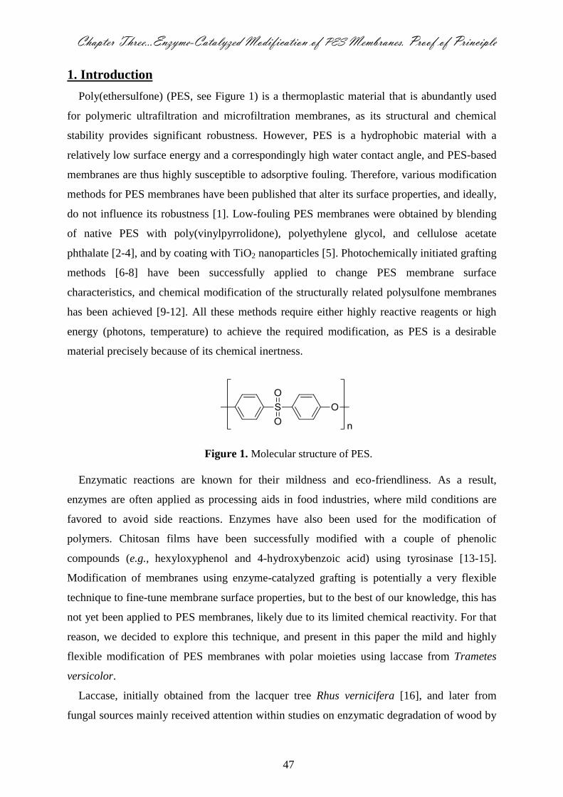
Chapter Three…Enzyme-Catalyzed Modification of PES Membranes. Proof of Principle
47
1. Introduction
Poly(ethersulfone) (PES, see Figure 1) is a thermoplastic material that is abundantly used
for polymeric ultrafiltration and microfiltration membranes, as its structural and chemical
stability provides significant robustness. However, PES is a hydrophobic material with a
relatively low surface energy and a correspondingly high water contact angle, and PES-based
membranes are thus highly susceptible to adsorptive fouling. Therefore, various modification
methods for PES membranes have been published that alter its surface properties, and ideally,
do not influence its robustness [1]. Low-fouling PES membranes were obtained by blending
of native PES with poly(vinylpyrrolidone), polyethylene glycol, and cellulose acetate
phthalate [2-4], and by coating with TiO2 nanoparticles [5]. Photochemically initiated grafting
methods [6-8] have been successfully applied to change PES membrane surface
characteristics, and chemical modification of the structurally related polysulfone membranes
has been achieved [9-12]. All these methods require either highly reactive reagents or high
energy (photons, temperature) to achieve the required modification, as PES is a desirable
material precisely because of its chemical inertness.
Figure 1. Molecular structure of PES.
Enzymatic reactions are known for their mildness and eco-friendliness. As a result,
enzymes are often applied as processing aids in food industries, where mild conditions are
favored to avoid side reactions. Enzymes have also been used for the modification of
polymers. Chitosan films have been successfully modified with a couple of phenolic
compounds (e.g., hexyloxyphenol and 4-hydroxybenzoic acid) using tyrosinase [13-15].
Modification of membranes using enzyme-catalyzed grafting is potentially a very flexible
technique to fine-tune membrane surface properties, but to the best of our knowledge, this has
not yet been applied to PES membranes, likely due to its limited chemical reactivity. For that
reason, we decided to explore this technique, and present in this paper the mild and highly
flexible modification of PES membranes with polar moieties using laccase from Trametes
versicolor.
Laccase, initially obtained from the lacquer tree Rhus vernicifera [16], and later from
fungal sources mainly received attention within studies on enzymatic degradation of wood by
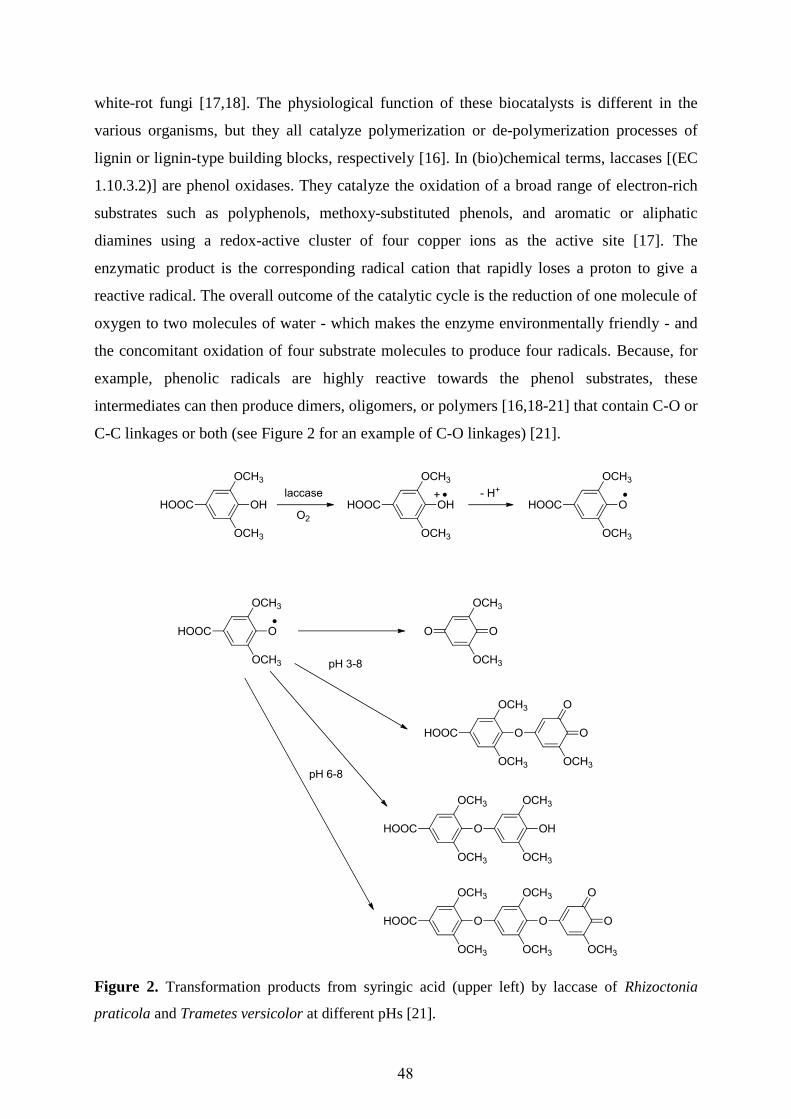
48
white-rot fungi [17,18]. The physiological function of these biocatalysts is different in the
various organisms, but they all catalyze polymerization or de-polymerization processes of
lignin or lignin-type building blocks, respectively [16]. In (bio)chemical terms, laccases [(EC
1.10.3.2)] are phenol oxidases. They catalyze the oxidation of a broad range of electron-rich
substrates such as polyphenols, methoxy-substituted phenols, and aromatic or aliphatic
diamines using a redox-active cluster of four copper ions as the active site [17]. The
enzymatic product is the corresponding radical cation that rapidly loses a proton to give a
reactive radical. The overall outcome of the catalytic cycle is the reduction of one molecule of
oxygen to two molecules of water - which makes the enzyme environmentally friendly - and
the concomitant oxidation of four substrate molecules to produce four radicals. Because, for
example, phenolic radicals are highly reactive towards the phenol substrates, these
intermediates can then produce dimers, oligomers, or polymers [16,18-21] that contain C-O or
C-C linkages or both (see Figure 2 for an example of C-O linkages) [21].
Figure 2. Transformation products from syringic acid (upper left) by laccase of Rhizoctonia
praticola and Trametes versicolor at different pHs [21].

Chapter Three…Enzyme-Catalyzed Modification of PES Membranes. Proof of Principle
49
The overall reaction rate depends both on the rate of the intermolecular electron transfer
between catalytic Cu sites, and on the rate of product release [22]. Oxidations by laccase can
be performed directly, i.e., the enzyme interacts with the administered substrate itself, or
indirectly, in which case the enzyme oxidizes a chemical mediator that acts as an intermediate
substrate. The oxidized radical of these mediators is subsequently able to react with, for
example, the bulky substrates or with compounds having a high oxidation potential [20]. This
stimulated us to precisely investigate PES-based materials, as the chemical inertness relates to
the relatively electron-poor nature of the aromatic moieties.
Recently, laccase-mediated coating of lignocellulosic surfaces with polyphenols has been
investigated, in order to obtain antibacterial performance [23]. The best conditions to make a
laccase-induced coating were determined based on the obtained coloration and color depth of
the formed layer, which depends on the nature of phenol (i.e., hydroquinone produces
colorless, 2-methoxy-5-nitrophenol produces pale yellow, ferulic acid produces orange, and
guaiacol produces a dark red layer). In a similar way, cellulose fibers were first functionalized
by amine functional groups and subsequently coated with poly(catechol) “in situ” synthesized
by T. villosa laccase [24]. In other applications, laccase-mediated grafting was used for
dyeing of cellulosic fibers [25,26], for attachment of anchoring groups for antifungals [27],
and phenol sulfonic acids [28], or to improve the water resistance of wool fabric [29].
This chapter presents a mild and highly flexible enzyme-catalyzed strategy for the
modification of PES-based membranes, via the enzyme-mediated formation of phenolic
radicals in the vicinity of PES membranes and subsequent covalent coupling of the radicals to
the PES polymer through C-O linkages, or by strong adsorption of the formed homopolymers.
This leads to modified PES membranes with more hydrophilic and tunable surface properties.
Laccase was used under mild reaction conditions (water, room temperature, nearly neutral
pH), and different phenol derivatives (see Figure 3) were used for the modification reaction.
The phenol derivatives were chosen because of their expected reactivity towards laccase
(laccase substrates), and because they contain a polar carbonyl group that will increase the
hydrophilicity of PES. The modified membranes were analyzed by color measurement, X-ray
photoelectron spectroscopy (XPS), infrared reflection absorption spectroscopy (IRRAS) and
nuclear magnetic resonance (NMR). The ionization potential of the substrates, the energies of
both the substrates and resulting radicals, and the spin densities in these radicals were
calculated by quantum chemical means (B3LYP/6-311+G(d,p)//B3LYP/6-311G(d,p)), to
further understand the modification reaction. Finally, membrane fluxes were checked to trace
any negative influence of the modification method.
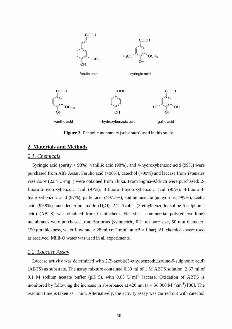
50
Figure 3. Phenolic monomers (substrates) used in this study.
2. Materials and Methods
2.1. Chemicals
Syringic acid (purity > 98%), vanillic acid (98%), and 4-hydroxybenzoic acid (99%) were
purchased from Alfa Aesar. Ferulic acid (>98%), catechol (>99%) and laccase from Trametes
versicolor (22.4 U·mg-1
) were obtained from Fluka. From Sigma-Aldrich were purchased: 2-
fluoro-6-hydroxybenzoic acid (97%), 3-fluoro-4-hydroxybenzoic acid (95%), 4-fluoro-3-
hydroxybenzoic acid (97%), gallic acid (>97.5%), sodium acetate (anhydrous, ≥99%), acetic
acid (99.9%), and deuterium oxide (D2O). 2,2′-Azobis (3-ethylbenzothiazoline-6-sulphonic
acid) (ABTS) was obtained from Calbiochem. Flat sheet commercial poly(ethersulfone)
membranes were purchased from Sartorius (symmetric, 0.2 µm pore size, 50 mm diameter,
150 μm thickness, water flow rate > 28 ml·cm-2
·min-1
at ∆P = 1 bar). All chemicals were used
as received. Milli-Q water was used in all experiments.
2.2. Laccase Assay
Laccase activity was determined with 2,2′-azobis(3-ethylbenzothiazoline-6-sulphonic acid)
(ABTS) as substrate. The assay mixture contained 0.33 ml of 1 M ABTS solution, 2.67 ml of
0.1 M sodium acetate buffer (pH 5), with 0.05 U·ml-1
laccase. Oxidation of ABTS is
monitored by following the increase in absorbance at 420 nm (ε = 36,000 M-1·
cm-1
) [30]. The
reaction time is taken as 1 min. Alternatively, the activity assay was carried out with catechol

Chapter Three…Enzyme-Catalyzed Modification of PES Membranes. Proof of Principle
51
as substrate, using 0.33 ml of 10 mM catechol solution, 2.67 ml of 0.1 M sodium acetate
buffer (pH 5), with 0.025 U·ml-1
laccase. Oxidation of catechol is monitored by following the
increase in absorbance at 400 nm (ε = 26,000 M-1
·cm-1
) [31], with a reaction time of 20 min.
One unit of laccase activity is defined as the amount of enzyme required to oxidize 1 µmol of
ABTS or catechol per min at 25 °C. The apparent kinetic parameters Km and Vmax were
determined by fitting the initial reaction rate (v) and substrate concentration [for ABTS Km =
258 mM & Vmax = 455·10-3
mM·min-1
, and for catechol Km = 8.4 mM & Vmax = 50.3·10-3
mM·min-1
]. These values are comparable to the values found for fungal laccases [32].
2.3. Color Measurements
The CIELAB coordinates for the modified membranes were measured with a Color Flex
(Hunter Lab, CIE L*, a*, b*, CIE L*, C*, h* at D 65/10°). The color values L* (lightness), a*
(red-green axes), b* (yellow-blue axes), and C* were determined as average of three readings.
Aperture size was 28 mm diameter. All parameters are determined relative to the unmodified
membrane (i.e., ∆L*, ∆a*, etc). ∆C* (color saturation), is a characteristic parameter indicating
the vibrancy or intensity of a color; a color with a high saturation will appear more intense
than the same color with less saturation. ∆E* is the degree of total color change, which is
calculated from [(∆L*)2 + (∆a*)
2 + (∆b*)
2]
0.5.
2.4. X-ray Photoelectron Spectroscopy (XPS) Analysis
A JEOL JPS-9200 X-ray Photoelectron Spectrometer (Japan) is used for surface analysis of
the elemental composition of the modified membranes to a depth of around 5 nm. High-
resolution spectra were obtained under UHV conditions using monochromatic Al K X-ray
radiation at 12 kV and 25 mA, using analyzer pass energy of 10 eV. All high-resolution
spectra were corrected with a linear background before fitting.
2.5. Infrared Reflection-Absorption Spectroscopy (IRRAS) Analysis
The IRRAS spectra of phenolic acid grafted-membranes were obtained with a Bruker
Tensor 27 FT-IR Spectrometer equipped with either an MCT-detector and the Auto-Seagull
reflection module from Harricks, or a Bruker Hyperion 2000 FT-IR microscope. The Auto-
seagull is working with a single angle (range from 85° till 10°) while the Hyperion is
equipped with a grazing IR-objective working with all angles in the range from 52 to 84° The
spectra of modified membranes are measured using the unmodified membrane as background.
Spectra are expressed as % of reflectance.

52
2.6. Scanning Electron Microscope (SEM)
Scanning of membrane cross sections were carried out using a Magellan 400 SEM (FEI,
Eindhoven, The Netherlands). The samples were prepared by fracturing in liquid nitrogen and
followed by coating with Pt. The applied voltage was 3 KV; the resolution was 1024 × 884
pixels.
2.7. Pure Water Flux and Membrane Hydraulic Resistance (Rm)
A dead-end stirred filtration cell (Millipore, Model 8050, active transport area 13.4 cm2)
was used to characterize the filtration performance of unmodified and modified membranes.
Pure water flux was measured at a constant trans-membrane pressure of 1 bar at 24 ± 1 ºC and
300 rpm.
The pure water flux is calculated with Equation 1, in which, Jw = water flux (m3·m
-2·s
-1), Q
= quantity of permeate collected (m3), ∆t = sampling time (s), and A = the membrane area
(m2). To determine the hydraulic resistance of the membrane (Rm), the pure water flux was
measured at different transmembrane pressures of 0.2, 0.4, 0.6, 0.8, 1, and 1.2 bar,
respectively. The resistance of the membrane follows from the slope of water flux versus
transmembrane pressure [33].
At
QJ w
(Eqn. 1)
2.8. Membrane Modification Experiments
For the basic screening experiments, flat membranes were incubated in 40 ml 0.1 M
sodium acetate buffer (pH 5) containing different concentrations of phenolic acids
(monomers) and enzyme. The samples were gently shaken, and after a specific modification
time, they were removed from the liquid. Ferulic acid, vanillic acid, syringic acid, 4-
hydroxybenzoic acid, and gallic acid were tested, all at concentrations of 0.6, 10, and 28.8
mM, respectively, and with modification times of 0.5, 2, 8, and 24 h. The enzyme
concentration tested was 0.5 U·ml-1
. After the modification time was completed, the
membranes were washed by strong flushing followed by repeated dipping in Milli-Q water.
The modified membranes were kept in glass-covered dishes in desiccators supplied with silica
gel for drying. Additional experiments were done with fluorohydroxybenzoic acids, to prove
their coupling to the membrane by XPS. The resulting membranes were treated in the same
way as described before.

Chapter Three…Enzyme-Catalyzed Modification of PES Membranes. Proof of Principle
53
2.9. Grafting Yield
The amount of phenolic acid grafted onto the membrane surface is calculated from the
weight of the membrane, before and after grafting, and the grafting yield is expressed as the
weight increase relative to the initial weight. Before grafting, all the membranes were kept for
24 h in glass-covered dishes in desiccators supplied with silica gel to remove any moisture.
2.10. Nuclear Magnetic Resonance (NMR) Studies
1H-NMR spectra were recorded on a Bruker AVANCE III NMR resonating at 400 MHz,
equipped with an inverse broadband gradient probe. A part of a membrane disk modified with
4-hydroxybenzoic acid (substrate concentration 28.8 mM, enzyme concentration 0.5 U·ml-1
,
sodium acetate buffer pH 5, 25°C, modification time 24 h) was completely dissolved in DMF-
d7 and subjected to NMR analysis.
2.11. Blank Experiments
2.11.1. Enzyme and Substrate Adsorption Tests
The unmodified membrane sample was incubated in 40 ml 0.1 M sodium acetate buffer
(pH 5, 24 ± 1 °C) containing 1 U·ml-1
enzyme in the absence of phenolic acid substrate, for
24 h. The amount of adsorbed enzyme was calculated from the weight difference. The same
test was performed with the monomer solution (without enzyme).
2.11.2. Homopolymer Adsorption Tests
The reaction of phenolic acid substrate and enzyme was carried out as described above, but
now without the PES membranes. The reaction time was 24 h for both 4-hydroxybenzoic acid
and gallic acid. The enzyme was inactivated by adding 5 ml of 0.1 M NaOH while stirring for
10 min. After re-adjusting the pH to 5 with concentrated HCl, the PES membranes were
incubated for 2 h in this solution. After the incubation time was completed, the membranes
were washed by strong flushing followed by repeated dipping in with Milli-Q water. The
membranes were kept in glass-covered dishes in desiccators supplied with silica gel for drying
before color measurements.
2.12. Molecular Modeling
The geometries of radical substrates were optimized by B3LYP density functional theory
calculations with the basis set (B3LYP/6-311G(d,p)) using the Gaussian03 program [34].
Frequency calculations were performed to ensure that the optimized geometry is a minimum
54
on the potential energy surface. Single-point calculations (B3LYP/6-311+G(d,p)//B3LYP/6-
311G(d,p)) were performed to obtain energies and spin densities, using the IOP settings
IOP(5/14=2,6/26=4) in the input file section; the resulting projected <S2> values were below
0.751 in all cases.
3. Results and Discussion
Incubation of the membrane with laccase and the substrates leads to visual changes of the
membrane, as is illustrated in Figure 4; depending on the substrate used, the color is more or
less intense. The reaction conditions were 28.8 mM substrate, 0.5 U·ml-1
enzyme
concentration, 24 h modification time, 24 ± 1 ºC, pH = 5 (0.1 M sodium acetate buffer); these
conditions are used throughout this chapter unless noted otherwise. The aqueous medium
solutions also colored with time (not shown) and this is attributed to the formation of
homopolymers, due to reaction of enzymatically formed phenolic radicals with phenol
monomers.
The experimental color changes show that reactions occur with the membrane, especially
when ferulic acid, 4-hydroxybenzoic acid, and gallic acid are used. Moreover, it is found that
the reaction rate (as qualitatively deduced from the rate of color change) is highest for gallic
acid, followed by ferulic acid and 4-hydroxybenzoic acid. For vanillic acid, a reaction
occurred albeit very slowly and at long modification time (a change in color is noticeable after
48 h modification time) and high monomer concentration. There was no notable color change
with syringic acid even at high monomer and enzyme concentration, or prolonged
modification time.
Figure 4. Color changes of modified circular membranes (wet membranes) after enzyme-
catalyzed modification with ferulic acid (F), 4-hydroxybenzoic acid (P), gallic acid (G), (B) is the
blank membrane.
As control experiments, it was shown that no notable reaction occurred between PES
membrane and any of the tested monomers in the absence of enzyme (no color change, no
weight change), even after 24 h modification time. Incubation of the membrane with enzyme

Chapter Three…Enzyme-Catalyzed Modification of PES Membranes. Proof of Principle
55
in the absence of substrate yielded some adsorption of enzyme on/in the PES membrane (142
mg·m-2
PES), but this did not lead to coloration or any other change of the membrane.
Because the colored membrane remains colored even after prolonged washing, covalent
modification of the membrane is likely (see below for investigations regarding this claim).
Presumably, the enzymatically generated radicals form covalent bonds to the membrane
material, yielding a surface-bound radical that can subsequently undergo further reactions to
form a surface-bound conjugated polymer. The resulting extended π-system gives rise to the
observed color change.
As mentioned, gallic acid, 4-hydroxybenzoic acid, and ferulic acid show high reactivity
towards the PES membrane. Therefore, our investigations initially focused on these three
compounds, in order to establish a proof of principle for laccase-catalyzed modification of
PES membranes under mild modification conditions. The amount of attached material was
first evaluated from color development as a function of time and substrate concentration, as
this turned out to be a quick method to evaluate a large number of samples. From the
CIELAB analysis, lightness (∆L*), color saturation (∆C*), and degree of total color change
(∆E*) were found indicative for the degree of modification. Obviously, the lowest ∆E* value
(greatest ∆L*, Table 1) was obtained for the unmodified membrane. For membranes treated
with ferulic acid, the ∆E* value increased with increasing modification time, but not with
increasing substrate concentration, thereby indicating that the used concentrations are in the
saturation range of the enzyme. It is assumed that the enzymatic reaction is the rate-limiting
step (i.e., the subsequent chemical reactions of the enzymatically formed radicals are much
faster than the enzymatic oxidation). The lower coloration at high concentration and longer
modification time is possibly due to enzyme inhibition and/or poor aqueous solubility of the
ferulic acid under the used reaction condition. Because of these uncertainties we decided to
continue our studies with just 4-hydroxybenzoic acid and gallic acid.
Membranes modified with 4-hydroxybenzoic acid showed an increase in ∆E* value, both
with increasing substrate concentration and modification time, indicating that the substrate
concentrations were below the saturation concentration of the enzyme. More polymer
attachment is thus expected at higher substrate concentrations. Membranes could also be
modified with gallic acid, as is evident from the color change. Free radical formation with
gallic acid is extremely fast; a color change in the reaction solution is already apparent after a
few seconds. At low gallic acid concentration, the difference in color change with increase of
modification time is less; apparently, all gallic acid has reacted already. An increase in the
gallic acid concentration allows the reaction to proceed for a longer time, resulting in

56
noticeable color change with longer modification times. These observations are in line with
the changes in both ∆L* and ∆C* values (see Table 1); it should be kept in mind, that an
increase in ∆E* corresponds to a decrease in ∆L* and an increase in ∆C*.
The observed color change of the membrane can be caused by covalent modification and/or
by physical adsorption of substrate homopolymer, formed in solution, to the membrane. In
order to study the latter possibility, enzymatic reactions were conducted in the absence of the
PES membrane. After 24 h of reaction, membranes were immersed in the colored solution for
2 hours, and the membrane color was determined, after extensive washing. Indeed some
coloration was found (see Table 1), indicating that homopolymer adsorption does occur and
apparently yields strong noncovalent bonds to the membrane.
Table 1. Lightness, degree of total color change, and color saturation response of modified PES
membranes as determined by CIELAB measurements.
Typical error limits: ± 0.1 for lightness (∆L*) and color saturation (∆C*) and ± 0.2 for the degree of total color change (∆E*)
Substrate
Modification conditions ∆L* ∆E* ∆C*
Substrate
concentration
(mM)
Modification
time
(h)
Unmodified membrane
– – 100.0
0.0
0.0
Ferulic acid
0.6 8 97.1 8.9 6.7
24 93.9 14.0 10.7
10.0
8 96.6 9.2 6.7
24 94.3 11.4 7.9
4-hydroxybenzoic acid
0.6
8 99.2 4.3 2.5
24 97.3 6.7 4.2
10.0
Homopolymer adsorption blank test
8 96.1 7.9 4.8
24 94.2 10.6 6.8
24 96.3 7.3 4.7
Gallic acid
0.6
8 98.8 3.0 0.8
24 98.1 3.2 0.9
10.0
Homopolymer adsorption blank test
8 96.4 4.8 1.3
24 94.3 8.9 5.0
24 94.7 7.8 3.8
Chapter Three…Enzyme-Catalyzed Modification of PES Membranes. Proof of Principle
57
For gallic acid it cannot be excluded that only physisorption accounts for the resulting
surface modification, as incubation with enzyme and membrane yields the same coloration as
polymerization followed by membrane immersion. The data for 4-hydroxybenzoic acid
indicate that covalent attachment onto the PES membrane also takes place, as physisorption
by itself cannot yield the same degree of coloration as observed from polymerization that
takes place in the presence of PES membranes.
To study the attached layer on the PES surface, XPS analysis was carried out. In Table 2,
the results for the unmodified and two modified membranes are shown. Most notable is the
decrease in sulfur content, as observed in the intensity of the S2p peak at 169.0 eV (-SO2-).
This decrease is a good indication for the formation of an overlayer on the membrane that
shields the underlying sulfur. For both carbon and oxygen, concentrations are found that are
notably different from the unmodified membrane, which is another indication for the
formation of an extra layer. These effects are pronounced for both phenolic acids; and the
XPS results are, in general, in good agreement with our initial observations on color change.
The presence of the nitrogen and the C=O peaks in the unmodified membrane are most
probably due to incomplete leaching out of used solvent during the phase-inversion
fabrication of the PES membrane or due to presence of other used additive materials like
polyvinylpyrrolidone. One would expect an increase in the C=O peak upon the covalent
coupling of substrate molecules, but apparently decarboxylation takes place (see Figure 2 for
an example from literature).
Table 2. Analysis of XPS spectra of modified PES membranes. Apart from the standard
modification time of 24 h, for gallic acid also 2 h modification was used.
* 2 h modification time.
Binding energy (eV)
C1s C1s
O1s N1s S2p
285.4
± 0.3
288.8
±0.5
533.2
±0.3
400.1
± 0.1
169.0
± 0.3
C─C C═O ─C─O─ ─N─ O═S═O
Sample Atomic %
Unmodified PES 77.01 16.99 15.56 2.15 5.30
4-hydroxybenzoic acid 76.18 03.9 19.79 1.98 2.05
Gallic acid 59307 .3.3 813.1 3389 93.7
Gallic acid* 59379 7390 80339 3399 .317
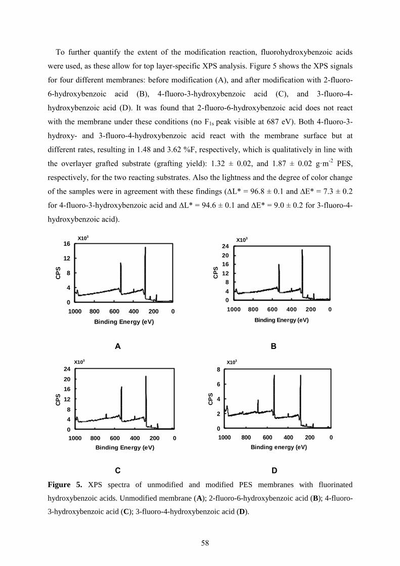
58
To further quantify the extent of the modification reaction, fluorohydroxybenzoic acids
were used, as these allow for top layer-specific XPS analysis. Figure 5 shows the XPS signals
for four different membranes: before modification (A), and after modification with 2-fluoro-
6-hydroxybenzoic acid (B), 4-fluoro-3-hydroxybenzoic acid (C), and 3-fluoro-4-
hydroxybenzoic acid (D). It was found that 2-fluoro-6-hydroxybenzoic acid does not react
with the membrane under these conditions (no F1s peak visible at 687 eV). Both 4-fluoro-3-
hydroxy- and 3-fluoro-4-hydroxybenzoic acid react with the membrane surface but at
different rates, resulting in 1.48 and 3.62 %F, respectively, which is qualitatively in line with
the overlayer grafted substrate (grafting yield): 1.32 ± 0.02, and 1.87 ± 0.02 g·m-2
PES,
respectively, for the two reacting substrates. Also the lightness and the degree of color change
of the samples were in agreement with these findings (∆L* = 96.8 ± 0.1 and ∆E* = 7.3 ± 0.2
for 4-fluoro-3-hydroxybenzoic acid and ∆L* = 94.6 ± 0.1 and ∆E* = 9.0 ± 0.2 for 3-fluoro-4-
hydroxybenzoic acid).
A B
C D
Figure 5. XPS spectra of unmodified and modified PES membranes with fluorinated
hydroxybenzoic acids. Unmodified membrane (A); 2-fluoro-6-hydroxybenzoic acid (B); 4-fluoro-
3-hydroxybenzoic acid (C); 3-fluoro-4-hydroxybenzoic acid (D).
0
4
8
12
16
02004006008001000
Binding Energy (eV)
CP
S
X103
0
4
8
12
16
20
24
02004006008001000
Binding Energy (eV)
CP
S
X103
0
4
8
12
16
20
24
02004006008001000
Binding Energy (eV)
CP
S
X103
0
2
4
6
8
02004006008001000
Binding energy (eV)
CP
S
X103

Chapter Three…Enzyme-Catalyzed Modification of PES Membranes. Proof of Principle
59
IRRAS spectra also confirmed that 4-hydroxybenzoic acid and gallic acid were bound to
the surface as grafted oligomers and/or adsorbed polymers. At 3400-3500 cm-1
an OH-peak
was present in the modified membranes, as shown in Figure 6.
A B
Figure 6. IRRAS spectra for 4-hydroxybenzoic acid (A) and gallic acid (B) modified membranes.
Reflectance is relative to that of the unmodified membrane.
The IR-GIR spectra of the 4-hydroxybenzoic acid grafted membranes obtained after
different modification times (0.5, 2, 8, and 24 h) are shown in Figure 7. A clear sharp C=O
group peak at 1708 cm-1
indicates the presence of carboxylic acid. The intensity of this peak
increases with increasing modification time, i.e., increased amount of grafting. The
characteristic bands due to aromatic C-H stretch around 3030-3080 cm-1
(benzene ring) and
the intense, broad band of OH around 3200-3500 cm-1
all refer to the newly attached phenolic
compounds. The increase in modification time produces stronger signals, because of increased
grafted amounts of 4-hydroxybenzoic acid.
For gallic acid, two different modification times (2 and 24 h, see Figure 8) were used; the
characteristic bands are more clear at lower modification time (2 h). This might be the result
of the reaction of the remaining OH-groups with PES and other gallic acid molecules inside
the modified membrane during prolonged incubation (i.e., gallic acid reacts much faster than
4-hydroxybenzoic acid).
0.3
0.5
0.7
0.9
1.1
150025003500
% R
efl
ecta
nce
Wavenumber (cm-1)
S-polarized Spectra
0.7
0.8
0.9
1
150025003500
% R
efl
ecta
nc
e
Wavenumber (cm-1)
S-polarized spectra
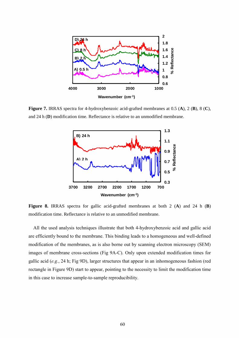
60
Figure 7. IRRAS spectra for 4-hydroxybenzoic acid-grafted membranes at 0.5 (A), 2 (B), 8 (C),
and 24 h (D) modification time. Reflectance is relative to an unmodified membrane.
Figure 8. IRRAS spectra for gallic acid-grafted membranes at both 2 (A) and 24 h (B)
modification time. Reflectance is relative to an unmodified membrane.
All the used analysis techniques illustrate that both 4-hydroxybenzoic acid and gallic acid
are efficiently bound to the membrane. This binding leads to a homogeneous and well-defined
modification of the membranes, as is also borne out by scanning electron microscopy (SEM)
images of membrane cross-sections (Fig 9A-C). Only upon extended modification times for
gallic acid (e.g., 24 h; Fig 9D), larger structures that appear in an inhomogeneous fashion (red
rectangle in Figure 9D) start to appear, pointing to the necessity to limit the modification time
in this case to increase sample-to-sample reproducibility.
0.6
0.8
1
1.2
1.4
1.6
1.8
2
1000200030004000
% R
efl
ecta
nce
Wavenumber (cm-1)
A) 0.5 h
B) 2 h
C) 8 h
D) 24 h
0.3
0.5
0.7
0.9
1.1
1.3
700120017002200270032003700
% R
efl
ecta
nce
Wavenumber (cm-1)
A) 2 h
B) 24 h

Chapter Three…Enzyme-Catalyzed Modification of PES Membranes. Proof of Principle
61
Figure 9. SEM images (30 000×) of cross-sections of blank membrane (A), and membranes
modified with 4-hydroxybenzoic acid (B, 24 h modification time), and gallic acid (C & D, 2 and
24 h modification time).
3.1. Linkage Type Exploration
The phenolic radicals produced by laccase can be O-centered or C-centered. Products
derived from both species have been found in literature [35-38]. Generally, it can be expected
that the radicals will react with carbon atoms of PES which have the highest electron density
(i.e., the carbon atoms ortho to the ether linkages). In addition, the substrate can be bound to
the PES as monomers or as oligomers as shown in Figure 10 for 4-hydroxybenzoic acid. The
strong coloration indicates an extended conjugated system, which would point to oligomeric
structures that link at least several benzene rings together.
To explore if the polymerization of gallic acid predominantly takes place through C-O or
C-C linkages, IR spectroscopy was carried out. Gallic acid shows peaks for O-H stretching
and bending at 3463 and 1103 cm-1
, respectively, as proven by deuterium exchange (i.e.,
gallic acid was dissolved in deuterium oxide (D2O) and freeze-dried to exchange the H by D
atom, see Figure 11) [39]. Upon binding to PES, these peaks are still visible after 2 h
modification time but diminish after 24 h modification time (see Figure 8). This is a strong
indication that gallic acid is enzymatically polymerized and bound to PES predominantly via
O-linked oligomers or polymers.
In another study, the membrane modified with 4-hydroxybenzoic acid was dissolved in
DMF-d7 and subjected to 1H-NMR analysis. Clear signals of the ortho and meta protons of the
grafted substrate were observed, but careful analysis showed that they were slightly shifted
towards higher field compared to free 4-hydroxybenzoic acid. Spiking of the membrane
solution with a small amount of 4-hydroxybenzoic acid clearly showed two sets of peaks: δ
7.922 and 6.953 (J = 8.66 Hz) for the membrane-bound 4-hydroxybenzoic acid, δ 7.927 and
6.957 (J = 8.61 Hz) for the free 4-hydroxybenzoic acid. Apparently the signals are derived
A B C D

62
from terminal O-linked 4-hydroxybenzoic acid units on the membrane. These results are
strong indications for O-linked coupling with a small fraction of C-C bond formation to
account for the coloration, leading to the mixed structure shown in Figure 10.
Figure 10. Schematic representation of a possible chemical structure of the PES surface after
modification with 4-hydroxybenzoic acid, containing mainly O-linked structures with some C-
linked compounds.
Also the grafting yield indicate that the new layer on the membrane consists of oligomers
or polymers rather than a monolayer. Assuming that one molecule (mass: 1×10-17
mg)
occupies about 1 nm2 of space, a dense monolayer would thus approximately yield a coverage
of 1×10-17
mg·nm-2
= 10 mg·m-2
. The obtained coverage of PES membranes by e.g.
hydroxybenzoic acid is in the range of hundreds of mg·m-2
, and thus is on average
significantly more than a monolayer.
Figure 11. FT-IR spectra for gallic acid (A) and deuterated gallic acid (B), as KBr pellets.
0
0.3
0.6
0.9
1.2
1.5
1.8
700170027003700
% T
ran
sm
itta
nc
e
Wavenumber (cm-1)
A) Gallic acid
B) Deuterated gallic acid

Chapter Three…Enzyme-Catalyzed Modification of PES Membranes. Proof of Principle
63
3.2. Molecular Modeling
Quantum chemical calculations were used to calculate electronic energies, spin densities,
and ionization potential of the radical substrates used. Thereby, the order of reactivity of
radical substrates, which coupled with the PES membrane, was determined. The experimental
results showed that the modification process with gallic acid is faster than with 4-
hydroxybenzoic acid (356.5 vs. 101.8 mg·m-2
; 4.8 mM substrate and 2 h modification time),
and this is in good agreement with the lower ionization potentials obtained in the calculations
(Table 3). Analysis of spin populations shows that there is a high spin density on the phenolic
oxygen atom compared to other atomic sites of the two substrates involved in the PES
modification reaction. In addition, the stability data for e.g. gallic acid mean that removal of
the H on the -OH para to the COOH yields a radical that is 6.7 kcal·mol-1
more stable than the
radical obtained by removal of the meta -OH hydrogen atom.
Table 3. Ionization potentials (IP) of the substrates and –O.spin densities of the resulting phenolic
radicals (B3LYP/6-311+G(d,p)//B3LYP/6-311G(d,p) level of theory).
Radical substrates
IP
(eV)
Spin density on -O.
4-hydroxybenzoic acid 7.09 0.333
Gallic acid (para to COOH group) 6.63 0.280
Gallic acid (meta to COOH group) 6.74 0.327
To obtain additional evidence about the reactivity of substrates with respect to the PES
membrane, the ionization potentials of the substrates were approximated by calculating the
negative of their HOMO energies (Koopmans’ theorem). As the one-electron oxidation is
likely rate limiting in aqueous media, this provides an easily calculable reactivity descriptor.
Gallic acid is more reactive than 4-hydroxybenzoic acid, as confirmed by the lower ionization
potential; the near-equal spin densities point to only a minor influence of that factor in
determining the relative reactivity. In order to get more insight into the stability of the PES-
coupled substrate, the relative electronic energy of 4-hydroxybenzoic acid attached to PES
(one unit) either through a phenolic oxygen atom or through an aromatic carbon atom (ortho
to the –O• moiety) was calculated. It is found that the oxygen-coupled product is more stable
(10.4 kcal·mol-1
) than the carbon-coupled one. (See Figure 12 for the optimized structures of
both PES-coupled substrates). Because it is likely that this difference is reflected in the
relative transition state energies, albeit to a smaller degree, the latter energy difference serves
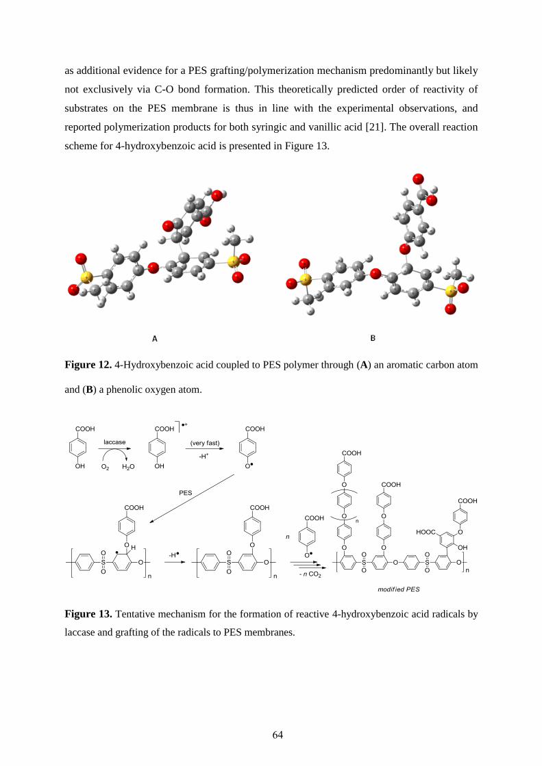
64
as additional evidence for a PES grafting/polymerization mechanism predominantly but likely
not exclusively via C-O bond formation. This theoretically predicted order of reactivity of
substrates on the PES membrane is thus in line with the experimental observations, and
reported polymerization products for both syringic and vanillic acid [21]. The overall reaction
scheme for 4-hydroxybenzoic acid is presented in Figure 13.
Figure 12. 4-Hydroxybenzoic acid coupled to PES polymer through (A) an aromatic carbon atom
and (B) a phenolic oxygen atom.
Figure 13. Tentative mechanism for the formation of reactive 4-hydroxybenzoic acid radicals by
laccase and grafting of the radicals to PES membranes.

Chapter Three…Enzyme-Catalyzed Modification of PES Membranes. Proof of Principle
65
4. Outlook for Laccase-Catalyzed Membrane Modification
We have shown that reactive radicals produced by laccase can be grafted onto the surface
of PES membranes by formation of a covalent C-O linkage (see Figure 13). Because of the
presence of many phenolic radicals in the reaction medium, extra monomers can be
oxidatively grafted to the firstly attached monomer, with concurrent elimination of both
hydrogen and carbon dioxide to form oligomers or polymers, as described elsewhere [40]. At
the same time, these extra monomers can also be oxidatively grafted to other monomers or
pre-formed oligomers to form homopolymers that can be partially adsorbed to the membrane
surface. The latter reaction route is especially pronounced for gallic acid. Water is the only
byproduct in this enzymatic reaction.
Interestingly enough, the flux of grafted membranes is not significantly influenced. At 1 bar
applied pressure, the 4-hydroxybenzoic acid-modified membrane [28.8 mM substrate, 0.5
U·ml-1
enzyme, 0.5 h modification time, 24 ± 1 °C, and pH 5 (0.1 M sodium acetate buffer)]
has a flux comparable to the unmodified membrane [25.9 ± 0.5 and 24.5 ± 0.5 ml·cm-2
·min-1
for unmodified and modified membrane, respectively]. Therefore, we expect, that through
medium engineering, (i.e., substrate concentration, enzyme concentration, reaction pH,
temperature, etc) the enzyme-catalyzed modification method for PES membranes can be
tailored to serve various separation purposes.
5. Conclusions
Laccase-catalyzed grafting of PES membranes can be carried out using a variety of
phenolic acids as substrates. For 4-hydroxybenzoic acid, it was shown that this compound is
directly coupled to the PES, and that substantial amounts can be grafted to the PES membrane
through O-centered coupling. For gallic acid, membrane modification mostly occurs via
polymerization and strong adsorption of the homopolymer to the membrane. Interestingly, the
fluxes of the modified membranes remained high (i.e., comparable to the unmodified
membrane).
From our findings can be concluded that any so-called ‘chemically inert’ membrane that
contains PES or a related polymer such as polysulfone can be modified enzymatically under
the mildest possible conditions (water, room temperature, pH 5), although clearly, further
optimization is needed to ‘fine-tune’ the modification layer to the separation for which it
needs to be designed.

66
Acknowledgments
The authors would like to thank ISPT for financial support of this research, and their
project partners from Norit X-Flow and Friesland Campina for lively scientific discussions.
We thank dr. Marcel Giesbers (XPS) and Tiny Franssen-Verheijen (SEM) for instrumental
assistance.

Chapter Three…Enzyme-Catalyzed Modification of PES Membranes. Proof of Principle
67
References
1. Rana, D.; Matsuura, T., Surface Modifications for Antifouling Membranes, Chem. Rev. 110
(2010) 2448-2471.
2. Lu, S.; Zhuang, L.; Lu, J., Homogeneous Blend Membrane Made from Poly(ethersulphone) and
Poly(vinylpyrrolidone) and its Application to Water Electrolysis, J. Membr. Sci. 300 (2007) 205-
210.
3. Idrisa, A; Zaina, N.M.; Noordin, M.Y., Synthesis, Characterization and Performance of
Asymmetric Polyethersulfone (PES) Ultrafiltration Membranes with Polyethylene Glycol of
Different Molecular Weights as Additives, Desalination 207 (2007) 324-339.
4. Rahimpour, A.; Madaeni, S.S., Polyethersulfone (PES)/Cellulose Acetate Phthalate (CAP) Blend
Ultrafiltration Membranes: Preparation, Morphology, Performance and Antifouling Properties, J.
Membr. Sci. 305 (2007) 299-312.
5. Bae, T.H.; Tak, T.M., Effect of TiO2 Nanoparticles on Fouling Mitigation of Ultrafiltration
Membranes for Activated Sludge Filtration, J. Membr. Sci. 249 (2005) 1-8.
6. Pieracci, J.; Wood, D.W.; Crivello, J.V.; Belfort, G., UV-Assisted Graft Polymerization of N-
vinyl-2-pyrrolidinone onto Poly(ethersulfone) Ultrafiltration Membranes: Comparison of Dip
versus Immersion Modification Techniques, Chem. Mater. 12 (2000) 2123-2133.
7. Taniguchi, M.; Belfort, G., Low Protein Fouling Synthetic Membranes by UV-Assisted Surface
Grafting Modification: Varying Monomer Type, J. Membr. Sci. 231 (2004) 147-157.
8. Liu, S.X.; Kim, J.T.; Kim, S., Effect of Polymer Surface Modification on Polymer-Protein
Interaction via Hydrophilic Polymer Grafting, J. Food Sci. 73 (2008) 143-150.
9. Higuchi, A.; Iwata, N.; Tsubaki, M.; Nakagawa, T, Surface-Modified Polysulfone Hollow Fibers,
J. Appl. Polym. Sci. 36 (1988) 1753-1767.
10. Michael D.; Guiver, M.D.; Kutowy, O.; Apsimon, J.W., Functional Group Polysulfones by
Bromination-Metalation, Polymer 30 (1989) 1137-1142.
11. Higuchi, A.; Nakagawa, T, Surface Modified Polysulfone Hollow Fibers. III. Fibers Having a
Hydroxide Group J. Appl. Polym. Sci. 41 (1990) 1973-1979.
12. Breitbach, L.; Hinke, E.; Staude, E., Heterogeneous Functionalizing of Polysulfone Membranes,
Die Angew. Makromol. Chem. 184 (1991) 183-196.
13. Payne, G.F.; Chaubal, M.V.; Barbari, T.A., Enzyme-Catalyzed Polymer Modification: Reaction of
Phenolic Compounds with Chitosan Films, Polymer 37 (1996) 4643-4648.
14. Chen, T.; Kumar, G.; Harris, M.T.; Smith, P.J.; Payne, G.F., Enzymatic Grafting of
Hexyloxyphenol onto Chitosan to Alter Surface and Rheological Properties, Biotechnol. Bioeng.
70 (2000) 564-573.
15. Chao, A.-C.; Shyu, S.-S.; Lin, Y.-C.; Mi, F.L., Enzymatic Grafting of Carboxyl Groups on to
Chitosan - To Confer on Chitosan the Property of a Cationic Dye Adsorbent, Bioresour. Technol.
91 (2004) 157-162.
16. Call, H.P.; Mucke, I., History, Overview and Applications of Mediated Lignolytic Systems,
Especially Laccase-Mediator-Systems (Lignozym®-Process), J. Biotechnol. 53 (1997) 163-202.
17. Claus, H., Laccases: Structure, Reactions, Distribution, Micron 35 (2004) 93-96.
18. Mayer, A.M.; Staples, R.C., Laccase: New Functions for an Old Enzyme, Photochemistry 60
(2002) 551-565.
19. Bourbonnais, R.; Paice, M.G.; Reid, L.D.; Lanthier, P.; Yaguchi, M., Lignin Oxidation by laccase
Isozymes from Trametes versicolor and Role of the Mediator 2,2'-azinobis(3-ethylbenzthiazoline-
6-sulfonate) in Kraft Lignin Depolymerization, Appl. Environ. Microbiol. 61 (1995) 1876-1880.
20. Riva, S., Laccases: Blue Enzymes for Green Chemistry, Trends Biotechnol. 24 (2006) 219-229.
21. Leonowicz, A.; Edgehili, R.U.; Bollag, J.M., The Effect of pH on the Transformation of Syringic
and Vanillic Acids by the Laccases of Rhizoctonia praticola and Trametes versicolor, Arch.
Microbiol. 137 (1984) 89-96.
22. Santucci, R.; Ferri, T.; Morpurgo, L.; Savini, I.; Avigliano, L., Unmediated Heterogeneous
Electron Transfer Reaction of Ascorbate Oxidase and Laccase at a Gold Electrode, Biochem. J.
332 (1998) 611-615.
23. Schroeder, M.; Aichernig, N.; Guebitz, G.M.; Kokol, V., Enzymatic Coating of Lignocellulosic
Surfaces with Polyphenols, Biotechnol. J. 2 (2007) 334-341.
24. Kim, S.Y.; Zille, A.; Murkovic, M.; Güebitz, G.; Cavaco-Paulo, A., Enzymatic Polymerization on
the Surface of Functionalized Cellulose Fibers, Enzyme Microb. Technol. 40 (2007) 1782-1787.

68
25. Kim, S.; Moldes, D.; Cavaco-Paulo, A., Laccases for Enzymatic Coloration of Unbleached Cotton,
Enzyme Microb. Technol. 40 (2007) 1788-1793.
26. Kim, S.; López, C.; Güebitz, G.; Cavaco-Paulo, A., Biological Coloration of Flax Fabrics with
Flavonoids Using Laccase from Trametes hirsute Eng. Life Sci. 8 (2008) 324-330.
27. Kudanga, T.; Prasetyo, E.N.; Sipilä, J.; Nousiainen, P.; Widsten, P.; Kandelbauer, A.; Nyanhongo,
G. S.; Guebitz, G., Laccase-Mediated Wood Surface Functionalization, Eng. Life Sci. 8 (2008)
297-302.
28. Schroeder, M.; Fatarella, E.; Kovač, J.; Guebitz, G. M.; Kokol, V., Laccase-Induced Grafting on
Plasma-Pretreated Polypropylene, Biomacromolecules 9 (2008) 2735-2741.
29. Hossain, Kh.M.G.; González, M.D.; Lozano, G.R.; Tzanov, T., Multifunctional Modification of
Wool Using An Enzymatic Process in Aqueous–Organic Media, J. Biotechnol. 141 (2009) 58-63.
30. Wolfenden, B.S.; Willson, R.L., Radical-Cations As Reference Chromogens in Kinetic Studies of
One-Electron Transfer Reactions: Pulse Radiolysis Studies of 2,2′-azinobis-(3-
ethylbenzthiazoline-6-sulphonate), J. Chem. Soc., Perkin Trans. 2 (1982) 805-812.
31. Singh, G.; Capalash, N.; Goel, R.; Sharma, P., A pH-stable Laccase from Alkali-Tolerant γ-
Proteobacterium JB: Purification, Characterization and Indigo Carmine Degradation, Enzyme
Microb. Technol. 41 (2007) 794-799.
32. Baldrian, P., Fungal Laccases Occurrence and Properties, FEMS Microbiol. Rev. 30 (2006) 215-
242.
33. Mahendran, R.; Malaisamy, R.; Mohan, D.R., Cellulose Acetate and Polyethersulfone Blend
Ultrafiltration Membranes. Part I: Preparation and Characterizations, Polym. Adv. Technol. 15
(2004) 149-157.
34. Frisch, M. J.; Trucks, G. W.; Schlegel, H. B.; G. E. Scuseria; Robb, M. A.; Cheeseman, J. R.; Jr., J.
A. M.; Vreven, T.; Kudin, K. N.; Burant, J. C.; Millam, J. M.; Iyengar, S. S.; Tomasi, J.; Barone,
V.; Mennucci, B.; Cossi, M.; Scalmani, G.; Rega, N.; Petersson, G. A.; Nakatsuji, H.; Hada, M.;
Ehara, M.; Toyota, K.; Fukuda, R.; Hasegawa, J.; M. Ishida; Nakajima, T.; Honda, Y.; Kitao, O.;
Nakai, H.; Klene, M.; Li, X.; Knox, J. E.; Hratchian, H. P.; Cross, J. B.; Adamo, C.; Jaramillo, J.;
Gomperts, R.; Stratmann, R. E.; Yazyev, O.; Austin, A. J.; Cammi, R.; Pomelli, C.; Ochterski, J.
W.; Ayala, P. Y.; Morokuma, K.; Voth, G. A.; Salvador, P.; Dannenberg, J. J.; Zakrzewski, V. G.;
Dapprich, S.; Daniels, A. D.; Strain, M. C.; Farkas, O.; Malick, D. K.; Rabuck, A. D.;
Raghavachari, K.; Foresman, J. B.; Ortiz, J. V.; Cui, Q.; Baboul, A. G.; Clifford, S.; Cioslowski, J.;
Stefanov, B. B.; Liu, G.; Liashenko, A.; Piskorz, P.; Komaromi, I.; Martin, R. L.; Fox, D. J.; Keith,
T.; Al-Laham, M. A.; Peng, C. Y.; Nanayakkara, A.; Challacombe, M.; Gill, P. M. W.; Johnson,
B.; Chen, W.; Wong, M. W.; Gonzalez, C.; Pople, J. A. Gaussian 03, Revision C.02; Gaussian,
Inc.: Wallingford CT, 2004.
35. Nicotra, S.; Cramarossa, M.R.; Mucci, M.; Pagnoni, U.M.; Riva, S.; Forti, L., Biotransformation of
Resveratrol: Synthesis of Trans-Dehydrodimers Catalyzed By Laccases From Myceliophtora
thermophyla and From Trametes pubescens, Tetrahedron 60 (2004) 595-600.
36. Ncanana, S.; Baratto, L.; Roncaglia, L.; Riva, S.; Burton, S.G., Laccase-Mediated Oxidation of
Totarol, Adv. Synth. Catal. 349 (2007) 1507-1513.
37. Ponzoni, C.; Beneventi, E.; Cramarossa, M.R.; Raimondi, S.; Trevisi, G.; Pagnoni, U.M.; Riva, S.;
Forti, L., Laccase-Catalyzed Dimerization of Hydroxystilbenes, Adv. Synth. Catal. 349 (2007)
1497-1506.
38. Gübitz, G.M.; Paulo, A.C., New Substrates for Reliable Enzymes: Enzymatic Modification of
Polymers, Curr. Opin. Biotechnol. 14 (2003) 577-582.
39. Tejedor-Tejedor, M.I.; Yost, E.C.; Anderson, M.A., Characterization of Benzoic and Phenolic
Complexes at the Goethite/Aqueous Solution Interface Using Cylindrical Internal Reflection
Fourier Transform Infrared Spectroscopy. Part 1. Methodology, Langmuir 6 (1990) 979-987.
40. Ikeda, R.; Sugihara, J.; Uyama, H.; Kobayashi, S., Enzymatic Oxidative Polymerization of 4-
Hydroxybenzoic Acid Derivatives to Poly(phenylene oxide)s, Polym. Int. 47 (1998) 295-301.
Chapter Four
Membrane Modification and Characterization
This chapter has been published as:
Laccase-Catalyzed Modification of PES Membranes with 4-Hydroxybenzoic Acid and
Gallic Acid, Norhan Nady, Karin Schroën, Maurice Franssen, Mohamed S. Mohy Eldin, Han
Zuilhof, and Remko Boom, J. Membr. Sci. 394-395 (2012) 69-79.

70
Laccase-Catalyzed Modification of PES Membranes with
4-Hydroxybenzoic Acid and Gallic Acid
Abstract
We here report on the performance of poly(ethersulfone) membranes modified with 4-
hydroxybenzoic acid and gallic acid as substrates, and using laccase as biocatalyst under
several modification conditions. The average flux of the base membrane was never reduced
more than 20% (mostly below 10% reduction) by modification with 4-hydroxybenzoic acid,
and not more than 9% for gallic acid. The mechanical and thermal properties of the membrane
were not adversely affected by the modification method. For 4-hydroxybenzoic acid, longer
modification times (i.e., hours) and higher substrate concentrations lead to modified
membranes with a better protein repellence. The reaction with gallic acid is faster, but less
effective in terms of the resulting protein repellence.
In conclusion, the laccase-catalyzed modification of poly(ethersulfone) membranes is a
mild method with low environmental impact that leads to effective protein repellence while
keeping the bulk properties of the base membrane intact. This makes laccase-catalyzed
modification an interesting alternative for currently used membrane modification methods.
Chapter Four…Membrane Modification and Characterization
71
1. Introduction
Membrane filtration is a key technology in many fields, such as food production,
downstream processing in biotechnological processes, and water treatment [1-8]. A major
obstacle is flux decline resulting from fouling, which necessitates frequent cleaning, and
occasional replacement of the membrane, which represents the largest operating cost [9,10].
Protein adsorption is often the initial step in biofouling [11], and may result from different
types of interactions with the membrane surface, such as Van der Waals interactions,
electrostatic interactions and hydrogen bonding [12,13]. The frequently used polymer-based
(e.g., poly(arylsulfone) as investigated in this paper) membranes are appreciated for their high
mechanical, thermal, and chemical stability, and their high flux-to-price performance.
However, these materials are all more or less hydrophobic, which makes these membranes
susceptible to fouling by proteins. In case of poly(ethersulfone) (PES), a blend with a
hydrophilic polymer is used (e.g., with polyvinylpyrrolidone, (PVP)), which renders the
surface of the membrane much more hydrophilic, but which fundamentally changes the
membrane structure as well [14]. In addition, use of PVP is not possible with every
membrane-forming polymer, and even membranes with PVP often show extensive fouling by
proteins [14,15]. Therefore, surface modification to influence and ideally prevent adsorption
on the membrane surface is an attractive route [14], as long as the modification method does
not influence the mechanical strength of the membrane.
Various investigations on surface grafting, e.g., with gas plasmas and UV, have shown that
an increased hydrophilicity of the membrane surface generally reduces membrane fouling
[15-18]. On the other hand, steric hindrance (entropic repulsion of coated/grafted polymer
chains) can drastically reduce protein adsorption, without influencing the overall
hydrophilicity of the surface [19]. Thus, both surface structure and hydrophilicity have
significant impact on membrane anti-fouling performance [20].
This thesis focuses on PES membranes. The incorporation of a wide variety of functional
groups to these membranes, e.g., through blending, coating, plasma and radiation induced-
grafting, has been reported [21]. In general, these methods offer only limited control over the
resulting surface structure, cannot be characterized as environmentally friendly due to the
required use of harmful solvents and reactive or persistent chemicals, and they may negatively
influence the mechanical properties of the membrane [14,21]. Enzyme-catalyzed, and in our
case laccase-catalyzed modification makes use of naturally occurring, non-toxic reactants
under mild reaction conditions in aqueous media [22].

72
The enzyme laccase from the mushroom Trametes versicolor is able to oxidize phenolic
compounds to yield reactive radicals [23-25]. These radicals can covalently bind to each other
(polymerization), but we have found that they can also covalently bind to a PES membrane
(grafting), mainly via their OH-groups [22]. In the current research we use 4-hydroxybenzoic
acid and gallic acid as substrate for laccase, and evaluate modified PES membranes based on
e.g. their flux and protein repellence. These substrates of laccase were chosen since their
binding will add ionic and hydrogen bond-forming properties to the PES membranes.
We report here the effects of laccase-catalyzed modification of PES membranes at various
modification times, substrate and enzyme concentrations, reaction temperatures, buffer
strengths, and reaction pHs on membrane properties such as flux and protein repellence. In
previous research [22], we found that PES membranes can be covalently modified with
phenolic acids by the action of laccases. However, the flux, protein repellence, and
(thermo)mechanical properties of the resulting membranes were not studied in detail yet.
Therefore we here report on the performance of the modified membranes focusing on 4-
hydroxybenzoic acid and gallic acid as modifiers. A variety of techniques was used to
characterize the grafting yield, including quantified color changes and gravimetrically
determined grafting yields. In addition, XRD, DSC, TGA, TMA, XPS, SEM and mechanical
analyses were used to further characterize the modified membranes. Besides, the stability of
the modification against strong base and acid was tested. All these aspects are brought
together in an outlook section presented at the end of the results.
2. Materials and Methods
2.1. Chemicals
All chemicals were used as received. 4-hydroxybenzoic acid (99%): Alfa Aesar (The
Netherlands), gallic acid (≥99.5%): Loba Chemie Pvt. Ltd (India), catechol (>98%): Oxford
Laboratory Reagent (India), sodium acetate (anhydrous, ≥99%): Polskie Odczynniki
Chemiczne S.A., (Poland), acetic acid (99.9%): Laboratory Chemicals (Egypt). Flat sheet
commercial poly(ethersulfone) membrane: Sartorius (symmetric, 0.2 µm pore size, 50 mm
diameter, 150 µm thickness, water flow rate > 28 ml·cm-2
·min-1
at ∆P = 1 bar). Laccase from
Trametes versicolor (>21.4 U·mg-1
, Fluka). Deionized water was used in all experiments. The
chemical structures of the membrane material and the substrates are shown in Figure 1.
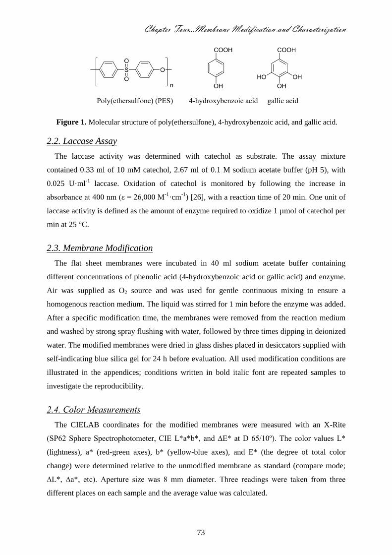
Chapter Four…Membrane Modification and Characterization
73
Figure 1. Molecular structure of poly(ethersulfone), 4-hydroxybenzoic acid, and gallic acid.
2.2. Laccase Assay
The laccase activity was determined with catechol as substrate. The assay mixture
contained 0.33 ml of 10 mM catechol, 2.67 ml of 0.1 M sodium acetate buffer (pH 5), with
0.025 U·ml-1
laccase. Oxidation of catechol is monitored by following the increase in
absorbance at 400 nm (ε = 26,000 M-1
·cm-1
) [26], with a reaction time of 20 min. One unit of
laccase activity is defined as the amount of enzyme required to oxidize 1 µmol of catechol per
min at 25 °C.
2.3. Membrane Modification
The flat sheet membranes were incubated in 40 ml sodium acetate buffer containing
different concentrations of phenolic acid (4-hydroxybenzoic acid or gallic acid) and enzyme.
Air was supplied as O2 source and was used for gentle continuous mixing to ensure a
homogenous reaction medium. The liquid was stirred for 1 min before the enzyme was added.
After a specific modification time, the membranes were removed from the reaction medium
and washed by strong spray flushing with water, followed by three times dipping in deionized
water. The modified membranes were dried in glass dishes placed in desiccators supplied with
self-indicating blue silica gel for 24 h before evaluation. All used modification conditions are
illustrated in the appendices; conditions written in bold italic font are repeated samples to
investigate the reproducibility.
2.4. Color Measurements
The CIELAB coordinates for the modified membranes were measured with an X-Rite
(SP62 Sphere Spectrophotometer, CIE L*a*b*, and ∆E* at D 65/10º). The color values L*
(lightness), a* (red-green axes), b* (yellow-blue axes), and E* (the degree of total color
change) were determined relative to the unmodified membrane as standard (compare mode;
∆L*, ∆a*, etc). Aperture size was 8 mm diameter. Three readings were taken from three
different places on each sample and the average value was calculated.

74
In order to remove any loosely bound material, the membranes were washed by filtration
with at least 1000 ml deionized water before the actual color change was measured.
2.5. Pure Water Flux
A dead-end stirred filtration cell (Millipore, Model 8050, active transport area 13.4 cm2)
was used to characterize the filtration performance of unmodified and modified membranes.
Pure water flux was measured at a constant trans-membrane pressure of 1 bar at 24 ± 1 ºC and
300 rpm.
The pure water flux is calculated with Equation 1, in which, Jw = water flux (m3·m
-2·s
-1), Q
= quantity of permeate collected (m3), ∆t = sampling time (s), and A = the membrane area
(m2).
At
QJ w
(Eqn. 1)
2.6. Grafting Yield
The amount of material grafted onto the membrane surface was calculated from the weight
of the membrane, before and after grafting; the grafting yield is expressed as the weight
increase relative to the initial weight. Before grafting, all the membranes were kept for 24 h in
glass-covered dishes in desiccators supplied with self-indicating blue silica gel to remove any
moisture.
To remove any loosely bound material, the grafted membrane was washed by filtration of
at least 1000 ml deionized water. Subsequently its weight was measured, after drying in a
desiccator.
2.7. BSA Adsorption
BSA was used as a model compound to evaluate protein adsorption on unmodified and
modified membranes. 1g·l-1
BSA solution at pH 7 was prepared using 0.1 M sodium acetate
buffer. The membranes were immersed in 50 ml BSA solution and gently shaken (200 rpm) at
25 °C for 24 h. BSA concentration in the solution was measured using a UV-Vis spectrometer
(280 nm), and from this the adsorbed amount was calculated. After completion of the
experiment, each membrane was intensively washed, first by spray flushing three times
followed by repeated (3×) immersion in deionized water and decantation.
The membrane flux after BSA adsorption was determined as follows. First, the membrane
was backwashed using 100 ml of deionized water to remove any unbound BSA, and then
forward washed using 100 ml of fresh deionized water. After that, fresh deionized water was

Chapter Four…Membrane Modification and Characterization
75
used in forward motion at 1 bar applied pressure, and the flux was determined. The reported
values are the average of three independent measurements.
2.8. Scanning Electron Microscope (SEM)
Unmodified and modified membranes were imaged using a Scanning Electron Microscope
(Jeol Jsm 6360LA, Japan). The membrane samples were cut using a very sharp shaving blade
and were then coated with Au/Pt, and imaged at a voltage of 20/30 KV, and a resolution of
1280 × 960 pixels.
2.9. X-ray Photoelectron Spectroscopy (XPS) Analysis
A JEOL JPS-9200 X-ray Photoelectron Spectrometer (Japan) is used for analysis of the
elemental composition of the modified membranes to a depth of around 5 nm. High-resolution
spectra were obtained under UHV conditions using monochromatic Al K X-ray radiation at
12 kV and 25 mA, using an analyzer pass energy of 10 eV. All high-resolution spectra were
corrected with a linear background before fitting. The elemental composition analyses were
done for a modified spin-coated PES layer on 1×1 cm2 silicon dioxide slides. For this, 0.25 wt
% PES in dichloromethane, was spun at 2500 rpm for 10 s, producing a PES layer with a
thickness of around 20 nm. The PES coated strips were heated for 60 min at 300 °C, and then
kept in glass-covered dishes within desiccators supplied with silica gel for drying. Three
samples were modified independently for each condition.
The modified membranes (i.e., not the spin-coated surfaces) were analyzed before and after
washing with 0.2 M HCl or 0.2 M NaOH (pH 1 and 13), respectively. Each modified
membrane was divided into three parts; two parts were washed with acid or base followed by
immersion in the respective liquid for 15 min, the third part was kept as a reference. The
modified PES membranes were kept in glass-covered dishes in desiccators containing silica
gel.
2.10. Differential Scanning Calorimetery (DSC)
The glass transition temperature (Tg) was measured with differential scanning calorimetry
(DSC, NETZSCH, DSC-200PC) at a heating rate of 10 °C·min-1
. The Tg was defined as the
onset temperature of the change in heat capacity during the heating cycle.

76
2.11. Thermal Gravimetric Analysis (TGA)
TGA analysis of the blank and the modified membranes was carried out using a Thermal
Gravimetric Analyzer (Shimadzu TGA-50, Japan). The samples were scanned over a
temperature range from 20 to 700 °C at a temperature gradient of 10 °C·min-1
under nitrogen
flow.
2.12. X-Ray Diffraction (XRD)
X-ray diffraction patterns for polymeric membranes were obtained on a Shimadzu XRD-
7000 X-ray diffractometer using a CuKα radiation source operating at 40 kV and 30 mA. The
samples were analyzed as such.
2.13. Thermomechanical Analysis (TMA)
The change in membrane thickness while subjected to increased load at a fixed temperature
was measured with the Thermomechanical Analyzer (TMA-60H Shimadzu). Temperature
range was 20-1500 C and the force range was 0 gf).
2.14. Mechanical Properties
Samples were cut in a dog-bone-like shape. The total length of each sample was 37 mm,
the gauge length of the samples was about 16 mm; the width was 13 mm at the top and 7.2
mm (narrowest) at the middle of the sample, to force a fracture in the middle of the sample.
Tensile testing of the films was performed with the Texture Analyzer T2 (Stable Micro
Systems, Ltd., Surrey, United Kingdom), at a constant crosshead speed of 0.1 mm·s-1
until
breaking. Stress-strain curves were calculated from load-elongation curves measured for two
samples from each film. The tensile strength, elongation at break, and Young’s modulus were
calculated from the stress-strain curves.
3. Results and Discussion
In this section we present the results obtained with 4-hydroxybenzoic acid and gallic acid
as substrates (reactants) for laccase-catalyzed modification of PES membranes. We first
discuss the substrates individually regarding flux, grafting yield, protein repellence, color
change, and appearance. Next we compare the mechanical and thermal properties of the
modified membranes, with culminates into an outlook for laccase-catalyzed modified
membranes.
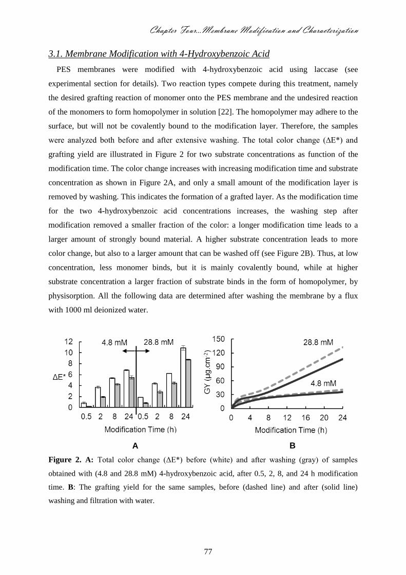
Chapter Four…Membrane Modification and Characterization
77
3.1. Membrane Modification with 4-Hydroxybenzoic Acid
PES membranes were modified with 4-hydroxybenzoic acid using laccase (see
experimental section for details). Two reaction types compete during this treatment, namely
the desired grafting reaction of monomer onto the PES membrane and the undesired reaction
of the monomers to form homopolymer in solution [22]. The homopolymer may adhere to the
surface, but will not be covalently bound to the modification layer. Therefore, the samples
were analyzed both before and after extensive washing. The total color change (∆E*) and
grafting yield are illustrated in Figure 2 for two substrate concentrations as function of the
modification time. The color change increases with increasing modification time and substrate
concentration as shown in Figure 2A, and only a small amount of the modification layer is
removed by washing. This indicates the formation of a grafted layer. As the modification time
for the two 4-hydroxybenzoic acid concentrations increases, the washing step after
modification removed a smaller fraction of the color: a longer modification time leads to a
larger amount of strongly bound material. A higher substrate concentration leads to more
color change, but also to a larger amount that can be washed off (see Figure 2B). Thus, at low
concentration, less monomer binds, but it is mainly covalently bound, while at higher
substrate concentration a larger fraction of substrate binds in the form of homopolymer, by
physisorption. All the following data are determined after washing the membrane by a flux
with 1000 ml deionized water.
Figure 2. A: Total color change (ΔE*) before (white) and after washing (gray) of samples
obtained with (4.8 and 28.8 mM) 4-hydroxybenzoic acid, after 0.5, 2, 8, and 24 h modification
time. B: The grafting yield for the same samples, before (dashed line) and after (solid line)
washing and filtration with water.
A B

78
The gravimetric grafting yield was determined together with the color, the clean water flux,
and the amount of BSA that adsorbs to the surface. The total change of color (∆E*, Figure
3A) increased gradually with increasing grafting yield up to 30 µg·cm-2
. At grafting yields
that are much higher than those shown in Figure 3, the color increased more strongly with
grafting yield, most probably due to formation of dense layers that are more saturated in color
(i.e., have higher extinction coefficients).
For all reaction conditions tested, the actual values obtained for color, grafting yield, flux
and BSA adsorption are given in the Supporting Information (Appendices A and B). It is
worth mentioning that the color developed more strongly at higher enzyme concentrations and
temperatures (i.e. going from 25 to 55 °C), due to a faster overall reaction [27], but at
temperatures above 55 °C, the enzyme is inactivated. The pH has a pronounced effect on the
grafting yield: for example, at pH 4 the grafting yield after 2 h modification is 4.0 µg·cm-2
,
while at pH 5 it is 11.7 µg·cm-2
and 12.7 µg·cm-2
at pH 7 (while the fluxes are virtually the
same as shown in Appendix A). This is attributed to the ionization of 4-hydroxybenzoic acid
at higher pH, which leads to a lower oxidation potential, and therewith in a higher reaction
rate and grafting yield [27-29]. The buffer strength [30] had minor influence on the grafting
yield. The grafting yield did not influence the membrane flux to a great extent, as illustrated
in Figure 3B. Typically the clean water flux reduction was below 10%; the highest clean
water flux reduction at the highest grafting yield (107 µg·cm-2
) was found to be 21.4% for
28.8 mM substrate in combination with 24 h modification (this point is not shown in Figure 3
but is included in Table 1). This implies that the original pores of the base membrane (0.2 μm)
were only minimally constricted by the modification layer, in line with the formation of only
relatively thin chemisorbed layers. The effect of a similarly sized modification layer will be
bigger for small pores, and the resulting pore size distribution will be dominated more by the
larger pores; however, given the thickness of the layer (<10 nm) and the average pore size
(0.2 μm) the effect on pore size distribution is not expected to be very great.

Chapter Four…Membrane Modification and Characterization
79
Figure 3. Total color change (∆E*) (A), Clean water flux (B), and BSA adsorption (C) as a
function of grafting yield; the reference modification condition is 4.8 mM 4-hydroxybenzoic
acid, 0.5 U·ml-1 enzyme, 2 h modification time, 25 °C, pH 5, and 0.1 M sodium acetate buffer.
The following parameters were varied: modification time (0.5, 2, 8 and 24 h) at 4.8 mM 4-
hydroxybenzoic acid, modification time (0.5, 2, and 8 h) at 28.8 mM 4-hydroxybenzoic
acid, 4-hydroxybenzoic acid concentration (0.6, 1.2, 4.8, and 28.8 mM) at 8 h modification
time, enzyme concentration (0.25, 0.75, and 1 U·ml-1), reaction temperature (25, 35, 45,
55, and 65 °C), reaction pH (4-7), buffer strength (0.05, 0.1, and 0.5 M). Typical error: ±
0.2 for total color change (∆E*), ± 0.3 m3∙m-2∙h-1 for flux, and ± 0.2 µg∙cm-2 for BSA
adsorption.
A
B
C

80
One could in fact give a sort of effective pore constriction, as the flux depends on the fourth
power of the pore radius: assuming that all flux reduction is caused by narrowing of the pores
due to deposition of the modification layer, the effective modification layer thickness can be
estimated at 5 nm for 10% flux reduction to 12 nm for 21.4% flux reduction, for a membrane
having 0.2 µm pores. Note that the values of the layer thicknesses used in this coarse estimate
are in good agreement with those measured by ellipsometry (for further use in foulant studies
carried out by reflectometry on non-porous flat PES surfaces), which is further discussed in
chapter 5.
The adsorbed amount of bovine serum albumin (BSA, see Figure 3C) decreases with
increasing grafting yield, and is close to zero after 8 h modification time at 28.8 mM 4-
hydroxybenzoic acid. Similar effects were also observed in the previously mentioned follow-
up reflectometry study in which we used non-porous surfaces; the observed effects are clearly
due to surface modification, and not (so much) due to a reduction of hydrodynamic forces.
The blank membranes that did not receive any modification are prone to flux reduction
(24%) due to BSA adsorption; modified membranes always showed higher residual fluxes as
shown in Table 1. If the total flux loss (flux loss due to modification plus flux loss due to
BSA adsorption) is compared to the original flux, most modified membranes give higher
residual fluxes than the blank membrane, in spite of the (small) inherent flux reduction by the
modification itself.
SEM images of membranes modified with 4.8 mM 4-hydroxybenzoic acid were used to
investigate the change in morphology of the membranes as a result of the modification. As
shown in Figure 4 (A - E), the thickness of the lamellae between the pores seems to increase
with increasing modification time. With longer modification time (2 h), individual domains
with clear edges can be seen, most probably consisting of homopolymer that was formed in
solution and adsorbed to the membrane. At 8 and 24 h modification, these domains become
more pronounced. On membranes modified with 28.8 mM 4-hydroxybenzoic acid for 24 h,
film formation was noted, most probably as a combination of surface modification and
homopolymer deposition (SEM image not shown). This latter membrane has the strongest
measured flux reduction (21.4%). Low reaction pH (4) results in the formation of a very thin
layer over the membrane surface (see image F in Figure 4), while increasing the pH from 5 to
7 seems to result in binding of small lumps of material (images G and H), we interpret to be
most likely homopolymer.
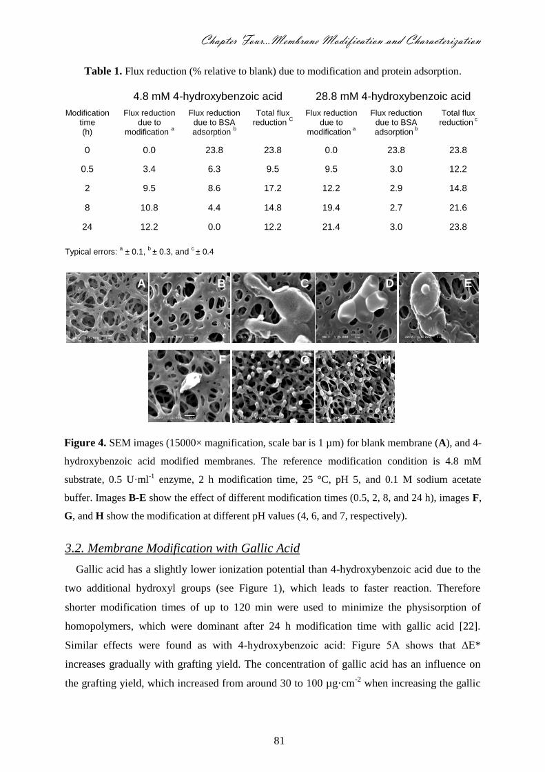
Chapter Four…Membrane Modification and Characterization
81
Table 1. Flux reduction (% relative to blank) due to modification and protein adsorption.
4.8 mM 4-hydroxybenzoic acid 28.8 mM 4-hydroxybenzoic acid
Modification time (h)
Flux reduction due to
modification a
Flux reduction due to BSA adsorption
b
Total flux reduction
C
Flux reduction due to
modification a
Flux reduction due to BSA adsorption
b
Total flux reduction
c
0 0.0 23.8 23.8 0.0 23.8 23.8
0.5 3.4 6.3 9.5 9.5 3.0 12.2
2 9.5 8.6 17.2 12.2 2.9 14.8
8 10.8 4.4 14.8 19.4 2.7 21.6
24 12.2 0.0 12.2 21.4 3.0 23.8
Typical errors: a ± 0.1,
b ± 0.3, and
c ± 0.4
3.2. Membrane Modification with Gallic Acid
Gallic acid has a slightly lower ionization potential than 4-hydroxybenzoic acid due to the
two additional hydroxyl groups (see Figure 1), which leads to faster reaction. Therefore
shorter modification times of up to 120 min were used to minimize the physisorption of
homopolymers, which were dominant after 24 h modification time with gallic acid [22].
Similar effects were found as with 4-hydroxybenzoic acid: Figure 5A shows that ∆E*
increases gradually with grafting yield. The concentration of gallic acid has an influence on
the grafting yield, which increased from around 30 to 100 µg·cm-2
when increasing the gallic
Figure 4. SEM images (15000× magnification, scale bar is 1 µm) for blank membrane (A), and 4-
hydroxybenzoic acid modified membranes. The reference modification condition is 4.8 mM
substrate, 0.5 U·ml-1 enzyme, 2 h modification time, 25 °C, pH 5, and 0.1 M sodium acetate
buffer. Images B-E show the effect of different modification times (0.5, 2, 8, and 24 h), images F,
G, and H show the modification at different pH values (4, 6, and 7, respectively).
H
A B C D E
F G

82
acid concentration from 4.8 to 28.8 mM (actual experimental values are shown in appendix
B).
In spite of the high grafting yield, the membrane flux was hardly influenced. The maximum
clean water flux loss is 9% (effective layer thickness 5 nm) and the clean water flux even
slightly increased in case of using a high concentration of gallic acid (28.8 mM) as shown in
Figure 5B. This surprising result may be attributed to a better wetting of the pores by an
increase in the number of OH groups inside the membrane pores (internal surface), opening
up a larger fraction of all pores for permeation. A similar effect has been seen by Mathias
Ulbricht and co-workers [31] when using high concentrations of three-armed cross linker with
photo-initiated grafted PEGMA, which did not occur when using a two-armed cross linker.
This suggests a change in the polymer structure.
The flux reduction due to protein adsorption of membranes modified with gallic acid was
always lower than with unmodified membranes (Table 2). However, it did not reduce BSA
adsorption to the same extent as 4-hydroxybenzoic acid, as shown in Figure 5C.
This can be understood as follows: phenolic acid radicals generated by laccase mainly react
via their hydroxyl groups [22]. Gallic acid has three adjacent hydroxyl groups on its aromatic
ring and as a result the formed radical can be further oxidized to an o-quinone [32]. This
oxidation can occur inside the active site of the enzyme, or in solution by a disproportionation
reaction between two radicals. Though they are less reactive than radicals, o-quinones still
readily react via addition reactions in a Michael-type fashion [33] (see Figure 6).
If this happens to gallic acid (derivatives) on the membrane, extensive cross-linking may
occur leading to rather compact layers (see Figure 7 for illustration of the effect). This is also
supported by the XPS analyses of blank and modified spin-coated PES layers on silicon
dioxide slides, and ellipsometry analysis (for experimental details see [34]; actual XPS graphs
not shown here), which allows measurement of the elemental composition of the layer; results
are summarized in Figure 7. In case of gallic acid, the sulfur signal is shielded by the
modification layer, even though the layer is rather thin. The grafting yield increases with
increasing modification time from 7 min to 2 h; however, the thickness of the layer (Th.) is
reduced from 5.5 to 3.3 ± 0.2 nm, which may be attributed to collapse of the poly(gallic acid)
layer due to cross-linking, and possibly to adsorption of polymers containing o-quinones. In
spite of these differences, the sulfur signal is very comparable for both samples.

Chapter Four…Membrane Modification and Characterization
83
Figure 5. Total color change (∆E*) (A), Clean water flux (B), and BSA adsorption (C) as a
function of grafting yield; the reference modification condition is 0.5 U·ml-1 enzyme, 25 °C, pH
5, and 0.1 M sodium acetate buffer with 4.8 and 28.8 mM gallic acid. The following parameters
were varied: modification time (10, 20, 30, 60 and 120 min) with 4.8 mM gallic acid, and
with 28.8 mM gallic acid, gallic acid concentration (1.2, 4.8, and 28.8 mM) at 120 min
modification time, enzyme concentration (0.25, 0.5, 0.75, and 1 U·ml-1) at 4.8 mM gallic acid
and 10 min modification time, reaction temperature (25, 35, 45, 55, and 65 °C) at both 4.8
mM gallic acid and 10 min modification time and 28.8 mM gallic acid and 120 min
modification time, reaction pH (4, 5, 6, and 7) at 28.8 mM gallic acid and 120 min
modification time, buffer strength (0.05, 0.1, and 0.5 M) at 28.8 mM gallic acid and 120 min
modification time. Typical errors: ± 0.2 for total color change (∆E*), ± 0.1 m3∙m-2∙h-1 for flux,
and ± 0.3 µg∙cm-2 for BSA adsorption.
A
B
C
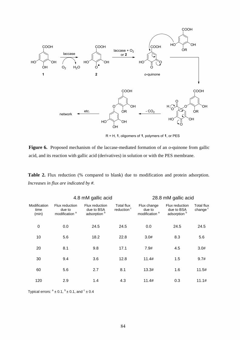
84
Table 2. Flux reduction (% compared to blank) due to modification and protein adsorption.
Increases in flux are indicated by #.
4.8 mM gallic acid 28.8 mM gallic acid
Modification time (min)
Flux reduction due to
modification a
Flux reduction due to BSA adsorption
b
Total flux reduction
c
Flux change due to
modification a
Flux reduction due to BSA adsorption
b
Total flux change
c
0 0.0 24.5 24.5 0.0 24.5 24.5
10 5.6 18.2 22.8 3.0# 8.3 5.6
20 8.1 9.8 17.1 7.9# 4.5 3.0#
30 9.4 3.6 12.8 11.4# 1.5 9.7#
60 5.6 2.7 8.1 13.3# 1.6 11.5#
120 2.9 1.4 4.3 11.4# 0.3 11.1#
Typical errors: a ± 0.1,
b ± 0.1, and
c ± 0.4
Figure 6. Proposed mechanism of the laccase-mediated formation of an o-quinone from gallic
acid, and its reaction with gallic acid (derivatives) in solution or with the PES membrane.
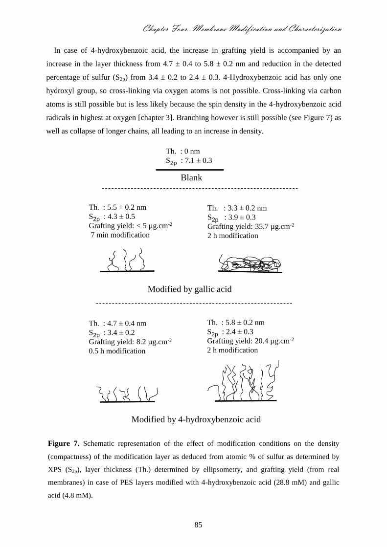
Chapter Four…Membrane Modification and Characterization
85
In case of 4-hydroxybenzoic acid, the increase in grafting yield is accompanied by an
increase in the layer thickness from 4.7 ± 0.4 to 5.8 ± 0.2 nm and reduction in the detected
percentage of sulfur (S2p) from 3.4 ± 0.2 to 2.4 ± 0.3. 4-Hydroxybenzoic acid has only one
hydroxyl group, so cross-linking via oxygen atoms is not possible. Cross-linking via carbon
atoms is still possible but is less likely because the spin density in the 4-hydroxybenzoic acid
radicals in highest at oxygen [chapter 3]. Branching however is still possible (see Figure 7) as
well as collapse of longer chains, all leading to an increase in density.
Figure 7. Schematic representation of the effect of modification conditions on the density
(compactness) of the modification layer as deduced from atomic % of sulfur as determined by
XPS (S2p), layer thickness (Th.) determined by ellipsometry, and grafting yield (from real
membranes) in case of PES layers modified with 4-hydroxybenzoic acid (28.8 mM) and gallic
acid (4.8 mM).
Th. : 3.3 ± 0.2 nm
S2p : 3.9 ± 0.3
Grafting yield: 35.7 µg.cm-2
2 h modification
Th. : 0 nm
S2p : 7.1 ± 0.3
Blank
Modified by gallic acid
Th. : 5.5 ± 0.2 nm
S2p : 4.3 ± 0.5
Grafting yield: < 5 µg.cm-2
7 min modification
Modified by 4-hydroxybenzoic acid
Th. : 5.8 ± 0.2 nm
S2p : 2.4 ± 0.3
Grafting yield: 20.4 µg.cm-2
2 h modification
Th. : 4.7 ± 0.4 nm
S2p : 3.4 ± 0.2
Grafting yield: 8.2 µg.cm-2
0.5 h modification

86
The layer itself has a relatively loose structure, as the polymer itself has a good interaction
with water, and will function to some degree as an entropic brush. Onto a compact layer as
formed with gallic acid, this steric repulsion between extended chains and proteins is less
effective, and the reduction of adsorption is mainly due to an increase in hydrophilicity. Thus,
BSA will adsorb better on layers made from gallic acid. For that reason, a low grafting yield
may be preferred with gallic acid, because it will keep more open structure with more
hydroxyl groups on the surface, and possibly reduced BSA adsorption. For 4-hydroxybenzoic
acid, the amount of grafted material seems to determine the length and density of the entropic
brush and therewith its efficiency in protein repellence.
SEM images of the grafted PES membranes do not show the big lumps that were observed
with 4-hydroxybenzoic acid at any of the chosen conditions (see Figure 8). The reaction of
gallic acid is very fast and the modification process seems to be initiated across the entire
membrane surface at the same time. Some tiny lumps appeared at higher temperatures
(images I and J) and under other conditions at which homopolymer is most likely to be
formed (higher substrate concentrations with long modification time, see images E and F), but
in general no lumps were observed, which is in contrast to the SEM images in Figure 4 for 4-
hydroxybenzoic acid.
Figure 8. SEM images (15000× magnification, scale bar is 1 µm) for the unmodified membrane
(image A), and the gallic acid modified membranes. The reference modification condition is 0.5
U·ml-1 enzyme, 25 °C, pH 5, and 0.1 M sodium acetate buffer. Images B-F show the effect of
different modification times (10, 20, 30, 60 and 120 min) with 28.8 mM gallic acid. The effect of
modification temperature (35, 45, 55, 65 °C) with 4.8 mM gallic acid and 10 min modification
time is given in images G-J, and with 28.8 mM gallic acid and the same temperatures at 120 min
modification time in images K-N.
A B C D E F
G H I J
K L M N
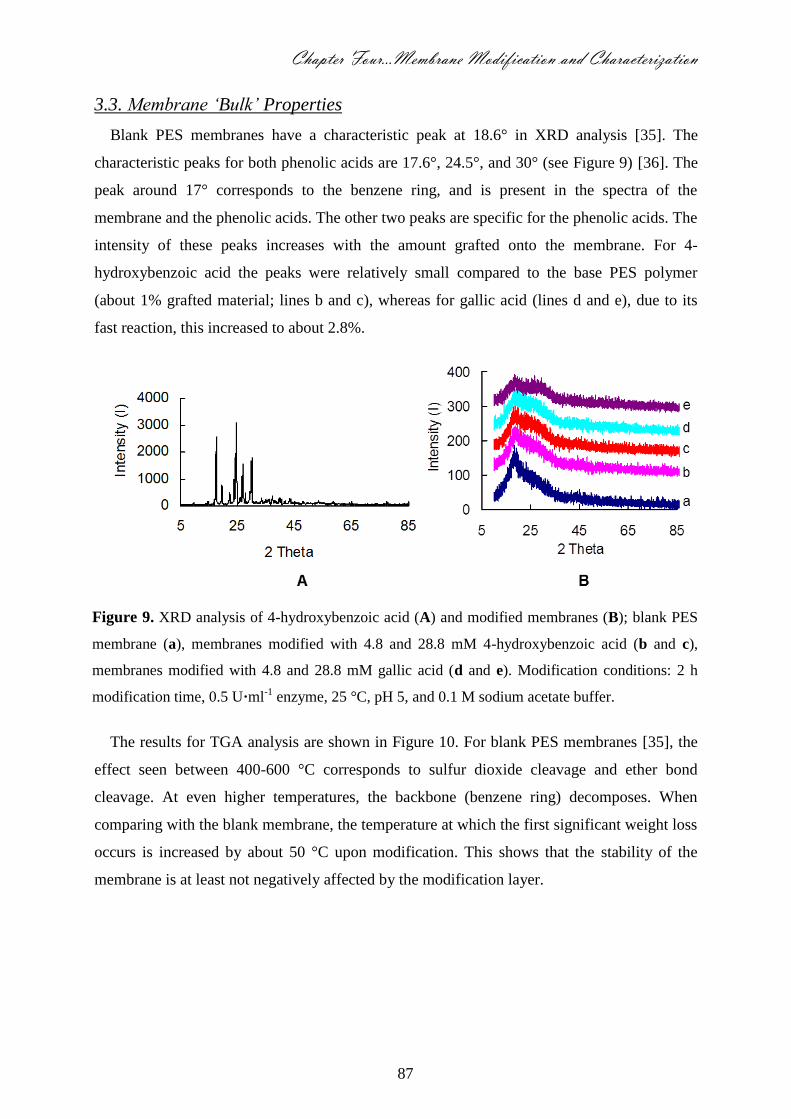
Chapter Four…Membrane Modification and Characterization
87
3.3. Membrane ‘Bulk’ Properties
Blank PES membranes have a characteristic peak at 18.6° in XRD analysis [35]. The
characteristic peaks for both phenolic acids are 17.6°, 24.5°, and 30° (see Figure 9) [36]. The
peak around 17° corresponds to the benzene ring, and is present in the spectra of the
membrane and the phenolic acids. The other two peaks are specific for the phenolic acids. The
intensity of these peaks increases with the amount grafted onto the membrane. For 4-
hydroxybenzoic acid the peaks were relatively small compared to the base PES polymer
(about 1% grafted material; lines b and c), whereas for gallic acid (lines d and e), due to its
fast reaction, this increased to about 2.8%.
Figure 9. XRD analysis of 4-hydroxybenzoic acid (A) and modified membranes (B); blank PES
membrane (a), membranes modified with 4.8 and 28.8 mM 4-hydroxybenzoic acid (b and c),
membranes modified with 4.8 and 28.8 mM gallic acid (d and e). Modification conditions: 2 h
modification time, 0.5 U·ml-1 enzyme, 25 °C, pH 5, and 0.1 M sodium acetate buffer.
The results for TGA analysis are shown in Figure 10. For blank PES membranes [35], the
effect seen between 400-600 °C corresponds to sulfur dioxide cleavage and ether bond
cleavage. At even higher temperatures, the backbone (benzene ring) decomposes. When
comparing with the blank membrane, the temperature at which the first significant weight loss
occurs is increased by about 50 °C upon modification. This shows that the stability of the
membrane is at least not negatively affected by the modification layer.
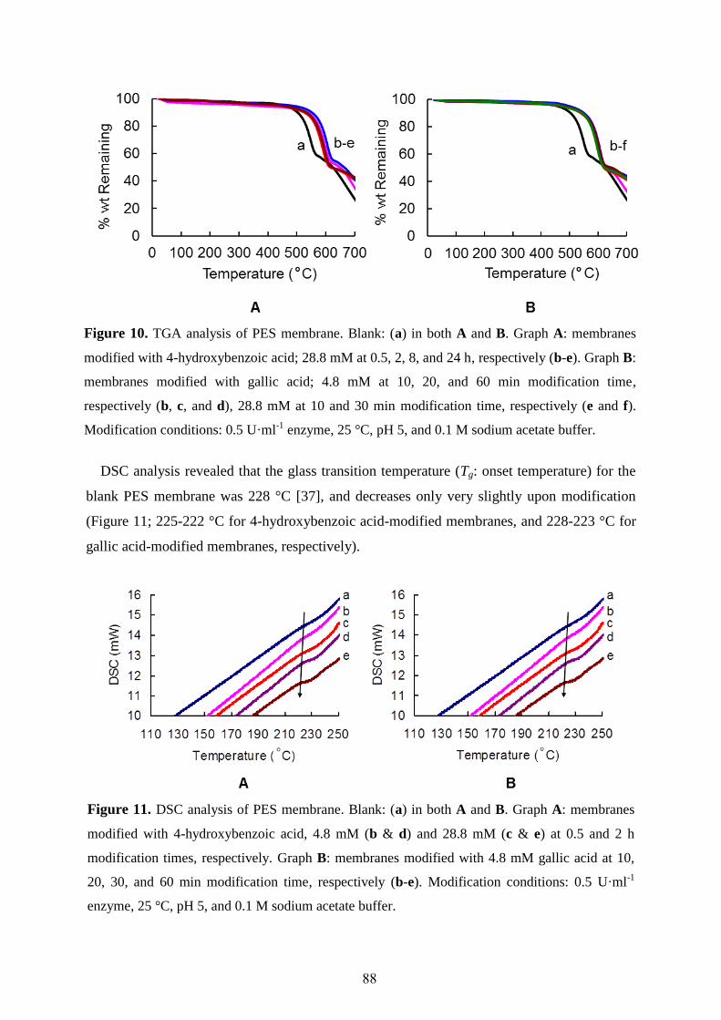
88
Figure 10. TGA analysis of PES membrane. Blank: (a) in both A and B. Graph A: membranes
modified with 4-hydroxybenzoic acid; 28.8 mM at 0.5, 2, 8, and 24 h, respectively (b-e). Graph B:
membranes modified with gallic acid; 4.8 mM at 10, 20, and 60 min modification time,
respectively (b, c, and d), 28.8 mM at 10 and 30 min modification time, respectively (e and f).
Modification conditions: 0.5 U·ml-1 enzyme, 25 °C, pH 5, and 0.1 M sodium acetate buffer.
DSC analysis revealed that the glass transition temperature (Tg: onset temperature) for the
blank PES membrane was 228 °C [37], and decreases only very slightly upon modification
(Figure 11; 225-222 °C for 4-hydroxybenzoic acid-modified membranes, and 228-223 °C for
gallic acid-modified membranes, respectively).
Figure 11. DSC analysis of PES membrane. Blank: (a) in both A and B. Graph A: membranes
modified with 4-hydroxybenzoic acid, 4.8 mM (b & d) and 28.8 mM (c & e) at 0.5 and 2 h
modification times, respectively. Graph B: membranes modified with 4.8 mM gallic acid at 10,
20, 30, and 60 min modification time, respectively (b-e). Modification conditions: 0.5 U·ml-1
enzyme, 25 °C, pH 5, and 0.1 M sodium acetate buffer.
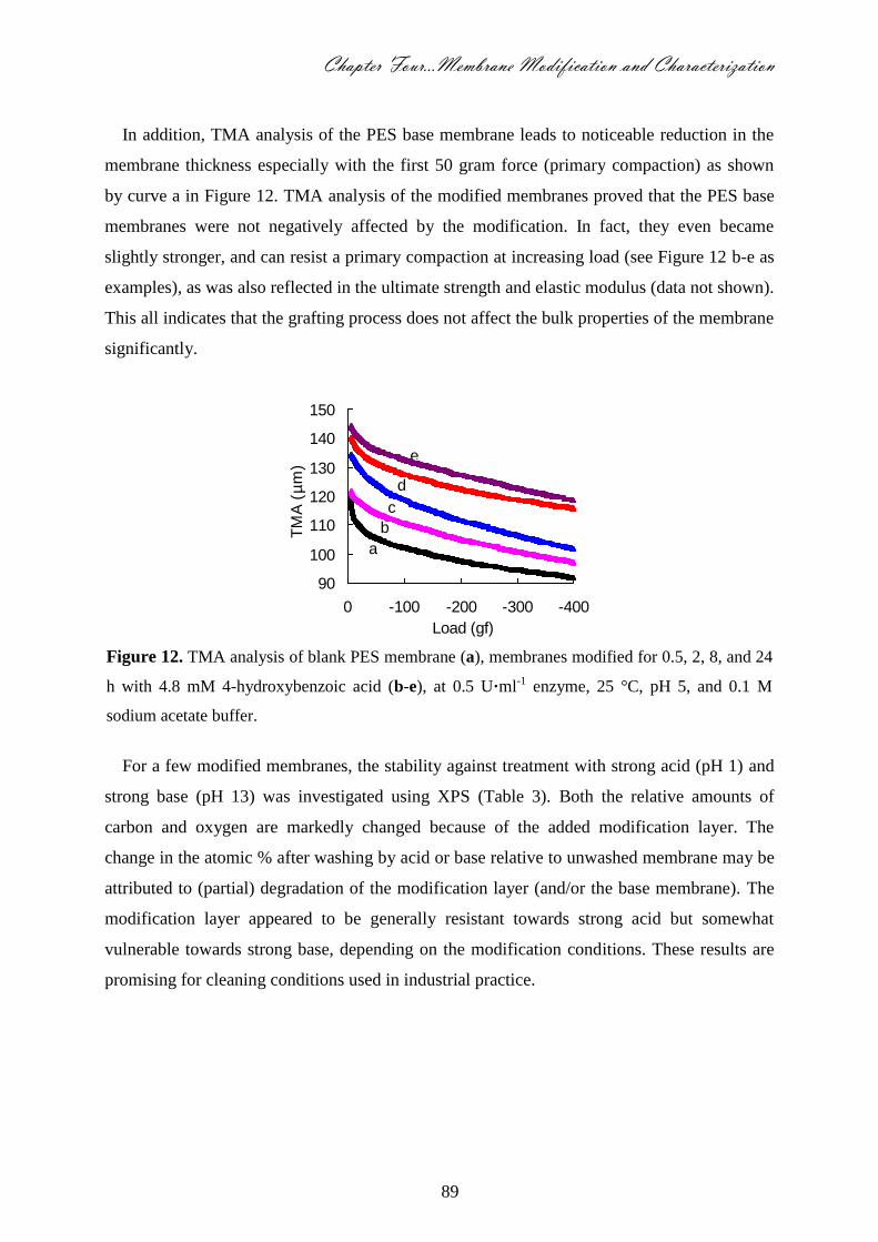
Chapter Four…Membrane Modification and Characterization
89
In addition, TMA analysis of the PES base membrane leads to noticeable reduction in the
membrane thickness especially with the first 50 gram force (primary compaction) as shown
by curve a in Figure 12. TMA analysis of the modified membranes proved that the PES base
membranes were not negatively affected by the modification. In fact, they even became
slightly stronger, and can resist a primary compaction at increasing load (see Figure 12 b-e as
examples), as was also reflected in the ultimate strength and elastic modulus (data not shown).
This all indicates that the grafting process does not affect the bulk properties of the membrane
significantly.
Figure 12. TMA analysis of blank PES membrane (a), membranes modified for 0.5, 2, 8, and 24
h with 4.8 mM 4-hydroxybenzoic acid (b-e), at 0.5 U·ml-1 enzyme, 25 °C, pH 5, and 0.1 M
sodium acetate buffer.
For a few modified membranes, the stability against treatment with strong acid (pH 1) and
strong base (pH 13) was investigated using XPS (Table 3). Both the relative amounts of
carbon and oxygen are markedly changed because of the added modification layer. The
change in the atomic % after washing by acid or base relative to unwashed membrane may be
attributed to (partial) degradation of the modification layer (and/or the base membrane). The
modification layer appeared to be generally resistant towards strong acid but somewhat
vulnerable towards strong base, depending on the modification conditions. These results are
promising for cleaning conditions used in industrial practice.
90
100
110
120
130
140
150
-400-300-200-1000
Load (gf)
a
b
c
d
e
TM
A (
µm
)
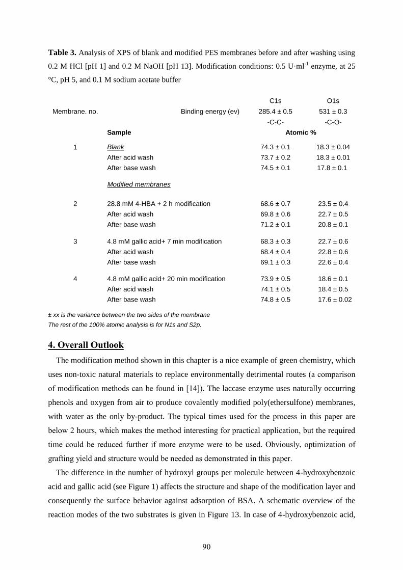
90
Table 3. Analysis of XPS of blank and modified PES membranes before and after washing using
0.2 M HCl [pH 1] and 0.2 M NaOH [pH 13]. Modification conditions: 0.5 U·ml-1 enzyme, at 25
°C, pH 5, and 0.1 M sodium acetate buffer
Membrane. no.
Binding energy (ev)
C1s O1s
285.4 ± 0.5 531 ± 0.3
-C-C- -C-O-
1
Sample Atomic %
Blank 74.3 ± 0.1 18.3 ± 0.04
After acid wash 73.7 ± 0.2 18.3 ± 0.01
After base wash 74.5 ± 0.1 17.8 ± 0.1
Modified membranes
2 28.8 mM 4-HBA + 2 h modification 68.6 ± 0.7 23.5 ± 0.4
After acid wash 69.8 ± 0.6 22.7 ± 0.5
After base wash 71.2 ± 0.1 20.8 ± 0.1
3 4.8 mM gallic acid+ 7 min modification 68.3 ± 0.3 22.7 ± 0.6
After acid wash 68.4 ± 0.4 22.8 ± 0.6
After base wash 69.1 ± 0.3 22.6 ± 0.4
4 4.8 mM gallic acid+ 20 min modification 73.9 ± 0.5 18.6 ± 0.1
After acid wash 74.1 ± 0.5 18.4 ± 0.5
After base wash 74.8 ± 0.5 17.6 ± 0.02
± xx is the variance between the two sides of the membrane
The rest of the 100% atomic analysis is for N1s and S2p.
4. Overall Outlook
The modification method shown in this chapter is a nice example of green chemistry, which
uses non-toxic natural materials to replace environmentally detrimental routes (a comparison
of modification methods can be found in [14]). The laccase enzyme uses naturally occurring
phenols and oxygen from air to produce covalently modified poly(ethersulfone) membranes,
with water as the only by-product. The typical times used for the process in this paper are
below 2 hours, which makes the method interesting for practical application, but the required
time could be reduced further if more enzyme were to be used. Obviously, optimization of
grafting yield and structure would be needed as demonstrated in this paper.
The difference in the number of hydroxyl groups per molecule between 4-hydroxybenzoic
acid and gallic acid (see Figure 1) affects the structure and shape of the modification layer and
consequently the surface behavior against adsorption of BSA. A schematic overview of the
reaction modes of the two substrates is given in Figure 13. In case of 4-hydroxybenzoic acid,
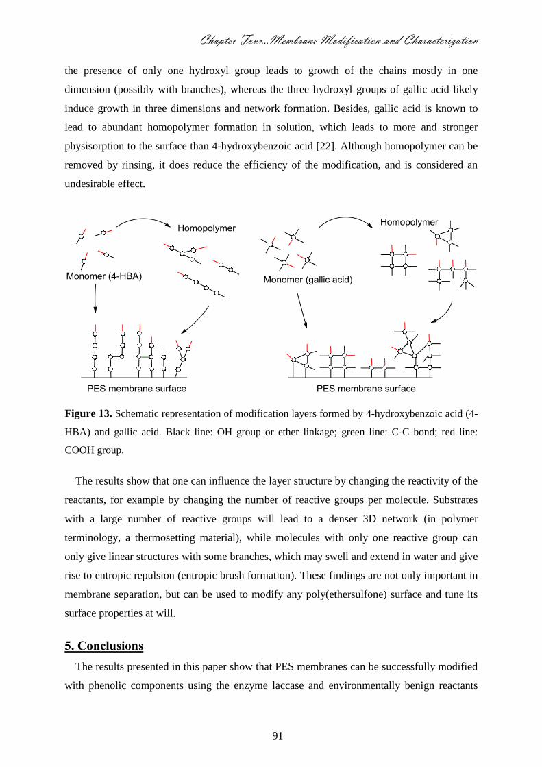
Chapter Four…Membrane Modification and Characterization
91
the presence of only one hydroxyl group leads to growth of the chains mostly in one
dimension (possibly with branches), whereas the three hydroxyl groups of gallic acid likely
induce growth in three dimensions and network formation. Besides, gallic acid is known to
lead to abundant homopolymer formation in solution, which leads to more and stronger
physisorption to the surface than 4-hydroxybenzoic acid [22]. Although homopolymer can be
removed by rinsing, it does reduce the efficiency of the modification, and is considered an
undesirable effect.
Figure 13. Schematic representation of modification layers formed by 4-hydroxybenzoic acid (4-
HBA) and gallic acid. Black line: OH group or ether linkage; green line: C-C bond; red line:
COOH group.
The results show that one can influence the layer structure by changing the reactivity of the
reactants, for example by changing the number of reactive groups per molecule. Substrates
with a large number of reactive groups will lead to a denser 3D network (in polymer
terminology, a thermosetting material), while molecules with only one reactive group can
only give linear structures with some branches, which may swell and extend in water and give
rise to entropic repulsion (entropic brush formation). These findings are not only important in
membrane separation, but can be used to modify any poly(ethersulfone) surface and tune its
surface properties at will.
5. Conclusions
The results presented in this paper show that PES membranes can be successfully modified
with phenolic components using the enzyme laccase and environmentally benign reactants
92
under very mild conditions. The modified membranes showed a considerably reduced protein
adsorption (and in some cases even no detectable protein adsorption at all), while neither the
clean water flux nor the bulk properties were significantly influenced.
The nature of the substrate molecule can be used to control the morphology and
functionality of the modified PES surface. Relatively open, swollen layers are obtained with
4-hydroxybenzoic acid; denser layers are obtained with gallic acid. Even though the latter
membrane shows less reduction in protein adsorption, the clean water flux remains stable and
high. The flexibility of this very mild modification method opens a broad area of interesting
applications for poly(ethersulfone) membranes and other processes in which poly(arylsulfone)
is used.
Acknowledgments
The authors thank the team (Mahmoud Darwesh, Nahla Taha and Nagla Roushdy) of the
central labs in the Advanced Technology and New Materials Research Institute (ATNMRI),
Alexandria, Egypt for performing SEM, DSC, TGA, TMA and XRD analyses, and dr. Marcel
Giesbers (WUR) for performing XPS analyses. Finally, we thank ISPT for financial support
of this research.

Chapter Four…Membrane Modification and Characterization
93
References
1. Saha, N.K.; Balakrishnan, M.; Ulbricht, M., Polymeric Membrane Fouling in Sugarcane Juice
Ultrafiltration: Role of Juice Polysaccharides, Desalination 189 (2006) 59-70.
2. Hernández-Campos, F.J.; Brito-De La Fuente, E.; Torrestiana-Sánchez, B., Purification of Egg
Yolk Immunoglobulin (IgY) by Ultrafiltration: Effect of pH, Ionic Strength, and Membrane
Properties, J. Agric. Food Chem. 58 (2010) 187-193.
3. Amin, I.N.H.M.; Mohammad, A.W.; Markom, M.; Peng, L.C.; Hilal, N., Flux Decline Study
During Ultrafiltration of Glycerin-Rich Fatty Acid Solutions, J. Membr. Sci. 351 (2010) 75-86.
4. Krishnan, N.N.; Lee, H.J.; Kim, H.J.; Kim, J.Y.; Hwang, I.; Jang, J.H.; Cho, E.A.; Kim, S.K.;
Henkensmeier, D.; Hong, S.A.; Lim, T.H., Sulfonated Poly(ethersulfone)/Sulfonated
Polybenzimidazole Blend Membrane for Fuel Cell Applications, Eur. Polym. J. 46 (2010) 1633-
1641.
5. Zhang, L.; Su, Y.; Zheng, Y.; Jiang, Z.; Shi, J.; Zhu, Y.; Jiang, J., Sandwich-Structured Enzyme
Membrane Reactor for Efficient Conversion of Maltose Into Isomaltooligosaccharides, Bioresour.
Technol. 101 (2010) 9144-9149.
6. Ould-Dris, A.; Paullier, P.; Griscom, L.; Legallais, C.; Leclerc, E., Analysis of the Mass Transfers
in an Artificial Kidney Microchip, J. Membr. Sci. 352 (2010) 116-125.
7. Tyszlera, D; Zytner, R.G.; Batsch, A.; Brügger, A.; Geissler, S.; Zhou, H.; Klee, D.; Melin, T.,
Reduced Fouling Tendencies of Ultrafiltration Membranes in Wastewater Treatment by Plasma
Modification, Desalination 189 (2006) 119-129.
8. Maximous, N.; Nakhla, G.; Wong, K.; Wan, W., Optimization of Al2O3/PES Membranes for
Wastewater Filtration, Sep. Purif. Technol. 73 (2010) 294-301.
9. Greenlee, L.F.; Lawler, D.F.; Freeman, B.D.; Marrot, B.; Moulin, P., Reverse Osmosis
Desalination: Water Sources, Technology, and Today’s Challenges, Water Res. 43 (2009) 2317-
2348.
10. Shao, L.; Low, B.T.; Chung, T.S.; Greenberg, A.R., Polymeric Membranes for the Hydrogen
Economy: Contemporary Approaches and Prospects for the Future, J. Membr. Sci. 327 (2009) 18-
31.
11. Al-Ahmad, M.; Abdul Aleem, F.A.; Mutiri, A.; Ubaisy, A., Biofouling in RO Membrane Systems.
Part 1: Fundamentals and Control, Desalination 132 (2000) 173-179.
12. Norde, W., Adsorption of Protein from Solution at the Solid-Liquid Interface, Adv. Colloid
Interface Sci. 25 (1986) 267-340.
13. Reihanian, H.; Robertson, C.R.; Michaels, A.S., Mechanisms of Polarization and Fouling of
Ultrafiltration Membranes by Proteins, J. Membr. Sci. 16 (1983) 237-258.
14. Nady, N.; Franssen, M.C.R.; Zuilhof, H.; Mohy Eldin, M.S.; Boom, R.; Schroën, K., Modification
Methods for Poly(arylsulfone) Membranes: A Mini-Review Focusing on Surface Modification,
Desalination 275 (2011) 1-9.
15. Zhou, M.; Liu, H.; Kilduff, J.E.; Langer, R.; Anderson, D.G.; Belfort, G., High-Throughput
Membrane Surface Modification to Control NOM Fouling, Environ. Sci. Technol. 43 (2009) 3865-
3871.
16. Van der Bruggen, B., Chemical Modification of Polyethersulfone Nanofiltration Membranes: A
Review, J. Appl. Polym. Sci. 114 (2009) 630-642.
17. Saha, N.K.; Balakrishnan, M.; Ulbricht, M., Fouling Control in Sugarcane Juice Ultrafiltration
with Surface Modified Polysulfone and Polyethersulfone Membranes, Desalination 249 (2009)
1124-1131.
18. Malaisamy, R.; Berry, D.; Holder, D.; Raskin, L.; Lepak, L.; Jones, K.L., Development of Reactive
Thin Film Polymer Brush Membranes to Prevent Biofouling, J. Membr. Sci. 350 (2010) 361-370.
19. Schroën, C.G.P.H.; Cohen Stuart, M.A.; van der Voort Maarschalk, K.; van der Padt, A.; van’t
Riet, K., Influence of Preadsorbed Block Copolymers on Protein Adsorption: Surface Properties,
Layer Thickness, and Surface Coverage, Langmuir 11 (1995) 3068-3074
20. Minko, S., Grafting on Solid Surfaces: “Grafting to” and “Grafting from” Methods, in: Manfred, S.
(Ed.), Polymer Surfaces and Interfaces: Characterization, Modification and Applications, E-
Publishing, Springerlink.com, Berlin/Heidelberg (2008) 215-234.
21. Rana, D.; Matsuura, T., Surface Modifications for Antifouling Membranes, Chem. Rev. 110
(2010) 2448-2471.
94
22. Nady, N.; Schroën, K.; Franssen, M.C.R.; van Lagen, B.; Murali, S.; Boom, R.M.; Mohy Eldin,
M.S.; Zuilhof, H., Mild and Highly Flexible Enzyme-Catalyzed Modification of Poly(ethersulfone)
Membranes, ACS Appl. Mater. Interfaces 3 (2011) 801-810.
23. Claus, H., Laccases: Structure, Reactions, Distribution, Micron 35 (2004) 93-96.
24. Riva, S., Laccases: Blue Enzymes for Green Chemistry, Trends Biotechnol. 24 (2006) 219-226.
25. Baldrian, P., Fungal Laccases Occurrence and Properties, FEMS Microbiol Rev. 30 (2006) 215-
242.
26. Singh, G.; Capalash, N.; Goel, R.; Sharma, P.; A pH-Stable Laccase from Alkali-Tolerant γ-
Proteobacterium JB: Purification, Characterization and Indigo Carmine Degradation, Enzyme
Microb. Technol. 41 (2007) 794-799.
27. Xu, F., Effects of Redox Potential and Hydroxide Inhibition on the pH Activity Profile of Fungal
Laccases, J. Biol. Chem. 272 (1997) 924-928.
28. Kim, Y.J.; Nicell, J.M., Impact of Reaction Conditions on the Laccase-Catalyzed Conversion of
Bisphenol A, Bioresour. Technol. 97 (2006) 1431-1442.
29. Tejedor-Tejedor, M.I.; Yost, E.C.; Anderson, M.A., Characterization of Benzoic and Phenolic
Complexes at the Goethite/Aqueous Solution Interface Using Cylindrical Internal Reflection
Fourier Transform Infrared Spectroscopy. Part 1. Methodology, Langmuir 6 (1990) 979-987.
30. Noubigh, A.; Abderrabba, M.; Provost, E., Temperature and Salt Addition Effects on the Solubility
Behavior of some Phenolic Compounds in Water, J. Chem. Thermodyn. 39 (2007) 297-303.
31. Peeva, P.D.; Pieper, T.; Ulbricht, M., Tuning the Ultrafiltration Properties of Anti-Fouling Thin-
Layer Hydrogel Polyethersulfone Composite Membranes by Suited Crosslinker Monomers and
Photo-Grafting Conditions, J. Membr. Sci. 362 (2010) 560-568.
32. Witayakran, S.; Ragauskas, A.J., Synthetic Applications of Laccase in Green Chemistry, Adv.
Synth. Catal. 351 (2009) 1187-1209
33. Land, E.J.; Ramsden, C.A.; Riley, P.A., Quinone Chemistry and Melanogenesis, Meth. Enzymol.
378 (2004) 88-109.
34. Schroën, C.G.P.H.; Roosjen, A.; Tang, K.; Norde, W.; Boom, R.M., In Situ Quantification of
Membrane Foulant Accumulation by Reflectometry, J. Membr. Sci. 362 (2010) 453-459.
35. Basri, H.; Ismail, A.F.; Aziz, M.; Nagai, K.; Matsuura, T.; Abdullah, M.S.; Ng, B.C., Silver-Filled
Polyethersulfone Membranes for Antibacterial Applications - Effect of PVP and TAP Addition on
Silver Dispersion, Desalination 261 (2010) 264-271.
36. Habenschuss, A.; Varma-Nair, M.; Kwon, Y.K.; Ma, J.; Wunderlich, B., The Phase Diagram of
Poly(4-Hydroxybenzoic Acid) and Poly(2,6-Hydroxynaphrhoic Acid) and their Copolymers from
X-ray Differection and Thermal Analysis, Polymer 47 (2006) 2369-2380.
37. Meier-Haack, J.; Vogel, C.; Butwilowski, W.; Lehmann, D., Sulfonated Poly(ethersulfone)s for
Fuel Cells by Solvent-Free Polymerization, Pure Appl. Chem. 79 (2007) 2083-2093.
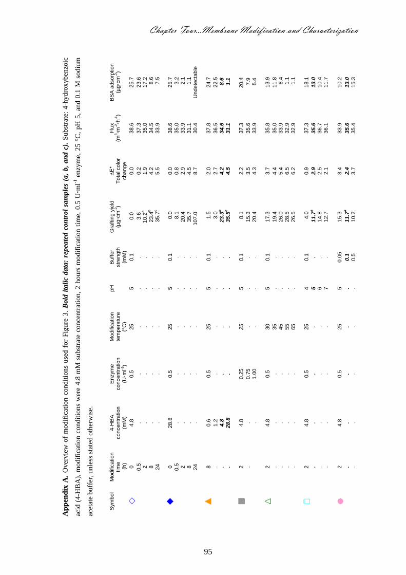
Chapter Four…Membrane Modification and Characterization
95
Ap
pen
dix
A.
Over
vie
w o
f m
odif
icat
ion c
ondit
ions
use
d f
or
Fig
ure
3.
Bold
ita
lic
data
: re
pea
ted c
on
trol
sam
ple
s (a
, b,
an
d c
). S
ubst
rate
: 4
-hydro
xyben
zoic
acid
(4
-HB
A),
modif
icat
ion c
ondit
ions
wer
e 4.8
mM
subst
rate
conce
ntr
atio
n,
2 h
ours
modif
icat
ion t
ime,
0.5
U·m
l-1 e
nzy
me,
25 °
C,
pH
5,
and 0
.1 M
sodiu
m
acet
ate
buff
er, unle
ss s
tate
d o
ther
wis
e.
S
ym
bol
Modific
ation
tim
e
(h)
4-H
BA
concentr
ation
(mM
)
Enzym
e
concentr
ation
(U·m
l-1)
Modific
ation
tem
pera
ture
(°
C)
pH
B
uff
er
str
ength
(m
M)
Gra
ftin
g y
ield
(µ
g·c
m-2)
∆E*
Tota
l colo
r change
Flu
x
(m3·m
-2·h
-1)
BS
A a
dsorp
tion
(µg·c
m-2)
0
4.8
0.5
25
5
0.1
0
.0
0.0
38.6
25.7
0.5
.
. .
. .
3.6
0.2
37.3
23.6
2
. .
. .
. 10.2
a
1.9
35.0
17.2
8
. .
. .
. 2
3.4
b
4.2
34.5
8.6
24
. .
. .
. 3
5.7
c
5.5
33.9
7.5
0
28.8
0.5
25
5
0.1
0.0
0.0
38.6
25.7
0.5
.
. .
. .
8.1
0.8
35.0
3.2
2
. .
. .
. 20.4
2.9
33.9
2.1
8
. .
. .
. 35.7
4.5
31.1
1.1
24
. .
. .
.
107.0
8.7
30.4
U
ndete
cta
ble
8
0.6
0.5
25
5
0.1
1.5
2.0
37.8
24.7
.
1.2
.
. .
.
3.0
2.7
36.5
22.5
. 4.8
.
. .
. 2
3.3
b
4.2
34.6
8
.6
.
28.8
.
. .
. 3
5.5
c
4.5
31.1
1
.1
2
4.8
0.2
5
25
5
0.1
8.1
2.2
37.3
20.4
.
. 0.7
5
. .
. 15.3
3.5
35.6
7
.9
. 1.0
0
. .
. 20.4
4.3
33.9
5
.4
2
4.8
0.5
30
5
0.1
17.3
3.7
35.8
13.9
.
. .
35
. .
19.4
4.4
35.0
11.8
. .
. 45
. .
26.0
5.4
33.9
6.4
. .
. 55
. .
28.5
6.5
32.9
1.1
. .
. 65
. .
26.5
6.2
32.9
1.1
2
4.8
0.5
25
4
0.1
4.0
0.9
37.3
18.1
.
. .
. 5
. 1
1.7
a
2.9
35.6
13.0
. .
. .
6
. 14.8
2.5
36.7
10.4
. .
. .
7
. 12.7
2.1
36.1
11.7
2
4.8
0.5
25
5
0.0
5
15.3
3.4
33.9
10.2
.
. .
. .
0.1
1
1.7
a
2.4
35.6
13.0
. .
. .
. 0.5
10.2
3.7
35.4
15.3

96
Ap
pen
dix
B.
Over
vie
w o
f m
odif
icat
ion
condit
ions
use
d f
or
Fig
ure
5.
Bold
ita
lic
data
: re
peate
d c
on
trol
sam
ple
s (a
, b,
c,
an
d d
). S
ubst
rate
: G
alli
c ac
id,
modif
icat
ion
condit
ions
wer
e 0.5
U·m
l-1 e
nzy
me,
25 °
C, pH
5, an
d 0
.1 M
sodiu
m a
ceta
te b
uff
er, unle
ss s
tate
d o
ther
wis
e.
Sym
bol
Modific
ation
tim
e
(min
)
Galli
c a
cid
concentr
ation
(m
M)
Enzym
e
concentr
ation
(U
·ml-1
)
Modific
ation
te
mpera
ture
(°
C)
pH
B
uff
er
str
ength
(m
M)
Gra
ftin
g y
ield
(µ
g·c
m-2
)
∆E*
Tota
l colo
r change
Flu
x
(m3·m
-2·h
-1)
BS
A a
dsorp
tion
(µ
g·c
m-2
)
0
4.8
0.5
25
5
0.1
0.0
0.0
32.9
37.2
10
.
. .
. .
7
.6a
2.3
31.1
35.7
20
.
. .
. .
14.3
2.4
30.3
26.5
30
.
. .
. .
24.9
3.0
29.9
8.1
60
.
. .
. .
22.4
2.7
31.1
7.6
120
.
. .
. .
35.7
b
6.0
32.0
6.5
0
28.8
0.5
25
5
0.1
0.0
0.0
32.9
37.2
10
.
. .
. .
10.0
2.0
33.9
32.1
20
.
. .
. .
18.3
2.8
35.5
24.4
30
.
. .
. .
24.8
3.0
36.7
7.6
60
.
. .
. .
34.4
4.2
37.3
6.1
120
.
. .
. .
86.6
c
10.9
36.7
5.0
120
1.2
0.5
25
5
0.1
13.6
2.0
32.0
25.7
.
4.8
.
. .
. 3
5.6
b
6.0
32.0
6.5
. 28.8
.
. .
. 8
6.6
c
10.9
36.7
5.0
10
4.8
0.2
5
25
5
0.1
5.5
2.2
30.8
36.0
.
. 0.5
0
. .
.
7.6
a
2.3
31.1
35.7
. .
0.7
5
. .
.
8.7
2.4
32.9
20.4
. .
1.0
0
. .
. 11.9
2.8
32.9
18.2
10
4.8
0.5
25
5
0.1
7.5
a
2.3
31.1
35.7
.
. .
30
.
. 12.7
3.6
32.0
28.9
. .
. 35
.
. 10.7
3.7
32.9
25.8
. .
. 45
.
. 15.2
3.9
33.9
22.5
. .
. 55
.
. 18.4
4.1
34.9
20.4
. .
. 65
.
. 21.6
4.3
35.0
17.2
120
28.8
0.5
25
5
0.1
86.6
c
10.9
36.7
5.0
.
. .
35
.
. 9
1.8
9
.4
36.7
11.6
. .
. 45
.
.
102.0
10.2
37.0
9.6
. .
. 55
.
.
110.0
11.2
37.3
6.4
. .
. 65
.
.
114.3
12.2
37.3
5.4
120
28.8
0.5
25
4
0.1
7
9.7
8.1
36.1
23.6
.
. .
. 5
.
86.6
c
10.9
36.7
5.0
. .
. .
6
. 9
6.6
8.1
35.0
19.3
. .
. .
7
. 105.2
9.0
35.5
18.3
120
28.8
0.5
25
5
0.0
5
69.8
8.6
35.5
22.5
.
. .
. .
0.1
86.6
c
10.9
36.7
5.0
. .
. .
. 0.5
8
1.5
9.7
36.1
10.7

Chapter Five
Adsorption on Modified Model PES Surfaces
This chapter will be submitted as:
Enzyme-Catalyzed Modification of PES Surfaces: Reduction in Adsorption of BSA,
Dextrin and Tannin, Norhan Nady, Karin Schroën, Maurice Franssen, Remco Fokkink,
Mohamed Mohy Eldin, Han Zuilhof, and Remko Boom.

98
Enzyme-Catalyzed Modification of PES Surfaces:
Reduction in Adsorption of BSA, Dextrin and Tannin
Abstract
Poly(ethersulfone) (PES) can be modified in a flexible manner using mild, environmentally
benign components such as 4-hydroxybenzoic acid and gallic acid, which can be attached to
the surface via catalysis by the enzyme laccase. This leads to grafting of mostly linear
polymeric chains (for 4-hydroxybenzoic acid, and for gallic acid at low concentration and
short modification time) and of networks (for gallic acid at high concentration and long
exposure time). The reaction is stopped at a specific time, and the modified surfaces are tested
for adsorption of BSA, dextrin and tannin using in-situ reflectometry and AFM imaging.
At short modification times, the adsorption of BSA, dextrin and tannin is significantly
reduced. However, at longer modification times, the adsorption increases again for both
substrates. As the contact angle on modified surfaces at short modification times is reduced
(indicative of more hydrophilic surfaces), and keeps the same low values at longer
modification times, hydrophilicity is not the only determining factor for the measured
differences. At longer modification times, intra-layer reactivity will increase the amount of
cross-linking (especially for gallic acid), branching (for 4-hydroxybenzoic acid) and/or
collapse of the polymer chains. This leads to more compact layers, which leads to increased
protein adsorption.
The modifications were shown to have clear potential for reduction of fouling by proteins,
polysaccharides, and polyphenols, which could be related to the surface morphology.
Chapter Five…Adsorption on Modified Model PES Surfaces
99
1. Introduction
Membrane fouling is a serious problem in membrane filtration. The first step of membrane
fouling is adsorption of components from the feed. This adsorption process depends on the
nature of the components, the (surface) material of the membrane, and on the operating
conditions [1-5]. Poly(ethersulfone) (PES) is a popular material for membranes, as it lends
itself well for membrane preparation through phase inversion, and yields mechanically and
chemically robust membranes. However, its hydrophobicity makes it (as usually proposed)
intrinsically susceptible to adsorption by e.g. proteins [4,6,7]. Besides proteins, also other
foulants such as polysaccharides and polyphenols pose problems in practice, and have been
investigated individually [8-10] or in combination [11,12].
Obtaining a (more) hydrophilic membrane surface has been the aim of many researchers in
order to reduce (or even prevent) adsorption [13-15], but the precise mechanism of fouling by
adsorption is complex. Hydrogen bonding, hydrophobic interactions, π-π stacking and
changes in water structure at the membrane have been proposed as (parts of) adsorptive
mechanisms [9,10,16]. In addition, a decrease in the Gibbs energy (G) of the system (i.e.,
protein, surface, and solvent) leads to adsorption of foulants. For that, any change in the
enthalpy, entropy, and the system temperature should affect the adsorption process [17,18].
For example, the release of water from the surface or protein molecules (i.e., dehydration
processes) with a concomitant large entropy gain leads to increased protein adsorption and an
overall decreased Gibbs energy [17-19]. On the other hand, the surface structure is also
sometimes considered as a factor to influence protein adsorption [5,17,20-22]. Steric
hindrance and the osmotic effect of hydrated coated/grafted polymer branches on the surface
contributes to the reduction of adsorption by keeping the foulant molecules at a distance
behind a barrier of adsorbed water molecules (i.e., hydration layer). Moreover, the strength
and thickness of this hydration layer has been used to effectively repel proteins from surfaces
covered with zwitterionic polymer brushes [23]. In addition to these parameters, the
adsorption process can also be affected by the protein structure, protein stability,
concentration, pH, and ionic strength [3,17,19].
While proteins may initially adsorb due to hydrophilic interactions, their irreversible
adsorption is typically caused by hydrophobic interactions. For polysaccharides, hydrophobic
interactions with the membrane surface are important, while hydrogen bonding is primarily
responsible for the adsorption of polyphenols on hydrophilic membrane surfaces [16,24]. To
100
cover a wide range of fouling interactions, this study thus includes a selection of model
foulants from each category.
The enzymatic modification of PES membranes allows the formation of hydrophilic layers
onto the membrane surface from monomers (4-hydroxybenzoic acid, gallic acid, and other
phenolic compounds) using oxidative enzymes such as laccase [25]. This very mild procedure
(room temperature, neutral pH) only involves natural chemicals and the enzyme. The
resulting surface is rather polar, and its low contact angle (52-62º) suggested the potential use
as antifouling surface [25,26]. However, the exact influence of the surface modification on the
adsorption of fouling constituents has not yet been evaluated.
In this work we aim to quantify these antifouling effects with reflectometry and
ellipsometry. We use a PES layer on top of a silicon dioxide surface as a model PES surface
for characterization of the adsorption of various components. These PES model surfaces were
then further modified with the enzyme laccase and phenolic acids as described previously
[25,26]. Reflectometry is a widely used technique to investigate the initial adsorption of
solutes [16,27-31] onto silicon dioxide surfaces: the technique allows the in-situ measurement
of the dynamics and total quantity of the (ir)reversible adsorption of foulant monolayers. The
adsorption of BSA, dextrin, and tannin was used as model materials for proteins,
polysaccharides, and polyphenols, respectively. Ellipsometry was used to measure the
thickness and refractive index of each layer (silicon dioxide, PES, and modification layer) on
the silicon substrate. Contact angle measurements were used to characterize the hydrophilicity
of the surface, and AFM to evaluate the surface structure in detail. This complete set of data
allows a general delineation of the potential of modified PES layers as antifouling surfaces.
2. Materials and Methods
2.1. Chemicals
Poly(ethersulfone) (PES) (Ultrason, E6020P) was obtained from BASF (Ludwigshafen,
Germany). Prime grade 150 mm silicon wafers of type P/B <100> orientation, thickness 660-
690 µm, with a 2.5 nm native oxide layer were purchased from Wafer Net Inc (San Jose, CA,
USA). From Sigma-Aldrich were purchased: catechol (>99%), sodium acetate (anhydrous,
≥99%), sodium phosphate dibasic anhydrous (>99%), acetic acid (99.9%), 4-hydroxybenzoic
acid (99%), dichloromethane ACS (stabilized, 99.9%), gallic acid (>97.5%), sodium
phosphate monobasic (>98%), Bovine Serum Albumin, Cohan Analog (>98%), and laccase
from Trametes versicolor (>20 U·mg-1
). Tannic acid powder (>98%) was obtained from
Chapter Five…Adsorption on Modified Model PES Surfaces
101
Riedel-de Haën (Seelze, Germany), and dextrin 5 (from maize starch, >95% ) was purchased
from Fluka (Neu-Ulm, Germany). All chemicals were used as received. Milli-Q water was
used throughout.
2.2. Laccase Assay
The laccase activity was determined with catechol as substrate. The assay mixture
contained 0.33 ml of 10 mM catechol, 2.67 ml of 0.1 M sodium acetate buffer (pH 5), with
0.025 U·ml-1
laccase. Oxidation of catechol is monitored from the absorbance at 400 nm (ε =
26,000 M-1
·cm-1
) [32] after 20 min. One unit of laccase activity is defined as the amount of
enzyme required to oxidize 1 µmol of catechol per min at 25 °C.
2.3. Preparation of Model Surfaces and Layer Thickness
The thickness of the native silicon dioxide layer (2.5 nm) was increased to approximately
70 ± 2 nm (triplicate measurements on ≥ 3 positions per strip) by heating at 1000 °C for 100
min. Then, the wafers were cut into strips of 1 × 4.5 cm. The strips were sonicated in ethanol
for 15 min, washed with water and ethanol, and dried in flowing nitrogen. The thickness and
refractive index of silicon dioxide layer were determined with a computer-controlled null
ellipsometer (Sentech instruments Gmbh) at λ = 632.8 nm and angle of incidence 70°. Values
of 3.85 and 0.02 were used for the refractive index (n) and the imaginary refractive index (k)
of silicon [27,28], respectively. Subsequently the strips were given a plasma treatment (PDC-
32G, Harrick at RF-level high) for 10 min. After the removal of any dust by using a flow of
nitrogen, the strips were used as substrate for the model PES surfaces by spin coating them
with 0.25% w/w PES solution in dichloromethane for 10 s at 2500 rpm. The spin coating was
performed immediately after plasma cleaning. After spin coating, the PES coated strips were
put at 300 °C for 60 min.
The thickness and refractive index of PES layers deposited on silicon dioxide were
determined by ellipsometry. To ensure homogenous PES layers of around 20 nm, the
thickness was tested at 3 different locations; the variation was never more than 2 nm. The
spin-coated PES model surfaces were then modified using laccase (see respective section).
The surfaces were kept for 24 h in glass-covered dishes in desiccators supplied with silica gel
to remove any moisture. After drying, the thickness and refractive index of the modification
layer was determined by ellipsometry, as the average of values measured at three different
locations. The refractive index initial values used for the silicon dioxide, PES, and
modification layers were 1.46, 1.65, and 1.57, respectively [27,33].
102
2.4. Modification Experiments
The spin-coated PES model surfaces were incubated in 20 ml 0.1 M sodium acetate buffer
containing different concentrations of phenolic acids (substrates or monomers) and laccase
enzyme. A flow of air was used for mixing and as oxygen source. After a specific
modification time, the modified model surfaces were removed from the liquid and washed by
strong flushing with Milli-Q water. Gallic acid and 4-hydroxybenzoic acid were used as
enzyme substrates, at two concentrations (4.8 and 28 mM), together with various modification
times ranging from a few minutes to 24 h [25,26]. The modified PES model surfaces were
kept in glass-covered dishes in desiccators supplied with silica gel for drying.
2.5. Static Water Contact Angle
Blank and modified PES model surfaces were characterized by static water contact-angle
measurement using a Krüss DSA 100 apparatus. Drops of demineralized water (7 µL) were
deposited on three different spots of each surface, and the contact angle was calculated from
three different measurements taken on each of two independently modified surfaces.
2.6. Atomic Force Microscopy (AFM)
Spin coated PES model surfaces were prepared on strips (1 × 6 cm) that were half covered
with polymer solution. In this way, blank and modified surfaces are present on the same strip,
and images of both were obtained with an MFP-3D AFM from Asylum Research (Santa
Barbara, CA). Imaging was performed in AC mode in air using OMCL-AC240 silicon
cantilevers (Olympus Corp.). In order to investigate the effect of the modification layer on
protein adsorption, the half-modified strips (surfaces) were incubated in 50 ml of 0.1 g·l-1
BSA solution for 1 h followed by immersion and flushing with Milli-Q water, and stored as
described before. The root mean squares (RMS) profiles (i.e., roughness) of the two parts
were calculated before and after exposure to BSA solution.
2.7. Adsorption Measurements
In Figure 1A, the reflectometry set-up is schematically depicted. A monochromatic light
beam (1) (He-Ne laser; 632.8 nm) is linearly polarized and passes a 45° glass prism (2). This
beam arrives at the interface (3) with an angle of incidence close to 71° for the
solvent/substrate interface. After reflection at the interface and refraction at the prism, the
beam is split into its parallel and perpendicular components (p- and s-polarized) of which the
intensities are measured continuously (Ip and Is) relative to the plane of incidence by means of
Chapter Five…Adsorption on Modified Model PES Surfaces
103
a beam splitter (4). Both components are detected separately by two photodiodes (5) and the
ratio between the intensity of the parallel and perpendicular components is recorded. The
output signal S is the ratio between the two intensities (Ip/Is) (6). The surface is inside a
stagnation point flow-cell allowing homogeneous adsorption on surfaces. Two 4-way valves
(7) are used to switch between the sample solutions.
Solutions of BSA (0.1 g·l-1
), dextrin, and tannin (both 0.2 g·l-1
) were prepared in 10 mM
phosphate buffer pH 7. Before the start of the adsorption experiments, each slide (model
surface) was incubated for 45 min in buffer to avoid artefacts due to initial surface wetting.
All solutions were degassed using ultrasound. After placing the slide inside the flow cell, the
buffer solution was injected until the output signal was nearly constant. This value was taken
as the base signal, S0. Each adsorption experiment was conducted in three stages: 15 min
buffer injection (base-line) followed by 15 min foulant solution injection (adsorption process;
the maximum adsorbed amount was mostly reached within 15 min) and then 15 min buffer re-
injection (desorption process). The change in intensity of the reflected polarized laser that
followed was converted into adsorbed mass using a 5-layer matrix model [27,28]. This model
uses equation (1), where Γ is the adsorbed amount (mg·m-2
), Qf is a sensitivity factor (mg·m-
2), and S0 is the base (blank) signal before introducing the foulant, and ∆S is the recorded
difference in signal before and after introduction of the foulant:
0S
SQΓ f
(1)
The value of Qf depends on the thickness and refractive index, which were determined by
ellipsometry. Refractive index increments dn/dc of 0.185, 0.128, and 0.172 ml.g-1
were used
for BSA, dextrin, and tannin respectively [16,34]. All the experiments were carried out at
least in duplicate, using a flow rate of 0.8-1.2 ml·min-1
. The increase in signal in the first 60 s
was used to determine the initial adsorption rate by using the 5-layer matrix model as
previously described. This measure is indicative for the kinetics of the adsorption process (see
Figure 1B for a schematic impression of the reflectometry experiment and the various
parameters that were derived from it).

104
Figure 1. A) Schematic representation of the fixed angle reflectometer, consisting of (1) laser
beam, (2) glass prism, (3) sample, (4) beam splitter, (5) photodiodes, (6) detection unit. The actual
inlet consists of two (7) 4-way valves (only 1 is drawn here), in order to allow sample to be
exposed to up to 3 liquids. B) Schematic representation of the reflectometer output.
2.8. Homopolymer Adsorption
Although the enzyme covalently binds the phenolic acid substrate compounds to the
surface, homopolymer is also formed in solution, which can then physisorb to the surface. To
investigate this, the reaction of phenolic acid substrate and enzyme was carried out as
Inlet a Inlet b
Detection
Cell
ParallelPerpendicular5
3
22
4
5
7
6
1
Slope ≈ Initial adsorption rate
Adsorbed amount
(reversible + irreversible)Residual amount
(irreversible)
Time
Adsorption phase Desorption phase
Desorbed amount
(reversible)
Ad
so
rbed
Am
ou
nt
A
B
Chapter Five…Adsorption on Modified Model PES Surfaces
105
described before in 40 ml, but now without the PES surfaces present. The reaction time was 2
h and 24 h for 4-hydroxybenzoic acid (28.8 mM), and 7 min, 2 h and 24 h for gallic acid (4.8
mM, and with 24 h reaction time also at 28.8 mM). The enzyme was inactivated by adding 5
ml of 0.1 M NaOH while stirring for 10 min. After re-adjusting the pH to 5 with concentrated
HCl, the homopolymer solutions were diluted 10× with Milli-Q water in order to use them in
the reflectometer. The refractive index increment dn/dc of tannin (0.172 ml.g-1
) was used for
the homopolymer, since they are structurally quite similar.
3. Results and Discussion
The research described in this chapter relates to the repellence of foulants (BSA, dextrin,
and tannin) by PES model surfaces, modified by phenolic acids by the action of laccase. First,
the results that were obtained for the two substrates (4-hydroxybenzoic acid and gallic acid)
separately will be discussed, followed by an overview in the overall outlook section. The
choice of the reaction conditions are based on previous research [26], in which we evaluated
the effect of BSA on ‘real’ membranes through flux analysis and weight increase. The
reaction conditions that were used to modify the model surfaces can be found in the appendix
(Tables A1 and A3), both for 4-hydroxybenzoic acid and gallic acid.
3.1. Model Surfaces Modified with 4-Hydroxybenzoic Acid
The unmodified PES surface (see Table 1; modification conditions of the different surfaces
are given in appendix A) shows a BSA adsorption of around 2 mg·m-2
, most of which is
irreversibly bound (1.8 mg·m-2
remains adsorbed after desorption). The modified surfaces
show a lower protein adsorption and in some cases better reversibility (max. 50%).
Interestingly enough, the surfaces that were most protein repellent, do not correspond with the
most hydrophilic surfaces (low contact angle) as is often assumed to be the case. This will be
discussed in more detail in the AFM section (vide infra).
Based on the results in Table 1, modification conditions used for surface number 1 (2 h
modification using 28.8 mM 4-hydroxybenzoic acid, 0.5 U·ml-1
enzyme, at 25 °C and pH 5)
were identified as suitable for further research.
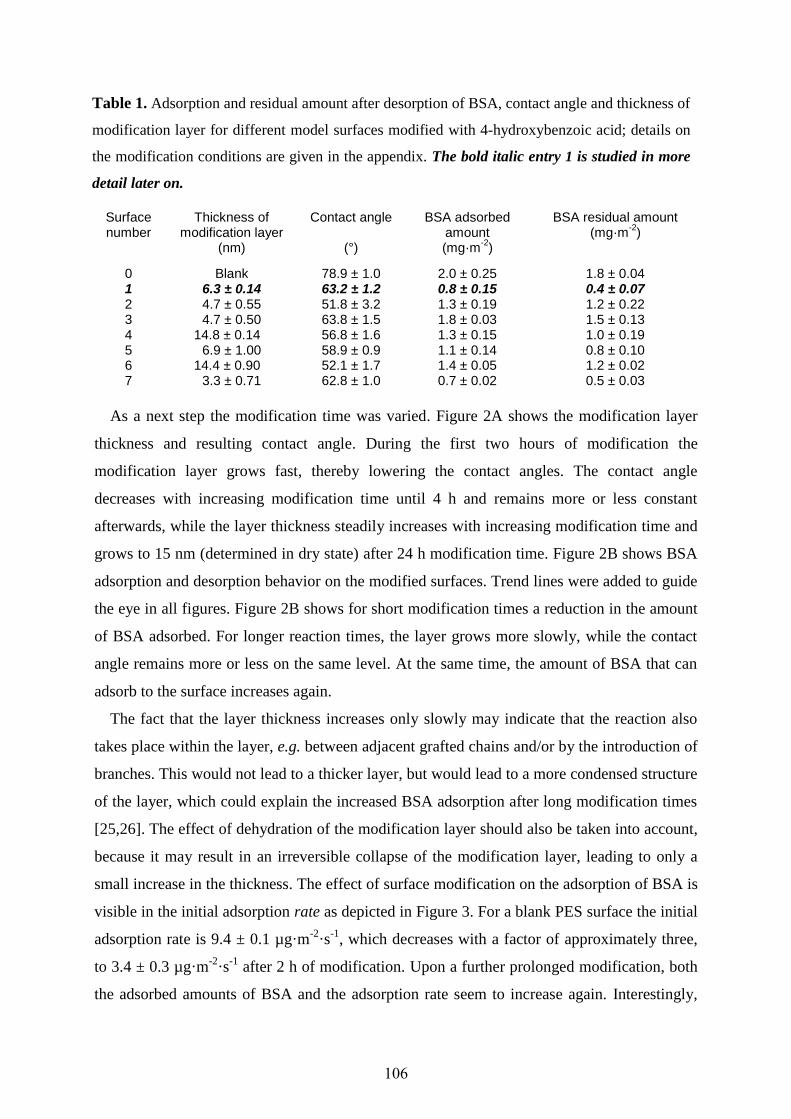
106
Table 1. Adsorption and residual amount after desorption of BSA, contact angle and thickness of
modification layer for different model surfaces modified with 4-hydroxybenzoic acid; details on
the modification conditions are given in the appendix. The bold italic entry 1 is studied in more
detail later on.
Surface number
Thickness of modification layer
(nm)
Contact angle
(°)
BSA adsorbed amount (mg·m
-2)
BSA residual amount (mg·m
-2)
0 Blank 78.9 ± 1.0 2.0 ± 0.25 1.8 ± 0.04 1 6.3 ± 0.14 63.2 ± 1.2 0.8 ± 0.15 0.4 ± 0.07 2 4.7 ± 0.55 51.8 ± 3.2 1.3 ± 0.19 1.2 ± 0.22 3 4.7 ± 0.50 63.8 ± 1.5 1.8 ± 0.03 1.5 ± 0.13 4 14.8 ± 0.14 56.8 ± 1.6 1.3 ± 0.15 1.0 ± 0.19 5 6.9 ± 1.00 58.9 ± 0.9 1.1 ± 0.14 0.8 ± 0.10 6 14.4 ± 0.90 52.1 ± 1.7 1.4 ± 0.05 1.2 ± 0.02 7 3.3 ± 0.71 62.8 ± 1.0 0.7 ± 0.02 0.5 ± 0.03
As a next step the modification time was varied. Figure 2A shows the modification layer
thickness and resulting contact angle. During the first two hours of modification the
modification layer grows fast, thereby lowering the contact angles. The contact angle
decreases with increasing modification time until 4 h and remains more or less constant
afterwards, while the layer thickness steadily increases with increasing modification time and
grows to 15 nm (determined in dry state) after 24 h modification time. Figure 2B shows BSA
adsorption and desorption behavior on the modified surfaces. Trend lines were added to guide
the eye in all figures. Figure 2B shows for short modification times a reduction in the amount
of BSA adsorbed. For longer reaction times, the layer grows more slowly, while the contact
angle remains more or less on the same level. At the same time, the amount of BSA that can
adsorb to the surface increases again.
The fact that the layer thickness increases only slowly may indicate that the reaction also
takes place within the layer, e.g. between adjacent grafted chains and/or by the introduction of
branches. This would not lead to a thicker layer, but would lead to a more condensed structure
of the layer, which could explain the increased BSA adsorption after long modification times
[25,26]. The effect of dehydration of the modification layer should also be taken into account,
because it may result in an irreversible collapse of the modification layer, leading to only a
small increase in the thickness. The effect of surface modification on the adsorption of BSA is
visible in the initial adsorption rate as depicted in Figure 3. For a blank PES surface the initial
adsorption rate is 9.4 ± 0.1 µg·m-2
·s-1
, which decreases with a factor of approximately three,
to 3.4 ± 0.3 µg·m-2
·s-1
after 2 h of modification. Upon a further prolonged modification, both
the adsorbed amounts of BSA and the adsorption rate seem to increase again. Interestingly,
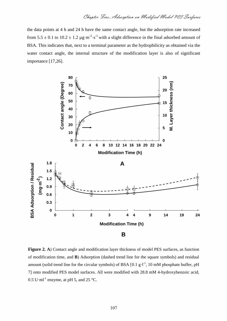
Chapter Five…Adsorption on Modified Model PES Surfaces
107
the data points at 4 h and 24 h have the same contact angle, but the adsorption rate increased
from 5.5 ± 0.1 to 10.2 ± 1.2 µg·m-2
·s-1
with a slight difference in the final adsorbed amount of
BSA. This indicates that, next to a terminal parameter as the hydrophilicity as obtained via the
water contact angle, the internal structure of the modification layer is also of significant
importance [17,26].
Figure 2. A) Contact angle and modification layer thickness of model PES surfaces, as function
of modification time, and B) Adsorption (dashed trend line for the square symbols) and residual
amount (solid trend line for the circular symbols) of BSA [0.1 g·l-1, 10 mM phosphate buffer, pH
7] onto modified PES model surfaces. All were modified with 28.8 mM 4-hydroxybenzoic acid,
0.5 U·ml-1 enzyme, at pH 5, and 25 °C.
0
0.3
0.6
0.9
1.2
1.5
1.8
0 1 2 3 4 4 9 14 19 24
Modification Time (h)
BS
A A
ds
orp
tio
n /
Re
sid
ua
l
(mg
·m)
-2
B
0
5
10
15
20
25
0
10
20
30
40
50
60
70
80
0 2 4 6 8 10 12 14 16 18 20 22 24
M.
La
ye
r th
ickn
ess
(n
m)
Co
nta
ct
an
gle
(D
eg
ree)
Modification Time (h)
A

108
Figure 3. Initial BSA adsorption rate measured on model PES surfaces modified with 28.8 mM
4-hydroxybenzoic acid, 0.5 U·ml-1 enzyme, at pH 5, and 25 °C as function of modification time.
AFM experiments carried out with half-modified PES model surfaces (i.e., half of the
surface is used as a blank and the other half is modified, see Figure 4), were used to study the
morphology of the modification layer. 4-Hydroxybenzoic acid can couple to the PES surface
forming grafted oligomers or polymers [25,26]. This is reflected in the roughness of the
surface: the RMS height differences increased from 0.7 nm for the blank surface to 2 nm for
the modified one.
The half-modified model PES surfaces were also used to visualize the differences in protein
adsorption. On the blank surface, white dots appear upon exposure to protein (images E and
G, in Figure 4), and the roughness increases considerably from 1.1 to 3.8 nm. On the modified
surface, it was difficult to differentiate between the BSA and the grafted oligomers/polymers
because the modification itself also leads to the formation of white dots (image F).
Furthermore, the RMS before and after adsorption was very similar (1.5 versus 1.0 nm,
images F and H). However, the large white patches of protein aggregates that are clearly
visible on the blank PES surface (G) were not found on the modified PES surface (H). This in
combination with the noted difference in roughness between modified and unmodified PES
surface after exposure to BSA, is an indication that the modification layer does lower the
adsorbed amount of BSA, as was found with reflectometry.
0
3
6
9
12
0 5 10 15 20 25
Modification Time (h)
Init
ial
BS
A A
dso
rpti
on
Rate
(µg
·m-2
·s-1
)

Chapter Five…Adsorption on Modified Model PES Surfaces
109
Unmodified PES model surface
(Blank side)
Modified PES model surface
(Modified side)
3D: 10 µm (A and B) & 200 nm (C and D)
2D: 2 µm before (E and F) and after (G and H) immersion in BSA
Figure 4. AFM images and height profiles of half-modified PES model surface (both
unmodified and modified model PES surfaces are on the same slide) prepared at 28.8 mM 4-
hydroxybenzoic acid A, 2 h modification time, 0.5 U·ml-1 laccase, 25 °C, and pH 5. 3D: blank
side [10 µm (A) and 200 nm (C)], modified side [10 µm (B) and 200 nm (D)]. 2D; 2 µm: blank
side before (E) and after (G) immersion in BSA solution, modified side before (F) and after (H)
immersion in BSA solution.
A B
C D
Overall
Detail
G
E F
H
Before BSA
Adsorption
After BSA
Adsorption
110
The adsorption of dextrin and tannin (model foulants for polysaccharides and polyphenols)
shows similar characteristics to that of BSA adsorption (see Figure 5). Dextrin adsorption is
sharply reduced from 0.71 mg·m-2
down to 0.03 mg·m-2
after 2 h of reaction time. After that
the amount adsorbed remains low, though the same increasing trend is visible as was seen
with BSA. Tannins initially adsorbed much faster, but are rinsed off easily. The development
of the residual amount left after rinsing is again similar to that observed for BSA: a sharp
reduction at modification times up to 2 h, and then a gradual increase again.
Once more, this general behavior may well be related to the internal structure of the layer.
In the first 2 h, the layer quickly grows, but after that the growth is much slower. Thus, the
initial layer will be built up by grafting onto the surface, while at later stages much of the
reaction may take place within the layer (e.g., leading to more cross-linking and/or branching
and/or collapse of the polymer chains).
Figure 5. Adsorption (dashed trend line) and residual amount (solid trend line) of dextrin (blue
square symbols) and tannin (red circular symbols) (both 0.2 g·l-1) in 10 mM phosphate buffer,
pH 7, on modified model PES surfaces as function of modification time [28.8 mM 4-
hydroxybenzoic acid, 0.5 U·ml-1 enzyme, pH 5, and 25 °C].
In addition, there is also polymerization in the bulk, leading to 4-hydroxybenzoic acid
homopolymer in solution, which may then be physisorbed to the model surface. This effect
may well disturb our measurements, and therefore we investigated this separately. By only
adding the PES model surface after the polymerization, surface grafting can be avoided. Table
2 shows that indeed homopolymer is formed, which adsorbs to the unmodified surface as
soon as it is available. As is to be expected with polymer adsorption (usually through multiple
adsorption points along the polymeric chain), it is not rinsed off. However, it should be noted
that the modified surface hardly shows any adsorption of the homopolymer, and of what is
adsorbed most can be removed.
0
0.3
0.6
0.9
1.2
1.5
0 1 2 3 4 4 9 14 19 24
Ad
so
rpti
on
/ R
esid
ua
l
(mg
∙m)
Modification Time (h)

Chapter Five…Adsorption on Modified Model PES Surfaces
111
Table 2. Adsorption and residual amount after desorption of 4-hydroxybenzoic acid homopolymer
on blank and PES model surface modified for 2 h with 28.8 mM 4-hydroxybenzoic acid, 0.5 U·ml-1
laccase, at pH 5 (0.1 M sodium acetate buffer), and 25 °C.
Reaction time for homopolymer
formation (h)
Blank PES model surface Modified PES model surface [2 h modification time]
Homopolymer adsorbed amount
mg·m-2
Homopolymer residual amount
mg·m-2
Homopolymer adsorbed amount
mg·m-2
Homopolymer residual amount
mg·m-2
2 1.0 ± 0.2 0.9 ± 0.2 0.08 ± 0.03 0.03 ± 0.03
24 1.4 ± 0.1 1.3 ± 0.1 0.02 ± 0.01 0.00 ± 0.00
3.2. Model Surfaces Modified with Gallic Acid
As was the case for 4-hydroxybenzoic acid, all surfaces modified with gallic acid showed
less protein adsorption than the blank model surface (Table 3). There is once more no obvious
relation between BSA adsorption and the contact angle of the modified model surfaces, which
again indicates that hydrophilicity is not the only factor involved. In general, the modification
process proceeds faster with gallic acid than with 4-hydroxybenzoic acid. Although this is a
positive aspect, homopolymer formation also takes place more quickly. We therefore decided
to focus on short reaction times and low substrate concentrations, as was used for surface
number 2 (italic bold data in Table 3: 4.8 mM gallic acid, 0.5 U·ml-1
enzyme, pH 5, and 25
°C).
Table 3. Adsorption and residual amount after desorption of BSA, contact angle and thickness
of modification layer for different reaction conditions during modification with gallic acid;
details on the reaction conditions are given in the appendix. The bold italic bold entry 2 is
investigated in more detail later on.
Surface number
Thickness of modification layer
(nm)
Contact angle
(°)
BSA adsorbed amount (mg·m
-2)
BSA residual amount (mg·m
-2)
0 Blank 78.9 ± 1.0 2.1 ± 0.12 1.8 ± 0.04 1 5.5 ± 0.91 69.7 ± 1.3 0.8 ± 0.01 0.7 ± 0.03 2 7.6 ± 0.75 64.2 ± 0.6 0.5 ± 0.14 0.3 ± 0.03 3 9.5 ± 1.00 67.6 ± 0.8 0.9 ± 0.13 0.8 ± 0.08 4 4.6 ± 0.41 64.4 ± 1.3 1.2 ± 0.17 0.9 ± 0.08 5 10.2 ± 0.76 70.9 ± 1.0 0.8 ± 0.04 0.7 ± 0.04 6 10.3 ± 0.58 62.8 ± 0.6 0.8 ± 0.09 0.7 ± 0.08
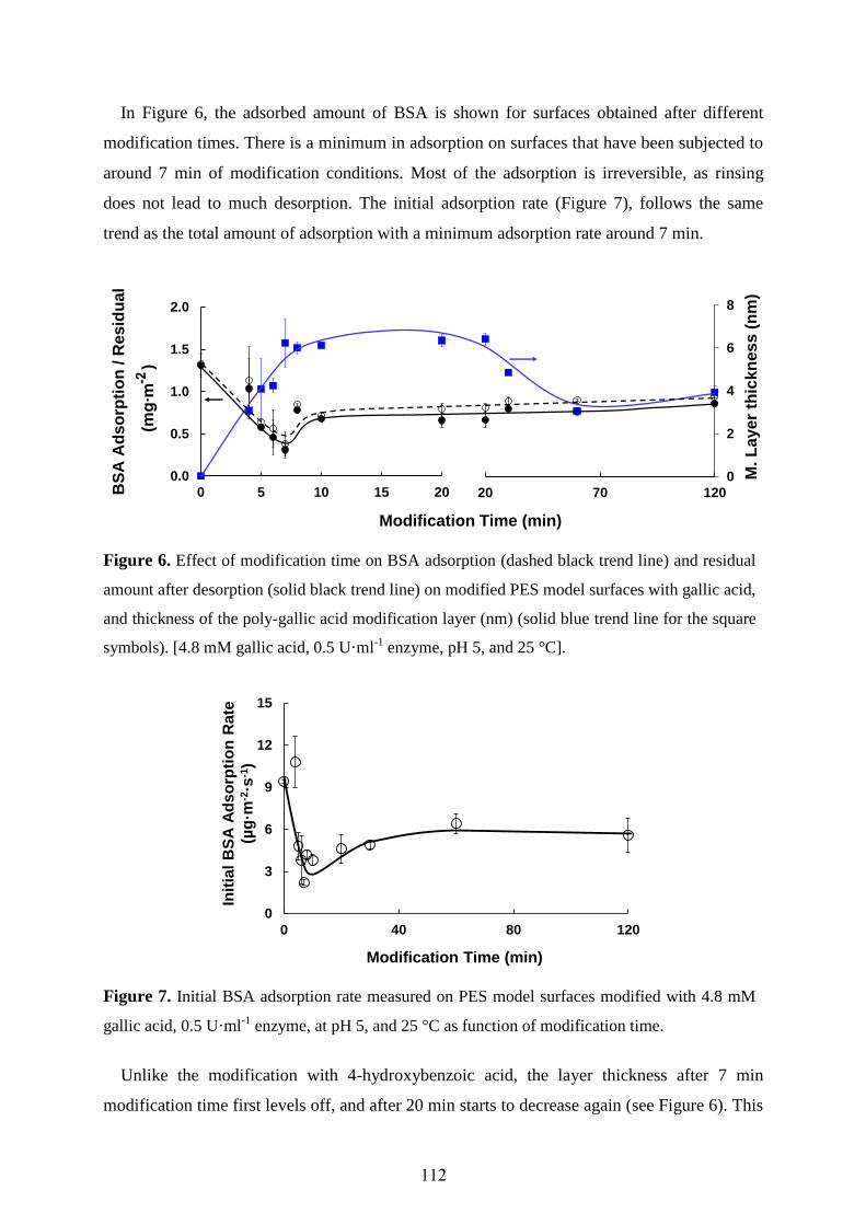
112
In Figure 6, the adsorbed amount of BSA is shown for surfaces obtained after different
modification times. There is a minimum in adsorption on surfaces that have been subjected to
around 7 min of modification conditions. Most of the adsorption is irreversible, as rinsing
does not lead to much desorption. The initial adsorption rate (Figure 7), follows the same
trend as the total amount of adsorption with a minimum adsorption rate around 7 min.
Figure 6. Effect of modification time on BSA adsorption (dashed black trend line) and residual
amount after desorption (solid black trend line) on modified PES model surfaces with gallic acid,
and thickness of the poly-gallic acid modification layer (nm) (solid blue trend line for the square
symbols). [4.8 mM gallic acid, 0.5 U·ml-1 enzyme, pH 5, and 25 °C].
Figure 7. Initial BSA adsorption rate measured on PES model surfaces modified with 4.8 mM
gallic acid, 0.5 U·ml-1 enzyme, at pH 5, and 25 °C as function of modification time.
Unlike the modification with 4-hydroxybenzoic acid, the layer thickness after 7 min
modification time first levels off, and after 20 min starts to decrease again (see Figure 6). This
0
2
4
6
8
0.0
0.5
1.0
1.5
2.0
0 5 10 15 200
2
4
6
8
0.0
0.5
1.0
1.5
2.0
20 70 120BS
A A
dso
rpti
on
/ R
es
idu
al
(mg
∙m)
Modification Time (min)
M. L
aye
r th
ick
ne
ss
(n
m)
0
3
6
9
12
15
0 40 80 120
Init
ial
BS
A A
dso
rpti
on
Rate
(µg
·m-2
·s-1
)
Modification Time (min)

Chapter Five…Adsorption on Modified Model PES Surfaces
113
may well be due to reactions taking place mostly within the modification layer, leading to
crosslinks. IRRAS analyses showed characteristic bands of OH moieties at short modification
times [25], but these bands were hardly visible anymore upon prolonged modification. This is
indicative for the formation of a cross-linked layer. Moreover, the sulfur signal in XPS
analysis [26], which becomes appreciably smaller while the layer thickness showed only a
marginal increase, provides an extra argument for cross-linking. The cross-linked
modification layer loses its protein-repellent properties, as is further illustrated in the AFM
section.
Figure 8 shows that the morphology of the modification layer is different from that
obtained with 4-hydroxybenzoic acid. The grafted gallic acid appears as small islands after 7
min modification time, which then grow together and become denser and rougher upon longer
reaction times. Notice that the layer thickness for 10 and 20 min is very similar (Figure 6),
while it is reproducibly thinner at 30 min but looks as a corrugated dense layer. This may be
due to excessive cross-linking. Further, it is interesting to note that the RMS roughness of the
blank PES surface is much higher after BSA adsorption than for any modified PES surface.
2D
3D
Modification
time (min)
Blank
7
10
20
30 Layer
thickness
(nm)
7
6
6
5
RMS (nm)
before BSA
adsorption
0.3
2.6 1.3
1.7
0.7
RMS (nm)
after BSA
adsorption
5.2 1.1 0.6 1.9 1.0
Figure 8. AFM images (2D and 3D), average modification layer thickness, and RMS value
before and after BSA adsorption onto blank and gallic acid modified PES model surfaces [4.8
mM gallic acid, 0.5 U·ml-1 enzyme, at pH 5, and 25 °C] obtained after 7, 10, 20, and 30 min
modification time.

114
Figure 9 shows that gallic acid modification does yield less adsorption of tannin and dextrin
compared to the blank PES model surfaces, but the observed decrease is much smaller as
compared to the adsorption of BSA. In case of tannin, the difference between initially
adsorbed amount and residual amount is rather large, as was the case for 4-hydroxybenzoic
acid, implying that any adsorbed tannin is only weakly adsorbed.
Also for gallic acid, the adsorption of homopolymer was investigated. Again (see Table 4),
the homopolymer attached well on the blank PES model surfaces, which was almost
completely avoided by modification. The only exception is the modification layer formed at
high substrate concentration and modification time, onto which the homopolymer adheres
readily, though reversibly.
Figure 9. Effect of modification time on dextrin (square symbols) adsorption (dashed blue trend
line), and residual amount after desorption (solid blue trend line), tannin (circular symbols)
adsorption (dashed red trend line), and residual amount after desorption (solid red trend line) on
PES model surfaces [4.8 mM gallic acid, 0.5 U·ml-1 enzyme, pH 5, and 25 °C].
Table 4. Adsorbed and residual amount of homopolymer on blank and modified PES model
surface. 4.8 mM gallic acid, 0.5 U·ml-1 laccase, pH 5 (0.1 M sodium acetate buffer), and 25 °C.
Reaction time for homopolymer
formation
Blank PES model surface Modified PES model surface [7 min modification time]
Homopolymer adsorbed amount
mg.m-2
Homopolymer residual amount
mg·m-2
Homopolymer adsorbed amount
mg.m-2
Homopolymer residual amount
mg·m-2
7 min 0.8 ± 0.13 0.7 ± 0.15 0.01 ± 0.01 0.00 ± 0.00 2 h 0.6 ± 0.06 0.6 ± 0.02 0.05 ± 0.01 0.02 ± 0.01 24 h 0.5 ± 0.05 0.4 ± 0.03 0.01 ± 0.01 0.01 ± 0.00
28.8 mM gallic acid + 24 h
1.3 ± 0.23 1.2 ± 0.27 1.7* 0.05*
*only one slide was investigated
0.0
0.5
1.0
1.5
0 5 10 15 20 20 70 120
Modification Time (min)
Ad
so
rpti
on
/ R
es
idu
al
(mg
∙m)

Chapter Five…Adsorption on Modified Model PES Surfaces
115
4. General Discussion
The adsorption of BSA, dextrin and tannin to the PES surface was reduced with laccase-
catalyzed modification using both substrates (4-hydroxybenzoic acid and gallic acid).
However, the extent of foulant repellence differs depending on the structure of the substrate
and the reaction conditions. At short modification times, the adsorption of the three foulants
was reduced, whereas at longer modification times, the adsorption increases again for both
substrates.
We found that the more hydrophilic surfaces were not necessarily best in protein
repellence; apparently, the internal polymer structure adds to the created effects. This is in
good agreement with recently published research about the design of novel antifouling
surfaces using surface-initiated atom transfer radical polymerization (SI-ATRP) to grow
poly[N-(2-hydroxypropyl) methacrylamide] brushes on a gold-coated substrate [35]. This
brush structure shows decreased adsorption of proteins from blood plasma to a level below
the detection limit of surface plasmon resonance (SPR, 0.03 ng.cm-2
), in spite of its only
moderate wettability (advancing and receding θ are 40° and 21°, respectively, 17 nm layer
thickness). This research and our results show that there is no direct relationship between
fouling of a surface and its hydrophilicity, which is in contrast to the currently proposed
theory for the design of protein-resistant surfaces. It is clear that the process of protein
adsorption to surfaces is still not well understood.
All the results suggest that at short reaction times, the surface becomes grafted, leading to a
thin layer that is more hydrophilic than PES. This layer is effective in reducing any strong
adsorption of protein, polysaccharide and polyphenol. At longer reaction times, other effects
become more prominent. One effect is that as more material is grafted on the membrane
surface, the enzyme and/or enzyme-generated radicals will also be active within the layer,
leading to cross-linking between adjacent chains (especially for gallic acid), branching and/or
collapse of the chains. This will lead to a modifying layer that is less swollen and denser.
In addition, further grafting may lead to a more irregular surface of the modification layer.
Any protrusions from the film will have faster access to the reactants in the bulk, and thus will
grow somewhat faster than their surroundings. This will lead to the protrusions to grow and
become more prominent. This well-known effect ultimately leads to rougher modification
layers, which then will increase the adsorption onto the film, since it gives more anchoring
points for the adsorbents to attach.
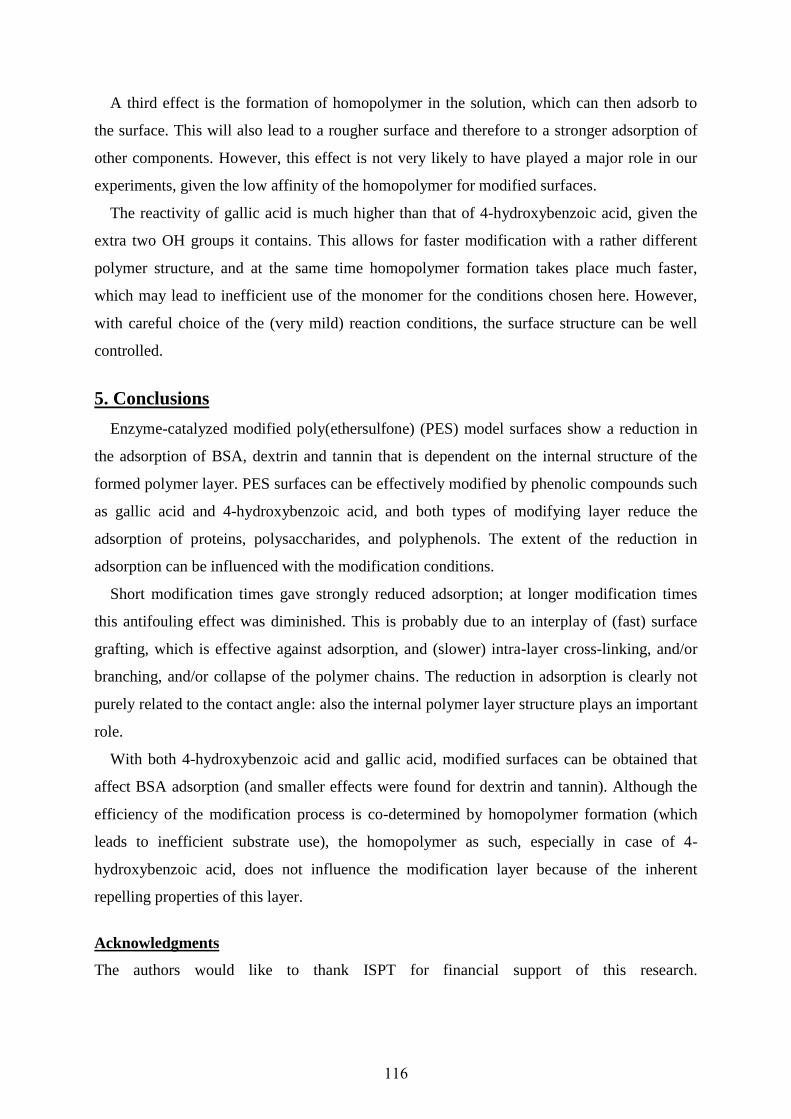
116
A third effect is the formation of homopolymer in the solution, which can then adsorb to
the surface. This will also lead to a rougher surface and therefore to a stronger adsorption of
other components. However, this effect is not very likely to have played a major role in our
experiments, given the low affinity of the homopolymer for modified surfaces.
The reactivity of gallic acid is much higher than that of 4-hydroxybenzoic acid, given the
extra two OH groups it contains. This allows for faster modification with a rather different
polymer structure, and at the same time homopolymer formation takes place much faster,
which may lead to inefficient use of the monomer for the conditions chosen here. However,
with careful choice of the (very mild) reaction conditions, the surface structure can be well
controlled.
5. Conclusions
Enzyme-catalyzed modified poly(ethersulfone) (PES) model surfaces show a reduction in
the adsorption of BSA, dextrin and tannin that is dependent on the internal structure of the
formed polymer layer. PES surfaces can be effectively modified by phenolic compounds such
as gallic acid and 4-hydroxybenzoic acid, and both types of modifying layer reduce the
adsorption of proteins, polysaccharides, and polyphenols. The extent of the reduction in
adsorption can be influenced with the modification conditions.
Short modification times gave strongly reduced adsorption; at longer modification times
this antifouling effect was diminished. This is probably due to an interplay of (fast) surface
grafting, which is effective against adsorption, and (slower) intra-layer cross-linking, and/or
branching, and/or collapse of the polymer chains. The reduction in adsorption is clearly not
purely related to the contact angle: also the internal polymer layer structure plays an important
role.
With both 4-hydroxybenzoic acid and gallic acid, modified surfaces can be obtained that
affect BSA adsorption (and smaller effects were found for dextrin and tannin). Although the
efficiency of the modification process is co-determined by homopolymer formation (which
leads to inefficient substrate use), the homopolymer as such, especially in case of 4-
hydroxybenzoic acid, does not influence the modification layer because of the inherent
repelling properties of this layer.
Acknowledgments
The authors would like to thank ISPT for financial support of this research.
Chapter Five…Adsorption on Modified Model PES Surfaces
117
References
1. Belfort, G.; Altena, F.W., Towards an Inductive Understanding of Membrane Fouling,
Desalination 47 (1983) 105-127.
2. Liao, B.Q.; Bagley, D.M.; Kraemer, H.E.; Leppard, G.G.; Liss, S.N., A Review of Biofouling and
Its Control in Membrane Separation Bioreactors, Water Environ. Res. 76 (2004) 425-436.
3. Norde, W., Adsorption of Proteins from Solution at the Solid-Liquid Interface, Adv. Colloid
Interface Sci. 25 (1986) 267-340.
4. Tsapikouni, T.S.; Missirlis, Y.F., Protein-Material Interactions: From Micro-to-Nano Scale, Mater.
Sci. Eng., B 152 (2008) 2-7.
5. Ulbricht, M., Advanced Functional Polymer Membranes, Polymer 47 (2006) 2217-2262.
6. Arkhangelsky, E.; Levitsky, I.; Gitis, V., Electrostatic Repulsion as a Mechanism in Fouling of
Ultrafiltration Membranes, Water Sci. Technol. 58 (2008) 1955-1965.
7. Arai, T.; Norde, W., The Behavior of some Model Proteins at Solid-Liquid Interfaces. 1.
Adsorption from Single Protein Solutions, Colloids Surf. 51 (1990) 1-15.
8. Le-Clech, P.; Marselina, Y.; Ye, Y.; Stuetz, R.A.; Chen, V., Visualization of Polysaccharide
Fouling on Microporous Membrane Using Different Characterization Techniques, J. Membr. Sci.
290 (2007) 36-45.
9. Susanto, H.; Franzka, S.; Ulbricht, M., Dextran Fouling of Polyethersulfone Ultrafiltration
Membranes - Causes, Extent and Consequences, J. Membr. Sci. 296 (2007) 147-155.
10. Susanto, H.; Feng, Y.; Ulbricht, M., Fouling Behavior of Aqueous Solutions of Polyphenolic
Compounds During Ultrafiltration, J. Food Eng. 91 (2009) 333-340.
11. Susanto, H.; Arafat, H.; Janssen, E.M.L.; Ulbricht, M., Ultrafiltration of Polysaccharide-Protein
Mixtures: Elucidation of Fouling Mechanisms and Fouling Control By Membrane Surface
Modification, Sep. Purif. Technol. 63 (2008) 558-565.
12. Zator, M.; Ferrando, M.; López, F.; Güell, C., Microfiltration of Protein/Dextran/Polyphenol
Solutions: Characterization of Fouling and Chemical Cleaning Efficiency Using Confocal
Microscopy, J. Membr. Sci. 344 (2009) 82-91.
13. Nady, N.; Franssen, M.C.R.; Zuilhof, H.; Mohy Eldin, M.S.; Boom, R.; Schroën, K., Modification
Methods for Poly(arylsulfone) Membranes: A Mini-Review Focusing on Surface Modification,
Desalination 275 (2011) 1-9.
14. Rana, D.; Matsuura, T., Surface Modifications for Antifouling Membranes, Chem. Rev. 110
(2010) 2448-2471.
15. Liu, S.X.; Kim, J.-T.; Kim, S., Effect of Polymer Surface Modification on Polymer-Protein
Interaction via Hydrophilic Polymer Grafting, J. Food Sci. 73 (2008) 143-150.
16. Schroën, C.G.P.H.; Roosjen, A.; Tang, K.; Norde, W.; Boom, R.M., In Situ Quantification of
Membrane Foulant Accumulation By Reflectometry, J. Membr. Sci. 362 (2010) 453-459.
17. Haynes, C.A.; Norde, W., Globular Proteins at Solid/Liquid Interface, Colloids Surf., B 2 (1994)
517-566.
18. Haynes, C.A.; Norde, W., Structures and Stabilities of Adsorbed Proteins, J. Colloid Interface Sci.
169 (1995) 313-328.
19. Lee, S.H.; Ruckenstein, E., Adsorption of Proteins onto Polymeric Surfaces of Different
Hydrophilicities - A Case Study with Bovine Serum Albumin, J. Colloid Interface Sci. 125 (1988)
365-379
20. Minko, S., Grafting on Solid Surfaces: “Grafting to” and “Grafting from” Methods, in: Manfred, S.
(Ed.), Polymer Surfaces and Interfaces: Characterization, Modification and Applications, E-
Publishing, Springerlink. com, Berlin/Heidelberg (2008) 215-234.
21. Schroën, C.G.P.H.; Cohen Stuart, M.A., van der Voort Maarschalk, K.; van der Padt, A.; van’t
Riet, K., Influence of Preadsorbed Block Copolymers on Protein Adsorption: Surface Properties,
Layer Thickness, and Surface Coverage, Langmuir 11 (1995) 3068-3074.
22. Currie, E.P.K.; Norde, W.; Cohen Stuart, M.A., Tethered Polymer Chains: Surface Chemistry and
their Impact on Colloidal and Surface Properties, Adv. Colloid Interface Sci. 100-102 (2003) 205-
265.
23. Nguyen, A.T.; Baggerman, J.;Paulusse, J.M.J.; van Rijn, C.J.M.; Zuilhof, H., Stable Protein-
Repellent Zwitterionic Polymer Brushes Grafted from Silicon Nitride, Langmuir 27 (2011) 2587-
2594.
118
24. Russo, C., A New Membrane Process for the Selective Fractionation and Total Recovery of
Polyphenols, Water and Organic Substances from Vegetation Waters (VW), J. Membr. Sci. 288
(2007) 239-246.
25. Nady, N.; Schroën, K.; Franssen, M.C.R.; van Lagen, B.; Murali, S.; Boom, R.M.; Mohy Eldin,
M.S.; Zuilhof, H., Mild and Highly Flexible Enzyme-Catalyzed Modification of Poly(ethersulfone)
Membranes, ACS Appl. Mater. Interfaces, 3 (2011) 801-810.
26. Nady, N.; Schroën, K.; Franssen, M.C.R.; Mohy Eldin, M.S.; Zuilhof, H.; Boom, R.M., Laccase-
Catalyzed Modification of PES Membranes with 4-Hydroxybenzoic Acid and Gallic Acid, J.
Membr. Sci. 394-395 (2012) 69-79 (Chapter 4).
27. Dijt, J.C.; Cohen Stuart, M.A.; Hofman, J.E.; Fleer, G.J., Kinetics of Polymer Adsorption in
Stagnation Point Flow, Colloids Surf. A. 51 (1990) 141-158.
28. Dijt, J.C.; Cohen Stuart, M.A.; Fleer, G.J., Reflectometry as a Tool for Adsorption Studies, Adv.
Colloid Interface Sci. 50 (1994) 79-101.
29. Böhmer, M.R.; van der Zeeuw, E.A.; Koper, G.J.M., Kinetics of Particle Adsorption in Stagnation
Point Flow Studied By Optical Reflectometry, J. Colloid Interface Sci. 197 (1998) 242-250.
30. Riquelme, B.D.; Valverde, J.R.; Rasia, R.J., Kinetic Study of Antibody Adhesion on a Silicon
Wafer By Laser Reflectometry, Optics and Lasers in Engineering 39 (2003) 589-598.
31. Bosker, W.T.E.; Iakovlev, P.A.; Norde, W.; Cohen Stuart, M.A., BSA Adsorption on Bimodal
PEO Brushes, J. Colloid Interface Sci. 286 (2005) 496-503.
32. Singh, G.; Capalash, N.; Goel, R.; Sharma, P.; A pH-Stable Laccase from Alkali-Tolerant γ-
proteobacterium JB: Purification, Characterization and Indigo Carmine Degradation, Enzyme
Microb. Technol. 41 (2007) 794-799.
33. Walsh, D.J.; Bhanu Singh, V., The Phase Behavior of a Poly(ethersulfone) with Poly(ethylene
oxide), Makromol. Chem. 185 (1984) 1979-1989.
34. Ball, V.; Ramsden, J.J., Buffer Dependence of Refractive Index Increments of Protein Solutions,
Biopolymers, 46 (1998) 489-492.
35. Rodriguez-Emmenegger, C.; Brynda, E.; Riedel, T.; Houska, M.; Šubr, V.; Alles, A.B.; Hasan, E.;
Gautrot, J.E.; Huck, W.T.S., Polymer Brushes Showing Non-Fouling in Blood Plasma Challenge
the Currently Accepted Design of Protein Resistant Surfaces, Macromol. Rapid Commun. 32
(2011) 952-957.

Chapter Five…Adsorption on Modified Model PES Surfaces
119
Appendix A. Modification conditions used in Tables 1 and 3; surfaces obtained after treatment
given in bold italic print are used as starting point for experiments explained in detail in the text.
Table A (1). Modification conditions used for 4-hydroxybenzoic acid.
Surface number
Concentration 4-hydroxybenzoic
acid (mM)
Concentration laccase (U·ml
-1)
Modification time (h)
Modification temperature
(°C)
pH
0 Blank 1 28.8 0.5 2 25 5 2 4.8 1.0 2 25 5 3 4.8 0.5 24 25 5 4 28.8 0.5 24 25 5 5 4.8 0.5 2 55 5 6 28.8 0.5 2 55 5 7 4.8 0.5 2 25 6
Table A (3). Modification conditions used for gallic acid.
Surface number
Concentration gallic acid
(mM)
Concentration laccase (U·ml
-1)
Modification time (h)
Modification temperature
(°C)
pH
0 Blank 1 4.8 0.5 30 25 5 2 4.8 0.5 120 25 5 3 28.8 0.5 120 25 5 4 4.8 0.75 120 25 5 5 28.8 0.5 120 55 5 6 28.8 0.5 120 25 6

120
Chapter Six
Listeria Repellence by Modified PES Surfaces
Part of this chapter will be published as:
Listeria monocytogenes Repellence by Enzyme-Catalyzed Modified PES Surfaces,
Norhan Nady, Stijn van der Veen, Karin Schroën, Maurice Franssen, Mohamed Mohy Eldin,
Tjakko Abee, Han Zuilhof, and Remko Boom.
Introduction
Materials and Methods
Results and Discussion
Conclusions
1
2
3
4

122
Listeria monocytogenes Repellence by Enzyme-Catalyzed
Modified PES Surfaces
Abstract
In this chapter, we evaluate the effect of the enzyme-catalyzed modification of
poly(ethersulfone) (PES) on the adhesion of Listeria monocytogenes, which is chosen as a
model bacterium. It is known to cause serious problems in both food industries and water
treatment.
PES was modified with 4-hydroxybenzoic acid and gallic acid, because the resulting layers
showed repellence of various foulants as described in the previous chapters. In addition, PES
was modified with ferulic acid, which in literature is claimed to have an antimicrobial effect.
Two strains of Listeria monocytogenes were applied: the commonly used EGD-e strain and
the well-known biofilm forming strain LR-991.
Listeria monocytogenes adhesion and biofilm growth was investigated both under static and
dynamic conditions. The surfaces modified with any of the three tested compounds repelled
Listeria adhesion up to 70% (static conditions) or 95% (dynamic conditions). Biofilm growth
was inhibited up to about 70%, depending on temperature (presence/absence of flagella), and
the conditions and substrate used for modification.

Chapter Six… Listeria Repellence by Modified PES Surfaces
123
1. Introduction
Listeria monocytogenes (L. monocytogenes) is a gram-positive bacterium. People infected
by this bacteria get a disease known as listeriosis [1,2], which can cause miscarriage, newborn
infection, dangerous illnesses such as meningitis and septicaemia, and even death. Listeria
species can attach to all kinds of surfaces including plastics, rubber, stainless steel, glass, etc.
[3,4]. This pathogen can survive and grow at a very wide temperature range (below freezing
point up to 46 °C), high salinity (grows up to 13% and remains alive up to 30%), and wide pH
range (below 5 and up to 9). The reason of survival and growth of these bacteria in such
severe conditions is attributed to their ability to adapt themselves to changing environments
[5,6].
L. monocytogenes is associated with foods like raw milk, cheese, ice cream, and raw and
smoked fish [7,8]. It also occurs in sea, sewage, and river water [9-13]. The factors that affect
adhesion of L. monocytogenes to surfaces are not completely understood yet. As influential
factors bacterial cell surface properties, the properties of the substratum (inert) surface, and
the local conditions have been mentioned [5,14]. Although in literature it has widely been
suggested that hydrophilic, negatively charged, and smooth surfaces are more effective in
reducing the initial adhesion of live cells [15], many researchers have reported that neither
initial adhesion of L. monocytogenes nor biofilm formation depend on the surface roughness
[16]. Also, the effect of surface hydrophilicity on attachment of Listeria on polymeric
surfaces is limited (less than one order of magnitude) [17]. On the other hand, it is known that
growth conditions may be of great influence on cell adhesion. For example, at temperatures
below 30 °C Listeria cells produce a flagellum, a tail-like projection that is used for
locomotion. The flagellum is rich in negatively charged proteins that strongly influence the
physicochemical (electrostatic) properties of the microbial cell surface and consequently its
adhesion to surfaces [18].
Poly(ethersulfone) (PES) membranes are widely used in the food industry and for water
treatment (separation and purification purposes). However, the drawback of this type of
membranes is the significant adhesion of proteins and living cells, with a severe reduction in
membrane performance (flux and selectivity) as a consequence. The resulting membrane
replacement that needs to take place regularly forms the largest operating cost in any
membrane separation process [19].
In our previous research [20], we showed that it is possible to covalently link phenolic
compounds such as 4-hydroxybenzoic acid and gallic acid via their oxygen atoms to PES
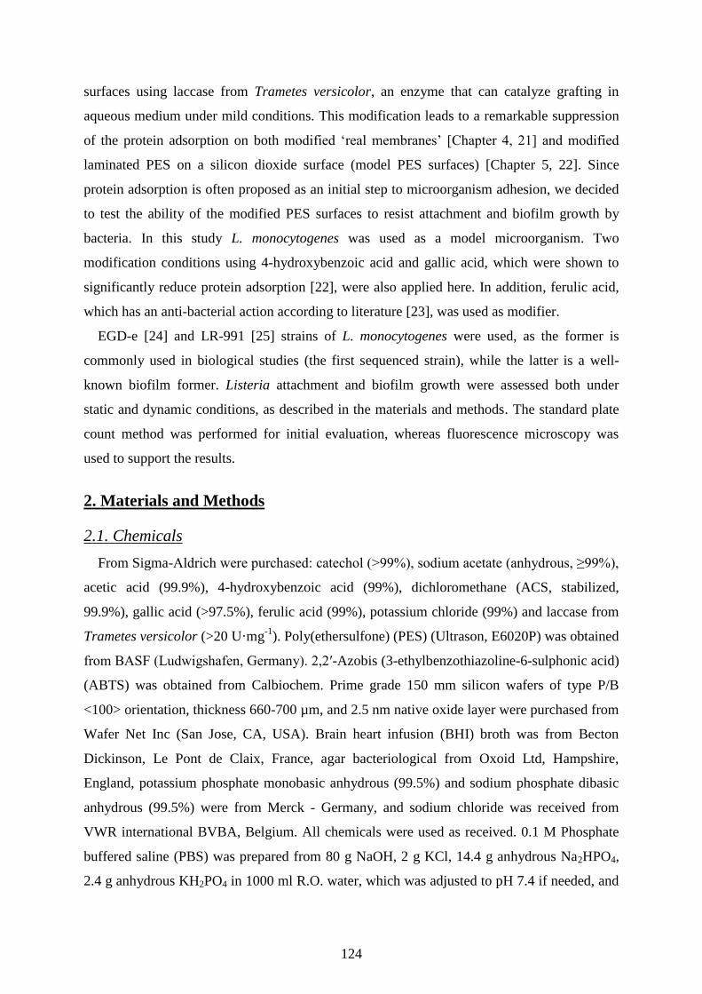
124
surfaces using laccase from Trametes versicolor, an enzyme that can catalyze grafting in
aqueous medium under mild conditions. This modification leads to a remarkable suppression
of the protein adsorption on both modified ‘real membranes’ [Chapter 4, 21] and modified
laminated PES on a silicon dioxide surface (model PES surfaces) [Chapter 5, 22]. Since
protein adsorption is often proposed as an initial step to microorganism adhesion, we decided
to test the ability of the modified PES surfaces to resist attachment and biofilm growth by
bacteria. In this study L. monocytogenes was used as a model microorganism. Two
modification conditions using 4-hydroxybenzoic acid and gallic acid, which were shown to
significantly reduce protein adsorption [22], were also applied here. In addition, ferulic acid,
which has an anti-bacterial action according to literature [23], was used as modifier.
EGD-e [24] and LR-991 [25] strains of L. monocytogenes were used, as the former is
commonly used in biological studies (the first sequenced strain), while the latter is a well-
known biofilm former. Listeria attachment and biofilm growth were assessed both under
static and dynamic conditions, as described in the materials and methods. The standard plate
count method was performed for initial evaluation, whereas fluorescence microscopy was
used to support the results.
2. Materials and Methods
2.1. Chemicals
From Sigma-Aldrich were purchased: catechol (>99%), sodium acetate (anhydrous, ≥99%),
acetic acid (99.9%), 4-hydroxybenzoic acid (99%), dichloromethane (ACS, stabilized,
99.9%), gallic acid (>97.5%), ferulic acid (99%), potassium chloride (99%) and laccase from
Trametes versicolor (>20 U·mg-1
). Poly(ethersulfone) (PES) (Ultrason, E6020P) was obtained
from BASF (Ludwigshafen, Germany). 2,2′-Azobis (3-ethylbenzothiazoline-6-sulphonic acid)
(ABTS) was obtained from Calbiochem. Prime grade 150 mm silicon wafers of type P/B
<100> orientation, thickness 660-700 µm, and 2.5 nm native oxide layer were purchased from
Wafer Net Inc (San Jose, CA, USA). Brain heart infusion (BHI) broth was from Becton
Dickinson, Le Pont de Claix, France, agar bacteriological from Oxoid Ltd, Hampshire,
England, potassium phosphate monobasic anhydrous (99.5%) and sodium phosphate dibasic
anhydrous (99.5%) were from Merck - Germany, and sodium chloride was received from
VWR international BVBA, Belgium. All chemicals were used as received. 0.1 M Phosphate
buffered saline (PBS) was prepared from 80 g NaOH, 2 g KCl, 14.4 g anhydrous Na2HPO4,
2.4 g anhydrous KH2PO4 in 1000 ml R.O. water, which was adjusted to pH 7.4 if needed, and

Chapter Six… Listeria Repellence by Modified PES Surfaces
125
autoclaved at 121 °C for 15 min. Milli-Q water was used throughout the preparation of the
model PES surfaces and sterilized reverse osmosis (R.O) water was used throughout the
biofouling tests.
2.2. Laccase Assay
Laccase activity was determined with 2,2′-azobis(3-ethylbenzothiazoline-6-sulphonic acid)
(ABTS) as substrate. The assay mixture contained 0.33 ml of 1 M ABTS solution, 2.67 ml of
0.1 M sodium acetate buffer (pH 5), with 0.05 U·ml-1
laccase. Oxidation of ABTS is
monitored by following the increase in absorbance at 420 nm (ε = 36,000 M-1
·cm-1
) [26]. The
reaction time taken is 1 min. One unit of laccase activity is defined as the amount of enzyme
required to oxidize 1 µmol of ABTS per min at 25 °C.
2.3. Preparation of Modified PES Surfaces
Silicon wafers (with approximately 70 nm silicon dioxide layer) were cut into strips of 1 ×
5 cm (static conditions) or 1.5 × 5.5 cm (dynamic conditions). The strips were sonicated in
ethanol for 15 min, washed with water and ethanol, and dried in flowing nitrogen.
Subsequently the strips were given a plasma treatment (PDC-32G, Harrick at RF-level high)
for 10 min. Immediately after plasma cleaning and removal of any dust by using a flow of
nitrogen, the strips were spin coated with 0.25 % w/w PES solution in dichloromethane for 10
s at 2500 rpm. The PES coated strips were put at 300 °C for 60 min. The spin-coated PES
surfaces were then incubated in 20 ml (1 × 5 cm strips) or 33 ml (1.5 × 5.5 cm strips) of 0.1 M
sodium acetate buffer containing different concentrations of phenolic acids
(substrates/monomers) and laccase. A flow of air was used for mixing and as oxygen source.
4-hydroxybenzoic acid (28 mM, 2 h modification), ferulic acid (4.8 mM, 1 h and 24 h
modification), and gallic acid (4.8 mM, 7 min modification) were used as enzyme substrates
[20-22]. Common modification conditions were 0.5 U·ml-1
enzyme concentration,
temperature 23 ± 2 ºC, pH 5 in 0.1 M sodium acetate buffer. After completing the reaction,
the modified surfaces were removed from the liquid and washed by strong flushing with
Milli-Q water. The modified PES surfaces were kept 24 h in glass-covered dishes in
desiccators supplied with silica gel for drying.
2.4. L. monocytogenes Strains and Culture Conditions
L. monocytogenes strains EGD-e and LR-991 [24,25] were stored in brain heart infusion
(BHI) broth (Becton Dickinson, Le Pont de Claix, France) containing 15% (v/v) sterile

126
glycerol (Fluka, Buchs, Switzerland) at -80 °C. Single colonies were inoculated in 10 ml BHI
in 50 ml tubes (Greiner Bio-One, Frickenhausen, Germany) and were grown overnight (static
incubation) at 30 °C or 37 °C.
Overnight-grown cultures were used to inoculate (1%) 25 ml BHI in 50 ml tubes and
incubated for 24 h at 30 °C or 37 °C. Cells were harvested by centrifugation at 5000 rpm for
15 min at 20 °C and subsequently re-suspended in 1 ml PBS (attachment) or BHI (biofilm
formation).
2.5. L. monocytogenes Attachment and Biofilm Formation
2.5.1. Static Conditions
To determine attachment of bacteria, modified PES slides were immersed in 20 ml PBS in
petri dishes and the bacterial suspension was added (approximately 109 total cfu). The PES
slides were incubated for 2.5 h at room temperature (23 ± 2 °C). After washing twice in PBS,
the adhered bacteria were collected using a sterile cotton swab. The swab was then placed in 1
ml PBS and vigorously vortexed. The suspended bacteria were serially diluted in PBS and
plated. The plates were incubated at 30 °C or 37 °C for 48 h colonies were enumerated.
Experiments were performed in duplicate in two biological independent replicates.
Biofilm formation on the modified PES slides was determined following a similar
procedure, with the exception that PES slides immersed in BHI and the slides were incubated
for 24 h.
2.5.2. Dynamic Conditions
Bacterial attachment under dynamic conditions was tested using a flow cell. A 1.5 x 5.5 cm
size slide was loaded on the sample support inside the flow cell (sample support size: 1.6 cm
width x 5.7 cm length x 1 mm depth). The bacterial suspension was diluted in 500 ml PBS
Prior to each experiment (approximately 109 total cfu), the whole system (connection tubes
and the flow cell) was washed with PBS for 10 minutes and subsequently filled with the
bacterial suspension. The bacterial suspension was circulated (0.038 m·s-1
- Reynolds number
is 38 in case of water at 20 °C) through the system for 2.5 h. The PES slide was recovered and
bacteria were harvested and enumerated as described for static conditions. Experiments were
performed in three independent biological replicates.

Chapter Six… Listeria Repellence by Modified PES Surfaces
127
2.6. Fluorescence Microscopy
Fluorescence microscopy experiments were performed on a BX41 microscope (Olympus,
Zoeterwoude, the Netherlands). Images were acquired using a XC30 camera (Olympus) and
Olympus Cell^B software. After washing the modified PES slides twice with PBS, it was
placed on a microscope slide (76 x 26 mm), and a square cover glass (18 mm) was placed on
top of the sample. Green fluorescent protein (GFP) was visualized using a MNIBA3 filter
(Olympus).
Statistics. Microsoft Excel was used to apply t-test on the obtained data; differences were
considered to be statistically significant at P < 0.05.
3. Results and Discussion
The overall purpose of this study was to examine the effect of laccase-catalyzed
modification of PES surfaces on the adhesion and biofilm formation of L. monocytogenes
under both static conditions and dynamic conditions. The obtained results were presented in
two ways: the number of bacteria cells per unit area (in log scale) and as percentage of
reduction relative to the blank (unmodified) PES surface.
3.1. Static Conditions
Under static conditions, the ability of modified PES surfaces to resist the attachment of L.
monocytogenes cells from EGD-e and LR-991 strains grown at 30 °C (i.e. flagella are present)
was determined and depicted in Figure 1 (white bars; P < 0.05 for all samples). The results
show approximately 60% reduction in the number of attached Listeria cells from both strains
relative to the blank surface. The high reduction found on PES surfaces modified by 4-
hydroxybenzoic acid (63%, P < 0.004) could be attributed to the brush-like structure of the
modification layer [22]. Possibly the effect is similar as demonstrated in other studies [27-29],
where the brush-like structure of the modifying layer on different surfaces e.g. stainless steel,
glass, polyamide and polyester also reduced bacterial adhesion. The effectiveness of these
structures against bacterial attachment is caused by steric hindrance that keeps the bacterial
cells at a distance from the surface, which results in weakening of the (e.g., van der Waals)
interactions [29].

128
Figure 1. Effect of PES modification on cell adhesion (white bars) and biofilm formation (grey
bars) under static conditions, shown as the number of L. monocytogenes cells adhering per unit
area, and as the percentage of reduction relative to the blank surface. Error bars represent the
standard deviation (St.d.) over four model surfaces, using two separate bacterial cultures.
6.0
6.4
6.8
7.2
7.6
8.0
8.4
1 2 3 4 5
Log (
cfu
.cm
-2)
Model Surface No.
1. Blank
2. Modified by 4-hydroxybenzoic acid (28.8 mM + 2 h modification)
3. Modified by ferulic acid (4.8 mM + 1 h modification)
4. Modified by ferulic acid (4.8 mM + 24 h modification)
5. Modified by gallic acid (4.8 mM + 7 min modification)
62.8 (5.4)
47.4 (5.2)57.2 (5.3)
63.3 (2.4)
68.2 (10.8)
45.9 (5.3)58.2 (6.2)
58.2 (3.0)
% Reduction relative to the blank (St.d.)
Bacteria attachment
Biofilm growth
Experimental Conditions: 0.5 U·ml-1 enzyme, 25 C, and pH 5 [ 0.1 M sodium acetate buffer]
EGD-e Strain
6.0
6.5
7.0
7.5
8.0
8.5
9.0
9.5
1 2 3 4 5
Log (
cfu
.cm
-2)
71.6 (9.6)
19.5 (4.8)58.7 (6.0)
62.7 (2.0)
57.7 (13.4)
40.1 (16.6)58.7 (3.6)
63.5 (0.8)
% Reduction relative to the blank (St.d.)
Bacteria attachment
Biofilm growth
LR-991 Strain
Model Surface No.

Chapter Six… Listeria Repellence by Modified PES Surfaces
129
This interpretation is also plausible for gallic acid; at the chosen reaction conditions, a
brush-like structure is formed [21,22]. PES surfaces modified with ferulic acid also show
inhibition of bacterial adhesion. However, the structure of the layer is still not known and is in
need of further investigation. Possibly at short reaction time, a brush type layer is formed that
later may cross-link as was the case for gallic acid.
Also under static conditions, biofilm formation of L. monocytogenes EGD-e and LR-991
strains grown at 30 °C (flagella are present) on modified surfaces was determined (Figure 1,
grey bars; P < 0.05 for all samples except the modification using 4-hydroxybenzoic acid with
LR-991 strain). The PES surfaces modified with ferulic acid and gallic acid exhibit
percentages of reduction close to their percentages of reduction in the attachment test (~60%).
This is may be attributed to the same phenomena (steric hindrance) to be involved in both cell
adhesion and biofilm formation, however the biofilm formation by the LR-991 strain was
hardly affected by modification of PES with 4-hydroxybenzoic acid (19.5%; P >> 0.05).
Changing the time that PES surfaces were modified with 4-hydroxybenzoic acid has an
effect on the inhibition of biofilm formation for both strains (static conditions), as is
visualized in Figure 2.
Figure 2. Effect of the modification time of PES surfaces modified with 4-hydroxybenzoic acid
on Listeria biofilm formation. The number of L. monocytogenes cells adhering per unit area and
the percentage of reduction relative to the blank surface were determined using static test
conditions. White bars: EGD-e strain, grey bars: LR-991 strain. Error bars represent the standard
deviation (St.d.) over four model surfaces, using two separate bacterial cultures.
6.0
6.4
6.8
7.2
7.6
8.0
8.4
8.8
0 0.25 0.5 1 2 24
Lo
g (
cfu
.cm
-2)
Modification Time (h)
73.1 (1.9)
44.7 (4.8)53.4 (4.1)
42.6 (5.2)51.3 (7.8)
37.0 (4.7)52.9 (6.2)
26.3. (7.2)
% Reduction relative to the blank (St.d.)
EGD-e strain
LR-991 strain33.0 (12)
38.1 (6.2)

130
It seems that the percentage of reduction was highest at short modification times. This
indicates that the formed structure needs to be accurately tuned to the properties of the
bacterial interaction to optimize the antimicrobial effect, similarly as was observed for protein
adsorption (see chapter 4 and 5). While this aspect is currently still poorly understood, it
displays significant potential to be used effectively against biofilm formation.
From a microbiological point of view, it seems there is no significant difference between
the used substrates: all the modified surfaces resist the attachment of bacteria cells and
consequently the biofilm formation to some extent. This reduction is not large enough for
pathogens because a decimal reduction of at least 2 (ideally 4) is needed here. However, our
results may be acceptable for non-pathogenic strains of Listeria or other bacteria.
3.2. Dynamic Conditions
The effect of flowing L. monocytogenes suspension in PBS over the model surfaces
(dynamic conditions, only bacterial attachment was tested) at 30 and 37 °C was studied for
both strains; the results are depicted in Figure 3. The data are the output of three surface sets
using three different cultures prepared on three different days. Modification of the surfaces
led to a reduction of the adhered cells compared to the static measurements, especially at a
growth temperature of 30 °C, at which the cells have flagella. The presence of flagella may
assist in electrostatic repulsion between the cells and the modification layers, because both of
them carry the same groups (COO-). At 37 °C, the cells do not possess the negatively charged
flagella so inhibition of adhesion is less prominent, but still significant. It seems that if
bacteria are not immediately attached to the surface, they can be carried away from the
surface by the cross-flowing liquid, possibly as a consequence of lift forces that may even
prevent them from coming into frequent contact with the surface [30]. However, at the blank
surface, considerable amounts of bacterial cells are present, indicating that the modification
layer has a significant role in the reduction. This could be a direct effect on prevention of
adhesion, or in a delay of adhesion (less fast) that allows lift forces to remove loosely adhered
bacteria.

Chapter Six… Listeria Repellence by Modified PES Surfaces
131
Figure 3. The number of L. monocytogenes cells adhering per unit area and percentage of
reduction relative to the blank surface was investigated using dynamic testing conditions at 30
(white bars, with flagella) and 37 °C (grey bars, without flagella). Error bars represent the
average standard deviation (St.d.) of three surfaces from three separate cultures.
3.3. Fluorescence Imaging
Fluorescence images for two modified surfaces are shown in Figure 4. Although these
images are to be treated with care, since they can give a false impression of the actual effect
Log (
cfu
.cm
-2)
Model Surface No.
1. Blank
2. Modified by 4-hydroxybenzoic acid (28.8 mM + 2 h modification)
3. Modified by ferulic acid (4.8 mM + 1 h modification)
4. Modified by gallic acid (4.8 mM + 7 min modification)
93.2 (0.2)
89.1 (2.8)91.6 (1.2)
82.5 (3.5)
95.2 (0.8)
85.4 (2.2)
% Reduction relative to the blank (St.d.)
At 30 C
At 37 C
EGD-e Strain
Log (
cfu
.cm
-2)
LR-991 Strain
4.5
5.0
5.5
6.0
6.5
7.0
7.5
1 2 3 4
88.3 (1.7)
78.2 (2.7)86.3 (0.9)
72.3 (3.8)
71.4 (2.6)
75.6 (3.5)
% Reduction relative to the blank (St.d.)
At 30 C
At 37 C
4.5
5.0
5.5
6.0
6.5
7.0
7.5
1 2 3 4
Model Surface No.
Experimental Conditions: 0.5 U·ml-1 enzyme, 25 C, and pH 5 [ 0.1 M sodium acetate buffer]
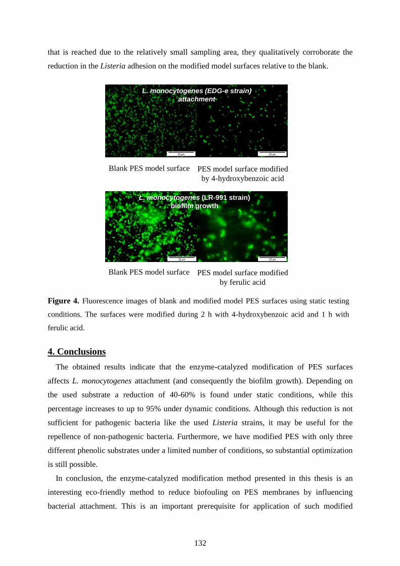
132
that is reached due to the relatively small sampling area, they qualitatively corroborate the
reduction in the Listeria adhesion on the modified model surfaces relative to the blank.
Figure 4. Fluorescence images of blank and modified model PES surfaces using static testing
conditions. The surfaces were modified during 2 h with 4-hydroxybenzoic acid and 1 h with
ferulic acid.
4. Conclusions
The obtained results indicate that the enzyme-catalyzed modification of PES surfaces
affects L. monocytogenes attachment (and consequently the biofilm growth). Depending on
the used substrate a reduction of 40-60% is found under static conditions, while this
percentage increases to up to 95% under dynamic conditions. Although this reduction is not
sufficient for pathogenic bacteria like the used Listeria strains, it may be useful for the
repellence of non-pathogenic bacteria. Furthermore, we have modified PES with only three
different phenolic substrates under a limited number of conditions, so substantial optimization
is still possible.
In conclusion, the enzyme-catalyzed modification method presented in this thesis is an
interesting eco-friendly method to reduce biofouling on PES membranes by influencing
bacterial attachment. This is an important prerequisite for application of such modified
Blank PES model surface PES model surface modified
by 4-hydroxybenzoic acid
Blank PES model surface
L. monocytogenes (EDG-e strain)
attachment
PES model surface modified
by ferulic acid
L. monocytogenes (LR-991 strain)
biofilm growth
Chapter Six… Listeria Repellence by Modified PES Surfaces
133
membranes in water treatment as well as in food processing, because the anti-biofouling
effects can strongly reduce the replacement costs that are currently a major hurdle.

134
References
1. Gray, M.L.; Killinger, A.H., Listeria monocytogenes and Listeria Infections, Bacteriol. Rev. 30
(1966) 309-382.
2. Doorduyn, Y.; de Jager, C.M.; van der Zwaluw, W.K.; Wannet, W.J.B.; van der Ende, A.;
Spanjaard, L.; van Duynhoven, Y.T.H.P., Invasive Listeria monocytogenes Infections in the
Netherlands, 1995-2003, Eur. J. Clin. Microbiol. Infect. Dis. 25 (2006) 433-442.
3. Briandat, R.; Leriche, V.; Carpentier, B.; Bellon-Fontaine, M.N, Effect of the Growth Procedure
on the Surface Hydrophilicity of Listeria monocytogenes Cells and their Adhesion to Stainless
Steel, J. Food Protect. 62 (1999) 994-998.
4. Chae, M.S.; Scharft, H.; Hansen, L.T.; Mackereth, R., Effect of Physicochemical Surface
Characteristics of Listeria monocytogenes Strains on Attachment to Glass, Food Microbiol. 23
(2006) 250-259.
5. Silva, S.; Teixeira, P.; Oliveria, R.; Azeredo, J., Adhesion to and Viability of Listeria monocytogenes on Food Contact Surfaces, J. Food Protect. 71 (2008) 1379-1385.
6. Bereksi, N.; Gavini, F.; Bénézech, T.; Faille, C., Growth, Morphology and Surface Properties of
Listeria monocytogenes Scott A and LO28 under Saline and Acid Environments, J. Appl.
Microbiol. 92 (2002) 556-565.
7. Lovett, J.; Francis, D.W.; Hunt, J.M., Listeria monocytogenes in Raw Milk: Detection, Incidence,
and Pathogenicity, J. Food Protect. 50 (1987) 188-192.
8. Bhagwat, A.A.; Saftner, R.A.; Abbott, J.A., Evaluation of Wash Treatments for Survival of
Foodborne Pathogens and Maintenance of Quality Characteristics of Fresh-Cut Apple Slices, Food
Microbiol. 21 (2004) 319-326.
9. Watkins, J.; Sleath, K.P., Isolation and Enumeration of Listeria monocytogenes from Sewage,
Sewage Sludge and River Water, J. Appl. Bacteriol. 50 (1981) 1-9.
10. El-Shenawy, M.A.; El-Shenawy, M.A, Listeria spp. in the Coastal Environment of the Aqaba Gulf,
Suez Gulf and Red Sea, Epidemiol. Infect. 134 (2006) 752-757.
11. El-Taweel, G.E.; Shaban, A.M., Microbiological Quality of Drinking Water at Eight Water
Treatment Plants, Int. J. Environ. Health Res. 11 (2001) 285-290.
12. Bremer, P.J.; Osborne, C.M.; Kemp, R.A.; Smith, J.J., Survival of Listeria monocytogenes in Sea
Water and Effect of Exposure on Thermal Resistance, J. Appl. Microbiol. 85 (1998) 545-554.
13. El-Shenawy, M.A.; El-Shenawy, M.A, Listeria species in Some Aquatic Environments in
Alexandria, Egypt, Int. J. Environ. Health Res. 6 (1996) 131-140.
14. Dickson, J.S.; Koohmarie, M., Cell Surface Charge Characteristics and their Relationship to
Bacterial Attachment to Meat Surfaces, Appl. Environ. Microbiol. 55 (1989) 832-836.
15. Kang, S.; Hoek, E.M.V.; Choi, H.; Shin, H., Effect of Membrane Surface Properties During the
Fast Evaluation of Cell Attachment, Sep. Sci. Technol. 41 (2006) 1475-1487.
16. Rodriguez, A.; Autio, W.R.; McLandsborough, L.A., Effect of Surface Roughness and Stainless
Steel Finish on Listeria monocytogenes Attachment and Biofilm Formation, J. Food Protect. 71
(2008) 170-175.
17. Cunliffe, D.; Smart, C.A.; Tsibouklis, J.; Young, S.; Alexandria, C.; Vulfson, E.N., Bacterial
Adsorption to Thermoresponsive Polymer Surfaces, Biotechnol. Lett. 22 (2000) 141-145.
18. Briandet, R.; Meylheuc, T.; Maher, C.; Bellon-Fontaine, M.N, Listeria monocytogenes Scott A:
Cell Surface Charge, Hydrophobicity, and Electron Donor and Acceptor Characteristics under
Different Environmental Growth Conditions, Appl. Environ. Microb. 65 (1999) 5328-5333.
19. Mansouri, J.; Harrisson, S.; Chen, V., Strategies for Controlling Biofouling in Membrane Filtration
Systems: Challenges and Opportunities, J. Mater. Chem. 20 (2010) 4567-4586.
20. Nady, N.; Schroën, K.; Franssen, M.C.R.; van Lagen, B.; Murali, S.; Boom, R.M.; Mohy Eldin,
M.S.; Zuilhof, H., Mild and Highly Flexible Enzyme-Catalyzed Modification of Poly(ethersulfone)
Membranes, ACS Appl. Mater. Interfaces 3 (2011) 801-810.
21. Nady, N.; Schroën, K.; Franssen, M.C.R.; Mohy Eldin, M.S.; Boom, R.M.; Zuilhof, H., Laccase-
Catalyzed Modification of PES Membranes with 4-Hydroxybenzoic Acid and Gallic Acid, J.
Membr. Sci. 394-395 (2012) 69-79 (Chapter Four).
22. Nady, N.; Schroën, K.; Franssen, M.C.R.; Fokkink, R.; MohyEldin, M.S.; Zuilhof, H.; Boom,
R.M., Enzyme-Catalyzed Modification of PES Surfaces: Reduction in Adsorption of BSA, Dextrin
and Tannin, to be submitted (Chapter Five).

Chapter Six… Listeria Repellence by Modified PES Surfaces
135
23. Schroeder, M.; Aichernig, N.; Guebitz, G.M.; Kokol, V., Enzymatic Coating of Lignocellulosic
Surfaces with Polyphenols, Biotechnol. J. 2 (2007) 334-341.
24. Glaser, P.; Frangeul, L.; Buchrieser, C.; Rusniok, C.; Amend, A.; Baquero, F.; Berche, P.;
Bloecker, H.; Brandt, P.; Chakraborty, T.; Charbit, A.; Chetouani, F.; Couvé, E.; de Daruvar, A.;
Dehoux, P.; Domann, E.; Domínguez-Bernal, G.; Duchaud, E.; Durant, L.; Dussurget, O.; Entian,
K.-D.; Fsihi, H.; Garcia-Del Portillo, F.; Garrido, P.; Gautier, L.; Goebel, W.; Gómez-López, N.;
Hain, T.; Hauf, J.; Jackson, D.; Jones, L.-M.; Kaerst, U.; Kreft, J.; Kuhn, M.; Kunst, F.; Kurapkat,
G.; Madueňo, E.; Maitournam, A.; Mata Vicente, J.; Ng, E.; Nedjari, H.;Nordsiek, G.; Novella, S.;
de Pablos, B.; Pérez-Diaz, J.-C.; Purcell, R.; Remmel, B.; Rose, M; Schlueter, T.; Simoes, N.;
Tierrez, A.; Váquez-Boland, J.-A.; Voss, H.; Wehland, J.; Cossart, P., Comparative Genomics of
Listeria Species, Science 294 (2001) 849-852.
25. van der Veen, S.; Moezelaar, R.; Abee, T.; Wells-Bennik, M.H., The Growth Limits of a Large
Number of Listeria monocytogenes Strains at Combinations of Stresses Show Serotype- and
Niche-Specific Traits. J. Appl. Microbiol. 105 (2008) 1246-1258.
26. Wolfenden, B.S.; Willson, R.L., Radical-cations as Reference Chromogens in Kinetic Studies of
One-electron Transfer Reactions: Pulse Radiolysis Studies of 2,2′-azinobis-(3-
ethylbenzthiazoline-6-sulphonate), J. Chem. Soc., Perkin Trans. 2 (1982) 805-812.
27. Dong, B.; Jiang, H.; Manolache, S.; Wong, A.C.L.; Denes, F.S., Plasma-Mediated Grafting of
Poly(ethylene glycol) on Polyamide and Polyester Surfaces and Evaluation of Antifouling Ability
of Modified Substrates, Langmuir 23 (2007) 7306-7313.
28. Dong, B.; Manolache, S.; Somers, E.B.; Wong, A.C.L.; Denes, F.S., Generation of Antifouling
Layers on Stainless Steel Surfaces by Plasma-Enhanced Crosslinking of Polyethylene Glycol, J.
Appl. Polym. Sci. 97 (2005) 485-497.
29. Roosjen, A.; Norde, W.; Van der Mei, H.C.; Busscher, H.J., The Use of Positively Charged or Low
Surface Free Energy Coatings versus Polymer Brushes in Controlling Biofilm Formation, Progr.
Colloid Polym. Sci. 132 (2006) 138-144.
30. Dinther, A.M.C. van ; Schroën, C.G.P.H.; Boom, R.M., High-Flux Membrane Separation Using
Fluid Skimming Dominated Convective Fluid Flow, J. Membr. Sci. 371 (2011) 20-27.

136

Chapter Seven
General Discussion and Outlook
Current Status
Versatility and Future Developments
Practical Aspects
1
2
3
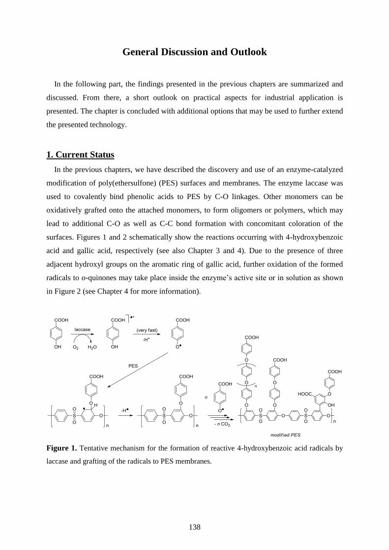
138
General Discussion and Outlook
In the following part, the findings presented in the previous chapters are summarized and
discussed. From there, a short outlook on practical aspects for industrial application is
presented. The chapter is concluded with additional options that may be used to further extend
the presented technology.
1. Current Status
In the previous chapters, we have described the discovery and use of an enzyme-catalyzed
modification of poly(ethersulfone) (PES) surfaces and membranes. The enzyme laccase was
used to covalently bind phenolic acids to PES by C-O linkages. Other monomers can be
oxidatively grafted onto the attached monomers, to form oligomers or polymers, which may
lead to additional C-O as well as C-C bond formation with concomitant coloration of the
surfaces. Figures 1 and 2 schematically show the reactions occurring with 4-hydroxybenzoic
acid and gallic acid, respectively (see also Chapter 3 and 4). Due to the presence of three
adjacent hydroxyl groups on the aromatic ring of gallic acid, further oxidation of the formed
radicals to o-quinones may take place inside the enzyme’s active site or in solution as shown
in Figure 2 (see Chapter 4 for more information).
Figure 1. Tentative mechanism for the formation of reactive 4-hydroxybenzoic acid radicals by
laccase and grafting of the radicals to PES membranes.

Chapter Seven…General Discussion and Outlook
139
Figure 2. Tentative mechanism for the laccase-mediated formation of an o-quinone from gallic
acid, and its reaction with gallic acid (derivatives) in solution or with the PES membrane.
The layer structure can be tuned by the modification conditions and the choice of substrate.
In general, substrates with more reactive groups (like gallic acid) will lead to denser 3D
networks, while molecules with only one hydroxyl group give linear or branched structures,
which swell and extend in water to give rise to entropic repulsion as shown in Figure 3 for 4-
hydroxybenzoic (4-HBA) and gallic acid (Chapter 4). Besides the surface reactions,
monomers can take part in oxidative grafting to other monomers or oligomers to form
homopolymers in solution that can adsorb to the membrane surface. This homopolymer is not
desirable, because it leads to inefficient substrate use. Furthermore, the short chains of this
homopolymer may physisorb to the surface, and if desorption takes place during the filtration
process, the permeate and/or the retentate may be contaminated. However, good washing by
e.g. a water flux after modification should be sufficient to remove the (weakly adsorbed)
homopolymers.
The various structures that can be formed during laccase-catalyzed modification of PES
surfaces have very different effects on the adhesion of model foulants such as proteins,
polysaccharides and polyphenols (Chapter 5), and Listeria monocytogenes (Chapter 6). It was
shown that surface hydrophilicity is not the only factor responsible for (prevention of)
adhesion.
The average flux of the base membrane was never reduced more than 20% (mostly below
10%) in case of modification with 4-hydroxybenzoic acid, and less than 9% for gallic acid,

140
while the mechanical properties and thermal stability of the membrane were not adversely
affected (Chapter 4).
Figure 3. Schematic representation of modification layers formed by 4-hydroxybenzoic acid (4-
HBA) and gallic acid. Black line: OH group or ether linkage; green line: C-C bond; red line:
COOH group.
In conclusion, the laccase-catalyzed modification of PES membranes (surfaces) is a method
that can be applied to flexibly create surface structures that can be used to effectively prevent
(or possibly facilitate as will be discussed later) attachment of various components to the
modified surface. The method can be carried out under very mild reaction conditions, and
when applied to PES membranes the resulting membranes retain a high flux. Modified PES
membranes possess mechanical properties and thermal resistances that are similar or superior
to those of the base membrane, as was illustrated in Chapter 4. All these aspects make the
presented method an interesting alternative for currently used modification methods, as was
illustrated in Chapter 2.
2. Practical Aspects
In this section, practical aspects needed to bring the developed method to large-scale
application are discussed, starting with the first results on hollow fibre membranes, which are
presented together with an assessment of the costs involved in the method, and a list of other
aspects.
The first results for hollow fibres were obtained with single high flush fibre modules which
were kindly provided by Pentair X-flow (0.79 mm inner diameter and pore size that is

Chapter Seven…General Discussion and Outlook
141
expected to be around 10 nanometers). The modification procedure was as follows. First,
Milli-Q water was pressurized through the membrane for 20 min (dead-end), and the flux was
measured three times at 4 bar. Then, 10 ml of 28.8 mM 4-hydroxybenzoic acid (pH 5, 24 ± 1
°C) was rinsed through the fibre by gravity (4 cm height difference between both ends of the
module; outlet flow ~1 ml·min-1
). Next, 20 ml of fresh well-mixed 4-hydroxybenzoic acid and
laccase (28.8 mM and 0.5 U·ml-1
, respectively) were rinsed through the module also by
gravity. Freshly mixed reactants replaced the previous mixture every 15 min during a total
modification time of 2 h. The modified membrane was cleaned by forward and backward
washing with Milli-Q water (as described in Chapter 4) at 4 bar and the flux was measured.
Next, 1 g·l-1
BSA was pressurized at 4 bar (pH 7, 0.1 M sodium acetate buffer, 24 ± 1 °C)
through the membrane in dead-end mode and both flux (after washing with Milli-Q) and
concentration of BSA in the outlet were determined. The same procedure was applied for
blank membranes (unmodified membranes).
Table 1. Specification and performance of the blank and modified high flush surface hollow
single fibre PES lab modules.
Blank module
no.1
Blank module
no.2
Modified module
no.1
Modified module
no.2
Length (cm) 23.7 24 23.6 24
Area (cm
2)
5.88 5.96 5.86 5.96
Flux (m
3·m
-2·h
-1)
0.3 ± 0.01 0.28 ± 0.01 0.32 ± 0.01 0.33 ± 0.01
Flux after modification (m
3·m
-2·h
-1)
0.072 ± 0.001 0.1 ± 0.001
Flux during BSA filtration
(m3·m
-2·h
-1)
0.04 ± 0.001 0.039 ± 0.001 0.043 ± 0.001 0.046 ± 0.001
BSA rejection (%) 99 ± 0.8 99.1 ± 0.8 50.7 ± 0.9 50 ± 2.5
Flux after BSA adsorption
(m3·m
-2·h
-1)
0.04 ± 0.001 0.04 ± 0.001 0.043 ±0.001 0.043 ± 0.001
Table 1 shows an overview of the results. The flux of modified membranes is typically 20-
30% of that of the blank membrane, probably due to the presence of the relatively large
modification layers compared to the pore size (in previous chapters larger pores were used:
0.2 µm). Upon exposure to BSA, the flux of the blank membrane was reduced to

142
approximately 12% of its original value due to pore blocking by BSA. For the modified
membrane, slightly higher fluxes were found; obviously the relative flux decrease due to BSA
was much less for these membranes. The most striking difference between the membranes is
in BSA rejection, which was around 50% for modified membranes where it was almost 100%
for the blank (unmodified) PES membrane. For unmodified membranes, accumulation and
aggregation of protein will take place, which reduces the pore size and increases rejection.
The lower rejection of the modified membranes is expected to be a result of reduced protein
adsorption on/into the membrane pores (schematically illustrated in Figure 4), which allows
passage of the protein through the membrane without sticking onto the pore mouth or surface.
In this it is noteworthy that protein rejection of the membrane surfaces is different from
protein rejection by the membrane itself: a perfectly protein-repellent surface will correlate
with a completely non-rejecting membrane if the pore size allows unhindered passage of the
protein. In this case, although the pore size of around 10 nm may be reduced (as reflected in
flux reduction) upon modification by growth of a brush-like layer of around 3 nm on each
side, the size of the modified pore may still be sufficient for passage of an individual protein
molecule (BSA size is around 3-4 nm) [1]. This phenomenon opens an opportunity for size-
based separation of the same protein type.
Figure 4. Schematic representation of the effect of modification of PES membrane pores on
rejection of proteins.
For industrial application, an assessment of the costs involved in the enzyme-catalyzed
modification method is needed. Both the enzyme and the used substrates are available
commercially and not expensive, and it is possible to recycle the enzyme, substrate, and
buffer. Table 2 shows an indication for the price of modification of one square meter of
membrane with 4-hydroxybenzoic acid (28.8 mM modifier, 0.5 U·ml-1
enzyme) according to
prices in the Sigma-Aldrich catalogue 2011. Based on the prices of the largest packages
Modified
pore
Unmodified
pore
BSA solution

Chapter Seven…General Discussion and Outlook
143
available in the catalogue, the cost of modification chemicals is 120-130 euro per m2.
However, for larger scale production this price can likely be significantly reduced to a
calculated cost of approximately 10 euro·m-2
when applied on industrial scale.
From the list it is clear that the enzyme cost is prominent, and when produced at large scale
this cost will be reduced considerably too. For a commercial enzyme that is e.g. used in
washing powder, a typical price would be 100 euro per kg of crude enzyme. The activity of
such enzyme will be less as the one we used in our experiments, but even if the enzyme
activity is a factor of 10 lower, the price for modification would still reduce with
approximately a factor of 50. Alternatively the reaction could also be carried out at lower
enzyme concentration, although this would require longer reaction times, which typically
increases the overall costs.
Table 2. Price indication for modification of one m2 of poly(ethersulfone) membrane.
Material
Sigma-Aldrich 2011
Used amount/0.002 m2
membrane.
Cost/m2 membrane
(euro/m2)
Sodium acetate 43.1 euro/1 kg 0.11 g 2.4
Acetic acid 13.5 euro/1 l 0.04 ml 0.3
Enzyme 38.6 euro/1 g 6 mg 115.4
4-hydroxybenzoic acid 48 euro/1 kg 0.2 g 4.8
Total cost/m2
122
On industrial scale the price can be significantly reduced, leading to a price
per m2 of:
*The costs related to oxygen needed for modification are negligible.
10
Besides the direct costs illustrated in Table 2, this enzyme-catalyzed modification has many
positive aspects. Just to name a few, the method is simple, and does not require specific
expensive equipment. It can be carried out under very mild conditions (with low impact on
the environment), and also on hollow fibre modules, which is an important step for large-scale
application. The method is reproducible as illustrated in the grafting yield and performance
shown in chapter 4. Apart from this, the residual flux after modification is high, and
especially protein repellence is an important feature of these modified membranes. Together
with the stability of the modification layer at low pH and to some extent at high pH, this may
lead to reduced replacement costs.

144
3. Versatility and Future Developments
The enzymatic modification method can be taken one step further and applied to other
substrates and surfaces. In the previous chapters, only one monomer has been used
simultaneously, but obviously the technology is not limited to this. Using different substrates
in consecutive (or simultaneous) reactions would allow multi-functionalization. For example,
ferulic and 4-hydroxybenzoic acid can be used in combination, to see whether it is possible to
make use of the anti-biofouling properties of ferulic acid and the anti-protein fouling
properties of 4-hydroxybenzoic acid. Also, further functionalization of the modification layer
can be obtained by using substrates that can subsequently react with other components, i.e.,
with phenolic amines [2] as illustrated in Figure 5.
Figure 5. Proposed schematic representation of attachment of phenolic amines to the
poly(ethersulfone) surface .
Besides PES, we found that other poly(aryl) surfaces such as poly(ether ether ketone)
(PEEK, Victrex@
PEEKTM
Film Technology, 1000-150G, 150 micron thickness, see Figure 6)
can be modified using 4-hydroxybenzoic acid, gallic acid, or ferulic acid via the same
modification procedure.
O O C
n
O
Figure 6. The structure of poly(ether ether ketone) (PEEK).
Color formation took place and the static water contact angle decreased, which indicates
coupling of the phenolic acids to the polymer. For example, the contact angle of blank PEEK
surface: 99.2 ± 4.3 (glossy side), 84.8 ± 1.5 (matte side) was measured. After modification
using 4-hydroxybenzoic acid (28.8 mM), the contact angle becomes 57.9 ± 1.2 (glossy side),
51.1 ± 1.6 (matte side); after modification using gallic acid (4.8 mM) it becomes 60.3 ± 1.8
(glossy side), 58.8 ± 0.6 (matte side), and after modification using ferulic acid (4.8 mM) it
Poly(ethersulfone) Surface
Chapter Seven…General Discussion and Outlook
145
becomes 51.1 ± 1.0 (glossy side), 48.7 ± 3.2 (matte side). The previously mentioned results
were obtained with the following modification conditions: 24 h modification time, 0.5 U.ml-1
enzyme, 24 ±1 °C, and pH 5 (0.1 M sodium acetate buffer). Clearly, the enzyme-catalyzed
modification method can be used for other poly(aryl) surfaces, but to what extent needs to be
further investigated.
From this section, it is clear that laccase-catalyzed modification is a versatile method that
can be used for other substrates, facilitates further functionalization, and can be used for other
poly(aryl) surfaces. This all indicates that the method has the potential to be widely used in
various fields of surface science and technology.
In summary, for the two applications targeted in this thesis, food and water treatment, the
modified membranes seem to be well suited, and can still be further developed. Using a
natural non-toxic compound (substrate) as modifier may show great benefits regarding safety
issues for application in the food industry. Besides, it is expected that these modified
membranes will be interesting for other applications, such as proton exchange membranes in
fuel cells. Adding charged groups, such as carboxylic acids, to poly(arylsulfone) membranes
will increase their proton conductivity (while keeping good water permeability), which makes
these membranes an interesting alternative for the costly perfluorosulfonate membranes
currently used, especially for application at high temperatures [3].

146
Reference
1. Su, T.J.; Lu, J.R.; Thomas, R.K.; Cui, Z.F.; Penfold, J., The Conformational Structure of Bovine
Serum Albumin Layers Adsorbed at the Silica-Water interface J. Phys. Chem. B 102 (1998) 8100-
8108.
2. Kudanga, T.; Prasetyo, E.N.; Sipilä, J.; Nousiainen, P.; Widsten, P.; Kandelbauer, A.; Nyanhongo,
G.S.; Guebitz, G., Laccase-Mediated Wood Surface Functionalization, Eng. Life Sci. 8 (2008)
297-302.
3. Hickner, M.A.; Ghassemi, H.; Kim, Y.S.; Einsla, B.R.; McGrath J.E., Alternative Polymer System
for Proton Exchange Membranes (PEMs), Chem. Rev. 104 (2004) 4589-4612.

Summaries
Summary
Samenvatting
1
2
3
148
Summary
Poly(ethersulfone) (PES) is the thermoplastic material of choice for the manufacture of
ultrafiltration and microfiltration membranes, due to its structural and chemical stability.
Unfortunately, the separation performance of PES membranes often deteriorates because of
membrane fouling, which is attributed to the intrinsic hydrophobic character of this material.
Therefore, introduction of different polar functional groups to the PES membrane surfaces
through e.g. blending, coating, and radiation induced-grafting has been reported in literature.
Although successful to some extent, these methods only offer random control over the
resulting surface structure and may be environmentally adverse.
This study presents enzyme-initiated grafting of PES membranes as the first successful
example of an environmentally friendly modification of PES membranes. Various phenolic
acids, such as 4-hydroxybenzoic acid and gallic acid (3,4,5-trihydroxybenzoic acid), were
coupled to the membrane in aqueous medium at room temperature using laccase from
Trametes versicolor as catalyst. This enzyme is able to oxidize phenolic compounds to their
corresponding free radicals that are subsequently grafted onto PES membranes, introducing
polar groups (OH, COOH) on the membrane surface.
This thesis consists of seven chapters. Chapter One contains three sections: the first
section concerns membrane separation processes and its drawbacks, with emphasis on fouling
by proteins and microbial cells (biofouling). Also membrane materials are briefly presented in
this section. The second section presents the enzyme laccase and the progress in its
application from delignification to surface modification. Finally, the aim and the outline of
the thesis are highlighted in the third section.
In Chapter Two, (surface) modification methods for poly(arylsulfone) [i.e., polysulfone
and poly(ethersulfone)] membranes are reviewed. All modification methods are compared on
various aspects such as flux after modification, simplicity, reproducibility, environmental
aspects, and cost effectiveness. In this review, enzyme-catalyzed modification is introduced as
an environmentally benign alternative for other methods.
The principle and mechanism of the enzyme-catalyzed modification method is presented in
Chapter Three. The modified membranes are studied using different analytical techniques
(XPS, IRRAS, NMR). Reactive radicals produced by the action of laccase are grafted onto the
surface of PES membranes by formation of a covalent C-O linkage as was proven by spin
density calculations and IRRAS. Also, because of the presence of many phenolic radicals in

Summaries
149
the reaction medium, extra monomers can be oxidatively grafted to the firstly attached
monomer, with concurrent loss of both hydrogen atoms and carbon dioxide to form oligomers
or polymers. At the same time, these extra monomers can also be oxidatively grafted to other
monomers or pre-formed oligomers to form homopolymers that can be partially adsorbed to
the membrane surface, which is especially relevant for fast-reacting phenolic acids such as
gallic acid. Water is the only by-product in this enzymatic reaction. The flux of grafted
membranes is not significantly influenced by the modification.
The performance of laccase-catalyzed modified PES membranes using 4-hydroxybenzoic
acid and gallic acid as monomers/modifiers is evaluated in Chapter Four. Modified
membranes show considerably reduced bovine serum albumin (BSA) adsorption. For 4-
hydroxybenzoic acid, longer modification times and higher substrate concentrations lead to
better protein repellence. Reactions with gallic acid are faster but less effective in terms of
protein repellence. The difference in the number of hydroxyl groups per molecule between 4-
hydroxybenzoic acid and gallic acid affects the structure of the modification layer and
consequently the surface behavior against BSA. Attachment of 4-hydroxybenzoic acid, which
has only one reactive hydroxyl group, leads mostly to linear structures (possibly with
branches and/or collapsing especially at long modification times), which swell and extend in
water and give rise to entropic repulsion (entropic brush formation). The three hydroxyl
groups of gallic acid likely induce growth in three dimensions and network formation (i.e.,
cross-linking and/or collapsing especially at high concentration and/or long modification
times), which is less effective in protein repellence. Neither the clean water flux nor the
thermal and mechanical properties of the modified membranes were significantly influenced.
The latter is attributed to the low amount of grafted material (average 1% grafted material,
increasing to around 2.5% at both long modification time and high phenolic acid
concentrations). In addition, the modification layer is quite resistant against treatment at low
and high pH.
Chapter Five explores the ability of enzymatically modified PES surfaces to repel
foulants. BSA, dextrin, and tannin were used as model components for protein,
polysaccharide, and polyphenol foulants. The effect of surface modification on the reversibly
and irreversibly adsorbed amounts of foulants, in addition to the rate of adsorption, is studied
using reflectometry. The obtained results show that at short modification times, the adsorption
of the three foulants is reduced. However, at longer modification times, the adsorption
increases again for both modifiers (4-hydroxybenzoic acid and gallic acid). The contact angle
is reduced at short modification times but stays at the same low values at longer modification
150
times, which indicates that surface hydrophilicity is not the only determining factor for the
measured differences. AFM imaging of the gallic acid-grafted PES surface as obtained using
low phenolic concentration shows small islands at very short modification times (around 7
min), which then grow together and become denser and rougher upon longer modification
times (>20 min). Such a compact layer apparently is less effective in foulant repellence as the
more brush-like structure expected for 4-hydroxybenzoic acid. Apparently, formation of an
effective foulant-repellent modification layer depends both on the structure of the used
substrate (e.g. phenolic acid) and the modification conditions. Via each of these, the extent of
(fast) surface grafting and (slower) intra-layer cross-linking, and/or branching, and/or collapse
of the polymer chains can be controlled.
Chapter Six evaluates the ability of the modified model PES surfaces to resist both
attachment and biofilm growth of the pathogenic bacterium Listeria monocytogenes under
static and dynamic conditions. 4-Hydroxybenzoic acid, gallic acid, and ferulic acid were used
as modifiers; the EGD-e and LR-991 strains of L. monocytogenes were used as the standard
and the well-known biofilm former strains, respectively. The standard plate count method was
used for initial evaluation, and fluorescence microscopy was used to illustrate the results. The
surfaces modified with the three tested compounds repelled Listeria adhesion up to 70%
(static conditions) or 95% (dynamic conditions). Biofilm growth was inhibited up to about
70%, depending on the temperature (presence/absence of flagella), the modifier and the
modification time. It is expected that further optimization will lead to better results.
Chapter Seven consists of three sections. The first section is a general discussion and an
overview of the enzyme-catalyzed modification method of poly(ethersulfone) membranes and
surfaces. An outlook for industrial scale and future developments are presented in the second
and third section. It is shown that the costs for the enzyme-catalyzed surface modification
method on industrial scale are reasonable and the proposed method can also be used to
modify other poly(aryl) surfaces.
In conclusion, PES membranes can be modified with phenolic acids using laccase in water
at ambient temperature. The enzyme-catalyzed modification method shows a remarkable
flexibility, and allows careful tuning of the membrane properties in such a way that
membrane fouling can be suppressed. Besides, the modification method does not influence
the bulk properties of the membrane adversely, which makes this modification method an
interesting eco-friendly alternative to currently used methods.
Summaries
151
Samenvatting
Poly(ethersulfon) (PES) is een thermoplastisch materiaal dat vanwege zijn grote structurele
en chemische stabiliteit veel gebruikt wordt voor de fabricage van membranen voor
ultrafiltratie en microfiltratie. Helaas gaat het scheidend vermogen van PES membranen vaak
snel achteruit vanwege vervuiling, die wordt toegeschreven aan het intrinsiek hydrofobe
karakter van dit materiaal. Daarom wordt bijvoorbeeld gebruikt gemaakt van menging
(blending), of bedekking (coating) met andere polymeren en covalente modificatie (grafting)
met behulp van ioniserende straling. Hoewel er successen behaald zijn met deze methoden,
hebben zij als nadelen dat ze beperkte controle over de oppervlaktestructuur geven en
milieuonvriendelijk zijn.
In dit onderzoek wordt enzymatisch modificatie van PES membranen gepresenteerd als
eerste succesvol voorbeeld van een milieuvriendelijke modificatie methode voor deze
oppervlakken. Diverse fenolische zuren zoals 4-hydroxybenzoëzuur en galluszuur (3,4,5-
trihydroxybenzoëzuur) worden gekoppeld aan PES in waterig milieu bij kamertemperatuur
met behulp van laccase uit Trametes versicolor als biokatalysator. Dit enzym is in staat om
fenolen te oxideren tot de overeenkomstige vrije radicalen die op hun beurt covalent binden
aan PES membranen. Hierdoor worden er polaire groepen (OH, COOH) geïntroduceerd op
het membraanoppervlak.
Het proefschrift bestaat uit zeven hoofdstukken. Hoofdstuk Een bestaat uit drie delen. Het
eerste deel bespreekt scheidingsprocessen met membranen en de nadelen daarvan, met nadruk
op vervuiling door eiwitten en microbiële cellen (‘biofouling’). Ook membraanmaterialen
komen kort aan de orde in dit deel. In het tweede gedeelte wordt het enzym laccase voor het
voetlicht gebracht, inclusief de vorderingen die er gemaakt zijn in de toepassingen van
laccase, van delignificatie tot en met oppervlaktemodificatie. Tot slot worden het doel van het
onderzoek en de indeling van het proefschrift gepresenteerd.
In Hoofdstuk Twee worden de bekende (oppervlakte-) modificatiemethoden voor
polyarylsulfonen [polysulfon en poly(ethersulfon)] oppervlakken besproken. De methoden
worden vergeleken op basis van diverse aspecten zoals flux na modificatie, eenvoud van
uitvoering, reproduceerbaarheid, milieuvriendelijkheid en kosten. In dit overzicht wordt
enzymgekatalyseerde modificatie geïntroduceerd als milieuvriendelijk alternatief voor andere
methoden.

152
Het principe en het mechanisme van de enzymgekatalyseerde modificatie van PES is het
onderwerp van Hoofdstuk Drie. De gemodificeerde membranen zijn bestudeerd met
verschillende analytische technieken (XPS, IRRAS, NMR). Uit IRRAS en
spindichtheidsberekeningen bleek dat de door het laccase geproduceerde reactieve radicalen
zijn gekoppeld aan het oppervlak van PES membranen d.m.v. een covalente C-O binding.
Vervolgens kunnen er additionele fenolzuren oxidatief gekoppeld worden aan het
oorspronkelijke adduct, onder gelijktijdige afsplitsing van twee waterstofatomen en CO2,
waarbij oligomeren of polymeren ontstaan. Gelijker tijd kunnen de monomeren ook oxidatief
gekoppeld worden aan andere monomeren of oligomeren in oplossing, waarbij
homopolymeren worden gevormd die aan het membraanoppervlak kunnen adsorberen. Dit is
met name relevant voor snel reagerende fenolzuren zoals galluszuur. Water is het enige
bijproduct van deze enzymatische reactie. De flux door de gemodificeerde membranen is niet
significant veranderd door de koppelingsreactie.
De prestaties van de PES membranen die m.b.v. laccase gemodificeerd zijn met 4-
hydroxybenzoëzuur en galluszuur worden geëvalueerd in Hoofdstuk Vier. De
gemodificeerde membranen vertonen aanzienlijk minder adsorptie van runder serum albumine
(BSA) dan de ongemodificeerde membranen. Bij 4-hydroxybenzoëzuur leiden langere
modificatietijden en hogere substraatconcentraties tot betere eiwitafstoting. De reacties met
galluszuur zijn sneller maar minder effectief in termen van eiwitafstoting. Het verschillende
aantal hydroxylgroepen in 4-hydroxybenzoëzuur en galluszuur heeft gevolgen voor de
structuur van de modificatielaag en daardoor voor het adsorptiegedrag van het oppervlak
m.b.t. BSA. De structuur van oligomeren en polymeren van 4-hydroxybenzoëzuur, dat slechts
één hydroxylgroep heeft, is vooral lineair, maar de vorming van vertakkingen en omgeklapte
structuren is zeker mogelijk, met name bij langere modificatietijden. De lineaire structuren
zwellen en strekken zich uit in water en leiden tot entropische afstoting (vorming van
‘entropische borstels’). De drie hydroxylgroepen van galluszuur daarentegen zullen eerder
driedimensionale netwerken geven, waarbij crosslinking en inklinken van structuren vooral
zullen voorkomen bij hogere concentraties en/of langere modificatietijden, waardoor deze
structuren minder effectief zijn in eiwitafstoting. In alle gevallen heeft de modificatie
nauwelijks effect op de schoon-water-flux van het membraan en de thermische en
mechanische eigenschappen. Dit komt doordat er maar weinig materiaal op het PES
membraan vastgehecht wordt: gemiddeld 1%, oplopend tot 2.5% bij lange incubatietijd met
hoge fenolconcentraties. Bovendien is de modificatielaag redelijk goed bestand tegen
behandeling met media van hoge en lage pH.

Summaries
153
In Hoofdstuk Vijf wordt onderzocht hoe goed de enzymatisch gemodificeerde PES
oppervlakken in staat zijn om adsorptie van vervuilende stoffen tegen te gaan. BSA, dextrine
en tannine zijn gebruikt als voorbeelden voor eiwit, polysacharide en polyfenol vervuiling.
Het effect van oppervlaktemodificatie op de reversibel en irreversibel gebonden hoeveelheid
foulant, als ook de snelheid waarmee dit plaatsvindt, is onderzocht m.b.v. reflectometrie. De
verkregen resultaten tonen aan dat de adsorptie van de drie modelstoffen verminderd wordt
bij korte modificatietijden; echter, bij langere modificatietijden stijgt de adsorptie weer, voor
zowel 4-hydroxybenzoëzuur als galluszuur als enzymsubstraat. De watercontacthoek is lager
bij korte modificatietijd en blijft op hetzelfde lage niveau, ook bij langere modificatie,
hetgeen aantoont dat hydrofiliciteit niet de enige factor van belang is voor adsorptie van
macromoleculen. AFM imaging van PES oppervlakken gemodificeerd met lage concentraties
galluszuur toont kleine eilandjes bij korte modificatietijd (ongeveer 7 minuten), die
vervolgens aan elkaar groeien, dichter en ruwer worden bij langere tijd (> 20 minuten). Zo’n
compacte laag is kennelijk minder effectief in de afstoting van vervuilende stoffen dan de
meer borstelachtige structuren die verwacht worden bij 4-hydroxybenzoëzuur. Blijkbaar hangt
de vorming van een effectieve foulant-afstotende laag van fenolzuren af van zowel de
structuur van het gebruikte substraat (fenolzuur) als de modificatiecondities. Door variatie van
beiden is volledige controle mogelijk over de mate van (snelle) oppervlaktemodificatie en
(langzame) inwendige crosslinking en/of vertakking en/of inklappen van de polymeerketens.
Hoofdstuk Zes is gewijd aan het effect van enzymatische PES modificatie op het
verhinderen van adhesie en biofilm vorming van de pathogene bacterie Listeria
monocytogenes, zowel onder statische als dynamische condities. 4-Hydroxybenzoëzuur,
galluszuur en ferulazuur zijn gebruikt als enzymsubstraten. Van L. monocytogenes is zowel de
veelgebruikte EGD-e stam genomen als de LR-991 stam, die berucht is vanwege biofilm
vorming. Voor initiële evaluatie is de plaat tel methode gebruikt; de resultaten zijn
geïllustreerd m.b.v. fluorescentiemicroscopie. De oppervlakken die gemodificeerd zijn met de
drie geteste verbindingen verminderen Listeria aanhechting tot maximaal 70% (statische
condities) resp. 95% (dynamische condities). Biofilm vorming wordt voor maximaal 70%
geremd, afhankelijk van de temperatuur (aan/afwezigheid van flagella), het gebruikte
enzymsubstraat en de modificatietijd. Verdere optimalisatie zal leiden tot betere resultaten.
Hoofdstuk Zeven is verdeeld in drie secties. Het eerste deel bestaat uit een algemeen
overzicht en discussie betreffende de enzymgekatalyseerde modificatiemethode van
poly(ethersulfon) membranen en oppervlakken. Onze visie op industriële opschaling en
toekomstige ontwikkelingen wordt gepresenteerd in het tweede en derde deel, respectievelijk.
154
De kosten van de enzymgekatalyseerde modificatiemethode lijken mee te vallen op
industriële schaal, en verder kunnen ook andere poly(aryl) oppervlakken behandeld worden.
Concluderend, PES membranen kunnen gemodificeerd worden met fenolzuren m.b.v.
laccase in water bij kamertemperatuur. De enzymgekatalyseerde modificatiemethode vertoont
een verrassende flexibiliteit, waardoor membraaneigenschappen kunnen worden afgestemd op
verminderde membraanvervuiling. Bovendien beïnvloedt de modificatiemethode de
basiseigenschappen van het membraanmateriaal niet negatief, zodat het een interessant
milieuvriendelijk alternatief is voor de bestaande methoden.

Summaries
155
على مقاومة ارتباط بكتيريا الليسترية )ر سلفونيثإ(لة للبولي بتقييم قدرة الأسطح النموذجية المعد الفصل السادس يقوم
-4المستوحدة الممرضة وتكدسها في شكل غلاف حيوي جرثومي في ظل الظروف الثابتة والمتغيرة. تم استخدام حمض
لة، بينما تم استخدام فصائل من LR-991و EGD-eهيدروكسي بنزويك وحمض الغال وحمض الفيروليك كمواد معدِّ
للفصائل المكونة للغلاف الحيوي الجرثومي، على التوالي. تم استخدام نتشرةكتيريا الليسترية المستوحدة كنماذج تقليدية ومب
التصاق الليسترية ث بِّط خدم الفحص المجهري التألقي في عرض النتائج.ي، كما است ئلتقييم المبدفي اطريقة التعداد التقليدية
( أو محلول ثابت\% )في الظروف الثابتة07تركيبها بالمركبات الثلاثة المختبرة، بنسبة تصل إلى الأسطح التي تم تعديل في
%، الأمر الذي يعتمد على 07(. تم تثبيط تكوين الغلاف الحيوي الجرثومي بنسبة محلول جار \% )في الظروف المتغيرة59
لةالملمادة على اغياب السياط في البكتيريا(، و\درجة الحرارة )وجود وفترة التعديل. ومن المتوقع أن إجراء المزيد من عدِّ
التحسينات على العملية سيؤدي إلى نتائج أفضل.
بالإنزيمة طريقة التعديل المحفز ل ا عام ا واستعراض إلى ثلاثة أجزاء. يتضمن الجزء الأول مناقشة الفصل السابع ينقسم
لنطاق الصناعي ا مناقشة إمكانية التطبيق على. أما في الجزئين الثاني والثالث، فيتم (إيثر سلفون)لأغشية وأسطح البولي
تعد معقولة على بالإنزيمة طريقة تعديل الأسطح المحفز لفة استخدام. ثبت أن تكمستقبلا التي يمكن إجراؤها اتحديثتالو
.(ريل)أأسطح أخرى للبولي ستخدم كذلك لتعديلالنطاق الصناعي، كما يمكن للطريقة المقترحة أن ت
( في Laccaseبالأحماض الفينولية باستخدام إنزيم اللاكاز ) )إيثر سلفون(، يمكن تعديل أغشية البولي وكنتيجة للدراسة
مرونة ملحوظة، وتتيح الضبط الدقيق بالإنزيمة . توفر طريقة التعديل المحفز بيئة المحيطةالماء، وفي درجة حرارة ال
ء بشكل يسمح بالحد من تلوثه. بالإضافة إلى ذلك، لا تؤثر طريقة التعديل هذه على الخصائص الأساسية لخصائص الغشا
لطرق المستخدمة حاليا . مقارنة باللغشاء بشكل سلبي، مما يجعلها بديلا مثيرا للاهتمام وصديقا للبيئة

156
المنتج الثانوي الوحيد في هذا وه اءعد الميصلة بشكل خاص بالأحماض الفينولية سريعة التفاعل مثل حمض الغال. وأمر ذ
التعديل. بعدبشكل كبير المطعمةالتفاعل الإنزيمي. ولا يتأثر تدفق الأغشية
هيدروكسي -4( باستخدام حمض Laccaseالمحفزة بإنزيم اللاكاز )والمعدلة )إيثر سلفون(تم تقييم أداء أغشية البولي
لة في \نومراتبنزويك وحمض الغال كمو في امتزاز ألبومين ا كبير ا ة انخفاضدل. تظهر الأغشية المعالفصل الرابعمواد معدِّ
تؤدي على لهتركيزات الأالهيدروكسي بنزويك، فإن فترات التعديل الأطول و-4(. وبالنسبة لحمض ASBالمصل البقري )
تكون طبقة معدلة . وتكون التفاعلات مع حمض الغال أسرع ولكنهابفاعلية لبروتيناطرد تكوين طبقة معدلة قادرة على إلى
-4أقل فاعلية فيما يتعلق بطرد البروتين. يؤثر الاختلاف بين عدد المجموعات الهيدروكسيلية لكل جزيء بين حمض
دلة وبالتالي السلوك السطحي في مقابل ألبومين المصل البقري. الطبقة المع تكوينهيدروكسي بنزويك وحمض الغال على
والذي يحتوي على مجموعة هيدروكسيلية متفاعلة واحدة فقط، تؤدي في أغلب هيدروكسي بنزويك،-4إن إضافة حمض
التي تتزايد وخاصة في فترات التعديل الطويلة(، و تهدم أو\ات وعيتفر وجود مع ربماطولية )و تكوين مركباتالأوقات إلى
(. تقوم مجموعات الهيدروكسيل الثلاثة في شبيه بالفرشاةوتمتد في الماء وتزيد من التنافر الإنتروبي )التكوين الإنتروبي ال
خاصة في تهدم،أو \حمض الغال على الأرجح بتحفيز النمو ثلاثي الأبعاد وتكوين شبكة جزيئية )أي تحفيز التشابك و
. لم يكن هناك تأثير ملحوظ اتالبروتين طردترات التعديل الطويلة(، مما يجعله أقل فعالية في أو ف\التركيزات المرتفعة و
غشية المعدلة. يرجع عدم تأثر الخصائص الحرارية الخصائص الحرارية والميكانيكية للأعلى على تدفق المياه النظيفة ولا
% في فترة التعديل 5.9المتوسط، وتزيد إلى حوالي ة فيطعم% مادة م 1ة )طعموالميكانيكية إلى قلة كمية المادة الم
لة تقاوم إلى حد كبير المعالجة في الطويلة والتركيزات المرتفعة للحمض الفينولي(. بالإضافة إلى ذلك، فإن الطبقة المعد
درجات الحمضية المنخفضة والمرتفعة.
نزيميا على طرد الملوثات. تم استخدام الألبومين المعدلة إ )إيثر سلفون(قدرة أسطح البولي الفصل الخامس يستكشف
. تالسكريات والبولي فينولا اتوعديد اتالبروتينمن نوع ( والدكسترين والتانين كنماذج لملوثات ASBالمصلي البقري )
، استرجاعيأو دائم )غير استرجاعي(على كميات الملوثات الممتزة بشكل التركيب السطحيتتم دراسة أثر تعديل
لإضافة إلى معدل الامتزاز، وذلك عن طريق مقياس الانعكاس. ت ظهر النتائج التي تم الحصول عليها أنه في فترات با
في فترات التعديل الطويلة، يزيد فالتعديل القصيرة، ينخفض معدل امتزاز أنواع الملوثات الثلاثة. وبالرغم من ذلك،
لين )حمض هيدروكسي بنزويك وحمض الغال(. تقل زاوية التماس في فترات -4الامتزاز مجددا لكلا المركبين المعدِّ
مما يشير إلى أن خاصية السطح المحبة تبقى بنفس قيمها المنخفضة في فترات التعديل الطويلة،التعديل الطويلة، إلا أنها
لسطح البولي ( BFAللماء ليست هي العامل المحدد الوحيد للاختلافات التي تم قياسها. إن التصوير بمجهر الطاقة الذرية )
تكوينات تشبه ي ظهر بحمض الغال والذي تم الحصول عليه باستخدام تركيزات فينولية منخفضة، المعالج -)ر سلفونيثإ(
أكثر كثافة وتصبح تنمو تلك الجزر بعد ذلك وتندمج معا دقائق(، 0صغيرة في فترات التعديل القصيرة )حوالي ا جزر
تكون أقل فعالية في طرد مدمجةدقيقة(. من الواضح أن مثل هذه الطبقة ال 57التعديل الطويلة )< وخشونة مع فترات
، أن تكوين من الواضحهيدروكسي بنزويك. -4حمض استخدام ع مع الملوثات من التركيب الأكثر شبها بالفرشاة والمتوق
لةيعتمد على كل من تركيب المادة ال ،فعالة في طرد الملوثات دلةطبقة مع )الحمض الفينولي على سبيل المثال( معدِّ
طبقي )الأكثر ال-السطح )السريع( والتشابك بين تطعيميمكن التحكم في مدى ا،مهمن من خلال كلا .وظروف عملية التعديل
ع، و\بطئا ( و سلاسل البوليمر. تهدمأو \أو التفر

Summaries
157
ملخص
غشية الترشيح فائق الدقة في صناعة أ الأكثر شيوعا اد الثيرموبلاستيكية والمإحدى )إيثر سلفون( هي بوليالإن
إيثر(بولي الوالكيميائي. ولسوء الحظ، فإن الأداء الفاصل لأغشية تكوينيوالترشيح الدقيق، وذلك بفضل استقرارها ال
كارهة للماء. بكونها هذه المادة ل المميزةخاصية الغالبا ما يتدهور بسبب تلوث الغشاء، وهو الأمر الذي ي عزى إلى )سلفون
منها على ،عدة طرقبواسطة )فونإيثر سل(ولذلك، فقد تم إدخال مجموعات وظيفية قطبية مختلفة على أسطح أغشية البولي
الرغم من كون هذه على . وبحسب ما ورد في المنشورات العلميةالمستحث بالإشعاع التطعيم الدمج والتغليف و سبيل المثال
السطح الناتج كما أنها قد تكون ضارة من الناحية تكوينإلى حد ما، إلا أن كل ما تقدمه هو تحكم عشوائي في الطرق ناجحة
البيئية.
صديق للبيئة باعتباره أول مثال ناجح على تعديل )سلفونر إيث(بالإنزيم لأغشية البولي حفزالم التطعيمتقدم هذه الدراسة
بنزويك وحمض الغال هيدروكسي -4تم إقران العديد من الأحماض الفينولية، مثل حمض .)إيثر سلفون(لأغشية البولي
روكسي بنزويك(، مع الغشاء في وسط مائي في درجة حرارة الغرفة وذلك باستخدام إنزيم اللاكاز دحمض تريهي -9،4،5)
(Laccase) فطر المستخرج منrTlo i e v TeterarT كمادة محفزة. حيث يتمكن هذا الإنزيم من أكسدة المركبات
إدخال مجموعات قطبية التالي بو، )إيثر سلفون(أغشية البولي علىها طعيمت الفينولية إلى جذورها الحرة والتي يتم
)الهيدروكسيل، الكربوكسيل( على سطح الغشاء.
ثلاثة أقسام: يتعلق القسم الأول بعملية الفصل بالأغشية الفصل الأول يتضمن من سبعة فصول. الرسالةوتتكون هذه
مواد عرضامتزاز البروتينات والخلايا الميكروبية )التلوث الحيوي(. كما يتم عنوسلبياتها، مع التأكيد على التلوث الناجم
الذي يحدث في عملية طور( والتLaccaseالقسم الثاني إنزيم اللاكاز ) عرضالأغشية بصورة موجزة في هذا القسم. وي
لرسالةا من تسليط الضوء على الهدفيتم ، استخدامه بدءا من إزالة التخشب وحتى تعديل التركيب السطحي. وفي الختام
في القسم الثالث. محتوياتهاو
، يتم استعراض طرق تعديل )التركيب السطحي( لأغشية البولي)أريل سلفون( ]أي، البولي سلفون الفصل الثانيفي
تتم مقارنة جميع طرق التعديل من العديد من الجوانب مثل مدى التدفق بعد التعديل والبساطة وقابلية .والبولي)إيثر سلفون([
غير عملية التعديل المحفزة بالإنزيم بوصفها بديلا تقديمفي هذا الاستعراض، يتم وإعادة الإنتاج والجوانب البيئية والتكلفة.
من الناحية البيئية للطرق الأخرى. ضار
. وتتم دراسة الأغشية المعدلة باستخدام الفصل الثالثطريقة التعديل المحَفَز بالإنزيم في عمل وآلية فكرةعرض يتم
، التحليل الطيفي لامتصاص (XPS) أساليب تحليلية مختلفة )التحليل الطيفي الضوئي الإليكتروني باستخدام الأشعة السينية
ور المتفاعلة الناتجة عن نشاط ذالج طعيم. يتم ت((NMR) المغناطيسي ، الرنين النووي(IRRAS) الأشعة تحت الحمراء
لمجموعة تكوين(ن ترابط تساهمي يمن خلال تكو )إيثر سلفون(( فوق سطح أغشية البولي Laccaseإنزيم اللاكاز )
الطيفي لامتصاص الأشعة تحت الحمراء. ليلوالتح الكثافة المغزلية، حيث تم إثبات ذلك من خلال حسابات )كربونيل
مونومرات إضافية عن يمكن تطعيمنتيجة لوجود العديد من الجذور الفينولية في وسط التفاعل، فو بالإضافة إلى ذلكو
ت الهيدروجين وثاني أكسيد الكربون االمضاف في البداية، مع الفقدان المتزامن لكل من ذر نومرطريق الأكسدة على المو
علىالإضافية عن طريق الأكسدة نومراتهذه المو يتم تطعيممرات أو بوليمرات. وفي نفس الوقت، فقد وأوليج لتكوين
لتكوين هوموبوليمرات يمكن امتزازها جزئيا إلى سطح الغشاء، وهو مرات متكونة مسبقا وأخرى خاصة بأوليج نومراتمو

158
Publication List
159
Publication List
Grafting of Poly(arylsulfone) and a Process of Grafted Poly(arylsulfone) Norhan Nady, Karin Schroën, Maurice Franssen, Remko Boom, and Han Zuilhof, European
Patent No. 10169842.1-1214, (2010).
Modification of PES Membranes: An Environmentally Friendly Approach Norhan Nady, Maurice Franssen, Remko Boom, Mohamed Mohy Eldin, Han Zuilhof, and Karin
Schroën, 13th Aachener Membrane Kolloquium (2010) 417-421.
Modification Methods for Poly(arylsulfone) Membranes: A Mini-Review Focusing on Surface
Modification Norhan Nady, Maurice Franssen, Han Zuilhof, Mohamed Mohy Eldin, Remko Boom, and Karin
Schroën, Desalination 275 (2011) 1-9.
Mild and Highly Flexible Enzyme-Catalyzed Modification of Poly(ethersulfone) Membranes Norhan Nady, Karin Schroën, Maurice Franssen, Barend van Lagen, Sukumaran Murali,
Mohamed Mohy Eldin, Remko Boom, and Han Zuilhof, H., ACS Appl. Mater. Interfaces 3
(2011) 801-810.
Laccase-Catalyzed Modification of PES Membranes with 4-Hydroxybenzoic Acid and Gallic
Acid Norhan Nady, Karin Schroën, Maurice Franssen, Mohamed Mohy Eldin, Han Zuilhof, and
Remko Boom, J. Membr. Sci. 394-395 (2012) 69-79.
Enzyme-Catalyzed Modification of PES Surfaces: Reduction in Adsorption of BSA, Dextrin
and Tannin Norhan Nady, Karin Schroën, Maurice Franssen, Remco Fokkink, Mohamed Mohy Eldin, Han
Zuilhof, and Remko Boom, to be submitted.
Listeria monocytogenes Repellence by Enzyme-Catalyzed Modified PES Surfaces Norhan Nady, Stijn van der Veen, Karin Schroën, Maurice Franssen, Mohamed Mohy Eldin,
Tjakko Abee, Han Zuilhof, and Remko Boom, in preparation.

160

Completed Activities
161
Overview of Completed Training Activities
This list has been approved by VLAG
Discipline Specific Activities
Courses
Bioreactors Design and Operation, the Netherlands (VLAG, 2008)
Nanoparticle Technology, the Netherlands (OSPT, 2009)
NanoMemCourse EA3: Nano-Structured Materials and Membranes in the Food Industry,
Italy. (ITM-CNR, 2010, Poster)
Conferences, Workshops, and Symposia
1st Conference on Contemporary Environmental Issues in Arid and Semi-Arid Regions,
Egypt (Alexandria University, 2008)
Workshop on your Factory and the Environment: Industrial Wastewater and
Environmental Laws, Egypt (ATNMRI, 2009)
Food Process Engineering Internal Symposium, the Netherlands (2009,Oral)
NPS9: Process Technology at the Interface, the Netherlands (2009, Oral)
Meeting of KNCV "Design & Synthesis", "Structure & Reactivity" and "Biomolecular
Chemistry", the Netherlands (2010)
BerlinFOOD2010, Germany (2010, Oral)
Nano4water Workshop, Germany (2010)
13th Aachener Membrane Kolloquium, Germany (2010, Poster)
Food Process Engineering Internal Symposium, the Netherlands (2010/11)
International Congress on Membranes and Membrane Processes, the Netherlands
(ICOM 2011, Oral)
Desalination Workshop, the Netherlands (2011)
NPS 11: From Plan to Plant, the Netherlands (2011, Oral)
Netherlands MicroNanoConference '11, the Netherlands (2011, Oral)
General Courses
Information Literacy, including Introduction EndNote (WGS, 2009)
Project and Time Management (WGS, 2009)
Techniques for Writing and Presenting a Scientific Paper (WGS, 2009)
Philosophy and Ethics of Food Science & Technology (VLAG, 2010)
English Course (Eurolinguist taleninstituut, 2011)
Optionals
Preparation PhD Research Proposal (2008)
Research Progress Meetings (2008-11)
Food Process Engineering Group Meetings (2009-11)
Organic Chemistry Group Meetings (2009-11)
PhD-trip Food Process Engineering (USA, 2010)

162

Acknowledgements
163
Acknowledgements
I owe my deepest gratitude to my family for their continuous support during my life. Your
continuous encouragement and advice were the inspiration needed to achieve this work. My
darlings, you are in my life, I own you the entire world! To my family I dedicate this thesis.
I am indebted to my daily supervisors, Assoc. Prof. Karin Schroën and Assoc. Prof.
Maurice Franssen. Dear Karin and Maurice, I cannot find enough suitable words to express
my appreciation for the massive amount of time you dedicated in the form of guidance and
assistance during these four years of continuous work. I appreciate your great efforts in
explaining things clearly and simply, which enabled me to develop the subject and to lend
words of encouragement. I feel motivated and encouraged every time I attend your meetings.
Throughout my thesis-writing period, you provided advice, teaching, and lots of good ideas. I
would have been lost without both of you. Without your encouragement and guidance this
project would not have materialized. Also thanks to Prof. M.S. Mohy Eldin for his
encouragement.
I would like to show my greatest appreciation to the thesis supervisors; Prof. Han Zuilhof
and Prof. Remko Boom. Dear professors, I can't thank you enough for your valuable
comments, ideas, and insight, which have been of great value to me and motivated this work.
My sincere thanks to Remco Fokkink and Barend van Lagen for the fruitful discussions and
valuable contributions to this work. Also, thanks to Astrid Roosjen for coaching me on
ellipsometry and reflectometry and to Stijn van der Veen for his support in the Food
Microbiology Lab. Thanks are also expressed to Naglaa Roushdy, Nahala Taha, Mahmoud
Darwish, Mohamed Fahmy, Marcel Giesbers, Tiny Franssen-Verheijen, Frank Claassen,
Elbert van der Klift, Jacob Baggerman, and Xandra Bakker-de Haan for their instrumental
assistance in both Egypt and the Netherlands.
I would like to thank all the members of the ShIFT project (FO-00-01) and its leader André
Mepschen for critical discussions and encouragement during the past two years.
This thesis would not have been possible without the essential and gracious support of
many individuals: Joyce Krauss, Jos Sewalt and Maurice Strubel; thanks a lot for your help. I
would like to thank all my colleagues in both Egypt and the Netherlands who supported me
during this work. I am deeply indebted to Hassan Sawalha and his wife Amal Samaaneh,
Wasma Al-Husainy and Lina Abu Ghunmi. I am very grateful for your gracious support and
encouragement. Thanks a lot.

164
Last but not least, I offer my regards and blessings to all of those who supported me in any
respect during the completion of this thesis.
About the Author
165
About the Author
Norhan Nady Ibrahim Mohamed Kotb was born in Cairo, Egypt on 16th
of July 1977. After
graduating from secondary school, she moved with her family to Alexandria. In June 2002,
she obtained the BSc. degree in Chemical Engineering from Alexandria University (Overall
grade: very good, graduation project: distinction). In September 2002, she started her MSc
project in membrane fabrication with title “Novel Asymmetric Membranes Suitable for
Ultrafiltration and Reverse Osmosis”. In April 2003, she worked at Alexandria Shipyard as a
Production/Chemical Engineer in the fiber glass workshop. In December 2004, she worked at
Polymeric Material Research Department, Advanced Technology and New Material Research
Institute (ATNMRI), Alexandria as a Research Assistant. Norhan defend her MSc. in May
2007 and raised her academic position to Assistant Researcher. In October 2007, she was
accepted as PhD student “Sandwich System” in Wageningen University to start her research
between Egypt (ATNMRI) and the Netherlands (Food Process Engineering Group -
Laboratory of Organic Chemistry) in February 2008. The results of her research are presented
in this thesis.
Norhan can be reached through her e-mail address: [email protected]

166
The research described in this thesis was funded by ISPT.
The cover: Green global image has been printed with a permission from istockphoto.
Translation into Arabic: "Arabic Localizer" www.arabiclocalizer.com.
Printed by GVO printers & designers B.V./ Ponsen & Looijen, Ede.


