© 2008 Angehrn et al, publisher and licensee Dove Medical Press Ltd. This is an Open Access article which permits unrestricted noncommercial use, provided the original work is properly cited.
Clinical Interventions in Aging 2008:3(1) 175–182 175
O R I G I N A L R E S E A R C H
Extracorporeal shock waves as curative therapy for varicose veins?
Fiorenzo Angehrn1
Christoph Kuhn1
Ortrud Sonnabend2
Axel Voss3
1Klinik Piano, Biel, Switzerland; 2Pathodiagnostics, Herisau, Switzerland; 3SwiTech Medical AG, Kreuzlingen, Switzerland
Correspondence: Fiorenzo AngehrnKlinik Piano, Gottstattstrasse 24, CH-2504 Biel, SwitzerlandTel +41 32 344 40 40Fax +1 41 32 344 40 42Email [email protected]
Abstract: In this prospective design study the effects of low-energy partially focused
extracorporeal generated shock waves (ESW) onto a subcutaneous located varicose vein – left
vena saphena magna (VSM) – are investigated. The treatment consisted of 4 ESW applications
within 21 days. The varicose VSM of both sides were removed by surgery, and samples analyzed
comparing the treated and untreated by means of histopathology. No damage to the treated
varicose vein in particular and no mechanical destruction to the varicose vein’s wall could be
demonstrated. However, an induction of neo-collagenogenesis was observed. The thickness of the
varicose vein’s wall increased. Optimization of critical application parameters by investigating
a larger number of patients may turn ESW into a non-invasive curative varicose treatment.
Keywords: curative therapy, extra-cellular matrix (ECM), histopathologic changes of varicose
veins, extracorporeal shock wave (ESW), progenitor cells
IntroductionNormal veins and varicose veinsFrom the lumen to the periphery of a normal vein one can recognize (Somers and
Knaapen 2006):
(i) tunica intima: endothelium, a thin layer of smooth muscle cells (SMC) embedded
in sub-endothelial connective tissue; the elastic fi bers tend to be parallel to the
inner elastic lamina.
(ii) tunica media: inconspicuous inner layer of longitudinally oriented SMC, and
a more prominent outer layer of circular oriented SMC, both embedded in an
extracellular matrix (collagen); the elastin pattern formation around individual
SMC indicates spiral connection of contractile-elastic units of vein wall; the
thickness varies from the proximal to the distal region of the vein according to
the number of circumferentially orientated SMC whorls; the outer elastic lamina
demarcates the end of the tunica media.
(iii) tunica adventitia: clusters of longitudinally oriented SMC, fi broblasts, capillar-
ies (vasa vasorum), bundles of collagen and elastic fi bers interwoven with each
other to provide strength and resilience.
Varicose veins are found mainly at the lower extremities of the body, especially
on the back of the calf or on the inside of the thighs and may involve saphenous
veins, saphenous tributaries, or non-saphenous superfi cial leg veins and perforators.
Varicose veins are usually tortuous, but tubular saphenous veins with demonstrated
refl ux may be classifi ed as varicose veins. The prevalence of varicose veins is defi nitely
underestimated and affects up to 40% of men and up to 51% of women (Somers and
Knaapen 2006).
The vein wall distensibility is controlled by collagen, elastin, and SMC, the content
of collagen is 47% and elastin is 7% of the normal venous vessel wall (Somers and
Clinical Interventions in Aging 2008:3(1)176
Angehrn et al
Knaapen 2006). Dilatation and distensibility of a varicose
vein is due to a defi ciency in smooth muscle cells (these lose
normal fusiform shape and gain marked phagocytic activity)
and elastic fi bers, and also to a disorganization of the elastin/
collagen lattice network and a disproportionate increase in
amorphous and fi brous tissue (Wali et al 2003). The lesions
are not concentric (hypertrophy with atrophic areas), which
explains the bulgings of varicosis.
The amount of collagen increases with senescence, a
phenomenon known as phlebosclerosis (Leu et al 1991).
Aging is defi ned as a genetic physiological process associated
with morphological and functional changes in cellular and
extra-cellular components (Drubaix et al 1998) aggravated by
injury throughout life, resulting in a progressive imbalance
of the control regulatory systems of the organism, including
hormonal, auto-crine, neuro-endocrine, and immune homeo-
static mechanisms (King 1988). According to this defi nition
varicose veins could then be regarded more as an aging of
the veins than as a disease.
Treatment options of varicose veinsTreatments of varicose veins have the common aim reducing
venous recirculation and correcting the venous blood-fl ow.
Table 1 shows the principal therapies.
In all these forms of therapy even fundamental mea-
sures are valued differently (eg, the necessity of cross-
ectomy). In addition there is little known about the reason
why long-term therapy results are essentially disappoint-
ing (predisposition, technical problems, delayed interven-
tion, neoangiogenesis). Minkiewicz stated in 1862: “The
plain number of surgically and therapeutically practices
being proposed as healing methods against specific dis-
eases is evidence that (i) either these practices are still
not enough explored or (ii) that the real healing method
is yet to be found. This analysis is very true for varicose
veins and their usual surgery methods” (Minkiewicz
1862, 1869).
Extracorporeal shock wave therapy (ESWT)The extracorporeal generated shock wave therapy (ESWT)
is the gold standard worldwide to treat urolithiasis. This
method is used now for many other indications, eg, of
musculoskeletal diseases (such as calcaneal spur, tennis-
elbow, golf-arm, lime-shoulder, patella-tip syndrome,
ectopic calcifi cation, and pseudarthrosis) (Siebert and Buch
1997; Wang et al 2006) and of soft-tissue diseases (such
as cellulite and venous ulcus cruris) as well as burns, and
arterial and diabetic ulcus (Siems et al 2005; Sparsa et al
2005; Schaden et al 2006). The general positive response
from our patients to ESWT, the observation of tissue
strengthening and “regeneration” (Angehrn et al 2007;
Kuhn et al 2007), as well as new methods and new results
on effects, led us to investigate the effects of ESWT on
varicose veins.
Posing the problemWhat are the effects of ESWT on the varicose vein? No
experimental or clinical data on this issue are available. Can
this technique achieve a “restitutio ad integrum” of varicose
veins? No healing procedures are known for varicose veins.
The following hypotheses were tested (using adjustments of
ESW device parameters as defi ned in Table 4) on changes
in the varicose vena saphena magna (VSM):
• ESWT does not cause tissue injury.
• ESWT does not cause changes to the luminal wall of veins
that would lead to thrombosis or occlusion of the vein.
• ESWT strengthens veins and stabilizes vein walls.
• ESWT improves venous function.
The physics of ESWShockwaves are acoustic pressure pulses of some micro-
seconds duration that transmit energy from the place of
generation, such as lightning strikes, to distant areas, where
they are audibly perceived as loud thunder (as in lightening
strikes). Shock waves (Figure 1) are presented by a single,
positive pressure pulse arising from ambient pressure within
nanoseconds to large amplitude up to more than 100 MPa
followed by an exponential descent. Subsequently there is a
comparatively small tensile wave component below ambi-
ent pressure with negative pressure up to 20% of the value
reached by the positive pressure peak and of comparable long
duration (2000 nanosec) (Gerdersmeyer et al 2002; Urhahne
2005; Wess 2006).
The bio-medical effects of ESW (Neuland et al 2004)
depend on the form and amplitude of the shock wave at the
Table 1 Diverse measures to relieve from venous disease, no curative therapy
• Relieve venous load by elevation and by compression (compression socks, elastic bandage)
• Eliminating veins by surgery (crossectomy, stripping, phlebectomy by Muller, Trivex-process)
• Closing the lumen of veins (sclerotherapy, endovenous LASER and endovenous radio-frequency therapy)
• Correcting and preserving veins (external valvuloplasty, CHIVA-method)

Clinical Interventions in Aging 2008:3(1) 177
Extracoporeal shock waves for varicose veins
place of interest within the tissue. The pressure-time curve
at a specifi c location in the tissue is mainly dependent (i) on
how the shock wave is generated (device-specifi c parameters)
and (ii) on the path through the tissue: effects that make
the pressure pulse even steeper due to non-linearities in the
propagation medium as well as phenomena such as refraction
and diffraction at acoustic interfaces have to be taken into
consideration. Besides changing the energy fl ow density and
the frequency of shock waves, their focus can be adjusted
to generate defocused, partially focused, or focused shock
waves. Defocused shock waves distribute radially into the
tissue, where the intensity diminishes with the third potency
of distance covered (1/r3). A focused shock wave, on the other
hand, is generated by an elliptic acoustic mirror where again
each component shock wave distributes radially into the tis-
sue, but the sum of all component shock waves add up to a
specifi c focus within the tissue, where the intensity peaks.
The biological effects of ESWShock waves traveling through the human body lose
mechanical energy, especially at the rear interface between
tissues with different acoustical impedance (Table 2). The
fragmentation of a kidney stone is a good example of how
the shock wave hits the interface kidney/kidney-stone (Hoff
and Behrend 1973).
Cavitation bubbles are generated within the propagation
medium by the tensile component of the shock wave (Hae-
ussler and Kiefer 1971; Wolfrum et al 2003). These cavita-
tion bubbles collapse after passage of the shock wave, and
transform into micro-jets which subsequently may add up to
large jet-streams with velocities as high as 800 m/sec.
It has been observed that ESWT applied to bone marrow
promotes the generation of progenitor cells (Wang et al 2002;
Schmidt et al 2007; Yang et al 2007). Progenitor cells have
a great regeneration capacity and, in contrast to stem cells,
their future tissue function is predetermined. This behavior
is not at all a drawback but is ideal for the regeneration of the
specifi c tissue of an organ. If activation of progenitor cells
is conjectured to be a general effect of low energy, partially
focused ESW, it may also achieve a curative “restitutio ad
integrum” in case of varicose veins.
On the other hand a higher energy fl ow density may
achieve total disintegration and thus a destruction of a
strongly degenerated vein. Future research on treating
varicose veins with ESW will show if a similar reaction takes
place as for the sclero-therapy, radio-wave-therapy or as for
the endoluminal laser-therapy. If so, ESW could be applied
for strongly advanced varicose veins. The great advantage
of such a method is its total non-invasiveness and the fact
no blood vessel needs to be opened.
Case study: methods, materials, and human resourcePatientA woman aged 50 had a symptomatic varicose equally on both
sides (CEAP-classifi cation: C2EpAs2As3As5Ap17Ap18Pr)
and surgery was indicated. She gave informed consent to
be treated for the cellulite with ESWT on the left thigh
at defi ned time-intervals 4 times (Table 3) before venous
surgery. No other treatment (such as medical stockings,
phlebotropic drugs) was carried out during the study period.
Table 2 Acoustical impedance values of different human body tissues
Substance tissue Impedance [103 kg/s m2] (lower and upper limits)
Air 429Water 1480Fat 138Lung 260 —— 460Brain 1600Blood 1620Kidney 1630Liver 1650Muscle 1650 —— 1740Bone 3200 —— 7400Kidney-stone 5600 —— 14400
Figure 1 Pressure-time curve of a shock wave.
pressure
timegeneration
of shock-wave
positive pressure duration (200 to 500 nsec)
negative pressure peak(10% to 20% of positive pressure peak)
shock-wave duration(2 to 5 sec)
pressure rising time (1 to 500 nsec)
positive pressure peak (5 to 120 MPa)
Am
bien
t pre
ssur
e (0
MPa
)
Table 3 ESW application regime
Start 1st ESW application 1\4 length of VSM7th day 2nd ESW application 1\2 length of VSM14th day 3rd ESW application 3\4 length of VSM21st day 4th ESW application Full length of VSM
Clinical Interventions in Aging 2008:3(1)178
Angehrn et al
The left (sections with 1, 2, 3, and 4 ESW treatments)
and right (no treatment) VSM were removed as non-
traumatically as possible by non-invaginated stripping
(which includes crossectomy, inserting a fi lament into
the lumen of the VSM, and extracting the vein by cutting
with a hemispherical blade which was fi xed on top of the
fi lament). They were then fi xed in 4% buffered formalin
and processed for histology by slicing uniformly the whole
material and analyzing each slide.
ESW deviceThe shock waves were produced by electro-hydraulic means
with the device ActiVitor-Derma®, the probe ActiVitor Probe
D0 (SwiTech Medical AG), with the adjustments shown in
Table 4.
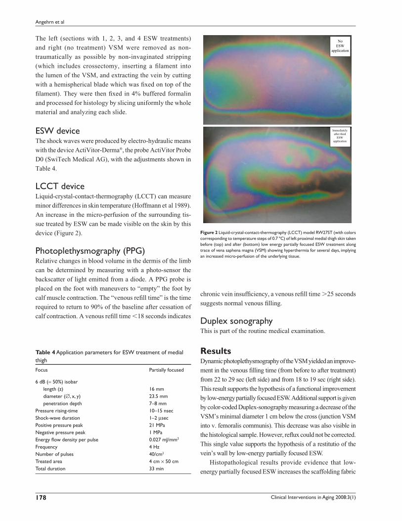
LCCT deviceLiquid-crystal-contact-thermography (LCCT) can measure
minor differences in skin temperature (Hoffmann et al 1989).
An increase in the micro-perfusion of the surrounding tis-
sue treated by ESW can be made visible on the skin by this
device (Figure 2).
Photoplethysmography (PPG)Relative changes in blood volume in the dermis of the limb
can be determined by measuring with a photo-sensor the
backscatter of light emitted from a diode. A PPG probe is
placed on the foot with maneuvers to “empty” the foot by
calf muscle contraction. The “venous refi ll time” is the time
required to return to 90% of the baseline after cessation of
calf contraction. A venous refi ll time �18 seconds indicates
chronic vein insuffi ciency, a venous refi ll time �25 seconds
suggests normal venous fi lling.
Duplex sonographyThis is part of the routine medical examination.
ResultsDynamic photoplethysmography of the VSM yielded an improve-
ment in the venous fi lling time (from before to after treatment)
from 22 to 29 sec (left side) and from 18 to 19 sec (right side).
This result supports the hypothesis of a functional improvement
by low-energy partially focused ESW. Additional support is given
by color-coded Duplex-sonography measuring a decrease of the
VSM’s minimal diameter 1 cm below the cross (junction VSM
into v. femoralis communis). This decrease was also visible in
the histological sample. However, refl ux could not be corrected.
This single value supports the hypothesis of a restitutio of the
vein’s wall by low-energy partially focused ESW.
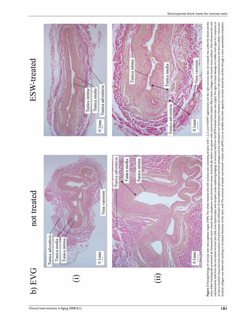
Histopathological results provide evidence that low-
energy partially focused ESW increases the scaffolding fabric
Table 4 Application parameters for ESW treatment of medial thigh
Focus Partially focused
6 dB (= 50%) isobar length (z) 16 mmdiameter (∅, x, y) 23.5 mmpenetration depth 7–8 mm
Pressure rising-time 10–15 nsecShock-wave duration 1–2 µsecPositive pressure peak 21 MPaNegative pressure peak 1 MPaEnergy fl ow density per pulse 0.027 mJ/mm2
Frequency 4 HzNumber of pulses 40/cm2
Treated area 4 cm × 50 cmTotal duration 33 min
NoESW
application
Immediatelyafter third
ESWapplication
Figure 2 Liquid-crystal-contact-thermography (LCCT) model RW27ST (with colors corresponding to temperature steps of 0.7 ºC) of left proximal medial thigh skin taken before (top) and after (bottom) low energy partially focused ESW treatment along trace of vena saphena magna (VSM) showing hyperthermia for several days, implying an increased micro-perfusion of the underlying tissue.

Clinical Interventions in Aging 2008:3(1) 179
Extracoporeal shock waves for varicose veins
of the varicose vein’s wall, particularly collagen. The amount
of elastic fi bers and smooth muscle cells probably increased
as well. The thickness of the varicose vein’s wall increased
(Figure 3). The vein’s endothel is seen to be damaged because
of stripping, whereas the endothel of the vasa vasorum are
seen to be undamaged by ESW.
DiscussionThe degenerative changes of varicose veins correlate with
those of the patient’s skin (Sansilvestri-Morel 2007) and the
tissues of other organs (Forster et al 2006). This observation
has led to the concept of the “impairment of the connective
tissue” and to the interpretation that varicose veins can be seen
as a consequence of a hereditary impairment of the connective
tissue (Tsukanov and Tsukanov 2004). This hereditary aspect
is supported by the evidence of FOXC2 gene-mutation of
patients with insuffi ciency of the vein’s valves (Mellor et al
2007) and the changes in the expression of tenascin-C of the
varicose patient in comparison to patients with normal veins
(Kirsch et al 1999). Research on the extra-cellular matrix of
varicose veins by performing immuno-histochemistry has
shown that the primary cause of varicose is an increase of
expression of metalloproteinase and a decrease of elastic
fi bers and their fragmentation (Michliels et al 2001; Somers
and Knaapen 2006; Jeanneret et al 2007). The insuffi ciency of
the vein’s valves is then seen as the consequence of the impair-
ment of the vein’s wall. Because the structure of skin’s col-
lagen is improved by ESW application (Angehrn et al 2007;
Kuhn et al 2007), we considered that similar positive results
can be achieved by application of ESW on varicose veins.
Reported effects of ESWT at the cellular level are diverse.
Light microscopy or electron microscopy show either no
changes within cells, or a complete destruction of cells. Pub-
lished results show changes in the cell’s membrane structure
(Seidl et al 1994), edema of the cell, increase of vacuoles
within the cytoplasm, dilatation of the endoplasmatic reticu-
lum, peri-nuclear cysternae, enlargement or destruction of
mitochondria, or even lethal damage of the cell (Seidl et al
1994). On the human endothel there is evidence of changes
in the permeability and even dissection of the endothel’s cells
(Seidl et al 1994). ESW with energy fl ow density of more
of than 0.3 mJ/mm2 was found to damage vascular walls
(Steinbach et al 1993; Verna et al 2006).
Some evidence of the effects of ESWT on the molecular
level is known (positive effect of ESW on the proliferation
of endothelial progenitor cells [Aicher et al 2006] or the
increased proliferation of osteoprogenitor cells [Wang et al
2002]). These experiments with cells from connective tissue
and supporting tissue showed in fi broblasts a decrease in the
survival rate proportional to the energy fl ow density. After a
few days there was an increase in the proliferation rate of bone
cells (osteoplasts), a sign of regeneration starting. Altogether
the clinical studies show an osteoneogenetic effect of ESW
application. Correct application of ESW causes no clinically
relevant or ongoing damage.
Unwanted side effects on the large venous vessels after
ESWT for ureterolithiasis are rare, but these incidences
cannot be taken for comparison because of the usage of
diverse energy fl ow densities (deep thrombosis of the femoral
vein in case of activated protein C [APC or factor V Leiden]
resistance [Brodmann et al 1998], thrombosis of the iliacal
vein [Desmet et al 1989], thrombosis of the portal vein in the
case of hypofi brinolysis [Abecassis et al 1991]).
The significant result of this study is that in vivo
application of low-energy partially focused ESW with
an energy fl ow density of 0.027 mJ/mm2 increased the
scaffolding fabric of the varicose vein’s wall, particularly
collagen, and probably also the elastic fi bers and smooth
muscle cells.
ConclusionSince the apparently encouraging results on neocollageno-
genesis and possibly neoelastino-genesis and neogenesis of
smooth muscle cells were obtained from a single case, their
clinical relevance cannot be deduced conclusively. However,
the results suggest that further research with a larger group
of patients is essential and worthwhile to show:
(i) the adequate dosage, frequency, and focus attributes of
ESW application to generate selectively constituents for
recovery of the varicose vein’s wall and thereby to open
the way for a proper curative and non-invasive therapy
for varicose vein (our conception);
(ii) that ESW application leads to even more inclusion
of collagen as in terms of phlebosclerosis with an
adequate dosage, frequency, and focus attributes to
obstruct varicose veins safely and in a controlled way.
In doing so, ESW would open a way to therapy that is
as effective as other therapies such as sclero-therapy,
radiowave-therapy, or endoluminar laser-therapy, but
is completely non-invasive.
AcknowledgmentsSarah Baccolini, Clinic Piano, Biel (organisation); SwiTech
Medical AG, Kreuzlingen (providing device for low energy
ESWT ActiVitor Ortho/Derm); Pathodiagnostics, Herisau,
Switzerland (providing histology laboratory material).

Clinical Interventions in Aging 2008:3(1)180
Angehrn et al
Clinical Interventions in Aging 2008:3(1) 181
Extracoporeal shock waves for varicose veins
Figu
re 3
His
topa
thol
ogy
of v
aric
ose
vein
: ven
a sa
phen
a m
agna
(V
SM). T
he w
hole
mat
eria
l (th
e le
ft va
rico
se v
ein
cons
istin
g of
par
ts w
ith 1
, 2, 3
, and
4 E
SWT
app
licat
ions
and
the
rig
ht v
aric
ose
vein
, not
tre
ated
) w
as u
nifo
rmly
slic
ed a
nd t
he
man
y sl
ides
his
tolo
gica
lly p
roce
ssed
. a)
hem
atox
ylin
eos
in (
nucl
ei: b
lue;
cyt
opla
sma
and
conn
ectiv
e tis
sue:
red-
pink
), b)
ela
stin
van
gie
son
with
res
orci
n-fu
chsi
n (e
last
ic fi
bers
: bla
ck, c
olla
gen:
red
, mus
cle
tissu
e: ye
llow
). O
ne c
hara
cter
istic
and
re
pres
enta
tive
slid
e fr
om th
e un
trea
ted
vari
cose
vei
n (le
ft, n
ote:
hype
rtro
phy
with
atr
ophi
c ar
eas
expl
aini
ng b
ulgi
ngs
of v
aric
osis
) and
from
the
ESW
-tre
ated
var
icos
e ve
in (r
ight
). N
o si
gns
of ti
ssue
dam
age
are
visi
ble.
Suc
h si
gns
of a
res
pons
e to
an
inju
ry w
ould
be
tissu
e ne
cros
is, e
xtra
vasa
tion
of e
ryth
rocy
tes,
the
infi l
trat
ion
of n
eutr
ophi
ls ly
mph
ocyt
es a
nd m
acro
phag
es, a
nd th
e su
bseq
uent
scar
form
atio
n. H
owev
er, a
n in
crea
se in
the
vein
’s co
nnec
tive
tissu
e, th
e ex
tra-
cellu
lar
mat
rix,
par
-tic
ular
ly c
olla
gen,
was
obs
erve
d re
sulti
ng in
incr
ease
d th
ickn
ess
of t
he v
ein’
s w
all. T
he a
mou
nts
of e
last
in a
nd s
moo
th m
uscl
e ce
lls (
SMC
) ar
e so
var
iabl
e th
at t
heir
app
aren
t in
crea
se s
houl
d be
ver
ifi ed
thr
ough
a m
ore
quan
titat
ive
eval
uatio
n.
Clinical Interventions in Aging 2008:3(1)182
Angehrn et al
DisclosuresThe authors have no confl icts of interest to disclose.
ReferencesAbecassis IP, Delaitre B, Morel MP, et al. 1991. Portal vein thrombosis after
extracorporeal shock wave lithotripsy. Lancet, 338:316–7.Aicher A, Heeschen C, Sasaki K, et al. 2006. Low-energy shock wave for
enhancing recruitment of endothelial hind limb ischemia. Circulation, 114:2823–30.
Angehrn F, Kuhn C, Voss A. 2007. Can cellulite be treated with low-energy extracorporeal shock wave therapy? Clin Interv Aging. In press.
Brodmann M, Ramschak H, Schreiber F, et al. 1998. Venous thrombosis after extracorporeal shock-wave lithotripsy in a patient with heterozygous APC-resistance. Thromb Haemost, 80:861.
Desmet W, Baert L, Vandeursen H, et al. 1989. Iliac vein thrombosis after extracorporeal shock wave lithotripsy. N Engl J Med, 321:907–8.
Drubaix I, Giakoumakis A, Robert L, et al. 1998. Preliminary data on the age-dependent decrease in basic fi broblast growth factor and platelet-derived growth factor in the human vein wall and their infl uence on cell proliferation. Gerontology, 44:9–14.
Forster OV, Tsarev OA, Shvarts IG. 2006. Interrelationship between lower limb varicosity, the grade of connective tissue dysplasia and arterial fi brillation in patients with coronary artery disease. Angiol Sosud Khir, 12:17–21.
Gerdersmeyer L, Maier M, Haake M, et al. 2002. Physikalisch-technische Grundlagen der extrakorporalen Stoßwellentherapie (ESWT). Der Orthopäde, 31:610–17.
Jeanneret C, Baldi T, Hailemariam S, et al. 2007. Selective loss of extra-cellular matrix proteins is linked to biophysical properties of varicose veins assessed by ultra-sonography. British J Surg, 94:449–56.
Haeussler E, Kiefer W. 1971. Anregung von Stoßwellen in Flüssigkeiten durch Hochgeschwindigkeits-Wassertropfen. Verhandlungen Dtsch Phys Gesellschaft, (VI) 6:786–9
Hoff G, Behrend A. 1973. Einrichtung zum Zertrümmern von im Körper eines Lebewesens befi ndlichen Konkrementen. DP 2351247.2–35.
Hoffmann R, Brutsch H-P, Largiader F, et al. 1989. Liquid-crystal-contact thermography – a new diagnostic method for determination of skin circulation. Helv Chir Acta, 56:263–6.
King DW. 1988. Pathology of aging.In Kent B, Butler R eds. Human aging research: concepts and techniques. New York NY: Raven Press, pp. 325–40.
Kuhn C, Angehrn F, Sonnabend O, et al. 2007. Impact of extracorporeal shock waves on the human skin with cellulite. Clini Interv Aging. In press.
Leu HJ, Vogt M, Pfrunder H, et al. 1991. Phlebosclerosis: disorder or disease? Vasa, 20:230–6.
Mellor RH, Brice G, Stanton AW, et al. 2007. Mutation in FOXC2 are strongly associated with primary valve failure in veins of lower limb. Circulation, 115:1912–20.
Michliels C, Bouaziz N, Pemacle J. 2001. Role of the endothelium and blood stasis in the appearance of varicose veins . Int Angiol, 20:1–8.
Minkiewicz J. 1862, 1869. Vergleichende Studien über alle gegen Varices empfohlenen Operationsverfahren. Arch pathol Anat u Physiol u klin Med, 25:193–267 (1862); 45:409–44 (1869).
Neuland H, Kesselman-Evans Z, Duchstein H-J, et al. 2004. Outline of the molecularbiological effects of the extracorporal. shockwaves (ESW) on the human organism. Orthopädische Praxis, 9:488–92.
Sansilvestri-Morel P, Fioretti F, Rupin A, et al. 2007. Comparison of extra-cellular matrix in skin and saphenous veins from patients with varicose: does the skin refl ect venous matrix changes? Clin Sci, 112:229–39.
Schaden W, Thiele R, Kölpl C, Pusch A. 2006. Extracorporeal shock wave therapy (ESWT) in skin lesions. 9th International Congress of the Inter-national Society for Musculoskeletal Shockwave Therapy (ISMST). News Letter ISMST, 2:13–14.
Schmidt A, Delhasse Y, Steingen C. 2007. The fi rst non-invasive way of inducing migration in mesenchymal stem cells (MSC). 10th Inter-national Congress of the International Society for Musculoskeletal Shockwave Therapy (ISMST). Toronto Canada, June 6–9.
Seidl M, Steinbach P, Wörle K, et al. 1994. Induction of stress fi bers and intercellular gaps in human vascular endothelium by shock-waves. Ultrasonics, 32:397–400.
Seidl M, Steinbach P, Hofstädter F. 1994. Shock wave induced endothelial damage. In situ analysis by confocal laser scanning microscopy. Ultra-sound Med Biol, 20:571–8.
Siebert W, Buch M. 1997. Extracorporeal shockwaves in orthopedics. Berlin: Springer.
Siems W, Grune T, Voss P, et al. 2005. Anti-fi brosclerotic effects of shock wave therapy in lipedema and cellulite. BioFactors, 24:275–82.
Somers P, Knaapen M, 2006. The histopathology of varicose vein disease. Angiology, 57:546–55.
Sparsa A, Lesaux N, Kessler E, et al. 2005. Treatmernt of cutaneus calcinosis in CREAST syndrome by extracorporal shock wave lithotripsy. J Am Acad Dermatol, 53:263–5.
Steinbach P, Hofstaedter F, Nicolai H. 1993. Determination of the energy-dependent extent of vascular damage caused by high-energy shock-waves in an umbilical cord model. Urol Res, 21:279–2.
Tsukanov IuT, Tsukanov AIu. 2004. Varicosis of the lower extremities as a consequence of connective tissue dysplasia. Angiol Sosud Khir, 10:84–9.
Urhahne P. 2005. Klinische Studie zur Behandlung häufi ger Erkrankungen des Bewegungsapparates des Pferdes mittels fokussierter extrakorpo-raler Stoßwellentherapie (ESWT). Dissertation München
Verna M, Turner TA, Hayden DW. 2006. Short-term effects of non-focused extracorporeal shockwave therapy on the palmar digital vasculature. Dentistry, surgery, and lameness. AAEP Proceedings, 52:580–2.
Wali MA, Dewan M, Eid RA. 2003. Histopathological changes in the wall of varicose veins. Int Angiol, 22:188–93.
Wang C-J, Wang F-S, Yang KD. 2006. Biological mechanism of muscu-loskeletal shockwaves. 9th International Congress of the International Society for Musculoskeletal Shockwave Therapy (ISMST). News Letter ISMST, 1:5–11.
Wang FS, Yang KD, Chen RF. 2002. Extracorporeal shock wave promotes growth and differentiation of bone-marrow stromal cells towards osteo-progenitors associated with induction of TGF-β1. J Bone Joint Surg, 84-B:457–61.
Wess O. 2006. Physics and technology of shock wave and pressure wave therapy. 9th International Congress of the International Society for Musculoskeletal Shockwave Therapy (ISMST). News Letter ISMST, 2:2–12.
Wolfrum B, Ohl C-D, Mettin R, et al. 2003. Die Bedeutung von Kavitations-blasen für transiente Membranpermeabilisierung und Zellschädigung. Fortschritte der Akustik – DAGA 2003, Aachen, 826–7, M. Vorländer, Deutsche Gesellschaft für Akustik e.V. (DEGA) Oldenburg.
Yang KD, Chiu C-C, Chang W-C, et al. 2007. Shock wave treatment enhances osteogenesis of mesenchymal stem cells from the blood or Wharton jelly of human umbilical cord. 10th International Congress of the International Society for Musculoskeletal Shockwave Therapy (ISMST). Toronto Canada, June 6–9.


