Download - Harvest and Staining Protocols - KSU
By: Sahar AlSubaie
Harvest and Staining Protocols

Arresting cells at metaphase stage .
Hypotonic treatment.
Cells fixation.
Harvest Protocols:
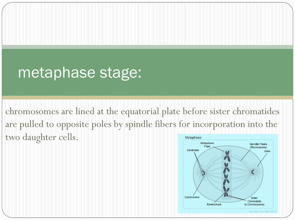
chromosomes are lined at the equatorial plate before sister chromatides
are pulled to opposite poles by spindle fibers for incorporation into the
two daughter cells.
metaphase stage:

Done by adding mitotic arrestant such as Colcemid.
Arresting cells at metaphase stage .

preventing spindle fibers formation and thereby preventing pulling the
two sister chromatides to the daughter cells.
Mechanism of Colcemid action:
Using hypotonic solution results in :
Swelling of the cells :
Thinning cytoplasm & enhance chromosome spreading
Lysis of RBCs.
Hypotonic treatment:

Fixative is added to kill and preserve the cells.
It removes the water from the cell (dehydrate them).
hardens membranes and chromatin.
Cells fixation:

1st few drops of fixative stop the action of hypotonic solution and
prepare cells for next higher fixative concentrations
The brown supernatant is the color of the methemoglobin.(formed
after the addition of fixative)
Multiple fixative steps should be carried out. (until a clear
supernatant is obtained)
Cells fixation:

after incubation , add 50 µl of colcemid.
Incubate for 15 mins at 37 ℃ .
Centrifuge the tube for 8 mins at 1200 rpm speed .
Discard the supernatant without touching the buffy coat ( because cells are there )
resuspend pellets :Mix thoroughly using the your palm hand , continue until the buffy coat disappears.
Add 2 ml of pre-warmed hypotonic solution(0.075 M KCl) drop by drop while mixing using vortex .
Add 8 ml of hypotonic solution without mixing .
Incubate at 37 ℃ for 15 mins.
1st wash : Add 1 ml of fixative(3:1 methanol: acetic acid) dropwise
Centrifuge for 8 min at 1200 rpm.
Procedure:

Remove the supernatant but not completely .
Mix thoroughly as the cells are so sticky
Add 2 ml of fixative drop by drop using vortex .
Add more fixative ( up to 10 ml) without using vortex.
Centrifuge for 8 min at 1200 rpm.
Now the 2nd wash: starts after removing the supernatant.
Mix to detach the cells from the tube wall
Add 10 ml of fixative
Centrifuge for 8 min at 1200 rpm.
Don’t remove the supernatant now.
Keep at 4 ℃ until you can do the dropping and staining
Procedure:

Dropping Protocol:
1. Replace the fixative with a new one. Using a fine glass pasture pipette make a gentle mix without making air bubbles, as this will cause cells to clump.
2. Clean the slides with methanol, that will help to get a uniformity in spreading.
3. Carry the slide in 45 degrees and drop the sample from about one meter apart.
4. As a test slide drop about 4 drops on the slide, leave to air dry completely and observe the mitotic index and metaphases compactness.
Aging: Put the slides in the oven at 60C over night or at 90C for 90 minutes, that will driving off water and get better banding pattern.
Procedure:
Staining :
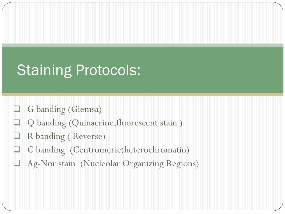
G banding (Giemsa)
Q banding (Quinacrine,fluorescent stain )
R banding ( Reverse)
C banding (Centromeric(heterochromatin)
Ag-Nor stain (Nucleolar Organizing Regions)
Staining Protocols:
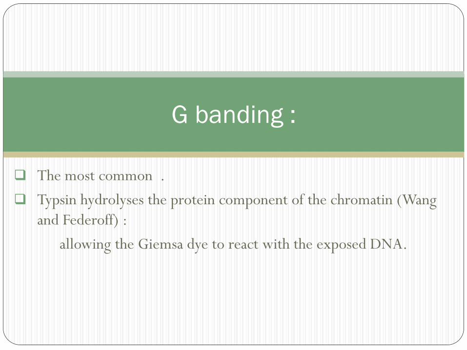
The most common .
Typsin hydrolyses the protein component of the chromatin (Wang
and Federoff) :
allowing the Giemsa dye to react with the exposed DNA.
G banding :
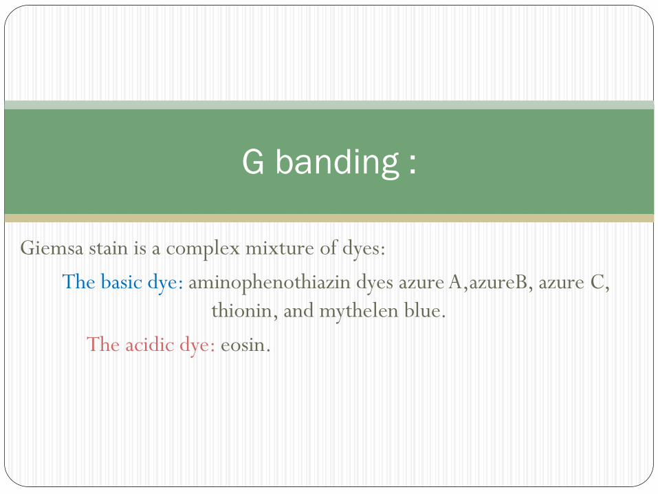
Giemsa stain is a complex mixture of dyes:
The basic dye: aminophenothiazin dyes azure A,azureB, azure C,
thionin, and mythelen blue.
The acidic dye: eosin.
G banding :
Immersing slides in fetal calf serum to stop the activity of the
trypsin since the serum contains α1-antitrypsin
G banding :

Appropriately stained chromosomes
neither too dark nor too pale.
Under-trypsinized chromosomes
have indistinct bands and little contrast. i.e.fuzzy in appearance.
G banding :
Over-trypsinized chromosomes
have sharp bands and often appear frazzled at the ends.
Eventually overtrypsinized chromosomes
are very pale after staining and may appear ghost-like and very swollen.
G banding :

Each chromosomes characterized by dark & light bands
400 bands in the haploid genome
Light bands are genetically active sites
Dark bands are gene poor rich in AT pairs.
G banding :

Chromosomes 13,18,21gene poor (very dark chromosomes)
Chromosome 21 is smaller than Chromosome 22
Chromosome 21(200 genes) is half as many as Chromosome22 (400 genes)
G banding :
Slides must cool down to RT
Immerse in trypsin solution 10-15 sec
Wash in phosphate buffer or serum ( to end trypsin activity)
Transfer to Gimsa stain 4 mins.
All the staining solution must be kept at 37 ℃ water bath during staining procedure.
Raines in D-water
Leave to dry completely
Examine under microscope
Procedure :

Count chromosomes in 10-15 metaphases
Count 30 if mosaicism suspected
Detailed analysis of 3-5metaphase
Analysis:
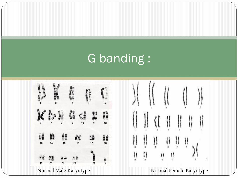
G banding :
Normal Male Karyotype Normal Female Karyotype

Used for Y chromosome abnormalities or mosaicism
Similar to G band ( but It can detect polymorphism)
Examined under fluorescent microscope
Q banding

Used to identify X chromosome abnormality
Heat chromosomes before treating with Gimsa
Light and dark bands are reversed
R banding:

To identify centromere/ heterochromatin
Heterochromatic region:
-contains highly repetitive DNA sequence
-highly condense chromatin fibres
-genetically inactive (structural elements)
•Treated chromosomes:
-Acid
-Alkaline
-Then G band
C banding
Organization of the chromosomes of an individual, lined up using
computer image processing according to:
Size, the largest to smallest
Location of centromere
Banding pattern
Karyotyping:
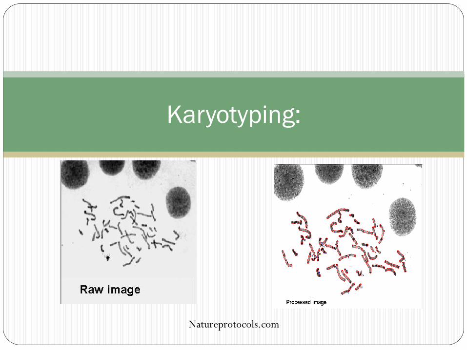
Karyotyping:
Natureprotocols.com

Karyotyping:
Natureprotocols.com
A ‘band’ is defined as that part of a chromosome which is clearly
distinguishable from its adjacent segments by appearing darker or lighter
with one or more banding techniques.
Banding pattern:
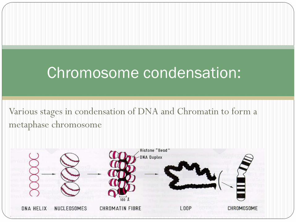
Various stages in condensation of DNA and Chromatin to form a
metaphase chromosome
Chromosome condensation:
prometaphase and prophase chromosomes
treatment of the cultures with chemical agent to
produce cell synchrony such as (Methotrexate).
then adding a release agent to prevent
contraction such as (Thymidine).
High resolution banding:
High resolution banding:
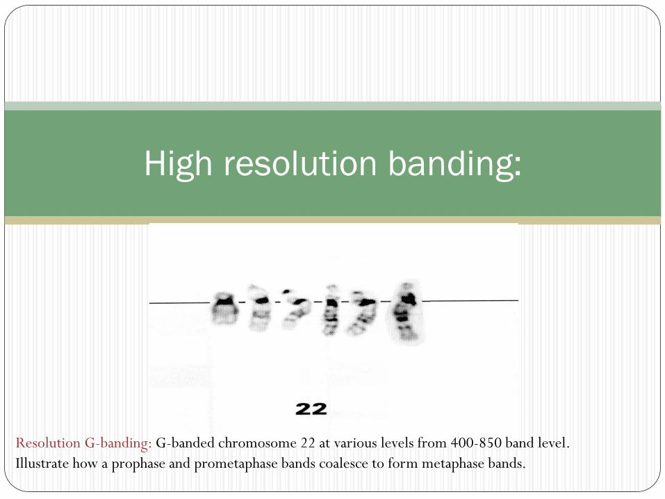
Resolution G-banding: G-banded chromosome 22 at various levels from 400-850 band level.
Illustrate how a prophase and prometaphase bands coalesce to form metaphase bands.


