Download - Health Assessment.Cardiac & PVS

8/10/2019 Health Assessment.Cardiac & PVS
http://slidepdf.com/reader/full/health-assessmentcardiac-pvs 1/56
heartchapter 19

8/10/2019 Health Assessment.Cardiac & PVS
http://slidepdf.com/reader/full/health-assessmentcardiac-pvs 2/56
blood flow
• know flow of blood &
anatomy• any questions about
flow of blood or
anatomy of heart?

8/10/2019 Health Assessment.Cardiac & PVS
http://slidepdf.com/reader/full/health-assessmentcardiac-pvs 3/56
cardiac cycle
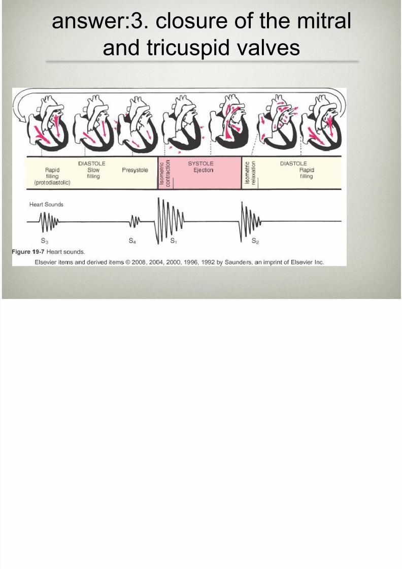
• what is happening during s1, s2, s3 &
s4?• s1: systole-mitral & tricuspid valves
close
• s2:systole-aortic and pulmonic valvesclose
• s3: blood empties from atria to
ventricles; passive filling of ventricles-diastole
• s4:at end of diastole-atria contract and
push last of blood into ventricles (atrial
kick)

8/10/2019 Health Assessment.Cardiac & PVS
http://slidepdf.com/reader/full/health-assessmentcardiac-pvs 4/56
cardiac cycle
• s1 sound is created when AV valves
close-where is this sound heard loudest?
• at apex - where is this?
• s2 sound is created when SL
valves close-where is this sound
heard loudest?
• at base - where is this?• what is a split s1/s2?
http://www.blaufuss.org/arrow/S2.html
http://www.blaufuss.org/arrow/S1.html
Clear explanation of heart sounds s1/s2, where they are heard & what is happening-follow links

8/10/2019 Health Assessment.Cardiac & PVS
http://slidepdf.com/reader/full/health-assessmentcardiac-pvs 5/56
extraneous heart sounds• **s3 when do we hear this? physiologic
& pathologic reasons• physiologic: young adults, children,
pregnancy
• patho: heart failure, regurgitation - V gallop• **s4 when do we hear this?
physiologic & pathologic
reasons
• physiologic: 45/50’s after exercise
• patho: heart dz, HTN - A gallop
http://www.blaufuss.org/arrow/S4.html
Clear explanation of heart sound s4, where they are heard & what is happening-follow links

8/10/2019 Health Assessment.Cardiac & PVS
http://slidepdf.com/reader/full/health-assessmentcardiac-pvs 6/56
murmur• *be able to define
• how do we document these sounds? p
478
• timing
• loudness - graded
• pitch
• pattern
• quality
• location- where heard
• radiation - travel?
• position - of pt

8/10/2019 Health Assessment.Cardiac & PVS
http://slidepdf.com/reader/full/health-assessmentcardiac-pvs 7/56
precordium
• know & be able to define
heave/lift/pulsations/apical
impulse/thrill/murmur
• when/how you note these & when they willoccur (during inspection, palpation,
auscultation, etc)
• which part of the stethoscope will we use for
s1, s2, s3 and s4?
• which set of pts will we have a difficult time
palpating the apical impulse?

8/10/2019 Health Assessment.Cardiac & PVS
http://slidepdf.com/reader/full/health-assessmentcardiac-pvs 8/56
auscultation heart sound
locations
• **know location of each -
may have to draw on blank
picture & label
• **read over Z pattern on pg
475
• which pulse is associatedwith s1? carotid
• what is a pulse deficit?

8/10/2019 Health Assessment.Cardiac & PVS
http://slidepdf.com/reader/full/health-assessmentcardiac-pvs 9/56
other terms
• angina-we ask about chest “discomfort” here-pain is subjective-
this occurs when the hearts own blood supply cannot keep up
with metabolic demand
• DOE-dyspnea on exertion-shortness of breath-when does this
happen?
• PND-paroxysmal nocturnal dyspnea-occurs with heart failure-
lying down increases pressure on heart workload-pt feels short
of air
• nocturia-increased urinary frequency during the night-due to
fluid reabsorption when lying down occurring with heart failure
• pericardial friction rub-inflammation of pericardial sac
surrounding the heart-sounds like rubber against leather-best
heard pt sitting up and learning forward breath held on expiration

8/10/2019 Health Assessment.Cardiac & PVS
http://slidepdf.com/reader/full/health-assessmentcardiac-pvs 10/56
Cardiovascular DZ
• list modifiable vs non-modifiable risk
factors
• warning signs of hypoxia

8/10/2019 Health Assessment.Cardiac & PVS
http://slidepdf.com/reader/full/health-assessmentcardiac-pvs 11/56
neck vessel assessment
• why do we palpate carotid artery one at a
time?
• what is a bruit & what does it indicate?(*difference between bruit & murmur)
• be able to describe the inspection of the
jugular vein - slide 21/pg 472 - always read
specific inspection instructions in book if onslide r/t test questions

8/10/2019 Health Assessment.Cardiac & PVS
http://slidepdf.com/reader/full/health-assessmentcardiac-pvs 12/56
PVSchapter 20
8/10/2019 Health Assessment.Cardiac & PVS
http://slidepdf.com/reader/full/health-assessmentcardiac-pvs 13/56
PVS
• look over anatomy
• this HH is VERY similar to cardiacassessment questions except for HPI
• **VTE risk assessment (select all that
apply would be applicable here) knowdifferences between modifiable and
non-modifiable-slide 30 & pg

8/10/2019 Health Assessment.Cardiac & PVS
http://slidepdf.com/reader/full/health-assessmentcardiac-pvs 14/56
occlusion of
arteries vs veins
• slide 38, 40; chart on page 521
• veins=towards (deoxygenated)
• painful! • large veins, edema pitting/non, thick skin, warm, erythema,
thrombosis, etc
• homans sign
• arteries=away (oxygenated)
• 5 P’s - pain, pallor, pulseless, paresthesia, paralysis
• loss of hair, thick nails, thin skin, cap refill, cold extremities, etc
• allen test

8/10/2019 Health Assessment.Cardiac & PVS
http://slidepdf.com/reader/full/health-assessmentcardiac-pvs 15/56
signs
• be able to describe what the test is determining, the
process, and what a positive (or negative) sign
indicates
• allen test-evaluate the adequacy of collateral ARTERIAL circulation in the hand-read over steps on
pg. 509-POSITIVE test is when the blood flow does
NOT return to the hand within 2-5 seconds
• homans sign-may indicate DVT or superficialthrombophlebitis-read over steps on slide 40-
POSITIVE sign means potential for DVT or
thrombophlebitis

8/10/2019 Health Assessment.Cardiac & PVS
http://slidepdf.com/reader/full/health-assessmentcardiac-pvs 16/56
terms
• ischemia-deficient supply of oxygenated arterial blood to a
tissue caused by obstruction of a blood vessel-partial blockage
creates insufficient supply and ischemia may be apparent only
at exercise when oxygen needs increase (claudication occurs
with activity, relieved with rest) • edema-swelling-occurs in both extremities when right sided HF
is present-unilaterally when there is an obstruction
• orthostatic hypotension-drop in systolic BP >20 mmHg and
diastolic of >10 mmHg-results in feeling lightheaded or
dizziness-results primarily from blood pooling in lowerextremities which results in decreased venous return and
decreased cardiac output

8/10/2019 Health Assessment.Cardiac & PVS
http://slidepdf.com/reader/full/health-assessmentcardiac-pvs 17/56
A patient has been diagnosed with Right-Sided
Congestive Heart Failure and is confused about return
of deoxygenated blood from the tissue. To clarify the
confusion, which chamber of the heart receives blood
from systemic circulation?
• 1. Left atrium
• 2. Right atrium
• 3. Right ventricle• 4. Left ventricle

8/10/2019 Health Assessment.Cardiac & PVS
http://slidepdf.com/reader/full/health-assessmentcardiac-pvs 18/56
answer: 2 right atrium

8/10/2019 Health Assessment.Cardiac & PVS
http://slidepdf.com/reader/full/health-assessmentcardiac-pvs 19/56
nurse is listening to client's heartbeat & focusing on 2nd
heart sound, which heart valves produce this sound?
• 1. Pulmonic & Mitral
• 2. Aortic & Pulmonic
• 3. Mitral & Tricuspid
• 4. Tricuspid & Aortic
8/10/2019 Health Assessment.Cardiac & PVS
http://slidepdf.com/reader/full/health-assessmentcardiac-pvs 20/56
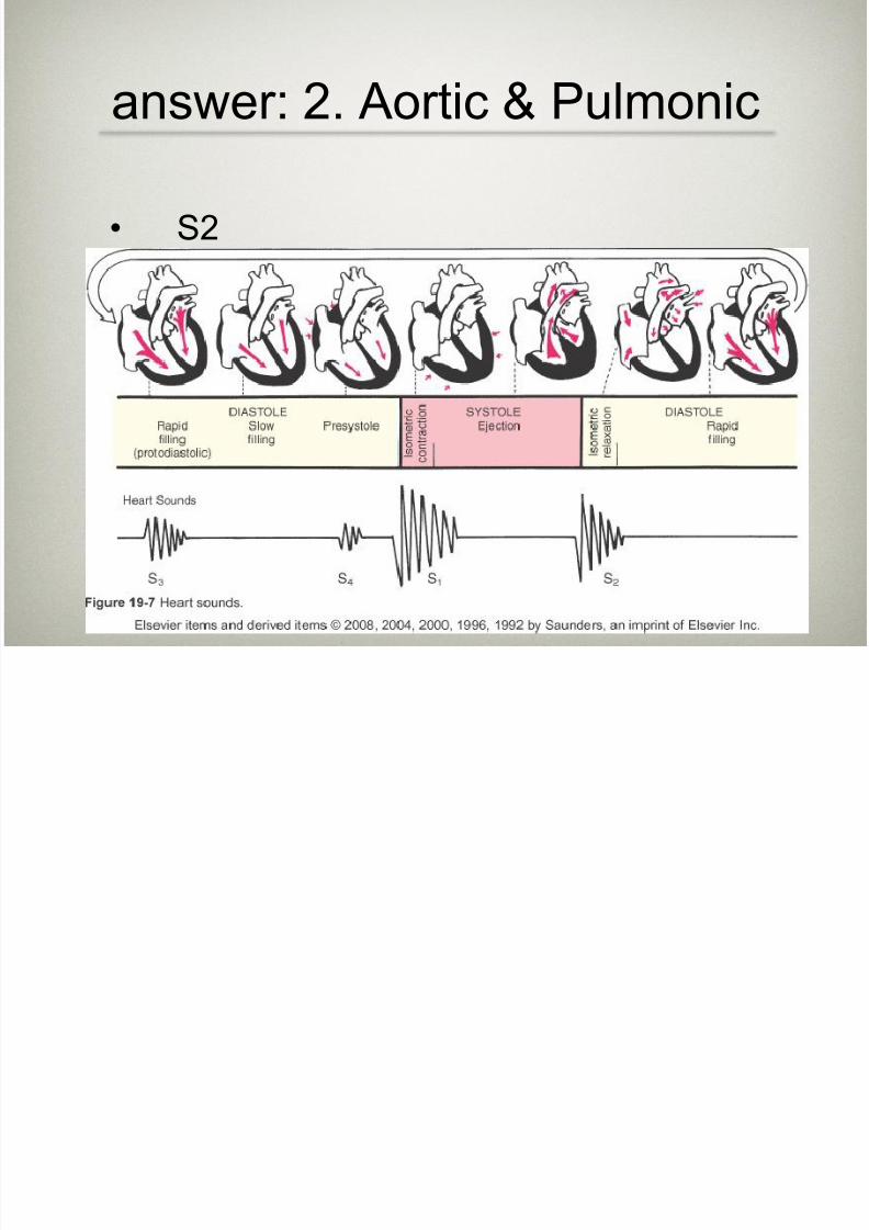
answer: 2. Aortic & Pulmonic
• S2
8/10/2019 Health Assessment.Cardiac & PVS
http://slidepdf.com/reader/full/health-assessmentcardiac-pvs 21/56
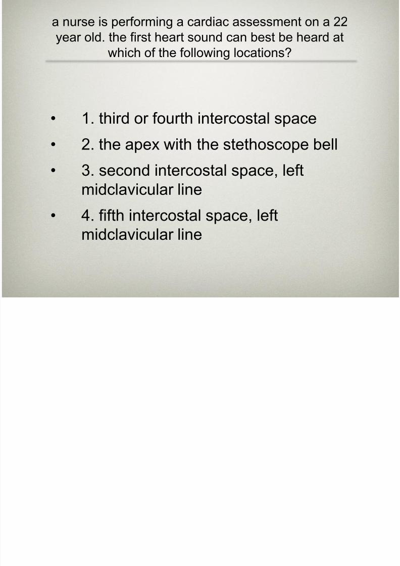
a nurse is performing a cardiac assessment on a 22
year old. the first heart sound can best be heard at
which of the following locations?
• 1. third or fourth intercostal space
• 2. the apex with the stethoscope bell
• 3. second intercostal space, left
midclavicular line
• 4. fifth intercostal space, leftmidclavicular line

8/10/2019 Health Assessment.Cardiac & PVS
http://slidepdf.com/reader/full/health-assessmentcardiac-pvs 22/56
answer: 4 fifth intercostal space,
left midclavicular line
• S1 can best be heard at the fifthintercostal space, midclavicular line.
• this is just knowledge of where heart
sounds can be heard-on powerpoint
h lt ti th h t hi h f th f ll i
8/10/2019 Health Assessment.Cardiac & PVS
http://slidepdf.com/reader/full/health-assessmentcardiac-pvs 23/56
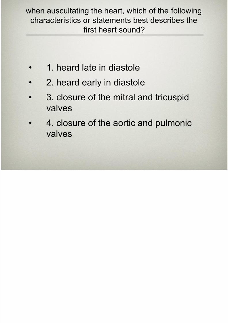
when auscultating the heart, which of the following
characteristics or statements best describes the
first heart sound?
• 1. heard late in diastole
• 2. heard early in diastole
• 3. closure of the mitral and tricuspid
valves
• 4. closure of the aortic and pulmonicvalves
answer:3 closure of the mitral
8/10/2019 Health Assessment.Cardiac & PVS
http://slidepdf.com/reader/full/health-assessmentcardiac-pvs 24/56
answer:3. closure of the mitral
and tricuspid valves
S

8/10/2019 Health Assessment.Cardiac & PVS
http://slidepdf.com/reader/full/health-assessmentcardiac-pvs 25/56
A 12 year old client has an S3 heart
sound. The nurse knows:
• 1. she can document this as an
abnormal finding and continue the
assessment• 2. physiologic S3 is common in children
and young adults
• 3. she must contact the attendingphysician immediately, something is
wrong
8/10/2019 Health Assessment.Cardiac & PVS
http://slidepdf.com/reader/full/health-assessmentcardiac-pvs 26/56
answer:2. physiologic S3 is common in
children and young adults
• An S3 heart sound, also called a
ventricular gallop, occurs early indiastole when blood is flowing from the
atria into the ventricles and causes
vibrations. S3 is a physiologic heart
sound in children, young adults, andpregnant females
Which of the following risk factors for

8/10/2019 Health Assessment.Cardiac & PVS
http://slidepdf.com/reader/full/health-assessmentcardiac-pvs 27/56
g
coronary artery disease cannot be corrected?
(non-modifiable)
• 1. Cigarette smoking• 2. DM
• 3. Heredity
• 4. HTN

8/10/2019 Health Assessment.Cardiac & PVS
http://slidepdf.com/reader/full/health-assessmentcardiac-pvs 28/56
answer: 3 heredity
• Because “heredity” refers to our genetic
makeup, it can’t be changed. Cigarette
smoking cessation is a lifestyle changethat involves behavior modification.
Diabetes mellitus is a risk factor that
can be controlled with diet, exercise,
and medication. Altering one’s diet,exercise, and medication can correct
hypertension.
A murmur is heard at the second left intercostal
8/10/2019 Health Assessment.Cardiac & PVS
http://slidepdf.com/reader/full/health-assessmentcardiac-pvs 29/56
A murmur is heard at the second left intercostal
space along the left sternal border. Which valve
area is this?
• 1. Aortic• 2. Mitral
• 3. Pulmonic
• 4. Tricuspid

8/10/2019 Health Assessment.Cardiac & PVS
http://slidepdf.com/reader/full/health-assessmentcardiac-pvs 30/56
answer: 3 pulmonic
• Abnormalities of the pulmonic valve are
auscultated at the second left intercostal
space along the left sternal border. Aortic
valve abnormalities are heard at the second
intercostal space, to the right of the sternum.
Mitral valve abnormalities are heard at the
fifth intercostal space in the midclavicular
line. Tricuspid valve abnormalities are heard
at the third and fourth intercostal spaces
along the sternal border.
Wh i i h ld h l h h d f h b d i

8/10/2019 Health Assessment.Cardiac & PVS
http://slidepdf.com/reader/full/health-assessmentcardiac-pvs 31/56
What position should the nurse place the head of the bed in to
obtain the most accurate reading (in our case, to visualize it)of
jugular vein distention?
• 1. High-fowler’s • 2. Raised 10 degrees
• 3. Raised 30 degrees
• 4. Supine position

8/10/2019 Health Assessment.Cardiac & PVS
http://slidepdf.com/reader/full/health-assessmentcardiac-pvs 32/56
answer: 3. Raised 30 degrees
• 30-45 is ideal. Inclined pressure can’t
be seen when the client is supine or
when the head of the bed is raised 10degrees because the point that marks
the pressure level is above the jaw
(therefore, not visible). In high Fowler’s
position, the veins would be barely
discernible above the clavicle.
The client is diagnosed with pericarditis. When
i th li t th i bl

8/10/2019 Health Assessment.Cardiac & PVS
http://slidepdf.com/reader/full/health-assessmentcardiac-pvs 33/56
assessing the client, the nurse is unable
to auscultate a friction rub. Which action should the
nurse implement?
• 1. Notify the health-care provider.
• 2. Document that the pericarditis has
resolved.
• 3. Ask the client to lean forward and
listen again.
• 4. Prepare to insert a unilateral chest
tube

8/10/2019 Health Assessment.Cardiac & PVS
http://slidepdf.com/reader/full/health-assessmentcardiac-pvs 34/56
answer: 3. Ask the client to lean forward and
listen again.
• pericarditis is best heard when the
patient is sitting up and learning
forward
Two nurses are taking an apical radial pulse and note a

8/10/2019 Health Assessment.Cardiac & PVS
http://slidepdf.com/reader/full/health-assessmentcardiac-pvs 35/56
Two nurses are taking an apical-radial pulse and note a
difference in pulse rate of 8 beats per minute. The nurse
would document this difference as which of the following?
• 1. Pulse deficit • 2. Pulse amplitude
• 3. Ventricular rhythm
• 4. Heart arrhythmia
8/10/2019 Health Assessment.Cardiac & PVS
http://slidepdf.com/reader/full/health-assessmentcardiac-pvs 36/56
answer: 1. Pulse deficit
• when the apical heart rate and the
radial heart rate do not coincide, this is
termed pulse deficit

8/10/2019 Health Assessment.Cardiac & PVS
http://slidepdf.com/reader/full/health-assessmentcardiac-pvs 37/56
When evaluating a client's circulation the nurse should
include which assessments? Select all that apply.
• 1. Palpation of pulses
• 2. Skin temperature of bilateralextremities
• 3. Skin color
• 4. Moles & freckles• 5. Hair on the legs and feet
8/10/2019 Health Assessment.Cardiac & PVS
http://slidepdf.com/reader/full/health-assessmentcardiac-pvs 38/56
Answer: 1, 2, 3 & 5
• 1. Palpation of pulses
• 2. Skin temperature of bilateral
extremities
• 3. Skin color
• 5. Hair on the legs and feet
• These are indicative of circulation to
and from the extremities
A 65-year-old patient with a history of heart failure comes to
8/10/2019 Health Assessment.Cardiac & PVS
http://slidepdf.com/reader/full/health-assessmentcardiac-pvs 39/56
y p y
the clinic with complaints of "being awakened from sleep with
shortness of breath." Which action by the nurse is most
appropriate?
• 1. Obtain a detailed history of the patient's
allergies and history of asthma.
• 2. Tell the patient to sleep on his or her
right side to facilitate ease of respirations.
• 3. Assess for other signs and symptoms of
paroxysmal nocturnal dyspnea.
• 4. Assure the patient that this is normal
and will probably resolve within the next
week.
8/10/2019 Health Assessment.Cardiac & PVS
http://slidepdf.com/reader/full/health-assessmentcardiac-pvs 40/56
answer: 3. Assess for other signs and symptoms of
paroxysmal nocturnal dyspnea.
• The patient is experiencing paroxysmalnocturnal dyspnea: being awakened
from sleep with shortness of breath and
the need to be upright to achieve
comfort.
During an assessment of a 68-year-old man with a recent onset of
right sided weakness the nurse hears a blowing swishing sound
8/10/2019 Health Assessment.Cardiac & PVS
http://slidepdf.com/reader/full/health-assessmentcardiac-pvs 41/56
right-sided weakness, the nurse hears a blowing, swishing sound
with the bell of the stethoscope over the left carotid artery. This
finding would indicate:
• 1. a valvular disorder.
• 2. blood flow turbulence.
• 3. fluid volume overload.
• 4. ventricular hypertrophy
8/10/2019 Health Assessment.Cardiac & PVS
http://slidepdf.com/reader/full/health-assessmentcardiac-pvs 42/56
answer:2. blood flow turbulence.
• A bruit is a blowing, swishing sound
indicating blood flow turbulence;
normally none is present.
Th i i t lt t f h t

8/10/2019 Health Assessment.Cardiac & PVS
http://slidepdf.com/reader/full/health-assessmentcardiac-pvs 43/56
The nurse is preparing to auscultate for heart
sounds. Which technique is correct?
• 1. Listen to the sounds at the aortic,
tricuspid, pulmonic, and mitral areas.
• 2. Listen by inching the stethoscope in a
rough Z pattern, from the base of the heartacross and down, then over to the apex.
• 3. Listen to the sounds only at the site
where the apical pulse is felt to be the
strongest.
• 4. Listen for all possible sounds at a time at
each specified area.
answer:B) Listen by inching the stethoscope in a rough Z

8/10/2019 Health Assessment.Cardiac & PVS
http://slidepdf.com/reader/full/health-assessmentcardiac-pvs 44/56
pattern, from the base of the heart across and down, then
over to the apex.
• Do not limit auscultation of breath
sounds to only four locations. Sounds
produced by the valves may be heardall over the precordium. Inch the
stethoscope in a rough Z pattern from
the base of the heart across and down,
then over to the apex. Or, start at the
apex and work your way up.
When performing a peripheral vascular assessment on a patient
8/10/2019 Health Assessment.Cardiac & PVS
http://slidepdf.com/reader/full/health-assessmentcardiac-pvs 45/56
When performing a peripheral vascular assessment on a patient,
the nurse is unable to palpate the ulnar pulses. The patient's skin
is warm and capillary refill time is normal. The nurse should next:
• 1. check for the presence of claudication.
• 2. refer the individual for further evaluation.
• 3. consider this a normal finding and
proceed with the peripheral vascular
evaluation.
• 4. ask the patient if he or she hasexperienced any unusual cramping or
tingling in the arm.

8/10/2019 Health Assessment.Cardiac & PVS
http://slidepdf.com/reader/full/health-assessmentcardiac-pvs 46/56
answer:3. consider this a normal finding and proceed with
the peripheral vascular evaluation.
• It is not usually necessary to palpate
the ulnar pulses. The ulnar pulses are
often not palpable in the normal
person. The other responses are not
correct.

8/10/2019 Health Assessment.Cardiac & PVS
http://slidepdf.com/reader/full/health-assessmentcardiac-pvs 47/56
When using a Doppler ultrasonic stethoscope, the nurse
recognizes arterial flow when which sound is heard?
• 1. Low humming sound
• 2. Regular "lub, dub" pattern
• 3. Swishing, whooshing sound
• 4. Steady, even, flowing sound

8/10/2019 Health Assessment.Cardiac & PVS
http://slidepdf.com/reader/full/health-assessmentcardiac-pvs 48/56
answer:3. Swishing, whooshing sound
• When using the Doppler ultrasonicstethoscope, the pulse site is found
when one hears a swishing, whooshing
sound.
During an assessment of an older adult, the nurse should expect

8/10/2019 Health Assessment.Cardiac & PVS
http://slidepdf.com/reader/full/health-assessmentcardiac-pvs 49/56
to notice which finding as a normal physiologic change associated
with the aging process?
• 1. Hormonal changes causing vasodilation and a
resulting drop in blood pressure
• 2. Progressive atrophy of the intramuscular calfveins, causing venous insufficiency
• 3. Peripheral blood vessels growing more rigid with
age, producing a rise in systolic blood pressure
• 4. Narrowing of the inferior vena cava, causing lowblood flow and increases in venous pressure
resulting in varicosities
3 P i h l bl d l i i id ith

8/10/2019 Health Assessment.Cardiac & PVS
http://slidepdf.com/reader/full/health-assessmentcardiac-pvs 50/56
answer: 3. Peripheral blood vessels growing more rigid with
age, producing a rise in systolic blood pressure
• Peripheral blood vessels grow more
rigid with age, resulting in a rise in
systolic blood pressure. Aging
produces progressive enlargement of
the intramuscular calf veins, not
atrophy. The other options are notcorrect.
during a cardiovascular assessment the nurse finds a bluish
tinge on the clients lips fingers and toes what is the

8/10/2019 Health Assessment.Cardiac & PVS
http://slidepdf.com/reader/full/health-assessmentcardiac-pvs 51/56
tinge on the clients lips, fingers, and toes. what is the
appropriate documentation for this finding?
• 1. blue tinged extremities
• 2. central and peripheral cyanosis
• 3. bad circulation
• 4. central and peripheral pallor

8/10/2019 Health Assessment.Cardiac & PVS
http://slidepdf.com/reader/full/health-assessmentcardiac-pvs 52/56
ANSWER: 2. central and peripheral cyanosis
• inadequate blood flow to the peripherymay be due to several different things
but will result in central & peripheral
cyanosis
A Nurse assesses the client for the presence of homan's

8/10/2019 Health Assessment.Cardiac & PVS
http://slidepdf.com/reader/full/health-assessmentcardiac-pvs 53/56
A Nurse assesses the client for the presence of homan s
sign- which one indicates that this sign is positive?
• 1. no pain
• 2. pain on dorsiflexion of the foot
• 3. pain on plantar flexion of the foot
• 4. pain when bringing knee to chest
answer: 2.pain on dorsiflexion
f f t

8/10/2019 Health Assessment.Cardiac & PVS
http://slidepdf.com/reader/full/health-assessmentcardiac-pvs 54/56
of foot
• Homan’s sign has to do with pain in the
calf area indicating possible DVT/VTE
A client has a 1+/0-4+ dorsalis pedis pulse on the right. The
lower leg is cool pale and painful This description is most

8/10/2019 Health Assessment.Cardiac & PVS
http://slidepdf.com/reader/full/health-assessmentcardiac-pvs 55/56
lower leg is cool, pale, and painful. This description is most
consistent with:
• 1. venous insufficiency
• 2. arterial insufficiency
• 3. normal finding
t i l i ffi i

8/10/2019 Health Assessment.Cardiac & PVS
http://slidepdf.com/reader/full/health-assessmentcardiac-pvs 56/56
answer: arterial insufficiency
• Arterial insufficiency is inadequate
circulation in the arterial system, whichresults in diminished pulses; cool, shiny
skin; deep muscle pain; absence of hair
on the toes; pallor on elevation; and a
red color when dependent.


