
Intravitreal Injection –
The Role of the
Comprehensive
Ophthalmologist Michael Rauser, MD
Department Chairman
Associate Professor
Loma Linda University Eye Institute

Medicare Enrollment
0.0
5.0
10.0
15.0
20.0
25.0
30.0
35.0
40.0
45.0
50.0
55.0
1995 2000 2005 2010 2015
Benefi
cari
es
(M)
Year
Medicare
Traditional
Advantage
Used with permission - Source- Kevin Corcoran

Ophthalmologist Trends
18000
19000
20000
21000
22000
23000
24000
25000
26000
1995 2000 2005 2010 2015 2020 2025
Op
hth
alm
olo
gis
ts
Year
Ophthalmologists Trends
Expected Oph
Required Oph
Sources: DHHS Physician Supply and Demand Projections to
2020
Source- Kevin
Corcoran- Used
with permission
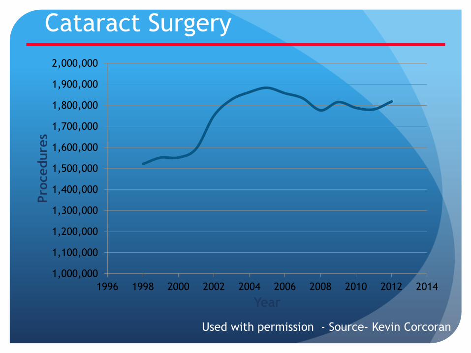
Cataract Surgery
1,000,000
1,100,000
1,200,000
1,300,000
1,400,000
1,500,000
1,600,000
1,700,000
1,800,000
1,900,000
2,000,000
1996 1998 2000 2002 2004 2006 2008 2010 2012 2014
Pro
cedure
s
Year
Used with permission - Source- Kevin Corcoran
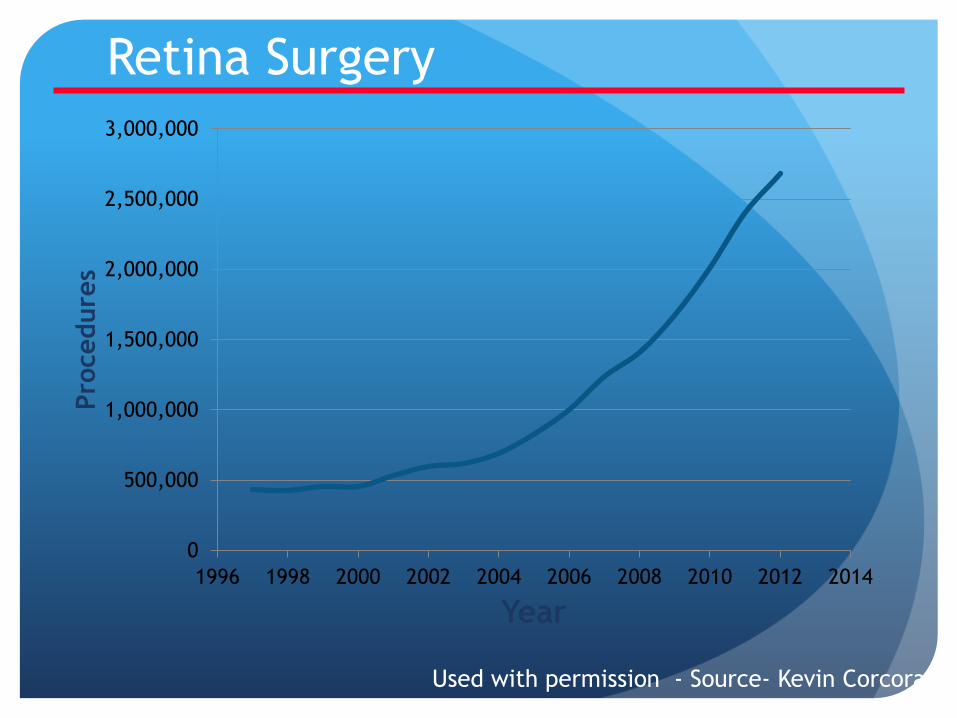
Retina Surgery
0
500,000
1,000,000
1,500,000
2,000,000
2,500,000
3,000,000
1996 1998 2000 2002 2004 2006 2008 2010 2012 2014
Pro
cedure
s
Year
Used with permission - Source- Kevin Corcoran

Medicare Utilization – 67028 – IV Injection
0
500,000
1,000,000
1,500,000
2,000,000
2,500,000
1996 1998 2000 2002 2004 2006 2008 2010 2012
Pro
cedu
res
67028 Utilization
Source: CMS data (1998 – 2012), 18 – Ophthalmology
Used with permission -
Source- Kevin Corcoran

Common Ophthalmic Surgery Medicare Utilization Patterns (18 - Ophthalmology)
CPT Procedure λ CPT Procedure λ
67028 Intravitreal injection
11% 68761
Punctum plugs 1%
66984 Cataract & IOL
8% 67228 67210
Retina laser 1%
66821 YAG 3% 67820
Epilation 1%
66761 65855
Glaucoma laser
1% 15823
Blepharoplasty 1%
Frequency is per 100 office visits (%) on Medicare beneficiaries
Source: CMS data (2012), 18 – Ophthalmology
Used with
permission -
Source- Kevin
Corcoran

Top 10 Ophthalmic Procedures Medicare Utilization Patterns Ophthalmology (18)
Rank
CPT Procedure Rank CPT Procedure
1 67028 Intravitreal Injection
6 66982 Complex Cataract
2 66984 Cataract w/IOL 7 65855 Lx Trabeculoplasty
3 66821 YAG capsulotomy
8 67210 Focal Laser
4 68761 Punctum plug 9 15823 Blepharoplasty
5 67820 Epilation 10 67228 PRP
Source: CMS data 2012, 18 - Ophthalmology Used with permission -
Source- Kevin Corcoran

Resident experience with IV injections
The Case for Comprehensive Ophthalmologists
Performing IV Injections
Technically an easy procedure to teach and perform
Current residents receive extensive experience
Strong safety profile
Injection volume is high and growing
Meets a community need
Cost effective use of manpower within a managed care environment
Patient convenience
Travel considerations

What to do 1st?
Diagnosis – including testing
FA – assists with diagnosis confirmation
Color photos- baseline documentation
Spectral Domain ( SD) Retina OCT-
Diagnosis confirmation
Quantitative and qualitative assessment
Response to treatment
Diseases Worth Treatment Consideration
Pseudophakic CME
Macular edema associated with Retinal Vein Occlusion
Diabetic Macular Edema
Exudative ARMD
Different “ treatment model” could be used for each condition
IV Injection Treatment Plan Models
Injector alone model
Injector “ plus” model
“Do all you possibly can” model
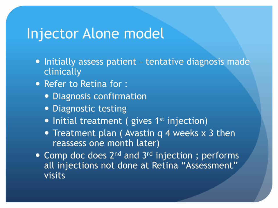
Injector Alone model
Initially assess patient – tentative diagnosis made clinically
Refer to Retina for :
Diagnosis confirmation
Diagnostic testing
Initial treatment ( gives 1st injection)
Treatment plan ( Avastin q 4 weeks x 3 then reassess one month later)
Comp doc does 2nd and 3rd injection ; performs all injections not done at Retina “Assessment” visits

Injector „Plus” model
Initially assess patient and diagnosis made. Includes some or all of the diagnostic testing ( includes Retina OCT)
Refer to Retina for :
Diagnosis confirmation
Possibly some diagnostic testing ( FA / Color photos )
Treatment plan ( Avastin q 4 weeks x 3 then reassess one month later)
Comp doc treats with OCT guided follow-up
Periodic reassessment with Retina to tailor management
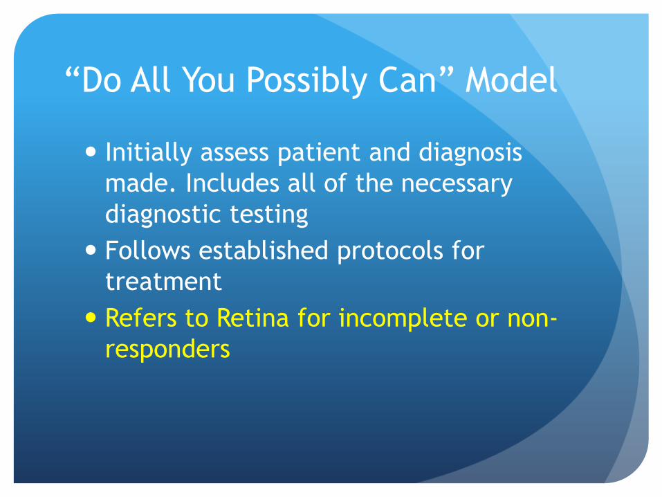
“Do All You Possibly Can” Model
Initially assess patient and diagnosis
made. Includes all of the necessary
diagnostic testing
Follows established protocols for
treatment
Refers to Retina for incomplete or non-
responders

Pre-op Assessment
No active inflammation / Infection
Note pre-op IOP / Glaucoma history
Consent for procedure

Instruments needed on Sterile
Tray Alcaine
5% Betadine solution
10% Betadine swabs
2% lidocaine plain on 3 cc syringe
Eyelid specula
Caliper
Marking pen
Sterile Q –tips
1cc syringe with 19g & 30g needles
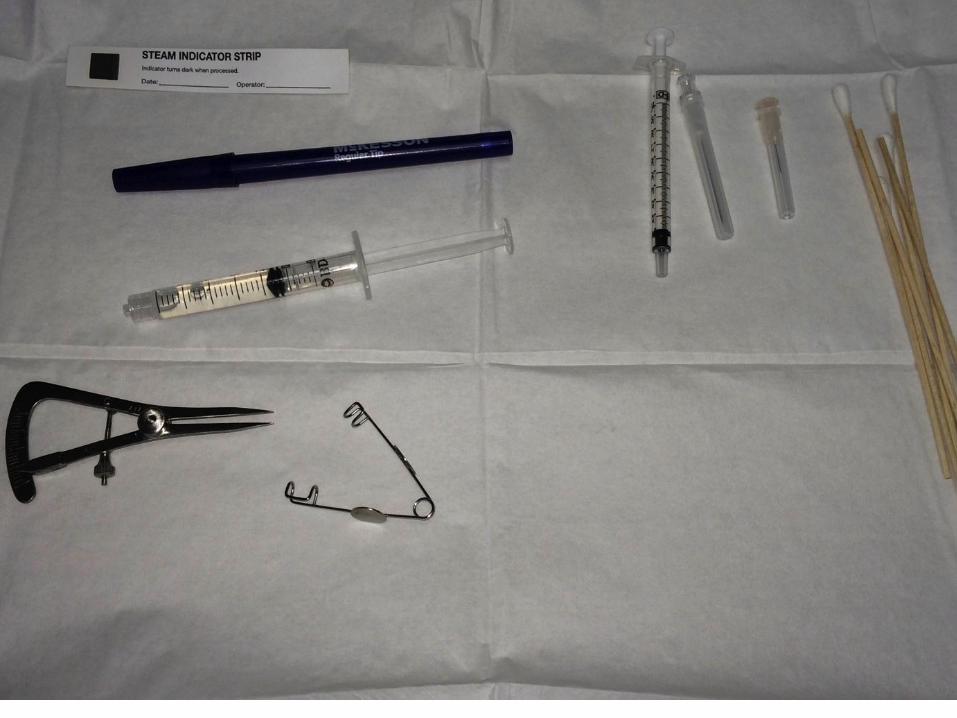
Photo of Tray
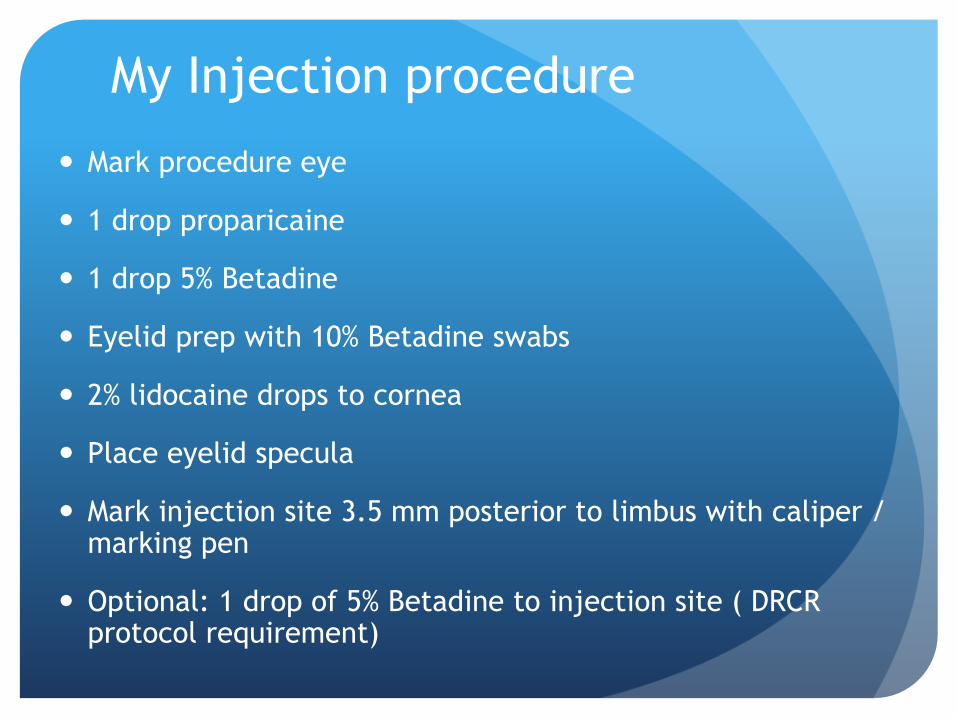
My Injection procedure
Mark procedure eye
1 drop proparicaine
1 drop 5% Betadine
Eyelid prep with 10% Betadine swabs
2% lidocaine drops to cornea
Place eyelid specula
Mark injection site 3.5 mm posterior to limbus with caliper / marking pen
Optional: 1 drop of 5% Betadine to injection site ( DRCR protocol requirement)
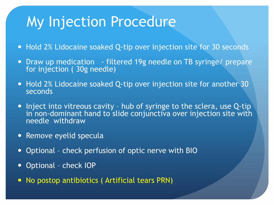
My Injection Procedure
Hold 2% Lidocaine soaked Q-tip over injection site for 30 seconds
Draw up medication - filtered 19g needle on TB syringe/ prepare for injection ( 30g needle)
Hold 2% Lidocaine soaked Q-tip over injection site for another 30 seconds
Inject into vitreous cavity – hub of syringe to the sclera, use Q-tip in non-dominant hand to slide conjunctiva over injection site with needle withdraw
Remove eyelid specula
Optional – check perfusion of optic nerve with BIO
Optional – check IOP
No postop antibiotics ( Artificial tears PRN)

What to Treat With?
Use 0.05 cc dosing:
Avastin 1.25mg
Lucentis 0.3mg ; 0.5mg
Eylea 2mg
Triescence 2 mg

Treatment Protocols
PRN therapy
Treat only if active disease / edema is present
Fixed interval
Ex: Monthly treatment x 1-2 years of Lucentis / Avastin as in the monthly CATT study arms
Treat and extend
Treat at each clinic visit, extend treatment interval if no active disease/ dry macula
If/ when recurrence develops – tighten treatment interval then fixed interval “ for a while” .

Diseases Worth Treating
Pseudophakic CME
Macular edema associated with
Retinal Vein Occlusion
Diabetic Macular Edema
Exudative ARMD

Pseudophakic CME - Ideal 1st Treatment Case
Occurs in 1-2 % of cases
All cataract surgeons encounter this condition
Self limiting disease
Avoids the trip to Retina
Not all cases respond to 1st line topical NSAIDs / Steroid
compliance
inflammation not fully controlled
Most cases respond to one dose of Avastin (2nd line Tx)
“combined” CME/ DME cases often require multiple treatments
Intravitreal Steroid ( 2mg Triescence ) – 3rd line therapy

Macular Edema Associated with RVO
Treatment options
Avastin – works in almost all cases
FDA approved- Eylea, Lucentis 0.5mg
Use for center –involved macular edema
Almost all cases respond to any ANTI- VEGF agent
Early treatment provides better final visual acuity outcome versus delayed treatment
Treat as soon as center is involved
Treat every 4-6 weeks until dry, then PRN or treat and extend
Some cases resolve in several months

Case #1 Va (RG) – Va 20/100
CMT- 644um

RG – 7 months of Avastin Tx – Va
20/40 Pre- treatment Post- treatment
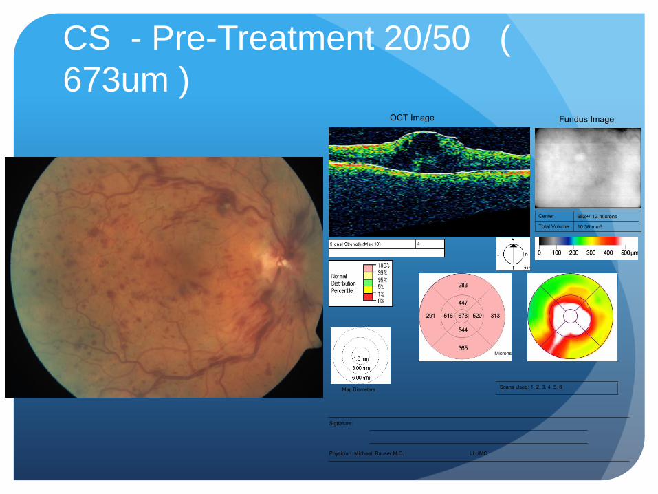
CS - Pre-Treatment 20/50 (
673um )
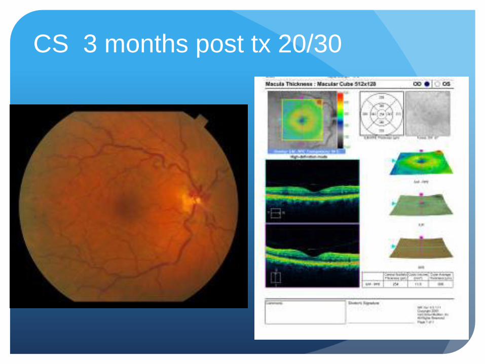
CS 3 months post tx 20/30

Pretx Avastin (EM) Va – 20/200 ( 653um)

EM 3 months after Avastin x 2 –
Va 20/60 (440 um Heidelburg
OCT)
Diabetic Macular Edema
Center-involved DME requires Treatment with Anti-VEGF therapy
Tx options: Avastin or Lucentis 0.3mg (FDA approved)
DRCR-I study results
Average 8 injections within the 1st year with monthly follow-up and treatmemt of foveal DME
Average 2-3 injections in the 2nd year
Early, aggressive therapy is best
Avastin or Lucentis 0.3mg every 4-6 weeks – treat until dry
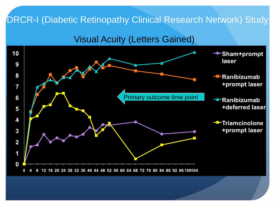
DRCR-I (Diabetic Retinopathy Clinical Research Network) Study
Visual Acuity (Letters Gained)

DRCR-I Patient – Pretreatment ( March
2008)
20/160 20/100

Pre-Treatment Imaging: OCT
OD
773 um
OS
790 um
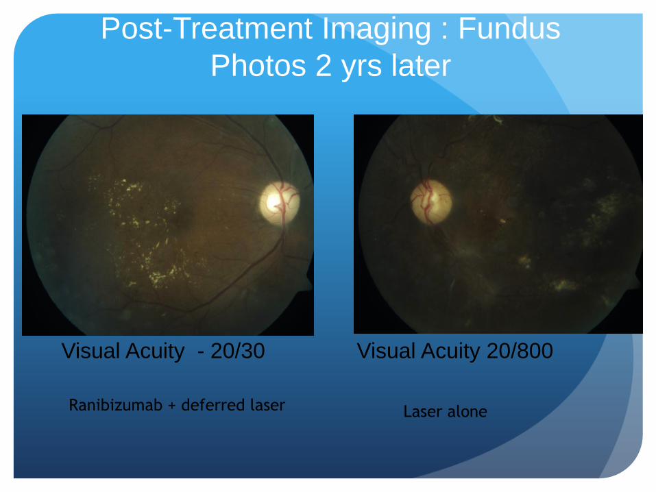
Post-Treatment Imaging : Fundus
Photos 2 yrs later
Visual Acuity - 20/30 Visual Acuity 20/800
Ranibizumab + deferred laser Laser alone
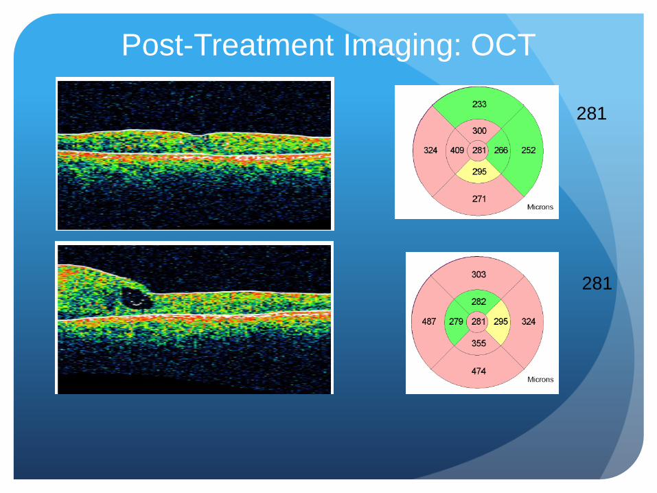
Post-Treatment Imaging: OCT
281
281
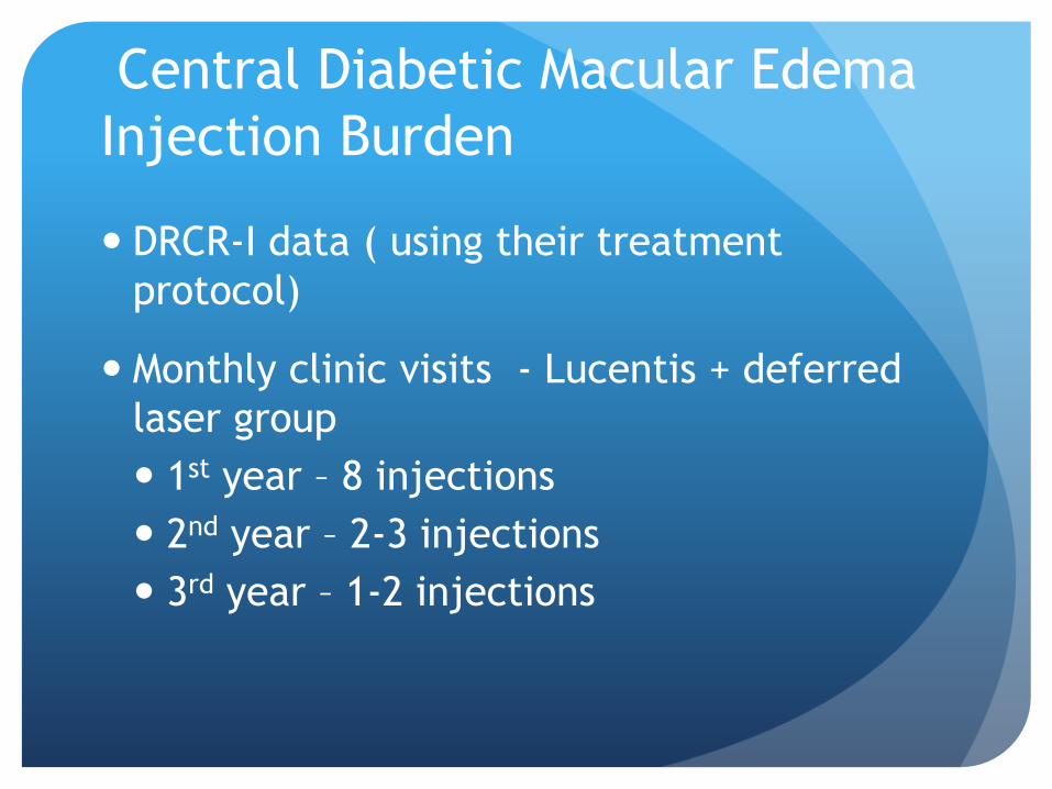
Central Diabetic Macular Edema
Injection Burden
DRCR-I data ( using their treatment
protocol)
Monthly clinic visits - Lucentis + deferred
laser group
1st year – 8 injections
2nd year – 2-3 injections
3rd year – 1-2 injections
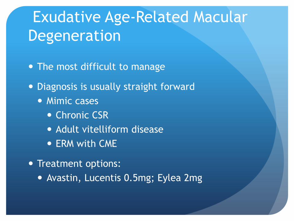
Exudative Age-Related Macular
Degeneration
The most difficult to manage
Diagnosis is usually straight forward
Mimic cases
Chronic CSR
Adult vitelliform disease
ERM with CME
Treatment options:
Avastin, Lucentis 0.5mg; Eylea 2mg
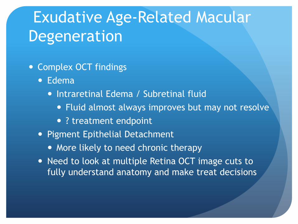
Exudative Age-Related Macular
Degeneration
Complex OCT findings
Edema
Intraretinal Edema / Subretinal fluid
Fluid almost always improves but may not resolve
? treatment endpoint
Pigment Epithelial Detachment
More likely to need chronic therapy
Need to look at multiple Retina OCT image cuts to
fully understand anatomy and make treat decisions

Note: Vertical bars are ± one standard error of the mean.
Anchor Study: Secondary Endpoint:
Mean Change in Visual Acuity Over Time
+11.3
+8.5
–9.5
20.8
letter
difference*
18.0
letter
difference*
* P < 0.0001
PDT (n=143) Ranibizumab 0.3 mg (n=140) Ranibizumab 0.5 mg (n=139)
ET
DR
S lett
ers
Month -15
-10
-5
0
5
10
15
0 1 2 3 4 5 6 7 8 9 10 11 12

CATT Study ( Comparison of Age-Related
Macular Degeneration Treatment Trial) Comparison of Avastin vs Lucentis ( Ranubizimab) for wet AMD therapy
Multicenter clinical trial sponsored by National Eye Institute ( not Genentech!)
Designed to study the comparative efficacy of Avastin vs Lucentis, and also evaluate the “current practice” of PRN therapy vs monthly treatment regimens
1107 patients ; monthly follow-up
4 treatment groups
Monthly Lu x1 yr ; re-randomization to monthly vs PRN dosing Monthly Avastin x1 yr ; re-randomization to monthly vs PRN dosing
Lu PRN dosing x 2 yrs; after initial tx monthly evaluation and treat based signs of lesion activity
Avastin PRN dosing x 2 yrs; after initial tx monthly evaluation and treat based signs of lesion activity

CATT Study Summary
Lucentis 0.5mg vs Avastin 1.25mg were equivalent in final visual outcome at 1 and 2 years
Lucentis was better than Avastin in achieving a “dry” macula
Monthly dosing ( Lucentis / Avastin ) vs PRN therapy by 2 years
Achieved slightly better final visual outcome vs PRN
Higher rate of geographic atrophy development
Higher rate of endophthalmitis
10/11 pts in monthly treatment arm
0.06% endophthalmitis injection rate

View Study Results
Eylea q 4 week or q 8 week dosing interval ( after
monthly losing dose x 3) non-inferior to monthly
Lucentis 0.5mg
Dry macula at 4 months:
Lucentis 0.5mg- 70%
Eylea – 80%
Eylea “Persistent edema” patients – better VA
outcome with q 4 week therapy vs q 8 week
therapy
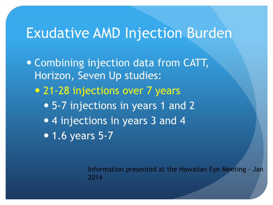
Exudative AMD Injection Burden
Combining injection data from CATT,
Horizon, Seven Up studies:
21-28 injections over 7 years
5-7 injections in years 1 and 2
4 injections in years 3 and 4
1.6 years 5-7
Information presented at the Hawaiian Eye Meeting - Jan
2014
Exudative Age-Related Macular
Degeneration
Given more complex decision making
process and evolving treatment paradigm –
Injector alone model with Retina co-
management may be best option
Summary
Intravitreal injection therapies have revolutionized
the treatment of retinal disorders
Frequent injections are needed to stabilize
disorders – many patients need chronic therapy
The number of annual injections performed per
year remains on a significant growth curve
Comprehensive ophthalmologists will play an
important role in the co-management of these
patients


