MINISYMPOSIUM: IMAGING OF CHILDHOOD TUBERCULOSIS
Standardized radiographic interpretation of thoracictuberculosis in children
Nathan David P. Concepcion1,2& Bernard F. Laya1,2 & Savvas Andronikou3
&
Pedro A. N. Daltro4 & Marion O. Sanchez5 & Jacqueline Austine U. Uy1 &
Timothy Reynold U. Lim2
Received: 27 January 2017 /Accepted: 9 April 2017 /Published online: 26 August 2017# The Author(s) 2017. This article is an open access publication
Abstract There is a lack of standardized approach and termi-nology to classify the diverse spectrum of manifestations intuberculosis. It is important to recognize the different clinicaland radiographic patterns to guide treatment. As a result ofchanging epidemiology, there is considerable overlap in theradiologic presentations of primary tuberculosis and post-primary tuberculosis. In this article we promote a standardizedapproach in clinical and radiographic classification for chil-dren suspected of having or diagnosed with childhood tuber-culosis. We propose standardized terms to diminish confusionand miscommunication, which can affect management. In ad-dition, we present pitfalls and limitations of imaging.
Keywords Children . Computed tomography . Ghon focus .
Progressiveprimary tuberculosis .Radiography .Tuberculosis
Introduction
Numerous articles have been published regarding childhoodtuberculosis but there is still a lack of standardized approachand terminology to classify the diverse spectrum of manifes-tations in tuberculosis. It is important to recognize the differ-ent clinical and radiographic patterns because management ofeach condition is varied. A classification of disease manifes-tation that is common to all will help to facilitate communica-tion and understanding among scientific communities.
Generally the pathological changes in childhood tuberculosisare pauci-bacillary, and thus the diagnosis of intrathoracic tuber-culosis depends largely on chest imaging [1]. Pulmonary tuber-culosis has been classically classified as primary tuberculosis inchildren and post-primary tuberculosis in adults. However, be-cause of the changing epidemiology there is a considerableoverlap in the radiologic presentations of these entities [2].
In this article we promote a standardized combined clinicaland radiographic approach for children suspected of having ordiagnosed with childhood tuberculosis. We propose standard-ized terms to diminish confusion and miscommunication,which can affect management. In addition, we present pitfallsand limitations of imaging in childhood tuberculosis diagno-sis. The atypical radiologic patterns seen in immunocompro-mised children, however, are not discussed in this article.
Clinical categories for intrathoracic tuberculosisin children [3–5]
Intrathoracic tuberculosis has been classified based on clini-cal, laboratory and radiologic evidence. The child should pres-ent at least one sign or symptom suggestive of tuberculosis butwithout any other plausible etiology. These signs and
* Savvas [email protected]
1 Section of Pediatric Radiology, Institute of Radiology,St. Luke’s Medical Center, Bonifacio Global City,Taguig City, Philippines
2 Section of Pediatric Radiology, Institute of Radiology,St. Luke’s Medical Center, Quezon City, Philippines
3 Department of Paediatric Radiology,Bristol Royal Hospital for Children and the University of Bristol,Bristol, UK
4 Section of Pediatric Radiology,Clínica de Diagnóstico por Imagem, Rio de Janeiro,Brazil
5 Section of Pediatric Pulmonology,Institute of Pulmonary Medicine, St. Luke’s Medical Center,Quezon City, Philippines
Pediatr Radiol (2017) 47:1237–1248DOI 10.1007/s00247-017-3868-z
symptoms include any of (a) persistent cough; (b) weightloss/failure to thrive; (c) persistent unexplained fever, or (d)persistent, unexplained lethargy or reduced activity. The fol-lowing sections are definitions that have been proposed toindicate the degree of certainty of the diagnosis oftuberculosis.
Confirmed tuberculosis
Tuberculosis is confirmed when the culture from a specimenrepresentative of intrathoracic disease (e.g., sputum,nasopharyngeal/gastric aspirate, pleural fluid) is positive,and more recently when Xpert MTB/RIF — a rapid test tosimultaneously detect Mycobacterium tuberculosis and resis-tance to rifampicin — from any specimen is positive.
The Xpert MTB/RIF is not only sensitive and specific fordiagnosing pediatric pulmonary mycobacterial tuberculosisbut is also effective in detecting rifampicin resistance [6, 7].The World Health Organization in 2013 [8] strongly recom-mended Xpert MTB/RIF for use rather than conventional mi-croscopy, culture and drug-susceptibility testing as the initialdiagnostic test in children suspected of having multidrug-resistant-tuberculosis or human immunodeficiency virus(HIV)-associated tuberculosis. This test can also be used (con-ditional recommendation) rather than conventional microsco-py and culture as the initial diagnostic test in all childrensuspected of having tuberculosis.
Probable tuberculosis
Children in this category have chest radiographs showingfindings consistent with intrathoracic tuberculosis disease,and at least one of the following:
(a) positive clinical response to anti-tuberculosis therapy,(b) documented exposure/close contact with a known tuber-
culosis patient, or(c) positive tuberculin skin test or interferon-gamma release
assay.
Possible tuberculosis
There are two scenarios in this category. One is when the chestradiograph is not consistent with tuberculous disease, but atleast one of the criteria in the prior section is present. The otherpossibility is when the chest radiography is consistent withtuberculosis disease but none of the criteria in the prior sectionis present.
Some children are symptomatic but have chest radiographyfindings that are not consistent with tuberculous disease andhave none of the criteria mentioned in probable tuberculosis.These children are categorized as either unlikely tuberculosis
if no alternative diagnosis is established or not tuberculosis ifan alternative diagnosis is established such as cardiac disease,foreign body aspiration or asthma. Those who have docu-mented exposure or close contact with a known tuberculosispatient but are asymptomatic and have negative tuberculinskin test and chest radiography are considered tuberculosisexposed. No treatment is necessary for these children.
Tuberculous infection and tuberculous disease have to bedifferentiated because treatments for these two entities aredifferent. We propose simple definitions. When a child has apositive tuberculin skin test but does not show any of theprobable tuberculosis symptoms and has a normal chest radio-graph, this might be classified as tuberculous infection andcould be treated with one-drug therapy [9]. However whenthe findings in the chest radiographs are consistent with tuber-culosis, this is considered tuberculous disease and warrantstreatment with at least three drugs [9–11]. In the next sectionswe discuss chest radiography findings that are consistent withtuberculous disease seen in possible or probable tuberculosis.
Primary pulmonary tuberculous disease
The major route of Mycobacterium tuberculosis infection isby inhalation [1]. Infection begins when infected droplets aredeposited in the terminal airway or alveoli, followed by alocalized parenchymal inflammation or pneumonic processcalled the primary (Ghon) focus. There is then spread viadraining lymphatic vessels, usually to the ipsilateral centralor regional lymph nodes, which then enlarge. The upper lobesdrain to the ipsilateral paratracheal nodes, while the rest of thelung drains to the perihilar nodes. The parenchymal focus andthe enlarged lymph nodes are called the primary (Ranke orGhon) complex [1, 2, 12–17] (Figs. 1 and 2).
Incubation can be up to 6 weeks from exposure, duringwhich time chest radiographs are normal. After 1–3 monthsfrom exposure, hilar or mediastinal adenopathy can be visual-ized in 50–70% of cases [18–20]. Primary tuberculosis re-flects a patient’s conversion from insensitivity to having theantigens of the tubercle bacilli [2, 14].
Regional (perihilar or paratracheal) lymphadenopathy isthe radiologic hallmark of primary infection in childhood [1,13] (Figs. 3 and 4). Anteroposterior and lateral views are re-quired for optimal lymph node visualization [13, 21], but itcan remain difficult to visualize enlarged lymph nodes withcertainty [1, 22]. The most common sites of nodal involve-ment are the right paratracheal and hilar regions [13, 17].
The prevalence of adenopathy decreases with age; it is100% in children <3 years of age and 88% in older children.The prevalence of parenchymal involvement detectable onradiographs, however, is significantly lower in children<3 years of age (51%) as compared with that in older chil-dren (78%) [13].
1238 Pediatr Radiol (2017) 47:1237–1248
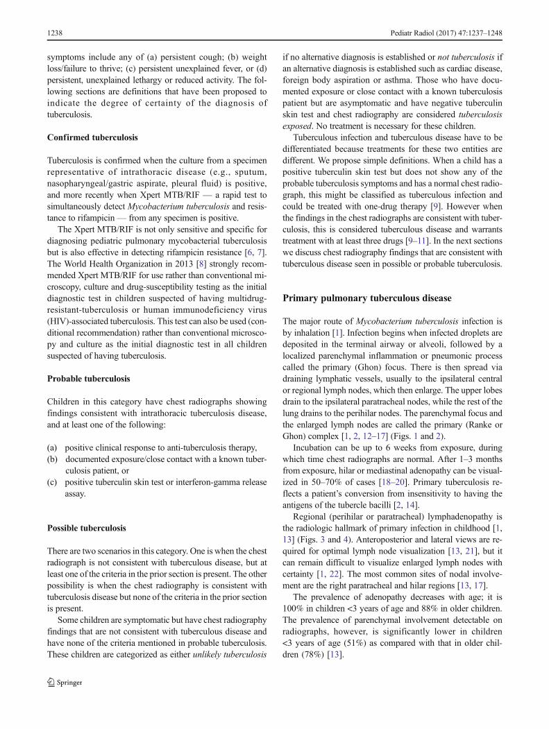
If the child is immunocompetent, the lesions heal and be-come dormant while still causing continuous antigenic stimu-lation for maintenance of hypersensitivity to tuberculous anti-gen. Thus the tuberculin skin test is positive in 95% of cases.This has been referred to as latent tuberculous infection. Thecaseating necrosis within the Ghon focus and infected lymphnode frequently calcifies [1, 2]. Calcification can occur from6 months to 4 years after infection, occurring earlier in youngchildren [1]. The parenchymal focus, called pulmonarytuberculomas (Fig. 5), which are identified radiographically,represent sharply defined ovoid granulomas, solitary or mul-tiple, ranging in size from 0.4 cm to 5 cm in diameter [2, 23].Children with latent tuberculous infection can be treated witha one-drug therapy provided there was no prior treatment.
If immunity is inadequate, the disease progresses eitherlocally or in other parts of the lung or body, with spread of
infection via the airways, lymphatics or bloodstream [15, 16].Clinically active tuberculous disease can develop within5 years after infection. This is called progressive primary tu-berculosis [2, 24].
Progressive primary tuberculous disease
Progression from infection to disease usually occurs within1 year after the primary infection in more than 90% of cases.It is bimodal in age distribution, with children younger than5 years and adolescents being at increased risk [1, 17, 25].
Early disease progression can happen 2–6 months fromexposure, when homogeneous consolidation can occur(Fig. 6). Obstructive atelectasis or overinflation can result fromcompression by an adjacent enlarged node. Distribution is
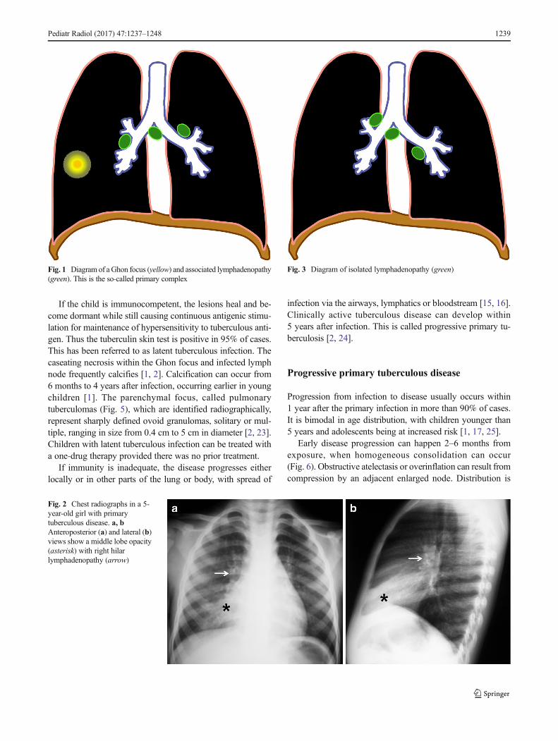
Fig. 2 Chest radiographs in a 5-year-old girl with primarytuberculous disease. a, bAnteroposterior (a) and lateral (b)views show a middle lobe opacity(asterisk) with right hilarlymphadenopathy (arrow)
Fig. 3 Diagram of isolated lymphadenopathy (green)Fig. 1 Diagram of aGhon focus (yellow) and associated lymphadenopathy(green). This is the so-called primary complex
Pediatr Radiol (2017) 47:1237–1248 1239
typically on the right side at the level of the right lobar bron-chus or bronchus intermedius. Fibrosis and destruction of thelung parenchyma result in traction bronchiectasis and forma-tion of cavities (Fig. 7), respectively [2]. This is known asprogressive Ghon focus [18–20].
Three possible mechanisms are involved in the formationof cavities: (1) progressive primary spread of disease withextensive and bilateral pulmonary cavities, (2) cavitiescaused by bronchial obstruction by lymph nodes or (3)post-primary tuberculosis showing cavities that are usuallysingle and unilateral in the upper lobe. These have fairlyequal incidence [26].
Lymph nodes can continue to enlarge 4–12 months fromexposure and can cause progression of the disease affectingthe airways, pleura and pericardium, which are discussed inthe next sections. On contrast-enhanced CT, involved lymphnodes often measure more than 2 cm and show a very char-acteristic, but not pathognomonic, rim sign consisting of alow-density center surrounded by a peripheral enhancing rim[17, 22, 27] (Fig. 6). The esophagus, lymphatic duct and
phrenic nerve might also be affected, producingtracheoesophageal fistula, chylothorax and diaphragmatic pal-sy, respectively [1, 18–20]. Hematogenous miliary dissemina-tion can also occur in this stage.
Miliary tuberculosis
Miliary tuberculosis is seen in 8% of cases [25], usually in theyounger age group because of immature immune function[28]. It is an acute hematogenously disseminated infectionpresenting as innumerable ≤2-mm non-calcified nodulesscattered in both lungs [1, 2, 18–20, 28–30] (Figs. 8 and 9).There is no pathognomonic finding for tuberculosis except formiliary tuberculosis [28], and it can be seen in primary andpost-primary disease [2].
In 25–40% cases, chest radiographs are initially normal [1,30]. CT is more sensitive for miliary disease before it becomesradiographically apparent. The tiny nodules can be sharply orpoorly defined, and are seen in a diffuse, random distribution,often with intra- and interlobular septal thickening [16].
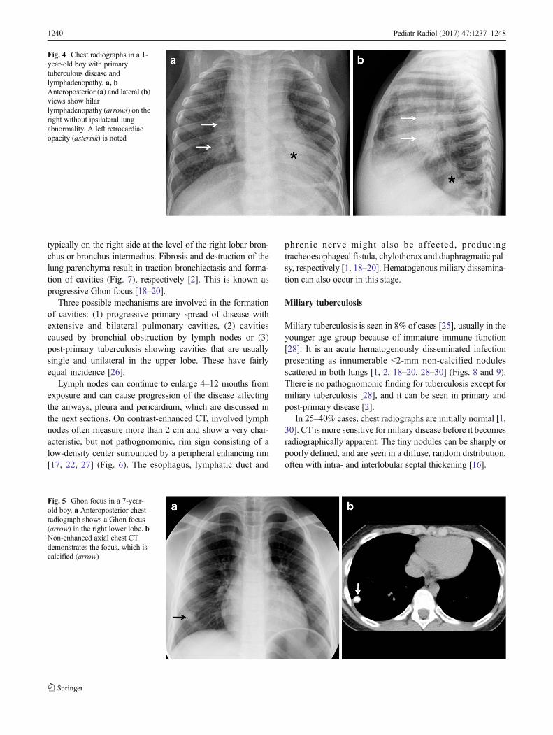
Fig. 4 Chest radiographs in a 1-year-old boy with primarytuberculous disease andlymphadenopathy. a, bAnteroposterior (a) and lateral (b)views show hilarlymphadenopathy (arrows) on theright without ipsilateral lungabnormality. A left retrocardiacopacity (asterisk) is noted
Fig. 5 Ghon focus in a 7-year-old boy. a Anteroposterior chestradiograph shows a Ghon focus(arrow) in the right lower lobe. bNon-enhanced axial chest CTdemonstrates the focus, which iscalcified (arrow)
1240 Pediatr Radiol (2017) 47:1237–1248
Lymphobronchial/lymphotracheobronchial tuberculosis
Lymphobronchial or lymphotracheobronchial involvement isa complication in 2–4% of tuberculosis cases [2, 31].Lymphadenopathy is seen in chest radiographs in 63–95%[26] and on CT in up to 96–100% of tracheobronchial tuber-culosis cases [32, 33]. The enlarged nodes compress the adja-cent trachea or bronchi, causing luminal narrowing andresulting in lung hyperinflation from partial obstruction withcheck-valve effect (Fig. 10), or atelectasis due to a completeobstruction (Figs. 11 and 12). These nodes subsequentlyerode, perforate and discharge caseous material into the air-ways manifesting as obstructive pneumonia [1, 2, 13, 14,17–20, 33–36]. Lymphogenic and hematogenous spread intothe large airways have also been reported [2].
Radiographic manifestations in lymphotracheobronchialtuberculosis are nonspecific, and a normal chest radiographdoes not rule out airway involvement. Involvement of thecentral airways can be easily missed on radiographs.Persistent segmental or lobar collapse, lobar hyperinflationand obstructive pneumonia are seen as complications of theairway compression [31, 33].
Enhancement and enlargement (usually >2 cm) of ad-jacent mediastinal lymph nodes are common findings atCT in the active stage of stenosis. The enlarged lymphnodes are commonly identified in the subcarinal [37],paratracheal and perihilar (infrahilar) regions closely abut-ting or compressing the airways [33].
The most commonly involved airway is the bronchusintermedius, followed by the left main bronchus and trachea[37]. Bronchial narrowing can be smooth or irregular, withmural thickening [33, 38]. Smooth bronchial narrowing iscaused by compression by an adjacent node, and the irregularnarrowing correlates with significant mucosal irregularity, ca-seation, granuloma formation or even perforation [33].
The obstructive infiltrates can be resorbed or calcify,fibrose with traction bronchiectasis (Fig. 13) or cause lungdestruction [2]. There is often excessive inflammation, whichcan result in dense alveolar consolidation and eventual paren-chymal breakdown [1]. Cicatricial bronchostenosis can man-ifest as concentric narrowing, uniform wall thickening, andinvolvement of a long bronchial segment after healing [31].
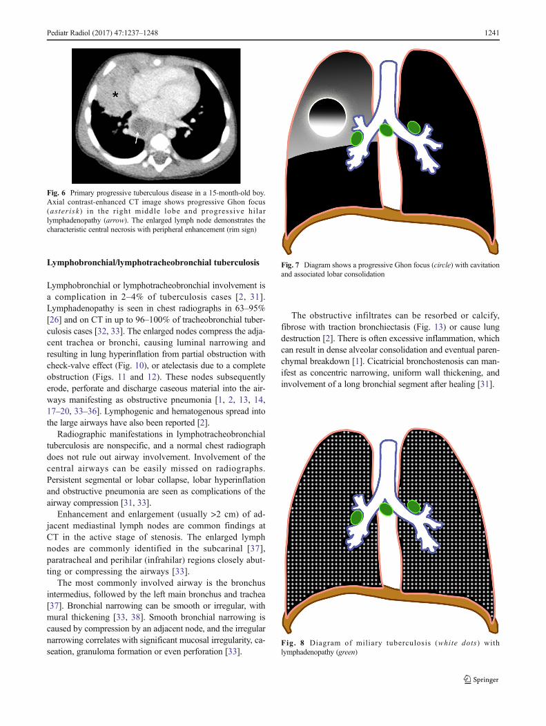
Fig. 7 Diagram shows a progressive Ghon focus (circle) with cavitationand associated lobar consolidation
Fig. 8 Diagram of miliary tuberculosis (white dots) withlymphadenopathy (green)
Fig. 6 Primary progressive tuberculous disease in a 15-month-old boy.Axial contrast-enhanced CT image shows progressive Ghon focus(asterisk) in the right middle lobe and progressive hi larlymphadenopathy (arrow). The enlarged lymph node demonstrates thecharacteristic central necrosis with peripheral enhancement (rim sign)
Pediatr Radiol (2017) 47:1237–1248 1241
Pleural tuberculous disease
Another site of extrapulmonary involvement, aside fromlymph nodes, is the pleurae [25]. Its prevalence increaseswith age. Pleural effusion (Figs. 14 and 15) most oftenresults from obstruction of the lymphatic drainage or hy-persensitivity reaction than from direct seeding into thepleura. This explains why pleural fluid cultures are mostlynegative [28]. Spread to the pleura might also come froma caseating granuloma near the pleura or via hematoge-nous dissemination [2].
As a complication of primary tuberculosis, pleural involve-ment is most frequently observed in older children and ado-lescents. It can occur 3–6 months after infection and is some-times asymptomatic [2]. It is also typically appreciated in as-sociation with parenchymal or nodal disease [39]. Pleural ef-fusions are associated with air-space consolidation in 29% andcould be bilateral or loculated in 6% of cases [17]. This isusually self-limiting and prognosis is good. Residual pleuralcalcifications appear in some cases [2].
The effusion can complicate into an exudative effusion,empyema or infiltration of the thoracic duct [18–20].Contrast-enhanced CT scan shows smooth thickening of vis-ceral and parietal pleura (“split-pleura” sign) [40]. An Air-fluid level in the pleural space indicates presence ofbronchopleural fistula [41]. The empyema can also spreadbeyond the parietal pleura to produce a subcutaneous abscess,called empyema necessitatis [42].
Pericardial disease
Tuberculous pericarditis is a relatively uncommon complica-tion of primary tuberculosis. It has been reported in 1% ofcases. It is commonly caused by direct extension of lymphnodes into the posterior pericardial sac [28], although miliaryspread has been reported [28, 43]. CT shows lymphadenopa-thy and pericardial thickening with or without effusion.Constrictive pericarditis with fibrous or calcified pericardialthickening of usually >3 mm occur in about 10% of patients
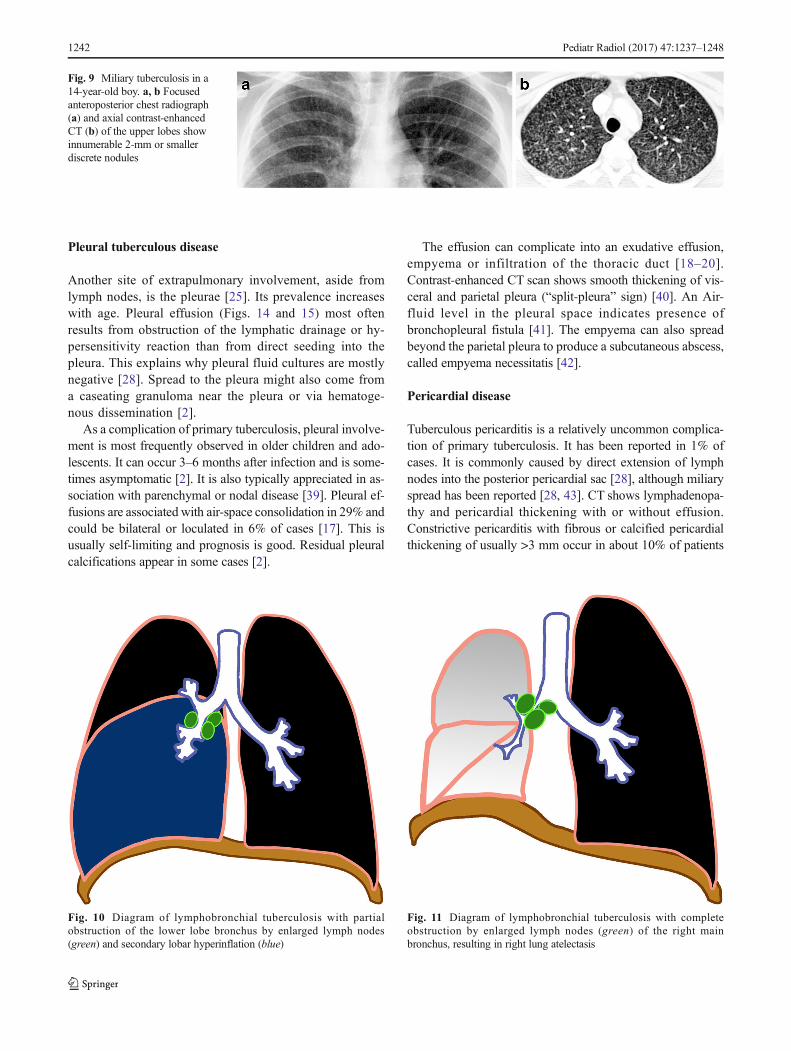
Fig. 11 Diagram of lymphobronchial tuberculosis with completeobstruction by enlarged lymph nodes (green) of the right mainbronchus, resulting in right lung atelectasis
Fig. 10 Diagram of lymphobronchial tuberculosis with partialobstruction of the lower lobe bronchus by enlarged lymph nodes(green) and secondary lobar hyperinflation (blue)
Fig. 9 Miliary tuberculosis in a14-year-old boy. a, b Focusedanteroposterior chest radiograph(a) and axial contrast-enhancedCT (b) of the upper lobes showinnumerable 2-mm or smallerdiscrete nodules
1242 Pediatr Radiol (2017) 47:1237–1248
[43]. Pericardial effusion (Figs. 15, 16 and 17), commonlyserous type [28], can result in globular enlargement of theheart shadow (water bottle sign) [1].
Post-primary tuberculosis
Post-primary tuberculosis is also known as adult-type, reacti-vation or secondary tuberculosis and sometimes phthisis [2].This results from the reactivation of dormant foci. It is further
observed in the pediatric age group, mostly in adolescents [2,13]. It is considered a late disease progression of the primaryinfection, which can occur 8–24 months from exposure and inchildren as young as 8 years [18–20].
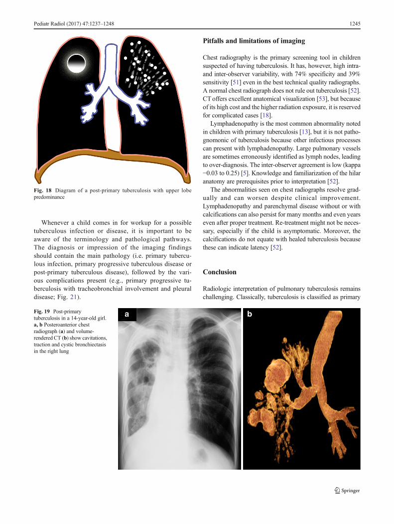
The most commonly affected locations are the apical andposterior segments of the upper lobes and the apical segmentof the lower lobes because of higher oxygen tension (Fig. 18).Initially theremight be cloudy opacification in a segment beforecoalescence and parenchymal breakdown. Complications in-clude cavitation, bronchogenic spread with bronchopneumonicconsolidation, exudative pleuritis, cicatrization atelectasis of the
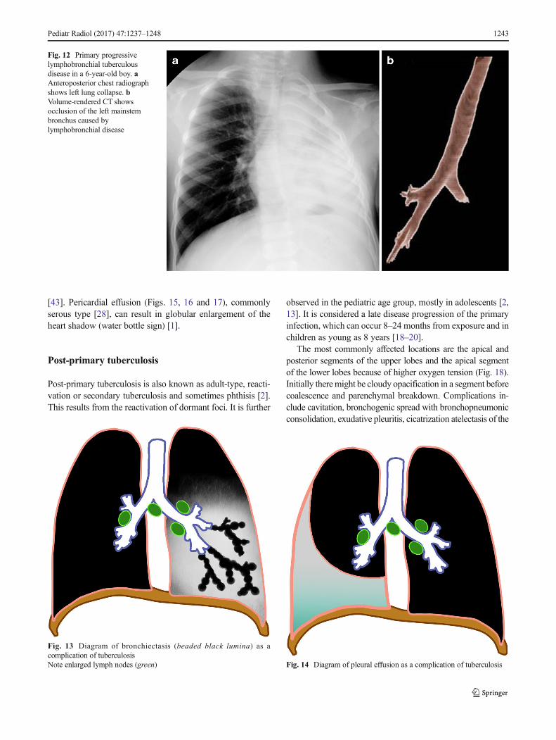
Fig. 12 Primary progressivelymphobronchial tuberculousdisease in a 6-year-old boy. aAnteroposterior chest radiographshows left lung collapse. bVolume-rendered CT showsocclusion of the left mainstembronchus caused bylymphobronchial disease
Fig. 13 Diagram of bronchiectasis (beaded black lumina) as acomplication of tuberculosisNote enlarged lymph nodes (green) Fig. 14 Diagram of pleural effusion as a complication of tuberculosis
Pediatr Radiol (2017) 47:1237–1248 1243
upper lobe with retraction of hilum and formation of tractionbronchiectasis [1, 2, 44–46]. Lymph node enlargement is notcommon in comparison to primary tuberculosis [18–20].
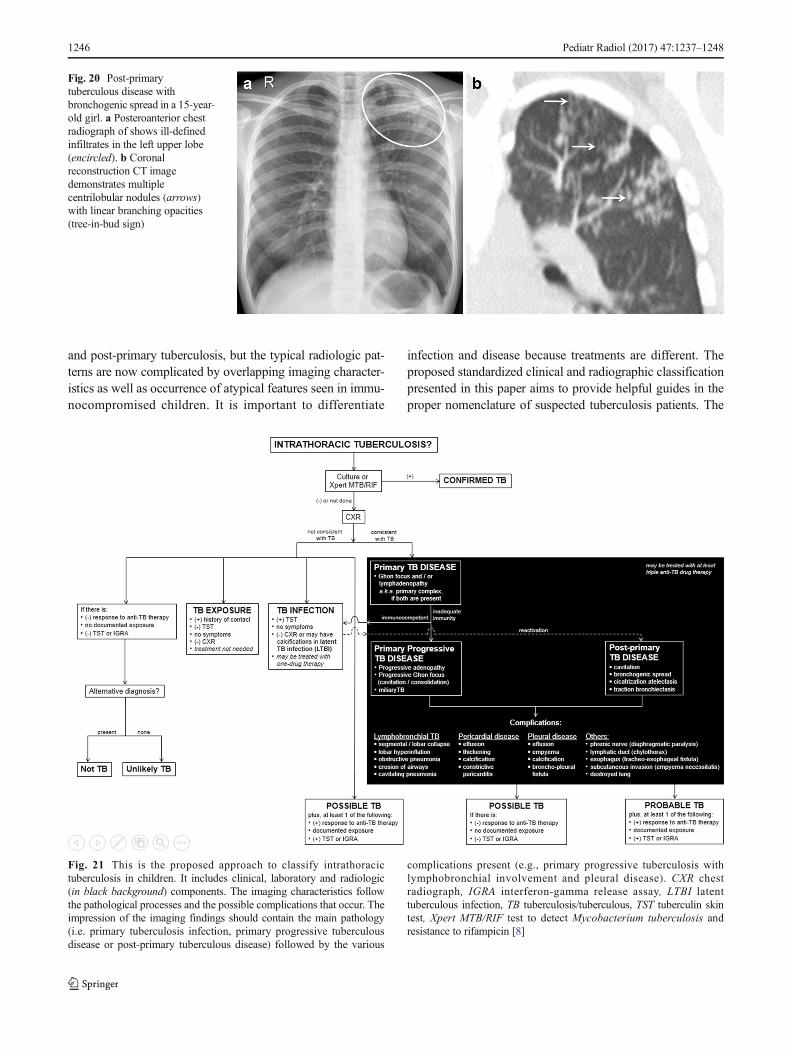
Cavitation is radiographically evident in 40% of cases ofpost-primary disease. The walls of the cavities might appearthin and smooth or thick and nodular. It is difficult to distin-guish thin-walled cavities from bullae, cysts or pneumatoceles.Cystic bronchiectasis should also be considered when multiplecavities are present [47] (Figs. 18 and 19).
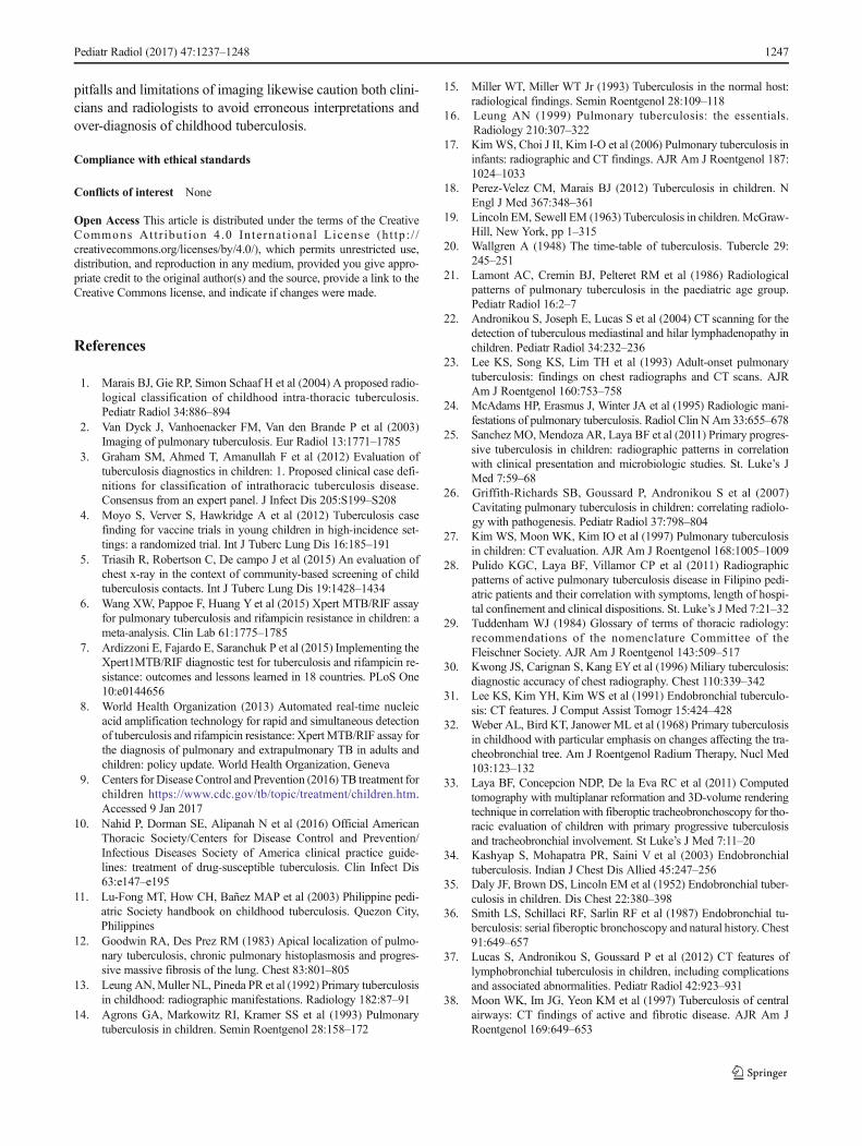
In 20% of post-primary tuberculosis cases, bronchogenicspread appears on radiographs as multiple, ill-definedmicronodules in a segmental or lobar distribution, typicallyin the lower-lung zones [48]. High-resolution CT, the modal-ity of choice, demonstrates centrilobular nodules ranging 2–
4 mm with linear branching opacities (“tree-in-bud” sign)which represent caseous necrosis at and around terminal andrespiratory bronchioles [49] (Fig. 20). Complete destructionof the entire lung or a large part of a lung is not uncommon inthe end stage of tuberculosis. Secondary pyogenic or fungalinfection can occur [47]. Miliary tuberculosis andtuberculomas might also be encountered in post-primary tu-berculosis [2, 50].
Approach to classification of intrathoracictuberculosis
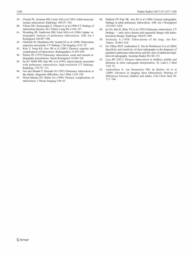
We adapted the clinical and laboratory components of theclassification from Graham et al. [3], Moyo et al. [4] andTriasih [5]. An imaging approach to interpretation is summa-rized in Fig. 21. The radiologic side of the algorithm is basedon the pathological processes that occur in tuberculosis thatresult in various complications.
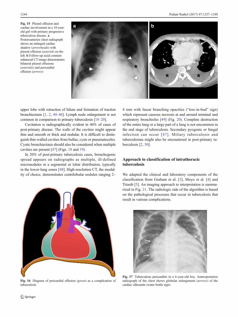
Fig. 15 Pleural effusion andcardiac involvement in a 10-year-old girl with primary progressivetuberculous disease. aPosteroanterior chest radiographshows an enlarged cardiacshadow (arrowheads) withpleural effusion (asterisk) on theleft. b Follow-up axial contrast-enhanced CT image demonstratesbilateral pleural effusions(asterisks) and pericardialeffusion (arrows)
Fig. 16 Diagram of pericardial effusion (green) as a complication oftuberculosis
Fig. 17 Tuberculous pericarditis in a 6-year-old boy. Anteroposteriorradiograph of the chest shows globular enlargement (arrows) of thecardiac silhouette (water bottle sign)
1244 Pediatr Radiol (2017) 47:1237–1248
Whenever a child comes in for workup for a possibletuberculous infection or disease, it is important to beaware of the terminology and pathological pathways.The diagnosis or impression of the imaging findingsshould contain the main pathology (i.e. primary tubercu-lous infection, primary progressive tuberculous disease orpost-primary tuberculous disease), followed by the vari-ous complications present (e.g., primary progressive tu-berculosis with tracheobronchial involvement and pleuraldisease; Fig. 21).
Pitfalls and limitations of imaging
Chest radiography is the primary screening tool in childrensuspected of having tuberculosis. It has, however, high intra-and inter-observer variability, with 74% specificity and 39%sensitivity [51] even in the best technical quality radiographs.A normal chest radiograph does not rule out tuberculosis [52].CT offers excellent anatomical visualization [53], but becauseof its high cost and the higher radiation exposure, it is reservedfor complicated cases [18].
Lymphadenopathy is the most common abnormality notedin children with primary tuberculosis [13], but it is not patho-gnomonic of tuberculosis because other infectious processescan present with lymphadenopathy. Large pulmonary vesselsare sometimes erroneously identified as lymph nodes, leadingto over-diagnosis. The inter-observer agreement is low (kappa−0.03 to 0.25) [5]. Knowledge and familiarization of the hilaranatomy are prerequisites prior to interpretation [52].
The abnormalities seen on chest radiographs resolve grad-ually and can worsen despite clinical improvement.Lymphadenopathy and parenchymal disease without or withcalcifications can also persist for many months and even yearseven after proper treatment. Re-treatment might not be neces-sary, especially if the child is asymptomatic. Moreover, thecalcifications do not equate with healed tuberculosis becausethese can indicate latency [52].
Conclusion
Radiologic interpretation of pulmonary tuberculosis remainschallenging. Classically, tuberculosis is classified as primary
Fig. 19 Post-primarytuberculosis in a 14-year-old girl.a, b Posteroanterior chestradiograph (a) and volume-rendered CT (b) show cavitations,traction and cystic bronchiectasisin the right lung
Fig. 18 Diagram of a post-primary tuberculosis with upper lobepredominance
Pediatr Radiol (2017) 47:1237–1248 1245
and post-primary tuberculosis, but the typical radiologic pat-terns are now complicated by overlapping imaging character-istics as well as occurrence of atypical features seen in immu-nocompromised children. It is important to differentiate
infection and disease because treatments are different. Theproposed standardized clinical and radiographic classificationpresented in this paper aims to provide helpful guides in theproper nomenclature of suspected tuberculosis patients. The
Fig. 21 This is the proposed approach to classify intrathoracictuberculosis in children. It includes clinical, laboratory and radiologic(in black background) components. The imaging characteristics followthe pathological processes and the possible complications that occur. Theimpression of the imaging findings should contain the main pathology(i.e. primary tuberculosis infection, primary progressive tuberculousdisease or post-primary tuberculous disease) followed by the various
complications present (e.g., primary progressive tuberculosis withlymphobronchial involvement and pleural disease). CXR chestradiograph, IGRA interferon-gamma release assay, LTBI latenttuberculous infection, TB tuberculosis/tuberculous, TST tuberculin skintest, Xpert MTB/RIF test to detect Mycobacterium tuberculosis andresistance to rifampicin [8]
Fig. 20 Post-primarytuberculous disease withbronchogenic spread in a 15-year-old girl. a Posteroanterior chestradiograph of shows ill-definedinfiltrates in the left upper lobe(encircled). b Coronalreconstruction CT imagedemonstrates multiplecentrilobular nodules (arrows)with linear branching opacities(tree-in-bud sign)
1246 Pediatr Radiol (2017) 47:1237–1248
pitfalls and limitations of imaging likewise caution both clini-cians and radiologists to avoid erroneous interpretations andover-diagnosis of childhood tuberculosis.
Compliance with ethical standards
Conflicts of interest None
Open Access This article is distributed under the terms of the CreativeCommons At t r ibut ion 4 .0 In te rna t ional License (h t tp : / /creativecommons.org/licenses/by/4.0/), which permits unrestricted use,distribution, and reproduction in any medium, provided you give appro-priate credit to the original author(s) and the source, provide a link to theCreative Commons license, and indicate if changes were made.
References
1. Marais BJ, Gie RP, Simon Schaaf H et al (2004) A proposed radio-logical classification of childhood intra-thoracic tuberculosis.Pediatr Radiol 34:886–894
2. Van Dyck J, Vanhoenacker FM, Van den Brande P et al (2003)Imaging of pulmonary tuberculosis. Eur Radiol 13:1771–1785
3. Graham SM, Ahmed T, Amanullah F et al (2012) Evaluation oftuberculosis diagnostics in children: 1. Proposed clinical case defi-nitions for classification of intrathoracic tuberculosis disease.Consensus from an expert panel. J Infect Dis 205:S199–S208
4. Moyo S, Verver S, Hawkridge A et al (2012) Tuberculosis casefinding for vaccine trials in young children in high-incidence set-tings: a randomized trial. Int J Tuberc Lung Dis 16:185–191
5. Triasih R, Robertson C, De campo J et al (2015) An evaluation ofchest x-ray in the context of community-based screening of childtuberculosis contacts. Int J Tuberc Lung Dis 19:1428–1434
6. Wang XW, Pappoe F, Huang Y et al (2015) Xpert MTB/RIF assayfor pulmonary tuberculosis and rifampicin resistance in children: ameta-analysis. Clin Lab 61:1775–1785
7. Ardizzoni E, Fajardo E, Saranchuk P et al (2015) Implementing theXpert1MTB/RIF diagnostic test for tuberculosis and rifampicin re-sistance: outcomes and lessons learned in 18 countries. PLoS One10:e0144656
8. World Health Organization (2013) Automated real-time nucleicacid amplification technology for rapid and simultaneous detectionof tuberculosis and rifampicin resistance: Xpert MTB/RIF assay forthe diagnosis of pulmonary and extrapulmonary TB in adults andchildren: policy update. World Health Organization, Geneva
9. Centers for Disease Control and Prevention (2016) TB treatment forchildren https://www.cdc.gov/tb/topic/treatment/children.htm.Accessed 9 Jan 2017
10. Nahid P, Dorman SE, Alipanah N et al (2016) Official AmericanThoracic Society/Centers for Disease Control and Prevention/Infectious Diseases Society of America clinical practice guide-lines: treatment of drug-susceptible tuberculosis. Clin Infect Dis63:e147–e195
11. Lu-Fong MT, How CH, Bañez MAP et al (2003) Philippine pedi-atric Society handbook on childhood tuberculosis. Quezon City,Philippines
12. Goodwin RA, Des Prez RM (1983) Apical localization of pulmo-nary tuberculosis, chronic pulmonary histoplasmosis and progres-sive massive fibrosis of the lung. Chest 83:801–805
13. Leung AN,Muller NL, Pineda PR et al (1992) Primary tuberculosisin childhood: radiographic manifestations. Radiology 182:87–91
14. Agrons GA, Markowitz RI, Kramer SS et al (1993) Pulmonarytuberculosis in children. Semin Roentgenol 28:158–172
15. Miller WT, Miller WT Jr (1993) Tuberculosis in the normal host:radiological findings. Semin Roentgenol 28:109–118
16. Leung AN (1999) Pulmonary tuberculosis: the essentials.Radiology 210:307–322
17. KimWS, Choi J II, Kim I-O et al (2006) Pulmonary tuberculosis ininfants: radiographic and CT findings. AJR Am J Roentgenol 187:1024–1033
18. Perez-Velez CM, Marais BJ (2012) Tuberculosis in children. NEngl J Med 367:348–361
19. Lincoln EM, Sewell EM (1963) Tuberculosis in children. McGraw-Hill, New York, pp 1–315
20. Wallgren A (1948) The time-table of tuberculosis. Tubercle 29:245–251
21. Lamont AC, Cremin BJ, Pelteret RM et al (1986) Radiologicalpatterns of pulmonary tuberculosis in the paediatric age group.Pediatr Radiol 16:2–7
22. Andronikou S, Joseph E, Lucas S et al (2004) CT scanning for thedetection of tuberculous mediastinal and hilar lymphadenopathy inchildren. Pediatr Radiol 34:232–236
23. Lee KS, Song KS, Lim TH et al (1993) Adult-onset pulmonarytuberculosis: findings on chest radiographs and CT scans. AJRAm J Roentgenol 160:753–758
24. McAdams HP, Erasmus J, Winter JA et al (1995) Radiologic mani-festations of pulmonary tuberculosis. Radiol Clin N Am 33:655–678
25. Sanchez MO, Mendoza AR, Laya BF et al (2011) Primary progres-sive tuberculosis in children: radiographic patterns in correlationwith clinical presentation and microbiologic studies. St. Luke’s JMed 7:59–68
26. Griffith-Richards SB, Goussard P, Andronikou S et al (2007)Cavitating pulmonary tuberculosis in children: correlating radiolo-gy with pathogenesis. Pediatr Radiol 37:798–804
27. Kim WS, Moon WK, Kim IO et al (1997) Pulmonary tuberculosisin children: CT evaluation. AJR Am J Roentgenol 168:1005–1009
28. Pulido KGC, Laya BF, Villamor CP et al (2011) Radiographicpatterns of active pulmonary tuberculosis disease in Filipino pedi-atric patients and their correlation with symptoms, length of hospi-tal confinement and clinical dispositions. St. Luke’s J Med 7:21–32
29. Tuddenham WJ (1984) Glossary of terms of thoracic radiology:recommendations of the nomenclature Committee of theFleischner Society. AJR Am J Roentgenol 143:509–517
30. Kwong JS, Carignan S, Kang EYet al (1996) Miliary tuberculosis:diagnostic accuracy of chest radiography. Chest 110:339–342
31. Lee KS, Kim YH, Kim WS et al (1991) Endobronchial tuberculo-sis: CT features. J Comput Assist Tomogr 15:424–428
32. Weber AL, Bird KT, Janower ML et al (1968) Primary tuberculosisin childhood with particular emphasis on changes affecting the tra-cheobronchial tree. Am J Roentgenol Radium Therapy, Nucl Med103:123–132
33. Laya BF, Concepcion NDP, De la Eva RC et al (2011) Computedtomography with multiplanar reformation and 3D-volume renderingtechnique in correlation with fiberoptic tracheobronchoscopy for tho-racic evaluation of children with primary progressive tuberculosisand tracheobronchial involvement. St Luke’s J Med 7:11–20
34. Kashyap S, Mohapatra PR, Saini V et al (2003) Endobronchialtuberculosis. Indian J Chest Dis Allied 45:247–256
35. Daly JF, Brown DS, Lincoln EM et al (1952) Endobronchial tuber-culosis in children. Dis Chest 22:380–398
36. Smith LS, Schillaci RF, Sarlin RF et al (1987) Endobronchial tu-berculosis: serial fiberoptic bronchoscopy and natural history. Chest91:649–657
37. Lucas S, Andronikou S, Goussard P et al (2012) CT features oflymphobronchial tuberculosis in children, including complicationsand associated abnormalities. Pediatr Radiol 42:923–931
38. Moon WK, Im JG, Yeon KM et al (1997) Tuberculosis of centralairways: CT findings of active and fibrotic disease. AJR Am JRoentgenol 169:649–653
Pediatr Radiol (2017) 47:1237–1248 1247
39. Choyke PL, Sostman HD, Curtis AM et al (1983) Adult-onset pul-monary tuberculosis. Radiology 148:357–362
40. Yilmaz MU, Kumcuoglu Z, Utkaner G et al (1998) CT findings oftuberculous pleurisy. Int J Tuberc Lung Dis 2:164–167
41. Woodring JH, Vandiviere HM, Fried AM et al (1986) Update: ra-diographic features of pulmonary tuberculosis. AJR Am JRoentgenol 146:497–506
42. Glicklich M, Mendelson DS, Gendal ES et al (1990) Tuberculousempyema necessitatis: CT findings. Clin Imaging 14:23–25
43. Kim Y, Song KS, Goo JM et al (2001) Thoracic sequelae andcomplications of tuberculosis. Radiographics 21:839–858
44. Palmer PE (1979) Pulmonary tuberculosis: usual and unusual ra-diographic presentations. Semin Roentgenol 14:204–242
45. Im JG, Webb WR, Han MC et al (1991) Apical opacity associatedwith pulmonary tuberculosis: high-resolution CT findings.Radiology 178:727–731
46. Van den Brande P, Demedts M (1992) Pulmonary tuberculosis inthe elderly: diagnostic difficulties. Eur J Med 1:224–229
47. Winer-Muram HT, Rubin SA (1990) Thoracic complications oftuberculosis. J Thorac Imaging 5:46–63
48. Hadlock FP, Park SK, Awe RJ et al (1980) Unusual radiographicfindings in adult pulmonary tuberculosis. AJR Am J Roentgenol134:1015–1018
49. Im JG, Itoh H, Shim YS et al (1993) Pulmonary tuberculosis: CTfindings — early active disease and sequential change with antitu-berculous therapy. Radiology 186:653–660
50. Sochocky S (1958) Tuberculoma of the lung. Am RevTuberc 78:403–410
51. De Villiers RVP, Andronikou S, Van de Westhuizen S et al (2004)Specificity and sensitivity of chest radiographs in the diagnosis ofpaediatric pulmonary tuberculosis and the value of additional high-kilovolt radiographs. Australas Radiol 48:148–153
52. Laya BF (2011) Thoracic tuberculosis in children: pitfalls anddilemma in chest radiograph interpretation. St. Luke’s J Med7:69–76
53. Andronikou S, van Hoenacker FM, de Backer AI et al(2009) Advances in imaging chest tuberculosis: blurring ofdifferences between children and adults. Clin Chest Med 30:717–744
1248 Pediatr Radiol (2017) 47:1237–1248


