
Received: 24 May 2017 | Accepted: 24 May 2017
DOI: 10.1002/jcp.26031
ORIGINAL RESEARCH ARTICLE
The nucleus is irreversibly shaped by motion of cellboundaries in cancer and non-cancer cells
Vincent J. Tocco1 | Yuan Li1 | Keith G. Christopher1 | James H. Matthews2 |
Varun Aggarwal1 | Lauren Paschall1 | Hendrik Luesch2 | Jonathan D. Licht3 |
Richard B. Dickinson1 | Tanmay P. Lele1,4
1Department of Chemical Engineering,
University of Florida, Gainesville, Florida
2Department of Medicinal Chemistry, Center
for Natural Products, Drug Discovery and
Development (CNPD3), University of Florida,
Gainesville, Florida
3Division of Hematology and Oncology,
Department of Medicine, University of Florida
Health Cancer Center, Gainesville, Florida
4Department of Anatomy and Cell Biology,
University of Florida College of Medicine,
Gainesville, Florida
Correspondence
Tanmay P. Lele, Department of Chemical
Engineering, Bldg. 723, University of Florida,
Gainesville, FL 32611.
Email: [email protected]
Funding information
National Science Foundation, Grant number:
CMMI 1437395; National Institutes of Health,
Grant numbers: R01 EB014869, R01
GM102486, U54CA193419, R01 CA172310
Actomyosin stress fibers impinge on the nucleus and can exert compressive forces on
it. These compressive forces have been proposed to elongate nuclei in fibroblasts, and
lead to abnormally shaped nuclei in cancer cells. In these models, the elongated or
flattened nuclear shape is proposed to store elastic energy. However, we found that
deformed shapes of nuclei are unchanged even after removal of the cell with micro-
dissection, both for smooth, elongated nuclei in fibroblasts and abnormally shaped
nuclei in breast cancer cells. The lack of shape relaxation implies that the nuclear shape
in spread cells does not store any elastic energy, and the cellular stresses that deform
the nucleus are dissipative, not static. During cell spreading, the deviation of the
nucleus from a convex shape increased inMDA-MB-231 cancer cells, but decreased in
MCF-10A cells. Tracking changes of nuclear and cellular shape on micropatterned
substrata revealed that fibroblast nuclei deform only during deformations in cell shape
and only in the direction of nearbymoving cell boundaries.We propose that motion of
cell boundaries exert a stress on the nucleus, which allows the nucleus to mimic cell
shape. The lack of elastic energy in the nuclear shape suggests that nuclear shape
changes in cells occur at constant surface area and volume.
K E YWORD S
cell forces, nuclear mechanics, nuclear shape
1 | INTRODUCTION
The nucleus undergoes changes in its shape as a cell migrates through
tissue and tight spaces (Denais et al., 2016; McGregor, Hsia, &
Lammerding, 2016), but how the nuclear deformation is caused by
changes in cell shape is not well understood. Yet, the mechanism of
nuclear deformation is important, given that the extent of nuclear
deformation limits cell migration through extracellular matrix (Petrie,
Koo, & Yamada, 2014), may change chromatin architecture, and
consequently gene expression (Gupta, Marcel, Sarin, & Shivashankar,
2012; Thomas, Collier, Sfeir, & Healy, 2002; Vergani, Grattarola, &
Nicolini, 2004).
Furthermore, in many forms of cancer, the nucleus commonly has
shape abnormalities, including lobes, invaginations and folds (Chow,
Factor, & Ullman, 2012). Abnormalities in nuclear shape occur early in
tumor progression (Boyd, Pienta, Getzenberg, Coffey, & Barrett, 1991)
and help improve the accuracy of prognosis in clinical settings
(Bloom & Richardson, 1957). Nuclear shape abnormalities correlate
with the biological behavior and clinical prognosis of different cancersVincent J. Tocco and Yuan Li contributed equally to this work.
J Cell Physiol. 2017;1–9. wileyonlinelibrary.com/journal/jcp © 2017 Wiley Periodicals, Inc. | 1

(Bussolati, Marchiò, Gaetano, Lupo, & Sapino, 2008; Elston & Ellis,
1991). Despite the strong diagnostic and prognostic importance of
nuclear shape abnormalities (Giardina et al., 1996; Haroske et al.,
1996), the mechanism by which the nucleus becomes abnormally
deformed in cancer is not understood.
Nuclear deformation is a response of the nucleus to mechanical
stresses. In some models explaining the correlation between nuclear
shape and cell shape, impinging actomyosin impinging actomyosin
stress fibers exert compressive stress on the nucleus. The stress
exerted by these fibers causes an elastic deformation of the nucleus
such that it can become elongated starting from an approximately
spherical shape (Khatau et al., 2009; Versaevel, Grevesse, & Gabriele,
2012). In this model, nuclear shape is determined by the distribution
and magnitude of the forces generated by the stress fibers and the
elastic properties of the nucleus. No time history of the stresses is
required to explain the current nuclear shape; instantaneous cell shape
determines the instantaneous nuclear shape.
Here, we show that both fibroblast nuclei and cancer nuclei in
spread cells do not store elastic energy, challenging the current models
of nuclear shaping. Further, we show that a change in cell shape is
required for deformation of the nucleus during cell migration. Based on
these results, we propose that motion of the cell boundary transmits a
stress to the nuclear surface to shape the normal nucleus and to
amplify shape abnormalities in the cancer nucleus.
2 | MATERIALS AND METHODS
2.1 | Cell culture and transfection
All cells were maintained in a humidified incubator at 37°C and 7%
CO2. Mouse fibroblasts (MEF and NIH 3T3) were cultured in
Dulbecco’s modified Eagle’s medium (DMEM) with 4.5 g/L of glucose
(Mediatech, Manassas, VA), supplemented with 10% donor bovine
serum (DBS; Gibco, Grand Island, NY) and 1% penicillin/streptomycin
(Mediatech). Human breast cancer cells (MDA-MB-231) were cultured
in 4.5 g/L glucose DMEMwithout HEPES and L-glutamine (Invitrogen,
Carlsbad, CA), supplementedwith 10% (v/v) donor bovine serum (DBS,
Gibco), 1% (v/v) 100×MEM non-essential amino acid (Mediatech), 1%
(v/v) 200mM L-glutamine (Fisher Scientific, Hampton, NH), and 1%
Penicillin-Streptomycin (Mediatech). Human breast epithelial cells
(MCF-10A) were cultured in DMEM/F12 (Invitrogen), supplemented
with 5% (v/v) horse serum (Invitrogen), 1% Penicillin-Streptomycin
(Mediatech), 20 ng/ml epidermal growth factor (EGF, Peprotech,
Rocky Hill, NJ), 0.5 μg/ml hydrocortisone (Sigma–Aldrich, St. Louis,
MO), 100 ng/ml cholera toxin (Sigma–Aldrich), and 10 μg/ml insulin
(Sigma–Aldrich). Cells were seeded onto 35mm glass-bottom dishes
(WPI, Sarasota, FL) treated with 5 μg/ml fibronectin (BD Biosciences,
San Jose, CA) or hydrophilic polymer tissue culture dishes (described
below; Ibidi, Martinsreid, Germany). For 3D culture, cells were
encapsulated in 3mg/ml rat-tail collagen I gels (Ibidi), as specified by
the manufacturer. Briefly, 5 mg/ml solution of collagen was thawed,
diluted, and brought to physiological pH with 1M sodium hydroxide.
The cells were suspended in this solution and transferred to a culture
dish. Collagen fibers were allowed to polymerize at 37°C for 30min
before cell culture. Transfections of were performed with Lipofect-
amine 3000 (ThermoFisher Scientific, Waltham, MA) in OptiMEM
serum-free media (ThermoFisher) following the manufacturer’s pro-
tocols. Dendra2-H3.3-N-14 construct was a gift from Michael
Davidson (Addgene, plasmid #57725). RFP-Lifeact was obtained
from Ibidi (#60102). EGFP-Tubulin was a gift from PatriciaWadsworth
(Addgene, plasmid #12298). GFP-NLS was a gift from Alexander Ishov
and GFP-KDEL and GFP-SUN1L-KDEL were gifts from Kyle Roux.
2.2 | Nuclear excision
The nucleus was removed from NIH 3T3 or MDA-MB-231 cells by
using a 0.5 μm micropipette tip (Femtotip; Eppendorf North America,
Hauppauge, NY) as a scalpel. The micropipette tip was controlled with
an Eppendorf InjectMan micromanipulator system. SYTO11 dye
(ThermoFisher) was used at manufacturer-recommended concentra-
tions to label the nuclei of MDA-MB-231 cells.
2.3 | Microcontact printing
Hydrophilic polymer tissue culture dishes (Ibidi, Martinsreid, Germany)
were patterned by microcontact printing as previously described
(Csucs, Michel, Lussi, Textor, & Danuser, 2003; Théry & Piel, 2009).
Briefly, specified features were etched on a silicon wafer surface using
standard photolithography techniques. Then, Sylgard 184 (Polydime-
thylsiloxane Elastomer Kit; Dow Corning, Midland, MI) was mixed in a
10:1 base-to-crosslinking agent ratio and cured at 60°C for 1 hr on the
silicon wafer. The cured elastomer was peeled from the silicon wafer
and cut into stamps. Rhodamine-conjugated fibronectin (Cytoskele-
ton, Denver, CO) was diluted to 50 μg/ml in deionized water and a
20 μl drop was applied to adsorb on the stamp surface for 1 hr. Culture
dishes were treated briefly with a hand-held corona treater (Model
BD-20; Electro-Technic Products, Inc., Chicago, IL) to increase
fibronectin transfer before printing the surface with rinsed and dried
stamps. Non-printed regions of the dishwere backfilledwith 0.2 μg/ml
poly-L-lysine grafted with poly-ethylene glycol (PLL-g-PEG Surface
Solutions, Dübendorf, Switzerland) in phosphate buffered saline (PBS)
for 1 hr to prevent unwanted protein adsorption. Substrates were
stored for up to 1 week at 4°C in PBS.
2.4 | Cell staining, and fixed and live cell imaging
For fixed-cell experiments, cells were fixed in 4% paraformaldehyde at
room temperature (25°C) for 10min, washed with PBS, and stained
with Hoechst 33342 and fluorescent phalloidin to label DNA and
F-actin, respectively. Imaging was done using a Nikon A1 laser
scanning confocal microscope (Nikon, Melville, NY) with a 60×/1.4NA
oil immersion objective. For live cell imaging, cells were maintained at
37°C and 5% CO2 in a humidified chamber. Z-stacks were acquired
with a 0.3-μm axial step size for fixed cell imaging, and 1–2-μm axial
step size for live cell imaging.
2 | TOCCO ET AL.
2.5 | High-content imaging
Both MDA-MB-231 and MCF-10A cells were seeded on 384-well
plates and then fixed at approximately 95% confluence. The nuclei
were stained with Hoechst 33342 dye and imaged with an Operetta
CLS system (PerkinElmer, Waltham, MA) with a consistent intensity of
UV light. The images were analyzed by Cell Profiler software
(Carpenter Lab, Boston, MA) to acquire data of nuclear morphology
and signal intensity.
2.6 | Image analysis and nuclear measurements
Fiji software (Schindelin et al., 2012) was used for image processing
and nuclear measurements. For 2D nuclear measurements, either the
nuclear outline was traced manually (for experiments without
fluorescence) or the nuclear parameters determined automatically
from applying an intensity threshold to maximum intensity XY nuclear
projections of fluorescent nuclei. Nuclear contour ratio of cancer cells
was calculated as 4πA/P2, where A is the area and P is the perimeter of
the maximum intensity projection. To measure nuclear 3D irregularity,
nuclear volumewasmeasured in FIJI with the “3Dobjects counter”with
applied intensity threshold and the x-y coordinate of all pixels counted
for measurement of nuclear volume were exported by FIJI. With these
coordinates, a convex hull was fit to thenucleus usingAnaconda Python
with integrated code. 3D irregularity is the difference in the convex hull
volume and the nuclear volume, normalized by the nuclear volume.
3 | RESULTS
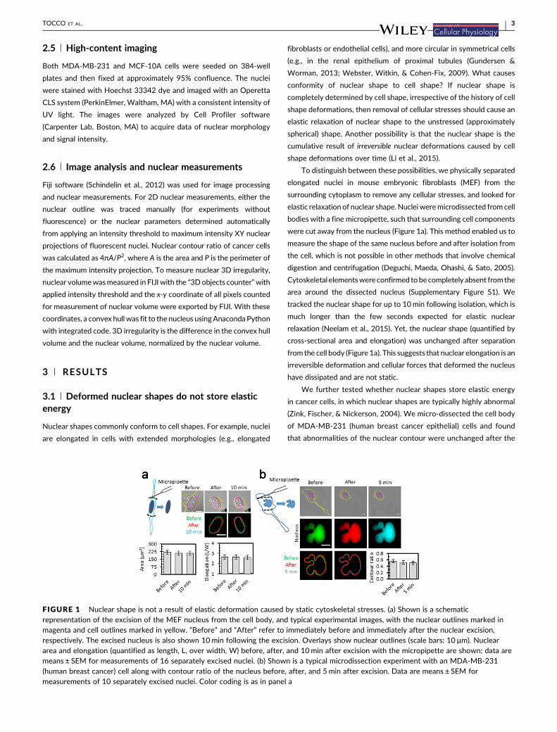
3.1 | Deformed nuclear shapes do not store elasticenergy
Nuclear shapes commonly conform to cell shapes. For example, nuclei
are elongated in cells with extended morphologies (e.g., elongated
fibroblasts or endothelial cells), and more circular in symmetrical cells
(e.g., in the renal epithelium of proximal tubules (Gundersen &
Worman, 2013; Webster, Witkin, & Cohen-Fix, 2009). What causes
conformity of nuclear shape to cell shape? If nuclear shape is
completely determined by cell shape, irrespective of the history of cell
shape deformations, then removal of cellular stresses should cause an
elastic relaxation of nuclear shape to the unstressed (approximately
spherical) shape. Another possibility is that the nuclear shape is the
cumulative result of irreversible nuclear deformations caused by cell
shape deformations over time (Li et al., 2015).
To distinguish between these possibilities, we physically separated
elongated nuclei in mouse embryonic fibroblasts (MEF) from the
surrounding cytoplasm to remove any cellular stresses, and looked for
elastic relaxation of nuclear shape. Nucleiweremicrodissected from cell
bodies with a fine micropipette, such that surrounding cell components
were cut away from the nucleus (Figure 1a). This method enabled us to
measure the shape of the same nucleus before and after isolation from
the cell, which is not possible in other methods that involve chemical
digestion and centrifugation (Deguchi, Maeda, Ohashi, & Sato, 2005).
Cytoskeletal elementswereconfirmed tobecompletely absent fromthe
area around the dissected nucleus (Supplementary Figure S1). We
tracked the nuclear shape for up to 10min following isolation, which is
much longer than the few seconds expected for elastic nuclear
relaxation (Neelam et al., 2015). Yet, the nuclear shape (quantified by
cross-sectional area and elongation) was unchanged after separation
from the cell body (Figure 1a). This suggests that nuclear elongation is an
irreversible deformation and cellular forces that deformed the nucleus
have dissipated and are not static.
We further tested whether nuclear shapes store elastic energy
in cancer cells, in which nuclear shapes are typically highly abnormal
(Zink, Fischer, & Nickerson, 2004). We micro-dissected the cell body
of MDA-MB-231 (human breast cancer epithelial) cells and found
that abnormalities of the nuclear contour were unchanged after the
FIGURE 1 Nuclear shape is not a result of elastic deformation caused by static cytoskeletal stresses. (a) Shown is a schematicrepresentation of the excision of the MEF nucleus from the cell body, and typical experimental images, with the nuclear outlines marked inmagenta and cell outlines marked in yellow. “Before” and “After” refer to immediately before and immediately after the nuclear excision,respectively. The excised nucleus is also shown 10min following the excision. Overlays show nuclear outlines (scale bars: 10 μm). Nucleararea and elongation (quantified as length, L, over width, W) before, after, and 10min after excision with the micropipette are shown; data aremeans ± SEM for measurements of 16 separately excised nuclei. (b) Shown is a typical microdissection experiment with an MDA-MB-231(human breast cancer) cell along with contour ratio of the nucleus before, after, and 5min after excision. Data are means ± SEM formeasurements of 10 separately excised nuclei. Color coding is as in panel a
TOCCO ET AL. | 3
surrounding cell body was cut away (Figure 1b). We compared the
nuclear contour ratio (a measure of abnormality; see methods)
before and after nuclear isolation, and found no statistically
significant effect of the isolation on the contour ratio (Figure 1b).
The lack of elastic relaxation upon removal of the surrounding cell
implies that the deformed nuclear shape in these different cell types
stores no elastic energy and that the deformation is not an elastic
response to the instantaneous cell shape-dependent cytoskeletal
forces on the nucleus.
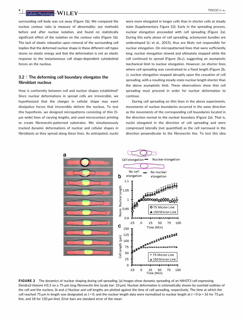
3.2 | The deforming cell boundary elongates thefibroblast nucleus
How is conformity between cell and nuclear shapes established?
Since nuclear deformations in spread cells are irreversible, we
hypothesized that the changes in cellular shape may exert
dissipative forces that irreversibly deform the nucleus. To test
this hypothesis, we designed micropatterns consisting of thin (5-
μm wide) lines of varying lengths, and used microcontact printing
to create fibronectin-patterned substrates. We simultaneously
tracked dynamic deformations of nuclear and cellular shapes in
fibroblasts as they spread along these lines. As anticipated, nuclei
were more elongated in longer cells than in shorter cells at steady
state (Supplementary Figure S2). Early in the spreading process,
nuclear elongation proceeded with cell spreading (Figure 2a).
During this early phase of cell spreading, actomyosin bundles are
undeveloped (Li et al., 2015), thus are likely not responsible for
nuclear elongation. On micropatterned lines that were sufficiently
long, nuclear elongation slowed and ultimately stopped while the
cell continued to spread (Figure 2b,c), suggesting an asymptotic
mechanical limit to nuclear elongation. However, on shorter lines
where cell spreading was constrained to a fixed length (Figure 2b,
c), nuclear elongation stopped abruptly upon the cessation of cell
spreading, with a resulting steady-state nuclear length shorter than
the above asymptotic limit. These observations show that cell
spreading must proceed in order for nuclear deformation to
continue.
During cell spreading on thin lines in the above experiments,
movements of nuclear boundaries occurred in the same direction
as the movements of the corresponding cell boundaries located in
the direction normal to the nuclear boundary (Figure 2a). That is,
nuclei elongated in the direction of cell spreading and were
compressed laterally (not quantified) as the cell narrowed in the
direction perpendicular to the fibronectin line. To test this idea
FIGURE 2 The dynamics of nuclear shaping during cell spreading. (a) Images show dynamic spreading of an NIH3T3 cell expressingDendra2-histone H3.3 on a 75-μm long fibronectin line (scale bar: 10 μm). Nuclear deformation is schematically shown by overlaid outlines ofthe cell and the nucleus. (b and c) Nuclear and cell lengths are plotted against the time of cell spreading, respectively. The time at which thecell reached 75 μm in length was designated as t = 0, and the nuclear length data were normalized to nuclear length at t = 0 (n = 16 for 75 μmline, and 18 for 150 μm line). Error bars are standard error of the mean
4 | TOCCO ET AL.

further, we next examined the correlation between the deforma-
tions in the nuclear and cellular shapes as cells migrated between
thin and wide fibronectin patterns. We tracked cells along a
fibronectin micropattern (Chang, Guo, Kim, & Wang, 2013)
consisting of a 400 μm line terminating in a 50 × 100 μm2 (“2D”)
rectangle (Figure 3a). As the cell crawled onto the rectangular
region, the nuclear shape was initially unchanged, despite widening
of the cell shape at the cell front (Figure 3b, left panels; compare
−28′ with −56′). Only when the cell width expanded laterally (i.e.,
perpendicular to the cell axis, 0′) in the region adjacent to the
nuclear surface did the nucleus begin to widen (Figure 3b, left
panels, and movie 1). Similarly, for cells moving from wide to thin
fibronectin regions, the nucleus compressed laterally only when
the adjacent cell edge moved laterally towards the nucleus
(Figure 3b, right panels; compare −28′ with 0′ and 28′). To
quantify these observations, we measured nuclear width and
compared it with cell width at two positions (at the nuclear midline
and 35 μm from the nucleus into the leading edge) as the nucleus
progressed from the line to the 2D region (Figure 3c). Nuclear
width did not change despite the increase in cell width at the cell
front; rather, the expansion of the nuclear width precisely
coincided with the increase in cell width lateral to the nucleus.
Nuclear deformation occurred only when the cell deformed;
moreover, global changes in cell shape did not affect the nuclear
shape unless a nearby membrane was moving in a direction normal
to the nuclear surface.
To examine the generality of these observations, we tracked
deformations in nuclear shape of NIH 3T3 fibroblasts migrating in
reconstituted collagen gels, and observed similar instances of nuclear
widening in direct response to cell widening (Supplementary Figure
S3). There, a narrow, elongated cell became wider as it migrated in the
local extracellular matrix environment, and the deformations in the
nuclear shape coincided with the deformations in cell shape
perpendicular to the nuclear surface. Such observations are consistent
FIGURE 3 Dynamic deformation of the fibroblast nucleus during cell migration. (a) Shown is an example of a rhodamine-fibronectinmicropattern consisting of a 5 μm wide line connected to a 2D rectangle measuring 100 × 50 μm2 (scale bar: 50 μm), along with time lapseimages (b) of cells migrating between micropattern regions, with narrow-to-wide (left) and wide-to-narrow (right) transitions. Nuclei (blackdotted lines), cell outlines (blue), and the fibronectin pattern (red dotted lines) are outlined for clarity. The time at which the nucleus crossedthe interface of the fibronectin regions was designated as t = 0 (scale bar: 20 μm). Below: Overlaid nuclear outlines at different time points(different color for each time). (c) Nuclear width (“nucleus”), cell width at the nuclear midline (“cell, nucleus”), and cell width 35 μm from thenuclear midline toward the leading cell edge (“cell, 35 μm”) were measured as shown in the schematic diagram on the left. The data areplotted against the distance between the nuclear midline and edge of the 2D region (dark gray arrow, top). The origin of the x-axiscorresponds to the nuclear midline reaching the edge of the 2D region. Data are means pooled from seven narrow-to-wide transitions and allwidth measurements are normalized to the minimum nuclear width. Error bars represent SEM for all data points, binned into increments of5 μm of nucleus to 2D distance
TOCCO ET AL. | 5

with the transitions observed on micropatterned lines and again
suggest that local motion of the cell boundary is required for changing
nuclear shape.
3.3 | The deforming cell shape amplifies cancernuclear abnormalities
We quantitatively measured nuclear shape abnormalities during cancer
cell spreading by imaging nuclei expressing GFP-NLS in MCF-10A and
MDA-MB-231cells (Figure4a).Wemeasured thedeviationof thenucleus
from a convex shape to calculate 3D irregularity (see Figure 4b and
Methods). The 3D irregularity increased in cancer nuclei during
spreading of cancer cells, but decreased in MCF-10A cells (Figure 4b).
We asked if abnormal cancer shapes are heritable. We classified
regular and irregularnuclei inMDA-MB-231cells, tracked them through
mitosis, and quantified the proportion of regular or irregular daughter
nuclei (Supplementary Figure S4). Cells with irregular nuclei were more
likely to divide into daughter cellswith irregular nuclei; cells with regular
nuclei were more likely to divide into daughter cells with regular nuclei
(Supplementary Figure S4). These results suggest that nuclear
abnormalities are heritable from mother to daughter cells. An obvious
reason for this observation could be the degree of ploidy in cells.
Therefore, we quantified the DNA content inMCF-10A andMDA-MB-
231 nuclei using high-content imaging of Hoechst 33342 DNA.
Computing histograms for total fluorescence intensity showed a
bimodal distribution in MCF-10A cells, consistent with proportions
expected from thecell cycle,whileMDA-MB-231cells expectedly hada
higher DNA content (Supplementary Figure S5a). However, we found
no dependence of nuclear irregularity on total DNA content in either
MCF-10A or MDA-MB-231 cells (Supplementary S5b). Therefore,
ploidy is not necessarily a factor in determining abnormal cancer nuclear
shapes.
4 | DISCUSSION
The shape of the nucleus inside the cell has been commonly assumed to
store elastic energy. That is, the rounded nucleus in a suspended cell
becomes elongated or flattened in a spreading cell through an elastic
(i.e., reversible) deformation of the nucleus. Such a model predicts that
removing cytoplasmic forces on the nucleus should restore the original
rounded shape of the nucleus. Similarly, nuclear lobes and invaginations
characteristic of cancer nuclei might be considered to be elastically
deformed in response to cytoskeletal forces (Funkhouser et al., 2013).
Herewe showed thatmicro-dissection of the nucleus from the cell does
not cause a relaxation of the nuclear shape. The lack of elastic relaxation
upon removal of the surrounding cell implies that the deformed nuclear
shape in these different cell types stores no elastic energy and that the
deformation is not an elastic response to the instantaneous cell shape-
dependent cytoskeletal forces on the nucleus.
Pajerowski, Dahl, Zhong, Sammak, and Discher (2007) aspirated
nuclei and cells into a micropipette and measured deformed nuclear
shapes. Upon ejecting the nucleus and cell from the pipette, they found
that the nuclear shape remained deformed. However, inside cells, the
nucleus does not undergo rapid and large deformations like those
applied in that study (100–500% strain in a few seconds). Therefore,
these experiments do not necessarily explain the irreversibility of
elongated nuclear shapes in cells that are established by the process of
cell spreading and migration over longer time scales (∼1 hr). Harada
et al. (2014) have previously suggested irreversible deformation of the
nucleus in cells. In their experiments, nuclei in migrating cells must
elongate to squeeze through tiny pores, and these elongated shapes
persist in cells that express a high amount of A-type lamins to
B-type lamins. However, it is possible that cells that migrate through
pores remain elongated, causing the elongated nuclear shape to
persist. This illustrates the difficulty in interpreting elongated nuclear
FIGURE 4 Cell spreading amplifies cancer nuclear abnormalities. (a) Shown is an example of nuclear shape changes during spreading of anMCF-10A and MDA-MB-231 cell. (b) Schematic shows the calculation of 3D irregularity defined here as the difference in volume between thenucleus and a convex hull fit to the nucleus divided by nuclear volume. Plot shows 3D irregularity plotted against time of spreading in MCF-10Aand MDA-MB-231 cells. (n > 30 for both MDA-MB-231 and MCF-10A cells at each time point, error bars are standard error of the mean)
6 | TOCCO ET AL.

shapes as evidence for irreversible nuclear deformation without
accounting for the cell shape.
Deguchi et al. (2005) quantified nuclear shapes after isolation
from cells. However, their method involved chemical isolation and
centrifugation; therefore, comparison of the same nucleus before and
after isolation was not possible. Cao et al. (2016) modeled the nucleus
as a hyperelastic shell containing permeable elastic chromatin. To
explain the large plastic deformations of nuclei observed by Pajerowski
et al. (2007) in cells lacking lamin A/C, they modeled chromatin as a
plastic material surrounded by a much softer hyperelastic shell. But, as
we point out here, the nucleus in wild-type fibroblasts—which contain
normal levels of lamins (Alam et al., 2016)—is irreversibly deformed at
steady state. The assumption of the elastic nucleus has not been
previously tested by removing the forces on the nuclear surface (i.e.,
isolating the nucleus) and then tracking its shape. Yet several models
have assumed that nuclear shapes reflect an elastic deformation
(Allena, Thiam, Piel, & Aubry, 2015; Jean, Gray, Spector, & Chen, 2004;
Kim et al., 2016; Li, Kumar, Makhija, & Shivashankar, 2014; Versaevel
et al., 2012).
In our experiments, we excised the cell away from the nucleus,
which exposed the nucleus to the cell culture medium. Although this
may create an osmotic driving force for water flux into the nucleus
(Finan, Chalut,Wax, &Guilak, 2009), such a change in osmotic pressure
would cause an even faster relaxation of the nucleus to its original
shape. Thus, an osmotic pressure—even if present—is not a
confounding variable in our analysis.
We have modeled the mechanical behavior of the nucleus in cells
by accounting for its bulk compressibility (volume elasticity) and the
extensional modulus of the nuclear lamina (Li et al., 2015). While we
cannot rule out a small change in nuclear volume in our excision
experiments, we have previously shown that the volume of the
nucleus stays approximately constant during nuclear flattening
(Li et al., 2015). Similarly, Davidson, Sliz, Isermann, Denais, and
Lammerding (2015) have reported that nuclear volume remains
constant during nuclear deformations in cells lacking lamin A/C as
they migrate through narrow pores. The lamina of a rounded nucleus
has excess surface area stored in undulations and wrinkles (Li et al.,
2015) and these decrease during the process of nuclear flattening
(Neelam, Hayes, Zhang, Dickinson, & Lele, 2016). We therefore
suggest that during nuclear flattening, the initially folded lamina
becomes taut such that the surface area of the lamina remains
constant. The lamina does not resist changes in nuclear shape until it
becomes taut. Only when there is no more excess area in the lamina,
does the lamina resist further nuclear elongation or flattening.
Because a steady-state nuclear shape is reached upon cell spreading,
it is likely that the taut lamina is too stiff to undergo extensions under
typical cellular forces (Li et al., 2015). Consistent with this idea,
Stephens, Banigan, Adam, Goldman, and Marko (2017) have found
that the nucleus is initially soft to longitudinal strain due to
mechanical resistance from chromatin, but is stiff beyond a threshold
strain because of the nuclear lamina. We note that the elasticity of
the nuclear lamina in cells (Dahl, Kahn, Wilson, & Discher, 2004) can
become apparent when an external force stretches the (taut) nuclear
lamina as is observed in short-time scale nuclear forcing experiments
(Neelam et al., 2015; Pajerowski et al., 2007). But, it has not been
demonstrated that forces generated by cells are sufficient to
discernably extend the lamina.
Because nuclear deformation requires a change in cell shape, we
speculate that this occurs due to a mechanical stress caused by
frictional resistance in the cytoskeletal elements to expansion or
contraction as the cell boundary moves relative to the nuclear surface
(Li et al., 2015). What is the molecular mechanism of this transmission
of stress? We have previously shown that cell spreading is necessary
and sufficient to drive nuclear flattening in fibroblasts under a wide
range of conditions (Li et al., 2015), including in the absence of
microtubules, vimentin intermediate filaments, and myosin activity.
Therefore, we speculate that it is primarily the F-actin network that
transmits stress from the moving cell boundary to the nucleus.
However, once F-actin is completely disrupted with pharmacological
agents, any further changes in cell shape are not possible (Li et al.,
2015), which makes it difficult to test the role of F-actin in mediating
nuclear response to deformations in cell shape. Experiments which
cause deformations in cell shape over time scales of minutes (typical
time scales ofmigration and spreading) in the absence of F-actin will be
needed to test whether the F-actin network is responsible for
transmitting the dissipative stresses. Alternatively, it is possible that
any of the three cytoskeletal filaments may transmit stresses to the
nucleus, and disruption of one of these allows other structures to
transmit the stresses. If there is such redundancy, then identifying the
molecular structures that coordinate nuclear and cell shape is likely to
remain a fundamental challenge.
Irrespective of the molecular source of the stress transmission,
motion of the nuclear surface in response to motion of the cell boundary
naturally allows the nucleus tomimic the cell shape. Thus, thismechanism
can easily explain how the nucleus adopts an “hour-glass” shape when
cells develop a similar shape during migration through narrow pores and
constrictions (Friedl, Wolf, & Lammerding, 2011; Denais et al., 2016).
Although it is possible that such extreme shapes result in stored elastic
energy in the lamina, this is not required to explain the nuclear shape.We
also note that once the nuclear shape is established during cell spreading
or migration, actomyosin stress fibers can indent the apical surface of the
nucleus as has been observed by others (Versaevel et al., 2014), and actin
polymerization can constrict nuclei through narrow pores as observed in
somecell types likedendritic cells (Thiamet al., 2016). Thus,multiple types
of stresses can act in combination with stresses generated by motion of
the cell boundary. However, it has not been shown that the resulting
nuclear deformationsby these typesof stresses are reversible (i.e., elastic).
Similarly, as shown here, the highly abnormal shapes of cancer nuclei are
not necessarily an elastic response to cytoskeletal forces. Rather, the
process of cell spreading can irreversibly amplify these abnormalities.
Why does dynamic cell spreading amplify nuclear abnormalities in
cancer cells, but not in non-cancer cells (Figure 4d)? It has been suggested
that the localization of lamins may be spatially inhomogeneous in cancer
nuclei resulting in a mechanically “patchy” nuclear surface (Funkhouser
et al., 2013). Thus, certain regions of the heterogeneous nuclear surface
may be more compliant in response to mechanical stresses, resulting in
TOCCO ET AL. | 7

abnormal nuclear shapes. We note that knockdown of nuclear proteins
other than the nuclear lamins (Lammerding et al., 2006) can also result in
abnormalities—these include the chromatin remodeling protein Brg1
(Imbalzano, Imbalzano, & Nickerson, 2013) and endosome regulator
protein Wash (Verboon et al., 2015).
Because gene expression and protein synthesis potentially depend
on the physical properties of the nucleus (Cremer&Cremer, 2001; Swift
et al., 2013; Thomas et al., 2002), irreversible deformations of the
nucleus may trigger expression of genes during cell migration by
modulating chromatin compaction (Tajik et al., 2016; Xiao, Freedman,
Miller, Heald, & Marko, 2012). Cell-shape-dependent nuclear deforma-
tions may therefore enable (or at least contribute to) the known
relationshipbetweencell shapeandcell function (Chen,Mrksich,Huang,
Whitesides, & Ingber, 1997). Therefore, knowing how dynamic cell
shape information is transmitted to the nucleuswill be key to a complete
understanding of the relationship between cell shape and cell function.
ACKNOWLEDGMENTS
This work was supported by funding from the National Institutes of
Health (R01 GM102486 to TPL; R01 EB014869 to TPL and RBD;
U54CA193419 to JDL; R01 CA172310 to HL) and the National
Science Foundation (CMMI 1437395 to TPL).
COMPETING FINANCIAL INTERESTS
The authors declare no competing or financial interests.
REFERENCES
Alam, S. G., Zhang, Q., Prasad, N., Li, Y., Chamala, S., Kuchibhotla, R., . . . Lele,T. P. (2016). The mammalian LINC complex regulates genometranscriptional responses tosubstrate rigidity.ScientificReports,6, 38063.
Allena, R., Thiam, H., Piel, M., & Aubry, D. (2015). A mechanical model toinvestigate the role of the nucleus during confined cell migration.
Computer Methods in Biomechanics and Biomedical Engineering, 18(Suppl1), 1868–1869.
Bloom, H. J., & Richardson, W. W. (1957). Histological grading andprognosis in breast cancer; a study of 1409 cases of which 359 havebeen followed for 15 years. British Journal of Cancer, 11(3), 359–377.
Boyd, J., Pienta, K. J., Getzenberg, R. H., Coffey, D. S., & Barrett, J. C. (1991).Preneoplastic alterations in nuclear morphology that accompany loss oftumor suppressor phenotype. Journal of the National Cancer Institute,83(12), 862–866.
Bussolati, G., Marchiò, C., Gaetano, L., Lupo, R., & Sapino, A. (2008).
Pleomorphism of the nuclear envelope in breast cancer: A newapproach to an old problem. Journal of Cellular and Molecular Medicine,12(1), 209–218.
Cao, X., Moeendarbary, E., Isermann, P., Davidson, P. M., Wang, X., Chen,
M. B., . . . Shenoy, V. B. (2016). A chemomechanical model for nuclearmorphology and stresses during cell transendothelial migration.Biophysical Journal, 111(7), 1541–1552.
Chang, S. S., Guo, W. H., Kim, Y., & Wang, Y. L. (2013). Guidance of cellmigration by substrate dimension. Biophysical Journal, 104(2), 313–321.
Chen, C. S., Mrksich, M., Huang, S.,Whitesides, G. M., & Ingber, D. E. (1997).Geometric control of cell life and death. Science, 276(5317), 1425–1428.
Chow, K. H., Factor, R. E., & Ullman, K. S. (2012). The nuclear envelopeenvironment and its cancer connections. Nature Reviews Cancer, 12(3),196–209.
Cremer, T., & Cremer, C. (2001). Chromosome territories, nucleararchitecture and gene regulation in mammalian cells. Nature ReviewsGenetics, 2(4), 292–301.
Csucs, G., Michel, R., Lussi, J. W., Textor, M., & Danuser, G. (2003).
Microcontact printing of novel co-polymers in combination withproteins for cell-biological applications. Biomaterials, 24(10),1713–1720.
Dahl, K. N., Kahn, S. M., Wilson, K. L., & Discher, D. E. (2004). The nuclearenvelope lamina network has elasticity and a compressibility limitsuggestive of a molecular shock absorber. Journal of Cell Science 117(Pt20), 4779–4786.
Davidson, P. M., Sliz, J., Isermann, P., Denais, C., & Lammerding, J. (2015).Design of a microfluidic device to quantify dynamic intra-nucleardeformation during cell migration through confining environments.Integrative Biology (Cambridge), 7(12), 1534–1546.
Deguchi, S., Maeda, K., Ohashi, T., & Sato, M. (2005). Flow-inducedhardening of endothelial nucleus as an intracellular stress-bearing
organelle. Journal of Biomechanics, 38(9), 1751–1759.Denais, C. M., Gilbert, R. M., Isermann, P., McGregor, A. L., te Lindert, M.,
Weigelin, B., . . . Lammerding, J. (2016). Nuclear envelope rupture andrepair during cancer cell migration. Science, 352(6283), 353–358.
Elston, C. W., & Ellis, I. O. (1991). Pathological prognostic factors in breast
cancer. I. The value of histological grade in breast cancer: Experiencefrom a large study with long-term follow-up. Histopathology, 19(5),403–410.
Finan, J. D., Chalut, K. J., Wax, A., & Guilak, F. (2009). Nonlinear osmoticproperties of the cell nucleus. Annals of Biomedical Engineering, 37(3),
477–491.Friedl, P., Wolf, K., & Lammerding, J. (2011). Nuclear mechanics during cell
migration. Current Opinion in Cell Biology, 23(1), 55–64.Funkhouser, C., Sknepnek, R., Shimi, T., Goldman, A., Goldman, R., & de la
Cruz, M. (2013). Mechanical model of blebbing in nuclear lamin
meshworks. Proceedings of the National Academy of Sciences of theUnited States of America, 110(9), 3248–3253.
Giardina, C., Renzulli, G., Serio, G., Caniglia, D. M., Lettini, T., Ferri, C., . . .Delfino, V. P. (1996). Nuclear morphometry in node-negative breast
carcinoma. Analytical and Quantitative Cytology and Histology, 18(5),374–382.
Gundersen, G. G., &Worman, H. J. (2013). Nuclear positioning. Cell, 152(6),1376–1389.
Gupta, S., Marcel, N., Sarin, A., & Shivashankar, G. V. (2012). Role of actin
dependent nuclear deformation in regulating early gene expression.PLoS One, 7(12), 53031.
Harada, T., Swift, J., Irianto, J., Shin, J. W., Spinler, K. R., Athirasala, A., . . .Discher, D. E. (2014). Nuclear lamin stiffness is a barrier to 3Dmigration,but softness can limit survival. Journal of Cell Biology, 204(5), 669–682.
Haroske, G., Dimmer, V., Friedrich, K., Meyer, W., Thieme, B., Theissig, F., &
Kunze, K. D. (1996). Nuclear image analysis of immunohistochemicallystained cells in breast carcinomas. Histochemistry and Cell Biology,
105(6), 479–485.Imbalzano, A. N., Imbalzano, K. M., & Nickerson, J. A. (2013). BRG1, a SWI/
SNF chromatin remodeling enzyme ATPase, is required for mainte-nance of nuclear shape and integrity. Communicative & IntegrativeBiology, 6(5), e25153.
Jean, R. P., Gray, D. S., Spector, A. A., & Chen, C. S. (2004). Characterizationof the nuclear deformation caused by changes in endothelial cell shape.Journal of Biomechanical Engineering, 126(5), 552–558.
Khatau, S. B., Hale, C. M., Stewart-Hutchinson, P. J., Patel, M. S., Stewart,C. L., Searson, P. C., . . . Wirtz, D. (2009). A perinuclear actin capregulates nuclear shape. Proceedings of the National Academy of Sciencesof the United States of America, 106(45), 19017–19022.
8 | TOCCO ET AL.

Kim, D. H., Li, B., Si, F., Phillip, J. M., Wirtz, D., & Sun, S. X. (2016). Volumeregulation and shape bifurcation in the cell nucleus. Journal of CellScience, 129(2), 457.
Lammerding, J., Fong, L. G., Ji, J. Y., Reue, K., Stewart, C. L., Young, S. G., &Lee, R. T. (2006). Lamins A and C but not lamin B1 regulate nuclearmechanics. Journal of Biological Chemistry, 281(35), 25768–25780.
Li, Q., Kumar, A., Makhija, E., & Shivashankar, G. V. (2014). The regulation of
dynamic mechanical coupling between actin cytoskeleton and nucleusby matrix geometry. Biomaterials, 35(3), 961–969.
Li, Y., Lovett, D., Zhang, Q., Neelam, S., Kuchibhotla, R. A., Zhu, R., . . .Dickinson, R. B. (2015). Moving cell boundaries drive nuclear shapingduring cell spreading. Biophysical Journal, 109(4), 670–686.
McGregor, A. L., Hsia, C. R., & Lammerding, J. (2016). Squish and squeeze-the nucleus as a physical barrier during migration in confinedenvironments. Current Opinion in Cell Biology, 40, 32–40.
Neelam, S., Chancellor, T. J., Li, Y., Nickerson, J. A., Roux, K. J., Dickinson,R. B., & Lele, T. P. (2015). Direct force probe reveals the mechanics of
nuclear homeostasis in the mammalian cell. Proceedings of the NationalAcademy of Sciences of the United States of America, 112(18),5720–5725.
Neelam, S., Hayes, P. R., Zhang, Q., Dickinson, R. B., & Lele, T. P. (2016).Vertical uniformity of cells and nuclei in epithelial monolayers. Scientific
Reports, 6, 19689.Pajerowski, J. D., Dahl, K. N., Zhong, F. L., Sammak, P. J., & Discher, D. E.
(2007). Physical plasticity of the nucleus in stem cell differentiation.Proceedings of the National Academy of Sciences of the United States of
America, 104(40), 15619–15624.Petrie, R. J., Koo, H., & Yamada, K. M. (2014). Generation of compartmen-
talized pressure by a nuclear piston governs cell motility in a 3D matrix.Science, 345(6200), 1062–1065.
Schindelin, J., Arganda-Carreras, I., Frise, E., Kaynig, V., Longair, M.,
Pietzsch, T., . . . Cardona, A. (2012). Fiji: An open-source platform forbiological-image analysis. Nature Methods, 9(7), 676–682.
Stephens, A. D., Banigan, E. J., Adam, S. A., Goldman, R. D., & Marko, J. F.(2017). Chromatin and lamin A determine two different mechanicalresponse regimes of the cell nucleus. Molecular Biology of the Cell, pp.
mbc-E16.Swift, J., Ivanovska, I. L., Buxboim, A., Harada, T., Dingal, P. C., Pinter, J., . . .
Discher, D. E. (2013). Nuclear lamin-A scales with tissue stiffness andenhancesmatrix-directed differentiation. Science, 341(6149), 1240104.
Tajik, A., Zhang, Y., Wei, F., Sun, J., Jia, Q., Zhou, W., . . . Wang, N. (2016).
Transcription upregulation via force-induced direct stretching of
chromatin. Nature Materials, 15(12), 1287–1296.Théry, M., & Piel, M. (2009). Adhesive micropatterns for cells: A
microcontact printing protocol. Cold Spring Harbor Protocols, 2009(7),
5255.
Thiam, H. R., Vargas, P., Carpi, N., Crespo, C. L., Raab, M., Terriac, E., . . . Piel,M. (2016). Perinuclear Arp2/3-driven actin polymerization enablesnuclear deformation to facilitate cell migration through complex
environments. Nature Communications, 7, 10997.Thomas, C. H., Collier, J. H., Sfeir, C. S., & Healy, K. E. (2002). Engineering
gene expression and protein synthesis by modulation of nuclear shape.Proceedings of the National Academy of Sciences of the United States of
America, 99(4), 1972–1977.Verboon, J. M., Rincon-Arano, H., Werwie, T. R., Delrow, J. J., Scalzo, D.,
Nandakumar, V., . . . Parkhurst, S. M. (2015). Wash interacts with laminand affects global nuclear organization.Current Biology,25(6), 804–810.
Vergani, L., Grattarola, M., & Nicolini, C. (2004). Modifications of chromatin
structure and gene expression following induced alterations of cellularshape. The International Journal of Biochemistry & Cell Biology, 36(8),1447–1461.
Versaevel, M., Braquenier, J. B., Riaz, M., Grevesse, T., Lantoine, J., &Gabriele, S. (2014). Super-resolution microscopy reveals LINC complex
recruitment at nuclear indentation sites. Scientific Reports, 4, 7362.
Versaevel, M., Grevesse, T., & Gabriele, S. (2012). Spatial coordination
between cell and nuclear shapewithin micropatterned endothelial cells.Nature Communications, 3, 671.
Webster, M., Witkin, K. L., & Cohen-Fix, O. (2009). Sizing up the nucleus:Nuclear shape, size and nuclear-envelope assembly. Journal of Cell
Science, 122(Pt 10), 1477–1486.
Xiao, B., Freedman, B. S., Miller, K. E., Heald, R., & Marko, J. F. (2012).
HistoneH1 compacts DNAunder force and during chromatin assembly.Molecular Biology of the Cell, 23(24), 4864–4871.
Zink, D., Fischer, A. H., &Nickerson, J. A. (2004). Nuclear structure in cancercells. Nature Reviews Cancer, 4(9), 677–687.
SUPPORTING INFORMATION
Additional Supporting Information may be found online in the
supporting information tab for this article.
How to cite this article: Tocco VJ, Li Y, Christopher KG,
et al. The nucleus is irreversibly shaped by motion of cell
boundaries in cancer and non-cancer cells. J Cell Physiol.
2017;1–9. https://doi.org/10.1002/jcp.26031
TOCCO ET AL. | 9


