
TOWARDS PRACTICAL APPLICATIONS FOR MOLECULAR LOGIC GATES: “AND” LOGIC AS AN ADDITIONAL LAYER OF SELECTIVITY IN SINGLET
OXYGEN RELEASE FOR PHOTODYNAMIC THERAPY
A THESIS SUBMITTED TO THE GRADUATE SCHOOL OF NATURAL AND APPLIED SCIENCES
OF MIDDLE EAST TECHNICAL UNIVERSITY
BY
SURİYE ÖZLEM
IN PARTIAL FULFILLMENT OF THE REQUIREMENTS
FOR
THE DEGREE OF MASTER OF SCIENCE IN
CHEMISTRY
JUNE 2008
Approval of the thesis:
TOWARDS PRACTICAL APPLICATIONS FOR MOLECULAR LOGIC GATES: “AND” LOGIC AS AN ADDITIONAL LAYER OF SELECTIVITY IN SINGLET
OXYGEN RELEASE FOR PHOTODYNAMIC THERAPY submitted by SURİYE ÖZLEM in partial fulfillment of the requirements for the degree of Master of Science in Chemistry Department, Middle East Technical University by,
Prof. Dr. Canan Özgen Dean, Gradute School of Natural and Applied Sciences
.Prof. Dr. Ahmet M. Önal Head of Department, Chemistry
Prof. Dr. Engin U. Akkaya Supervisor, Chemistry Dept., METU
Examining Committee Members: Prof. Dr. Ahmet M. Önal Chemistry Dept., METU
Prof. Dr. Engin U. Akkaya Chemistry Dept., METU
Prof. Dr. Metin Zora Chemistry Dept., METU
Prof. Dr. Özdemir DOGAN Chemistry Dept., METU Dr. Ö. Altan BOZDEMİR Researcher
Date:
iii
I hereby declare that all information in this document has been obtained and presented in accordance with academic rules and ethical conduct. I also declare that, as required by these rules and conduct, I have fully cited and referenced all material and results that are not original to this work.
Name, Last Name : SURİYE ÖZLEM
Signature :

iv
ABSTRACT
TOWARDS PRACTICAL APPLICATIONS FOR MOLECULAR LOGIC GATES:
“AND” LOGIC AS AN ADDITIONAL LAYER OF SELECTIVITY IN SINGLET
OXYGEN RELEASE FOR PHOTODYNAMIC THERAPY
Özlem, Suriye
M.S., Department of Chemistry
Supervisor: Prof. Dr. Engin Umut Akkaya
June 2008, 54 pages
There have been many examples of individual molecular logic gates and molecular
equivalents of more complex digital designs in recent years such as half adder, half
subtractor, multiplexer. Neverethless, the unresolved issues of addressability and lack of
communication between logic gates remain to be the Achille’s heel for molecular logic
gates. A few years ago we have demonstrated that appropriately decorated bodipy dyes
can be very efficient generators for singlet oxygen, thus act as a satisfactory
photodynamic agents. As a bonus, these dyes absorb very strongly at 660 nm which is
considered to be within the therapeutic window of mammalian tissue.
So, combining our earlier experience in molecular logic gates and rational design of
photodynamic agents, we proposed a photodynamic therapy agent that would release
singlet oxygen at a much larger rate when the cancer related cellular parameters are above
v
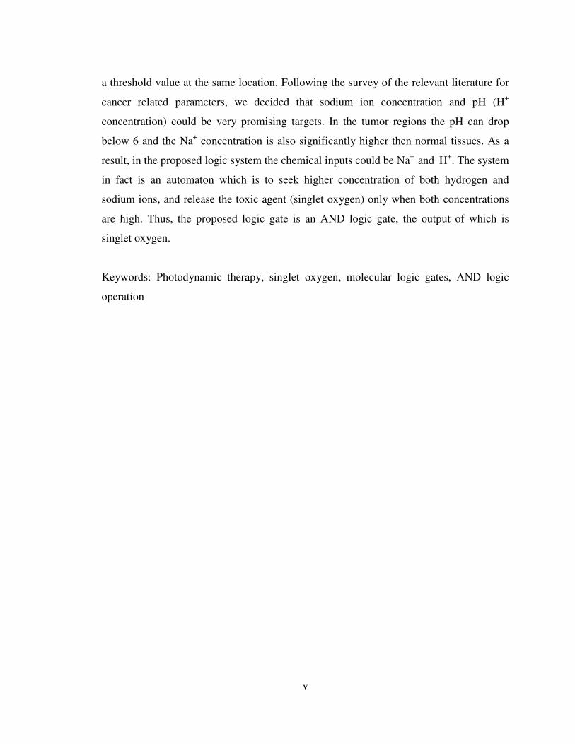
a threshold value at the same location. Following the survey of the relevant literature for
cancer related parameters, we decided that sodium ion concentration and pH (H+
concentration) could be very promising targets. In the tumor regions the pH can drop
below 6 and the Na+ concentration is also significantly higher then normal tissues. As a
result, in the proposed logic system the chemical inputs could be Na+ and H+. The system
in fact is an automaton which is to seek higher concentration of both hydrogen and
sodium ions, and release the toxic agent (singlet oxygen) only when both concentrations
are high. Thus, the proposed logic gate is an AND logic gate, the output of which is
singlet oxygen.
Keywords: Photodynamic therapy, singlet oxygen, molecular logic gates, AND logic
operation

vi
ÖZ
MOLEKÜLER MANTIK KAPISININ PRATİKTEKİ UYGULAMALARI: “VE”
KAPISININ FOTODİNAMİK TERAPİDEKİ SİNGLET OKSİJEN ÜRETİMİ İLE
İLİŞKİLENDİRİLMESİ
Özlem, Suriye
Yüksek Lisans, Kimya Bölümü
Tez Yöneticisi: Prof. Dr. Engin Umut Akkaya
Haziran 2008, 54 sayfa
Moleküler mantık kapılarıyla ilgili geçmiş yıllarda yapılan birçok çalışma mevcuttur,
bunlara toplama, çıkarma ve multiplekser başta olmak üzere daha da karmaşık tasarımlar
eşlik etmektedir. Ancak, bu güne kadar moleküler mantık kapılarının yaptığı işlemin
sonucunun herhangi bir işlev yerine getiriyor olmaması bu konunun en büyük
eksikliğidir. Birkaç yıl önce yaptığımız çalışmalarda düzgün tasarlanmış olan bodipylerin
çok verimli birer singlet oksijen kaynağı olduğunu ve dolayısıylada fotodinamik terapi
uygulayabildiğini göstermiş bulunmaktayız. Üstelik bu maddelerin absorpladığı dalga
boyu insan vücudunun geçirebildiği 660 nm dir.
Dolayısıyla, moleküler mantık kapıları ve fotodinamik terapiyle ilgili önceden
edindiğimiz deneyimlerimizi birleştirerek tasarladığımız fotodinamik terapi ajanı,
kanserli bölgedeki bazı madde miktarlarının belli bir değerin üzerinde olduğu zaman,
singlet oksijen üretimi en üst seviyede olmaktadır. Yaptığımız araştırmalarda gördük ki;
vii
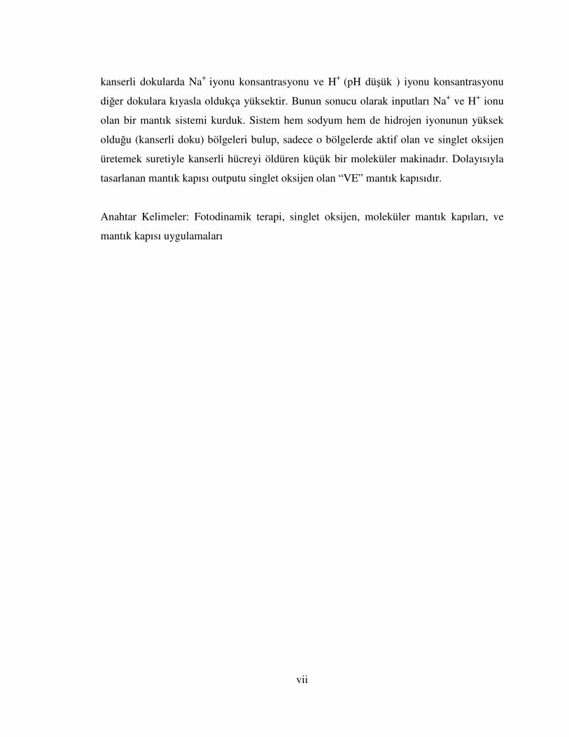
kanserli dokularda Na+ iyonu konsantrasyonu ve H+ (pH düşük ) iyonu konsantrasyonu
diğer dokulara kıyasla oldukça yüksektir. Bunun sonucu olarak inputları Na+ ve H+ ionu
olan bir mantık sistemi kurduk. Sistem hem sodyum hem de hidrojen iyonunun yüksek
olduğu (kanserli doku) bölgeleri bulup, sadece o bölgelerde aktif olan ve singlet oksijen
üretemek suretiyle kanserli hücreyi öldüren küçük bir moleküler makinadır. Dolayısıyla
tasarlanan mantık kapısı outputu singlet oksijen olan “VE” mantık kapısıdır.
Anahtar Kelimeler: Fotodinamik terapi, singlet oksijen, moleküler mantık kapıları, ve
mantık kapısı uygulamaları

viii
Dedicated to my parents and my brother. . .

ix
ACKNOWLEDGEMENTS
I would like to express my sincere thanks to my supervisor Prof. Dr. Engin U. Akkaya for
his guidance, support, endless imagination and patience.
I would like to express my gratitude to the NMR technician Fatoş Doğanel Polat and Seda
Karayılan for NMR spectra, excellent friendship and for their patience.
My special thanks go to my mother and father, especially to my brother Taner for their
continuous support, patience, encouragement and endless love.
I want to thank Ali Coşkun, Altan O. Bozdemir, Deniz Yılmaz for their guidance and
valuable friendship. I also would like to thank our group members Yusuf, Serdar, İlker,
Bora, Tuğba, Onur and rest of the SCL members and Gençay for his giving positive
energy.
.
x
TABLE OF CONTENTS
ABSTRACT………………………………………………………………..…….......iv
ÖZ………………………………………………………………………….…............vi
ACKNOWLEDGEMENTS………………………………………………….….........ix
TABLE OF CONTENTS…………………………………….…………....…….........x
LIST OF TABLES......................................................................................................xiii
LIST OF FIGURES……………………………………………………….…….......xiv
LIST OF ABBREVIATIONS…………………………………………….…….......xvii
CHAPTERS
1.INTRODUCTION......................................................................................................1
1.1 History of photodynamic therapy............................................................................1
1.2 What is photodynamic therapy?..............................................................................2
1.2.1 Cells, Tissues, and Light................................................................................4
1.2.2 Vital Requirements for an Ideal Photosensitizer............................................5
1.3 Types of photosensitizers........................................................................................6
1.3.1 First-generation photosensitizers: hematoporphyrin and its derivatives........6
1.3.2 Second generation photosensitizers(non-porphyrin
photosensitizers)......................................................................................................7
1.3.2.1 Meso-substituted Porphyrin................................................................8
1.3.2.2 Phthalocyanines and naphthalocyanines.............................................9
1.3.2.3 Synthetic Chlorins and Bacteriochlorins...........................................10
1.3.2.4 Texaphyrins.......................................................................................11
1.3.2.5 BF2 chelated Azadipyromethene dyes...............................................11
1.3.2.6 Borondipyrromethene (BODIPY) dyes.............................................13
1.3.2.7 Perylenediimide (PDIs) dyes.............................................................14
1.3.3 Third generation photosensitizers..................................................................15
1.4 Light Sources..........................................................................................................15
1.4.1. Arc lamps.....................................................................................................16
xi
1.4.2 Incandescent lamps.......................................................................................16
1.4.3 Light-emitting diodes (LEDs).......................................................................17
1.4.4 Lasers.............................................................................................................17
1.5 Heavy atom effect in PDT.....................................................................................17
1.6 Photophysical processes of a molecule..................................................................19
1.6.1 Excitation.......................................................................................................19
1.6.2 Internal Conversion.......................................................................................19
1.6.3 Fluorescence..................................................................................................20
1.6.4 Intersystem crossing......................................................................................20
1.6.5 Phosphorescence............................................................................................20
1.6.6 Quantum yield................................................................................................20
1.7 PET (Photoinduced Electron Transfer)...................................................................21
1.8 Advantages and limitations of photodynamic therapy............................................24
1.9 Logic gates..............................................................................................................25
1.9.1 Single-Input Molecular Logic........................................................................25
1.9.2 Multiple-Input Molecular Logic....................................................................26
1.9.2.1 AND gate..........................................................................................27
1.9.2.2 OR gate.............................................................................................28
1.9.2.3 INHIBIT gate....................................................................................28
1.9.2.4 NAND gate.......................................................................................29
2. EXPERIMENTAL PROCEDURES........................................................................31
2.1 General........... ......................................................................................................31
2.2 Singlet Oxygen Measurements..............................................................................31
2.3 Synthesis of benzo-15-crown [5] ..........................................................................31
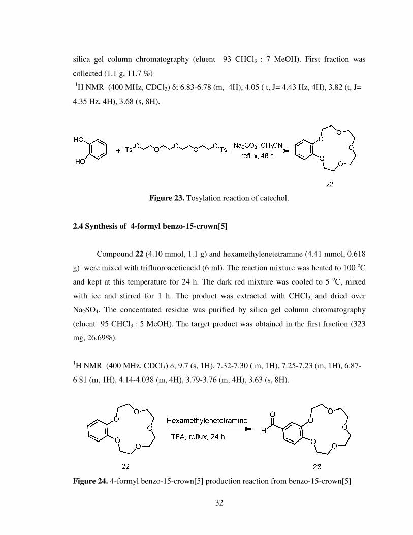
2.4 Synthesis of 4-formyl benzo-15-crown [5]...........................................................32
2.5 Synthesis of meso-Benzocrown appended BODIPY.............................................33
2.6 Synthesis of 2,6-Diiodo-substituted BODIPY.......................................................34
2.7 Synthesis of target compound 26...........................................................................35
3. RESULTS AND DISCUSSION ............................................................................36
3.1 Generation of a novel photosensitizer: 3, 5 Di-styryl
substituted boradiazaindacene dye.............................................................................36

xii
3.2 Singlet oxygen generation capacity of compound 26............................................38
4.CONCLUSION.........................................................................................................49
REFERENCES..........................................................................................................50
APPENDIX................................................................................................................53

xiii
LIST OF TABLES
TABLES
1. Truth table for some 2-input systems........................................................................26
2. AND truth table and coresponding AND gate for compound 26..............................48

xiv
LIST OF FIGURES
FIGURE
1. Modified Jablonski diagram. Photophysical processes: 1, absorption; 2, fluorescence;
3, internal conversion; 4, intersystem crossing; 5, phosphorescence; and 6, formation of
singlet oxygen 1O2 by energy transfer from T1 photosensitizer to triplet oxygen 3O2......................................................................................................................................3
2. Light interactions with a tissue.......................................................................................4
3. Structure of hematophorphyrin, 1...................................................................................7
4. Meso-substituted porphyrins that have been developed as potential photosensitizers for
PDT.....................................................................................................................................8
5. Phthalocyanine and naphthalocyanine derivatives that are relevant to PDT .................9
6. Structure of 5, 10, 15, 20-tetra (3-hydroxyphenyl)-2,3- dihydroporphyrin, 7, (mTHPC)
...........................................................................................................................................10
7. Texaphyrinato-Lu(III), a very stable hydrophilic photosensitizer.................................11
8. Structures of azadipyromethene dyes.............................................................................12
9. BF2-chelated azadipyrromethenes..................................................................................12
10. Structure of 2I-boron dipyrromethene..........................................................................13
11. An example of a BODIPY dye designed for PDT.......................................................14
12. Water-soluble green perylenediimide (PDI) dyes........................................................15
13. Positioning of the bromine heavy atom around the core sensitizer (blue)...................18
14. Simplified Jablonski diagram.......................................................................................21
15. Spaced fluorophore-receptor system and representive frontier orbital energy diagram
in the “off “ state................................................................................................................22
16. Spaced fluorophore-receptor system and representive frontier orbital energy diagram
in the “on “ state.................................................................................................................22
17. Crown containing fluorescent PET sensor 16.............................................................23
18. The molecular NOT logic gate 17; principles of operation and truth table.................26
19. The molecular AND logic gate 18: principles of operation and truth table.................27
20. Photoactive molecular system corresponding to an OR gate 19.................................28
xv
21. Photoactive molecular system corresponding to an INHIBIT gate 20........................28
22. Photoactive molecular system corresponding to an NAND gate 21...........................30
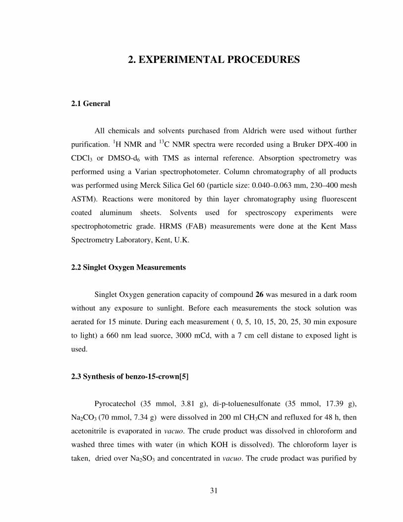
23. Tosylation reaction of catechol...................................................................................30
24. 4-formyl benzo-15-crown[5] production reaction from benzo-15-crown[5]..............32
25. Meso-benzocrown appended BODIPY synthesis reaction.........................................33
26. Iodination reaction of meso-benzocrown appended BODIPY..................................34
27. Formation of di-styrl pyridine unit on the bodipy framework....................................35
28. Chemical structure of the compound 26.....................................................................36
29. Absorbance spectra of target compound 26, p(in the presence of just photosensitizer-
target compound 26), Na (in the presence of NaClO4), TFA (in the presence of TFA),
Na+TFA (in the presence of both NaClO4 and TFA)........................................................37
30. Structure of the singlet oxygen trap 1,3-diphenylisobenzofuran (DPBF)....................38
31. Excitation of compound 26 with LED light in air saturated solution causes a
remarkable degradation of the selective singlet oxygen trap1, 3-diphenyl-iso-
benzofuran..........................................................................................................................38
32. Absorbance spectra of 1,3-diphenyl-isobenzofuran (DPBF concentration 100 µM) in
acetonitrile, without any exposure to 3000mCd light in the presence of 10 nM
photosensitizer(p) 26..........................................................................................................40
33. Absorbance spectra of 1,3-diphenyl-isobenzofuran (DPBF concentration 100 µM) in
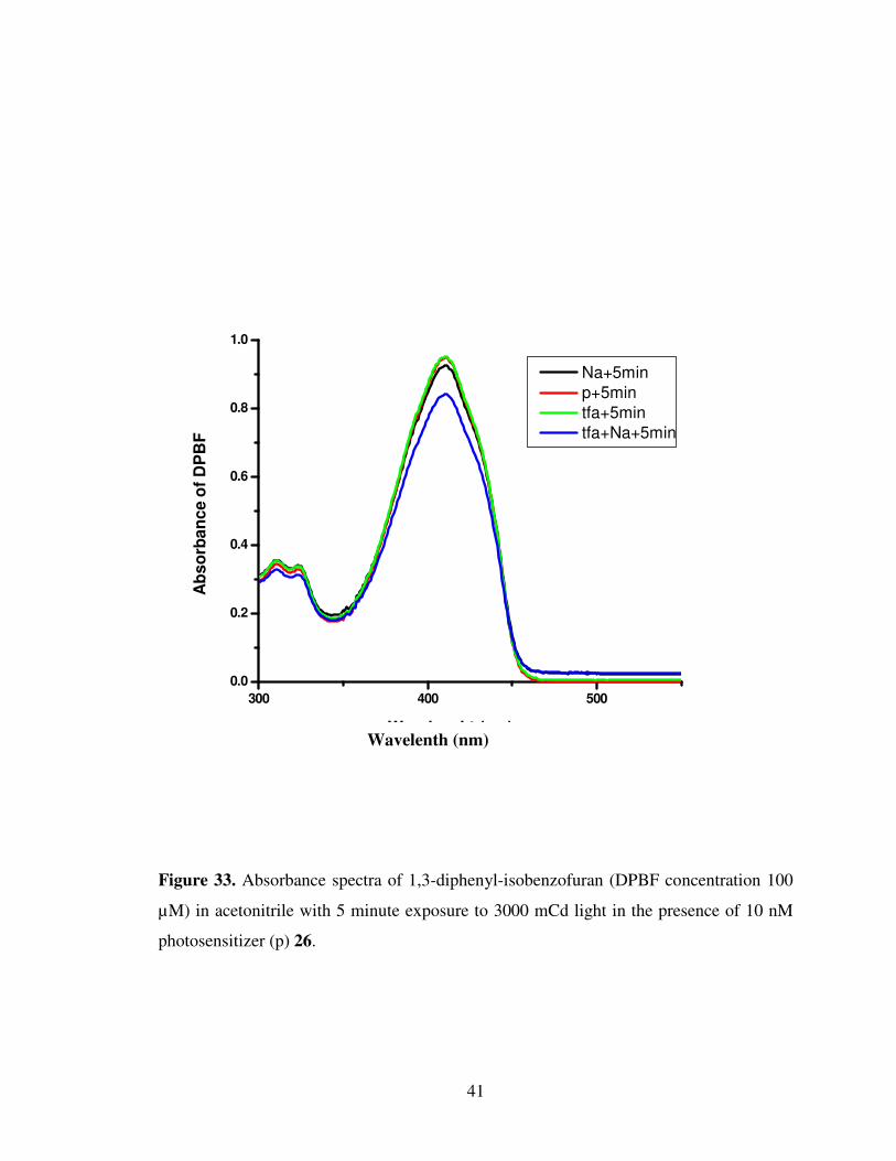
acetonitrile with 5 minute exposure to 3000mCd light in the presence of 10 nM
photosensitizer(p) 26..........................................................................................................41
34. Absorbance spectra of 1,3-diphenyl-isobenzofuran(DPBF concentration 100 µM) in
acetonitrile with 10 minute exposure to 3000 mCd light in the presence of 10 nM
photosensitizer(p) 26..........................................................................................................42
35. Absorbance spectra of 1,3-diphenyl-isobenzofuran (DPBF concentration 100 µM) in
acetonitrile with 15 minute exposure to 3000 mCd light in the presence of 10 nM
photosensitizer(p) 26..........................................................................................................43
36. Absorbance spectra of 1,3-diphenyl-isobenzofuran (DPBF concentration 100 µM) in
acetonitrile with 20 minute exposure to 3000 mCd light in the presence of 10 nM
photosensitizer(p) 26..........................................................................................................44
xvi
37. Absorbance spectra of 1,3-diphenyl-isobenzofuran (DPBF concentration 100 µM) in
acetonitrile with 25 minute exposure to 3000 mCd light in the presence of 10 nM
photosensitizer(p) 26..........................................................................................................45
38. Absorbance spectra of 1,3-diphenyl-isobenzofuran (DPBF concentration 100 µM) in
acetonitrile with 30 minute exposure to 3000 mCd light in the presence of 10 nM
photosensitizer(p) 26..........................................................................................................46
39. Absorbance of DPBF vs time graph with relative slopes
Slopes; P = 3x10⁻⁵, tfa = 3x10⁻⁵, Na = 2x10⁻⁴, Na+tfa = 4x10⁻⁴ .....................................47
40 1H NMR spectra of compound 22...............................................................................53
41 1H NMR Spectra of compound 23..............................................................................54
42 1H NMR Spectrum of compound 24...........................................................................55
43 13C NMR Spectrum of compound 24..........................................................................56
44 ESI-HRMS of compound 24......................................................................................57
45 1H NMR Spectrum of compound 25...........................................................................58
46 13C NMR Spectrum of compound 25..........................................................................59
47 ESI-HRMS of compound 25 ......................................................................................60
48 1H NMR Spectrum of compound 26............................................................................61
49 ESI-HRMS of compound 26.......................................................................................62

xvii
LIST OF ABBREVIATIONS
BODIPY Borondipyrromethene
PDT Photodynamic therapy
PS Photosensitizer
NIR Near infrared
ROS Reactive oxygen species
ISC Inter system crossing
PET Photoinduced electron transfer
LED Light-emitting diode
PDI Perylenediimide dye
TFA Trifluoroacetic acid
1
CHAPTERS
1. INTRODUCTION
1.1 History of Photodynamic Therapy
Photodynamic therapy (PDT) is an emerging treatment modality for a range of
disease classes, both cancerous and noncancerous [1]. It is a modality for the treatment of
a variety of oncological, cardiovascular, dermatological, and ophthalmic diseases [2]. The
utility of light as a therapeutic agent can be traced back over thousands of years when it
was used in Ancient Egypt, India and China to treat a variety of skin diseases like
psoriasis, vitiligo, rickets, cancer and psychosis. Whole body sun exposure (heliotherapy)
was considered important for the restoration of health in ancient Greece. Sunlight was
used in France during 18th and 19th centuries for treating various diseases like
tuberculosis, rickets, scurvy, rheumatism, paralysis, edema and muscle weakness. Danish
physician Niels Finsen successfully treated small pox using red light and went ahead with
the development of carbon arc phototherapy which made use of ultaviolet light for the
treatment of cutaneous tuberculosis for which he was awarded Nobel Prize in 1903. Administration of a photosensitizing agent followed by the irradiation of light on
tissues in which the agent is localized was another form of therapy. 3000 years ago
vitiligo was treated in India by employing psolarens and light while, Egyptians used
different psolarens in 12th century for the treatment of leucoderma [3]. In 1970,
ultraviolet A light (PUVA therapy) were used for the treatment of psoriasis and in
immunotherapy [4]. Thus it can either be by its direct action on tissue or indirectly by the
activation of a photosensitizer which in turn will bring about the therapeutic effect.

2
Inducing cytotoxicity by the interaction of light and chemical was first reported
by Oscar Raab, a medical student working with Professor Herman von Tappeiner, in 1900
while studying the effects of acridine on paramacium Infusoria . His pioneering work led
to the conclusion that such fluorescent dyes will have future medicinal applications. In
1903, Tappeiner along with dermatologist Jesionek used eosin and sunlight to treat skin
tumors . In 1907, Tappeiner together with Jodlbauer demonstrated the requirement of
oxygen in photosensitizing reactions and coined the term “photodynamic action” to
describe the phenomenon [3, 5].
1.2 What is Photodynamic Therapy?
Photodynamic therapy is based on the concept that light-sensitive species or
photosensitizers (PSs) can be preferentially localized in tumor tissues upon systemic
administration. When such photosensitizers are irradiated with an appropriate wavelength
of visible or near infrared (NIR) light, the excited molecules can transfer their energy to
molecular oxygen in the surroundings, which is normally in its triplet ground state. This
results in the formation of reactive oxygen species (ROSs), such as singlet oxygen (1O2)
or free radicals. ROSs are responsible for oxidizing various cellular compartments
including plasma, mitochondria, lysosomal, and nuclear membranes, etc., resulting in
irreversible damage of tumor cells. Therefore, under appropriate conditions,
photodynamic therapy offers the advantage of an effective and selective method of
destroying diseased tissues without damaging adjacent healthy ones [2].
3
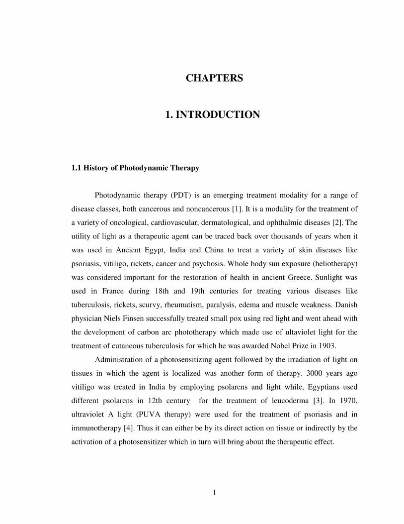
Figure 1. Modified Jablonski diagram. Photophysical processes: 1, absorption; 2,
fluorescence; 3, internal conversion; 4, intersystem crossing; 5, phosphorescence; and 6,
formation of singlet oxygen 1O2 by energy transfer from T1 photosensitizer to triplet
oxygen 3O2.
The photochemical and photophysical principles of PDT have been extensively
studied and are schematically represented in a modified Jablonski diagram shown in
Figure 1. Upon illumination, sensitizer molecule gets excited from its ground singlet state
(S0) to short-lived (~10–6 seconds) electronically excited singlet state (S1), which can
undergo radiative (fluorescence shown as route 2) and nonradiative (internal conversion
indicated as route 3) decay to come back to the ground state (S0). A good photosensitizer
will at this stage undergo a spin forbidden inter system crossing (ISC) (shown as route 4)
which requires a spin inversion, converting the photosensitizer to a triplet state (T1, 10-2
seconds) with high efficiency [4]. This transition is spin-forbidden, but a good
photosensitizer has nevertheless a high triplet-state yield. The T1-state is sufficiently
long-lived to take part in chemical reactions, and therefore the photodynamic action is
mostly mediated by the T1-state [6]. Triplet state relaxes back to ground state via spin
forbidden radiative pathway (phosphorescence shown as route 5 in Fig. 1) which imposes
relatively long life time for triplet state or by internal conversion (radiationless transitions
during collisions with other molecules). In oxygenated environments it undergoes
4
photochemical process (shown as route 6) which involves an energy transfer between
excited triplet state of photosensitizer and stable triplet oxygen (3O2) producing short
lived and highly reactive excited singlet oxygen (1O2).
Singlet oxygen is actually a highly polarized zwitterion and is considered to be
a proficient cytotoxic agent. The short lifetime of singlet oxygen (100-250 ns) limits the
diffussion range to approximately 45 nm in cellular medium and hence cannot diffuse
more than a single cell length (diameter of human cell ranges from 10-100 µm). Hence,
the primary generation of 1O2 warrants for the subcellular structures that can be accessed
and destroyed [3].
1.2.1 Cells, Tissues, and Light
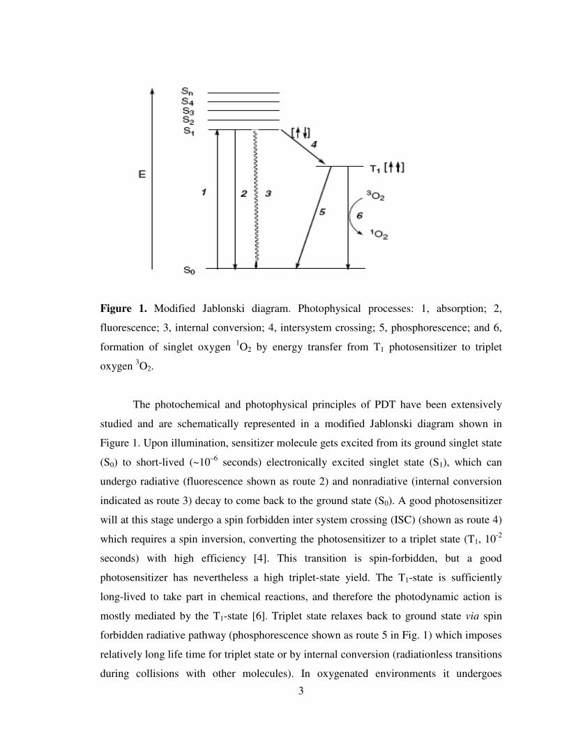
Light upon interaction with a tissue surface can be reflected, scattered,
transmitted, or absorbed (Figure 2) depending on optical features of the tissue and on the
light properties (absorption coefficient, photon energy, power density, exposition time,
etc.). The presence of water and highly absorbing endogenous dyes such as melanin and
hemoglobin strongly influences light penetration depth into the tissue. Therefore, light
penetration depth is highly dependent on the tissue type; however, in the case of most
tissues light of the spectral range 600-700 nm penetrates 50-200% deeper than light of the
range 400-500 nm. The maximum of skin permeability occurs in the range of 620-850
nm; thus, light of this spectral range (so-called “phototherapeutic window”) is
predominantly used in phototherapy [7].
Figure 2. Light interactions with a tissue
5
Photodynamic therapy (PDT) takes advantage of the interaction between light and
a photosensitizing agent to initiate apoptosis of cancer cells [8]. In PDT, instead of
directly reacting with cells and tissues the photosensitizing agent becomes activated by
light. It does that by transfering its triplet state energy to nearby oxygen molecules to
form reactive singlet oxygen (1O2) species, which cause cytotoxic reactions in the cells
[9].
1.2.2 Vital Requirements for an Ideal Photosensitizer
The choice of a photosensitizer and its subsequent phototherapeutic effect
depends on its physicochemical properties in the ground and excited states,
pharmacokinetic and pharmacodynamic behavior, and photoactivity in vivo [10,11].
Some of the properties essential for an ideal photosensitizer are listed below :
i) It should be chemically pure and of known specific composition with a reproducible
synthesis.
ii) It should have high quantum yield for singlet oxygen production for effective
destruction of tumor cells.
iii) It should have strong absorption with high extinction coefficient ε at longer
wavelength (red) region preferably between 700-800 nm where scattering of light is
minimum and tissue penetration is maximum and is sufficiently energetic enough to
produce singlet oxygen.
iv) It should have excellent photochemical reactivity, with high triplet state yields (φf) and
long triplet state life times (τ1) and be able to effectively produce 1O2 and other reactive
species. When the triplet energy of sensitizer is lower than 94 kJ/mol (1270 nm), it cannot
transfer its energy effectively from triplet state of sensitizer to the ground state triplet
oxygen and hence could not produce singlet oxygen.
v) It should possess minimal dark toxicity and only be cytotoxic in presence of light.
vi) It should have preferential retention by target tissue (tumor cells).
vii) It should be rapidly excreted from the body, thus inducing a low systemic toxicity.
viii) Finally it should be synthesizable from easily available precursors and should be
stable and easy to dissolve in the body’s tissue fluids [3].

6
ix) Preferably, the photosensitizer should not strongly absorb light of the region 400–600
nm, so that the risk of generalized photosensitivity caused by sunlight would be as small
as possible [6].
There is no sensitizer that can be named as ideal for every possible application,
but efforts are on in search of perfection and a host of second generation photosensitizers
are reported which has set right some of the drawbacks of first generation sensitizers [3].
1.3 Types of photosensitizers
A photosensitizer is a molecule that is activated to an excited singlet state when it
absorbs a photon of an appropriate wavelength; it can react with biological targets [12].
Since the first demonstration of the photodynamic action in 1900, great effort has been
devoted towards the development of photodynamic therapy agents, which have specific
light absorption and tissue distribution properties [13].
1.3.1 First-generation photosensitizers: hematoporphyrin and its derivatives
A first-generation photosensitizer that has been accepted for clinical use is the
hematophorphyrin (HpD) derivative, Photofrin, 1 [13]. The first-generation
photosensitizers are based on chemically modified natural hematoporphyrin [11,14]. They
possess certain limitations such as weak absorption in the phototherapeutic window as
well as a relatively poor specificity of uptake and retention with respect to malignant and
healthy tissues. In addition, they cause prolonged skin photosensitivity (usually 2-3
months) [15]. In view of the shortcomings encountered by the first generation
photosensitizers, researchers around the world are in search of more powerful, efficient
and maximum penetrating red light absorbing photosensitizers that can ensure effective
treatment for deep lying tumors with minimal side effects.
Photofrin has been accepted for treating various forms of cancer in many
countries such as early and late stage lung cancer, superficial and advanced oesophagal
cancer, bladder cancer, superficial and early stage gastric cancers, early stage cervical
cancer and cervical dysplasia [16]. Eventhough it fulfills certain criterion for ideal
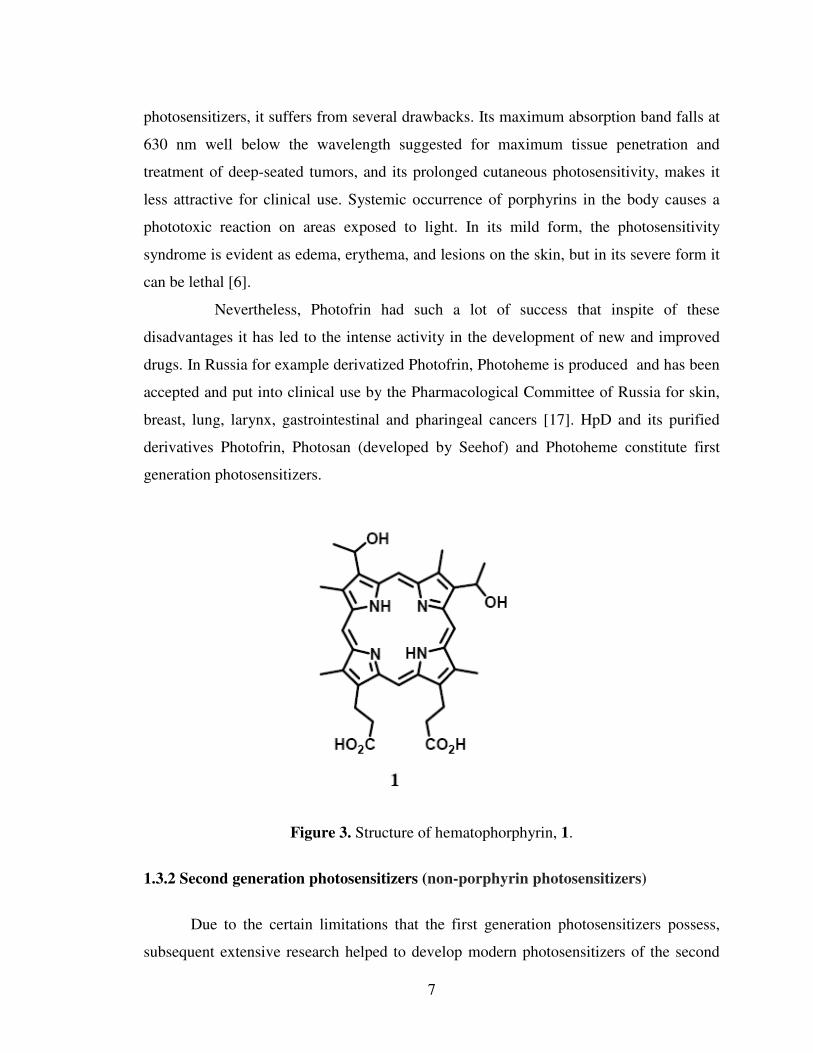
7
photosensitizers, it suffers from several drawbacks. Its maximum absorption band falls at
630 nm well below the wavelength suggested for maximum tissue penetration and
treatment of deep-seated tumors, and its prolonged cutaneous photosensitivity, makes it
less attractive for clinical use. Systemic occurrence of porphyrins in the body causes a
phototoxic reaction on areas exposed to light. In its mild form, the photosensitivity
syndrome is evident as edema, erythema, and lesions on the skin, but in its severe form it
can be lethal [6].
Nevertheless, Photofrin had such a lot of success that inspite of these
disadvantages it has led to the intense activity in the development of new and improved
drugs. In Russia for example derivatized Photofrin, Photoheme is produced and has been
accepted and put into clinical use by the Pharmacological Committee of Russia for skin,
breast, lung, larynx, gastrointestinal and pharingeal cancers [17]. HpD and its purified
derivatives Photofrin, Photosan (developed by Seehof) and Photoheme constitute first
generation photosensitizers.
Figure 3. Structure of hematophorphyrin, 1.
1.3.2 Second generation photosensitizers (non-porphyrin photosensitizers)
Due to the certain limitations that the first generation photosensitizers possess,
subsequent extensive research helped to develop modern photosensitizers of the second
8
and third generations. The second generation PDT sensitizers are mainly based on
engineered, synthetic, and semisynthetic porphyrins with various substituents at at the
pyrrole rings and the methylene bridges. They are structurally homogeneous compounds
with long-wavelength absorption bands of high intensity [7]. Second generation
photosensitizers are chemically pure, absorb light around 650 nm or longer and induce
significantly less skin photosensitivity [18].
The second generation photosensitizers used in clinical trials belong to the groups
of porphyrins, phthalocyanines, texaphyrins, chlorins, or bacteriochlorins. These
compounds have certain characteristics that make them especially suitable for PDT. An
important characteristic is their good ability to generate 1O2 [19].
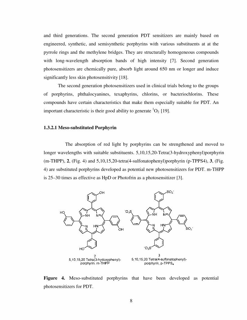
1.3.2.1 Meso-substituted Porphyrin
The absorption of red light by porphyrins can be strengthened and moved to
longer wavelengths with suitable substituents. 5,10,15,20-Tetra(3-hydroxyphenyl)porphyrin
(m-THPP), 2, (Fig. 4) and 5,10,15,20-tetra(4-sulfonatophenyl)porphyrin (p-TPPS4), 3, (Fig.
4) are substituted porphyrins developed as potential new photosensitizers for PDT. m-THPP
is 25–30 times as effective as HpD or Photofrin as a photosensitizer [3].
Figure 4. Meso-substituted porphyrins that have been developed as potential
photosensitizers for PDT.
9
The expansion of the macrocyclic p-system of porphyrins moves their absorption to
longer wavelengths and strengthens it [6].
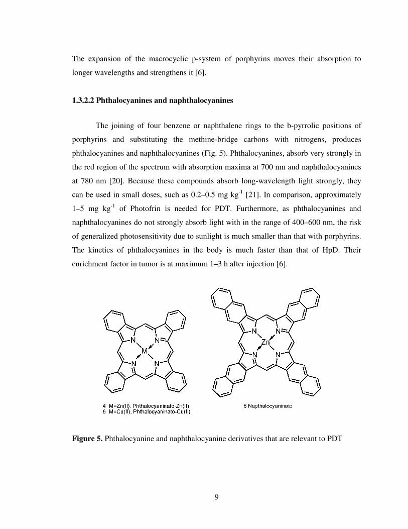
1.3.2.2 Phthalocyanines and naphthalocyanines
The joining of four benzene or naphthalene rings to the b-pyrrolic positions of
porphyrins and substituting the methine-bridge carbons with nitrogens, produces
phthalocyanines and naphthalocyanines (Fig. 5). Phthalocyanines, absorb very strongly in
the red region of the spectrum with absorption maxima at 700 nm and naphthalocyanines
at 780 nm [20]. Because these compounds absorb long-wavelength light strongly, they
can be used in small doses, such as 0.2–0.5 mg kg-1 [21]. In comparison, approximately
1–5 mg kg-1 of Photofrin is needed for PDT. Furthermore, as phthalocyanines and
naphthalocyanines do not strongly absorb light with in the range of 400–600 nm, the risk
of generalized photosensitivity due to sunlight is much smaller than that with porphyrins.
The kinetics of phthalocyanines in the body is much faster than that of HpD. Their
enrichment factor in tumor is at maximum 1–3 h after injection [6].
Figure 5. Phthalocyanine and naphthalocyanine derivatives that are relevant to PDT

10
1.3.2.3 Synthetic Chlorins and Bacteriochlorins
The reduction of a peripheral double bond of a porphyrin strengthens the longest-
wavelength absorption band and moves the maximum absorption further to the red up to
680 nm. The dihydroporphyrins thus obtained are called chlorins. In bacteriochlorins, two
double bonds on the opposite sides of the macrocycle are reduced, which strengthens and
moves their absorption even further to the red to 780 nm [19]. The synthetic chlorine
5,10,15,20-tetra(3-hydroxyphenyl)-2,3- dihydroporphyrin, 7, (mTHPC), is perhaps the
most useful photosensitizer of the synthetic chlorins. mTHPC (Foscan, Biolitec Pharma,
Scotland, U.K.), figure 6, has been approved in Europe for use against head and neck
cancer, and additional indications have been filed for prostate and pancreatic tumors [22].
7, mTHPC
Figure 6. Structure of 5, 10, 15, 20-tetra (3-hydroxyphenyl)-2,3- dihydroporphyrin, 7,
(mTHPC)
m-THPP and m-THPC differ only in the reduction degree of the tetrapyrrole ring. The
reduced form of m-THPC is more active than m-THPP in PDT [6].
11
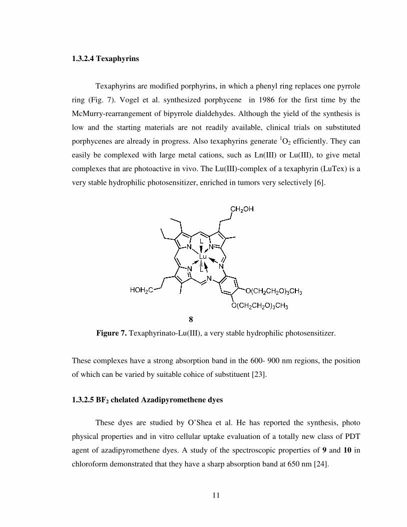
1.3.2.4 Texaphyrins
Texaphyrins are modified porphyrins, in which a phenyl ring replaces one pyrrole
ring (Fig. 7). Vogel et al. synthesized porphycene in 1986 for the first time by the
McMurry-rearrangement of bipyrrole dialdehydes. Although the yield of the synthesis is
low and the starting materials are not readily available, clinical trials on substituted
porphycenes are already in progress. Also texaphyrins generate 1O2 efficiently. They can
easily be complexed with large metal cations, such as Ln(III) or Lu(III), to give metal
complexes that are photoactive in vivo. The Lu(III)-complex of a texaphyrin (LuTex) is a
very stable hydrophilic photosensitizer, enriched in tumors very selectively [6].
8
Figure 7. Texaphyrinato-Lu(III), a very stable hydrophilic photosensitizer.
These complexes have a strong absorption band in the 600- 900 nm regions, the position
of which can be varied by suitable cohice of substituent [23].
1.3.2.5 BF2 chelated Azadipyromethene dyes
These dyes are studied by O’Shea et al. He has reported the synthesis, photo
physical properties and in vitro cellular uptake evaluation of a totally new class of PDT
agent of azadipyromethene dyes. A study of the spectroscopic properties of 9 and 10 in
chloroform demonstrated that they have a sharp absorption band at 650 nm [24].

12
Figure 8. Structures of azadipyromethene dyes
Another example about azadipyromethene dyes synthesized by O’Shea et al. is 11.
In this study, the hypotheses to be tested were whether (i) singlet oxygen generation could
be controlled by solution pH and (ii) cells could activate the photosensitizer and as a
consequence cause their own death. The ability of 11a-d to modulate singlet oxygen
generation in response to an acidic environmental stimulus was tested by trapping with
1,3-diphenylisobenzofuran (DPBF). Photosensitizer 11a, which does not contain an
amine receptor group, showed no significant variance in singlet oxygen generation when
analyzed in DMF or DMF with an aliquot of 0.05 M HCl. In comparison, singlet oxygen
generation by 11b, c and d displayed a marked reliance upon the presence or absence of a
proton source. This would substantiate a PET mechanism being the predominate mode of
singlet oxygen control.
Figure 9. BF2-chelated azadipyrromethenes.
13
With this study, they have outlined a new strategy to therapeutic selectivity in
which a supramolecular therapeutic agent could manufacture a cytotoxic agent (singlet
oxygen) in response to one exogenous stimulus (light) and one endogenous stimulus
(microenvironment pH). The absence of the specific endogenous stimulus would switch
off the therapeutic function of the photosensitizer thereby providing selectivity [25].
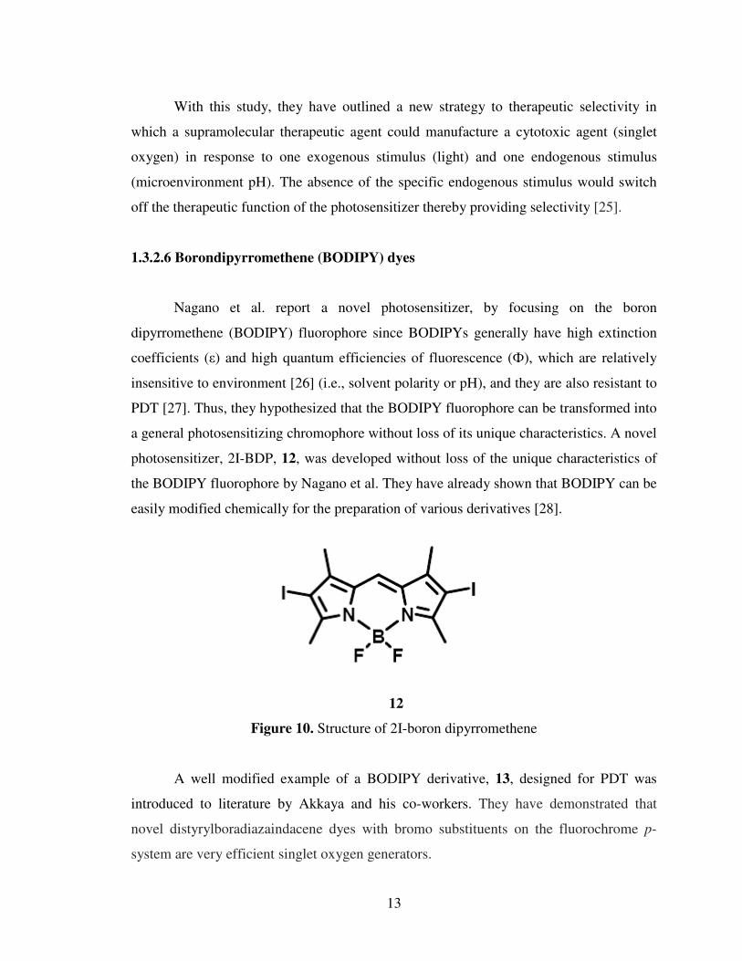
1.3.2.6 Borondipyrromethene (BODIPY) dyes
Nagano et al. report a novel photosensitizer, by focusing on the boron
dipyrromethene (BODIPY) fluorophore since BODIPYs generally have high extinction
coefficients (ε) and high quantum efficiencies of fluorescence (Ф), which are relatively
insensitive to environment [26] (i.e., solvent polarity or pH), and they are also resistant to
PDT [27]. Thus, they hypothesized that the BODIPY fluorophore can be transformed into
a general photosensitizing chromophore without loss of its unique characteristics. A novel
photosensitizer, 2I-BDP, 12, was developed without loss of the unique characteristics of
the BODIPY fluorophore by Nagano et al. They have already shown that BODIPY can be
easily modified chemically for the preparation of various derivatives [28].
12
Figure 10. Structure of 2I-boron dipyrromethene
A well modified example of a BODIPY derivative, 13, designed for PDT was
introduced to literature by Akkaya and his co-workers. They have demonstrated that
novel distyrylboradiazaindacene dyes with bromo substituents on the fluorochrome p-
system are very efficient singlet oxygen generators.

14
13
Figure 11. An example of a BODIPY dye designed for PDT.
In addition, these water soluble photosensitizers were shown to have spectacular
photoinduced cytotoxicity at very low concentrations and even under low fluence rate
LED irradiation. Dark toxicity was nil at the concentration range studied [29].
1.3.2.7 Perylenediimide (PDIs) dyes
PDIs are reddish dyes with very high quantum yields. As such they are not long
wavelength dyes. But, by single or double amine substitution on the perylene core, the
absorption maxima at these dyes can be shifted up to 750 nm with further appropriate
modifications; solubility can be improved [30].
Akkaya et al. have synthesized a series of water-soluble green
perylenediimide (PDI) dyes. On red light excitation, these dyes were shown to be
efficient generators of singlet oxygen, and in cell culture media, they were shown to
display significant light-induced cytotoxic effects on the human erythroleukemia cell line
(K-562) [31].

15
14
Figure 12. Water-soluble green perylenediimide (PDI) dyes.
1.3.3 Third generation photosensitizers
The third-generation photosensitizers consist of the photosensitizer moiety linked
to biomolecules such as polypeptide chains, monoclonal antibodies, proteins, etc., which
allow their selective delivery. This strategy overcomes difficulties in molecule
recognition and specific binding to the tumor [7].
1.4 Light Sources
The radiation used in PDT is light of the visible and near-infrared regions. The
wavelength of light required is dependent on the photosensitizer used. The longest
possible wavelength is chosen to reach a therapeutic effect as deep as possible [32]. The
light dose required to achieve a certain therapeutic effect is dependent on the
photosensitizer used and on the optical properties of the tissue. In Photofrin PDT, the
required light dose is normally 50–500 J cm-2, but when second-generation sensitizers
with stronger light absorption are used, 10 J cm-2 may suffice. To avoid excess heating,
the irradiation power should not exceed 200 mW cm-2 [6].

16
1.4.1. Arc lamps
The mercury arc lamp is the workhorse of organic photochemistry, and has some
applications in photomedicine. There are three main types. The low-pressure mercury arc
operates at room temperature and about 10-3 mm pressure: the main emission is a single
line at 253.7 nm. Medium-pressure mercury lamps operate at about 1 atmosphere: the
emission contains a number of lines, of which 366 nm and 546 nm are the principal ones.
The 366 nm line is commonly used to observe the red fluorescence of porphyrins and
other dye stuffs. The high-pressure mercury arc operates at ~100 atmospheres and is an
intense source: the emission is practically a continuum.
1.4.2 Incandescent lamps
Incandescent lamps are inexpensive and have been used in PDT. For example, in
the treatment of basal cell carcinoma using δ-aminolaevulinic acid as a pro-drug,
Kennedy and Pettier employed a projector lamp as the light source. Infrared and
ultraviolet light are filtered out with filters and a dichroic reflector to give illumination in
the required 400-550 nm region ( λmax 450 nm).
1.4.3 Light-emitting diodes (LEDs)
They are semiconductor based devices driven by an electric current. The emission
is not coherent (this is not a laser source), and is low power, so not much heat is
produced. Wavelength can be adjusted by changing the semiconductor, and the devices
are small, but they can be bunched together to fit a particular structure. They are finding
increasing application. Thus, the LED system recently announced by Diomed consists of
a close-packed array of LEDs in a water-cooled head which is designed to be kept in
contact with the area of treatment. The irradiation wavelength is 635 nm or 652 nm, with
a fluence rate of up 200 mWcm-2 [30].

17
1.4.4 Lasers
A laser (from the acronym Light Amplification by Stimulated Emission of
Radiation) is an optical source that emits photons in a coherent beam [30]. Lasers have
certain characteristics that make them especially suitable for use in PDT. Lasers are able
to deliver intense light with a high degree of monochromaticity, which makes the
focusing of the light beam into an optical fiber possible without a great loss of energy.
The employment of optical fibers in PDT enables treatment of internal tumors
endoscopically and placement of the light source interstitially into the tumor tissue. The
high power of laser light is of minor importance in PDT, because the light is used for
photosensitizer activation and not for tissue incision by strong heating. The use of lasers
as light sources has drawbacks, because they are expensive, complicated devices, which
are not readily transportable [6].
1.5 Heavy atom effect in PDT
As singlet oxygen is the key cytotoxic agent in the PDT therapeutic process. The
quantity of singlet oxygen generated by a photosensitizer is regulated by the efficiency of
a spin-forbidden electronic transition from a singlet to a triplet state (ISC). The
introduction of a heavy atom into a molecule is known to have an influence over the rates
of the ISC and is termed the heavy-atom effect [33].
Introduction of a heavy metal ion strongly affects intersystem crossing in the
complex via enhancement of spin-orbit coupling, which influences the formation and
decay of triplet states. As a result, the rate of intersystem crossing is enhanced [7]. An
electronic transition from a singlet to a triplet excited state within a molecule is a spin-
forbidden process and as such occurs inefficiently for many compounds. In order for a
transition between states of different spin multiplicities to occur effectively, a spin-orbit
perturbation is generally required [34]. Enhanced spin-orbit perturbations can be achieved
by the attachment of a heavy atom directly onto the molecule [35] (internal heavy-atom
effect) or placing the molecule in a surrounding environment containing heavy atoms [36]
(external heavy-atom effect).
18
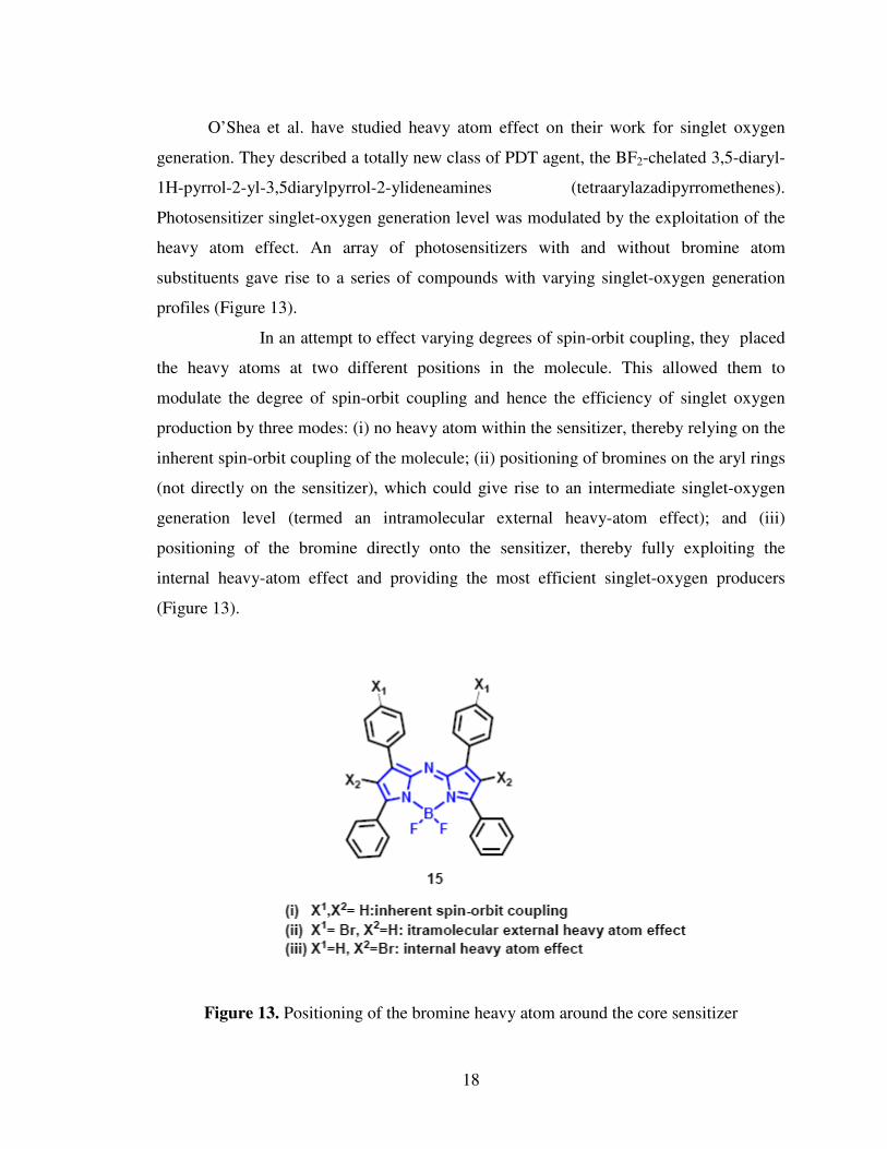
O’Shea et al. have studied heavy atom effect on their work for singlet oxygen
generation. They described a totally new class of PDT agent, the BF2-chelated 3,5-diaryl-
1H-pyrrol-2-yl-3,5diarylpyrrol-2-ylideneamines (tetraarylazadipyrromethenes).
Photosensitizer singlet-oxygen generation level was modulated by the exploitation of the
heavy atom effect. An array of photosensitizers with and without bromine atom
substituents gave rise to a series of compounds with varying singlet-oxygen generation
profiles (Figure 13).
In an attempt to effect varying degrees of spin-orbit coupling, they placed
the heavy atoms at two different positions in the molecule. This allowed them to
modulate the degree of spin-orbit coupling and hence the efficiency of singlet oxygen
production by three modes: (i) no heavy atom within the sensitizer, thereby relying on the
inherent spin-orbit coupling of the molecule; (ii) positioning of bromines on the aryl rings
(not directly on the sensitizer), which could give rise to an intermediate singlet-oxygen
generation level (termed an intramolecular external heavy-atom effect); and (iii)
positioning of the bromine directly onto the sensitizer, thereby fully exploiting the
internal heavy-atom effect and providing the most efficient singlet-oxygen producers
(Figure 13).
Figure 13. Positioning of the bromine heavy atom around the core sensitizer
19
The final step of the singlet-oxygen generation process is an energy transfer from
photosensitizer triplet state to ground-state oxygen. An additional consequence of
introducing heavy-atom substituents can be to give rise to nonradiative internal back
conversion to the ground state or inhibiting the photosensitizer triplet to ground-state
oxygen energy transfer. These competing pathways would give rise to loss of the excited-
state energy without the generation of singlet oxygen. This makes the position of the
heavy-atom within the sensitizer critical, as the atom(s) must be positioned to have a
substantial effect on the degree of spin-orbit coupling but not give rise to competing
excited-state energy loss pathways. As such an heavy atom can promote an S1 to T1
transition , ISC, though it may not necessarily result in enhanced singlet-oxygen
production. This is evidence that the heavy atom effect can be exploited to deliver
controlled levels of the key cytotoxic agent for photosensitizer class. The dramatically
enhanced singlet-oxygen production levels show that the inclusion of the heavy atom as a
substituent directly onto the central core of the photosensitizer has achieved the desired
aim and has not given rise to loss of excited-state energy by internal radiationless
transitions [1].
1.6 Photophysical processes of a molecule
1.6.1 Excitation
Excitation involves promoting an electron residing in a low-energy molecular
level to a higher level. The lifetime of excited species is low because several mechanisms
exist whereby an excited molecule can give up its excess energy and relax to its ground
state [37] (Figure 14).
1.6.2 Internal Conversion
Internal conversion is a non-radiative transition between two electronic states of
the same spin multiplicity. Therefore, internal conversion from S1 to S0 compete with
emission of photons (fluorescence) and intersystem crossing to the triplet state from
which emission of photons (phosphorescence) can possibly be observed [38].

20
1.6.3 Fluorescence
Emission of photons accompanying the S1 to S0 relaxation is called fluorescence.
The fluorescence spectrum is located at higher wavelengths (lower energy) than the
absorption spectrum because of the energy loss in the excited state due to the vibrational
relaxation [38].
1.6.4 Intersystem crossing
Intersystem crossing is a non-radiative transition between two isoenergetic
vibrational levels belonging to electronic states of different multiplicities. Crossing
between states of different multiplicity is in principle forbidden, but spin-orbit coupling
(i.e. coupling between the orbital magnetic moment and the spin magnetic moment) can
be large enough to make it possible. The probability of intersystem crossing depends on
the singlet and triplet states involved. It should also be noted that the presence of heavy
atoms (i.e. whose atomic number is large, for example Br, Pb) increases spin-orbit
coupling and thus favors intersystem crossing [38].
1.6.5 Phosphorescence
In solution at room temperature, non-radiative de-activation from the triplet state
T1, is predominant over radiative de-activation called phosphorescence. In fact, the
transition T1 to S0 is forbidden (but it can be observed because of spin-orbit coupling),
and the radiative rate constant is thus very low. The life time of the triplet state, may
under these conditions, is long enough to observe phosphorescence on a time-scale up to
seconds, even minutes or more [38].
1.6.6 Quantum yield
The tendency of a photosensitizer to reach the triplet state is measured by the
triplet state quantum yield, which measures the probability of formation of the triplet state
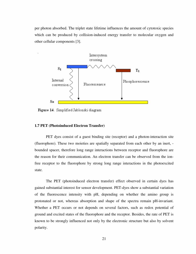
21
per photon absorbed. The triplet state lifetime influences the amount of cytotoxic species
which can be produced by collision-induced energy transfer to molecular oxygen and
other cellular components [3].
1.7 PET (Photoinduced Electron Transfer)
PET dyes consist of a guest binding site (receptor) and a photon-interaction site
(fluorophore). These two moieties are spatially separated from each other by an inert, -
bounded spacer, therefore long range interactions between receptor and fluorophore are
the reason for their communication. An electron transfer can be observed from the ion-
free receptor to the fluorophore by strong long range interactions in the photoexcited
state.
The PET (photoinduced electron transfer) effect observed in certain dyes has
gained substantial interest for sensor development. PET-dyes show a substantial variation
of the fluorescence intensity with pH, depending on whether the amino group is
protonated or not, whereas absorption and shape of the spectra remain pH-invariant.
Whether a PET occurs or not depends on several factors, such as redox potential of
ground and excited states of the fluorophore and the receptor. Besides, the rate of PET is
known to be strongly influenced not only by the electronic structure but also by solvent
polarity.
22
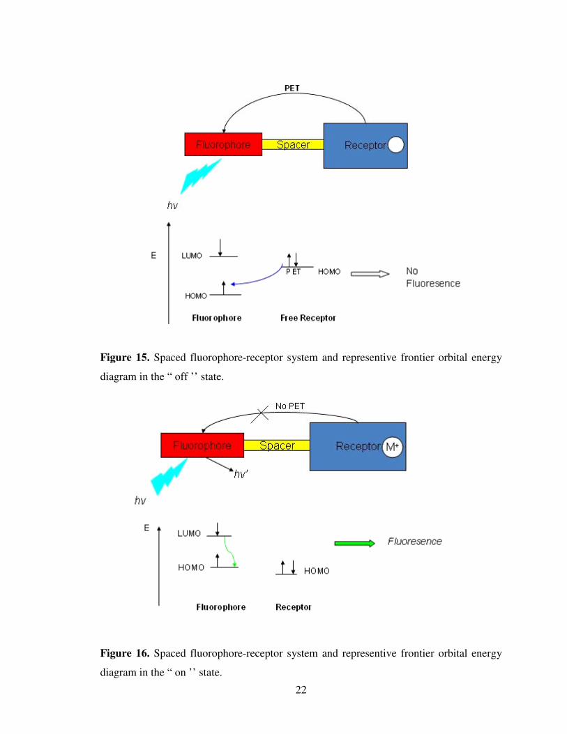
Figure 15. Spaced fluorophore-receptor system and representive frontier orbital energy
diagram in the “ off ’’ state.
Figure 16. Spaced fluorophore-receptor system and representive frontier orbital energy
diagram in the “ on ’’ state.

23
Fluorescent signaling via the PET strategy is distinguished by its intrinsically
supramolecular nature since distinct components perform each one (or more) of the
necessary functions. A fluorophore module is the site of both photonic transactions of
excitation and emission. A receptor module is responsible for guest complexation and
decomplexation. A spacer module holds the fluorophore and receptor close to, but
separate from each other. This also means that true molecular engineering applies, i.e.,
the optical, guest-binding, and redox properties of the components allow the quantitative
prediction of the signaling parameters of the supramolecular system. Further, PET
signaling systems have guest-induced “off-on” and “on-off” fluorescence. The pionering
work of Weller over a quarter of a century is the starting point, since this provides the
thermodynamic basis of PET. Figures 15 and 16 provide a summary in terms of frontier
orbital energies. It also shows how PET systems employ thermal back-electron transfer as
a self-repair mechanism following the potentially damaging PET process [39].
Several of the laboratories active in this field have summarized their own
contributions along with related material. These include the groups of Czarnik, Fabbrizzi,
Tsien, Kuhn, Rettig, Valeur and Shinkai [39]. Compound 16 is the simplest and first PET
sensor which has been synthesized by de Silva et al., its fluorescence quantum yield
increases from 0.003 to0.140 upon binding of K+ in methanol solution [40] .
Figure 17. Crown containing fluorescent PET sensor 16.
24
1.8 Advantages and limitations of photodynamic therapy
The most important application of PDT is in treatment of cancer. PDT has many
advantages as compared with the traditional cancer treatments. It is a selective treatment
that does not destroy healthy tissues. It is an effective, non-invasive treatment, which is at
its best easy, fast, and painless to the patient. There are no serious side effects and no
resistance normally develops, so that the treatment can be repeated if necessary. There is
also no cross-resistance with other cancer treatments. It can be used to treat many
different kinds of cancers, including cancers resistant to other treatments, and its efficacy
is not dependent on the cell cycle phase.
Tissues treated by PDT also heal well, probably because the collagen they contain
is not sensitive to photodynamic damage. The mechanical strength of tissues is mostly
maintained and their perforation is therefore unlikely. Further, because most
photosensitizers do not accumulate in the nuclei of normal cells, photodynamic damage is
unlikely. The risk of carcinogenesis or mutations is therefore small. Light of wavelengths
absorbed by the photosensitizer is needed for photodynamic action. Light that is normally
used in PDT does not penetrate deep into tissue, as it is scattered and absorbed by
endogenous chromophores. Therefore, PDT can be applied to treat tumors that are at the
most 2 cm thick, the precise thickness depending on the photosensitizer used. Such
tumors include cutaneous tumors and early stages of various other tumors.
PDT cannot be used when the destruction of the tumor would lead to a serious
medical crisis, as when the tumor forms a part of the chest wall. PDT can, however, be
combined with surgery; the tumor can be resected to a PDT treatable size, or PDT can be
applied to remove possible microscopic remains of the tumor after surgery. It may also be
combined with chemotherapy, radiation or hyperthermia. It can be used for palliation,
where the goal is to reduce the size of the tumor and thereby alleviate the symptoms and
increase the life expectancy of a terminal patient [6].
25
1.9 Logic gates
Logic gates are the workhorses of modern information technology, because
computation relies on arithmetic and logic units. Arithmetic units can be dissected into
simpler logic gate arrays. So, the challange is to pass on these properties and capabilities
to molecules. This challenge has been accepted with increasing frequency since 1993,
when it was demonstrated that molecular fluorescence signals can be switched under the
influence of simple chemical species. The forces that drive research in molecular logic
gates are identified as neutral science and computer technology. The longest established
design relies on chemical inputs (usually ionic) and fluorescence outputs.
Despite a lot of efforts and expectations, molecular-scale electronics/photonics is
undergoing a gradual evolution via wires, switches, and diodes with logic gates being
beyond the current horizon. On the other hand, it has been possible to shift directly to
molecular logic gates by combining chemical and photonic signals according to principles
of supramolecular chemistry. Both our brains and modern silicon-based electronics
technology rely heavily on integration [41].
1.9.1 Single-Input Molecular Logic
There are four possible output patterns arising from a single input. If the input is 0,
the output can be 0 or 1 (two choices). If the input is 1, the output can again be 0 or 1
(two choices). Each one of these four output bit patterns corresponds to a logic type:
PASS 0, YES, NOT and PASS 1. PASS 0 always outputs 0, whatever the input. PASS 1
always outputs 1. YES obediently follows the input (e.g., output 1 if input is 1). NOT
always opposes the input (e.g., output 0 if input is 1). For instance, a NOT gate with a
chemical input, fluorescence light output and excitation light for the power supply is 1
[43] (Figure 18). Any case of chemical-induced fluorescence quenching (of which there
are hundreds) would have served in its place, testifying to the generality of this approach.

26
Figure 18. The molecular NOT logic gate 17; principles of operation and truth table.
The H+-induced fluorescence quenching arises as follows. When the H+ level is
low (input 0), excitation of the fluorophore results in bright fluorescence (output 1) with
no substantial photochemistry intervening. However when the H+ level is high (input 1),
excitation of the fluorophore leads to photochemical intervention and, hence, the
fluorescence is quenched (output 0). This intervention is photoinduced electron transfer
(PET)]from the fluorophore to the protonated benzoate receptor (a rather electron-
deficient unit).
1.9.2 Multiple-Input Molecular Logic
In Multiple-Input Molecular Logic, when the number of inputs is increased from 1
to 2, the possible number of logic types will rise to 16 [43].
Table 1. Truth table for some 2-input systems.
INPUTS OUTPUTS
A B AND NAND OR INHIBIT
0 0 0 1 0 0
0 1 0 1 1 0
1 0 0 1 1 1
1 1 1 0 1 0
17

27
1.9.2.1 AND gate
AND logic, gives an output of 1 only if both inputs are 1. Since the first report of
molecular AND logic gate by de Silva et al., many individual logic gates have been
described on the basis of the spectral variations of molecular systems in response to
external stimulations [44]. A molecular-scale example of this is from de Silva’s group
again, 18 (Figure 19). This AND gate is made possible by utilising two discrete, selective
binding sites that are weakly coupled to a fluorophore. In this scheme,
thermodynamically controlled and guest-modulated PET processes from oxidizable
moieties located at each receptor serve to quench the fluorescence from the excited
fluorophore, across inert methylene spacers. Only if chemical species (Na+ and H+) are
present at both receptor1 and receptor2 are both oxidation potentials raised, such that the
electron transfer is diminished. Then the fluorescence output is restored (output 1), and
the truth table is satisfied. In this implementation, the power supply was provided by
excitation light, while inputs are chemical (Na+ and H+) and the output is fluorescence
light [45].
18
Figure 19. The molecular AND logic gate 18: principles of operation and truth table.
28
1.9.2.2 OR gate
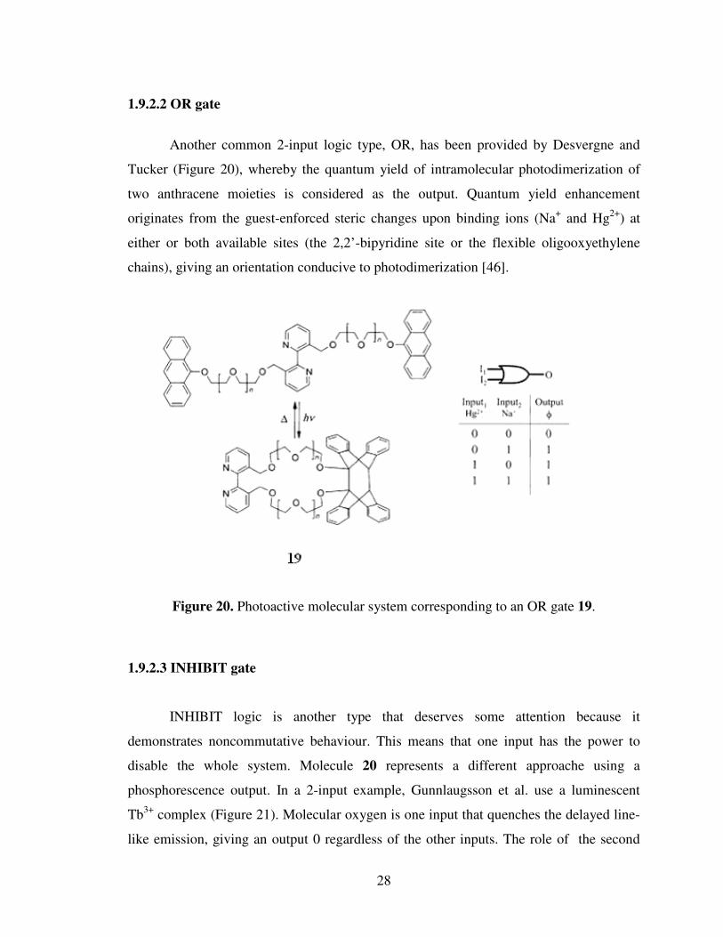
Another common 2-input logic type, OR, has been provided by Desvergne and
Tucker (Figure 20), whereby the quantum yield of intramolecular photodimerization of
two anthracene moieties is considered as the output. Quantum yield enhancement
originates from the guest-enforced steric changes upon binding ions (Na+ and Hg2+) at
either or both available sites (the 2,2’-bipyridine site or the flexible oligooxyethylene
chains), giving an orientation conducive to photodimerization [46].
Figure 20. Photoactive molecular system corresponding to an OR gate 19.
1.9.2.3 INHIBIT gate
INHIBIT logic is another type that deserves some attention because it
demonstrates noncommutative behaviour. This means that one input has the power to
disable the whole system. Molecule 20 represents a different approache using a
phosphorescence output. In a 2-input example, Gunnlaugsson et al. use a luminescent
Tb3+ complex (Figure 21). Molecular oxygen is one input that quenches the delayed line-
like emission, giving an output 0 regardless of the other inputs. The role of the second

29
input (H+) is to shift the absorption band of 20 into the range of the excitation light that
provides the power for this device. Luminescence (output 1) is only observed if O2 is
absent (input 0), in the presence of acid (input 1) [47].
Figure 21. Photoactive molecular system corresponding to an INHIBIT gate 20.
1.9.2.4 NAND gate
NAND gates have high value in electronics, since multiple copies of these can be
wired up to emulate all the other logic types. Akkaya and Baytekin use of a well-known
compound 21 is of particular interest (Figure 22). They use hydrogen-bonding
interactions for the first time alongside luminescence ideas for logic design with small
molecules. Molecule 21 is known to intercalate into adenine (A)-thymine (T) base-pair
regions of DNA. In aqueous/organic solvent mixtures they observe binding between the
A-T mononucleotide pair and 21, with the binding being signalled by luminescence
spectral changes. Careful choice of emission wavelength (455 nm) shows significant
reduction of luminescence intensity only when both A and T are present, that is, NAND
logic [48].

30
Figure 22. Photoactive molecular system corresponding to an NAND gate 21.
31
2. EXPERIMENTAL PROCEDURES
2.1 General
All chemicals and solvents purchased from Aldrich were used without further
purification. 1H NMR and 13C NMR spectra were recorded using a Bruker DPX-400 in
CDCl3 or DMSO-d6 with TMS as internal reference. Absorption spectrometry was
performed using a Varian spectrophotometer. Column chromatography of all products
was performed using Merck Silica Gel 60 (particle size: 0.040–0.063 mm, 230–400 mesh
ASTM). Reactions were monitored by thin layer chromatography using fluorescent
coated aluminum sheets. Solvents used for spectroscopy experiments were
spectrophotometric grade. HRMS (FAB) measurements were done at the Kent Mass
Spectrometry Laboratory, Kent, U.K.
2.2 Singlet Oxygen Measurements
Singlet Oxygen generation capacity of compound 26 was mesured in a dark room
without any exposure to sunlight. Before each measurements the stock solution was
aerated for 15 minute. During each measurement ( 0, 5, 10, 15, 20, 25, 30 min exposure
to light) a 660 nm lead suorce, 3000 mCd, with a 7 cm cell distane to exposed light is
used.
2.3 Synthesis of benzo-15-crown[5]
Pyrocatechol (35 mmol, 3.81 g), di-p-toluenesulfonate (35 mmol, 17.39 g),
Na2CO3 (70 mmol, 7.34 g) were dissolved in 200 ml CH3CN and refluxed for 48 h, then
acetonitrile is evaporated in vacuo. The crude product was dissolved in chloroform and
washed three times with water (in which KOH is dissolved). The chloroform layer is
taken, dried over Na2SO3 and concentrated in vacuo. The crude prodact was purified by
32
silica gel column chromatography (eluent 93 CHCl3 : 7 MeOH). First fraction was
collected (1.1 g, 11.7 %)
1H NMR (400 MHz, CDCl3) δ; 6.83-6.78 (m, 4H), 4.05 ( t, J= 4.43 Hz, 4H), 3.82 (t, J=
4.35 Hz, 4H), 3.68 (s, 8H).
Figure 23. Tosylation reaction of catechol.
2.4 Synthesis of 4-formyl benzo-15-crown[5]
Compound 22 (4.10 mmol, 1.1 g) and hexamethylenetetramine (4.41 mmol, 0.618
g) were mixed with trifluoroaceticacid (6 ml). The reaction mixture was heated to 100 oC
and kept at this temperature for 24 h. The dark red mixture was cooled to 5 oC, mixed
with ice and stirred for 1 h. The product was extracted with CHCl3, and dried over
Na2SO4. The concentrated residue was purified by silica gel column chromatography
(eluent 95 CHCl3 : 5 MeOH). The target product was obtained in the first fraction (323
mg, 26.69%).
1H NMR (400 MHz, CDCl3) δ; 9.7 (s, 1H), 7.32-7.30 ( m, 1H), 7.25-7.23 (m, 1H), 6.87-
6.81 (m, 1H), 4.14-4.038 (m, 4H), 3.79-3.76 (m, 4H), 3.63 (s, 8H).
Figure 24. 4-formyl benzo-15-crown[5] production reaction from benzo-15-crown[5]

33
2.5 Synthesis of meso-Benzocrown appended BODIPY
Compound 23 (1.09 mmol, 323 mg) and 2, 4-dimethyl pyrole (2.20 mmol, 207
mg) were dissolved in 300 ml absolute CH2Cl2 (argon was bubled through CH2Cl2 for 30
min) under argon atmosphere. One drop of tfa was added and and the solution stirred at
R. T until TLC control showed 50 % consumption ( about 24 h) of aldehyde. At this
point, a solution of tetrachlorobenzoquinone in 120 ml CH2Cl2 was added, stirring was
continued for 30 min followed by the addition of 3 ml of Et3N and 3 ml of BF3OEt2. After
stirring for 30 min the reaction mixture was washed three times with water , dried over
Na2SO4 and evaporated to dryness in vacuo. The residue was chromatographed on silica
gel (95 CHCl3 : 5 MeOH) . The target product was obtained in the second fraction (277
mg, 49.4 % ).
1H NMR (400 MHz, CDCl3) δ; 6.87 (d, J= 7.96 Hz, 1H), 6.71 ( d, J= 11.55 Hz, 2H), 5.90
(s, 2H), 4.11 (t, J= 3.94 Hz, 2H), 4.01 (t, J= 4.02 Hz, 2H), 3.88 (t, J= 3.77 Hz, 2H), 3.83
(t, J= 4.11 Hz, 2H), 3.70 ( d, J= 6.42 Hz, 8H), 2.47 ( s, 6H), 1.40 ( s, 6H) ;
13C NMR (100 MHz, CDCl3) δ; 155.3, 149.8, 143.2, 141.5, 131.7, 127.5, 121.1, 120.9,
113.9, 113.6, 71.0, 70.3, 69.4, 69.3, 69.0, 68.7, 61.0, 14.6, 14.5;
ESI-HRMS cald for [M+Na] 537.2354 found 537.2328 ∆=4.8 ppm.
Figure 25. Meso-benzocrown appended BODIPY synthesis reaction.

34
2.6 Synthesis of 2,6-Diiodo-substituted BODIPY
HIO3 ( 1.32 mmol, 232 mg) is dissolved in minimum amount of water and added
dropwise to a solution of 24 ( 0.33 mmol, 170 mg) and I2 ( 1.65 mmol, 419 mg) in 10 ml
EtOH. The mixture was heated to 60 oC for 20 min. Then, cooled down to room
temperature and extracted with sodium thiosulphate dissolved water and CH2Cl2. The
organic phase was dried over Na2SO4 and concentrated in vacuo. Then crude product was
purified by silica gel column chromatography ( eluent 95 CHCl3 : 5 MeOH). The target
product was obtained (95 mg, 37.5 %).
1H NMR (400 MHz, CDCl3) δ; 6.91 (d, J= 8.08 Hz, 1H), 6.71-6.66 ( m, 2H), 4.11 (t, J=
4.07 Hz, 2H), 4.01 (t, J= 4.17 Hz, 2H), 3.88 (t, J= 4.05 Hz, 2H), 3.83 (t, J= 4.12 Hz, 2H),
3.72 ( d, J= 7.35 Hz, 8H), 2.47 ( s, 6H), 1.40 ( s, 6H);
13C NMR (100 MHz, CDCl3) δ; 156.6, 150.1, 150.0, 145.4, 141.2, 131.6, 127.1, 120.8,
114.0, 113.3, 85.5, 71.0, 70.3, 70.2, 69.3, 69.2, 69.1, 68.7;
ESI-HRMS cald for [M+Na] 789.0286 found 789.0257 ∆=3.7 ppm.
Figure 26. Iodination reaction of meso-benzocrown appended BODIPY.

35
2.7 Synthesis of target compound 26
Compound 25 ( 0.124 mmol, 95 mg) and 4-pyridinecarboxaldehyde (0.45 mmol,
53 mg) were refluxed for 20 min in a mixture of benzene ( 25 ml), piperidine ( 275 µl)
and AcOH ( 230 µl). Any water formed during the reation, was removed azeotropically
by heating the mixture in a Dean - Stark apparatus. The reaction was monitored by TLC
(eluent 95 CHCl3 : 5 MeOH ). Organic phase was washed three times with NaHCO3
dissolved water, dried over Na2SO4 and concentrated in vacuo. The crude product was
purified by silica gel column chromatography (eluted first with 95 CHCl3 : 5 MeOH then
flash column chromatography of 1 EtOAc : 1 CHCl3 ). The product was collected ( 15
mg, 12.8 % ).
1H NMR (400 MHz, CDCl3) δ; 8.70-8.61 (m, 4H), 7.96 ( d, J= 16.72 Hz, 2H), 7.73 (d,
J= 16.73 Hz, 2H), 7.48-7.42 (m, 4H), 6.94 (d, J= 8.12 Hz, 1H), 6.78-6.72 (m, 2H), 4.25-
4.11 (m, 2H), 4.06-4.03 (m, 2H), 4.06-4.03 (m, 2H), 3.94-3.88 (m, 2H), 3.75-3.68 ( d,
8H), 1.51 ( s, 6H) ;
ESI-HRMS cald for [M+Na] 967.0812 found 967.0776 ∆=3.7 ppm.
Figure 27. Formation of di-styrl pyridine unit on the bodipy framework.

36
3. RESULTS AND DISCUSSION
3.1 Generation of a novel photosensitizer: 3,5-di-styryl substituted
boradiazaindacene dye
The design for the automaton includes the following elements; i) strong long
wavelenght absorption preferably beyond 650 nm; 3,5-di-styryl substituted bodipy dyes
are useful in this regard with typical absorption peaks ranging from 630-750 nm ii) an
acid sensitive absorption peak; we reasoned this could be best achieved by incorporating
di-methyl amino styryl or pyridyl ethenyl moities iii) in order to modulate PET efficiency
and intersystem crossing efficiency which is directly related to the PET process, we
utilized a crown ether based PET modulator which is only sensitive to sodium but not to
hydrogen ions. With this design we set out for the synthesis of compound 26.
Figure 28. Chemical structure of the compound 26.
Our synthesis starts with the formulation of benzo-15-crown[5]. Then, using
standard procedures and the resulting aldehyde together with 2,4-dimethyl pyrole, a
bodipy dye was obtained. To facilitate intersystem crossing 2 and 6 positions of the
26
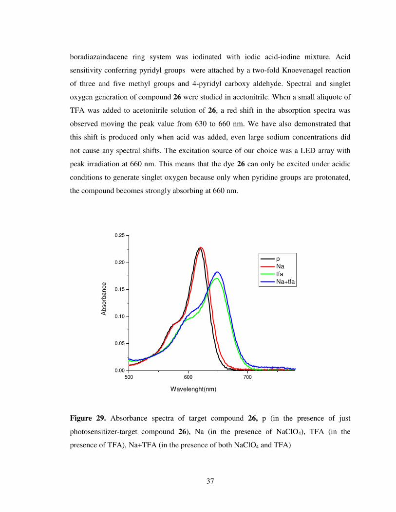
37
boradiazaindacene ring system was iodinated with iodic acid-iodine mixture. Acid
sensitivity conferring pyridyl groups were attached by a two-fold Knoevenagel reaction
of three and five methyl groups and 4-pyridyl carboxy aldehyde. Spectral and singlet
oxygen generation of compound 26 were studied in acetonitrile. When a small aliquote of
TFA was added to acetonitrile solution of 26, a red shift in the absorption spectra was
observed moving the peak value from 630 to 660 nm. We have also demonstrated that
this shift is produced only when acid was added, even large sodium concentrations did
not cause any spectral shifts. The excitation source of our choice was a LED array with
peak irradiation at 660 nm. This means that the dye 26 can only be excited under acidic
conditions to generate singlet oxygen because only when pyridine groups are protonated,
the compound becomes strongly absorbing at 660 nm.
Figure 29. Absorbance spectra of target compound 26, p (in the presence of just
photosensitizer-target compound 26), Na (in the presence of NaClO4), TFA (in the
presence of TFA), Na+TFA (in the presence of both NaClO4 and TFA)
500 600 700
0.00
0.05
0.10
0.15
0.20
0.25
Abso
rban
ce
Wavelenght(nm)
p
Na
tfa
Na+tfa
38
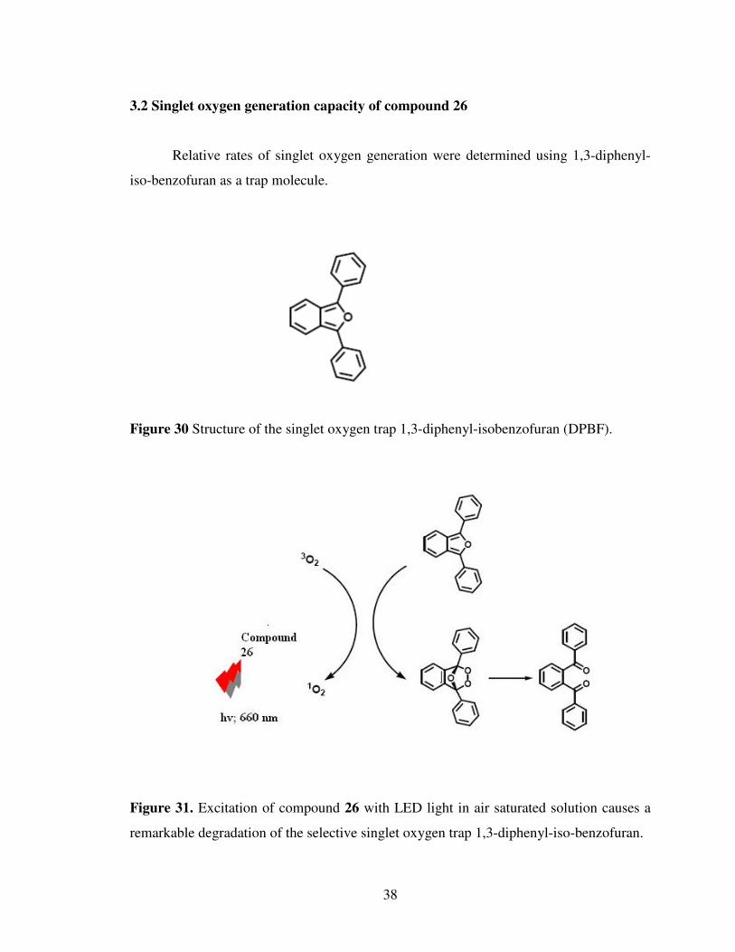
3.2 Singlet oxygen generation capacity of compound 26
Relative rates of singlet oxygen generation were determined using 1,3-diphenyl-
iso-benzofuran as a trap molecule.
Figure 30 Structure of the singlet oxygen trap 1,3-diphenyl-isobenzofuran (DPBF).
Figure 31. Excitation of compound 26 with LED light in air saturated solution causes a
remarkable degradation of the selective singlet oxygen trap 1,3-diphenyl-iso-benzofuran.

39
A few years ago we have demonstrated that appropriately decorated bodipy dyes
can be very efficient generators for singlet oxygen, thus act as a satisfactory
photodynamic agents. As a bonus these dyes absorbed very strongly at 660 nm which is
considered to be within the therapeutic window of mammalian tissue. So, combining our
earlier experience in molecular logic gates and rational design of photodynamic agents,
we proposed a photodynamic therapy agent that would release singlet oxygen at a much
larger rate when the cancer related cellular parameters are above a threshold value at the
same location. Thus, the proposed logic gate is an AND logic gate, the output of which is
singlet oxygen. Following the survey of the relevant literature for cancer related
parameters, we decided that sodium ion concentration and pH (H+) concentration could
be very promising targets. In the tumor regions the pH can drop below 6 and the Na+
concentration is also significantly higher then normal tissues. As a result in the proposed
logic system the chemical inputs could be Na+ and H+.

40
300 400 500
0.0
0.2
0.4
0.6
0.8
1.0
Ab
so
rban
ce o
f D
PB
F
Wavelenght(nm)
Na
p
tfa+Na
tfa
Figure 32. Absorbance spectra of 1,3-diphenyl-isobenzofuran (DPBF concentration 100
µM) in acetonitrile, without any exposure to 3000 mCd light in the presence of 10 nM
photosensitizer (p) 26, p (no Na+ or TFA is added), Na ( in the presence of 20 mM
NaClO4), TFA (in the presence of 1 µM TFA), Na+ + TFA (in the presence of both 20
mM NaClO4 and 1 µM TFA)
Wavelenth (nm)
41
300 400 500
0.0
0.2
0.4
0.6
0.8
1.0
Ab
so
rba
nc
e o
f D
PB
F
Wavelenght (nm)
Na+5min
p+5min
tfa+5min
tfa+Na+5min
Figure 33. Absorbance spectra of 1,3-diphenyl-isobenzofuran (DPBF concentration 100
µM) in acetonitrile with 5 minute exposure to 3000 mCd light in the presence of 10 nM
photosensitizer (p) 26.
Wavelenth (nm)

42
300 400 500
0.0
0.2
0.4
0.6
0.8
1.0
Ab
so
rban
ce o
f D
PB
F
Wavelenght (nm)
Na+10min
p+10min
tfa+10min
tfa+Na+10min
Figure 34. Absorbance spectra of 1,3-diphenyl-isobenzofuran (DPBF concentration 100
µM) in acetonitrile with 10 minute exposure to 3000 mCd light in the presence of 10 nM
photosensitizer (p) 26.
Wavelenth (nm)
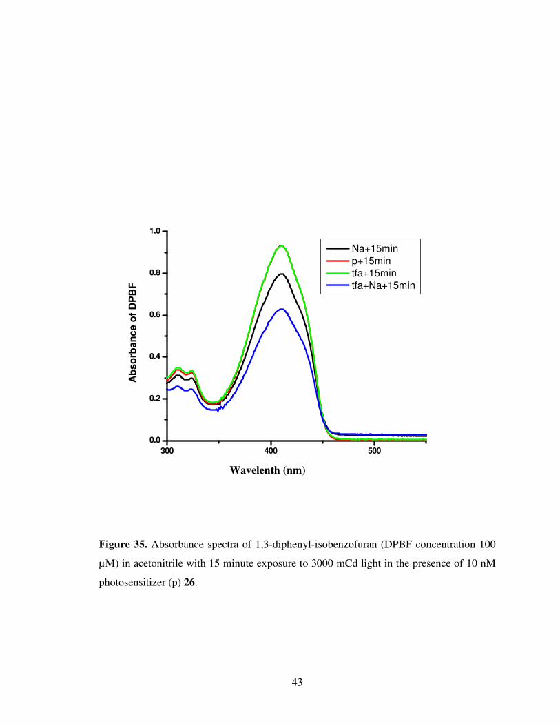
43
300 400 500
0.0
0.2
0.4
0.6
0.8
1.0
Ab
so
rba
nc
e o
f D
PB
F
Wavelenght(nm)
Na+15min
p+15min
tfa+15min
tfa+Na+15min
Figure 35. Absorbance spectra of 1,3-diphenyl-isobenzofuran (DPBF concentration 100
µM) in acetonitrile with 15 minute exposure to 3000 mCd light in the presence of 10 nM
photosensitizer (p) 26.
Wavelenth (nm)

44
300 400 500
0.0
0.2
0.4
0.6
0.8
1.0
Ab
so
rba
nc
e o
f D
PB
F
Wavelenght(nm)
Na+20min
p+20min
tfa+20min
tfa+Na+20min
Figure 36. Absorbance spectra of 1,3-diphenyl-isobenzofuran (DPBF concentration 100
µM) in acetonitrile with 20 minute exposure to 3000 mCd light in the presence of 10 nM
photosensitizer (p) 26.
Wavelenth (nm)
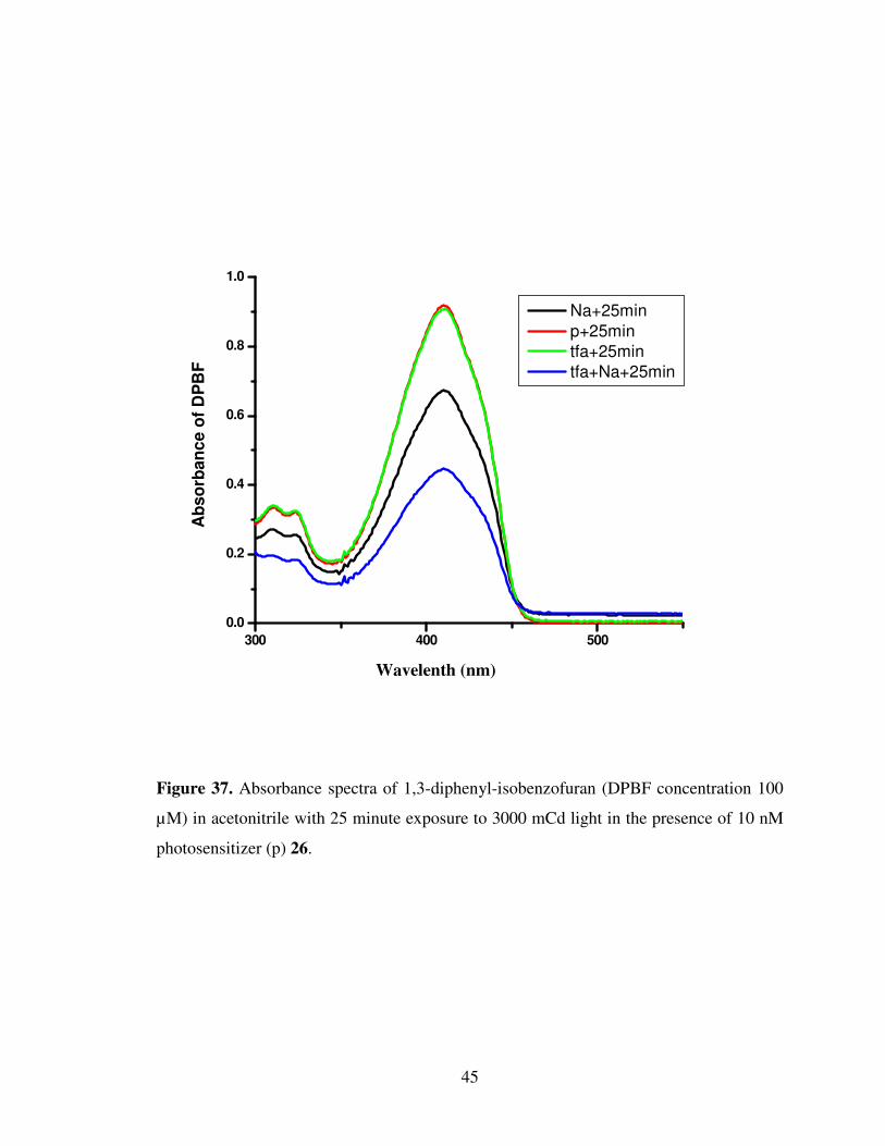
45
300 400 500
0.0
0.2
0.4
0.6
0.8
1.0
Ab
so
rban
ce o
f D
PB
F
Wavelenght(nm)
Na+25min
p+25min
tfa+25min
tfa+Na+25min
Figure 37. Absorbance spectra of 1,3-diphenyl-isobenzofuran (DPBF concentration 100
µM) in acetonitrile with 25 minute exposure to 3000 mCd light in the presence of 10 nM
photosensitizer (p) 26.
Wavelenth (nm)
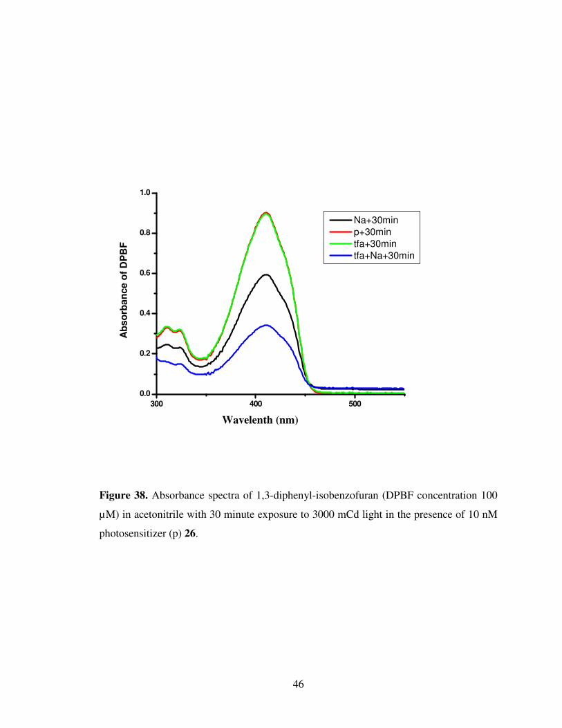
46
300 400 500
0.0
0.2
0.4
0.6
0.8
1.0
Ab
so
rba
nce
of
DP
BF
Wavelenght(nm)
Na+30min
p+30min
tfa+30min
tfa+Na+30min
Figure 38. Absorbance spectra of 1,3-diphenyl-isobenzofuran (DPBF concentration 100
µM) in acetonitrile with 30 minute exposure to 3000 mCd light in the presence of 10 nM
photosensitizer (p) 26.
Wavelenth (nm)

47
0
0,2
0,4
0,6
0,8
1
1,2
0 500 1000 1500 2000
Time(seconds)
Ab
so
rba
nc
e o
f D
PB
F
P
Tfa
Na
Na+Tfa
Figure 39. Absorbance of DPBF vs time graph with relative slopes.
Slopes; P = 3x10⁻⁵, tfa = 3x10⁻⁵, Na = 2x10⁻⁴, Na + tfa = 4x10⁻⁴ .
During the 0, 5, 10, 15, 20, 25, 30 minute exposure of the sample to 660 nm LED
source, the maximum decrease in 1,3 di-phenyl isobenzofuran absorption i.e.maximum
rate of singlet oxygen generation were observed when both TFA and Na+ ions were
present . When neither TFA (H+ ions) nor NaClO4 (Na+ ions) were present or when only
TFA was present a very low rate of production is in effect. Na+ alone results in a
somewhat higher rate, demonstrating the reciprocal relationship between PET and
intersystem crossing processes, at least for this molecule. This experimental results are in
accordance with AND logic operation. The system in fact is an automaton which is to
seek higher concentration of both hydrogen and sodium ions, and release the toxic agent
(singlet oxygen) only when both concentrations are high.

48
Table 2. AND truth table and corresponding AND gate for compound 26.
INPUT OUTPUT
A B A AND B
0 0 0
1 0 0
0 1 0
1 1 1
INPUT OUTPUT
Na+ H+ 1O2
0 0 Low ( 1.0 )
1 0 Low ( 6.6 )
0 1 Low ( 1.0 )
1 1 High (13. 3 )
49
4. CONCLUSION
In this study a novel photosensitizer 3,5-di-styryl substituted boradiazaindacene
dye was synthesized. This compound is a prototypic example of an automaton which
releases its lethal weapon following an analysis of enviromental parameters for sign of
disease and when necessary and sufficient conditions are met. This prototype also points
to the direction of further development; incorporation of additional functional units which
modulate singlet oxygen generation efficiency could result in therapeutic agents with very
high level of selective delivery potential. The notion of molecular logic gates assisted us
in the design of this particular PDT agent and it is very likely molecular medicine, could
be the first niche application of molecule based logic gates.
50
REFERENCES
[1] Gorman A., Killoran J., O’Shea C., Kenna T., Gallagher W. M. and O’Shea D. F., J.
Am. Chem. Soc., 2004, 126, 10619–10631.
[2] Roy I., Ohulchanskyy T. Y., Pudavar H. E, Bergey E. J., Oseroff A. R., Morgan J.,
Dougherty T. J., Prasad P. N., J. Am. Chem. Soc. , 2003, 125, 7860- 7865.
[3] Pushpan S. K., Venkatraman S., Anand V.G., Sankar J., Parmeswaran D., Ganesan S.,
Chandrashekar T.K., Curr. Med. Chem., 2002, 2, 187-207.
[4] Epstein, J. M. N. Engl. J. Med., 1990, 32, 1149-1151.
[5] Spikes J. D., Photochem. Photobiol., 65, 1997, 142S-147S.
[6] Nyman E. S., Hynninen P. H., J. Photochem. Photobio. 73, 2004, 1–28.
[7] Szacilowski, K., Macyk, M., Matuszek, A. D., Brindell, M., Stochel, G., Chm. Rev.
105, 2005, 2647-2694.
[8] Morris R. L., Azizuddin K., Lam M., Berlin J., Nieminen A., Kenney M. E., Samia A.
C. S., Burda C., Oleinick N. L., Cancer Res. 2003, 63, 5194-5196.
[9] Allen C. M., Sharman W. M., Van Lier J. E., J. Porphyrins Pthalocyanines 2001, 5,
161-163
[10] Ali H., van Lier, J. E. Chem. Rev. 1999, 99, 2379-2381.
[11] Nyman E. S., Hynninen P. H., J. Photochem. Photobiol., B: Biol, 2004, 73, 1-3.
[12] Capella M. A. M., Capella L. S., J. Biomed. Sci, 2003, 10, 361-366
[13] Dougherty T. J., Crit. Rev. Oncol. Hematol., 1984, 2, 83-85.
[14] Allison R. R., Downie G. H., Cuenca R., Hu X. H., Childs C. J. H., Sibata C. H.,
Photodiagn. Photodyn. Ther. 2004, 1, 27-29.
[15] Sharman W. M., Allen C. M., van Lier J. E., Drug. Discuss. Today, 1999, 4, 507-
509.
[16] Brown S., Int. Photodyn., 1996, 1, 1-3.
[17] Sobolev A. S., Stranadko E. F., Int. Photodyn., 1997, 1, 2-4.
[18] Evenson J. F., Sommer S., Rimington C., Moan J. Br., J. Cancer, 1987, 55, 483-485.
[19] Sternberg E. D., Dolphin D., Brückner C., Tet. Let., 1998, 54, 4151-4202.
51
[20] Paquette B., Boyle R. W., Ali, H., MacLennan A. H., Truscott T. G., van Lier J. E.
Sulfonated phthalimidomethyl aluminum phthalocyanine: The effect of hydrophobic
substituents on the in vitro phototoxicity of phthalocyanines.
[21] Jori G., J. Photochem. Photobiol., 1992, 62, 371-378.
[22] Wyss P., Schwarz V., Dobler-Girdziunaite D., Hornung R., Walt H., Degen A., Fehr
M. K., Int. J. Cancer , 2001, 93, 720- 724.
[23] Yukruk F., PhD thesis, METU, 2005.
[24] Killoran J., Allen L., Gallapher J. F., Gallapher W. M., O’Shea D. F.,
Chem.Commun., 2002, 1862-1863.
[25] Mc. Donell S. O., Hall M. J., Allen L. T., Byrne A., Gallagher W. M., O’Shea D. F.,
J. Am. Chem. Soc., 2005, 127, 16360-16361.
[26] Karolin J., Johansson L. B. A., Strandberg L., Ny T., J. Am. Chem. Soc. 1994, 116,
7801- 7806.
[27] Wittmershaus B. P., Skibicki J. J., McLafferty J. B., Zhang Y. Z., Swan S., J.
Fluoresc., 2001, 11, 119- 128.
[28] Yogo T., Urano Y., Ishitsuka Y., Maniwa F., Nagano T., J. Am. Chem. Soc. 2005,
127, 12162- 12163.
[29] Atılgan S., Ekmekçi Z., Doğan A. L., Güç Dicle, Akkaya E. U., Chem. Commun..,
2006, 4398-4400.
[30] Atılgan S., Ms Thesis, METU, 2006.
[31] Baytekin H. T., Akkaya E. U., Org. Lett., 2000, 2, 1725-1727.
[32] Dougherty T. J, Gomer C. J., Henderson B. W., Jori G., Kessel D., Korbelik M.,
Moan J., Peng Q., J. Natl. Cancer. Inst., 1998, 90, 889-905.
[33] Turro N. J. In Modern Molecular Photochemistry; University Science Books:
Sausalito, CA, 1991, 191- 195.
[34] Lower S. K., El-Sayed M. A., Chem. Rev., 1966, 66, 199-201.
[35] Yuster P., Weissman S. I., J. Chem. Phys. 1949, 17, 1182-1183.
[36] Koziar J. C., Cowan D. O., Acc. Chem. Res. 1978, 334-336.
[37] Douglas A. Skoog, Donald M. West, F. James Holler, Fundamentals of Analytical
Chemistry, 1996, 506-508.
[38] Valeur B., Molecular fluorescence principles and applications, Wiley, 2001, 34-47.
52
[39] De Silva A. P., Gunaratne H. Q. N., Gunnlaugsson T., Huxley A. J. M., McCoy C.
P., Rademacher J. T., Rice T. E., Chem. Rev. 1997, 97, 1515-1566.
[40] De Silva A. P., De Silva S.A., J. Chem. Soc. Chem. Commun., 1986, 1709-1711.
[41] Atwood J. L., Steed J. W., Encyclopedia of Supramolecular Chemistry, 2000, 893.
[42] De Silva A. P., De Silva S. A., Dissanayake A. S., Sandanayake K. R. A. S.,
J. Chem. Soc. Chem. Commun., 1989, 1054 -1056.
[43] Ben-Ari M., Mathematical Logic for Computer Science, Prentice- Hall, Hemel
Hempstead, 1993.
[44] De Silva, A. P., Gunaratne H. Q. N., McCoy C. P., Nature (London), 1993, 364-
366.
[45] De Silva A. P., Gunaratne H. Q. N., McCoy C. P., J. Am. Chem. Soc. 1997, 119,
7891 -7892;
[46] McSkimming G., Tucker J. H. R., Bouas-Laurent H., Desvergne J. P., Angew.
Chem., 2000, 112, 2251-2253;
[47] Gunnlaugsson T., Mac Donail D. A., Perker D., Chem. Commun., 2000, 93-94.
[48] Baytekin H. T., Akkaya E. U., Org. Lett. 2000, 2, 1725 -1727.

53
APPENDIX
Figure 40 1H NMR spectra of compound 22

54
Figure 41 1H NMR Spectra of compound 23
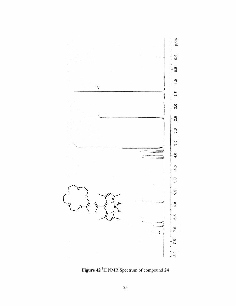
55
Figure 42 1H NMR Spectrum of compound 24
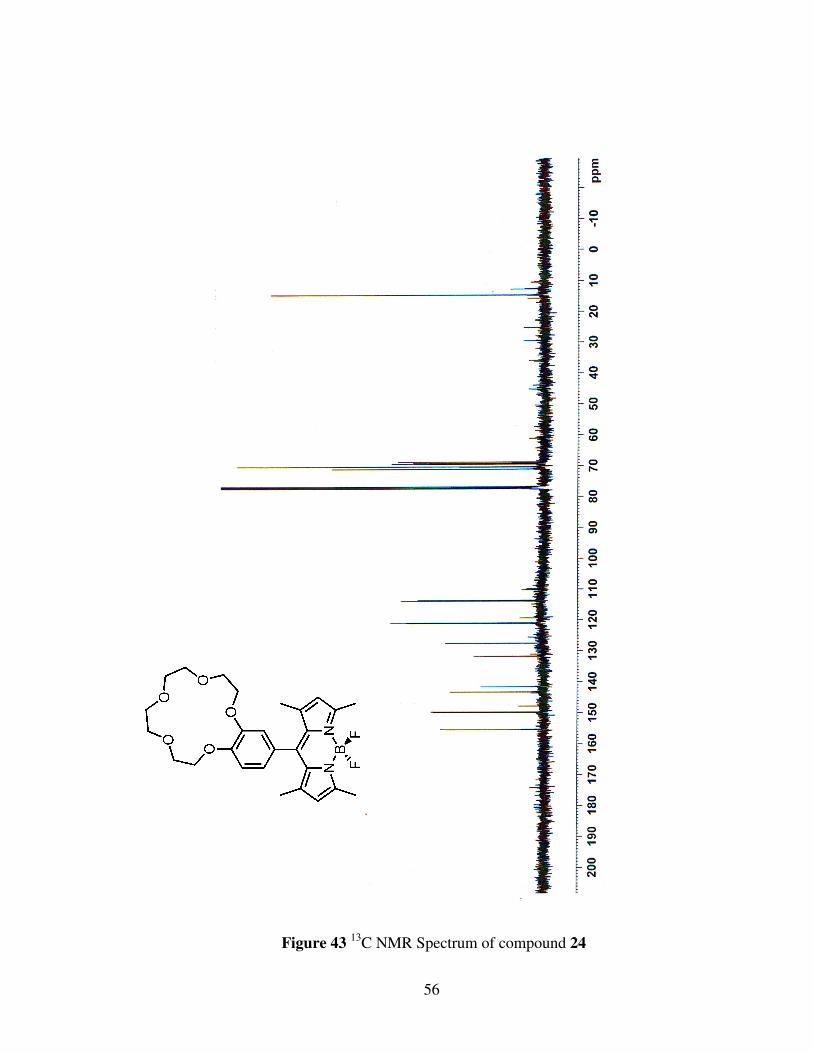
56
Figure 43 13C NMR Spectrum of compound 24
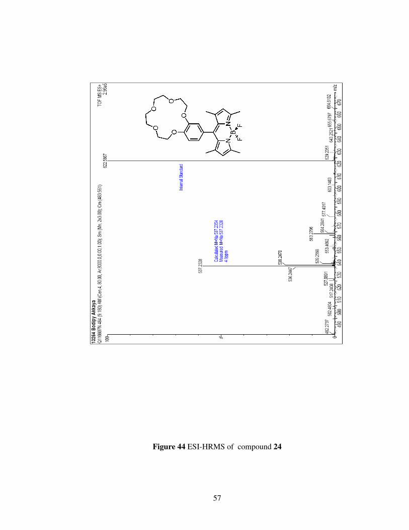
57
Figure 44 ESI-HRMS of compound 24
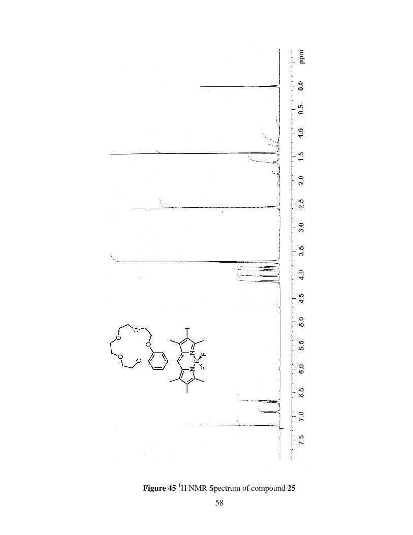
58
Figure 45 1H NMR Spectrum of compound 25

59
Figure 46 13C NMR Spectrum of compound 25

60
Figure 89 ESI-HRMS of compound 89
Figure 47 ESI-HRMS of compound 25
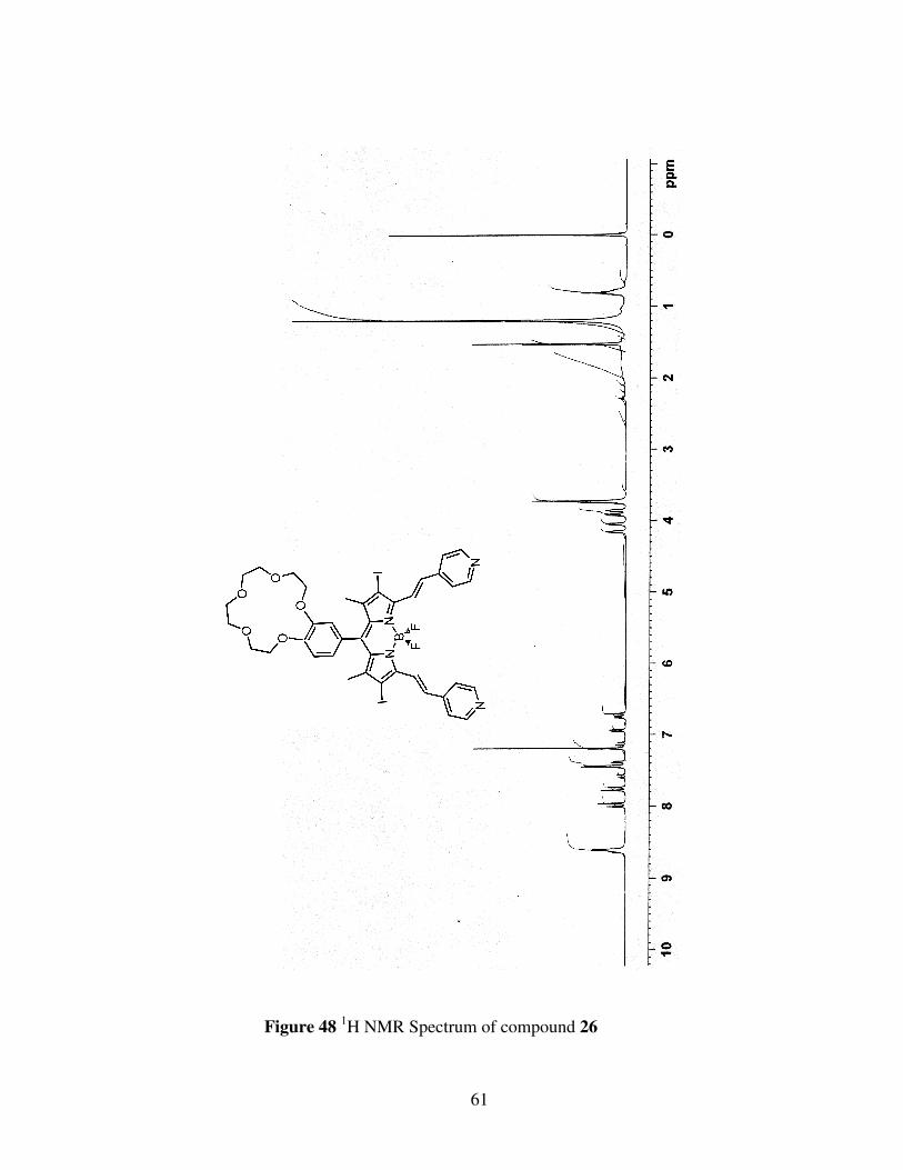
61
Figure 48 1H NMR Spectrum of compound 26
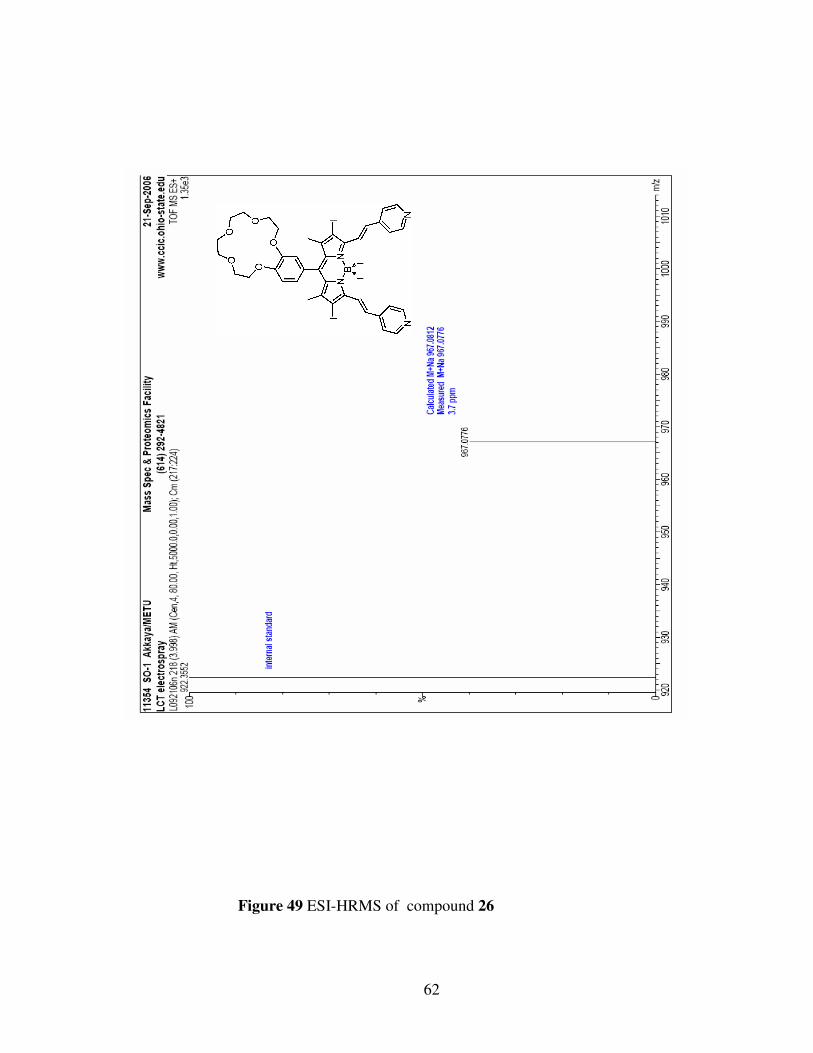
62
Figure 49 ESI-HRMS of compound 26


