1
Virus-specific memory CD8 T cells provide substantial protection from 1
lethal SARS-CoV infection 2
3
Rudragouda Channappanavar1, Craig Fett
1, Jincun Zhao
1, David Meyerholz
2 and 4
Stanley Perlman1#
5
Departments of 1Microbiology and
2Pathology, 6
University of Iowa, Iowa City, IA, USA 52242 7
8
#Corresponding author: Dr. Stanley Perlman, Department of Microbiology, University of Iowa, 9
Iowa City, IA 52242. Tele-319-335-8549; FAX number-319-335-9006; email: Stanley-10
12
Running title: SARS-CoV-specific memory CD8 T cells 13
14
Keywords: coronavirus, SARS-coronavirus, CD8 T cells, memory T cells, animal model 15
16
Word count. Abstract: 244 Text: 4438 17
18
19
20
21
22
JVI Accepts, published online ahead of print on 23 July 2014J. Virol. doi:10.1128/JVI.01505-14Copyright © 2014, American Society for Microbiology. All Rights Reserved.
on July 8, 2018 by guesthttp://jvi.asm
.org/D
ownloaded from
2
ABSTRACT: 23
The Severe Acute Respiratory Syndrome-coronavirus (SARS-CoV) caused an acute 24
human respiratory illness with high morbidity and mortality in 2002-2003. Several studies have 25
demonstrated the role of neutralizing antibodies induced by the spike (S) glycoprotein in protecting 26
susceptible hosts from lethal infection. However, the anti-SARS-CoV antibody response is short-27
lived in SARS-recovered patients making it critical to develop additional vaccine strategies. 28
SARS-CoV-specific memory CD8 T cells persisted for up to six years after SARS-CoV infection, 29
a time at which memory B cells and anti-virus antibodies were undetectable in SARS-recovered 30
individuals. Here, we assessed the ability of virus-specific memory CD8 T cells to mediate 31
protection against infection in the absence of SARS-CoV-specific memory CD4 T or B cells. We 32
demonstrate that memory CD8 T cells specific for a single immunodominant epitope (S436 or 33
S525) substantially protected 8-10 month old mice from lethal SARS-CoV infection. Intravenous 34
immunization with peptide-loaded dendritic cells (DCs) followed by intranasal boosting with 35
recombinant vaccinia virus (rVV) encoding S436 or S525 resulted in accumulation of virus-36
specific memory CD8 T cells in alveolar lavage fluid (BAL), lungs and spleen. Upon challenge 37
with a lethal dose of SARS-CoV, virus-specific memory CD8 T cells efficiently produced multiple 38
effector cytokines (IFN-γ, TNF-α and IL-2) and cytolytic molecules (granzyme B) and reduced 39
lung viral loads. Overall, our results show that SARS-CoV-specific memory CD8 T cells protect 40
susceptible hosts from lethal SARS-CoV infection, but also suggest that SARS-CoV specific CD4 41
T cell and antibody responses are necessary for complete protection. 42
43
44
on July 8, 2018 by guesthttp://jvi.asm
.org/D
ownloaded from

3
45 IMPORTANCE: 46
Virus-specific CD8 T cells are required for pathogen clearance following primary SARS-CoV 47
infection. However, the role of SARS-CoV-specific memory CD8 T cells in mediating protection 48
after SARS-CoV challenge has not been previously investigated. Here, using a prime-boost 49
immunization approach, we show that virus-specific CD8 T cells protect susceptible 8-10 month 50
old mice from lethal SARS-CoV challenge. Thus, future vaccines against emerging coronaviruses 51
should emphasize the generation of a memory CD8 T cell response for optimal protection. 52
53
INTRODUCTION: 54
Coronaviruses belong to a group of pathogens that periodically emerge from zoonotic 55
sources to infect human populations, often resulting in high rates of morbidity and mortality (1-3). 56
SARS-CoV and MERS (Middle East Respiratory Syndrome)-CoV are two notable examples of 57
novel coronaviruses that emerged during the last decade (1, 2, 4). Infection with these 58
coronaviruses can result in the acute respiratory distress syndrome (ARDS), which has a high rate 59
of morbidity and mortality (3, 5). SARS-CoV infected humans during 2002-03, caused a global 60
epidemic and spread rapidly to more than 30 countries killing approximately 800 people (3). Both 61
SARS-CoV and MERS-CoV infect airway and alveolar epithelial cells resulting in acute 62
respiratory illnesses (6). While there was 10% mortality among all SARS-CoV infected patients, 63
individuals aged 60 and above suffered worse outcomes with >50% deaths (3). On a similar note, 64
the newly emerging MERS-CoV infection is associated with an approximate 30% mortality in 65
humans (5). Although there has not been any known new incidence of SARS-CoV infection in 66
humans, recent emergence of MERS-CoV in humans and identification of SARS-like 67
on July 8, 2018 by guesthttp://jvi.asm
.org/D
ownloaded from

4
coronaviruses in bats and wild animals illustrate the potential threat of such life-threatening 68
pathogens. 69
Neutralizing (NT) antibody responses generated against spike (S) glycoprotein of SARS-70
CoV provide complete protection against SARS-CoV infection. Several potential vaccine 71
candidates such as attenuated virus vaccines, sub-unit constructs and recombinant DNA (rDNA) 72
plasmids were shown to be protective in mouse models of SARS-CoV infection, largely by 73
inducing a robust NT antibody response (7-11). Recent studies from our laboratory showed that 74
attenuated mouse-adapted SARS-CoV (MA15) (12), which lacks the E protein (rMA15-ΔE), was 75
safe and completely protective in susceptible 6 week and 12-month-old BALB/c mice. In addition 76
to inducing NT antibody responses, rMA15-ΔE induced strong T cell responses (11, 13, 14). 77
Cytotoxic T cells (CTL) play a crucial role in clearing respiratory viruses and can provide long-78
term protective cellular immunity (15, 16). SARS-CoV infection induces a potent and long-lived T 79
cell response in surviving humans (17, 18). The majority of immunodominant T cell-epitopes 80
reside primarily in three structural proteins, the S, M and N proteins of SARS-CoV. 81
Immunodominant CD8 T cell epitopes recognized in C57BL/6 (B6) mice include S525 and S436 82
(encompassing residues 525-533 and 426-444 of the spike protein) (19, 20). 83
Young 6-10-week B6 mice are resistant to MA15 infection; however, as mice age, there 84
is a steep increase in the susceptibility such that mice >6 months are highly susceptible to the 85
infection (21). As in many infections, virus-specific CD4 and CD8 T cells protect susceptible 86
young and aged BALB/c and aged B6 mice following MA15 infection (19, 21, 22). The age-87
dependent susceptibility to MA15 is associated with a poor anti-viral CD8 T cell response. We 88
showed that increased PGD2 levels in the lungs of aged mice after MA15 infection was 89
responsible, at least in part, for this poor T cell response by impairing migration of rDCs to 90
on July 8, 2018 by guesthttp://jvi.asm
.org/D
ownloaded from
5
draining lymph nodes (DLN). This led to reduced priming in the DLN and less MA15-specific 91
CD8 T cell accumulation in the lungs compared to young mice (21). Although MA15-specific 92
effector CD8 T cells are required for virus clearance during the acute infection, the role of memory 93
CD8 T cells in protecting the host against subsequent lethal challenge is not known. Interestingly, 94
SARS-CoV infection induced strong and long lasting virus-specific T cells that were detectable for 95
up to six-years in recovered patients (17, 23). Since the memory B cell response and neutralizing 96
antibodies are short lived in SARS-CoV infected patients, developing vaccines capable of 97
generating, long-lived memory CD8 T cells is desirable. 98
Antigen-specific memory CD8 T cells are categorized into three subpopulations. In 99
addition to antigen-specific ‘effector memory’ (T EM) and ‘central memory’ (TCM) CD8 T cells, a 100
third population of ‘tissue resident’ memory (TRM) memory CD8 T cells exist in the peripheral 101
tissues after a local pathogen encounter. TRM are non-migratory and persist at the site of infection 102
for a long period of time (24). These TRM mediate rapid virus clearance from the site of infection 103
upon pathogen challenge by secreting antiviral effector molecules, which limit virus replication 104
(25) and expressing chemokines that recruit additional memory CD8 T cells from the circulation 105
(26). An effective early T cell response to a respiratory virus challenge depends on the number of 106
antigen-specific memory CD8 T cells in different lung compartments (27, 28). Further, the number 107
and efficacy of virus-specific CD8 T cells in the lung airways correlates with the ability to clear a 108
secondary virus challenge (29, 30). 109
In the current study, we examined whether a SARS-CoV-specific memory CD8 T cell 110
response was sufficient to protect mice from lethal disease. Using a well-established prime-boost 111
strategy to boost the number of memory CD8 T cells in the respiratory tract, we show that SARS-112
CoV immunodominant epitope-specific memory CD8 T cells protected susceptible 8-10 month B6 113
on July 8, 2018 by guesthttp://jvi.asm
.org/D
ownloaded from
6
mice from a lethal MA15 infection. Mice were primed intravenously with DCs loaded with peptide 114
(S436 or S525) and then boosted intranasally with recombinant vaccinia virus (rVV) encoding 115
S436 or S525. MA15-specific memory CD8 T cells generated in the lungs provided a significant 116
level of protection from lethal MA15 challenge. 117
118
MATERIALS AND METHODS: 119
Mice and viruses: Pathogen-free female B6 mice (8-9 months) were purchased from the National 120
Cancer Institute (Frederick, MD). Mice were maintained in the University of Iowa animal care 121
facility. All animal experiments were approved by the University of Iowa Institutional Animal 122
Care and Use Committee. MA15, a kind gift from Kanta Subbarao (NIH, Bethesda, MD), was 123
propagated on Vero E6 cells (12). 124
Recombinant vaccinia virus (rVV) encoding S436 and S525 were engineered using the 125
following complementary oligonucleotides: S436- 5’-TCGACGCCACCATGTACAACTACAA 126
GTACAGGTACCTGTAAGGTAC; 3’-CTTACAGGTACCTGTACTTGTAGTTGTACATGGT 127
GGCG. S525- 5’-TCGACGCCACCATGGTGAACTTCAACTTCAACGGCCTGTAAGGTAC; 128
3’- CTTACAGGCCGTTGAAGTTGAAGTTCACCATGGTGGCG. The oligonucleotides were 129
annealed and ligated into PSC65 (a VV shuttle vector with a strong synthetic VV early/late 130
promoter, kindly provided by Dr. B. Moss, National Institutes of Health). 131
Prime-boost immunization: DC-peptide immunization: Spleen-derived DCs were isolated from 132
6-8 week old B6 mice previously inoculated subcutaneously with 1x10⁶ B16 cells expressing Flt3L 133
(provided by M. Prlic and M. Bevan, University of Washington). DCs were then harvested and 134
pulsed as described previously (31). Briefly, 106 LPS-matured DCs 1 µg of LPS/mouse, i.p. (S. 135
abortus equi, S-form, Enzo Lifesciences, Formingdale, NY) were coated with 1 µM peptide 136
on July 8, 2018 by guesthttp://jvi.asm
.org/D
ownloaded from
7
(S436 or S525) for two hours at 37C. Peptide-pulsed DCs were then intravenously injected into 8-137
9 month old B6 mice. Similar numbers of unpulsed DCs were injected into control mice. For 138
detection of antigen-specific CD8 T cells, PBL were obtained by retro-orbital bleeding at different 139
times post immunization and analyzed for intracellular IFN-γ expression as described below. 140
rVV-minigenome booster: At six days after DC-peptide immunization, mice were boosted by 141
intranasal (i.n.) inoculation of rVV encoding either S436 or S525 (2x10⁶
PFU in 50 µl DMEM). 142
Mice were then rested for 42-45 days for memory studies. 143
Challenge and survival studies: To assess the protective ability of virus-specific memory CD8 T 144
cells, prime-boost immunized mice were challenged after 42-45 days by intranasal inoculation of 145
5x10⁴ PFU of MA15 in 50 µl of DMEM. All infected mice were monitored daily for morbidity and 146
mortality. Mice that lost 30% of their initial body weight were euthanized as per institutional 147
IACUC guidelines. All challenge experiments were carried out in the ABSL-3 laboratory as per 148
approved guidelines. 149
Virus titers in the lungs: To obtain tissue for virus titers, mice were euthanized different days 150
post challenge, lungs were homogenized in PBS and titers were determined on Vero E6 cells. 151
Virus titers are represented as PFU/g of lung tissue. 152
Preparation of cells from lungs, BAL and spleen for FACS analysis: Mice were sacrificed at 153
the indicated time points and perfused via the right ventricle with 10 ml PBS. BAL, lungs and 154
spleen were obtained. Lungs were cut into small pieces and digested in HBSS buffer containing 155
2% FCS, 25 mM HEPES, 1 mg/ml Collagenase D (Roche), and 0.1 mg/ml DNase (Roche) for 30 156
minutes at room temperature. Digested tissues were then minced and passed through a 70-μm-157
nylon filter to obtain single cell suspensions. Cells were enumerated by 0.2% trypan blue 158
exclusion. 159
on July 8, 2018 by guesthttp://jvi.asm
.org/D
ownloaded from

8
Antibodies and flow cytometry: The following monoclonal antibodies were used for these 160
studies: rat anti-mouse CD4 (RM4-5), rat anti-mouse CD8α (53-6.7), PE-anti-IFN-γ (XMG1.2), 161
APC-anti-TNF-α (MP6-XT22), APC-anti-IL-2 (JES6-5H4), FITC-anti-CD107a/b and PE-anti-162
CD69 (H1.2F3) were procured from BD Biosciences. PE-cy7-anti-CD8 (53-6.7), rat anti-mouse 163
IFN-γ (XMG1.2), hamster PE-anti-CD103 (2E7), V510-rat anti-mouse CXCR3 (CXCR3-173) and 164
V450-anti-CD11a (M17/4) were purchased from e-Bioscience. 165
Intracellular cytokine staining: For intracellular cytokine staining, 1x10⁶ cells per well were 166
cultured in 96-well dishes at 37°C for 5 to 6 hours in the presence of Golgiplug (1µg) (BD 167
Biosciences). 106 cells were blocked with 1 μg anti-CD16/32 antibody and surface stained with the 168
indicated antibodies on ice. Cells were then fixed/permeabilized with Cytofix/Cytoperm Solution 169
(BD Biosciences), and labeled with anti–cytokine antibody. All flow cytometry data were acquired 170
on a BD FACSVerse (BD Biosciences) and were analyzed using FlowJo software (Tree Star Inc.). 171
Tetramer Staining: MHC class I/peptide tetramer, used to measure S436 and S525-specific CD8 172
T cells, were obtained from the NIH Tetramer Core Facility (Emory University, Atlanta, GA). A 173
total of 5x10⁵
to 1x10⁶
cells obtained from BAL, lungs and spleen of immunized or MA15-174
challenged mice were first incubated on ice with Fc block (anti-CD16/32 antibody) (BD 175
Biosciences) for 15 minutes followed by incubation with APC- conjugated tetramer at 4C for 30 176
min. Cells were then surface stained with PE-Cy7 anti-CD8 antibody. Flow cytometry data were 177
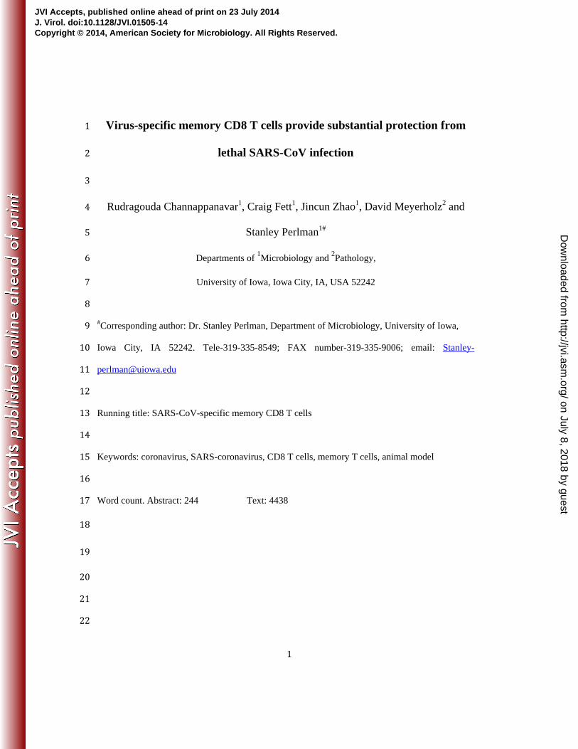
acquired and processed as described above. 178
In vivo cytotoxicity assay: In vivo cytotoxicity assays were performed on day 5 after infection, as 179
previously described (32). Briefly, splenocytes from naive CD45.1 (Ly5.2) mice were labeled with 180
either 1 μM or 100 nM CFSE (Molecular Probes). Labeled cells were then pulsed with the 181
indicated peptides (5 μM) at 37°C for 1 hour, and 5x10⁵ cells from each group (peptide-pulsed vs. 182
on July 8, 2018 by guesthttp://jvi.asm
.org/D
ownloaded from

9
non-pulsed) were mixed together. A total of 106
cells were transferred i.n. into challenged mice, 183
and total lung cells were isolated at 12 hours after transfer. Target cells were distinguished from 184
host cells on the basis of CD45.1 staining and from each other on the basis of CFSE staining. 185
Percent specific lysis was determined as previously described (32). 186
Lung Histology: Animals were anesthetized and transcardially perfused with PBS followed by 187
zinc formalin. Lungs were removed, fixed in zinc formalin, and paraffin embedded. Sections were 188
stained with hematoxylin and eosin and examined by light microscopy. 189
Statistical Analysis: Data were analyzed using Student’s t test. Results in the graphs are 190
represented as mean ± SEM. unless otherwise mentioned. p values of *P≤0.05, ** P≤0.01 and *** 191
P ≤0.001 192
193
194
195
196
197
198
199
200
201
202
203
204
205
on July 8, 2018 by guesthttp://jvi.asm
.org/D
ownloaded from
10
206 RESULTS: 207
Prime-boost immunization induces a strong CD8 T cell response. Recently, we and others 208
identified and validated several CD8 T cell epitopes located in SARS-CoV structural proteins (19, 209
20). S436-443 and S525-532 were found to be immunodominant in B6 mice and are used in this 210
study to evaluate virus-specific CD8 T cell responses. We adopted a prime-boost strategy to 211
generate large numbers of virus-specific memory CD8 T cells in the BAL and lungs (31). 8-9 212
month B6 mice were initially primed by intravenous injection of LPS-matured DCs loaded with 213
S436 or S525 peptide (Fig. 1A). DC-peptide immunization resulted in a higher percentage and 214
number of MA15-specific CD8 T cells in the peripheral blood (PBL) in comparison to mice 215
immunized with uncoated DCs. A kinetics study of DC-peptide immunization showed that a 216
significant S436 and S525-specific CD8 T cell response was detected on day 4 after immunization 217
and peaked at day 6 after DC-peptide immunization. The proportion and total number of epitope-218
specific CD8 T cells in PBL were similar in both the DC-S436 and DC-S525 immunized groups 219
(Fig. 1B and C). DC-peptide primed mice were boosted 6 days later by intranasal inoculation of 220
rVV-S436 or rVV-S525. Mice treated with uncoated DCs and boosted with rVV expressing GFP 221
(rVV-GFP) were used as controls. Intranasal boosting with rVV-S436 or rVV-S525 generated a 222
large pool of virus-specific effector CD8 T cells in the BAL, lungs and spleen with virus-specific 223
cells detected at highest frequency in the BAL (Fig. 1D and E). Additionally, the proportion and 224
number of S525-specific CD8 T cells were significantly higher in BAL, lungs and spleen in 225
comparison to the S436 immunized group (P<0.001) (Fig. 1D-F). Collectively, these data indicate 226
that prime-boost immunization resulted in the generation of large pools of S436 and S525-specific 227
CD8 T cells in BAL and lungs. 228
229
on July 8, 2018 by guesthttp://jvi.asm
.org/D
ownloaded from
11
Induction of a MA15-specific memory CD8 T cell response after DC priming and rVV 230
boosting. An early protective response of virus-specific CD8 T cells to a respiratory virus 231
challenge depends on the presence of an adequate number of antigen-specific memory CD8 T cells 232
in the BAL and lungs (15). To enumerate the number of virus-specific memory CD8 T cells, mice 233
were allowed to rest for 42-45 days after prime-boost immunization and the percentage and total 234
number of S436 and S525-specific memory CD8 T cells were determined in the BAL, lung and 235
spleen. The percentage and total number of S436 and S525-specific memory CD8 T cells were 236
similar and were significantly higher in BAL, lung and spleen in the S436 and S525 prime-boost 237
immunized groups, respectively, compared to mice immunized with rVV-GFP controls (Fig. 2A 238
and B). Additionally, the proportion of virus-specific CD8 T cells among the total CD8 T cell pool 239
was much higher for both S436 and S525 groups in the lung airways (30-40%) compared to the 240
lungs (4-5%) or spleen (1-1.5%) (Fig. 2A). Since the protective ability of lung resident memory 241
CD8 T cells to counter a local pathogen challenge depends upon their capacity to produce multiple 242
antiviral effector molecules (33), we determined the poly-functionality of the S436 and S525 243
memory CD8 T cells by assessing cytokine expression after ex-vivo stimulation capacity (33). As 244
shown in Fig. 2C, a high percentage of total CD8 T cells isolated from the BAL secreted multiple 245
cytokines followed by those in the lung and spleen in both the S436 and S525 groups. The 246
proportion of IFN-+ CD8 T cells co-expressing TNF- (double producers) and TNF- and IL-2 247
(triple producers) was much higher in the BAL and lungs in comparison to the spleen. In contrast, 248
virus-specific CD8 T cells from spleen were mostly single cytokine producers (IFN-+ only) (Fig. 249
2C). Together, these results suggest that cells in the BAL and lung are especially well positioned 250
to respond effectively after challenge. 251
on July 8, 2018 by guesthttp://jvi.asm
.org/D
ownloaded from
12
Virus-specific memory CD8 T cells in the lung airways, lung parenchyma and secondary 252
lymphoid organs are phenotypically distinct with surface expression of markers such as CD103, 253
CXCR3 and CD11a defining tissue resident versus non-resident memory CD8 T cells (16). To 254
phenotypically distinguish virus-specific memory CD8 T cells in the lung airways, lung 255
parenchyma and spleen, we examined the expression of several molecules associated with tissue 256
homing and activation. Expression of CXCR3, a molecule required for localization of memory T 257
cells to lung airways, was significantly higher on S436 and S525-specific memory CD8 T cells 258
from the lung airways (>95 %) as compared to those isolated from lung parenchyma (35-50%) and 259
spleen (40-50%) (Fig. 2D). CD103, another marker of resident memory T cells, was also 260
expressed on a high proportion of virus-specific CD8 T cells in the lung airway, followed by those 261
in the lung parenchyma with least CD103 expression on splenic CD8 T cells. CD11a and CD69, 262
required for T cell activation and migration to tissues, are also upregulated on tissue resident 263
memory T cells (34). As shown in Fig. 2D, a significantly higher proportion of S436 and S525-264
specific memory CD8 T cells in the lung parenchyma co-expressed CD11a and CD69 in 265
comparison to those in the lung airways and spleen, in agreement with previous studies (34). 266
267
Analysis of virus-specific memory CD8 T cells following SARS-CoV challenge: 268
The ability of pathogen-specific memory CD8 T cells to protect the host from lethal 269
challenge depends on their absolute number and on their ability to produce multiple effector 270
cytokines (IFN-, TNF- and IL-2) and cytotoxic molecules (Granzyme B and perforin) (35). To 271
determine the magnitude and effector function of virus-specific CD8 T cells after pathogen 272
exposure, prime-boost immunized mice (now 10-11 months old) were challenged with a lethal 273
dose (5x10⁴
PFU) of MA15 intranasally. Following MA15 challenge, the proportion and total 274
on July 8, 2018 by guesthttp://jvi.asm
.org/D
ownloaded from

13
number of S436 and S525-specific CD8 T cells were significantly higher in BAL and lungs of 275
S436 and S525-immunized groups at day 5 post infection (p.i.) in comparison to those in the 276
control group (Fig. 3A and B). Additionally, both the percentage and total number of virus-277
specific CD8 T cells were higher in the BAL and lungs of S525-immunized mice compared to 278
those immunized against S436 epitope 74% vs. 29% in BAL (P<0.001) and 40% vs. 24% in lungs 279
(P<0.001); 3.5x10⁵
vs. 0.4x10
5 in BAL (P<0.001) and 2.2 x10
6 vs. 0.4x10
6 in lungs (P<0.001) 280
(Fig. 3A and B). To assess the functionality of the virus-specific CD8 T cells, we measured the 281
ability of these cells to co-produce multiple effector cytokines. The majority of virus-specific CD8 282
T cells in both S436 and S525–immunized groups co-produced IFN- and TNF- but not IL-2 in 283
the BAL and lungs (Fig. 3C). Since the BAL and lungs of control immunized mice had much 284
lower percentages of virus-specific CD8 T cells (<1%) (Fig. 3A), we did not further analyze these 285
cells. 286
To assess the cytotoxic ability of these virus-specific CD8 T cells, we measured 287
granzyme B expression by S436 and S525-specific CD8 T cells in the BAL and lungs following 288
MA15 challenge. A significantly higher percentage of CD8 T cells from the BAL and lungs of the 289
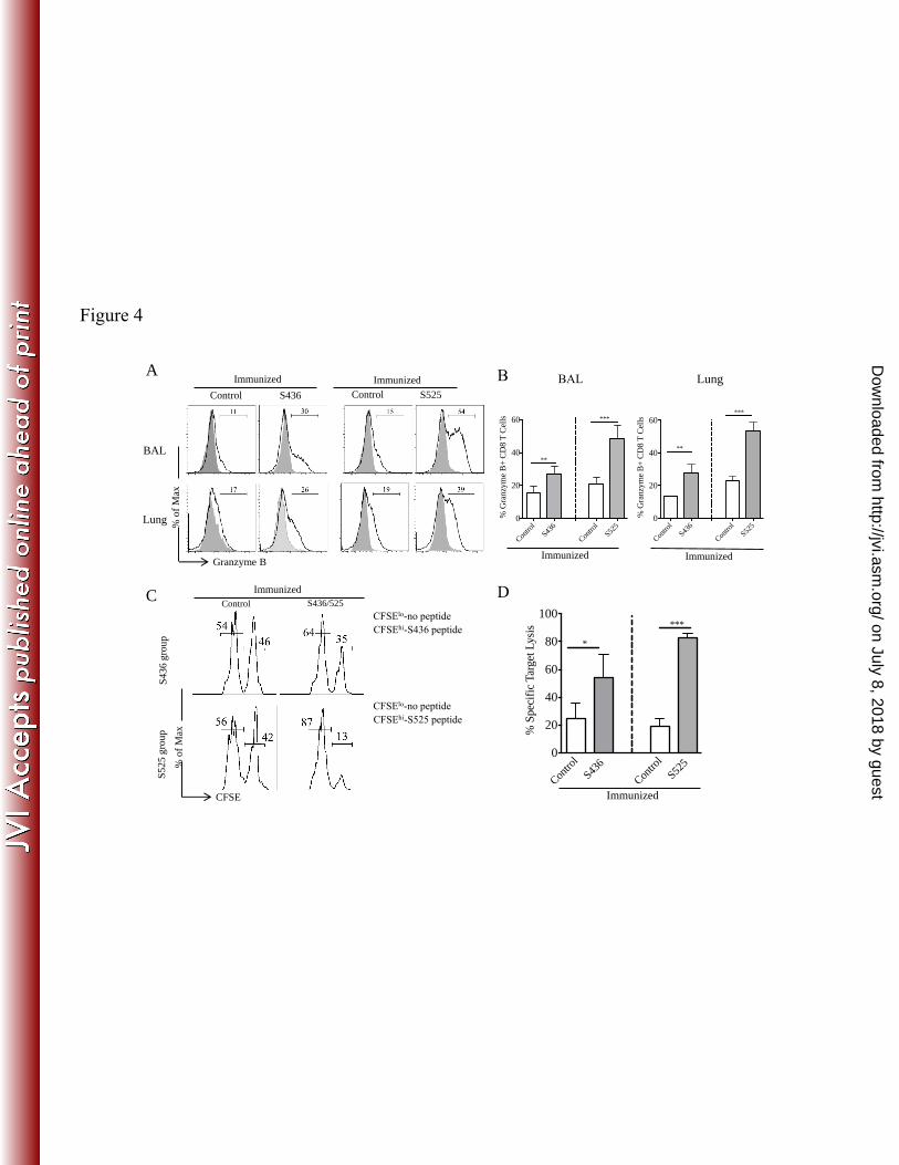
S436 and S525 immunized groups expressed granzyme B compared to control mice (Fig. 4A and 290
B). Further, CD8 T cells from S525-immunized compared to S436-immunized mice expressed 291
higher levels of granzyme B in both the BAL (48% vs. 27%, P<0.01) and lungs (53% vs. 27%, 292
P<0.001). Moreover, CD8 T cells from the S436 and S525-immunized groups exhibited higher in 293
vivo cytotoxic activity compared to those from the control group. Notably, virus-specific CD8 T 294
cells from the S525 immunized group were more cytotoxic than CD8 T cells from S436 295
immunized mice (P<0.005) (Fig. 4C and D). This may reflect both enhanced intrinsic 296
cytotoxicity and higher numbers of virus-specific CD8 T cells in the S525-immunized group (Fig. 297
on July 8, 2018 by guesthttp://jvi.asm
.org/D
ownloaded from
14
3B and 4A and B). Thus, the prime-boost regimen generated tissue resident memory CD8 T cells 298
that efficiently produced multiple effector cytokine and cytotoxic molecules upon MA15 299
challenge. 300
Virus-specific memory CD8 T cells protect mice from lethal MA15 infection: 301
To investigate the protective effect of virus-specific memory CD8 T cells, prime-boost 302
immunized mice were challenged with a lethal dose of MA15 and monitored for morbidity and 303
mortality. MA15-specific memory CD8 T cells protected immunized mice from lethal MA15 304
challenge to different extents. Consistent with the number of virus-specific memory CD8 T cells 305
(Fig. 3A), immunization against epitope S525 protected approximately 80% of mice, while nearly 306
60% of mice survived in the S436-immunized group. Mice from both the S525 and S436-307
immunized groups lost approximately 20% of their initial body weight (Fig. 5A and B). In 308
contrast, 100% of mice from the naïve group and 80% of mice from the control-immunized group 309
succumbed to the lethal infection (Fig. 5A and B). To determine whether protective efficacy 310
could be further enhanced, we immunized an additional group of mice with a mixture of DC-525 311
and DC-436 and boosted them with rVV-525 and rVV-436. After challenge, approximately 85% 312
of mice survived, only marginally different from the survival occurring after S525 immunization 313
alone. No differences in weight were observed when the S525 and S436+S525-immunized groups 314
were compared (Fig. 5A and B). 315
We next compared virus loads in lungs of immunized groups at different times after 316
MA15 challenge. Immunization with S436 and S525 resulted in more rapid virus clearance 317
compared to control VV-GFP-immunized mice. Mice immunized with S436 or S525 had reduced 318
viral burdens in the lungs as early as 4 days p.i. By 7 days post challenge, 100% of S525 319
immunized mice and 50% of S436-immunized mice cleared the virus from lungs while virus was 320
on July 8, 2018 by guesthttp://jvi.asm
.org/D
ownloaded from

15
not cleared in control mice (Fig. 5C). Histopathological examination of lungs of control mice on 321
day 4 post infection revealed marked alveolar edema, terminal bronchiolar epithelial sloughing and 322
thickening of interstitial septa, while that of S525-immunized mice revealed minimal amounts of 323
alveolar edema but increased peribronchial lymphocytic infiltration (Fig. 5D). S436-immunized 324
mice showed histological features intermediate to control and S525-immunized mice. On day 7 325
p.i., lungs of control-immunized mice had marked alveolar and bronchiolar edema with thickened 326
alveolar septa. At this time, pathological changes were reduced in both S436 and S525-immunized 327
mice especially in the latter group (Fig. 5D). Since S525 and S436+S525 immunization protected 328
mice equivalently, no additional studies were performed on the dually immunized group. In 329
summary, our results showed that memory CD8 T cells generated after prime-boost vaccination 330
enhanced virus clearance, limited lung pathology and protected susceptible B6 mice from lethal 331
MA15 challenge. 332
333
DISCUSSION: 334
Only a limited number of studies have addressed the role of the T cell-mediated immune 335
response in SARS-CoV infections. Previously, we demonstrated the ability of virus-specific CD8 336
T cells to protect susceptible young (BALB/C) and aged mice during a primary MA15 infection. 337
Here, we used a DC-rVV prime-boost regimen to generate a large number of virus-specific 338
memory CD8 T cells in the BAL and lungs. SARS-CoV-specific memory CD8 T cells in the lungs 339
exhibited a tissue resident memory CD8 T cell phenotype and produced multiple effector cytokines 340
and cytotoxic molecules. Our results show that SARS-CoV-specific memory CD8 T cells provided 341
substantial protection against lethal MA15 challenge with the extent of protection dependent on the 342
specific immunodominant epitope used for immunization. Of note, DC-rVV prime-boost 343
on July 8, 2018 by guesthttp://jvi.asm
.org/D
ownloaded from
16
immunization did not induce SARS-CoV neutralizing antibodies measured 45 days after 344
immunization, consistent with the notion that protection was mediated by memory CD8 T cells. In 345
this study, we analyzed 8-10 month old infected mice because, like middle-aged humans, these 346
mice were more susceptible to SARS-CoV than younger animals, but more immunocompetent 347
than very old mice. Following systemic primary immunization, effective CD8 T cell recall 348
responses to a localized challenge depends upon antigen presentation by DCs in the DLN (36). 349
Since DC migration to DLN is progressively impaired as mice age (21, 37), we adopted an 350
intranasal boosting regimen to generate lung resident memory CD8 T cells, thereby minimizing the 351
impact of DCs on the magnitude of CD8 T cell recall response. The expansion of tissue resident 352
memory CD8 T cells upon antigen re-challenge is largely independent of rDC migration to DLN, 353
as local antigen presentation by epithelial cells, lung resident DCs and recruited DCs drive 354
memory CD8 T cell expansion (38). Intravenous priming with DC-peptide and intranasal boosting 355
with rVV-minigene resulted in accumulation of SARS-CoV-specific memory CD8 T cells in BAL 356
and lungs (Fig. 2). 357
Interestingly, we observed a change in immunodominance patterns of S436 and S525 358
specific CD8 T cells after rVV boosting and challenge. The proportion and total number of S436 359
and S525-specific CD8 T cells were similar at 6 days after DC-peptide immunization in the PBL 360
and during the memory phase (42-45 days post rVV-minigene boost) in all tissues (Fig. 1B and C 361
and Fig. 2A and B). However, the proportion and the total number of S525-specific CD8 T cells 362
were significantly higher than that of S436-specific CD8 T cells after rVV-minigene boost (Fig. 363
1D-F) and after MA15 challenge (secondary effector response) (Fig. 3A and B). In mice infected 364
with influenza A virus (IAV), CD8 T cell responses to the NP366 and PA224 epitopes are co-365
dominant but upon rechallenge, the T cells response to epitope NP366 is dominant (39, 40). This 366
on July 8, 2018 by guesthttp://jvi.asm
.org/D
ownloaded from
17
change in epitope recognition was attributed to differences in antigen presentation (DCs vs. non 367
DC APCs) (41). Such a mechanism might also explain differences in responses to S525 and S436, 368
at least after MA15 challenge, although in this case, both epitopes are located on the same viral 369
protein. 370
We observed less protection against challenge in the S436-immunized compared to S525-371
immunized mice, which is likely due to the lesser number of S436-specific compared to S525-372
specific CD8 T cells in the BAL and lungs (Fig. 3A and B) (35, 42, 43). Additionally, both S436 373
and S525 immunized mice cleared virus rapidly and exhibited reduced lung pathology compared to 374
control mice. rVV boosting generated a substantial fraction of MA15-specific lung resident 375
memory CD8 T cells, which provided protection upon subsequent challenge. These results are in 376
agreement with recent studies demonstrating a critical role for lung resident virus-specific memory 377
CD8 T cells in protecting the host from a lethal IAV challenge (44). Thus, intranasal immunization 378
may be superior to systemic immunization because lung resident memory T cells are not generated 379
if immunogen is delivered systemically. Since systemic immunization does not result in the 380
generation of lung resident memory CD8 T cells, protection is dependent upon constant 381
replenishment from the periphery (27). Moreover, lung resident memory CD8 T cells generated 382
after intranasal priming are required for optimal heterosubtypic IAV immunity. Lung resident 383
memory CD8 T cells prevented extensive viral replication and limited alveolar damage, while 384
circulating cells failed to protect against heterosubtypic challenge (44). 385
The protective ability of immunodominant epitope-specific CD8 T cells is of 386
considerable significance, since SARS-CoV-specific antibody levels declined rapidly after 387
recovery. SARS-CoV-specific IgM and IgA responses lasted less than 6 months, while IgG titers 388
peaked at four months p.i. and markedly declined after one year (45-47). These studies suggested 389
on July 8, 2018 by guesthttp://jvi.asm
.org/D
ownloaded from

18
that SARS-CoV-specific IgG antibody response would eventually disappear, and the peripheral 390
memory B cell response would be insufficient to protect upon SARS-CoV re-infection. In contrast, 391
SARS-CoV-specific memory CD8 T cells persisted in SARS recovered patients for at least 6 years 392
(45). Consequently, SARS-CoV-specific CD4 and CD8 T cells are likely to play a vital role in 393
protecting patients upon SARS-CoV re-infection. Moreover, our results suggest that vaccine 394
strategies aimed at achieving elevated numbers of tissue resident memory virus-specific CD8 T 395
cells would be fruitful. 396
On note, whether the T cell response is protective or pathogenic depends on the specific 397
coronavirus and host strain (48). For example, following MA15, MERS-CoV or most strains of 398
mouse hepatitis virus (MHV) infection, virus-specific CD8 T cells are generally critical for virus 399
clearance both during primary infection and secondary challenge (49, 50). In contrast, in C3H/HeJ 400
mice infected with MHV-1 (MHV-1), a pneumotropic strain of MHV, T cells moderately 401
enhanced clinical illness and depletion of T cells ameliorated disease (51). Further, adoptive 402
transfer of MHV-1-specific memory CD8 T cells in the absence of anti-MHV-1 antibody induced 403
severe lung pathology and mortality in naive A/J and C3H/HeJ mice (51). In mice infected with 404
the JHM strain of MHV, T cell-mediated virus clearance resulted in myelin destruction (52). 405
Although SARS has not recurred since its last pandemic in 2002-03, the recent 406
emergence of MERS-CoV in humans and porcine epidemic diarrhea virus in pigs highlights the 407
need for coronavirus vaccines and anti-viral agents. Our results indicate that in addition to a strong 408
anti-SARS-CoV antibody response, an optimal memory CD8 T cell response will be an important 409
goal in vaccine design. 410
ACKNOWLEDGMENTS 411
on July 8, 2018 by guesthttp://jvi.asm
.org/D
ownloaded from
19
We thank the NIH Tetramer Facility for providing MHC class I/peptide tetramers. This work 412
was supported in part by grants from the National Institutes of Health (AI091322 and 413
AI060699). 414
REFERENCES: 415
1. Fouchier RA, Kuiken T, Schutten M, van Amerongen G, van Doornum GJ, van 416 den Hoogen BG, Peiris M, Lim W, Stohr K, Osterhaus AD. 2003. Aetiology: Koch's 417 postulates fulfilled for SARS virus. Nature 423:240. 418
2. Kuiken T, Fouchier RA, Schutten M, Rimmelzwaan GF, van Amerongen G, van 419 Riel D, Laman JD, de Jong T, van Doornum G, Lim W, Ling AE, Chan PK, Tam JS, 420 Zambon MC, Gopal R, Drosten C, van der Werf S, Escriou N, Manuguerra JC, 421 Stohr K, Peiris JS, Osterhaus AD. 2003. Newly discovered coronavirus as the 422 primary cause of severe acute respiratory syndrome. Lancet 362:263-270. 423
3. Peiris JS, Guan Y, Yuen KY. 2004. Severe acute respiratory syndrome. Nat Med 424 10:S88-97. 425
4. Zaki AM, van Boheemen S, Bestebroer TM, Osterhaus AD, Fouchier RA. 2012. 426 Isolation of a novel coronavirus from a man with pneumonia in Saudi Arabia. New 427 Engl J Med 367:1814-1820. 428
5. organization Wh. 2014. Middle East respiratory syndrome coronavirus (MERS-429 CoV) – update. http://www.who.int/csr/don/2014_03_25/en/. 430
6. Graham RL, Donaldson EF, Baric RS. 2013. A decade after SARS: strategies for 431 controlling emerging coronaviruses. Nat Rev Microbiol 11:836-848. 432
7. Gao W, Tamin A, Soloff A, D'Aiuto L, Nwanegbo E, Robbins PD, Bellini WJ, 433 Barratt-Boyes S, Gambotto A. 2003. Effects of a SARS-associated coronavirus 434 vaccine in monkeys. Lancet 362:1895-1896. 435
8. Yang ZY, Kong WP, Huang Y, Roberts A, Murphy BR, Subbarao K, Nabel GJ. 436 2004. A DNA vaccine induces SARS coronavirus neutralization and protective 437 immunity in mice. Nature 428:561-564. 438
9. Deming D, Sheahan T, Heise M, Yount B, Davis N, Sims A, Suthar M, Harkema J, 439 Whitmore A, Pickles R, West A, Donaldson E, Curtis K, Johnston R, Baric R. 440 2006. Vaccine efficacy in senescent mice challenged with recombinant SARS-CoV 441 bearing epidemic and zoonotic spike variants. PLoS Med 3:e525. 442
10. Graham RL, Becker MM, Eckerle LD, Bolles M, Denison MR, Baric RS. 2012. A 443 live, impaired-fidelity coronavirus vaccine protects in an aged, 444 immunocompromised mouse model of lethal disease. Nat Med 18:1820-1826. 445
11. Enjuanes L, Dediego ML, Alvarez E, Deming D, Sheahan T, Baric R. 2008. 446 Vaccines to prevent severe acute respiratory syndrome coronavirus-induced 447 disease. Virus Res 133:45-62. 448
12. Roberts A, Deming D, Paddock CD, Cheng A, Yount B, Vogel L, Herman BD, 449 Sheahan T, Heise M, Genrich GL, Zaki SR, Baric R, Subbarao K. 2007. A mouse-450 adapted SARS-coronavirus causes disease and mortality in BALB/c mice. PLoS 451 Pathog 3:e5. 452
on July 8, 2018 by guesthttp://jvi.asm
.org/D
ownloaded from
20
13. Fett C, DeDiego ML, Regla-Nava JA, Enjuanes L, Perlman S. 2013. Complete 453 protection against severe acute respiratory syndrome coronavirus-mediated lethal 454 respiratory disease in aged mice by immunization with a mouse-adapted virus 455 lacking E protein. J Virol 87:6551-6559. 456
14. DeDiego ML, Alvarez E, Almazan F, Rejas MT, Lamirande E, Roberts A, Shieh 457 WJ, Zaki SR, Subbarao K, Enjuanes L. 2007. A severe acute respiratory syndrome 458 coronavirus that lacks the E gene is attenuated in vitro and in vivo. J Virol 81:1701-459 1713. 460
15. Kohlmeier JE, Woodland DL. 2009. Immunity to respiratory viruses. Annu Rev 461 Immunol 27:61-82. 462
16. Woodland DL, Scott I. 2005. T cell memory in the lung airways. Proc Am Thorac 463 Soc 2:126-131. 464
17. Yang LT, Peng H, Zhu ZL, Li G, Huang ZT, Zhao ZX, Koup RA, Bailer RT, Wu CY. 465 2006. Long-lived effector/central memory T-cell responses to severe acute 466 respiratory syndrome coronavirus (SARS-CoV) S antigen in recovered SARS 467 patients. Clin Immunol 120:171-178. 468
18. Yang L, Peng H, Zhu Z, Li G, Huang Z, Zhao Z, Koup RA, Bailer RT, Wu C. 2007. 469 Persistent memory CD4+ and CD8+ T-cell responses in recovered severe acute 470 respiratory syndrome (SARS) patients to SARS coronavirus M antigen. J Gen Virol 471 88:2740-2748. 472
19. Zhao J, Zhao J, Perlman S. 2010. T cell responses are required for protection from 473 clinical disease and for virus clearance in severe acute respiratory syndrome 474 coronavirus-infected mice. J Virol 84:9318-9325. 475
20. Zhi Y, Kobinger GP, Jordan H, Suchma K, Weiss SR, Shen H, Schumer G, Gao G, 476 Boyer JL, Crystal RG, Wilson JM. 2005. Identification of murine CD8 T cell epitopes 477 in codon-optimized SARS-associated coronavirus spike protein. Virology 335:34-45. 478
21. Zhao J, Zhao J, Legge K, Perlman S. 2011. Age-related increases in PGD(2) 479 expression impair respiratory DC migration, resulting in diminished T cell 480 responses upon respiratory virus infection in mice. J Clin Invest 121:4921-4930. 481
22. Chen J, Lau YF, Lamirande EW, Paddock CD, Bartlett JH, Zaki SR, Subbarao K. 482 2010. Cellular immune responses to severe acute respiratory syndrome coronavirus 483 (SARS-CoV) infection in senescent BALB/c mice: CD4+ T cells are important in 484 control of SARS-CoV infection. J Virol 84:1289-1301. 485
23. Peng H, Yang LT, Wang LY, Li J, Huang J, Lu ZQ, Koup RA, Bailer RT, Wu CY. 486 2006. Long-lived memory T lymphocyte responses against SARS coronavirus 487 nucleocapsid protein in SARS-recovered patients. Virology 351:466-475. 488
24. Slifka MK, Whitton JL. 2000. Antigen-specific regulation of T cell-mediated 489 cytokine production. Immunity 12:451-457. 490
25. Kohlmeier JE, Cookenham T, Roberts AD, Miller SC, Woodland DL. 2010. Type I 491 interferons regulate cytolytic activity of memory CD8(+) T cells in the lung airways 492 during respiratory virus challenge. Immunity 33:96-105. 493
26. Schenkel JM, Fraser KA, Vezys V, Masopust D. 2013. Sensing and alarm function 494 of resident memory CD8(+) T cells. Nat Immunol 14:509-513. 495
27. Slutter B, Pewe LL, Kaech SM, Harty JT. 2013. Lung airway-surveilling CXCR3(hi) 496 memory CD8(+) T cells are critical for protection against influenza A virus. 497 Immunity 39:939-948. 498
on July 8, 2018 by guesthttp://jvi.asm
.org/D
ownloaded from

21
28. Wakim LM, Gupta N, Mintern JD, Villadangos JA. 2013. Enhanced survival of lung 499 tissue-resident memory CD8(+) T cells during infection with influenza virus due to 500 selective expression of IFITM3. Nat Immunol 14:238-245. 501
29. Hogan RJ, Usherwood EJ, Zhong W, Roberts AA, Dutton RW, Harmsen AG, 502 Woodland DL. 2001. Activated antigen-specific CD8+ T cells persist in the lungs 503 following recovery from respiratory virus infections. J Immunol 166:1813-1822. 504
30. Ely KH, Cookenham T, Roberts AD, Woodland DL. 2006. Memory T cell 505 populations in the lung airways are maintained by continual recruitment. J Immunol 506 176:537-543. 507
31. Pham NL, Badovinac VP, Harty JT. 2009. A default pathway of memory CD8 T cell 508 differentiation after dendritic cell immunization is deflected by encounter with 509 inflammatory cytokines during antigen-driven proliferation. J Immunol 183:2337-510 2348. 511
32. Barber DL, Wherry EJ, Ahmed R. 2003. Cutting edge: rapid in vivo killing by 512 memory CD8 T cells. J Immunol 171:27-31. 513
33. Seder RA, Darrah PA, Roederer M. 2008. T-cell quality in memory and protection: 514 implications for vaccine design. Nat Rev Immunol 8:247-258. 515
34. Teijaro JR, Turner D, Pham Q, Wherry EJ, Lefrancois L, Farber DL. 2011. Cutting 516 edge: Tissue-retentive lung memory CD4 T cells mediate optimal protection to 517 respiratory virus infection. J Immunol 187:5510-5514. 518
35. Harty JT, Badovinac VP. 2008. Shaping and reshaping CD8+ T-cell memory. Nat 519 Rev Immunol 8:107-119. 520
36. Zammit DJ, Cauley LS, Pham QM, Lefrancois L. 2005. Dendritic cells maximize the 521 memory CD8 T cell response to infection. Immunity 22:561-570. 522
37. Grolleau-Julius A, Harning EK, Abernathy LM, Yung RL. 2008. Impaired dendritic 523 cell function in aging leads to defective antitumor immunity. Cancer Res 68:6341-524 6349. 525
38. Wakim LM, Waithman J, van Rooijen N, Heath WR, Carbone FR. 2008. Dendritic 526 cell-induced memory T cell activation in nonlymphoid tissues. Science 319:198-202. 527
39. Belz GT, Stevenson PG, Doherty PC. 2000. Contemporary analysis of MHC-related 528 immunodominance hierarchies in the CD8+ T cell response to influenza A viruses. J 529 Immunol 165:2404-2409. 530
40. Belz GT, Xie W, Altman JD, Doherty PC. 2000. A previously unrecognized H-2D(b)-531 restricted peptide prominent in the primary influenza A virus-specific CD8(+) T-cell 532 response is much less apparent following secondary challenge. J Virol 74:3486-533 3493. 534
41. Crowe SR, Turner SJ, Miller SC, Roberts AD, Rappolo RA, Doherty PC, Ely KH, 535 Woodland DL. 2003. Differential antigen presentation regulates the changing 536 patterns of CD8+ T cell immunodominance in primary and secondary influenza 537 virus infections. J Exp Med 198:399-410. 538
42. Christensen JP, Doherty PC, Branum KC, Riberdy JM. 2000. Profound protection 539 against respiratory challenge with a lethal H7N7 influenza A virus by increasing the 540 magnitude of CD8(+) T-cell memory. J Virol 74:11690-11696. 541
43. Slutter B, Pewe LL, Lauer P, Harty JT. 2013. Cutting edge: rapid boosting of cross-542 reactive memory CD8 T cells broadens the protective capacity of the Flumist 543 vaccine. J Immunol 190:3854-3858. 544
on July 8, 2018 by guesthttp://jvi.asm
.org/D
ownloaded from
22
44. Wu T, Hu Y, Lee YT, Bouchard KR, Benechet A, Khanna K, Cauley LS. 2014. Lung-545 resident memory CD8 T cells (TRM) are indispensable for optimal cross-protection 546 against pulmonary virus infection. J Leuko Biol 95:215-224. 547
45. Tang F, Quan Y, Xin ZT, Wrammert J, Ma MJ, Lv H, Wang TB, Yang H, Richardus 548 JH, Liu W, Cao WC. 2011. Lack of peripheral memory B cell responses in recovered 549 patients with severe acute respiratory syndrome: a six-year follow-up study. J 550 Immunol 186:7264-7268. 551
46. Wu LP, Wang NC, Chang YH, Tian XY, Na DY, Zhang LY, Zheng L, Lan T, Wang LF, 552 Liang GD. 2007. Duration of antibody responses after severe acute respiratory 553 syndrome. Emerg Infect Dis 13:1562-1564. 554
47. Woo PC, Lau SK, Wong BH, Chan KH, Chu CM, Tsoi HW, Huang Y, Peiris JS, Yuen 555 KY. 2004. Longitudinal profile of immunoglobulin G (IgG), IgM, and IgA antibodies 556 against the severe acute respiratory syndrome (SARS) coronavirus nucleocapsid 557 protein in patients with pneumonia due to the SARS coronavirus. Clin Diagn Lab 558 Immunol 11:665-668. 559
48. Channappanavar R, Zhao J, Perlman S. 2014. T cell-mediated immune response to 560 respiratory coronaviruses. Immunol Res 2014 May 21. [Epub ahead of print]. 561
49. Williamson JS, Stohlman SA. 1990. Effective clearance of mouse hepatitis virus 562 from the central nervous system requires both CD4+ and CD8+ T cells. J Virol 563 64:4589-4592. 564
50. Zhao J, Li K, Wohlford-Lenane C, Agnihothram SS, Fett C, Zhao J, Gale MJ, Jr., 565 Baric RS, Enjuanes L, Gallagher T, McCray PB, Jr., Perlman S. 2014. Rapid 566 generation of a mouse model for Middle East respiratory syndrome. Proc Natl Acad 567 Sci U S A. 111:4970-4975. 568
51. Khanolkar A, Hartwig SM, Haag BA, Meyerholz DK, Epping LL, Haring JS, Varga 569 SM, Harty JT. 2009. Protective and pathologic roles of the immune response to 570 mouse hepatitis virus type 1: implications for severe acute respiratory syndrome. J 571 Virol 83:9258-9272. 572
52. Wu GF, Dandekar AA, Pewe L, Perlman S. 2000. CD4 and CD8 T cells have 573 redundant but not identical roles in virus-induced demyelination. J. Immunol 574 165:2278-2286. 575
576
577
on July 8, 2018 by guesthttp://jvi.asm
.org/D
ownloaded from

23
578 FIGURE LEGENDS: 579
Figure 1. Prime-boost immunization induces a strong CD8 T cell response in 8 month old 580
mice: (A) 8 month old B6 mice were immunized intravenously with DC-loaded peptides and 581
boosted 6 days later with rVV-S436 or rVV-S525 intranasally. Mice were rested for 42-45 days 582
and then challenged with lethal dose (5x10⁴ PFU) of MA15 (i.n). Lungs and BAL were harvested 583
5 days post-challenge for further analysis. (B) FACS plots show percentages of S436 and S525-584
specific IFN-+ CD8 T cells in the blood (after direct ex-vivo stimulation with respective peptides) 585
on 0, 4 and 6 days after DC-peptide immunization. (C) Mean percentage (above) and numbers 586
(below) of S436 and S525-specific CD8 T cells in the blood are shown. (D) FACS plots represent 587
percentage of S436 and S525-specific IFN-+ CD8 T cells (after direct ex-vivo stimulation with 588
respective peptides) in BAL, lungs and spleen, 8 days post rVV-minigene boost. (E and F) Bar 589
graphs show mean percentage (E) and number (F) of S436 and S525-specific IFN-+ CD8 T cells 590
(after in vitro stimulation with respective peptides) in BAL, lungs and spleen, 8 days post rVV-591
minigene boost. Data are representative of 2 independent experiments with 3-4 mice/group. * 592
P<0.05, ** P<0.01 and *** P<0.001 by unpaired two tailed Student’s t-test. 593
594
Figure 2. Identification of SARS-CoV-specific memory CD8 T cells: Prime-boost immunized 595
mice were rested for 42-45 days and the percentage and number of S436 and S525-specific 596
memory CD8 T cells were enumerated in BAL, lungs and spleen. (A and B) The bar graphs show 597
mean percentage (A) and number (B) of S436 and S525-specific IFN-+ CD8 T cells in the BAL, 598
lungs and spleen 42-45 days after rVV-minigene boost. (C) The bar graphs show mean percentage 599
of polyfunctional S436 and S525-specific IFN-+ CD8 T cells in the BAL, lungs and spleen. Data 600
on July 8, 2018 by guesthttp://jvi.asm
.org/D
ownloaded from

24
are means of cytokine-positive CD8 T cells obtained by Boolean gating. (D) Scatter plots represent 601
the percentage of S436 and S525 CD8 T cells that express the indicated marker. Data are 602
representative of 2-3 independent experiments with 3-4 mice/group. * P<0.05, ** P<0.01 and *** 603
P<0.001 by unpaired two tailed Student’s t-test. 604
605
Figure 3. Immunization induces polyfunctional secondary effector CD8 T cells: Prime-boost 606
immunized mice were rested for 42-45 days and then challenged with a lethal dose (5x10⁴ PFU) 607
of MA15 (i.n). Lung and BAL were harvested 5 days post-challenge and the percentage and 608
number of epitope-specific CD8 T cells were enumerated in the BAL and lung. (A and B) The bar 609
graphs show mean percentage (A) and number (B) of S436 and S525-specific IFN-+ CD8 T cells 610
(after direct ex-vivo stimulation with respective peptides) in the BAL and lungs 5 days post MA15 611
challenge. (C) IFN-+ CD8 T cells were further gated for TNF- and IL-2 expression to determine 612
polyfunctionality in the BAL and lungs. Data are representative of 3 independent experiments with 613
3-4 mice/group. * P<0.05, ** P<0.01 and *** P<0.001 by unpaired two tailed Student’s t-test. 614
615
Figure 4. Increased granzyme B production and cytotoxicity after challenge: (A and B) 616
Histograms represent percent granzyme B+ CD8 T cells in the BAL and lungs at day 5 post MA15 617
challenge (A). (B) Bar graphs represent mean percent of granzyme B+ CD8 T cells (after direct ex-618
vivo stimulation with respective peptide). (C and D) In vivo cytotoxicity assays were performed 5 619
days post MA15 challenge and the percentage of killing was calculated, as described in Materials 620
and Methods (numbers represent the percentage of cells labeled with different concentrations of 621
CFSE). n = 4–5 mice/group/experiment. Data are representative of 2 independent experiments. * 622
P<0.05, ** P<0.01 and *** P<0.001 by unpaired two tailed Student’s t-test. 623
on July 8, 2018 by guesthttp://jvi.asm
.org/D
ownloaded from
25
624
Figure 5. Virus-specific memory CD8 T cells protect mid-aged mice from lethal MA15 625
infection. Prime-boost immunized mice were rested for 42-45 days and then challenged with lethal 626
dose (5x10⁴ PFU) of MA15 intranasally. (A) Percent initial body weight and (B) survival curves 627
are shown. The experiment shows data combined from three independent experiments, with n=8 628
mice in the naïve group and n=15-16 in all other groups. Data are expressed as the percent initial 629
weight ± the SEM. (C) Virus titers in lungs. Data correspond to 4 mice per group ± SEM and are 630
representative of 2-3 independent experiments. (D) Lung sections from control and immunized 631
mice are shown at days 0, 4 and 7 post-challenge. Asterisks represent alveolar and bronchiolar 632
edema and arrowheads show peribronchial lymphocyte infiltration. * P<0.05, ** P<0.01 and *** 633
P<0.001 by unpaired Student’s t-test. 634
on July 8, 2018 by guesthttp://jvi.asm
.org/D
ownloaded from
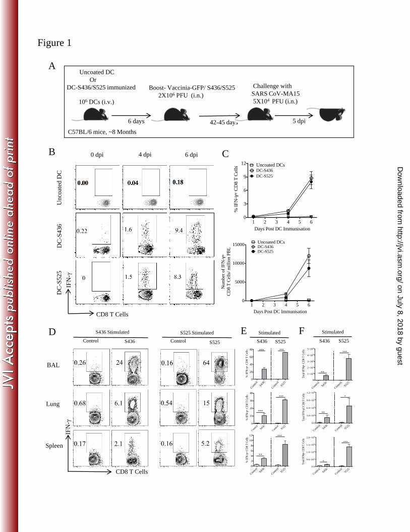
Figure 1
CD8 T Cells
IFN
-γ
6 dpi 4 dpi B
S436 S525 Control
CD8 T Cells
IFN
-γ
Lung
BAL
Spleen
Control
S436 Stimulated S525 Stimulated
DC
-S4
36
D
C-S
52
5
C
D
Uncoated DC
Or
DC-S436/S525 immunized
Challenge with
SARS CoV-MA15
5X10⁴ PFU (i.n.)
42-45 days
C57BL/6 mice, ~8 Months
6 days
Boost- Vaccinia-GFP/ S436/S525
2X10⁶ PFU (i.n.) 106 DCs (i.v.)
5 dpi
A
E
Un
coat
ed D
C
S436
S525
F
0 dpi
1 2 3 4 5 60
3
6
9
12
Days Post DC Immunisation
% I
FN
-g+
CD
8 T
Cel
ls
DC-S436
DC-S525
Uncoated DCs
1 2 3 4 5 60
5000
10000
15000
Days Post DC Immunisation
Num
ber
of
IFN
-g+
CD
8 T
Cel
ls/
mil
lion P
BL DC-S436
DC-S525
Uncoated DCs
Stimulated
S436
S525
Stimulated
Con
trol
S436
Con
trol
S525
0
20
40
60
80
% I
FN
-g+
CD
8 T
Cell
s ******
Contro
l
S436
Contro
l
S525
0
1×10⁵
2×10⁵
3×10⁵
4×10⁵
5×10⁵
Tot
al I
FN
g+ C
D8
T C
ells
***
**
Contro
l
S436
Contro
l
S525
0
10
20
30
40
% I
FN
-g+
CD
8 T
Cel
ls ***
***
Contro
l
S436
Contro
l
S5250.0
3.0×10⁵
6.0×10⁵
9.0×10⁵
1.2×10⁶
Tot
al I
FN
-g+
CD
8 T
Cel
ls
*
**
Con
trol
S436
Con
trol
S525
0
2
4
6
8
10
12
% I
FN
-g+
CD
8 T
Cel
ls
***
**
Control
S436
Control
S5250.0
5.0×10⁵
1.0×10⁶
1.5×10⁶
2.0×10⁶
Tot
al I
FN
g+ C
D8
T C
ells
***
*
on July 8, 2018 by guesthttp://jvi.asm
.org/D
ownloaded from

Figure 2
L
A CBS436 Stimulated
S525Stimulated
S436 Stimulated
S525Stimulated
D
40
50
T C
ells ***
***
3×10³
4×10³
8 T
Cel
ls
***
80
100
120
Tetra
mer
+ T
Cel
ls
******
****
100
Cel
ls
BALLungSpleen
S436
BA
L
Contro
lS43
6
Contro
lS52
50
10
20
30
% IF
N-γ
+ CD
8
Contro
lS43
6
Contro
lS52
50
1×10³
2×10³
Tota
l IFN
-γ+
CD
8
0
20
40
60
80
% C
XCR
3+ o
n T
mem
ory
CD
8
S436 S525
0
20
40
60
80
% IF
N-γ
+ C
D8
T C
***
****
****
Lung
2
4
6
8
IFN
-γ+
CD
8 T
Cel
ls ** *
1×10⁴
2×10⁴
3×10⁴
4×10⁴
al IF
N-γ
+ CD
8 T
Cells
*
**
20
40
60
80
100
CD
103+
on
Tetra
mer
+ m
emor
y C
D8
T C
ells
BAL LungSpleen
*
0IFN-γTNF-αIL-2
+++
+
+_
++_
+__
S525
Contro
lS43
6
Contro
lS52
50
%
2.0
2.5
T Ce
lls
**
Contro
lS43
6
Contro
lS52
50To
ta
1.0×10⁵
1.5×10⁵
D8
T Ce
lls
**
*
0
20
% C m
S436 S525
60
80
100
+ T
etra
mer
+ 8
T C
ells
* * *20
40
60
80
100
IFN
-γ+
CD
8 T
Cel
ls
**
*****
*****
Sple
en
Contro
lS43
6
Contro
lS52
50.0
0.5
1.0
1.5
% IF
N-γ
+ C
D8
Contro
lS43
6
Contro
lS52
50.0
5.0×10⁴
1.0 10
Tota
l IFN
-γ+
CD
0
20
40
60
% C
D11
a+ C
D69
mem
ory
CD
8
S436 S525
*0
20
%
IFN-γTNF-αIL-2
+++
+
+_
++_
+__
on July 8, 2018 by guesthttp://jvi.asm
.org/D
ownloaded from

BA
L
LU
NG
A
C
Figure 3
S436 Stimulated
S525 Stimulated
S436 Stimulated
S525 Stimulated
B
BA
L
LU
NG
Control
S436
Control
S525
0
1×10⁵
2×10⁵
3×10⁵
4×10⁵
5×10⁵
Tota
l IF
N-g
+ C
D8 T
Cel
ls
***
*
Immunized
Control
S436
Control
S525
0
20
40
60
80
100
% I
FN
-g+
CD
8 T
Cel
ls**
***
Immunized
Control
S436
Control
S525
0
1×10⁶
2×10⁶
3×10⁶
4×10⁶
Tota
l IF
N-g
CD
8 T
Cel
ls
**
*
Immunized
Control
S436
Control
S525
0
10
20
30
40
50
% I
FN
-g+
CD
8 T
Cel
ls
***
***
Immunized
S436 S5250
20
40
60
80
100
120
% I
FN
g+ C
yto
kin
e+
IFNγ+
IFNγ+TNF-α+
TNFa+
Immunized
S436 S5250
20
40
60
80
100
120
% I
FN
g+ C
yto
kin
e+
Immunized
0
20
40
60
80
100
120
% I
FN
g+ C
yto
kin
e+
IFNγ+
IFNγ+IL-2+
IL-2+
S436 S525
Immunized
S436 S5250
20
40
60
80
100
120
% I
FN
g+ C
yto
kin
e+
Immunized
on July 8, 2018 by guesthttp://jvi.asm
.org/D
ownloaded from
% o
f M
ax
Granzyme B
BAL
Lung
S436 S525 Control
A B Control
Immunized Immunized BAL Lung
C
% o
f M
ax
CFSE
Control
S5
25
gro
up
S436/525
S4
36
gro
up
CFSElo-no peptide
CFSEhi-S436 peptide
CFSElo-no peptide
CFSEhi-S525 peptide
D
Figure 4
Immunized
Con
trol
S436
Con
trol
S525
0
20
40
60
% G
ranzy
me
B+
CD
8 T
Cel
ls
**
***
Con
trol
S436
Con
trol
S525
0
20
40
60
% G
ranzy
me
B+
CD
8 T
Cel
ls
**
***
Immunized Immunized
Con
trol
S436
Con
trol
S525
0
20
40
60
80
100
% S
pec
ific
Tar
get
Lysi
s
*
***
Immunized
on July 8, 2018 by guesthttp://jvi.asm
.org/D
ownloaded from


