dr. uttam kumar kanp c1t vaucheria
TRANSCRIPT

BOTANY: SEM – I, PAPER
Vaucheria: Occurrence, Range of thallus organization, cell structure,
Reproduction and Life Cycle
Dr. Uttam Kumar Kanp
Systematic Position:
Class: Chlorophyceae
Order: Siphonales
Family: Vaucheriaceae
Genus : Vaucheria
Occurrence of Vaucheria:
Vaucheria is represented
Vaucheria is found mostly in fres
found on moist soil.
The terrestrial species like
places in green houses. V. amph
winter ice in U.S.A. The common
polysperma, V. sessilis and V. unci
Thallus Organization:
The thallus is made of long, cylindrical well branched filaments. The
coenocytic structure. The thallus is attached to
branched holdfast called the haptera. The thallus of V. mayyanadensis is differentiated in
subterranean branched rhizoidal system and an erect aerial system. The filaments are
interwoven and appear as dark green felt like structure.
Some species like V. debar
lateral or dichotomous. The filame
along the entire length of thallus t
(Fig. 1A, B). The septa formation
I, PAPER-C1T: PHYCOLOGY AND MICROBIOLOGY, UNIT
VAUCHERIA
Vaucheria: Occurrence, Range of thallus organization, cell structure,
Reproduction and Life Cycle
Dr. Uttam Kumar Kanp
by 54 species of which about 19 species a
esh water but about six species are marine and
V. sessilis and V. terrestris form green mats on
phibia is amphibous. V. jonesii was reported by
ommon Indian species of Vaucheria are V. amphibi
cinata etc.
The thallus is made of long, cylindrical well branched filaments. The filament is aseptate,
coenocytic structure. The thallus is attached to substratum by means of branched rhizoids or
haptera. The thallus of V. mayyanadensis is differentiated in
branched rhizoidal system and an erect aerial system. The filaments are
interwoven and appear as dark green felt like structure.
ryana show calcium carbonate incrustations. Th
ents are non-septate, the protoplasm with many n
thus the coenocytic Vaucheria thallus makes siph
on occurs only during reproduction or in Gongros
PHYCOLOGY AND MICROBIOLOGY, UNIT-5:
Vaucheria: Occurrence, Range of thallus organization, cell structure,
are found in India.
some are terrestrial
moist soil in shady
y Prescott (1938) in
ia, V. geminata, V.
filament is aseptate,
substratum by means of branched rhizoids or
haptera. The thallus of V. mayyanadensis is differentiated in
branched rhizoidal system and an erect aerial system. The filaments are rough,
e branching may be
nuclei is continuous
phonaceous structure
sira condition or for

BOTANY: SEM – I, PAPER
sealing of an injury.
The thallus structure is differentiated into cell wall and protoplasm. The
thallus is thin, weak and non-elastic. The
the inner layer is cellulosic. Inner to the
central vacuole filled with cell sap runs from one end of the filament to another forming
continuous canal or siphon.
In peripheral part of protop
chloroplasts which lack pyrenoids
chromatophores.
The chromatophores in Vaucheria contain pigments, chlorophyll a,
carotenoids and an unknown xanthophyll. The pigments in
Xanthophyceae as chlorophyll b the
Many small nuclei lie in the cytoplasm inner to the layer of chloroplasts.
nuclei with respect to chloroplasts is reversed at the
contains other membrane bound cell organelle such as mitochondria, small vesicles an
stored in form of oil. The growth of filament is apical, the filament increases in
growth of all the branches.
Nature of Thallus:
The thallus of Vaucheria is branched, non
single large cell but Vaucheria cannot be
I, PAPER-C1T: PHYCOLOGY AND MICROBIOLOGY, UNIT
VAUCHERIA
The thallus structure is differentiated into cell wall and protoplasm. The
elastic. The cell wall is made of two layers, the outer layer is pectic and
the inner layer is cellulosic. Inner to the cell wall there is thick layer of protoplasm. A very large
filled with cell sap runs from one end of the filament to another forming
oplasm are present a large number of small o
s (Fig. 1 B). Christensen (1952) reported presen
in Vaucheria contain pigments, chlorophyll a,
carotenoids and an unknown xanthophyll. The pigments in Vaucheria are like those of
Xanthophyceae as chlorophyll b the characteristic pigment of Chlorophyceae is absent.
the cytoplasm inner to the layer of chloroplasts. The arrangement of
nuclei with respect to chloroplasts is reversed at the time of zoospore formation. The cytoplasm also
bound cell organelle such as mitochondria, small vesicles an
in form of oil. The growth of filament is apical, the filament increases in
The thallus of Vaucheria is branched, non-septate and multinucleate structure which appears like
single large cell but Vaucheria cannot be
PHYCOLOGY AND MICROBIOLOGY, UNIT-5:
The thallus structure is differentiated into cell wall and protoplasm. The cell wall of
layers, the outer layer is pectic and
cell wall there is thick layer of protoplasm. A very large
filled with cell sap runs from one end of the filament to another forming a
oval or disc shaped
nce of pyrenoids in
in Vaucheria contain pigments, chlorophyll a, chlorophyll e,
Vaucheria are like those of
characteristic pigment of Chlorophyceae is absent.
The arrangement of
time of zoospore formation. The cytoplasm also
bound cell organelle such as mitochondria, small vesicles and food is
in form of oil. The growth of filament is apical, the filament increases in length by apical
structure which appears like

BOTANY: SEM – I, PAPER-
considered as single cell. As in multicellular forms
number of nuclei. The apical growth takes place. Hence
Vaucheria should be considered as
Reproduction in Vaucheria:
Reproduction in Vaucheria takes plac
(i) Vegetative Reproduction in Vaucheria:
The vegetative reproduction takes place by fragmentation. The thallus can
fragments due to mechanical injury or insect bites etc. A
to seal the injury. The broken fragment develops thick wall and later on develops into Vaucheria
thallus.
(ii) Asexual Reproduction in Vaucheria:
The asexual reproduction takes place by formation of zoospores,
(a) By Zoospores:
The zoospores formation is the most common method of reproduction in
terrestrial species it takes place when the plants are
favourable seasons or can be induced if aquatic species a
from running water to still water.
Zoospores are formed sing
development of zoosporangium b
large number of nuclei and chlorop
protoplasmic region becomes visi
cytoplasm of thallus.
Each separated protoplast secretes
cross wall. Inside zoosporangium the vacuole decreases, the
dense and round off. The change takes
nuclei become peripheral and chloroplasts enter in inner layer of cytoplasm.
The entire protoplasm of
each nucleus two flagella are pro
I, PAPER-C1T: PHYCOLOGY AND MICROBIOLOGY, UNIT
VAUCHERIA
considered as single cell. As in multicellular forms mitotic divisions take place increasing the
number of nuclei. The apical growth takes place. Hence the aseptate coenocytic structure of
Vaucheria should be considered as acellular coenocyte.
Reproduction in Vaucheria:
Reproduction in Vaucheria takes place by vegetative, asexual and sexual methods.
Vegetative Reproduction in Vaucheria:
The vegetative reproduction takes place by fragmentation. The thallus can
fragments due to mechanical injury or insect bites etc. A septum develops at the
fragment develops thick wall and later on develops into Vaucheria
Vaucheria:
The asexual reproduction takes place by formation of zoospores, aplanospores and akinetes
The zoospores formation is the most common method of reproduction in
terrestrial species it takes place when the plants are flooded. Zoospore formation takes place in
induced if aquatic species are transferred from light to darkness or
gly within elongated club shaped zoosporangium
begins with a club shaped swelling at the tip o
oroplasts along with the cytoplasm move into
ible at the base of cytoplasm and it is separate
Each separated protoplast secretes thin membrane and zoosporangium gets
cross wall. Inside zoosporangium the vacuole decreases, the contents of sporangium become very
dense and round off. The change takes place in relative position of chloroplasts and nuclei, the
peripheral and chloroplasts enter in inner layer of cytoplasm.
the zoosporangium contracts to form oval zoo
roduced making zoospore a multi-flagellate structu
PHYCOLOGY AND MICROBIOLOGY, UNIT-5:
place increasing the
the aseptate coenocytic structure of
methods.
The vegetative reproduction takes place by fragmentation. The thallus can break into small
septum develops at the place of breaking
fragment develops thick wall and later on develops into Vaucheria
aplanospores and akinetes
aquatic species. In
flooded. Zoospore formation takes place in
re transferred from light to darkness or
m (Fig. 2A, B). The
of a side branch. A
o it. A colourless
ed from rest of the
thin membrane and zoosporangium gets separated by a
contents of sporangium become very
place in relative position of chloroplasts and nuclei, the
zoospore. Opposite to
ucture. A terminal

BOTANY: SEM – I, PAPER-
aperture develops in zoosporangiu
aperture in morning hours (Fig. 2 C
Each zoospore is large yellow green, oval structure. It has a central vacuole
sap and may be traversed by cytoplasmic strands. The
towards the walls and chromatophores towards vacuoles. Two flagella arise opposite to each
nucleus. This part of cytoplasm can be regarded equivalent to one zoospore.
Fritsch (1948) regarded th
number of biflagellate zoospores h
According to Greenwood,
and whiplash type. The shorter fl
zoospore. The flagellar bases are u
According to Greenwood e
posterior region of the zoospores.
bodies and plastids are present i
zoospores.
The zoospores swim in w
significant period of rest. The zo
secrete thin walls (Fig. 2 E, F).
vegetative condition.
The two tube like outgrowt
elongates, branches to form colou
tubular coenocytic filament (Fig. 2
I, PAPER-C1T: PHYCOLOGY AND MICROBIOLOGY, UNIT
VAUCHERIA
ium by gelatinization of wall. The zoospore is
C, D).
Each zoospore is large yellow green, oval structure. It has a central vacuole
by cytoplasmic strands. The protoplasm outer to vacuole has many nuclei
chromatophores towards vacuoles. Two flagella arise opposite to each
nucleus. This part of cytoplasm can be regarded equivalent to one zoospore.
his kind of zoospore as compound zoospore or
have failed to separate from one another.
Manton and Clarke (1957) the flagella of a pa
flagellum of each pair is directed towards the an
united together in pairs and are firmly attached to
et. al (1957), there is large anterior vacuole and
Mitochondria are present in the peripheral layer
in the cytoplasm. Chlorophyll has also been
water for 5-15 minutes and germinate withou
zoospores get attached to the substratum, with
The chromatophores move outwards and nucl
owths develop in opposite directions. One of the
urless lobed holdfast and the other outgrowth fo
2 G, H).
PHYCOLOGY AND MICROBIOLOGY, UNIT-5:
s liberated through
Each zoospore is large yellow green, oval structure. It has a central vacuole which has cell
protoplasm outer to vacuole has many nuclei
chromatophores towards vacuoles. Two flagella arise opposite to each
or synzoospore as a
pair are heterokontic
anterior end of the
o the tip of nuclei.
and small ones in the
r of cytoplasm. Fat
reported from the
ut undergoing any
hdraw flagella and
uclei inwards as in
he two outgrowths
forms yellow-green

BOTANY: SEM – I, PAPER-
(b) By Aplanospores:
Aplanospores are commonly observed in species. V. geminata, V. uncinata
species V. pitoboloides. The aplanospores are generally
Aquatic species form aplanspores under unfavorable condition of drought.
are non-motile asexual spores formed in special
aplanospores are produced singly in cells at the terminal end of the short lateral or terminal
The protoplasm of aplanosporangium gets
aplanospore which is thin walled. In V.
apical pore formed by gelatinization.
In V. uncinata aplanospores are spherical and are liberated by rupture of
The formation and structure of aplanospores and
flagella. The aplanospores soon after liberation germinate into new thalli (Fig. 3D).
I, PAPER-C1T: PHYCOLOGY AND MICROBIOLOGY, UNIT
VAUCHERIA
Aplanospores are commonly observed in species. V. geminata, V. uncinata
species V. pitoboloides. The aplanospores are generally formed by terrestrial species.
Aquatic species form aplanspores under unfavorable condition of drought.
motile asexual spores formed in special structures called aplanosporangia (Fig. 3 A
produced singly in cells at the terminal end of the short lateral or terminal
The protoplasm of aplanosporangium gets metamorphosed into single
aplanospore which is thin walled. In V. germinate aplanospores are oval and are liberated from
gelatinization.
aplanospores are spherical and are liberated by rupture of the spo
The formation and structure of aplanospores and zoospores is similar except that the zoospores lack
aplanospores soon after liberation germinate into new thalli (Fig. 3D).
PHYCOLOGY AND MICROBIOLOGY, UNIT-5:
Aplanospores are commonly observed in species. V. geminata, V. uncinata and in marine
formed by terrestrial species.
Aquatic species form aplanspores under unfavorable condition of drought. The aplanospores
structures called aplanosporangia (Fig. 3 A-C). The
produced singly in cells at the terminal end of the short lateral or terminal branch.
metamorphosed into single multinucleate
aplanospores are oval and are liberated from
the sporangial wall.
zoospores is similar except that the zoospores lack
aplanospores soon after liberation germinate into new thalli (Fig. 3D).

BOTANY: SEM – I, PAPER-
(c) By Akinetes:
Akinetes are thick walled structures
low temperature. The akinetes have been commonly
uncinata.
The akinetes are formed on the terminal part of lateral branches where
to the tips followed by cross-wall formation (Fig. 4).
are called akinetes or hypnospores.
The akinetes by successive divisions may form numerous thin walled bodies
When many akinetes remain attached
I, PAPER-C1T: PHYCOLOGY AND MICROBIOLOGY, UNIT
VAUCHERIA
Akinetes are thick walled structures formed during unfavorable conditions
low temperature. The akinetes have been commonly observed in V. geminata, V. megaspora and V.
The akinetes are formed on the terminal part of lateral branches where protoplasm migrates
wall formation (Fig. 4). These multinucleate, thick walled segments
hypnospores.
The akinetes by successive divisions may form numerous thin walled bodies
When many akinetes remain attached to the parent thallus, the thallus gives the appearance of
PHYCOLOGY AND MICROBIOLOGY, UNIT-5:
formed during unfavorable conditions like drought, and
observed in V. geminata, V. megaspora and V.
protoplasm migrates
These multinucleate, thick walled segments
The akinetes by successive divisions may form numerous thin walled bodies called cysts.
thallus gives the appearance of
BOTANY: SEM – I, PAPER-
another alga Gongrosira.
Hence this stage of Vauch
akinetes and cysts develop into n
submerged parts of thallus develop
akinetes.
(iii) Sexual Reproduction in Vaucheria:
In Vaucheria sexual reproduction is of advanced oogamous type. The male
organs are antheridia and oogonia, respectively.
Majority of the freshwater species are monoecious or homothallic while
dichotoma, V. litorea and V. mayyanadensis are
of arrangement of antheridia and oogonia in homothallic species. The position, structure and
of antheridia are of taxonomic importance in Vaucheria.
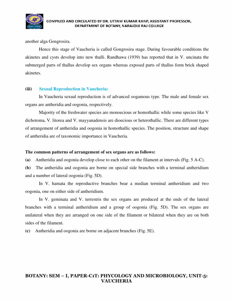
The common patterns of arrangement of sex organs are as
(a) Antheridia and oogonia develop close to each other on
(b) The antheridia and oogonia are borne on special side branches with
and a number of lateral oogonia (Fig.
In V. hamata the reproductive branches bear a median terminal
oogonia, one on either side of antheridium.
In V. geminata and V. terrestris the sex organs are produced at the ends of
branches with a terminal antheridium and a group of oogonia
unilateral when they are arranged
sides of the filament.
(c) Antheridia and oogonia are borne on adjacent branches (Fig.
I, PAPER-C1T: PHYCOLOGY AND MICROBIOLOGY, UNIT
VAUCHERIA
heria is called Gongrosira stage. During favoura
new thalli. Randhawa (1939) has reported that
op sex organs whereas exposed parts of thallus
Vaucheria:
In Vaucheria sexual reproduction is of advanced oogamous type. The male
are antheridia and oogonia, respectively.
Majority of the freshwater species are monoecious or homothallic while some species like V
dichotoma, V. litorea and V. mayyanadensis are dioecious or heterothallic. There are different types
heridia and oogonia in homothallic species. The position, structure and
of antheridia are of taxonomic importance in Vaucheria.
The common patterns of arrangement of sex organs are as follows:
Antheridia and oogonia develop close to each other on the filament at intervals (Fig. 5 A
The antheridia and oogonia are borne on special side branches with a terminal antheridium
and a number of lateral oogonia (Fig. 5D).
In V. hamata the reproductive branches bear a median terminal antheridium and two
oogonia, one on either side of antheridium.
In V. geminata and V. terrestris the sex organs are produced at the ends of
branches with a terminal antheridium and a group of oogonia (Fig. 5D). The sex organs are
unilateral when they are arranged on one side of the filament or bilateral when they are on both
Antheridia and oogonia are borne on adjacent branches (Fig. 5E).
PHYCOLOGY AND MICROBIOLOGY, UNIT-5:
able conditions the
in V. uncinata the
form brick shaped
In Vaucheria sexual reproduction is of advanced oogamous type. The male and female sex
some species like V
dioecious or heterothallic. There are different types
heridia and oogonia in homothallic species. The position, structure and shape
the filament at intervals (Fig. 5 A-C).
a terminal antheridium
antheridium and two
In V. geminata and V. terrestris the sex organs are produced at the ends of the lateral
(Fig. 5D). The sex organs are
of the filament or bilateral when they are on both
BOTANY: SEM – I, PAPER-
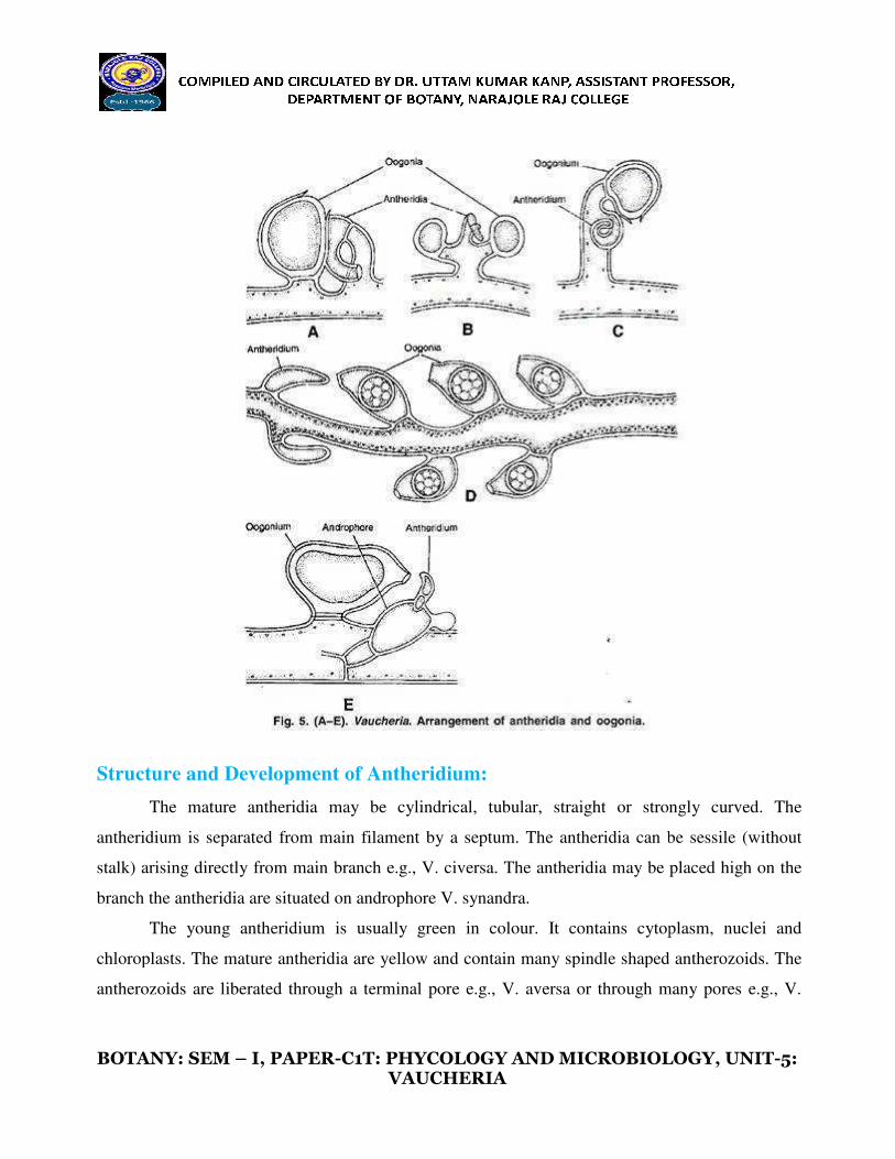
Structure and Development of Antheridium:
The mature antheridia may be cylindrical, tubular, straight
antheridium is separated from main filament by a septum. The
stalk) arising directly from main branch
branch the antheridia are situated on androphore V. synandra.
The young antheridium is usually green in colour. It contains cytoplasm,
chloroplasts. The mature antheridia are yellow and contain
antherozoids are liberated through
I, PAPER-C1T: PHYCOLOGY AND MICROBIOLOGY, UNIT
VAUCHERIA
Structure and Development of Antheridium:
The mature antheridia may be cylindrical, tubular, straight or strongly
antheridium is separated from main filament by a septum. The antheridia can be sessile (without
stalk) arising directly from main branch e.g., V. civersa. The antheridia may be placed high on the
on androphore V. synandra.
The young antheridium is usually green in colour. It contains cytoplasm,
chloroplasts. The mature antheridia are yellow and contain many spindle shaped antherozoids. The
antherozoids are liberated through a terminal pore e.g., V. aversa or through many pores e.g., V.
PHYCOLOGY AND MICROBIOLOGY, UNIT-5:
or strongly curved. The
antheridia can be sessile (without
e.g., V. civersa. The antheridia may be placed high on the
The young antheridium is usually green in colour. It contains cytoplasm, nuclei and
many spindle shaped antherozoids. The
ore e.g., V. aversa or through many pores e.g., V.
BOTANY: SEM – I, PAPER-
debaryana
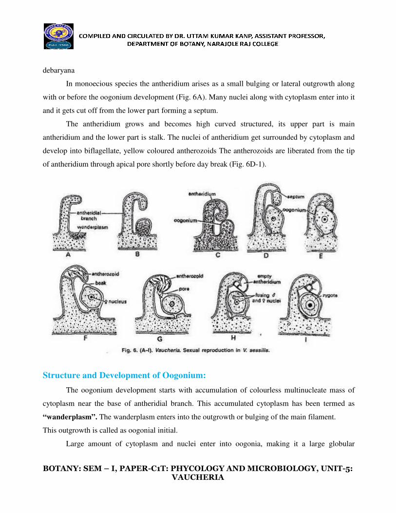
In monoecious species the antheridium arises as a small bulging or lateral
with or before the oogonium development (Fig. 6A). Many
and it gets cut off from the lower part forming a septum.
The antheridium grows
antheridium and the lower part is
develop into biflagellate, yellow c
of antheridium through apical pore
Structure and Development of Oogonium:
The oogonium development starts with accumulation of
cytoplasm near the base of antheridial branch. This
“wanderplasm”. The wanderplasm enters into the outgrowth or bulging of the main filament.
This outgrowth is called as oogonial ini
Large amount of cytoplasm and nuclei enter into oogonia, making it a large
I, PAPER-C1T: PHYCOLOGY AND MICROBIOLOGY, UNIT
VAUCHERIA
In monoecious species the antheridium arises as a small bulging or lateral
with or before the oogonium development (Fig. 6A). Many nuclei along with cytoplasm enter into it
part forming a septum.
and becomes high curved structured, its up
stalk. The nuclei of antheridium get surrounded
coloured antherozoids The antherozoids are libe
e shortly before day break (Fig. 6D-1).
Structure and Development of Oogonium:
The oogonium development starts with accumulation of colourless multinucleate mass of
cytoplasm near the base of antheridial branch. This accumulated cytoplasm has been termed as
wanderplasm enters into the outgrowth or bulging of the main filament.
This outgrowth is called as oogonial initial.
Large amount of cytoplasm and nuclei enter into oogonia, making it a large
PHYCOLOGY AND MICROBIOLOGY, UNIT-5:
In monoecious species the antheridium arises as a small bulging or lateral outgrowth along
nuclei along with cytoplasm enter into it
pper part is main
d by cytoplasm and
erated from the tip
multinucleate mass of
accumulated cytoplasm has been termed as
wanderplasm enters into the outgrowth or bulging of the main filament.
Large amount of cytoplasm and nuclei enter into oogonia, making it a large globular

BOTANY: SEM – I, PAPER-
structure called as oogonium (Fig. 6 B
branch by the development of septum
The nucleus of oogonium with protoplasm develops into a single egg.
The mature oogonia are globose, obovoid, hemispherical or pyriform in
may be sessile or stalked structure. The protoplast of
septum formation.
The entire protoplasm with single nucleus makes a central spherical mass
or ovum. In mature oogonium a distinct vertical or
beak develops a colourless receptive spot. A pore develops just opposite to receptive spot (Fig. 6 F).
Fertilization:
The oogonium secretes a gelatinous drop through a pore near the beak. A
liberated antherozoids stick to the drop. Many
antherozoids strike violently, fall
enters into the oogonium.
After its entry the membrane develops at the pore to stop the further entry
The male nucleus increases in size and fuses with the egg
zygote secretes a thick 3-7 layered wall
chromatophores degenerate and lie in the centre of the cell.
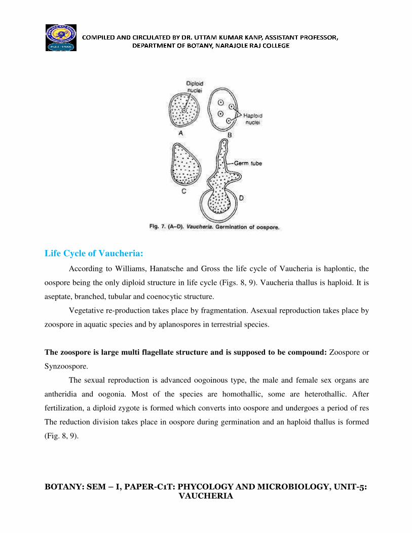
Germination of oospore:
The oospore undergoes a period of rest before germination. During
oogonial wall disintegrates and the oospore is
filaments.
Although the exact stage at which the reduction
clear, it is believed that reduction division occurs in first
oospore (Fig. 7 A-D). The oospore
I, PAPER-C1T: PHYCOLOGY AND MICROBIOLOGY, UNIT
VAUCHERIA
structure called as oogonium (Fig. 6 B-E). As the oogonium matures, it gets separated from main
branch by the development of septum at its base. The mature oogonium is uninucleate structure.
oogonium with protoplasm develops into a single egg.
The mature oogonia are globose, obovoid, hemispherical or pyriform in shape. The oogonia
may be sessile or stalked structure. The protoplast of oogonium is separated from main filament by
The entire protoplasm with single nucleus makes a central spherical mass
or ovum. In mature oogonium a distinct vertical or oblique beak develops in apical part. Opposite to
receptive spot. A pore develops just opposite to receptive spot (Fig. 6 F).
The oogonium secretes a gelatinous drop through a pore near the beak. A
liberated antherozoids stick to the drop. Many antherozoids push into the oogonium. The
back and push forward again and fall back. Only one antherozoid
After its entry the membrane develops at the pore to stop the further entry
e nucleus increases in size and fuses with the egg nucleus to make diploid zygote. The
7 layered wall and is now called as oospore (Fig. 6 G
and lie in the centre of the cell.
The oospore undergoes a period of rest before germination. During favourable season the
oogonial wall disintegrates and the oospore is liberated. The oospore germinates directly into new
Although the exact stage at which the reduction division takes place in
clear, it is believed that reduction division occurs in first nuclear division in the germinating
D). The oospore germinates to make haploid thallus of Vaucheria.
PHYCOLOGY AND MICROBIOLOGY, UNIT-5:
matures, it gets separated from main
is uninucleate structure.
shape. The oogonia
separated from main filament by-
called as oosphere
oblique beak develops in apical part. Opposite to
receptive spot. A pore develops just opposite to receptive spot (Fig. 6 F).
The oogonium secretes a gelatinous drop through a pore near the beak. A large number of
push into the oogonium. The
back and push forward again and fall back. Only one antherozoid
After its entry the membrane develops at the pore to stop the further entry of antherozoids.
nucleus to make diploid zygote. The
and is now called as oospore (Fig. 6 G-I). The
favourable season the
liberated. The oospore germinates directly into new
division takes place in Vaucheria is not
nuclear division in the germinating
germinates to make haploid thallus of Vaucheria.
BOTANY: SEM – I, PAPER-
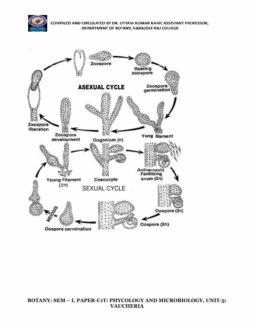
Life Cycle of Vaucheria:
According to Williams, Hanatsche and Gross the life cycle of Vaucheria is
oospore being the only diploid structure in life cycle (Figs. 8,
aseptate, branched, tubular and coenocytic structure.
Vegetative re-production takes place by fragmentation. Asexual
zoospore in aquatic species and by
The zoospore is large multi flagellate structure and is supposed
Synzoospore.
The sexual reproduction is advanced oogoinous type, the male and female
antheridia and oogonia. Most of the species are homothallic,
fertilization, a diploid zygote is formed which
The reduction division takes place in oospore during germination and an haploid thallus is formed
(Fig. 8, 9).
I, PAPER-C1T: PHYCOLOGY AND MICROBIOLOGY, UNIT
VAUCHERIA
ing to Williams, Hanatsche and Gross the life cycle of Vaucheria is
oospore being the only diploid structure in life cycle (Figs. 8, 9). Vaucheria thallus is haploid. It is
coenocytic structure.
production takes place by fragmentation. Asexual reproduction takes place by
zoospore in aquatic species and by aplanospores in terrestrial species.
The zoospore is large multi flagellate structure and is supposed to be compound:
The sexual reproduction is advanced oogoinous type, the male and female
antheridia and oogonia. Most of the species are homothallic, some are heterothallic. After
fertilization, a diploid zygote is formed which converts into oospore and undergoes a period of res
takes place in oospore during germination and an haploid thallus is formed
PHYCOLOGY AND MICROBIOLOGY, UNIT-5:
ing to Williams, Hanatsche and Gross the life cycle of Vaucheria is haplontic, the
9). Vaucheria thallus is haploid. It is
reproduction takes place by
to be compound: Zoospore or
The sexual reproduction is advanced oogoinous type, the male and female sex organs are
some are heterothallic. After
dergoes a period of res
takes place in oospore during germination and an haploid thallus is formed
BOTANY: SEM – I, PAPER-
ASEXUAL CYCLE
SEXUAL CYCLE
I, PAPER-C1T: PHYCOLOGY AND MICROBIOLOGY, UNIT
VAUCHERIA
ASEXUAL CYCLE
SEXUAL CYCLE
PHYCOLOGY AND MICROBIOLOGY, UNIT-5:

BOTANY: SEM – I, PAPER-
REFERENCES:
1. Studies in Botany by Mitra, Mitra and Chowdhury (2005) 7
2. Botany for Degree Students Part
3. https://www.biologydiscussion.com/algae/life
This information, including the figures, are collected from the
solely for academic purpose.
I, PAPER-C1T: PHYCOLOGY AND MICROBIOLOGY, UNIT
VAUCHERIA
Studies in Botany by Mitra, Mitra and Chowdhury (2005) 7th
Edition, Vol. I.
Botany for Degree Students Part-I ALGAE by Vashishta, Sinha and Singh (2002)
www.biologydiscussion.com/algae/life-cycle-algae/vaucheria-occurrence-
This information, including the figures, are collected from the above references and will be used
PHYCOLOGY AND MICROBIOLOGY, UNIT-5:
, Vol. I.
by Vashishta, Sinha and Singh (2002).
-reproduction-
above references and will be used