lab manual fm sem i - d.a.v college jalandhardavjalandhar.com/dbt/botany/sop-lab manual/bsc....
TRANSCRIPT
Department of Botany, DAV College, Jalandhar (PB.)
1
BOTANY
Lab Manual
BSc.-I Medical Semester I
Department of Botany, DAV College, Jalandhar (PB.)
2
Syllabus
Botany I-A
Botany I-B
Department of Botany, DAV College, Jalandhar (PB.)
3
1. Algae
1.1. General Information
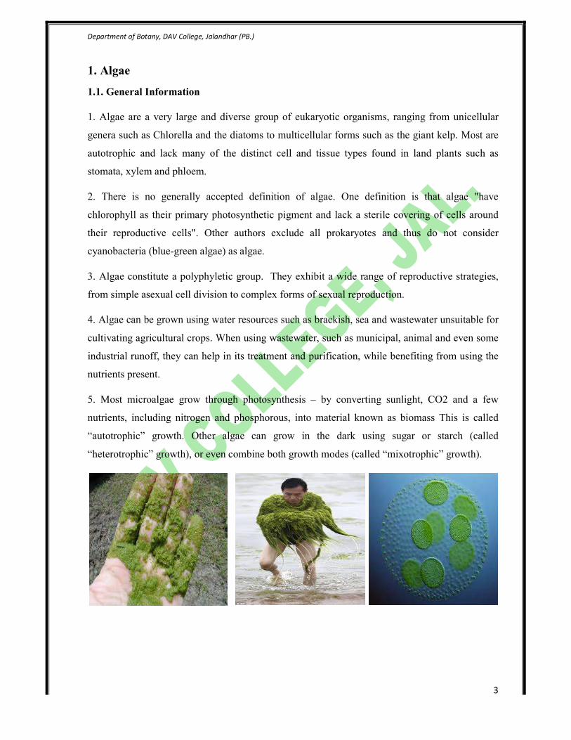
1. Algae are a very large and diverse group of eukaryotic organisms, ranging from unicellular
genera such as Chlorella and the diatoms to multicellular forms such as the giant kelp. Most are
autotrophic and lack many of the distinct cell and tissue types found in land plants such as
stomata, xylem and phloem.
2. There is no generally accepted definition of algae. One definition is that algae "have
chlorophyll as their primary photosynthetic pigment and lack a sterile covering of cells around
their reproductive cells". Other authors exclude all prokaryotes and thus do not consider
cyanobacteria (blue-green algae) as algae.
3. Algae constitute a polyphyletic group. They exhibit a wide range of reproductive strategies,
from simple asexual cell division to complex forms of sexual reproduction.
4. Algae can be grown using water resources such as brackish, sea and wastewater unsuitable for
cultivating agricultural crops. When using wastewater, such as municipal, animal and even some
industrial runoff, they can help in its treatment and purification, while benefiting from using the
nutrients present.
5. Most microalgae grow through photosynthesis – by converting sunlight, CO2 and a few
nutrients, including nitrogen and phosphorous, into material known as biomass This is called
“autotrophic” growth. Other algae can grow in the dark using sugar or starch (called
“heterotrophic” growth), or even combine both growth modes (called “mixotrophic” growth).

Department of Botany, DAV College, Jalandhar (PB.)
4
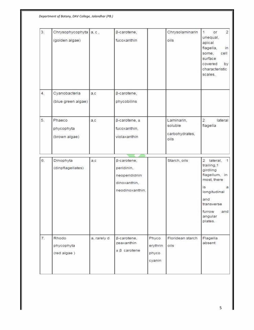
1.2. Algae Classification
The classification of algae into taxonomic groups is based upon the same rules that are used for
the classification of land plants, but the organization of groups of algae above the order level has
changed substantially since 1960. Research using electron microscopes has demonstrated
differences in features, such as the flagellar apparatus, cell division process, and organelle
structure and function that are important in the classification of algae. Similarities and
differences among algal, fungal, and protozoan groups have led scientists to propose major
taxonomic changes, and these changes are continuing.
The classes are distinguished by the structure of flagellate cells (e.g., scales, angle of flagellar
insertion, microtubular roots, and striated roots), the nuclear division process (mitosis), the
cytoplasmic division process (cytokinesis), and the cell covering. Many scientists combine the
Micromonadophyceae with the Pleurastrophyceae, naming the combined group the
Prasinophyceae. “Phylum” and “division” represent the same level of organization; the former is
the zoological term, the latter is the botanical term.
Department of Botany, DAV College, Jalandhar (PB.)
5

Department of Botany, DAV College, Jalandhar (PB.)
6
Division - Chlorophyta
The green algae are a diverse group of eukaryotic organisms classified in the phylum
Chlorophyta. They are considered eukaryotic because individual cells possess a prominent
structural feature known as a nucleus, which houses the chemicals responsible for heredity and
metabolic regulation. The phylum is one of several algal phyla in the kingdom Protista, where
algae are grouped based upon pigmentation, carbohydrate storage reserves, and cell wall
composition. Green algae are found in moist soils and fresh-water and saltwater habitats; most
are believed to be freshwater-dwelling. The phylum consists of at least eight thousand species.
Some estimates place this number at seventeen thousand species. Several shared characteristics
support the hypothesis that these organisms and terrestrial plants derived from a common
ancestor.
Members to be studied:
1. Volvox
2. Coleochaete
3. Oedogonium
Department of Botany, DAV College, Jalandhar (PB.)
7
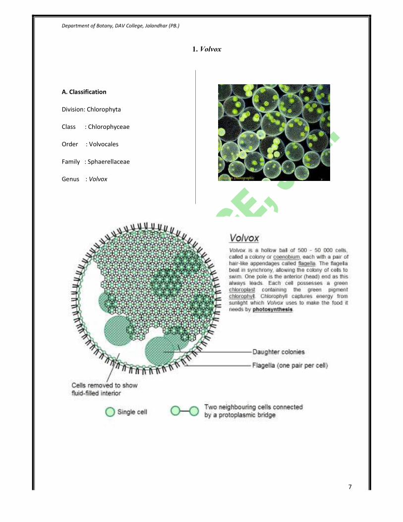
1. Volvox
A. Classification
Division: Chlorophyta
Class : Chlorophyceae
Order : Volvocales
Family : Sphaerellaceae
Genus : Volvox

Department of Botany, DAV College, Jalandhar (PB.)
8
Specimen to be studied:
Volvox Coenobium
- Each mature Volvox colony is composed of
numerous flagellate cells similar to
Chlamydomonas, up to 50,000 embedded in the
surface of a hollow sphere or coenobium.
- The vegetative cells comprise a single layer with
the flagella facing outward. The cells swim in a
coordinated fashion, with distinct anterior and
posterior poles. The cells have eyespots, more
developed near the anterior, which enable the
colony to swim towards light. - The individual algae
in some species are interconnected by thin strands
of cytoplasm, called protoplasmates.
Dughter Colonies
- Once the young coenobium attains maturity, a
few cells in the posterior half are pushed back into
the hollow cavity. These cells withdraw their
flagella and increase in size and become round in
shape. These are called Gonidia.
- The protoplasm of each gonidia divides by
successive longitudinal and horizontal divisions
and form daughter coenobium.
Department of Botany, DAV College, Jalandhar (PB.)
9
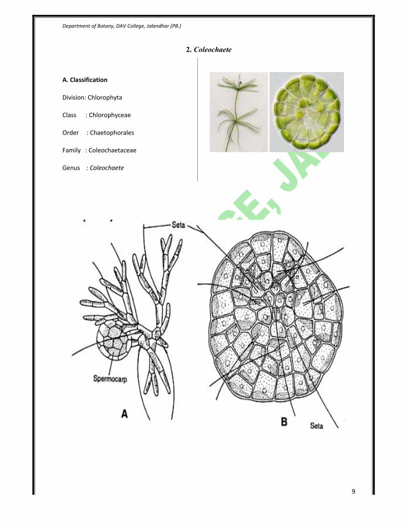
2. Coleochaete
A. Classification
Division: Chlorophyta
Class : Chlorophyceae
Order : Chaetophorales
Family : Coleochaetaceae
Genus : Coleochaete

Department of Botany, DAV College, Jalandhar (PB.)
10
Specimen to be studied:
Coleochaete Heterotrichous Habit
Coleochaete spermocarp
- After fertilization, the zygote which remains
inside the oogonium, increases in size and
secreates a thick wall around itself to become a
oospore.
- At the same time there is an up growth of
branches from the cell below the oospore and
from the neighbouring cells to form a
parenchymatous layer that completely encloses
the oogonoium. It later on becomes reddish brown
and is termed spermocarp.

Department of Botany, DAV College, Jalandhar (PB.)
11
3. Oedogonium
A. Classification
Division: Chlorophyta
Class : Chlorophyceae
Order : Oedogoniales
Family : Oedogoniaceae
Genus : Oedogonium

Department of Botany, DAV College, Jalandhar (PB.)
12
Specimen to be studied:
Oedogonium Filament
Oedogonium Oogonium
Department of Botany, DAV College, Jalandhar (PB.)
13
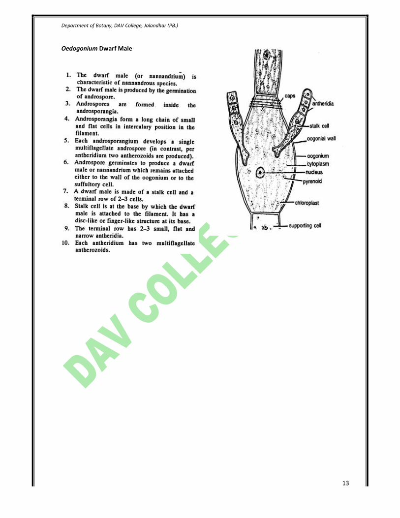
Oedogonium Dwarf Male
Department of Botany, DAV College, Jalandhar (PB.)
14
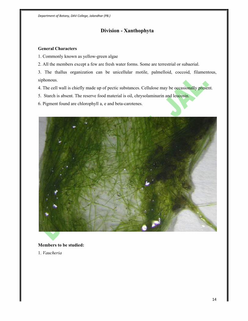
Division - Xanthophyta
General Characters
1. Commonly known as yellow-green algae
2. All the members except a few are fresh water forms. Some are terrestrial or subaerial.
3. The thallus organization can be unicellular motile, palmelloid, coccoid, filamentous,
siphonous.
4. The cell wall is chiefly made up of pectic substances. Cellulose may be occasionally present.
5. Starch is absent. The reserve food material is oil, chrysolaminarin and leucosin.
6. Pigment found are chlorophyll a, e and beta-carotenes.
Members to be studied:
1. Vaucheria

Department of Botany, DAV College, Jalandhar (PB.)
15
1. Vaucheria
A. Classification
Division: Xanthophyta
Class : Xanthophyceae
Order : Heterosiphonales
Family : Vaucheriaceae
Genus : Vaucheria
Vaucheria life cycle

Department of Botany, DAV College, Jalandhar (PB.)
16
Vaucheria Thallus
- Thalli are multinucleate and known as
coenocytic.
- The cytoplasm contains numerous small, discoid
chromatophores.
- The pyrenoids are absent.
- The chromatophores are arranged in the
peripheral layer of the cytoplasm.
- A large number of nuclei ar arranged next to
chromatophores towards central vacuole.
Vaucheria sessile sex organs
- The sessile organs are directly formed on the
main filament
- The antheridia and oogonia are born close to
each other, but they are sessile.
- Antheridia are terminal. These are strongly
curved, hook like and cylindrical.
- Antheridia are cut off from the main filament by
a transverse septum at its base.
Vaucheria geminata sex organs
- The sex organs are borne on certain special
branches.
- These branches are short and bear terminal
antheridium and lateral group of oogonia.
- Antheridial development begins slightly earlier
than oogonia.
Department of Botany, DAV College, Jalandhar (PB.)
17
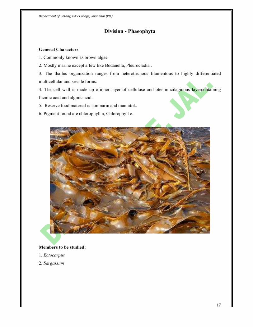
Division - Phaeophyta
General Characters
1. Commonly known as brown algae
2. Mostly marine except a few like Bodanella, Pleurocladia..
3. The thallus organization ranges from heterotrichous filamentous to highly differentiated
multicellular and sessile forms.
4. The cell wall is made up ofinner layer of cellulose and oter mucilaginous layercontaining
fucinic acid and alginic acid.
5. Reserve food material is laminarin and mannitol..
6. Pigment found are chlorophyll a, Chlorophyll c.
Members to be studied:
1. Ectocarpus
2. Sargassum
Department of Botany, DAV College, Jalandhar (PB.)
18
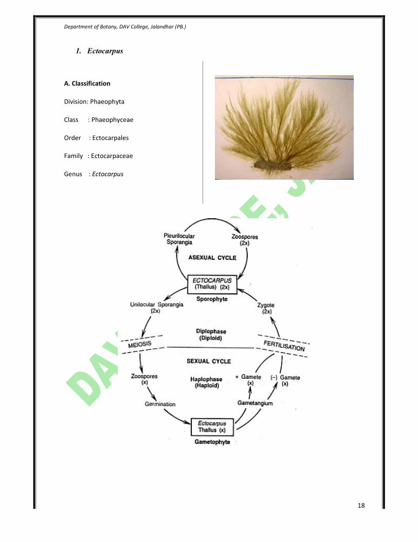
1. Ectocarpus
A. Classification
Division: Phaeophyta
Class : Phaeophyceae
Order : Ectocarpales
Family : Ectocarpaceae
Genus : Ectocarpus
Department of Botany, DAV College, Jalandhar (PB.)
19
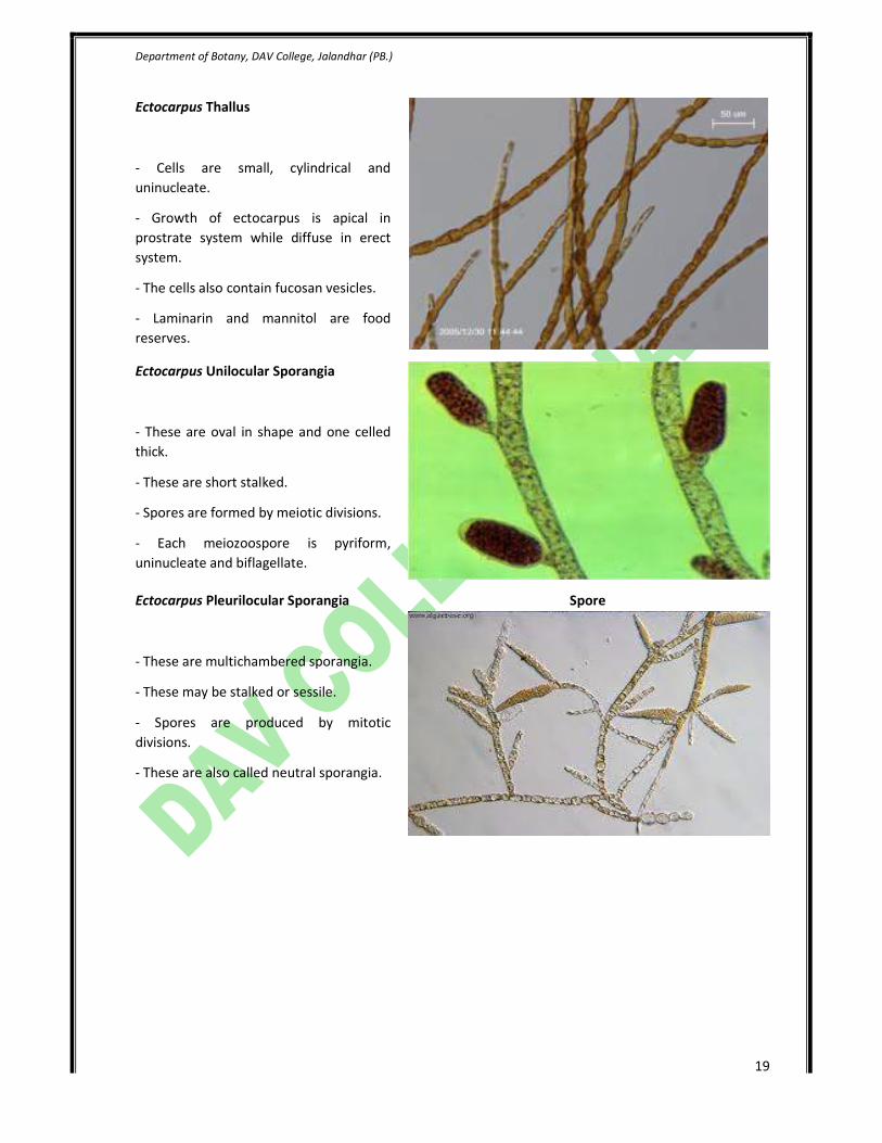
Ectocarpus Thallus
- Cells are small, cylindrical and
uninucleate.
- Growth of ectocarpus is apical in
prostrate system while diffuse in erect
system.
- The cells also contain fucosan vesicles.
- Laminarin and mannitol are food
reserves.
Ectocarpus Unilocular Sporangia
- These are oval in shape and one celled
thick.
- These are short stalked.
- Spores are formed by meiotic divisions.
- Each meiozoospore is pyriform,
uninucleate and biflagellate.
Ectocarpus Pleurilocular Sporangia
- These are multichambered sporangia.
- These may be stalked or sessile.
- Spores are produced by mitotic
divisions.
- These are also called neutral sporangia.
Spore
Department of Botany, DAV College, Jalandhar (PB.)
20
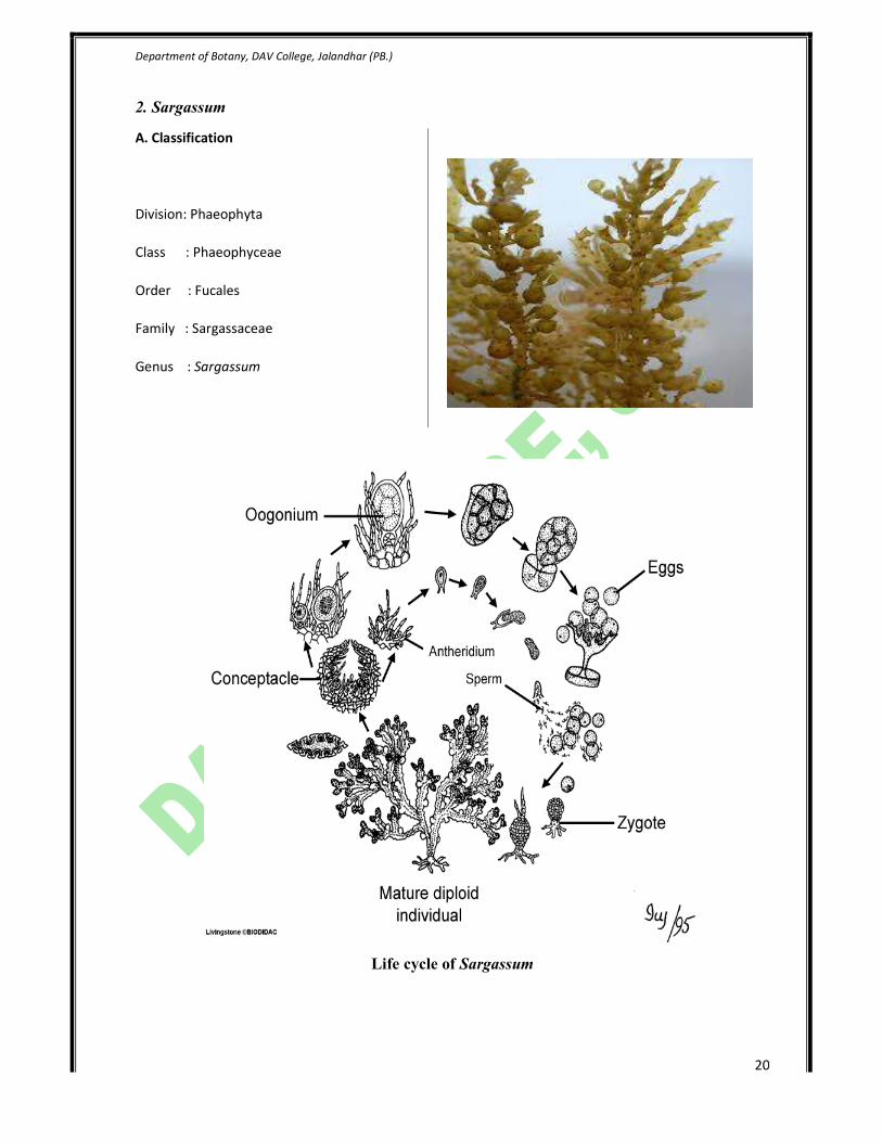
2. Sargassum
A. Classification
Division: Phaeophyta
Class : Phaeophyceae
Order : Fucales
Family : Sargassaceae
Genus : Sargassum
Life cycle of Sargassum

Department of Botany, DAV College, Jalandhar (PB.)
21
Sargasssum vegetative structure
- The plant body is perennial, erect and bushy in
habit.
- There is holdfast and main axis.
- The main axis bear primary laterals or branches
in spiral phyllotaxy.
- Primary laterals bear leaf like branches called
secondary laterals.
- Leaves bear cup shaped sterile cavities called
cryptostomata or sterile conceptacles.
Sargassum male conceptacle
- Conceptacle bearing antheridia are called male
conceptacles.
- Each antheridium is small, oval body.
- It contains 64 biflagellate structures.
- Antheridium are interrupted in between by
sterile filaments called paraphyses.
Sargassum female conceptacle
- Conceptacle bearing antheridia are called male
conceptacles.
- Oogonium is single celled structure, which is
sessile and remains embedded in female
conceptacle.
- At maturity, it is discharged through ostiole.

Department of Botany, DAV College, Jalandhar (PB.)
22
Division – Rhodophyta
General Characters
1. Commonly known as red algae
2. Mostly marine upto a depth of 30-90 meters.
3. The thallus organization ranges from unicellular, palmelloid, uniaxial filamentous branched,
uniaxial pseudoparenchymatous forms, multiaxial filamentous branched forms to multicellular
pseudoparenchymatous thallus.
4. The cell wall is two layered made up of inner layer of pectin and outer layer of cellulose.
5. Reserve food material is floridean starch.
6. Pigment found are chlorophyll a, Chlorophyll d, r-phycoerythrin and r-phycocyanin.
Members to be studied:
1. Polysiphonia

Department of Botany, DAV College, Jalandhar (
1. Polysiphonia
A. Classification
Division: Rhodophyta
Class : Rhodophyceae
Order : Ceramiales
Family : Rhodomelaceae
Genus : Polysiphonia
Department of Botany, DAV College, Jalandhar (PB.)
Polysiphonia life cycle
23

Department of Botany, DAV College, Jalandhar (PB.)
24
Polysiphonia thallus
- The plant body is bushy or feathery appearance.
- It reaches upto 25 cm in length.
- The thallus is multi-axial, filamentous, branched
and heterotrichous.
- Rhizoids are also present to fix plant to
substratum.
Polysiphonia spermatangia
- The male sex organs are called spermatangia
(antheridia).
- These are borne in clusters on special
trichoblasts.
- Pericentral cell divides to form spermatangial
mother cells which in turn give rise to
spermatangia.
Polysiphonia cystocarp
- After fertilization, a fruiting body is formed which
encloses gonimoblast initial, carposporangia and
carpospores.
- It encloses a diploid carpospores.
- It is oval or urn shaped attached to main axis.
- It has an opening called ostiole.
Department of Botany, DAV College, Jalandhar (PB.)
25
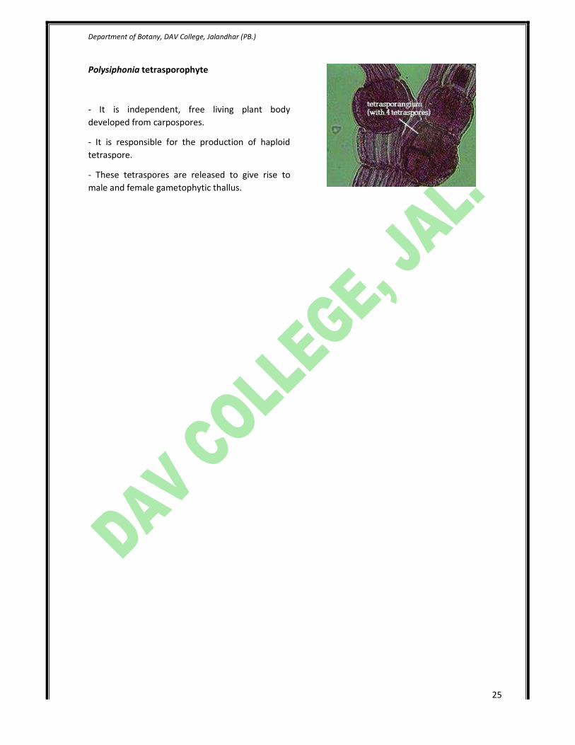
Polysiphonia tetrasporophyte
- It is independent, free living plant body
developed from carpospores.
- It is responsible for the production of haploid
tetraspore.
- These tetraspores are released to give rise to
male and female gametophytic thallus.
Department of Botany, DAV College, Jalandhar (PB.)
26
2. Fungi
Fungi are the eukaryotic, achlorophyllous, and unicellular or multicellular organisms, which may
reproduce by asexual and sexual spores.
1. All are eukaryotic - Possess membrane-bound nuclei (containing chromosomes) and a range of
membrane-bound cytoplasmic organelles (e.g. mitochondria, vacuoles, endoplasmic reticulum).
2. Most are filamentous - Composed of individual microscopic filaments called hyphae, which
exhibit apical growth and which branch to form a network of hyphae called a mycelium.
3. Some are unicellular - e.g. yeasts.
4. Protoplasm of a hypha or cell is surrounded by a rigid wall - Composed primarily of chitin and
glucans, although the walls of some species contain cellulose.
5. Many reproduce both sexually and asexually - Both sexual and asexual reproduction often
result in the production of spores.
6. Their nuclei are typically haploid and hyphal compartments are often multinucleate although,
the oomycota and some yeast possess diploid nuclei.
7. All are achlorophyllous - They lack chlorophyll pigments and are incapable of photosynthesis.
8. All are chemoheterotrophic (chemo-organotrophic) - They utilise pre-existing organic sources
of carbon in their environment and the energy from chemical reactions to synthesize the organic
compounds they require for growth and energy.
9. Possess characteristic range of storage compounds - e.g. trehalose, glycogen, sugar alcohols
and lipids.
10. May be free-living or may form intimate relationships with other organisms i.e. may be free-
living, parasitic or mutualistic (symbiotic).

Department of Botany, DAV College, Jalandhar (PB.)
27
2.1. Pythium
Division : Eumycota
Class : Oomycetes
Order : Peronosporales
Family : Pythiaceae
Genus : Pythium
Pythium vegetative structure
- The vegetative plant body consists of long,
filamentous, branched, aseptate and
multinucleate hyphae.
- It shows acellular coenocytic habit.
- Septa appear during differentiation of sex
organs.
- When parasitic mycelium is both intercellular
and intracellular, but no haustoria is produced.
- The hyphal cell wall is made up of cellulose
impregnated with chitin.
Damping off disease
- It is the most common disease of seed beds
and nurseries.
- It causes damping off disease of tobacco,
tomato, mustard, chillies and cress seedlings.
- Damping off disease may be pre-emergence
or post-emergence.
- In pre-emergence, the radical and plumule of
the germinating seed got rot and killed before
its emergence above the ground.
- In post emergence, the basal part of the stem
or hypocotyls of seedlings become soft and
droop and get killed.
Department of Botany, DAV College, Jalandhar (PB.)
28
2.2. Phytophthora
Division : Eumycota
Class : Oomycetes
Order : Peronosporales
Family : Pythiaceae
Genus : Phytophthora
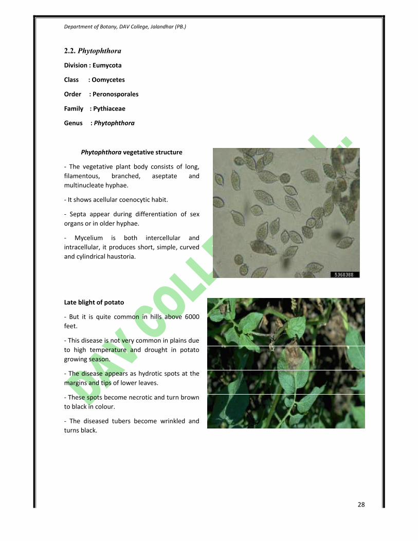
Phytophthora vegetative structure
- The vegetative plant body consists of long,
filamentous, branched, aseptate and
multinucleate hyphae.
- It shows acellular coenocytic habit.
- Septa appear during differentiation of sex
organs or in older hyphae.
- Mycelium is both intercellular and
intracellular, it produces short, simple, curved
and cylindrical haustoria.
Late blight of potato
- But it is quite common in hills above 6000
feet.
- This disease is not very common in plains due
to high temperature and drought in potato
growing season.
- The disease appears as hydrotic spots at the
margins and tips of lower leaves.
- These spots become necrotic and turn brown
to black in colour.
- The diseased tubers become wrinkled and
turns black.
Department of Botany, DAV College, Jalandhar (PB.)
29
2.3. Mucor
Division : Eumycota
Class : Zygomycetes
Order : Mucorales
Family : Mucoraceae
Genus : Mucor
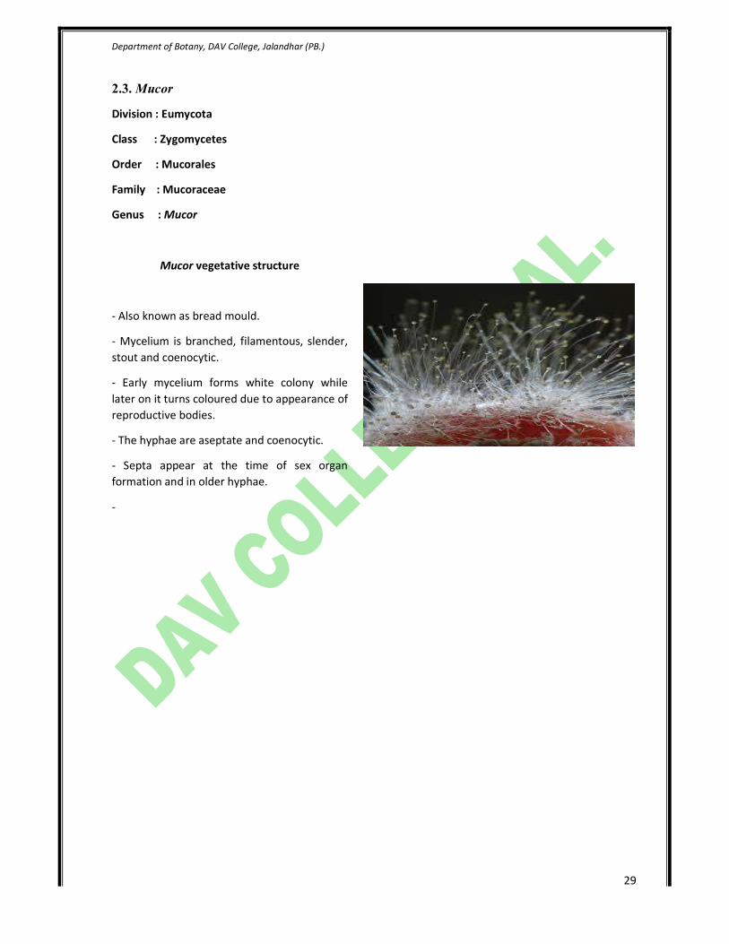
Mucor vegetative structure
- Also known as bread mould.
- Mycelium is branched, filamentous, slender,
stout and coenocytic.
- Early mycelium forms white colony while
later on it turns coloured due to appearance of
reproductive bodies.
- The hyphae are aseptate and coenocytic.
- Septa appear at the time of sex organ
formation and in older hyphae.
-

Department of Botany, DAV College, Jalandhar (PB.)
30
2.4. Saccharomycetes (Yeasts)
Division : Eumycota
Class : Hemiascomycetes
Order : Saccharomycetales
Family : Saccharomycetaceae
Genus : Saccharomycetes
Yeast vegetative structure
- The yeasts are ubiquitous and found almost
everywhere wherever there is sugary
substratum.
- These are unicellular organisms.
- These cells stick in chains and form
pseudomycelium or false colonies.
- They are generally spherical, oval, elliptical
and elongated.
Department of Botany, DAV College, Jalandhar (PB.)
31
2.5. Aspergillus (Eurotium)
Division : Eumycota
Class : Plectomycetes
Order : Eurotiales
Family : Eurotiaceae
Genus : Eurotium
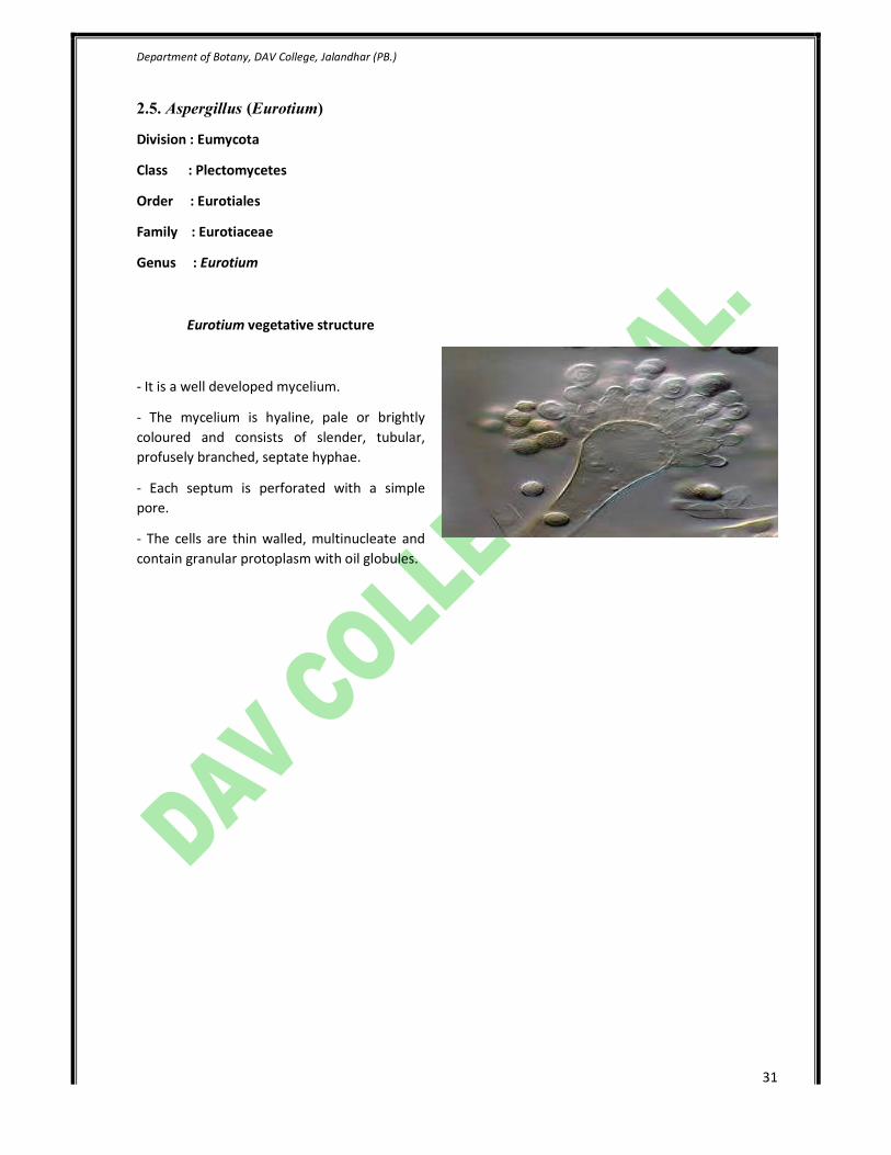
Eurotium vegetative structure
- It is a well developed mycelium.
- The mycelium is hyaline, pale or brightly
coloured and consists of slender, tubular,
profusely branched, septate hyphae.
- Each septum is perforated with a simple
pore.
- The cells are thin walled, multinucleate and
contain granular protoplasm with oil globules.

Department of Botany, DAV College, Jalandhar (PB.)
32
2.6. Penicillium (Green bread mould)
Division : Eumycota
Class : Plectomycetes
Order : Eurotiales
Family : Eurotiaceae
Genus : Penicillium
Penicillium vegetative structure
- It is a well developed mycelium which forms
a compact mass called sclerotium.
- The mycelium is hyaline, pale or brightly
coloured and consists of slender, tubular,
profusely branched, septate hyphae.
- Each septum is perforated with a simple
pore.
- The cells are thin walled, short and uni-bi or
multinucleated.

Department of Botany, DAV College, Jalandhar (PB.)
33
2.7. Chaetomium
Division : Eumycota
Class : Pyrenomycetes
Order : Chaetomiales
Family : Chaetomiaceae
Genus : Chaetomium
Chaetomium vegetative structure
- Mycelium is well developed profusely
branched and septate.
- It yields an antibiotic called chaetomin.
- It reproduces both by asexual and sexual
methods.
- It develops its food from rotten organic
matter.
Department of Botany, DAV College, Jalandhar (PB.)
34
2.7. Peziza (cup fungi)
Division : Eumycota
Class : Discomycetes
Order : Pezizales
Family : Pezizaceae
Genus : Peziza
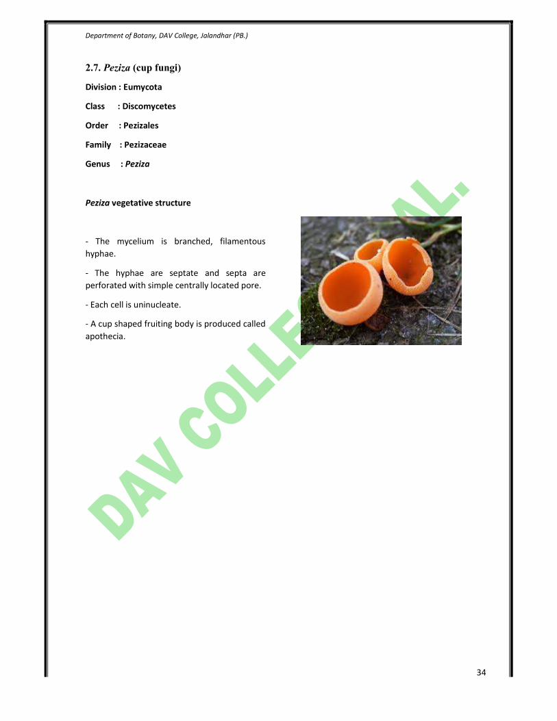
Peziza vegetative structure
- The mycelium is branched, filamentous
hyphae.
- The hyphae are septate and septa are
perforated with simple centrally located pore.
- Each cell is uninucleate.
- A cup shaped fruiting body is produced called
apothecia.
Department of Botany, DAV College, Jalandhar (PB.)
35
2.8. Puccinia
Division : Eumycota
Class : Teliomycetes
Order : Uredinales
Family : Pucciniaceae
Genus : Puccinia
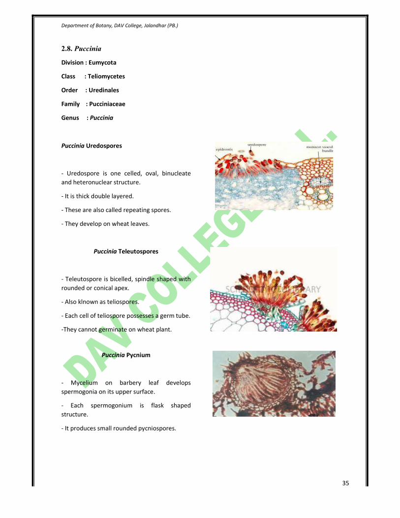
Puccinia Uredospores
- Uredospore is one celled, oval, binucleate
and heteronuclear structure.
- It is thick double layered.
- These are also called repeating spores.
- They develop on wheat leaves.
Puccinia Teleutospores
- Teleutospore is bicelled, spindle shaped with
rounded or conical apex.
- Also klnown as teliospores.
- Each cell of teliospore possesses a germ tube.
-They cannot germinate on wheat plant.
Puccinia Pycnium
- Mycelium on barbery leaf develops
spermogonia on its upper surface.
- Each spermogonium is flask shaped
structure.
- It produces small rounded pycniospores.
Department of Botany, DAV College, Jalandhar (PB.)
36
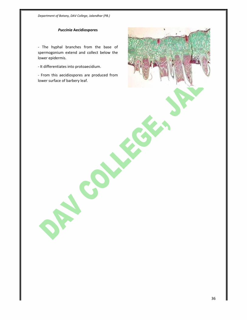
Puccinia Aecidiospores
- The hyphal branches from the base of
spermogonium extend and collect below the
lower epidermis.
- It differentiates into protoaecidium.
- From this aecidiospores are produced from
lower surface of barbery leaf.
Department of Botany, DAV College, Jalandhar (PB.)
37
2.9. Agaricus
Division : Eumycota
Class : Hymenomycetes
Order : Agaricales
Family : Agaricaceae
Genus : Agaricus
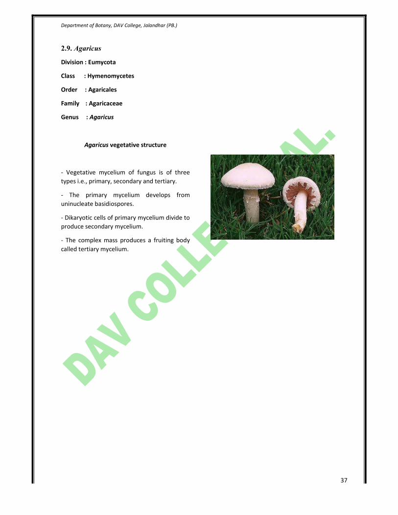
Agaricus vegetative structure
- Vegetative mycelium of fungus is of three
types i.e., primary, secondary and tertiary.
- The primary mycelium develops from
uninucleate basidiospores.
- Dikaryotic cells of primary mycelium divide to
produce secondary mycelium.
- The complex mass produces a fruiting body
called tertiary mycelium.

Department of Botany, DAV College, Jalandhar (PB.)
38
2.10. Cercospora
Division : Eumycota
Class : Hyphomycetes
Order : Moniliales
Family : Dimatiaceae
Genus : Cercospora
Cercospora vegetative structure
- Hyphae are colored branched and septate.
- Hyphae kill the cells they infect.
- Asexual reproduction occurs by conidia.
Tikka disease of Ground nut
- The spots appear as small pale areas on the
surface of older leaves.
- Spots later on turn brown.
- The foliage finally dries up and results
defoliation.
Department of Botany, DAV College, Jalandhar (PB.)
39
2.11. Colletotrichum
Division : Eumycota
Class : Coelomycetes
Order : Melanconiales
Family : Melanconiaceae
Genus : Colletotrichum
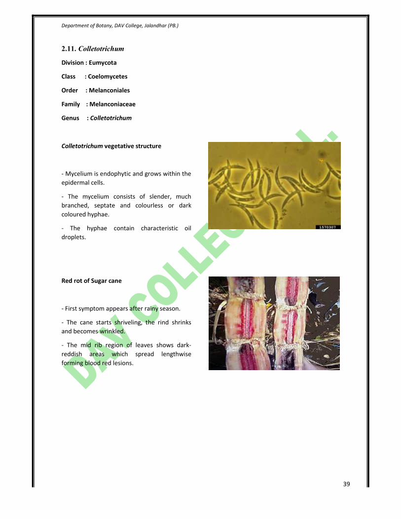
Colletotrichum vegetative structure
- Mycelium is endophytic and grows within the
epidermal cells.
- The mycelium consists of slender, much
branched, septate and colourless or dark
coloured hyphae.
- The hyphae contain characteristic oil
droplets.
Red rot of Sugar cane
- First symptom appears after rainy season.
- The cane starts shriveling, the rind shrinks
and becomes wrinkled.
- The mid rib region of leaves shows dark-
reddish areas which spread lengthwise
forming blood red lesions.

Department of Botany, DAV College, Jalandhar (PB.)
40
3. Bryophytes
The term "bryophyte" comes from Greek βρύον, bryon, "tree-moss, oyster-green" + φυτόν -
phyton "plant".
1. Bryophyte is a traditional name used to refer to all embryophytes (land plants) that do not have
true vascular tissue and are therefore called "non-vascular plants".
2. Some bryophytes do have specialized tissues for the transport of water; however, since these
do not contain lignin, they are not considered to be true vascular tissue.
3. As of 2014, it is uncertain whether bryophytes are a natural ormonophyletic group or a
paraphyletic group, but the name is convenient and remains in use as a collective term for
mosses, hornworts, and liverworts.
4. Bryophytes produce enclosed reproductive structures (gametangia and sporangia), but they
produce neither flowers norseeds, reproducing via spores.
Department of Botany, DAV College, Jalandhar (PB.)
41
3.1. Marchantia
Division : Bryophyta
Class : Hepaticopsida
Order : Marchantiales
Family : Marchantiaceae
Genus : Marchantia
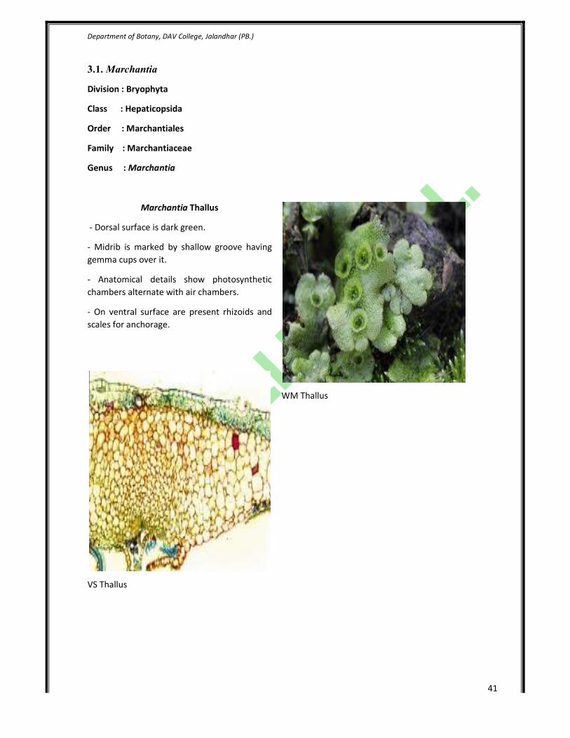
Marchantia Thallus
- Dorsal surface is dark green.
- Midrib is marked by shallow groove having
gemma cups over it.
- Anatomical details show photosynthetic
chambers alternate with air chambers.
- On ventral surface are present rhizoids and
scales for anchorage.
VS Thallus
WM Thallus

Department of Botany, DAV College, Jalandhar (PB.)
42
Gemma Cup
- Gemma cups are found on the dorsal surface
of the thallus.
- They are crescent shaped.
- Mature gemmae are found attached at the
base of gemma cup.
- Gemma are intermingled with mucilage hairs.
- They are means of vegetative reproduction.
Antheridiophore
- Antheridia are produced on special erect
branches called antheridiophores.
- It consists of 1-3 cm long stalk and a lobbed
disc at top.
- Disc is usually 8 lobed.
- Lobed disc is a result of repeated
dichotomies.
- Antheridia arise in the disc in acropetal
succession.
- Each antheridium produces numerous
antherozoids.
Archegoniophore
- Archegonia are produced on special erect
branches called archigoniophores.
- It consists of 1-3 cm long stalk and a lobbed
disc at top.
- It is usually longer than antheridiophore.
- Lobed disc is a result of repeated
dichotomies.
- Archegonia arise in the disc in acropetal
succession.
- Rays alternate with lobes.
Department of Botany, DAV College, Jalandhar (PB.)
43
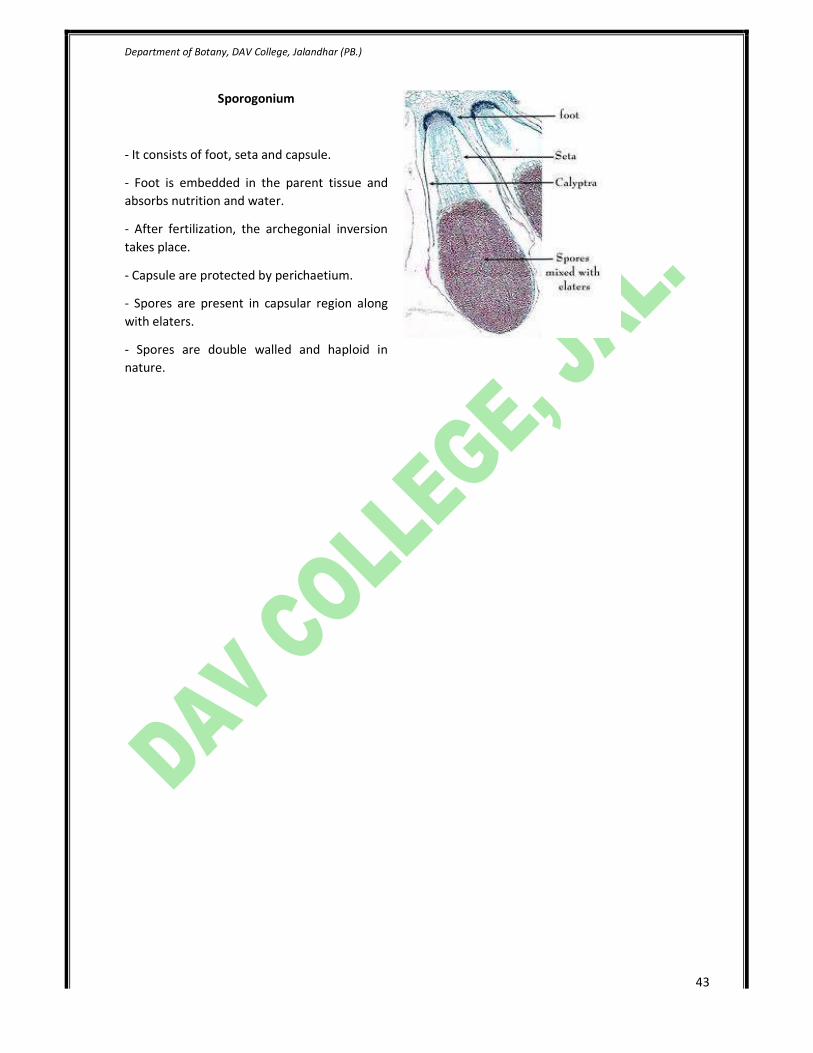
Sporogonium
- It consists of foot, seta and capsule.
- Foot is embedded in the parent tissue and
absorbs nutrition and water.
- After fertilization, the archegonial inversion
takes place.
- Capsule are protected by perichaetium.
- Spores are present in capsular region along
with elaters.
- Spores are double walled and haploid in
nature.

Department of Botany, DAV College, Jalandhar (PB.)
44
3.2. Anthoceros
Division : Bryophyta
Class : Anthocerotopsida
Order : Anthocerotales
Family : Anthocerotaceae
Genus : Anthoceros
Anthoceros Thallus
- Dorsal surface is gren and smooth.
- Mid rib is not distinct.
- Ventral surface bears unicellular, smooth
walled rhizoids.
- Tuberculated rhizoids, scales or mucilaginous
hair are absent.
- Bluish green spots are visible on ventral side.
- These spots contain Nostoc colonies and are
filled with mucilage.
- Nostoc colonies help in nitrogen fixation.
T.S of Anthoceros Thallus
- It lacks particular zonation.
- It is uniformly composed of thin walled
parenchymatous cells.
- The outermost layer is upper epidermis.
- Each cell contains a single large chloroplast.
- Each chloroplast is having a single pyrenoid.
Department of Botany, DAV College, Jalandhar (PB.)
45
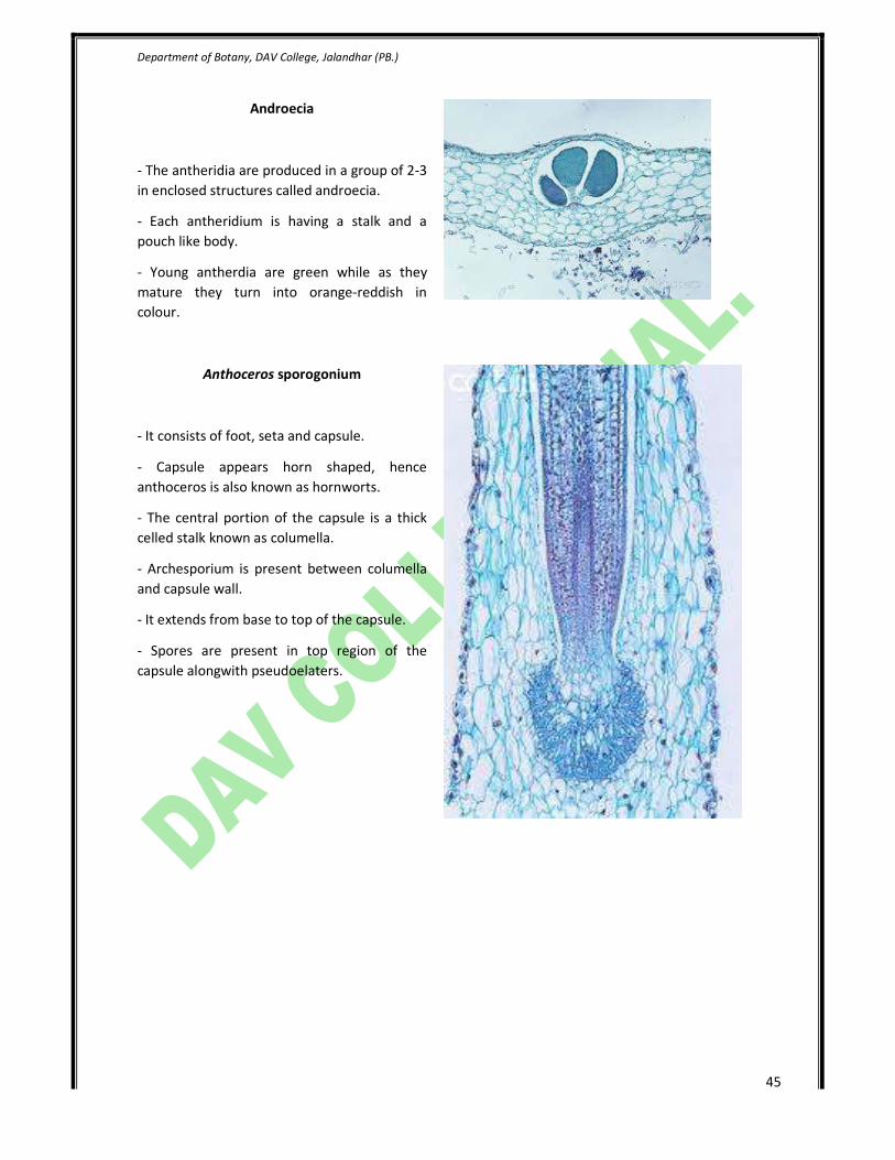
Androecia
- The antheridia are produced in a group of 2-3
in enclosed structures called androecia.
- Each antheridium is having a stalk and a
pouch like body.
- Young antherdia are green while as they
mature they turn into orange-reddish in
colour.
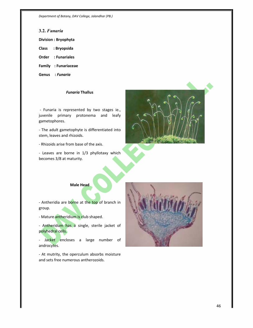
Anthoceros sporogonium
- It consists of foot, seta and capsule.
- Capsule appears horn shaped, hence
anthoceros is also known as hornworts.
- The central portion of the capsule is a thick
celled stalk known as columella.
- Archesporium is present between columella
and capsule wall.
- It extends from base to top of the capsule.
- Spores are present in top region of the
capsule alongwith pseudoelaters.
Department of Botany, DAV College, Jalandhar (PB.)
46
3.2. Funaria
Division : Bryophyta
Class : Bryopsida
Order : Funariales
Family : Funariaceae
Genus : Funaria
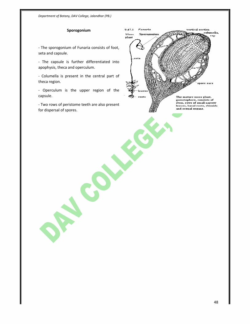
Funaria Thallus
- Funaria is represented by two stages ie.,
juvenile primary protonema and leafy
gametophores.
- The adult gametophyte is differentiated into
stem, leaves and rhizoids.
- Rhizoids arise from base of the axis.
- Leaves are borne in 1/3 phyllotaxy which
becomes 3/8 at maturity.
Male Head
- Antheridia are borne at the top of branch in
group.
- Mature antheridium is club shaped.
- Antheridum has a single, sterile jacket of
polyhedral cells.
- Jacket encloses a large number of
androcytes.
- At mutrity, the operculum absorbs moisture
and sets free numerous antherozoids.

Department of Botany, DAV College, Jalandhar (PB.)
47
Female Head
- Archegonia are borne in groups at top of the
archegonial branch.
- It is surrounded by perichaetial leaves.
- Mature archegonium consists of venter, neck
and egg cell.
- Venter wall is two layered and encloses a
venter canal cell and egg cell.
- Venter canal cell is situated below the neck
canal cell.
Peristome teeth
- Two rows of peristome teeth are present in
the operculum region of capsule in
sporogonium of Funaria.
- Inner and outer row of teeth are
distinguishable from each other in
morphological details.
- Outer row of teeth have striations all over it.
- Inner row of teeth are smoother.
Department of Botany, DAV College, Jalandhar (PB.)
48
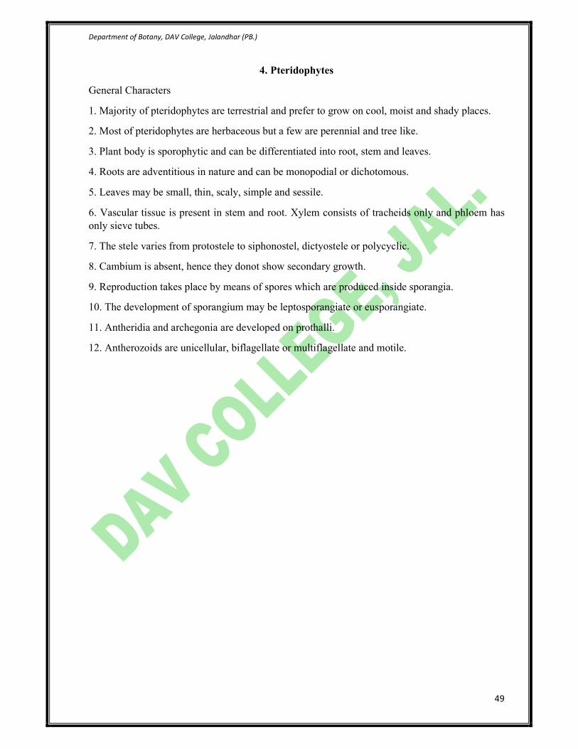
Sporogonium
- The sporogonium of Funaria consists of foot,
seta and capsule.
- The capsule is further differentiated into
apophysis, theca and operculum.
- Columella is present in the central part of
theca region.
- Operculum is the upper region of the
capsule.
- Two rows of peristome teeth are also present
for dispersal of spores.
Department of Botany, DAV College, Jalandhar (PB.)
49
4. Pteridophytes
General Characters
1. Majority of pteridophytes are terrestrial and prefer to grow on cool, moist and shady places.
2. Most of pteridophytes are herbaceous but a few are perennial and tree like.
3. Plant body is sporophytic and can be differentiated into root, stem and leaves.
4. Roots are adventitious in nature and can be monopodial or dichotomous.
5. Leaves may be small, thin, scaly, simple and sessile.
6. Vascular tissue is present in stem and root. Xylem consists of tracheids only and phloem has
only sieve tubes.
7. The stele varies from protostele to siphonostel, dictyostele or polycyclic.
8. Cambium is absent, hence they donot show secondary growth.
9. Reproduction takes place by means of spores which are produced inside sporangia.
10. The development of sporangium may be leptosporangiate or eusporangiate.
11. Antheridia and archegonia are developed on prothalli.
12. Antherozoids are unicellular, biflagellate or multiflagellate and motile.
Department of Botany, DAV College, Jalandhar (PB.)
50
4.1. Lycopodium
Division : Pteridophyta
Class : Eligulopsida
Order : Lycopodiales
Family : Lycopodiaceae
Genus : Lycopodium
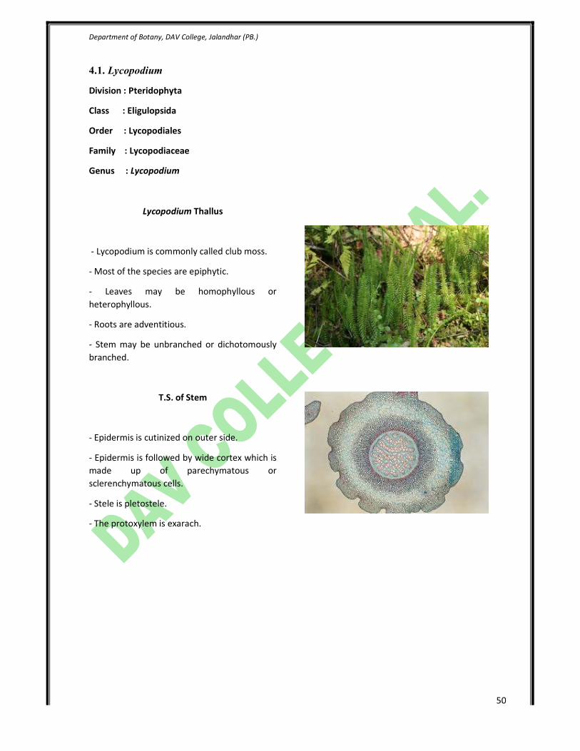
Lycopodium Thallus
- Lycopodium is commonly called club moss.
- Most of the species are epiphytic.
- Leaves may be homophyllous or
heterophyllous.
- Roots are adventitious.
- Stem may be unbranched or dichotomously
branched.
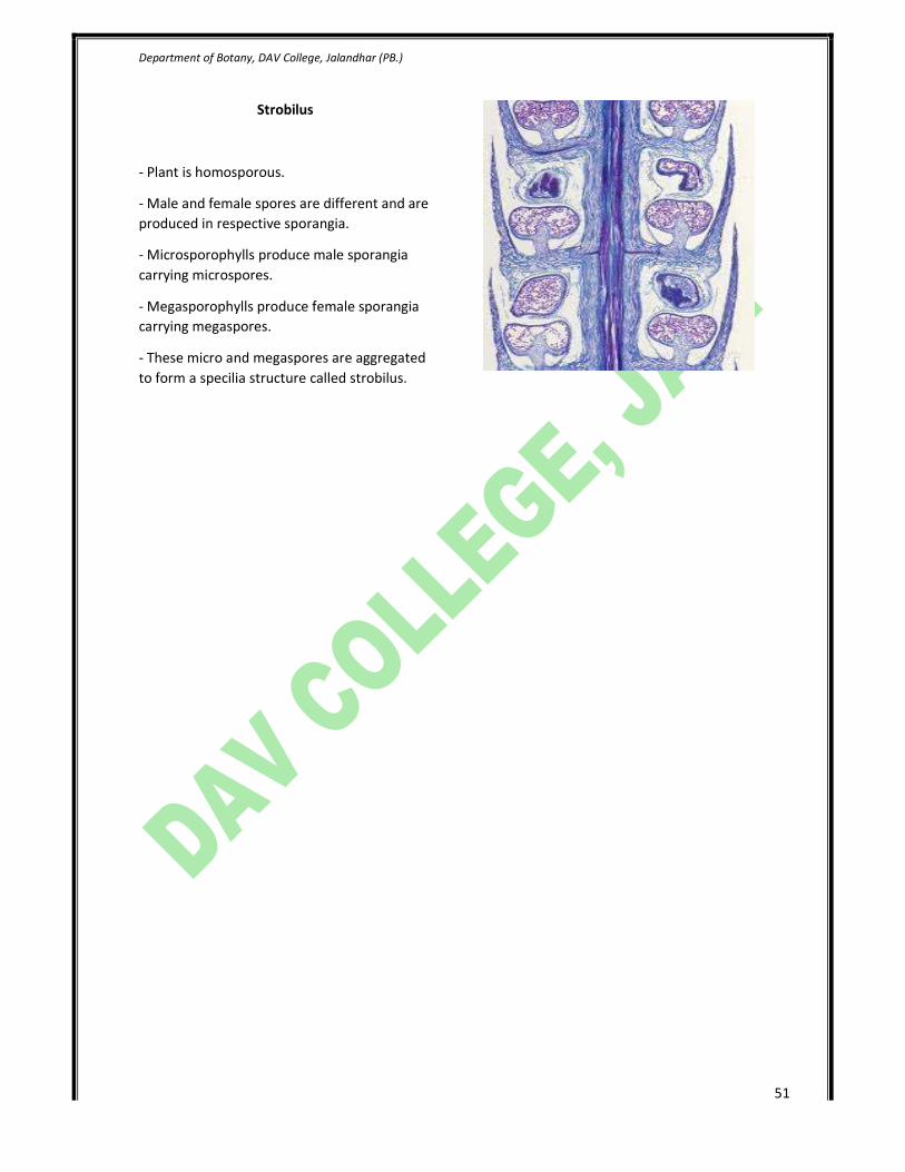
T.S. of Stem
- Epidermis is cutinized on outer side.
- Epidermis is followed by wide cortex which is
made up of parechymatous or
sclerenchymatous cells.
- Stele is pletostele.
- The protoxylem is exarach.
Department of Botany, DAV College, Jalandhar (PB.)
51
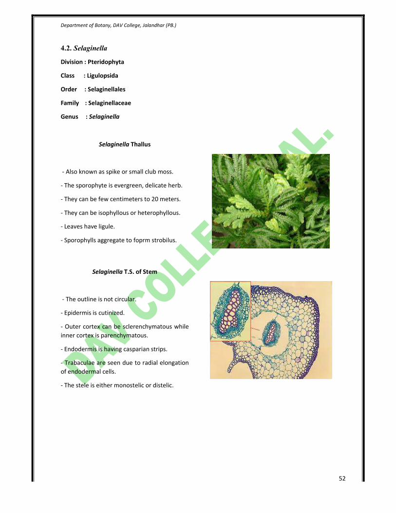
Strobilus
- Plant is homosporous.
- Male and female spores are different and are
produced in respective sporangia.
- Microsporophylls produce male sporangia
carrying microspores.
- Megasporophylls produce female sporangia
carrying megaspores.
- These micro and megaspores are aggregated
to form a specilia structure called strobilus.
Department of Botany, DAV College, Jalandhar (PB.)
52
4.2. Selaginella
Division : Pteridophyta
Class : Ligulopsida
Order : Selaginellales
Family : Selaginellaceae
Genus : Selaginella
Selaginella Thallus
- Also known as spike or small club moss.
- The sporophyte is evergreen, delicate herb.
- They can be few centimeters to 20 meters.
- They can be isophyllous or heterophyllous.
- Leaves have ligule.
- Sporophylls aggregate to foprm strobilus.
Selaginella T.S. of Stem
- The outline is not circular.
- Epidermis is cutinized.
- Outer cortex can be sclerenchymatous while
inner cortex is parenchymatous.
- Endodermis is having casparian strips.
- Trabaculae are seen due to radial elongation
of endodermal cells.
- The stele is either monostelic or distelic.
Department of Botany, DAV College, Jalandhar (PB.)
53
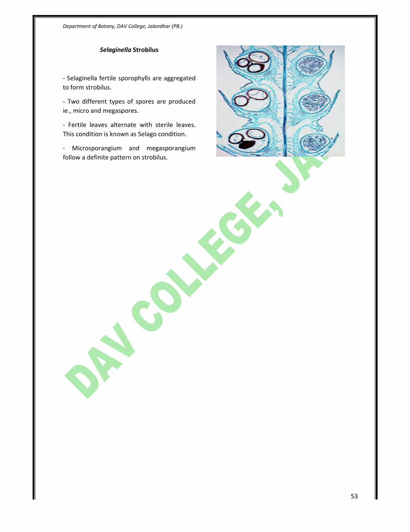
Selaginella Strobilus
- Selaginella fertile sporophylls are aggregated
to form strobilus.
- Two different types of spores are produced
ie., micro and megaspores.
- Fertile leaves alternate with sterile leaves.
This condition is known as Selago condition.
- Microsporangium and megasporangium
follow a definite pattern on strobilus.
Department of Botany, DAV College, Jalandhar (PB.)
54
4.3. Equisetum
Division : Sphenophyta
Class : Calmopsida
Order : Equisetales
Family : Equisetaceae
Genus : Equisetum
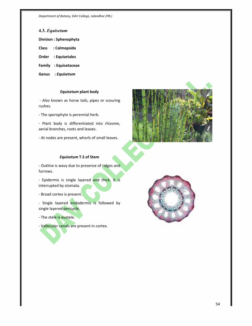
Equisetum plant body
- Also known as horse tails, pipes or scouring
rushes.
- The sporophyte is perennial herb.
- Plant body is differentiated into rhizome,
aerial branches, roots and leaves.
- At nodes are present, whorls of small leaves.
Equisetum T.S of Stem
- Outline is wavy due to presence of ridges and
furrows.
- Epidermis is single layered and thick. It is
interrupted by stomata.
- Broad cortex is present.
- Single layered endodermis is followed by
single layered pericycle.
- The stele is eustele.
- Vallecular canals are present in cortex.

Department of Botany, DAV College, Jalandhar (PB.)
55
Equisetum Strobilus
- Each strobilus consists of thick central axis.
- Around central axis are attached
spororangiophores.
- Sporangiophore is umbrella shaped,
differentiated into slender stalk and expanded
disc.
- Sac like sporangia are present under the disc.
Department of Botany, DAV College, Jalandhar (PB.)
56
4.4. Pteris
Division : Filicophyta
Class : Leptosporangiopsida
Order : Filicales
Family : Polypodiaceae
Genus : Pteris
Pteris Thallus
- The main plant body is sporophytic
differentiated into root, stem and leaves.
- Stem is modified into underground rhizome.
- Roots are adventitious.
- Leaves are macrophyllous. They are pinnately
compound to decompound.
- Each pinna is sessile and traversed by a
central midrib.
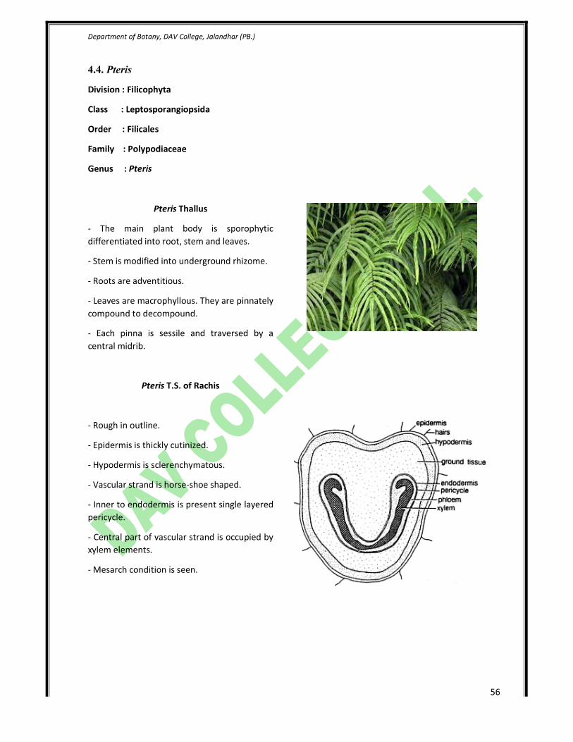
Pteris T.S. of Rachis
- Rough in outline.
- Epidermis is thickly cutinized.
- Hypodermis is sclerenchymatous.
- Vascular strand is horse-shoe shaped.
- Inner to endodermis is present single layered
pericycle.
- Central part of vascular strand is occupied by
xylem elements.
- Mesarch condition is seen.
Department of Botany, DAV College, Jalandhar (PB.)
57
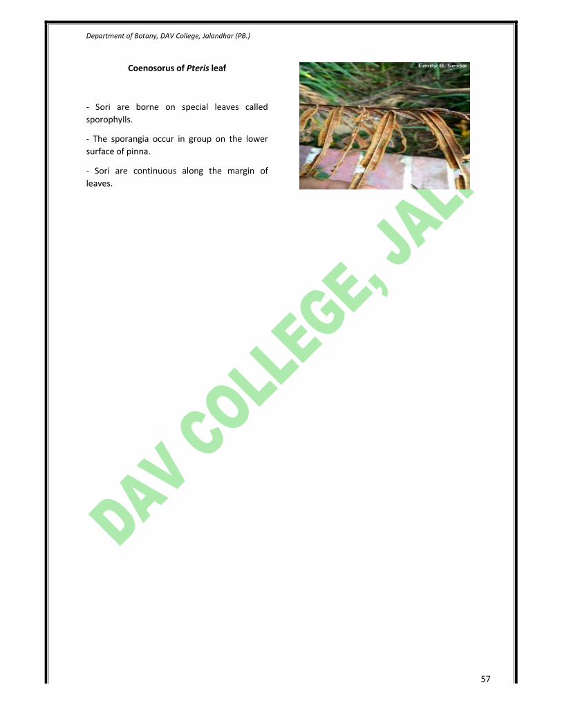
Coenosorus of Pteris leaf
- Sori are borne on special leaves called
sporophylls.
- The sporangia occur in group on the lower
surface of pinna.
- Sori are continuous along the margin of
leaves.

Department of Botany, DAV College, Jalandhar (PB.)
58
4.5. Marsilea
Division : Filicophyta
Class : Leptosporangiopsida
Order : Marsileales
Family : Marsileaceae
Genus : Marsilea
Marsilea plant body
- Also called pepper wort or water fern.
- The mature sporophyte is herbaceous.
- Plant body is differentiated into rhizome,
leaves and root.
- Rhizome creeps on or just below the soil.
- Young leaves show circinate vernation.
- Leaves also show sleeping movement.
- Roots are adventitious.
Marsilea Rhizome
- Epidermis is the outermost limiting layer.
- It is differentiated into outer, middle and
inner cortex.
- Outer cortex is parenchymatous, middle
cortex is aerenchymatous and inner cortex is
parenchymatous.
- Stele is amphiphloic siphonostele.
Department of Botany, DAV College, Jalandhar (PB.)
59
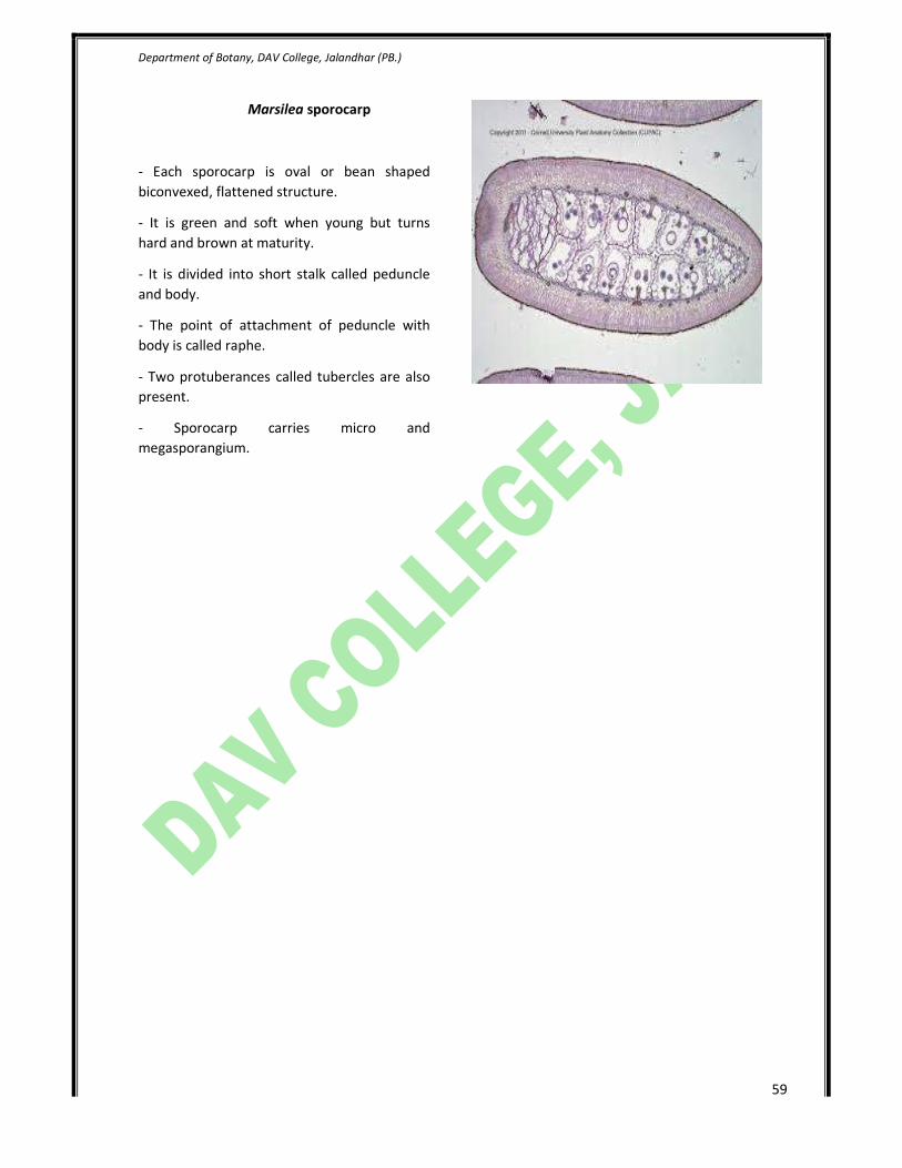
Marsilea sporocarp
- Each sporocarp is oval or bean shaped
biconvexed, flattened structure.
- It is green and soft when young but turns
hard and brown at maturity.
- It is divided into short stalk called peduncle
and body.
- The point of attachment of peduncle with
body is called raphe.
- Two protuberances called tubercles are also
present.
- Sporocarp carries micro and
megasporangium.
Department of Botany, DAV College, Jalandhar (PB.)
60
5. Lichens
Lichen is a composite organism that emerges from algae living among filaments of a fungus in a
mutually beneficial (symbiotic) relationship. It is of following types.
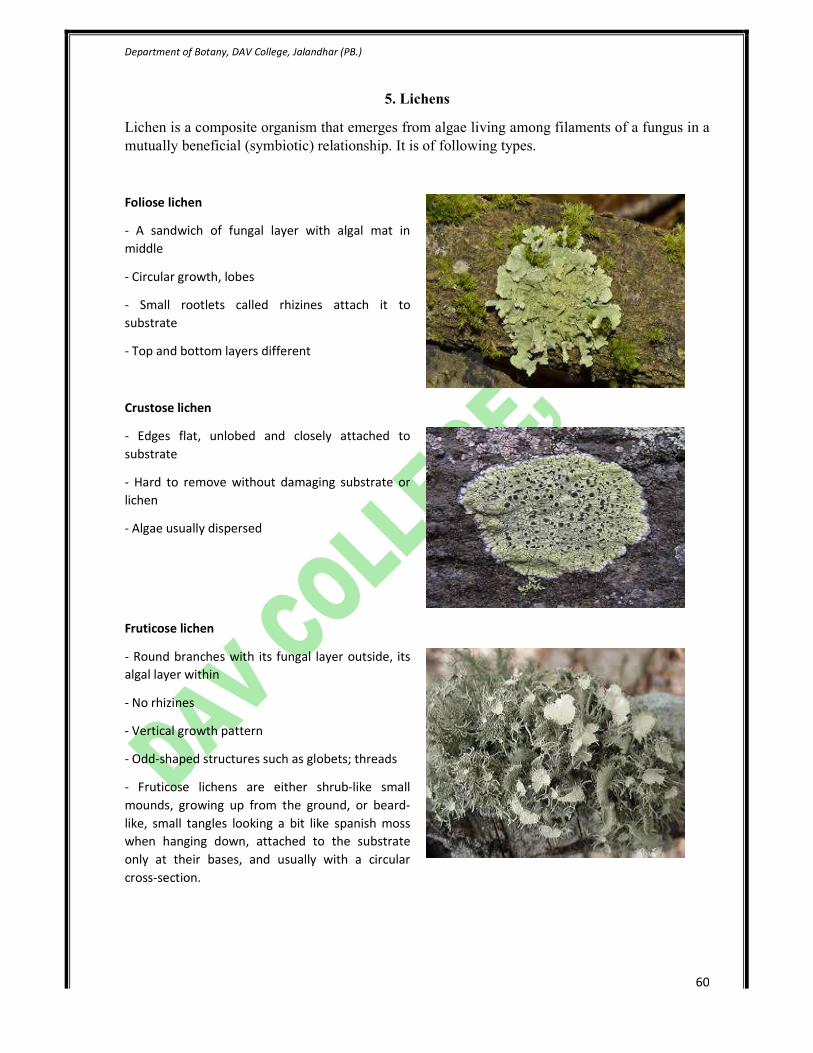
Foliose lichen
- A sandwich of fungal layer with algal mat in
middle
- Circular growth, lobes
- Small rootlets called rhizines attach it to
substrate
- Top and bottom layers different
Crustose lichen
- Edges flat, unlobed and closely attached to
substrate
- Hard to remove without damaging substrate or
lichen
- Algae usually dispersed
Fruticose lichen
- Round branches with its fungal layer outside, its
algal layer within
- No rhizines
- Vertical growth pattern
- Odd-shaped structures such as globets; threads
- Fruticose lichens are either shrub-like small
mounds, growing up from the ground, or beard-
like, small tangles looking a bit like spanish moss
when hanging down, attached to the substrate
only at their bases, and usually with a circular
cross-section.