drug-induced steatohepatitis
TRANSCRIPT
Drug-Induced Steatohepatit is
Vaishali Patel, MBBS, MD, Arun J. Sanyal, MBBS, MD*
KEYWORDS
� Nonalcoholic fatty liver disease � Nonalcoholic steatohepatitis� Microvesicular and macrovesicular steatosis � Drug-induced steatohepatitis
KEY POINTS
� Hepatic steatosis and steatohepatitis can arise from the interplay of several inciting fac-tors, including alcohol, drugs, and metabolic syndrome as nonalcoholic fatty liver disease(NAFLD).
� Drugs induce fat deposition in the liver in microvesicular or macrovesicular distribution.
� Most drugs implicated in steatosis and steatohepatitis can induce both to a variableextent.
� It is difficult to ascertain whether an implicated drug leads to de novo steatosis and/orsteatohepatitis versus worsening of underlying NAFLD.
� Thepathogenesisofdrug-inducedsteatohepatitis often involvesmitochondrial dysfunction.
DEFINITION OF NAFLD
Hepatic steatosis is defined as fat deposition within hepatocytes (Fig. 1). It is seenmicroscopically as vacuoles in a microvesicular or macrovesicular distribution. Micro-vesicular steatosis is characterized by multiple small, fat vesicles distributedthroughout the hepatocyte, whereas macrovesicular steatosis is characterized by alarge droplet of fat within the cytoplasm, which pushes the nucleus to the edge ofthe cell. Although microvesicular steatosis is more commonly seen in Reye syndromeand other forms of mitochondrial injury, nonalcoholic fatty liver disease (NAFLD) typi-cally has macrovesicular distribution of fat deposits. Several drugs can lead to bothforms of hepatic steatosis. Although the triggering events differ, each of these insultscan lead to excessive hepatic fat deposition, increased reactive oxygen species (ROS)formation, mitochondrial dysfunction, and Endoplasmic Reticulum (ER) stress that in-duces inflammation, cell death and eventually leads to fibrosis.
Disclosure: This work has been supported by the NIH T32 Training Grant.Division of Gastroenterology, Hepatology, and Nutrition, Virginia Commonwealth UniversityMedical Center, Virginia Commonwealth University, MCV Box 980341, Richmond, VA23298-0341, USA* Corresponding author.E-mail address: [email protected]
Clin Liver Dis 17 (2013) 533–546http://dx.doi.org/10.1016/j.cld.2013.07.012 liver.theclinics.com1089-3261/13/$ – see front matter � 2013 Elsevier Inc. All rights reserved.

Fig. 1. Histologic appearance of hepatic steatosis and steatohepatitis. (A) Simple steatosis.Vacuoles represent areas of fat accumulation (hematoxylin-eosin). (B) Steatohepatitisdemonstrating ballooning degeneration (arrow) and fat deposition (hematoxylin-eosin).(C) Steatohepatitis with hepatic perisinusoidal fibrosis (Prussian blue staining). Originalmagnification 200x.
Patel & Sanyal534
Traditionally, NAFLD is defined as fat infiltration in the liver parenchyma in peoplewho do not consume alcohol in quantities that are considered to be hepatotoxic.NAFLD is a manifestation of the metabolic syndrome and it is often associated withobesity, dyslipidemia, and type 2 diabetes mellitus.1,2 With the spread of the obesityepidemic, the disease burden of NAFLD is increasing, both in terms of geography andthe age of presentation. It is now the most common cause of chronic liver disease inthe United States, affecting about a third of the population.3,4
There are 2 principal phenotypes of NAFLD: (1) nonalcoholic fatty liver (NAFL) and(2) nonalcoholic steatohepatitis (NASH). NAFL is defined by the presence of steatosiswithout inflammation. NASH is defined by the presence of steatosis, inflammation andhepatocyte ballooning injury.2 NASH progresses to cirrhosis in up to a fifth of patients.Apart from this, several cases of cryptogenic cirrhosis are attributed to NAFLD.2–6
DIAGNOSING DRUG-INDUCED STEATOHEPATITIS
Drug-induced liver injury (DILI) is diagnosed in a person when worsening of the base-line liver function is caused by prescription or nonprescription drugs. The diagnosis ofDILI requires consideration of the following:
1. The biochemical and histologic pattern of liver injury2. Lead time between the initiation of the suspected drug and the onset of liver
disease3. Evidence of improvement of liver function after discontinuation of the drug
The diagnosis of DILI is challenging in many cases because there is no specificmaker for DILI. The literature is sparse for newer agents and herbal products. More-over, the clinical presentation and microscopic appearance of liver injury may benonspecific and rechallenge is not safe. Hence, several scoring systems have beendeveloped to objectively diagnose DILI, such as the Roussel-Uclaf Causality Assess-ment Method (RUCAM), the Maria and Victorino method, and the Naranjo scale.7
RUCAM is the most commonly used instrument for the diagnosis of DILI. Accordingto RUCAM, DILI is most likely when it develops within 90 days of the initiation of thedrug and improves within 15 to 30 days of discontinuation in cases of hepatocellular
Drug-Induced Steatohepatitis 535
and cholestatic patterns of injury, respectively. Drug-induced steatohepatitis mayoccur after many months of use and may not resolve within 15 days.8,9 RUCAM is,thus, suboptimal for the diagnosis of drug-induced steatohepatitis. The situation isfurther complicated by a high prevalence of NAFLD in the general population. Hence,even if the disease is previously undiagnosed, several patients have risk factors asso-ciated with NAFLD, making it difficult to differentiate drug-induced steatohepatitisfrom de novo NAFLD. It is possible that drugs may exacerbate preexistingNAFLD.10,11
PATHOGENESIS OF DRUG-INDUCED STEATOHEPATITISMitochondrial Structure: Link Between b-oxidation of Fatty Acids and Fuel Synthesis
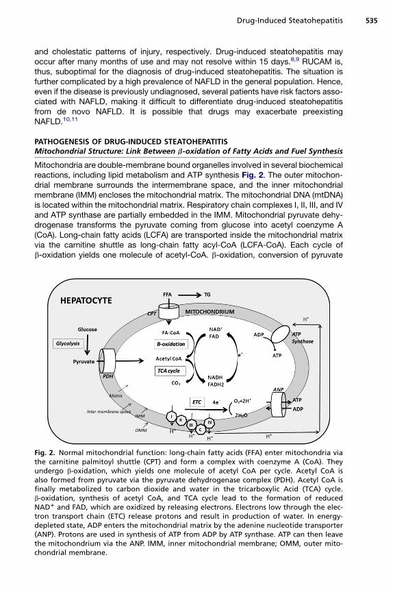
Mitochondria are double-membrane bound organelles involved in several biochemicalreactions, including lipid metabolism and ATP synthesis Fig. 2. The outer mitochon-drial membrane surrounds the intermembrane space, and the inner mitochondrialmembrane (IMM) encloses the mitochondrial matrix. The mitochondrial DNA (mtDNA)is located within the mitochondrial matrix. Respiratory chain complexes I, II, III, and IVand ATP synthase are partially embedded in the IMM. Mitochondrial pyruvate dehy-drogenase transforms the pyruvate coming from glucose into acetyl coenzyme A(CoA). Long-chain fatty acids (LCFA) are transported inside the mitochondrial matrixvia the carnitine shuttle as long-chain fatty acyl-CoA (LCFA-CoA). Each cycle ofb-oxidation yields one molecule of acetyl-CoA. b-oxidation, conversion of pyruvate
Fig. 2. Normal mitochondrial function: long-chain fatty acids (FFA) enter mitochondria viathe carnitine palmitoyl shuttle (CPT) and form a complex with coenzyme A (CoA). Theyundergo b-oxidation, which yields one molecule of acetyl CoA per cycle. Acetyl CoA isalso formed from pyruvate via the pyruvate dehydrogenase complex (PDH). Acetyl CoA isfinally metabolized to carbon dioxide and water in the tricarboxylic Acid (TCA) cycle.b-oxidation, synthesis of acetyl CoA, and TCA cycle lead to the formation of reducedNAD1 and FAD, which are oxidized by releasing electrons. Electrons low through the elec-tron transport chain (ETC) release protons and result in production of water. In energy-depleted state, ADP enters the mitochondrial matrix by the adenine nucleotide transporter(ANP). Protons are used in synthesis of ATP from ADP by ATP synthase. ATP can then leavethe mitochondrium via the ANP. IMM, inner mitochondrial membrane; OMM, outer mito-chondrial membrane.

Patel & Sanyal536
to acetyl CoA by the pyruvate dehydrogenase complex, and metabolism of acetyl-CoA by the tricarboxylic acid cycle produce NADH and FADH2. These moleculesare, in turn, oxidized by transferring their electrons to the mitochondrial respiratorychain. As electrons flow through the electron transport chain (ETC), 3 protons fromthemitochondrial matrix are pushed into the intermembrane space at the level of com-plexes I, III, and IV. This process increases the electrochemical gradient across theIMM. In an energy-depleted state, ADP enters the mitochondrial matrix via the adeninenucleotide translocator (ANT). The increased transmembrane potential allows ADP todrag along protons, which results in the generation of ATP via ATP synthase. ATP canthen leave the mitochondrion via the ANT. These processes are well coordinated andultimately result in the production of oxygen, water, and ATP. However, at the level ofcomplexes I and III, electrons can interact directly with protons and result in the pro-duction of ROS.10,12
Mechanisms of Hepatic Steatosis
Hepatic steatosis in NAFLD results predominantly from a combination of increaseddietary intake, peripheral lipolysis, and de novo fatty acid synthesis Fig. 3.13 Drugsthat lead to steatosis and steatohepatitis primarily interfere with mitochondrial respi-ration, b-oxidation, or both.14 It is important to understand that the ETC and b-oxida-tion of fatty acids are metabolically inter-connected. Hence, drugs affecting onepathway invariably inhibit the other.15 When hepatic mitochondrial b-oxidation isseverely inhibited, the impairment of fatty acyl-CoA b-oxidation increases fatty acyl-CoA and nonesterified fatty acids, which are converted into triglycerides resulting inhepatic steatosis.12 Besides inducing steatosis, the inhibition of b-oxidation andETC results in increased ROS formation and, in more severe cases, hepaticnecrosis.14,16–19
Fig. 3. Mechanisms of hepatic steatosis and injury in drug-induced steatohepatitis. Inhibi-tion of entry of long-chain fatty acids (FFA) via the carnitine palmitoyl shuttle (CPT) andinhibition of b-oxidation lead to increased free fatty acids, which are esterified into triglyc-erides. Transport of triglycerides (TG) as very low-density lipoprotein (VLDL) can be blockedby some drugs. Blocking the flow of electrons through the ETC leads to accumulation ofelectrons. These electrons can directly interact with oxygen to produce ROS. Certain drugsdirectly damage mtDNA and can induce mitochondrial permeability transition (MPT) poreformation.
Drug-Induced Steatohepatitis 537
Drugs that inhibit mitochondrial b-oxidation do so by several mechanisms:
� Inhibition of entry of LCFA into the mitochondrial matrix, which is seen with theantidiabetic drug troglitazone, which inhibits mitochondrial acyl-CoA synthase20
� Sequestration of CoA in the form of a drug-CoA thioester as seen in valproatetoxicity21
� Inhibition of enzymes catalyzing b-oxidation, for example, glucocorticoids inhibitacyl-CoA dehydrogenases10,22
Rarely, other mechanisms that result in increased hepatic fat are involved. Somedrugs can lead to increased synthesis or decreased secretion of hepatic triglyceridesas seen with protease inhibitors and dexamethasone, respectively.23,24 De novo fattyacid synthesis is transiently increased by some antipsychotic medications by an in-crease in the active form of sterol regulatory element binding protein-1c (SREBP-1c).25
Mechanisms of Steatohepatitis
The causes of the progression of steatosis to steatohepatitis in DILI are not well under-stood and mostly extrapolated from NASH literature.10–12 One of the most-studiedhypothesis is that mitochondrial dysfunction leads to and worsens drug-induceduncoupling of b-oxidation and phosphorylation, which results in the production ofROS. Reduced energy availability and direct damage form ROS, in turn, induce hepa-tocyte necrosis.12 ROS cause additional damage by interaction with nonesterifiedpolyunsaturated fatty acids to produce lipid peroxidation products, which have alonger half-life than ROS and cause damage by diffusing to surrounding cells. ROSalso lead to nuclear translocation of the transcription factor, nuclear factor-kb(NF-kb). NF-kb upregulates genes that promote insulin resistance26 and severalinflammatory cytokines, including tumor necrosis factor (TNF)–a25; interleukin (IL)-8,which promotes polymorphonuclear (PMN) infiltration26; and transforming growthfactor (TGF)-b. TGF-b, in turn, stimulates hepatic stellate cell proliferation andfibrosis.16,26 NF-kb also promotes apoptotic cell death by inducing transcriptionalexpression of the normally repressed fatty acid synthetase (FAS)-ligand. FAS ligandbinds to FAS on adjacent hepatocytes, leading to caspase-9 activation. Caspase-9then activates other caspases to promote apoptotic hepatocyte cell death.16
PATHOLOGIC SPECTRUM OF DRUG-INDUCED STEATOSIS AND STEATOHEPATITIS
Drugs implicated in hepatic steatosis can be divided on basis of hepatic microscopyinto those that cause microvesicular steatosis and those that predominantly lead tomacrovesicular steatosis (Table 1). Characteristics and mechanism of injury by indi-vidual drugs are described later.
Drugs Causing Microvesicular Steatosis
AspirinAspirin is metabolized to salicylic acid and then salicyl-CoA. In aspirin poisoning, thisprocess leads to excessive utilization of CoA, thus blocking the entry of LCFA entryinto mitochondria and arresting b-oxidation.27 Aspirin can also directly uncouple respi-ration. It promotes mitochondrial permeability transition pore formation that leads tomitochondrial death, which triggers cell death by apoptosis and necrosis.28 Aspirinuse in children with viral infections has been associated with Reye syndrome. Thepathogenesis of Reye syndrome involves widespread arrest of b-oxidation, increasedureagenesis and ketogenesis, and severe hypoglycemia caused by the inability toconvert lactate to glucose. Diffuse hepatic microvesicular steatosis is seen in this
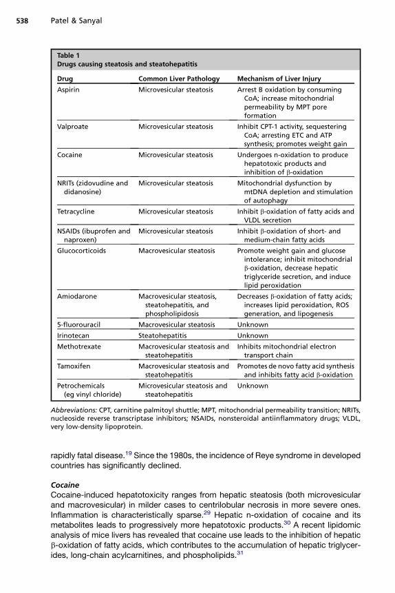
Table 1Drugs causing steatosis and steatohepatitis
Drug Common Liver Pathology Mechanism of Liver Injury
Aspirin Microvesicular steatosis Arrest B oxidation by consumingCoA; increase mitochondrialpermeability by MPT poreformation
Valproate Microvesicular steatosis Inhibit CPT-1 activity, sequesteringCoA; arresting ETC and ATPsynthesis; promotes weight gain
Cocaine Microvesicular steatosis Undergoes n-oxidation to producehepatotoxic products andinhibition of b-oxidation
NRITs (zidovudine anddidanosine)
Microvesicular steatosis Mitochondrial dysfunction bymtDNA depletion and stimulationof autophagy
Tetracycline Microvesicular steatosis Inhibit b-oxidation of fatty acids andVLDL secretion
NSAIDs (ibuprofen andnaproxen)
Microvesicular steatosis Inhibit b-oxidation of short- andmedium-chain fatty acids
Glucocorticoids Macrovesicular steatosis Promote weight gain and glucoseintolerance; inhibit mitochondrialb-oxidation, decrease hepatictriglyceride secretion, and inducelipid peroxidation
Amiodarone Macrovesicular steatosis,steatohepatitis, andphospholipidosis
Decreases b-oxidation of fatty acids;increases lipid peroxidation, ROSgeneration, and lipogenesis
5-fluorouracil Macrovesicular steatosis Unknown
Irinotecan Steatohepatitis Unknown
Methotrexate Macrovesicular steatosis andsteatohepatitis
Inhibits mitochondrial electrontransport chain
Tamoxifen Macrovesicular steatosis andsteatohepatitis
Promotes de novo fatty acid synthesisand inhibits fatty acid b-oxidation
Petrochemicals(eg vinyl chloride)
Microvesicular steatosis andsteatohepatitis
Unknown
Abbreviations: CPT, carnitine palmitoyl shuttle; MPT, mitochondrial permeability transition; NRITs,nucleoside reverse transcriptase inhibitors; NSAIDs, nonsteroidal antiinflammatory drugs; VLDL,very low-density lipoprotein.
Patel & Sanyal538
rapidly fatal disease.19 Since the 1980s, the incidence of Reye syndrome in developedcountries has significantly declined.
CocaineCocaine-induced hepatotoxicity ranges from hepatic steatosis (both microvesicularand macrovesicular) in milder cases to centrilobular necrosis in more severe ones.Inflammation is characteristically sparse.29 Hepatic n-oxidation of cocaine and itsmetabolites leads to progressively more hepatotoxic products.30 A recent lipidomicanalysis of mice livers has revealed that cocaine use leads to the inhibition of hepaticb-oxidation of fatty acids, which contributes to the accumulation of hepatic triglycer-ides, long-chain acylcarnitines, and phospholipids.31

Drug-Induced Steatohepatitis 539
ValproateValproate is a branched-chain fatty acid that causes microvesicular steatosis andcirrhosis. In one report, up to 60% of patients treated with valproate had ultrasoundevidence of hepatic steatosis.32 Valproate is initially metabolized by cytochromeP-450 enzymes to 4-ene-valproate. Both valproate and 4-ene-valproate then formcomplexes with CoA, sequestering CoA as well as competitively inhibiting carnitinepalmitoyl shuttle I activity. Valproate can release protons and, thus, arrest the ETCand ATP synthesis.21 Besides mitochondrial dysfunction, with prolonged use,valproate promotes weight gain and systemic insulin resistance, which may lead toworsening of the underlying NAFLD.32
Nucleoside reverse transcriptase inhibitors, thymidine analoguesAlthough hepatotoxicity can be seen with all groups of HIV antiretroviral therapy,nucleoside reverse transcriptase inhibitors (NRTIs) have been associated with hepaticsteatosis. With prolonged use, thymidine analogues, zidovudine and didanosine (butnot the cytidine analogue lamivudine) can lead to hepatic microvesicular steatosisand steatohepatitis. A few cases of acute liver failure have been reported.33,34 Thesedrugs deplete mtDNA and stimulate autophagy, which leads to ROS formation andfurther worsening of mitochondrial function.33–35 NRTI-related hepatic steatosis ismore common in obese patients and women.36 Hence, it is plausible that by inhibitionof autophagy, these drugs may worsen and/or unmask underlying NAFLD.35,37
TetracyclineIntravenous tetracyclines were discontinued in 1991 because of reports of rapid,fulminant, and often fatal hepatotoxicity. Histopathology of tetracycline injury is char-acterized by generalized microvesicular injury. Tetracyclines inhibit the secretion ofhepatic fat as very low-density lipoprotein by inhibiting microsomal triglyceride trans-fer protein. They also inhibit the mitochondrial b-oxidation of fatty acids.38
Nonsteroidal antiinflammatory drugs (ibuprofen, naproxen)Nonsteroidal antiinflammatory drugs (NSAIDS) are a leading cause of hepatotoxicity.NSAIDs can cause both cholestatic and hepatocellular patterns of liver injury and, insevere cases, lead to acute liver failure. Only a few NSAIDs have been reported toinduce hepatic steatosis. Naproxen and ibuprofen are commonly used NSAIDs inthe United States that can lead to microvesicular steatosis. The proposed mechanismis inhibition b-oxidation of short- and medium-chain fatty acids.39,40
Drugs Causing Macrovesicular Steatosis and Steatohepatitis
Most drugs leading to macrovesicular steatosis can also cause steatohepatitis to avarying degree. One exception to this is 5-fluorouracil (5-FU) because its use is asso-ciated with isolated macrovesicular steatosis. Individual drugs leading to macrovesic-ular steatosis and steatohepatitis are summarized later.
Drugs with true cause-effect relationship with steatosis and steatohepatitisAmiodarone Amiodarone is a potent antiarrhythmic agent that, over prolonged use,causes several adverse effects, including liver dysfunction; pulmonary fibrosis; neuro-toxicity; ocular complications; and, because it is structurally similar to thyroxin, thyroiddysfunction.41,42 These adverse effects are seen in up to 80% of patients taking thedrug. Twenty percent to 40% of patients need to discontinue its use because of theadverse effects.42 In some reports, up to 30% of patients taking the drug have anacute elevation of liver enzymes, usually within 24 hours of intravenous infusion. Liverenzymes may be up to 1.5 to 4.0 times the upper limit of normal even in asymptomatic

Patel & Sanyal540
patients.41 Although liver enzyme abnormalities are benign in about a fourth of the pa-tients, 1% to 2% develop symptomatic disease in the form of steatohepatitis. Othermore aggressive patterns of injury, including extensive hepatocellular necrosis,Reye syndrome–like illness, and cholestatic hepatitis, have also been reported.41,43,44
With chronic use, amiodarone is concentrated in the liver and can be visualized onimaging studies. With prolonged use, its hepatic levels can be 100 to 500 times higherthan serum. Chronic liver injury caused by amiodarone is a function of its cumulativedose. Hence, steatohepatitis can be seen with low daily doses.10,22 The histopatho-logic appearance of amiodarone-induced hepatotoxicity is similar to classic NASH.Mallory hyaline deposits and neutrophil infiltration with steatosis may be seen.Some patients develop a distinct pattern of lipid deposition inside lysosomes leadingto foamy-appearing hepatocytes and Kupffer cells. This condition is referred to asphospholipidosis, and it can be seen in the absence of steatohepatitis.45,46
Amiodarone promotes several enzymes involved in de novo fatty acid synthesis,including SREBP-1c, FAS, and ATP citrate lyase.47 It can inhibit b-oxidation ofLCFA by blocking their mitochondrial entry via the carnitine shuttle and by inhibitinglong-chain acyl-CoA dehydrogenase.48,49 It arrests mitochondrial respiration by inhib-iting enzymes of the ETC as well as by direct inhibition of electron transport by itsbenzofuran structure.50 It is important to note that because of hepatic concentrationand long half-life, amiodarone hepatotoxicity not only takes time to resolve but canalso occasionally manifest after drug discontinuation.44
Diethylamioethoxyhexestrol and perhexiline maleate Perhexiline maleate (Pexid) anddiethylamioethoxyhexestrol (Coralgil) caused steatohepatitis and phospholipido-sis.51–54 Both drugs have been removed from the market in the United States.
Chemotherapy-associated steatohepatitis Irinotecan, 5-FU, and oxaliplatin, alongwith the biologic agents cituximab (Erbitux) and bevacizumab (Avastin), haveimproved the survival of patients with colorectal cancer with metastasis.55–57 Whenused before surgery, they can downsize the tumor and allow resection in carefullyselected patients who otherwise have incurable disease.58,59 However, the use ofthese agents has proved to be challenging because they cause steatosis, steatohepa-titis, and sinusoidal obstruction syndrome, collectively referred to as chemotherapy-induced liver injury (CALI). 5-FU causes isolated hepatic steatosis. Steatohepatitis isseen following treatment with irinotecan and is referred to as chemotherapy-associated steatohepatitis. Oxaliplatin has been shown to cause sinusoidal obstruc-tion syndrome.60,61 According to one large study, steatosis involving more than30% of the hepatocytes was seen in more than 46% of patients and steatohepatitisin about 20% of patients who underwent neoadjuvant chemotherapy for colorectalliver metastasis.62 A recent consensus statement by the International Hepato-Pancreato-Biliary Association noted that hepatic steatosis and steatohepatitis areassociated with poor postoperative outcomes, including slower regeneration andincreased mortality.63 CALI interferes with decreases the accuracy of the preoperativeassessment of metastasis. It has been associated with poor surgical outcomes, suchas longer operating time, longer hospital stay, postoperative infections, and perioper-ative hemorrhage. Liver failure leading to portal hypertension and ascites is possiblebecause of poor functional reserve. However, there is evidence that shows no changein outcomes in patients with isolated hepatic steatosis. Another study has shownworse outcomes to be a function of the amount of resection and blood loss ratherthan the degree of steatosis.64,65 The problem in interpreting these studies is that itis unclear if those patients who got the drug were somehow different from those

Drug-Induced Steatohepatitis 541
who did not. A lack of prechemotherapy data biopsy for diagnoses and the higherprevalence of NAFLD add to the difficulty of establishing causality.63 The mechanismof hepatic fat accumulation and liver injury induced by these drugs remains to beelucidated.
Drugs leading to worsening of underlying NAFLDMethotrexate Methotrexate is a folate antagonist. It is used as a chemotherapeuticagent and as an immunosuppressant in the treatment of rheumatoid arthritis, psoria-sis, and inflammatory bowel disease. Methotrexate toxicity increases with cumulativedoses. Liver pathology ranges from simple steatosis, mild portal inflammation, andfocal necrosis to more severe forms of injury, including extensive necrosis, fibrosis,and cirrhosis. Methotrexate can independently cause steatohepatitis and lead toworsening of underlying NASH.66–68 The risk of developing liver disease with metho-trexate use is higher in those with underlying liver disease. The American Associationof Dermatology’s 2009 guidelines on methotrexate use in psoriasis recommend a liverbiopsy after cumulative doses of 3.5 to 4.0 g in patients with no underlying liver dis-ease or risk factors.66 Methotrexate targets mitochondrial respiration to induce stea-tohepatitis, and scarring may be caused by its effect on the canals of Hering.69,70
Tamoxifen Tamoxifen is a selective estrogen receptor modulator widely used for thetreatment of patients with breast cancer. Several forms of liver injury, both acute andchronic, have been reported with tamoxifen use. Among these, hepatic steatosis andsteatohepatitis are the most commonly seen on microscopic examination.71,72 Hepat-ic steatosis develops within 2 years of therapy in patients with breast cancer who aretreated with tamoxifen. Overall, about a third of patients develop steatosis.73,74 Rapidimprovement in both steatosis and steatohepatitis is seen with drug withdrawal.73,75
Several of these patients are obese and have other risk factors for metabolic syn-drome. Hence, it has been suggested that tamoxifen may accelerate the developmentof NAFLD.71–76 The mechanisms reported include the promotion of de novo fatty acidsynthesis and impairment of fatty acid b-oxidation.11,77
Corticosteroids Glucocorticoids are used widely as immunosuppressants in a varietyof autoimmune diseases. Their use over the long-term commonly leads to weight gain,dyslipidemia, and glucose intolerance. Hence, as expected, glucocorticoids lead tosteatosis and steatohepatitis by the worsening of metabolic syndrome. Glucocorti-coids also inhibit mitochondrial b-oxidation, decrease hepatic triglyceride secretion,and induce the peroxidation of lipids, thus independently causing steatohepatitis.24,78
MANAGEMENT OF PATIENTS WITH DRUG-INDUCED STEATOHEPATITIS
There are no guidelines and there is little evidence from controlled clinical trials thatcan be applied in the management of patients with drug-induced steatohepatitis. Asin other forms of DILI, if the implicated drug has already been discontinued at thetime of diagnosis, it should not be reintroduced because of the risk of developingmore aggressive liver injury. However, if patients are still on the drug, stop the drugwhenever possible and consider alternative forms of therapy, if available. If not, therisks and benefits of continuing the drug versus stopping it should be carefullyweighed. The patients’ background history and risk factors for NAFLD should bereviewed. If no risk factors for NAFLD can be identified, then steatosis/steatohepatitisin those patients can be exclusively attributed to the drug. In such scenarios, stoppingthe drug should be favored. Current literature supports that steatosis and steatohepa-titis both improve after stopping the implicated drug. Liver enzymes and imaging

Patel & Sanyal542
should be used to confirm improvement after stopping the drug. Magnetic resonanceimaging (MRI) is more specific than sonography as a marker of hepatic steatosis. Theinformation gained by anMRI may aid in future drug development and to guide therapyin other patients on that particular drug.
SUMMARY
Steatohepatitis is a complex disease with several possible etiologic factors. The clin-icopathological picture varies depending on the genetic makeup of an individual andthe contributing environmental factors, including nutrition excess and exposure totoxins, such drugs and alcohol. In the real world, several of these exposures areusually present simultaneously in the same individual. Hence, classifying steatohepa-titis by the cause, such as alcoholic liver disease, NAFLD, or drug-induced steatohe-patitis, creates false barriers that may not allow for a unifying diagnosis in an individualpatient. Nevertheless, an understanding of each contributing factor adds to our knowl-edge into the pathogenesis of the disease and can help us in developing individualizeddiagnostic and therapeutic tools.
REFERENCES
1. Marchesini G, Bugianesi E, Forlani G, et al. Nonalcoholic fatty liver, steatohepa-titis, and the metabolic syndrome. Hepatology 2003;37:917–23.
2. Chalasani N, Younossi Z, Lavine JE, et al. The diagnosis and management ofnon-alcoholic fatty liver disease: practice guideline by the American Associationfor the Study of Liver Diseases, American College of Gastroenterology, and theAmerican Gastroenterological Association. Hepatology 2012;55:2005–23.
3. Dam-Larsen S, Franzmann M, Andersen IB, et al. Long term prognosis of fattyliver: risk of chronic liver disease and death. Gut 2004;53:750–5.
4. Matteoni CA, Younossi ZM, Gramlich T, et al. Nonalcoholic fatty liver disease: aspectrum of clinical and pathological severity. Gastroenterology 1999;116:1413–9.
5. Angulo P. Diagnosing steatohepatitis and predicting liver-related mortality inpatients with NAFLD: two distinct concepts. Hepatology 2011;53:1792–4.
6. Caldwell SH, Oelsner DH, Iezzoni JC, et al. Cryptogenic cirrhosis: clinical char-acterization and risk factors for underlying disease. Hepatology 1999;29:664–9.
7. Rockey DC, Seeff LB, Rochon J, et al. Causality assessment in drug-inducedliver injury using a structured expert opinion process: comparison to theRoussel-Uclaf causality assessment method. Hepatology 2010;51:2117–26.
8. Benichou C, Danan G, Flahault A. Causality assessment of adverse reactions todrugs–II. An original model for validation of drug causality assessment methods:case reports with positive rechallenge. J Clin Epidemiol 1993;46:1331–6.
9. Danan G, Benichou C. Causality assessment of adverse reactions to drugs–I. Anovel method based on the conclusions of international consensus meetings:application to drug-induced liver injuries. J Clin Epidemiol 1993;46:1323–30.
10. Stravitz RT, Sanyal AJ. Drug-induced steatohepatitis. Clin Liver Dis 2003;7:435–51.
11. Farrell GC. Drugs and steatohepatitis. Semin Liver Dis 2002;22:185–94.12. Pessayre D, Fromenty B, Berson A, et al. Central role of mitochondria in drug-
induced liver injury. Drug Metab Rev 2012;44:34–87.13. Donnelly KL, Smith CI, Schwarzenberg SJ, et al. Sources of fatty acids stored in
liver and secreted via lipoproteins in patients with nonalcoholic fatty liverdisease. J Clin Invest 2005;115:1343–51.
Drug-Induced Steatohepatitis 543
14. Fromenty B, Pessayre D. Inhibition of mitochondrial beta-oxidation as a mecha-nism of hepatotoxicity. Pharmacol Ther 1995;67:101–54.
15. Watmough NJ, Bindoff LA, Birch-Machin MA, et al. Impaired mitochondrial beta-oxidation in a patient with an abnormality of the respiratory chain. Studies inskeletal muscle mitochondria. J Clin Invest 1990;85:177–84.
16. Koek GH, Liedorp PR, Bast A. The role of oxidative stress in non-alcoholic stea-tohepatitis. Clin Chim Acta 2011;412:1297–305.
17. Saudubray JM, Martin D, de Lonlay P, et al. Recognition and management offatty acid oxidation defects: a series of 107 patients. J Inherit Metab Dis1999;22:488–502.
18. Fromenty B, Grimbert S, Mansouri A, et al. Hepatic mitochondrial DNA deletionin alcoholics: association with microvesicular steatosis. Gastroenterology 1995;108:193–200.
19. Kimura S, Kobayashi T, Tanaka Y, et al. Liver histopathology in clinical Reye syn-drome. Brain Dev 1991;13:95–100.
20. Fulgencio JP, Kohl C, Girard J, et al. Troglitazone inhibits fatty acid oxidation andesterification, and gluconeogenesis in isolated hepatocytes from starved rats.Diabetes 1996;45:1556–62.
21. Eadie MJ, Hooper WD, Dickinson RG. Valproate-associated hepatotoxicity andits biochemical mechanisms. Med Toxicol Adverse Drug Exp 1988;3:85–106.
22. Goldman IS, Winkler ML, Raper SE, et al. Increased hepatic density and phos-pholipidosis due to amiodarone. AJR Am J Roentgenol 1985;144:541–6.
23. Lenhard JM, Croom DK, Weiel JE, et al. HIV protease inhibitors stimulate hepat-ic triglyceride synthesis. Arterioscler Thromb Vasc Biol 2000;20:2625–9.
24. Letteron P, Brahimi-Bourouina N, Robin MA, et al. Glucocorticoids inhibit mito-chondrial matrix acyl-CoA dehydrogenases and fatty acid beta-oxidation.Am J Physiol 1997;272:G1141–50.
25. Lauressergues E, Staels B, Valeille K, et al. Antipsychotic drug action onSREBPs-related lipogenesis and cholesterogenesis in primary rat hepatocytes.Naunyn Schmiedebergs Arch Pharmacol 2010;381:427–39.
26. Cai D, Yuan M, Frantz DF, et al. Local and systemic insulin resistance result-ing from hepatic activation of IKK-beta and NF-kappaB. Nat Med 2005;11:183–90.
27. Deschamps D, Fisch C, Fromenty B, et al. Inhibition by salicylic acid of the acti-vation and thus oxidation of long chain fatty acids. Possible role in the develop-ment of Reye’s syndrome. J Pharmacol Exp Ther 1991;259:894–904.
28. Oh KW, Qian T, Brenner DA, et al. Salicylate enhances necrosis and apoptosismediated by the mitochondrial permeability transition. Toxicol Sci 2003;73:44–52.
29. Wanless IR, Dore S, Gopinath N, et al. Histopathology of cocaine hepatotoxicity.Report of four patients. Gastroenterology 1990;98:497–501.
30. Roberts SM, Harbison RD, James RC. Human microsomal N-oxidative meta-bolism of cocaine. Drug Metab Dispos 1991;19:1046–51.
31. Shi X, Yao D, Gosnell BA, et al. Lipidomic profiling reveals protective function offatty acid oxidation in cocaine-induced hepatotoxicity. J Lipid Res 2012;53:2318–30.
32. Luef GJ, Waldmann M, Sturm W, et al. Valproate therapy and nonalcoholic fattyliver disease. Ann Neurol 2004;55:729–32.
33. Brivet FG, Nion I, Megarbane B, et al. Fatal lactic acidosis and liver steatosisassociated with didanosine and stavudine treatment: a respiratory chaindysfunction? J Hepatol 2000;32:364–5.
Patel & Sanyal544
34. Chariot P, Drogou I, de Lacroix-Szmania I, et al. Zidovudine-induced mitochon-drial disorder with massive liver steatosis, myopathy, lactic acidosis, and mito-chondrial DNA depletion. J Hepatol 1999;30:156–60.
35. Stankov MV, Panayotova-Dimitrova D, Leverkus M, et al. Autophagy inhibitiondue to thymidine analogues as novel mechanism leading to hepatocytedysfunction and lipid accumulation. AIDS 2012;26:1995–2006.
36. Osler M, Stead D, Rebe K, et al. Risk factors for and clinical characteristics ofsevere hyperlactataemia in patients receiving antiretroviral therapy: a case-control study. HIV Med 2010;11:121–9.
37. Neuman MG, Schneider M, Nanau RM, et al. HIV-antiretroviral therapy inducedliver, gastrointestinal, and pancreatic injury. Int J Hepatol 2012;2012:760706.
38. Letteron P, Sutton A, Mansouri A, et al. Inhibition of microsomal triglyceridetransfer protein: another mechanism for drug-induced steatosis in mice. Hepa-tology 2003;38:133–40.
39. Freneaux E, Fromenty B, Berson A, et al. Stereoselective and nonstereoselectiveeffects of ibuprofen enantiomers on mitochondrial beta-oxidation of fatty acids.J Pharmacol Exp Ther 1990;255:529–35.
40. Victorino RM, Silveira JC, Baptista A, et al. Jaundice associated with naproxen.Postgrad Med J 1980;56:368–70.
41. Lewis JH, Ranard RC, Caruso A, et al. Amiodarone hepatotoxicity: prevalenceand clinicopathologic correlations among 104 patients. Hepatology 1989;9:679–85.
42. Podrid PJ. Amiodarone: reevaluation of an old drug. Ann Intern Med 1995;122:689–700.
43. Kalantzis N, Gabriel P, Mouzas J, et al. Acute amiodarone-induced hepatitis.Hepatogastroenterology 1991;38:71–4.
44. Chang CC, Petrelli M, Tomashefski JF Jr, et al. Severe intrahepatic cholestasiscaused by amiodarone toxicity after withdrawal of the drug: a case report andreview of the literature. Arch Pathol Lab Med 1999;123:251–6.
45. Guigui B, Perrot S, Berry JP, et al. Amiodarone-induced hepatic phospholipido-sis: a morphological alteration independent of pseudoalcoholic liver disease.Hepatology 1988;8:1063–8.
46. Lewis JH, Mullick F, Ishak KG, et al. Histopathologic analysis of suspected amio-darone hepatotoxicity. Hum Pathol 1990;21:59–67.
47. Antherieu S, Rogue A, Fromenty B, et al. Induction of vesicular steatosis byamiodarone and tetracycline is associated with up-regulation of lipogenic genesin HepaRG cells. Hepatology 2011;53:1895–905.
48. Fromenty B, Fisch C, Labbe G, et al. Amiodarone inhibits the mitochondrialbeta-oxidation of fatty acids and produces microvesicular steatosis of the liverin mice. J Pharmacol Exp Ther 1990;255:1371–6.
49. Kennedy JA, Unger SA, Horowitz JD. Inhibition of carnitine palmitoyltransferase-1 in rat heart and liver by perhexiline and amiodarone. Biochem Pharmacol1996;52:273–80.
50. Fromenty B, Fisch C, Berson A, et al. Dual effect of amiodarone on mitochondrialrespiration. Initial protonophoric uncoupling effect followed by inhibition of therespiratory chain at the levels of complex I and complex II. J Pharmacol ExpTher 1990;255:1377–84.
51. Pessayre D, Mansouri A, Haouzi D, et al. Hepatotoxicity due to mitochondrialdysfunction. Cell Biol Toxicol 1999;15:367–73.
52. Pessayre D, Bichara M, Degott C, et al. Perhexiline maleate-induced cirrhosis.Gastroenterology 1979;76:170–7.

Drug-Induced Steatohepatitis 545
53. Kubo M, Hostetler KY. Metabolic basis of diethylaminoethoxyhexestrol-inducedphospholipid fatty liver. Am J Physiol 1987;252:E375–9.
54. Le Gall JY, Guillouzo A, Glaise D, et al. Perhexiline maleate toxicity on humanliver cell lines. Gut 1980;21:977–84.
55. Douillard JY, Cunningham D, Roth AD, et al. Irinotecan combined with fluoro-uracil compared with fluorouracil alone as first-line treatment for metastatic colo-rectal cancer: a multicentre randomised trial. Lancet 2000;355:1041–7.
56. Tournigand C, Andre T, Achille E, et al. FOLFIRI followed by FOLFOX6 or thereverse sequence in advanced colorectal cancer: a randomized GERCORstudy. J Clin Oncol 2004;22:229–37.
57. Levi F, Zidani R, Misset JL. Randomised multicentre trial of chronotherapy withoxaliplatin, fluorouracil, and folinic acid in metastatic colorectal cancer. Interna-tional Organization for Cancer Chronotherapy. Lancet 1997;350:681–6.
58. Adam R, Delvart V, Pascal G, et al. Rescue surgery for unresectable colorectalliver metastases downstaged by chemotherapy: a model to predict long-termsurvival. Ann Surg 2004;240:644–57 [discussion: 657–8].
59. Bismuth H, Adam R, Levi F, et al. Resection of nonresectable liver metastasesfrom colorectal cancer after neoadjuvant chemotherapy. Ann Surg 1996;224:509–20 [discussion: 520–2].
60. Gentilucci UV, Santini D, Vincenzi B, et al. Chemotherapy-induced steatohepa-titis in colorectal cancer patients. J Clin Oncol 2006;24:5467 [author reply:5467–8].
61. Rubbia-Brandt L, Audard V, Sartoretti P, et al. Severe hepatic sinusoidal obstruc-tion associated with oxaliplatin-based chemotherapy in patients with metastaticcolorectal cancer. Ann Oncol 2004;15:460–6.
62. Brouquet A, Benoist S, Julie C, et al. Risk factors for chemotherapy-associatedliver injuries: a multivariate analysis of a group of 146 patients with colorectalmetastases. Surgery 2009;145:362–71.
63. Schwarz RE, Berlin JD, Lenz HJ, et al. Systemic cytotoxic and biological thera-pies of colorectal liver metastases: expert consensus statement. HPB (Oxford)2013;15:106–15.
64. Cho JY, Suh KS, Kwon CH, et al. Mild hepatic steatosis is not a major risk factorfor hepatectomy and regenerative power is not impaired. Surgery 2006;139:508–15.
65. Kooby DA, Fong Y, Suriawinata A, et al. Impact of steatosis on perioperativeoutcome following hepatic resection. J Gastrointest Surg 2003;7:1034–44.
66. Menter A, Korman NJ, Elmets CA, et al. Guidelines of care for the managementof psoriasis and psoriatic arthritis: section 4. Guidelines of care for the manage-ment and treatment of psoriasis with traditional systemic agents. J Am AcadDermatol 2009;61:451–85.
67. Langman G, Hall PM, Todd G. Role of non-alcoholic steatohepatitis inmethotrexate-induced liver injury. J Gastroenterol Hepatol 2001;16:1395–401.
68. Berends MA, van Oijen MG, Snoek J, et al. Reliability of the Roenigk classi-fication of liver damage after methotrexate treatment for psoriasis: a clinico-pathologic study of 160 liver biopsy specimens. Arch Dermatol 2007;143:1515–9.
69. Hytiroglou P, Tobias H, Saxena R, et al. The canals of Hering might represent atarget of methotrexate hepatic toxicity. Am J Clin Pathol 2004;121:324–9.
70. Yamamoto N, Oliveira MB, Campello Ade P, et al. Methotrexate: studies on thecellular metabolism. I. Effect on mitochondrial oxygen uptake and oxidativephosphorylation. Cell Biochem Funct 1988;6:61–6.
Patel & Sanyal546
71. Ogawa Y, Murata Y, Nishioka A, et al. Tamoxifen-induced fatty liver in patientswith breast cancer. Lancet 1998;351:725.
72. Pratt DS, Knox TA, Erban J. Tamoxifen-induced steatohepatitis. Ann Intern Med1995;123:236.
73. Murata Y, Ogawa Y, Saibara T, et al. Unrecognized hepatic steatosis and non-alcoholic steatohepatitis in adjuvant tamoxifen for breast cancer patients. OncolRep 2000;7:1299–304.
74. Bruno S, Maisonneuve P, Castellana P, et al. Incidence and risk factors for non-alcoholic steatohepatitis: prospective study of 5408 women enrolled in Italiantamoxifen chemoprevention trial. BMJ 2005;330:932.
75. Nishino M, Hayakawa K, Nakamura Y, et al. Effects of tamoxifen on hepatic fatcontent and the development of hepatic steatosis in patients with breast cancer:high frequency of involvement and rapid reversal after completion of tamoxifentherapy. AJR Am J Roentgenol 2003;180:129–34.
76. Saphner T, Triest-Robertson S, Li H, et al. The association of nonalcoholic stea-tohepatitis and tamoxifen in patients with breast cancer. Cancer 2009;115:3189–95.
77. Cole LK, Jacobs RL, Vance DE. Tamoxifen induces triacylglycerol accumulationin the mouse liver by activation of fatty acid synthesis. Hepatology 2010;52:1258–65.
78. Letteron P, Fromenty B, Terris B, et al. Acute and chronic hepatic steatosis leadto in vivo lipid peroxidation in mice. J Hepatol 1996;24:200–8.