early development of five carangid fishes of the gulf … · hshei
TRANSCRIPT

EARLY DEVELOPMENT OF FIVE CARANGID FISHES OFTHE GULF OF MEXICO AND THE SOUTH ATLANTIC COAST
OF THE UNITED STATES
VIRGINIA L. APRIET01
ABSTRACT
Larvae of round scad, Decapterus punctatus; rainbow runner, Elagatis bipinnulata; banded rudderfish.Seriola zonata; lookdown, Selene vomer; and leatherjacket, Oligoplites saurus, collected in the Gulf ofMexico and offthe south Atlantic coast ofthe United States are described and illustrated. Larvae 2 to 3mm long show general family characteristics but generic and specific characters are differentiated inlater stages. Morphological features including supraoccipital crest, thickness of the first interhemalspine, and body indices; meristic characters; mode of development and modificRtion of the dorsal andpelvic fins; and patterns of pigmentation are useful in distinguishing the family, genera, and species.Information on distribution and spawning is included.
The family Carangidae consists of about 200species offishes which vary widely in form and aredistributed in tropical and subtropical waters.Various attempts by authors to divide the familyinto subfamilies proved unsatisfactory in view ofthe numerous, weak characters used for this purpose and the presence of many transition generawhich did not permit delineation of groups whichmay have been proposed as subfamilies (Ginsburg, 1952).
Twenty-eight species of carangids have beenfound along the Atlantic and Gulf coasts of theUnited States (Table 1). The larvae of some ofthese species occurred frequently in plankton andnekton collected in the Gulf of Mexico and off thesouth Atlantic coast of the United States duringthe multiship cruises in October to November1970 and May to October 1971 during continuingsurveys of marine biological communities conducted by the National Marine Fisheries Service(Southeast Fisheries Center) and cooperatingagencies. The larval development offive species isdescribed and illustrated in the present work.
Only a few studies dealing with early life history stages of North Atlantic carangids have beencarried out by American workers. Hildebrand andCable (1930) described larvae and early juvenilesof Decapterus punctatus and Seriola dumerili.
'College of Fisheries, University of the Philippines. QuezonCity, Philippines.
Manuscript accepted August 1973.
F1SHFRY BUIIFTlN: VOL. 72. NO.2. 1974.
Fields (1962) described postlarvae of these speciesof Trachinotus: T. carolinus, T. falcatus, and T.glaucus; McKenney, Alexander, and Voss (1958)described a rather complete larval series ofCaranx crysos; Berry (1959) described late-stagelarvae and juveniles of five species of Caranx,including: C. crysos, C. bartholomaei, C. ruber, C.hippos, and C. latus. None of the above series included eggs or yolk-sac larvae and the majoritylacked early-stage larvae as well.
Over a third of the carangids that occur off theeastern United States are wide-ranging species,and early life history series had been describedfrom other areas for the following: Selar crumenophthalmus by Delsman (1926) and Devanesan and Chidambaram (1941), Naucrates ductorby Sanzo (1931), Caranx dentex by Schnakenbeck(1931), Seriola dumerili by Sanzo (1933),Trachinotus glaucus by de Gaetani (1940), Caranxhippos by Chacko (1950) and Subrahmanyam(1964), Chloroscombrus chrysurus and Alectiscrinitus by Aboussouan (1968), and Elagatisbipinnulata by Okiyama (1970). Hence, early lifehistory series-some complete, some fragmentary-were known for 16 of 28 species of carangids that occur along the Atlantic and Gulf coastsof the United States.
A proper understanding of the early life historyof fishes, particularly those of species important toman, can never be overemphasized. The presenceof larvae is indicative of recent spawning, and
415

HSHEI<Y HUll ETlN: VOL 7~. NO,
TABLE I.-Meristic characters of adult carangids of the Gulf and Atlantic coasts of the United States.
Pectoral Verta-Species Dorsal fin Anal fin fin Gill rakers brae Source
A/ectis crinitus (VII)O; 1.18-19 (11)0; I, 15-16 18-20 5- 6+14-16 Ginsburg, 1952Alectis crinitus 24 Starks. 1911Caranx bartholomae; VIII; I. 25-28 II; I. 21-24 1.19-21 6- 9 +18-21 Berry, 1959Caranx bartholomaei 24 Miller and Jorgensen, 1973Caranx crysos VIII; I. 22-25 II; I, 19-21 1,19-23 10-14 '23-28 Berry, 1959Caranx crysos 25 Miller and Jorgensen, 1973Caranx crysos VIII; I. 23-25 McKenney et aI., 1958Caranx crysos 25 Starks, 1911Caranx hippos VIII; I, 19-21 II; I. 16-17 19-20 6- 9 +22-27 Berry, 1959Caranx hippos 24 Lane, 1965Caranx latus VIII; I, 19-22 II; I, 16-18 1,18-20 6- 7+16-18 Berry, 1959Caranx latus 20-21 16-18 18-20 16+17 24 Lane, 1965Caranx lugubris VIII; I. 22 II; I. 19 19 6,20 Berry, 1959Caranx lugubris 24 Lane, 1965
Caranx lugubris VIII; 1,22 II; 1,18 Fowler, 1936
Caranx ruber VIII; I. 26-30 II; 1,23-26 1,18-21 10-14+31-35 Berry, 1959
Caranx ruber 24 Miller and Jorgensen, 1973
Caranx dentex VIII; I, 25-26 II; I. 21·23 1,19-20 11-13 +26-28 Berry, 1959Chloroscombrus chrysurus VIII; I, 26-28 II; 1,25-27 19-20 9-11 +31-35 Ginsburg, 1952Chloroscombrus chrysurus 24 Miller and Jorgensen, 1973Chloroscombrus chrysurus VII-VIII; I, 24-26 \I; I, 25-26 9-10+32-35 FOWler, 1936Chloroscombrus chrysurus 24 Starks, 1911DecBpterus macarellus V\lI; I, 31-37 \I; I, 27-31 1,21-23 9-13+32-39 24 Berry, 1968Decapterus punctatus VIII; I, 29-34 II; I, 25-30 I. 18-20 11-16+32-44 25 Berry, 1968Decapterus punctatus VIII; I, 28-32 \I; I, 25-27 19-21 12-15+34-40 Ginsburg, 1952Decapterus punctatus VIII; 1,27-31 II; I. 24-27 12-15+35-40 FOWler, 1936Decapterus tabl VIII; I. 29-34 II; I, 24-27 I, 20-22 10-12+30-33 24 Berry, 1968Elagatis blpinnulata VI-I,25-26 0-11; I, 16-17 20-21 10-11 +25-26 Ginsburg, 1952Elagatis bipinnulata V-VI; I, 25-30 11,18-22 1,18-21 9-12,25-29 24 Berry, 1969Hemicaranx amblyrhynchus VII-VIII; I, 27-29 II; I, 23-25 19-22 8-10+19-23 Ginsburg, 1952Hemicaranx amblyrhynchus 26 Miller and Jorgensen, 1973Naucrates ductor III-IV; HI, 26-28 a-II; II, 15-16 6+18-19 FOWler, 1936Naucrates duetor 25 Starks, 1911OligopMes saurus V-VI; I, 19-21 II; I, 18-21 15-17 6- 9+ 13-15 Ginsburg, 1952Oligoplites saurus 26 Miller and Jorgensen, 1973Selar crumenophthalmus VIII; 1,24-26 II; I, 21-23 20-22 9-11 +27-30 Ginsburg, 1952Selar crumenophtha/mus 24 Miller and Jorgensen, 1973Selar crumenophthalmus VIII; 1,26 II; I, 22 10-12+28-31 FOWler, 1936Selar crumenophtha/mus 24 Starks, 1911Selene vomer VIII; I. 21-23 a-II; I, 18-20 20-21 6- 8+ 23-27 Ginsburg, 1952Selene vomer 24 Miller and Jorgensen, 1973Selene vomer VII-VIII; I. 21-23 II; I, 18-19 7- 8 +24-28 FOWler, 1936Seriola dumerili VII; I, 30-35 II; I, 19-22 19-22 2- 3 +11-13 Ginsburg, 1952Seriola dumerili 24 Miller and Jorgensen, 1973Seriola dumerili VII; I, 29-35 11-24 Mather, 1958Seriola fasciata VIII; I, 30-32 II; I. 19-20 19-20 7- 8,18-20 Ginsburg. 1952Seriola fasciata VII-VIII; I, 29-32 II; I, 18-21 6,15 Fowler, 1936Seriola fasciats VIII; I, 28-32 22-26 Mather, 1958Seriola rivoliana VII-VIII; I, 28-32 HI; I, 19-22 19-20 7- 8,16-18 Ginsburg, 1952Serio/a rivoliana 24 Miller and Jorgensen, 1973Seriola rivoliana VII; I. 29 II; I, 21 Fowler, 1936Seriola rivoUana VII; I, 27-33 18-28 Mather, 1958Seriola zonata VII-VIII; I, 33-40 I-II; 1,19-21 16-21 2- 3 t 11-13 Ginsburg, 1952Seriola zonata VIII; \, 38-40 12-13 Mather, 1958Seriola zonata 24 Starks, 1911frach/notus carof/nus V-VI; I, 23.-27 II; I, 20-23 17-19 , 7-11 GinSburg, 1952TrachinotlJs carolinus V-VI; I, 22-27 II; I, 20-23 I, 16-18 4- 71 6-13 Fields, 1962Trachinotus carolinus 24 Starks, 1911Trachinotus falcatus VI; I, 18-20 II; 1,17-18 18-20 1 9-13 Ginsburg, 1952Trachinotus fa/catus 24 Miner and Jorgensen, 1973Trachinotus lalcatus VI; I. 17-21 II; i, 16-19 I, 17-19 3- 8 t 12-14 Fields, 1962Trachinotus glaucus VI; I, 19-20 II; I, 16-18 16-19 +8-12 Ginsburg, 1952Trachinotus glaucus VI; I. 19-20 II; I, 16-18 1,15-19 3- 8+9-14 Fields, 1962Trachurus lathami VIII; I. 28-33 II; I, 26-30 21-22 12-14+34-39 Ginsburg, 1952Trachurus lathami VIII; 1,30 II; I, 28 15 '36 24 Merriman, 1943Uraspis hBidl VIII; I, 29 0-1,21 23 6+14 Ginsburg, 1952Vomer setapinnis VIII; I, 20-23 0-11; I, 17-19 17-19 5- 8125-29 GinSburg, 1952Vomer setapinnis VIII; I. 21-22 II; I, 18-20 6- 8+ 26-30 FOWler, 1936Vomer setapinnis 24 Starks, 1911
416

APRIFIO: FARLY DEVELOPMENT OF FIVE CARANGID FISHES
data derived from the study oflarvae provide useful tools in gaining insight into the abundance andfluctuation of the size of spawning populations(Farris, 1961). Patterns oflarval development andlarval structures, when sufficient groups arestudied, are potential keys to possible relationswhich often are not adequately illustrated in adultmorphology and osteology. The present paperaims to contribute to the understanding of theearly life stages of members of the family Carangidae.
MATERIALS, METHODS,AND TERMINOLOGY
Larvae, juveniles, and adults were largely inthe collections of the Miami Laboratory, Southeast Fisheries Center. The larvae and juvenileswere collected with 1-m bongo plankton (Posgay,Marak, and Hennemuth, 1968) and nekton nets onboard research vessels during oceanographic andbiological surveys and during the routine sampling for larval fish in the Gulf Stream ofT Miami.Descriptions of vessels, cruise tracks, and sampling methods are available at the MiamiLaboratory, Southeast Fisheries Center. Somespecimens were contributed from a private collection and from the fish museum of the Center. Onespecies was raised in the marine fish larvae rearing system of the Rosenstiel School of Marine andAtmospheric Science, University of Miami.
The larval development ofthe carangids in thiswork is based on 551 larval and early juvenilespecimens ofDecapterus punctatus, 94 ofElagatisbipinnulata, 86 of Selene vomer, 64 of Seriolazonata, and 31 of Oligoplites saurus. Meristiccharacters and sequence of ossification data weretaken from stained and cleared specimens. Thecomplete sequence of ossification was not observed, however, in Selene vomer and Oligoplitessaurus on account of the lack of transformingspecimens and poorly preserved rna terials, respectively.
The embryological and anatomical terms andmeasurements used in this study follow largelythose ofLagler, Bardach, and Miller (1962), Mansueti and Hardy (1967), and Moser and Ahlstrom(1970). Terms for ossification are those of Starks(1911), Suzuki (1962), and Weitzman (1962l.Chromatophore terminology is from Fujii (1969J.However, for clarity, certain terms are defined as
they relate to larvae of carangids.Growth stages beyond the yolk-sac stage are
defined according to Moser and Ahlstrom (1970),and the terms prolarva and postlarva of Hubbs(1943) are not used. The larval period lasts fromhatching to the attainment ofjuvenile characters.The transformation or metamorphosis of the larvae into juveniles is called the transitional periodand the individuals undergoing this process arecalled transforming, metamorphic, or transitionalspecimens. The fish is a juvenile when it has theessential features of the adult, particularly thecomplete fin ray counts. The juvenile period terminates with the attainment of sexual maturitywhen the fish is considered an adult.
The dynamic approach of Moser and Ahlstrom(1970) is adapted in the description of larval fish.Here, a complete or fairly complete series ofgrowth stages from the smallest differentiatedlarvae to the juvenile is assembled, and the development of each character is traced sequentially. The method used for determining apparentrelative abundance is based on Ahlstrom (1948).
The youngest specimens collected in the plankton were past the yolk-sac stage. While eggs werepresent in the collections, identification is uncertain in view of the conspicuous absence of theintervening yolk-sac stages. Perhaps, the yolk sacruptured or collapsed at capture due to mechanical stress.
All specimens used in this study are deposited inthe larval fish laboratory of the Miami Laboratory, Southeast Fisheries Center of the NationalMarine Fisheries Service.
DESCRIPTIONS
Rainbow runner, Elagatis bipinnulata(Quoy and Gaimard)
Figure 1
Literature
Larval stages of this species from theIndo-Pacific oceans were illustrated and describedby Okiyama (1970) who also traced their development. Berry (1969) illustrated an 18.5-mmjuvenile from the Straits of Florida. Schnakenbeck (1931) illustrated an 11.5-mm larva from theLesser Antilles under the name of Caranx helvalus.
417
Ji
-J"'C~h:';"
- -- --------~-
•A
I mm
B.'.. .... ~
'. '. p
I~~~- -.-/
/
/~/
10'0'
z<:J
0.5 ...,1'.'0'0'
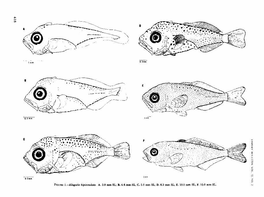
FIGURE l.-Elagatis bipinnulata. A. 3.8 mm SL; B. 4.6 mm SL; C. 5.5 mm SL; D. 6.3 mm SL; E. 10.0 mm SL; F. 15.0 mm SL.
APRIETO: FARLY DEVFLOPMENT OF FIVE CARANGID FlSHFS
Distinguishing Features
Larvae ofElagatis bipinnulata are distinct fromthose of other carangids in having only two spinesin the anal fin. Following transformation, theterminal dorsal and anal soft fin rays becomegradually separated from these fins. These larvaeare remarkably similar to those ofSeriola speciesin size, structure, and pigmentation. Unlike thelarvae of Seriola, however, they have a supraoccipital crest, serrations on the preopercularspines, and all the dorsal spines are about equal inlength. The first interhemal spine is only slightlyswollen and is not pressed nor fused with thehemal spine of the first caudal vertebra (Starks,1911). The larvae transform at 10 to 14 mm.the other interhemal and hemal spines. In many
carangids the first interhemal spine is much enlarged and pressed against or fused with thehemal spine of the first caudal vertebra (Starks,1911). The larvae transform at 10 to 14 mm.
Morphology
Larvae ofE. bipinnulata are deep-bodied. Bodydepth at the base of the pectoral fin is 32% of thestandard length at 3.8 mm; it attains a maximumof400/0 at initial notochord flexion and is never lessthan 33% during the entire period of larval development (Table 2).
The head is large and deep. Relative length ofthe head increases throughout the larval andtransition periods. Head length is 31.6% at 3.8mm, incrcases to 35 to 49% at notochord flexion,
TABLE 2.-Measurements (mm) of larvae and juveniles of Elagatis bipinnulata.
(Specimens between dashed lines are undergoing notochord flexion.)
Standard
length
383.9
Body depthSnout-to- at base of Orbit Snout to fin origin
anus Head Head pectoral Snout Orbitdistance length depth fin length diametel Predorsal Prepelvic Preanal
1.9 12 12 1.2 0.32 0301.96 1.25 1.25 1.2 .32 30
4.6 2.9 1.6 17 1.5 48 .55 2.0 20 2.050 3.5 2.0 2.0 2.1 58 .50 2.7 20 4.05.2 3.5 2.1 2.2 2.1 59 .60 2.7 2.2 365.9 3.5 2.2 2.3 22 .50 .65 28 24 36
---._~-------_._--~--------------------_._--p------------_.--------------------------.-----------------------------.--------------------------------------------------.------------------------------------.---------
6.1 3.9 2.1 2.2 23 .52 65 3.0 2.5 446.2 3.9 2.3 24 2.2 55 65 30 25 4.17.0 4.4 2.5 2.6 24 .60 70 .35 2.8 4.58.5 51 2.8 3.0 28 75 .77 40 32 5287 53 3.0 3.2 3.1 .75 .77 4.1 3.4 5.5
95 6.0 3.2 3.3 32 80 10 41 3.5 61
97 60 3.2 33 3.3 .85 1.0 4.5 36 62
'10.0 6.1 3.8 3.5 34 90 1.2 46 36 6.2'11.0 7.2 3.8 3.8 38 1.0 1.0 5.2 4.0 7.4
'11.2 72 4.2 40 3.5 1.1 1.2 5.5 43 74
'11.5 7.2 43 3.8 3.8 1.2 1.2 6.0 43 7.5
'11.8 73 40 3.6 3.6 1.1 12 60 4.3 7.5
'12.0 7.4 4.0 3.8 38 1.2 13 60 45 7.4
'12.5 75 4.2 38 3.8 11 14 5.9 48 7.8
'13.0 7.5 4.0 4.0 39 1.2 1.3 6.0 5.0 80'13.'2 78 4.0 4.0 39 10 14 5.8 4.8 80'13.3 8.1 45 4.1 42 12 13 6.1 50 86'140 8.5 53 4.9 4.5 1.5 13 63 5.5 9.0
'14.1 8.5 50 4.9 45 1.5 14 61 54 9.0
'14.5 9.5 5.0 48 4.6 1.7 1.3 60 5.2 9.8
'14.8 9.0 4.8 4.6 4.5 1.7 14 65 54 9.8
'15.2 9.5 53 4.7 43 16 1.5 6.5 5.3 99215.5 9.0 5.4 4.7 46 1.6 1.3 6.6 52 97
216.2 10.0 56 4.9 4.8 17 1.6 7.0 60 110216.8 10.2 57 50 4.7 1.8 1.5 80 70 106217.0 100 56 50 4.9 1.9 13 7.0 60 10.5'18.0 100 6.0 5.0 5.0 19 1.5 75 6.0 1102184 105 6.0 50 5.6 1.8 1.5 8.1 5.9 11.0
'Transforming.2Juveniles.
419

and averages 35% during the late larval and earlyjuvenile stages. The head is as deep as long at 3.8mm and is deeper than long for most of the larvalperiod. Head depth reaches a maximum of 110% ofthe head length at 5 mm and averages 101% in thelate larvae. The dorsal profile of the snout isslightly concave at 3.8 mm; at notochord flexion, itbecomes indented at the anterior and posteriormargins of the slightly swollen forebrain. Theseindentations disappear in older larvae and attransformation, the dorsal profile becomes convex.
The eyes are round and large. Relative eyediameter ranges from 26.3 to 31.3% of the headlength. A low orbital crest is formed above the eyesin early larvae but regresses at metamorphosis.
A supraoccipital crest is present throughout thelarval stages. At 15 mm, the crest is much reducedand is no longer visible externally but may beobserved in cleared and stained specimens.
There are two series of preopercular spines: onealong the margin and another on the lateral surface. Spines on the lower margin are bigger andmore serrated than those on the lateral surface.All preopercle spines gradually diminish in sizeand are completely overgrown by the expandingpreopercle bones following metamorphosis.
The gut is long and coiled in a single, roundedloop in larvae up to a length of12 mm; at 18 mm, asecond loop is added. Hypaxial muscles enclose thegut at 5 mm and completely cover the abdominalcavity except at the opening of the gut at 8 mm.
The number of myomeres is constant-10preanal plus 14 postanal-throughout the larvaland early juvenile periods.
The first scales formed are those found along thelateral line near the caudal peduncle. Lateral linescales ossify in a posteroanterior direction at 15mm. Regular body scales are not ossified until thejuveniles are at least 20 mm long.
Pigmentation
Larvae are among the most intensely pigmented of carangids. In the early larvae (3.8-4.6mm), the most conspicuous pattern of pigmentation includes melanophores along the bases of thedorsal and anal fins and along the lateral midline(Figure lA, Bl. Small patches ofmelanophores arepresent on the head,jaws, snout, and on the uppersides ofthe body. Internal pigmentation is concentrated on the dorsal wall ofthe peritoneum. At 5 to6 mm, melanophores develop profusely all overthe body leaving only a small unpigmented area at
420
FISHERY IJULLETIN: VOl. 72. NO.2
the caudal peduncle (Figure 1C,D). A row ofclosely spaced pigment cells along the midventralline below the gut is notable and distinguishesearly larvae from similarly pigmented larvae ofSeriola. Xanthophores (yellow) develop profuselyon the head and back in late larvae. Atmetamorphosis, iridiophores (reflecting), xanthophores, and melanophores cover the whole lengthof the body except on the jaws and fins, and anirregular row of large melanophores is formed onthe upper side of the body (Figure IE). Themelanophores are capable of expansion and contraction and the larvae are pale or dark dependingon the state of the pigment cells. Iridiophores andxanthophores fade upon preservation. The onlychromatophores apparent in preserved specimensare the melanophores.
Fin Development
Rudiments of all fins except the ventral fins arepresent in the smallest larva (3.8 mm) and aresituated in about the position they occupy at olderstages. The fins ossify in the following sequence: 1)caudal, 2) first dorsal and anal, 3) second dorsaland pectoral, 4) ventral. All fins are essentiallycomplete at metamorphosis (Table 3).
In the pectoral fins the dorsalmost rays are thefirst to ossify at 5 mm, and the rest of the rays areadded ventrally. The full complement of 18 to 21rays is present at 8 mm.
The pelvic fins ossify at 6 mm, and the full complement of 5 rays is present at 8 mm. The pelvic finrays grow fast, and at transformation they areabout as long as the pectoral fins.
Ossification of the dorsal and anal fin rays proceed in an anteroposterior direction in an orderlymanner. The anteriormost rays are the first toossify at 5 mm, and ossification continues posteriad. The last two fin rays are gradually separated beginning at metamorphosis in a mannerdescribed by Berry (1969). The full complement of7 spines and 25 to 30 soft rays is present at 10 mm.The dorsal spines are of almost uniform height inthe larvae. In early juveniles (17 mm), theirheight is about half that of the soft rays. The firstand second dorsal fins are continuous throughoutthe larval and transition stages. The full complement of 2 spines and 19 to 20 soft rays in the analfin is completed at 10 mm.
Caudal fin formation has begun in the smallestlarva (3.8 mm). This is indicated by the presence ofa thickening near the tip of the notochord. When

APRIFTO; FARlY DFVFLOPMFNT OF FlVF CARANGID FISHFS
TABLE 3.-Meristic characters of cleared and stained larvae and juveniles of Elagatis bipinnuiata.
72 2 0.5 77 8 0+11 88 8 2 4 2+13 89 8 4 5 4.15 79 8 8 9 5+18 69 8 9 9 5+ 18 89 8 9 9 6 +22 79 8 10 9 7 t 21 59 8 10 10 7 t 21 59 8 10 9 7 t 21 49 8 10 10 8+21 29 8 11 10 8 +23 29 8 11 10 8+22 29 8 10 11 8+23 19 8 11 11 8+ 22 19 8 11 10 8+24 19 8 11 11 8 t 24 19 8 10 10 9+ 25 1~ 8 11 10 8+27 1
Standardlength Dorsal fin Anal fin
3.8465A V; 18 1.106.0 V; 24 I. 1670 VI; 1,25 11880 VI; I, 24 I. 199.0 VI, I. 26 I. 20
lOA VI; I, 27 11,2011.5 VI; I. 26 11.1912.0 VI; I. 27 II, 2013.1 VI; I. 28 11,19147 VI; I, 28 11,1915.2 VI; 1,27 II. 19160 VI; I, 27 II 1917.1 VI; I, 27 II. 1918.0 VI; 1,26 11,19190 VI; I, 27 II, 1919.5 VI; I, 27 II. 1920.0 VI; I, 27 II. 1923.3 VI; I, 27 II. 19
Leftpectoral
fin
51317181919201920202020202019201921
Leftpelvic
fin
I. 2I. 41,5I. 5I. 5I 5I. 5I. 51,5I. 5I 5I. 51,5I. 51,5I. 51,5
Primary caudalfin rays
Dorsal Ventral
Secondary caudalfin rays
Dorsal Ventral
Gill rakers.left firstgill arch
Left preopercular
marginspines
the larvae are 4 to 5 mm long, the thickeningdifferentiates into hypural elements, thenotochord starts flexion, and the median caudalrays ossify, The hypural elements ossify at 6 mm,the full complement of9 dorsal and 8 ventral principal rays is present at 7 mm, and notochordflexion is completed at 8 mm. The secondary raysossify at 6 mm beginning with the posteriormostrays, The full complement of 10 to 11 dorsal and of10 to 11 ventral secondary rays is formed at 12mm.
The last 3 vertebrae, the hypural elements, and5 dorsal structures including the neural spine ofthe antepenultimate vertebra, 3 median epurals,and a specialized neural process support thecaudal fin, The supporting structures articulatewith the principal and secondary caudal rays.They are generally similar to those occuring inTrachurus symmetricus (Ahlstrom and Ball,1954).
Distribution and Spawning
Elagatis bipinnulata has a circumtropical distribution (Briggs, 1960), It has been previouslyreported from Texas, Florida, and Long Island(Ginsburg, 1952) and is also known from the WestIndies (Jordan and Evermann, 1896), Japan(Okada, 1966), Hawaii (Gosline and Brock, 1960),Africa (Fowler, 1936), Philippines (Herre, 1953),and the Great Barrier Reef (Marshall, 1965). Thelarvae were reported by Okiyama (1970) to be themost abundant form of epipelagic larval carangidin the tropical as well as in the subtropical
Indo-Pacific ocean where spawning occursthroughout the year with a peak in March.
In the present study, larvae and early juvenilesless than 20 mm have been taken in every monthexcept in May and December. While the specimens are too few to give conclusive information, itappears that spawning may occur throughout theyear. The larvae were taken mainly in offshorewaters in the eastern GulfofMexico, in the Santaren Channel, the Straits of Florida, and theCarolina Bight offNew Brunswick, Ga. (Figure 2).They occurred in 2.6% of the net stations and constituted 2.3% of the young carangids collected.
FIGURE 2.-Locations of collections of larval carangids duringtwo cruises of the Oregon II from July to August and fromOctober to November 1970 and a cruise ofthe Tursiops in August1971. Records of occurrences ofElagatis bipinnulata are shownas solid circles, those of Seriola zonata as solid triangles, andthose of Selene V01tU'r as solid squares. Open circles representother stations occupied,
421

During its larval life, Elagatis bipinnulata isplanktonic. Some larvae and juveniles become associated with the pelagic sargassum community(Dooley, 1972) and are carried along the FloridaCurrent and Gulf Stream.
Banded rudderfish,Seriola zonata (Mitchill)
Figure 3
Literature
Larvae of Seriola zonata have not been. previously described. Early juveniles of the bandedstage were described by Nichols (1946), Ginsburg(1952), and Mather (1958). Lutken (1880) illustrated an unbanded 20-mm juvenile.
As noted earlier, larvae ofSeriola dumerili weredescribed by Hildebrand and Cable (1930) andSanzo (1933). Japanese workers have describedlife history series of three species of Seriola. Themost detailed study of development from eggs tojuveniles was made on Seriola quinqueradiata[Uchida, Dotu et al. (1958), Uchida in Uchida,Imai et al. (1958), Mitani (1960), and Mito (1961)].Larvae and juveniles of two other Japanesespecies, S. aureovittata and S. purpurascens werecovered by Uchida in Uchida, Imai et al. (1958).
Distinguishing Features
Seriola larvae resemble most those of Elagatisbipinnulata in size, body structure, and pigmentation. Unlike E. bipinnulata, however, there is nosupraoccipital crest; the spines ofthe dorsal fin areof unequal length, the anterior and posterior onesbeing shorter; and the preopercular spines havesmooth sides until the transition period when thelongest spine develops 1 to 2 denticles. The larvaetransform at about 13 mm. They are deep-bodiedand robust. The head is massive and slightly depressed, and the eyes protrude slightly from theorbit at the dorsal side.
Early larvae ofSeriola zonata (3-7 mm) are differentiated from those of other species of Seriolaby the presence of5 to 6 large melanophores on themiddorsal line at the base ofthe dorsal fin (Figure3 B). These large melanophores which are apposedto the myomeres stand out among the morenumerous and smaller pigment spots on the backand sides. In older larvae these melanophores become embedded in the muscles and covered by
422
FISHERY HLJI.I.ETIN: VO!. 71. NO.2
superficial melanophores. When the full complement of dorsal fin rays is formed at about 8 mm,larvae ofS. zonata are distinct in having 35 to 40soft rays in the second dorsal fin, the highest second dorsal fin ray count of all species of Seriola.All dorsal fin rays are sharply visible even in unstained specimens.
The first interhemal spine of the first ventralpterygiophore is only slightly enlarged and doesnot press against the first hemal spine.
Morphology
Maximum body depth at 3.6 mm is 300/tc, of thestandard length. It increases to 37% at initialnotochord flexion and does not changesignificantly during larval and transition periods.In early juveniles, the body depth is never lessthan 30% of the standard length (Table 4).
Head length is 33% of standard length in thesmallest larva (3.6 mm) and attains a maximum of43% at 7.0 mm. Thereafter, head length decreasesgradually, with an average of 35% in earlyjuveniles 18.0 mm in length. Depth of head is 91%of the head length at 3.6 mm and attains a maximum of 122% at 5 mm. Thereafter, head depthdecreases slightly and is never less than 89% ofthe head depth throughout the larval and juvenileperiods. The dorsal profile of the snout is slightlyconcave at 3.6 mm but becomes straight at about 5mm and then convex in the older larvae andjuveniles.
The eyes are round and large, and the orbitdiameter increases in relation to head length. Relative orbit diameter ranges from 28 to 36% of thehead length in larvae and transforming specimensand gradually increases in early juveniles. A loworbital crest with a weak spine is present in theearly larvae and is resorbed at metamorphosis.
Marginal and lateral surface preopercularspines are present. The marginal angle spinewhich is the longest develops 1 or 2 denticles on itsdorsal side in transforming larvae and earlyjuveniles. All preopercular spines graduallydiminish in size and become overgrown by theexpanding preopercle.
Scales along the posterior end of the lateral linein front of the caudal peduncle are formed at 20mm. Subsequently, the scales along the anteriorportion ofthe lateral line ossify, followed by thoseon the head and sides of the body.
The slender gut is coiled in a single loop inlarvae up to 10 mm long. The number of loops

APRIETO: FARLY DEVELOPMENT OF FIVF CARANCiID FISHES
TABLE 4.-Measurements (mm) of larvae and juveniles of Seriola zonala.
(Specimens between dashed lines are undergoing notochord flexion.)
Stan- Snout-to-Body depthat base of Snout to fin origin
dard anus Head Head pectoral Snout Orbitlength distance length depth fin length diameter Predorsal Prepelvic Preanal
3.6 2.1 1.2 1.1 1.1 035 03637 23 1.25 1.15 1.2 .36 36
4.7 30 1.6 1.5 1.5 .42 .425.5 35 1.8 2.1 2.0 50 .60 2.5 2.1 3.656 35 1.8 2.2 2.0 .52 62 25 2.1 376.2 4.0 20 2.3 2.0 .65 .70 29 2.2 4.06.4 3.9 2.3 23 2.1 .65 70 29 25 4.065 4.0 2.5 25 23 .65 .72 30 25 4.268 4.5 2.8 2.6 2.4 67 .72 3.4 28 4670 46 3.0 2.8 2.5 .70 .75 3.4 2.9 4.87.1 4.7 30 3.0 2.6 .72 .75 35 2.8 5.075 4.8 30 30 2.5 .80 .85 3.8 3.0 5.0
80 52 30 28 2.5 .85 90 38 30 5382 5.2 31 3.1 2.8 .90 1.0 39 32 5.38.4 5.4 32 3.5 3.2 10 1.1 4.0 3.5 5591 58 3.5 3.5 32 1.0 1.2 4.1 39 5.99.5 62 35 3.6 3.2 1.0 1.2 4.3 39 6.498 62 40 38 3.5 1.2 1.3 43 4.1 6.4
'10.0 6.5 4.0 38 3.5 1.1 1.3 43 4.1 6.6'10.3 66 4.0 38 3.5 1.1 1.3 4.5 4.1 6.8'10.8 7.4 42 4.0 3.6 1.3 1.5 5.2 4.4 7.6'11.9 8.4 4.7 4.8 45 1.5 16 58 5.5 8.6'12.0 8.5 4.9 4.8 4.5 1.6 1.6 6.0 56 8.6'12.1 85 50 4.6 4.5 1.6 16 59 5.6 86'125 86 5.0 4.8 4.5 1.6 16 60 5.2 8.7'13.1 8.8 5.0 48 4.5 1.4 1.8 6.1 53 8.3'13.5 9.2 5.0 4.8 4.6 1.5 1.9 61 56 9.5'13.6 92 50 4.6 45 1.4 20 6.2 55 10.0'15.0 98 5.5 5.0 5.0 1.4 20 6.0 55 10.2'15.1 10.0 56 50 5.0 1.5 20 63 5.5 10.5'15.2 10.0 5.7 5.1 5.2 1.5 2.0 6.4 5.6 10.6'16.2 105 58 5.2 5.4 1.6 2.1 7.5 5.8 11.0'17.5 105 6.0 5.5 60 1.5 2.2 75 6.0 11.0'18.1 110 6.5 58 6.2 1.7 23 75 7.0 11.0
'Transforming.2Juveniles.
increases with growth and at 15 mm four loops arepresent. Snout-to-anus distance increases in relation to standard length. It is 58.3°k, at 3.6 mm andgradually increases to 700/0 at transformation.Hypaxial musculature develops at 6 mm and completely surrounds the abdominal cavity except atthe gut opening at 10 mm.
There are 24 myomeres-lO preanal plus 14postanal-throughout the larval and juvenilestages
Pigmentation
Larval pigmentation consists of conspicuousmelanophores along the bases of the dorsal andanal fins, on the lateral midline, and in theperitoneum lining the middorsal wall of the abdominal cavity. In a freshly preserved 9-mm larvathere are dense concentrations of xanthophores on
the head, preopercle, and on the back and uppersides of the body while iridiophores are profuse onthe sides of the body below the lateral midline. Inolder larvae, the melanophores are apparently actively expanding and contracting as most larvaehave either contracted melanophores and are palelooking, or are dark when the melanophores areexpanded. Other larvae have alternating patchesof expanded and contracted melanophores forming false bands (Figure 3F-H). In early juveniles(17 mm), the body definitely becomes banded. Abold color pattern develops including a distinctnuchal bar and 6 solid bands which extend to thedorsal and anal fins (Figure 31). In a youngjuvenile 23 mm long, the lobes ofthe unpigmentedcaudal fin have a brown spot developing at thetips. Alcohol-preserved metamorphic larvae andearly juveniles have chocolate-brown bands over asilvery background. With the exception of
423

lmm
to' .' •• ". : .•". .~.
Imm
.,.
. "- ~
c
I mm
." "" :."; :'". ~...:.=:----'", .,' ..... • 1'..,. . .
Imm

D
E
I ....
I ....
..... :,.........• e •.• ".
.. ::::. ;.....
FIGURE 3.-Seriokl zonata. A. 3.6 mm 8L; B. 4.8 mm 8L (dorsalview); C. 4.8 mm 8L (lateral view); D. 5.5 mm 8L; E. 6.5 mm 8L;F. 8.7 mm 8L; G.10.6 mm 8L; 8.13.5 mm 8L; 1.18.0 mm 8L.
)...:::~m);cc-<om<mcO...:::::mZ...,o.,.,.,.,:;::m
")-;c)Z
'"'o:!JVJ:tmVJ

melanophores, all other chromatophores fade onpreservation in formaldehyde solution.
Fin Development
The dorsal, anal, caudal, and pectoral finfoldsare present in the youngest larva (3.6 mm). Differentiation of the fin rays occurs in the followingsequence: 1) caudal, 2) first dorsal and anal, 3)second dorsal and pectoral, 4) ventral. All fins areessentially formed at 9 mm (Table 5).
The pectoral fin rays ossify at 6 mm beginningwith the most dorsal rays, and the rest ossify ventrally. The full complement of 19 to 20 rays isformed at 10 mm.
The pelvic fin rays differentiate at 7 mm, andthe full complement of 5 rays is present at 9 mm.
The dorsal and anal fin rays ossify anteroposteriorly. At 8 to 9 mm the full number of8 spines inthe first dorsal fin and 1 spine and 35 to 40 soft raysin the second dorsal is completed. The first dorsalfin becomes arch-shaped as the spines increase inheight and remains continuous with the seconddorsal fin until in early juveniles (15-17 mm) adeep notch demarcates the 2 fins. The anal fin raysbegin to ossify at 5 mm, and the full complement of3 spines and 19 to 22 soft rays is completed at 10mm.
The caudal fin begins to develop at 4 to 5 mm ina manner similar to that occurring in Elagatisbipinnulata. The full complement of 9 dorsal and8 ventral principal rays is present at 7 to 8 mm,while the full complement of 10 to 11 dorsal andof9 to 10 ventral secondary rays is completed at 9mm.
FISHERY BULLFTIN: VOL. 72. NO.
Distribution and Spawning
Juveniles of Seriola zonata (12-23 mm) havebeen reported to be regular summer visitors inCape Cod waters which appear to be their mostnorthernly record (Mather, 1952). Adults havebeen recorded from various points of the Atlanticcoast and in the Gulf of Mexico (Ginsburg, 1952).
Larvae and early juveniles up to 26 mm weretaken in all months except in February, April,September, and December. They were caught witha I-m plankton net in the Gulf Stream off Miami,with dip nets at the pier ofthe Southeast FisheriesCenter in Biscayne Bay, and with neuston nets inthe Gulf of Mexico, Yucatan Channel, Straits ofFlorida, and south Atlantic coast. The larvae occurred in 1.4% of the net stations and constituted1.8% of the larval carangids in the collection. Theoccurrence of the larvae is too erratic to indicatewhether or not the spawning period is continuousover 12 mo or broken into two parts, winter-springand fall. Spawning occurred mainly in offshorewaters in the eastern Gulf of Mexico, YucatanChannel, Santaren Channel, along the edge ofthecontinental shelf in the Straits of Florida, and inthe Carolina Bight off New Brunswick, Ga. (Figure 2). The planktonic larvae are presumably carried along the Florida Current and Gulf Streamand reach their northern limits as juveniles.
Round scad, Decapterus punctatus(Agassiz)
Figure 4Literature
The early growth of this species were described
TABLE 5.-Meristic characters of cleared and stained larvae and juveniles ofSeriola zonata.
Left LeftPrimary caudal Secondary caudal Left pre-
fin rays fin rays Gill rakers, opercularStandard pectoral pelvic left first margin
length Dorsal fin Anal fin fin fin Dorsal Ventral Dorsal Ventral gill arch spines
3.6 444 4 5 45.5 IV II; 5 8 7 0+6 56.5 VI; 22 II; 11 8 9 8 0+8 67.5 VII; 30 II; 16 10 1,3 9 8 1 2 0+ 11 784 VIII; 1,34 II; 19 13 1,4 9 8 3 3 3+13 79.5 VIII; I, 36 II; 20 17 1,5 9 8 5 5 4+15 7
10.3 VIII; 1,38 II; 1,20 18 1,5 9 8 7 8 5 +15 711.2 VIII; I, 39 II; I, 21 20 1,5 9 8 8 8 5+15 712.0 VIII; 1,36 II; I, 22 20 I, 5 9 8 9 8 5+15 513.0 VIII; I, 37 II; I, 20 20 1,5 9 8 9 9 5+16 314.2 VIII; 1,36 II; I, 19 20 1,5 9 8 10 9 6t 16 415.2 VIII; 1,38 II; I, 20 19 1,5 9 8 11 10 6+16 416.0 VIII; 1,40 II; I, 21 20 1,5 9 8 11 10 6+ 17 417.0 VIII; I, 38 II; I, 20 20 1,5 9 8 11 10 8+18 3
426

APRIETO: EARLY DEVELOPMENT OF FIVE CARANGID FISHES
by Hildebrand and Cable (1930) who also studiedthe abundance and distribution ofthe young up to50 mm taken from the coastal and offshore watersof Beaufort, N.C. In addition, life history serieswere described for two species ofDecapterus fromthe Indo-Pacific region: D. russelli by Vijayaraghavan (1958) andD. maruadsi by Shojima(1962). The latter series is quite similar to that ofD. punctatus.
Distinguishing Features
Larvae of D. punctatus are decidedly deepbodied although the adults are the most slender ofall carangids. The head has a supraoccipital andan orbital crest and preopercular spines. Duringmetamorphosis, the ultimate rays of the dorsaland anal fins become separated and modified intofinlets. The first interhemal spine is slightly swollen. Together with the hemal spine of the firstcaudal vertebra to which it is closely apposed itforms a strong brace at the posterior border of theabdominal cavity. There are 25 vertebrae-l0trunk plus 15 caudal. D. punctatus is one of thefew carangids with a vertebral count of 25, theusual number being 24; this is a useful characterfor separating the larvae from other species ofDecapterus. Berry (1968) used the scales, scutes,and lateral line spots in the identification of theolder juveni les from 90 mm long. These charactersare not yet formed in the larvae.
Morphology
Body depth decreases relative to standardlength and is 30.6 to 35.4% during the entire larval period (Table 6). In transforming larvae andearly juveniles, body depth decreased to 28%. Thehead is long and deep; relative head length increases throughout the larval and juvenile stages.It is 27% in the smallest larvae (3.0 mm) andattains a maximum of 35% in the larval period. Intransforming larvae and early juveniles, it isnever less than 31% of the standard length. Thehead is deeper than long in the early larval stagesup to notochord flexion and ranges from 108.3 to113%. Thereafter, head depth decreases graduallywith an average of 90% but is never less than 80%of the head length in the early juveniles. A supraoccipital crest is present during the larval periodbut is resorbed at metamorphosis. The snout isslightly concave in the youngest larvae (3.0 mm),but becomes straight at initial notochord flexion.
In older and transforming larvae, the snout develops a convex profile.
The eyes are large and round, and the orbitdiameter increases relative to head length. Eyeindex ranges from 30 to 46.1% during the larvaland transition periods and is highest at notochordflexion. A low orbital crest bearing a weak spine ispresent above the eyes in larvae 3 to 7 mm long. Inolder larvae, the crest is gradually resorbed and isno longer visible at 10 mm.
Marginal and lateral surface spines are presenton the preopercle. They increase in size andnumber during the larval period but are graduallyresorbed at metamorphosis. At 17 mm, the lateralsurface spines are gone and the margin spines arereduced to fine crenulations on the preopercularmargin.
The slender gut is coiled in a single loop in theearly larvae. A second loop is formed at transformation and a third is added in the early juveniles.Snout-to-anus distance increases relative to standard length; it is 52.3 to 59.5% of the standardlength during the entire larval and transitionperiods and does not change noticeably during theearly juvenile stages. Hypaxial muscles begin todevelop around the gut in 5-mm larvae, and at 7mm, the abdominal cavity is completely enclosedexcept at the anal opening of the gut.
The first scales to ossify are those at the posterior region of the lateral line in juveniles 17 mmlong. Ossification of the lateral line scales proceeds anteriad and the full complement of scalesand scutes is present at 20 mm when the bodyscales are formed.
Pigmentation
Chromatophores are slow to develop in the larvae and remain sparse until the early juvenilestages. There are a few melanophores on the backof the head, on the jaws, and infrequently on thesnout and cheeks. As in other carangid larvae,there is a row ofmelanophores on each side of thebases of the dorsal and anal fins, along the lateralline at the caudal region, and on the dorsal wall ofthe peritoneum. Compared to those of Seriolazonata and Elagatis bipinnulata, the larvae ofDecapterus punctatus are pale. Melanophores arenot profusely developed until metamorphosis, andthey are mostly located above the midline. In theearly juveniles, iridiophores spread all over thebody but are most dense below the lateral line,giving the fish a metallic sheen.
427

--=-=-----.0.5 mm
I
F
I",,,,.
c
""~~~.".
~::
G
Imm
z:)

APRIl-TO: EARLY DEVELOPMENT OF FIVE CARANGID FISHES
Fin Development
The dorsal, anal, caudal, and pectoral finfoldsare distinct in the smallest larvae (3.0 mm), butthe fin rays begin to ossify at 4 to 5 mm in thefollowing sequence: 1) caudal; 2) dorsal, anal, andpectoral; 3) ventral (Table 7).
The pectoral fin rays are differentiated at 5 mm,and the full complement of 19 to 21 rays is presentat 11 mm.
The pelvic fin buds are present at 4 to 5 mm, butthe fin rays ossify at 6 mm, and the full complement of 1 spine and 5 soft rays is formed at 7 mm.
As in E. bipinnulata and S. zonata, ossificationof the dorsal and anal fin rays proceed anteroposteriorly. The full complement of 8 spines in thefirst dorsal fin and of 1 spine and 31 to 34 soft raysin the second dorsal fin is present at 10 mm. At thisstage also, the ultimate fin rays gradually separate from the dorsal and anal fins and eachmodifies into a much branched finlet. The anal finrays start to ossify at 5 mm, and the full complement of 3 spines and 27 to 31 rays is completed at9mm.
The pattern of caudal fin formation is generallysimilar to that ofE. bipinnulata. Caudal fin structures initially develop at 4 mm, and all 17 principalrays are present at 6 mm. The full complement of9 dorsal and 9 ventral secondary rays is formed at13 mm. Unlike E. bipinnulata, only two medianepurals are normally developed as the centerepural is markedly reduced.
Distribution and Spawning
Adults ofD. punctatus have been reported fromboth sides of the Atlantic from Nova Scotia toBrazil (Jordan and Evermann, 1896) and WestAfrica (Fowler, 1936). The first record of the larvae was reported by Hildebrand and Cable (1930)in Beaufort, N. C. They noted that spawning occurred throughout the summer or from May toNovember with a peak from July to September.Larvae and juveniles 2 to 50 mm long were presentin inshore as well as offshore waters, possibly extending beyond the Gulf Stream, from the surface to the bottom up to a depth of 20 fathoms. Inthe present study, larvae and juveniles of D.punctatus were taken in all the months duringthe routine fish larvae sampling in the GulfStream off Miami and in numerous net stationsin the Gulf of Mexico and the south Atlanticcoast. Spawning occurs in pelagic inshore as well
429

FISHFRY BULLETIN: VOL. 72. NO.2
TABLE 6.-Measurements (mm) of larvae and juveniles of Decapterus punctatus.
(Specimens between dashed lines are undergoing notochord flexion.)
Body depthStan- Snout-to- at base of Snout to fin origindard anus Head Head pectoral Snout Orbit
length distance length depth fin length diameter Predorsal Prepelvic Preanal
3.0 1.7 0.8 0.9 10 0.26 0323.1 1.8 8 1.0 1.1 .30 .373.5 20 10 1.1 1.2 .37 .423.7 2.0 1.1 1.2 1.2 40 47
4.0 2.2 1.2 13 12 45 .5242 25 1.3 1.5 1.3 .47 .6048 2.6 15 1.6 1.6 .50 6552 3.0 1.6 1.8 1.8 .55 705.5 32 18 2.0 1.9 57 .7564 3.7 2.2 2.1 2.0 .62 82 2.8 2.265 38 22 2.1 2.0 62 82 2.9 2.16.7 3.9 2.2 22 2.1 .65 .80 28 2.07.1 4.0 2.2 24 2.2 67 85 3.1 247.5 4.1 2.5 2.5 24 .72 .90 32 2.378 4.2 2.5 25 2.5 .75 .92 32 2.6
3.83.8374.24.24.5
8.0 4.2 2.7 26 2.6 .75 95 32 2.683 4.5 28 26 26 .75 1.0 35 2.786 45 30 2.6 27 .80 10 3.2 2.698 5.5 3.2 30 3.0 85 1.2 39 30
10.0 5.8 35 3.0 3.0 1.0 1.1 45 4.0102 5.8 3.5 3.2 34 1.1 1.2 4.5 4.310.5 62 35 3.2 32 1.1 125 4.6 4.011.0 6.1 3.5 35 3.4 1.1 1.2 4.5 4.311.2 6.2 3.5 35 35 1.2 1.2 4.6 40114 6.4 3.7 37 36 1.2 1.2 4.6 4.5
'12.0 69 40 3.8 3.7 1.2 1.3 4.8 4.5'12.3 68 4.0 3.8 3.7 12 13 48 45'12.5 70 42 4.0 38 12 1.3 50 48'13.0 7.2 4.2 4.0 4.1 1.3 1.3 5.2 4.9'13.2 74 4.2 4.0 4.1 1.2 14 54 4.7'13.5 7.5 4.3 4.1 4.2 1.3 1.5 52 50'14.0 8.0 44 4.5 4.3 13 1.6 58 5.8'144 7.8 4.5 4.1 4.3 1.3 1.6 5.6 54'14.9 80 4.8 4.2 4.3 14 16 5.9 60'15.2 82 5.0 42 44 1.5 1.6 5.8 5.6'15.5 86 52 4.1 4.5 1.6 16 5.9 5.5'160 8.7 53 4.5 4.6 1.5 1.7 6.5 58'16.2 88 56 46 4.6 1.6 17 64 59'16.5 8.8 5.7 4.7 4.7 1.7 1.8 63 59'170 95 5.7 5.0 4.8 1.8 1.9 70 6.0'17.5 9.2 5.7 4.6 4.9 1.9 1.9 64 5.8'18.0 95 60 5.0 5.2 1.9 2.0 70 63'18.5 9.6 60 5.2 5.3 1.8 2.0 7.0 65'19.2 10.0 6.1 54 55 2.0 20 70 68'19.5 10.0 6.1 5.4 5.7 1.9 20 73 6.6
4.34.7465.6626.6636.6646.66.86.87.27.5767.88.583868.2839.0909.09.89.3
10010.011311.5
..•..as offshore waters and along the edge of the continental shelf (Figures 5, 6). In the Gulf of Mexico, the larvae appear to have their center ofabundance in the eastern area. They have thehighest frequency of occurrence and are the mostabundant among the larval carangids considered
'Transforming.2Juveniles.
FIGURI! 5.-Distribution and apparent relative abundance ofthelarvae of Decapterus punctatus in the Gulf of Mexico and theSouth Atlantic coast of the United States: a composite record ofoccurrences at stations occupied in October to November 1970 bythe Joie de Vivre and in August, October, and November 1971 bythe Dan BroTTUJn and Oregon II. Open circles indicate otherstations occupied.
430

APRIETO: EARLY DEVELOPMENT OF FIVE CARANGID FISHES
TABLE 7.-Meristic characters of cleared and stained larvae and juveniles of Decapterus punctatus.
Standardlength Dorsal fin Anal fin
Leftpectoral
fin
Leftpelvic
fin
Primary caudalfin rays
Dorsal Ventral
Secondary caudalfin rays
Dorsal Ventral
Gill rakers.left firstgill arch
Left preopercular
marginspines
3.13.54.25.36.67.68.29.0
10.411.512.513.514.415.116.517.019.023.028.2
V; 6VI; I. 19
VII; I. 20VIII; I, 25VIII; I. 30VIII; I, 31VIII; 1.23VIII; 1.34VIII; 1,32VIII; 1.32VIII; 1.33VIII; I, 32VIII; I, 31VIII; I, 34VIII; I. 32VIII; 1.31
II; 6II; I. 16II; I. 18II; I. 19II; 1.21II; 1,24II; I, 26II; I, 31II; I, 28II; I, 27II; I, 29II; I, 27II; I, 27II; I, 27II; I, 28II; I, 28
3101417181719192020201920202120
I, 5I, 31,51,51,51,51,5I, 51,5I, 5I, 51,5I, 5I, 51,5
56
4 3 0+ 6 86 0+12 109 8 3 2 0+14 119 8 4 4 0+16 129 8 5 6 0+17 129 8 7 8 0+17 119 8 8 8 3 +17 119 8 8 8 3+21 119 8 9 9 6+24 109 8 9 9 6+25 109 8 9 9 6+26 99 8 9 9 6+25 109 8 9 9 6+25 79 8 9 9 6 +27 59 8 ~ 9 6 +28 39 8 9 9 8+28 29 8 9 9 9+28 0
.•
FIGURE 6.-Distribution and apparent relative abundance ofthelarvae ofDecapterus punctatus in the GulfofMexico: a compositerecord of occurrences at stations occupied from May to August1972 by the Tursiops. Dan Braman, and Gerda.
here. They occurred in 18.5% of the net stationsand constituted 44.7% of the larval carangidscollected.
Lookdown, Selene vomer (Linnaeus)Figure 7
Literature
Larvae ofS. vomer are previously undescribed.Fowler (1936) illustrated a 15-mm juvenile andLutken (1880) a 28-mm juvenile.
Distinguishing Features
Larvae of S. vomer have extremely deep and
trenchant bodies. The advanced development ofthe dorsal and ventral fins is perhaps the mostnotable feature of their development; it is the earliest observed among carangid larvae. The secondand third dorsal spines develop into long filamentsoften twice the length ofthe body. The ventral finsare elongated, often extending to the anal fin. Thelarvae probably attain a maximum length of 12mm before transformation. The biggest larva inthe series in 9 mm long (Figure 7F) and the nextsize, 13.5 mm, (Figure 7G) is a transforming larvahatched from a planktonic egg and reared in anaquarium. The smallest juvenile is 23.9 mm longand has attained most adult features.
As in most larval carangids, there is a bony crestin the supraoccipital bones, two rows ofpreopercular spines, and a supraorbital crest. The distancefrom the snout to the occipitals is long and slopesinto an abrupt angle. The first interhemal spine ismuch enlarged and pressed against an equallyenlarged hemal spine.
Morphology
The larvae are among the most deep bodied ofall larval carangids. Relative body depth increases during the larval and early juvenile periods (Table 8). It is 32% at 2.5 mm and increasessteadily, attaining a maximum of96% at 23.9 mm.Thereafter, body depth gradually declines but isnever less than 74% of the body length in thejuveniles. Simultaneous with the deepening ofthebody is the enlargement of the first interhemaland hemal spine of the first caudal vertebra. The
431

~jz<o
zo

APRIETO: EARLY DEVELOPMENT 01' FIVE CARANGID EISHES
two spines fuse at the posterior wall of the abdominal cavity which becomes elongated vertically.Consequently, the long and slender gut forms vertical loops following the shape of the abdominalcavity. The gut opening is pushed anteriorly andlies adjacent to the base of the pelvic fins. Snoutto-anus distance decreases relative to standardlength. It is 56% at 2.5 mm and declines to 40% atthe end of the larval period. During the earlyjuvenile stages, snout-to-anus distance had an average of 38%.
The length and width of the head increases relative to standard length. Head length is 31.0% inthe smallest larva (2.5 mm) and does not increasesubstantially until the juvenile period when itattains a maximum of 41.9% at 23.9 mm. To obtain the true depth of the head and not the bodydepth at the head region, the measurement istaken from the posterior margin of the preoperclefrom the dorsal margin of the head to the articulation of the mandible and maxillary. Head depth is100% of the head length at 2.5 to 3.2 mm. Thereafter, head depth exceeds head length. During thelarval period, the head is deepest at 4.6 mm whenit is 14<Y)',; of the head length. This is, however,exceeded by the progressive deepening of the headduring the transition and juvenile stages with amaximum of 182% and an average of 158%. Thedorsal profile of the snout is slightly concave in theearly larval period (2.5-3.8 mm) but becomesstraight in older and transforming larvae. Thenasal, frontal, and supraoccipital bones becomemarkedly elongated and slope steeply, giving thehead an almost vertical anterior profile.
The eyes are round and large. Relative orbitdiameter increases during the larval and juveniledevelopment; it is 320/c at 2.5 mm and attains amaximum of 37% at notochord flexion with anaverage of 31% during the larval and transitionperiods. In the juveniles, orbit diameter decreasesto a range of 20 to 27%.
The orbital and supraoccipital crests are wellmarked in early larval stages up to 5 mm. Thecrests are gradually resorbed and are vaguely visible at metamorphosis. Preopercular marginalspines consist of4 to 7long and strong spines whilethe lateral surface spines are smaller and limitedto the lower surface. All preopercular spines areresorbed at transformation. Scales are absent.
Pigmentation
The common larval pattern of pigmentation in
433

FISHERY BULLETIN: VOl.. n. NO.
TABLE B.-Measurements (mm) of larvae and juveniles of Selene vomer.
(Specimens between dashed lines are undergoing notochord flexion.)
Body depthSnout-to- at base of Snout to fin origin
Standard anus Head Head pectoral Snout Orbitlength distance length depth fin length diameter Predorsal Prepelvic Preanal
2.5 1.4 0.78 0.7S O.SO 0.25 0.25 1.0 0.732 1.7 10 10 1.0 37 32 1.1 1.2 1.73.5 1.8 1.1 1.2 1.3 .37 35 1.2 1.0 1.83.8 18 12 1.4 15 .42 .42 1.2 135 20
4.0 20 1.2 1.5 1.5 .47 .37 13 1.2 2043 22 15 19 1.9 52 .50 15 1.6 1.94.4 2.1 1.5 2.0 2.1 55 .50 1.4 1.6 1.84.5 22 1.5 2.0 20 .52 .45 1.4 15 2.04.6 1.8 1.5 2.1 2.1 .55 .42 1.6 1.6 2.14.8 2.6 1.7 2.0 21 .55 .45 1.8 1.7 2.85.0 2.7 1.7 2.2 2.2 .52 .47 1.8 1.9 3.053 3.0 1.7 21 23 .55 .45 1.7 2.0 3.05.5 27 2.0 2.5 29 .62 eo 1.8 1.8 3.1
~-------------------------------------------------------------- -----------------------------------------------------.----------------------.---._---------------.-----------------61 2.8 20 3.0 3.27.5 3.6 26 3.5 3.98.2 4.5 3.0 3.8 4.8
'9.0 36 31 52 5.8'13.5 6.7 5.2 9.5 6.2
'23.9 87 10.0 14.0 23.0
'27.5 11.5 10.0 15.0 24.5
'30.0 12.0 110 170 270
'34.0 12.0 135 220 310
'38.0 14.0 14.5 20.0 31.0
'42.5 14.0 17.0 28.0 350'47.0 16.0 200 30.0 350
lTransforming2Juveniles
carangids, including melanophores along thebases of the dorsal and anal fins and along thelateral-midline, is present inS. vomer. In the earlylarvae (2.5-5.0 mm), a few melanophores developon the tips of the jaws, head, sides of the body,pelvic fin, dorsal fin, and base ofthe caudal fin. Theearliest patch of melanophores is formed on thelower side of the body anterior to the caudalpeduncle. In older larvae, the melanophoresgradually proliferate all over the body and formdiscrete patches which develop into broad spots attransformation and in the juvenile stages. Theheaviest concentrations of pigment cells comprisethose lining the dorsal wall of the peritoneum.Reglarly spaced melanophores similar to those inElagatis bipinnulata (Figure IB-D) are presentalong the midventral line in the trunk region.
Fin Development
The sequence offin formation and ossification isas follows: 1) pelvic; 2) first dorsal; 3) second dorsal, caudal, and anal; and 4) pectoral (Table 9).
434
.87 62 2.0 2.0 30
.90 85 2.4 2.5 4.01.2 .87 2.9 3.0 4.613 1.0 2.9 2.8 5.0?2 1.7 5.5 5.5 6.231 25 70 80 11.03.5 2.6 11.0 8.0 14.05.0 3.0 14.0 120 14.06.5 30 14.0 13.0 14.05.0 32 15.5 11.5 16055 3.9 170 150 14.080 4.2 16.0 18.0 17.0
The pectoral finbud is formed at 2.5 mm but therays are not differentiated until the larvae are 5 to6 mm. The full complement of 18 to 21 is formedat 9 mm.
The pelvic fins are fully formed in the smallestlarva (2.5 mm). They steadily increase in lengthand at metamorphosis extend beyond the origin ofthe anal fin.
The first 3 dorsal spines are ossified at 2.5 mm.The second and third spines progressively increase in length throughout the larval period,forming extremely long filaments. At metamorphosis, they are about twice the length ofthe body.The full complement of 8 spines in the first dorsalfin and of 1 spine and 20 to 22 soft rays in thesecond dorsal fin is present at 9 mm.
Rudiments of the anal fin are discernible at 3.2mm and the rays ossify at 4 to 5 mm. The fullcomplement of 3 spines and 16 to 18 soft rays ispresent at 6 mm.
The development and structure ofthe caudal finand supporting structures are similar to those ofE. bipinnulata. The full complement of 17 principal and 7 to 9 dorsal, and 7-8 ventral secondarycaudal rays is present at 9 mm.

APRIETO; EARLY DEVELOPMENT OF EIVE CARANGID EISHES
TABLE 9,-Meristic characters of cleared and stained larvae and juveniles of Selene vomer (Linnaeus),
Primary caudal Secondary caudal Left pre-Left Left fin rays fin rays Gill rakers, opercular
pectoral pelvic left first marginStandardDorsal fin Anal fin fin fin Dorsal Ventral Dorsal Ventral gill arch spineslength
2.5 III I. 53.2 V 1,53.4 V 1,53.5 VI 1,5
0+11 11,5 2 34.0 V;3 II0+11 24.4 VIII; I. 10 II; 10 1,5 5 6
1,5 7 7 0+ 16 24.9 VIII; I, 15 II; I. 141,5 7 8 3+16 25.2 VIII, I. 18 II, I, 161,5 7 8 3+18 355 VIII; I, 18 II; I, 17 6
4 +19 210 1,5 8 86.1 VIII; I, 19 II, I, 17I, 5 8 8 3 4 5+20 263 VIII; I, 20 II; I, 16 13
2I, 5 9 8 5 4 6+207.6 VIII; I, 21 11; I, 17 171,5 9 8 6 6 6+20 28.6 VIII; I, 20 II; I, 17 17
18 I, 5 9 8 7 8 6+22 19.0 VIII; I, 20 II; I, 181,5 9 8 8 7 6+2313.5 VIII; I, 21 II; I, 18 18
20 I, 5 9 8 9 8 6+2523.9 VIII; 1,21 II; I, 18
The Embryo
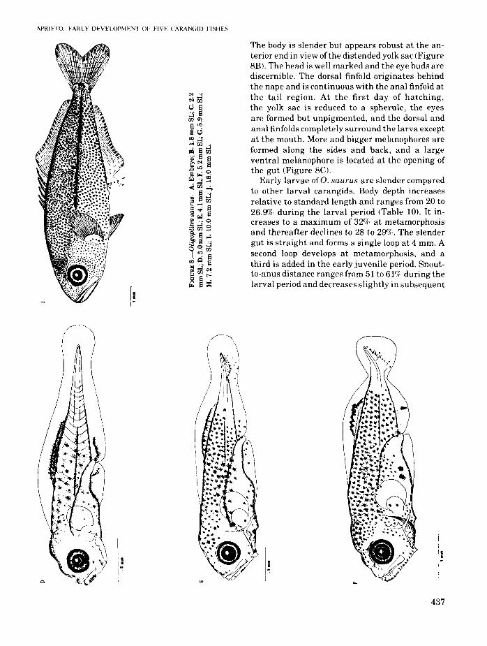
Two preserved eggs of O. saurus are 0.87 and0.88 mm in diameter. They have ventral, single oilglobules, 0.33 and 0.34 mm in diameter. T~e oilglobule consists of minute oil droplets and IS enclosed in a rather tough, pigmented capsule. Thepigmented yolk is bright yellow and unsegmented. The perivitelline space is narrow and theegg case smooth. The embryos are well developedand have stellate melanophores along the backand upper sides of the body. A large melanophoreis present at the posteroventral midline (Figure8A).
E. bipinnulata, the first interhemal spine is thickened and, as in S. zonata, the supraoccipital crestis lacking. Larvae of O. saurus are distinct fromthose of the two species mentioned in having anorbital crest with fine serrations, 1 to 3 dentideswhich appear early in the larval period on thedorsal side of the longest preopercular spine, and26 vertebrae-the highest vertebral count amongcarangids. The number of dorsal spines and pectoral fin rays formed is fewer than in most carangids, 5 to 6 and 15 to 17 respectively. Lar~al pigmentation is moderately profuse and, as 10 mostcarangid larvae, conspicuous melanophores arepresent along the bases ofthe dorsal and anal fins,on the lateral midline, and on the dorsal wall ofthe abdominal cavity. The larvae transform at 7 to10 mm.
Leatherjacket, Oligoplites saurus(Bloch and Schneider)
Figure 8Literature
Larvae of this species have not been describedpreviously.
Distribution and Spawning
Adults of Selene vomer have been recorded onboth coasts of the United States, from Cape Cod toBrazil and from Lower California to Peru (Jordanand Evermann, 1896). They have also been reported from the Gulf of Mexico (Ginsburg, 1952),the Bahamas (Bohlke and Chaplin, 1968), andWest Africa (Fowler, 1936).
Larval and early juveniles of S. vomer weretaken in all months except in June, October, andDecember. The monthly occurrence and distribution of the larvae is a composite of the records ofspecimens which include those taken from thecoastal waters of the eastern tropical Pacific fromBaja California to Costa Rica, the Gulf of Mexico,and the tropical Atlantic off Brazil and Liberia(Aprieto, 1973). In the Gulf of Mexico, larvae wereabundant mainly in the northeastern offshorewaters in August which suggests a short spawning period in that area (Figure 2). The larvae occurred in 2.20/0 of the net stations and constituted2.6% of the larval carangids collected in the Gulfof Mexico and the south Atlantic coast.
Distinguishing Features
Larvae ofO. saurus resemble those of Elagatisbipinnulata and Seriola zonata. Further, as in
Morphology
The larvae are 1.87 and 1.97 mm at hatching.
435

A
·s ••
@.....~t,.~.."". .... ,f' ~"
•
. 1 ••
APRIETO: EARLY DEVELOPMENT OF FlVF CARAN(iID FISHES
The body is slender but appears robust at the anterior end in view of the distended yolk sac (Figure8E). The head is well marked and the eye buds arediscernible. The dorsal finfold originates behindthe nape and is continuous with the anal finfold atthe tail region. At the first day of hatching,the yolk sac is reduced to a spherule, the eyesare formed but unpigmented, and the dorsal andanal finfolds completely surround the larva exceptat the mouth. More and bigger melanophores areformed along the sides and back, and a largeventral melanophore is located at the opening ofthe gut (Figure 8el.
Early larvae ofO. saurus are slender comparedto other larval carangids. Body depth increasesrelative to standard length and ranges from 20 to26.9% during the larval period (Table 10). It increases to a maximum of 329(: at metamorphosisand thereafter declines to 28 to 290/c. The slendergut is straight and forms a single loop at 4 mm. Asecond loop develops at metamorphosis, and athird is added in the early juvenile period. Snoutto-anus distance ranges from 51 to 61% during thelarval period and decreases slightly in subsequent
•• E
• • ~.. E..Q
437
FISHERY BULLETIN: VOL. 72. NO.
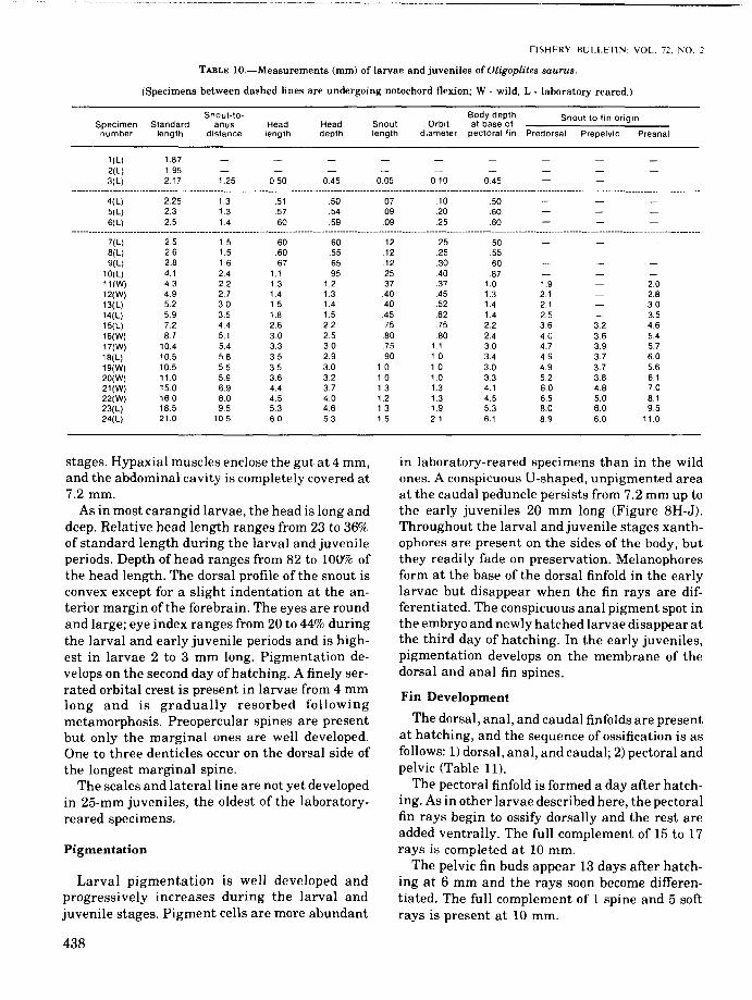
TABLE lO.-Measurements (mm) of larvae and juveniles ofOligoplites saurus.
(Specimens between dashed lines are undergoing notochord flexion; W . wild, L - laboratory reared.)
Snout-to- Body depth Snout to fin originSpecimen Standard anus Head Head Snout Orbit at base ofnumber length distance length depth length diameter pectoral fin Predorsal Prepelvic Preanal
1(L) 1.872(L) 1.953(L) 2.17 1.25 050 0.45 0.05 0.10 0.45
4(L)5(L)6(L)
2.252.32.5
1.31.31.4
51.57.60
.50
.54
.59
.07
.09
.09
.10
.2025
.50
.60
.60
7(L) 2.5 1.5 60 .60 .12 .25 .508(L) 2.6 1.5 .60 .55 .12 25 .559(L) 2.8 1.6 .67 .65 .12 .30 .60
10(L) 4.1 2.4 1.1 .95 25 .40 .8711(W) 4.3 22 1.3 1.2 .37 .37 1.0 1.9 2.012(W) 4.9 2.7 1.4 1.3 .40 .45 1.3 2.1 2.813(L) 5.2 3.0 1.5 1.4 .40 .52 1.4 2.1 3014(L) 5.9 3.5 1.8 1.5 .45 .62 1.4 2.5 3.515(L) 7.2 4.4 2.6 2.2 .75 .75 2.2 36 3.2 4.616(W) 8.7 5.1 3.0 2.5 .80 80 2.4 40 3.6 5.417(W) 10.4 5.4 3.3 30 .75 1.1 30 4.7 3.9 5.718(L) 10.5 5.8 35 2.9 .90 1.0 3.4 49 3.7 6.019(W) 10.5 5.5 35 30 1.0 1.0 30 4.9 3.7 5.620(W) 11.0 5.9 3.6 32 10 10 3.3 5.2 3.8 6121(W) 150 6.9 4.4 3.7 13 1.3 4.1 60 4.8 7.022(W) 16.0 8.0 4.5 4.0 1.2 1.3 4.5 6.5 5.0 8.123(L) 185 95 53 4.6 13 1.9 53 8.0 6.0 9.524(L) 21.0 10.5 6.0 5.3 1.5 2.1 6.1 89 6.0 11.0
stages. Hypaxial muscles enclose the gut at 4 mm,and the abdominal cavity is completely covered at7.2mm.
As in most carangid larvae, the head is long anddeep. Relative head length ranges from 23 to 36%of standard length during the larval and juvenileperiods. Depth of head ranges from 82 to 10<:Y*J ofthe head length. The dorsal profile of the snout isconvex except for a slight indentation at the anterior margin of the forebrain. The eyes are roundand large; eye index ranges from 20 to 44% duringthe larval and early juvenile periods and is highest in larvae 2 to 3 mm long. Pigmentation develops on the second day of hatching. A finely serrated orbital crest is present in larvae from 4 mmlong and is gradually resorbed followingmetamorphosis. Preopercular spines are presentbut only the marginal ones are well developed.One to three denticles occur on the dorsal side ofthe longest marginal spine.
The scales and lateral line are not yet developedin 25-mm juveniles, the oldest of the laboratoryreared specimens.
Pigmentation
Larval pigmentation is well developed andprogressively increases during the larval andjuvenile stages. Pigment cells are more abundant
438
in laboratory-reared specimens than in the wildones. A conspicuous U-shaped, unpigmented areaat the caudal peduncle persists from 7.2 mm up tothe early juveniles 20 mm long (Figure 8H-J).Throughout the larval and juvenile stages xanthophores are present on the sides of the body, butthey readily fade on preservation. Melanophoresform at the base of the dorsal finfold in the earlylarvae but disappear when the fin rays are differentiated. The conspicuous anal pigment spot inthe embryo and newly hatched larvae disappear atthe third day of hatching. In the early juveniles,pigmentation develops on the membrane of thedorsal and anal fin spines.
Fin Development
The dorsal, anal, and caudal finfolds are presentat hatching, and the sequence of ossification is asfollows: 1) dorsal, anal, and caudal; 2) pectoral andpelvic (Table 11).
The pectoral finfold is formed a day after hatching. As in other larvae described here, the pectoralfin rays begin to ossify dorsally and the rest areadded ventrally. The full complement of 15 to 17rays is completed at 10 mm.
The pelvic fin buds appear 13 days after hatching at 6 mm and the rays soon become differentiated. The full complement of 1 spine and 5 softrays is present at 10 mm.

APRIETO: EARLY DEVELOPMENT OF FIVE CARANGID FISHES
TABLE ll.-Meristic characters of cleared and stained larvae and juveniles of Oligoplites saurus.
Primary caudal Secondary caudal Left pre-Left Left fin rays fin rays Gill rakers, opercular
Standard pectoral pelvic left first marginlength Dorsal fin Anal fin fin fin Dorsal Ventral Dorsal Ventral gill arch spines
4.1 44.3 44.9 45.9 III; 8 II; 7 3 3 0+4 67.2 V; 18 II; I, 16 7 2 9 8 2 0+10 88.7 V; I, 17 II; I, 17 10 1,5 9 8 0+9 9
10.4 IV; I, 20 II; I, 19 13 I, 5 9 8 9 9 3+12 1211.0 V; I, 20 II; I, 18 14 1,5 9 8 9 9 3+11 912.2 V; I, 21 II; I, 19 14 I, 5 9 8 10 9 3+11 1013.0 V; I, 19 II; I, 18 14 1,5 9 8 9 9 3+10 915.1 V; I, 20 II; I, 19 14 I, 5 9 8 9 9 5+11 815.2 V; I, 21 II; I, 19 15 1,5 9 8 9 9 5+11 616 V; I, 21 II; I, 18 16 1,5 7 9 9 9 5+11 517 V; I, 21 II; I, 20 16 I, 5 9 8 9 10 5+12 618.5 V; I, 20 II; I, 20 16 1,5 9 8 9 8 5+13 419 V; I, 20 II; I, 18 15 1,5 9 8 10 9 5+13 3210 V; I, 21 II; I, 18 16 I, 5 9 8 ~ 9 5+13 3
The dorsal and anal fin rays differentiate simultaneously in an anteroposterior direction. Unlikepreviously described species, in which either themiddle or anterior spines are longer, the posteriorspine of the first dorsal fin is slightly longer thanthe rest. The full complement of6 spines and 19 to21 rays is present at 10 mm. The anal fin rays of 3spines and 18 to 20 soft rays are also complete at10mm.
Caudal fin formation is similar to that of theother species described. The full complement of 9to 10 dorsal and 8 to 10 ventral principal rays and18 to 20 secondaries is present at 10 mm.
Distribution and Spawning
Adults ofO. saurus are known from both coastsof Central America and in the West Indies (Jordanand Evermann, 1896). They also occur along theAtlantic coast of the United States from Massachusetts to Florida and in the Gulf of Mexico(Ginsburg, 1952). The wild larvae and juveniles inthe present work were taken from Escambia Bay,Fla., and at Sapelo and St. Simons Islands, Ga., inMay and July by means ofchannel nets and beachseines. The laboratory-reared larvae werehatched from planktonic eggs collected from Biscayne Bay. Larvae and juveniles were not collected in any of the net stations in the Gulf ofMexico and the south Atlantic coast. Distributionof the young in these regions is obscure, and abundance and frequency of occurrence in relation tothe other larval carangids could not be established. The wild larvae obtained were too few toderive conclusive information, but apparentlyspawning occurs in summer. Unlike the other
carangids which spawn in offshore pelagic waters,O. saurus spawns in inshore and shallow waters.Further investigation is necessary to establishwith certainty the spawning period and sites andthe distribution of the young.
Laboratory Rearing
Planktonic eggs ofO. saurus were collected in aI-m, 505-/-1 mesh plankton net at the pier of theRosenstiel School of Marine and AtomosphericScience on 15 July 1972, at 9:00 A.M., EDT. Atotal of 75 eggs was sorted from the plankton andincubated in a 50-liter glass aquarium. Theaquarium water was drawn from Biscayne Baythrough the School's seawater system. It was oxygenated and circulated with compressed airadded through airstones and lighted continuouslyby two cool, white, fluorescent bulbs. Temperatureranged from 23.9c to 28cC and salinity from 32 to36 %0 during the experiment. The larvae were fedwild plankton collected from Biscayne Bay as wellas nauplii of brine shrimp (Artemia salina). Adetailed description of the rearing technique employed is given in Houde and Palko (1970).
The eggs began hatching in the afternoon of theday of collection and after 24 h all the eggs werepresumed hatched. The larvae averaged 1.92 mmat hatching, were 5.2 mm 8 days after hatching,and about 21 mm at 34 days (Figure 9). Mortalityin the first 18 days included 2 eggs and 16 larvaepreserved for describing larval development. Sixyoung juveniles averaged 25 mm after 45 days.Thereafter, the juveniles failed to feed and all butone died at 51 days when the rearing experimentwas terminated.
439

FISHFRY BULl.FTIN: VOL 7"_ NO. "
Ossification
~ 12.0
..I 10.11
FIGURE 9.-Growth of Oligoplites SQurus larvae reared in thelaboratory at an average temperature of 26.0°C.
The sequence of ossification of the skull, axial,and appendicular skeleton is generally similaramong the four species in which ossification wasobserved (Table 12). Without exception, the premaxillaries, preopercular spines, and cleithra ossify in the smallest larvae (2.5-3.8 mm). Next toossify at 4 to 5 mm are the maxillaries, dentaries,parasphenoid, supraoccipital, articulars, frontals,angulars, and the branchial arches. The entiremaxillary arch is ossified before the larvae are 6mm long. Teeth are formed along the entire margin of the premaxillaries and anterior region ofthe dentaries in the youngest larvae following theossification of these elements. It is apparent thatthe bones related to feeding ossify early, and thisis consistent with the need of the larvae for foodfrom external sources following the absorption ofthe yolk.
Seven branchiostegal rays on each side are present in 3-mm larvae. Ossification begins with theposterior and longer rays and proceeds anteriad.The ceratohyal and epihyal to which the branchiostegal rays are attached ossify simultaneously with the rays. The rest of the hyoid archincluding the glassohyal, urohyal, and hypohyalossify at metamorphosis.
Aside from the quadrate and hyomandibularwhich ossify during the larval period, the rest ofthe palatine arch is not calcified until metamorphosis.
The branchial arches initially ossify in larvae 4to 5 mm long and all arches are calcified at 6 mm.The first branchial arch is the first to ossify starting from the center of the ceratobranchial towards
both ends. The epibranchial is the next to ossifybeginning from near the angle of the arch outward. Ossification of the other arches follows in asimilar sequence.
The gill rakers calcify following the ossificationof the elements to which they are attached. Thenumber of gill rakers increase as growth progresses but gill rakers are slow to ossify, and thefull complement usually is not completed until thetransition and early juvenile stages. The adultcount in Seriola zonata is fewer than is formed inthe juveniles due to the reduction of the terminalgill rakers into tubercles in the ceratobranchial.Patches of fine teeth are formed on the superiorpharyngeals of the third and fourth gill archeswhile the fifth and shortest gill arch has teethpatches for most of its length. Pharyngeal teethossify in larvae 6 to 8 mm long.
In the cranium, the parasphenoid, frontals, andsupraoccipitals ossify in the youngest larvae(2.5-3.8 mm). Except for the parietals which ossifyin the midlarval period, the rest of the cranium isnot ossified until the late larval and transitionperiods.
The cleithra, postcleithra, and posttemporalsare ossified in the early and midlarval stages, butthe rest of the pectoral girdle calcifies in late andtransforming larvae. From 2 to 4 posttemporalspines protrude from the myotomes during theearly larval period. These are small and hardlyvisible in most species except in stained specimens. These spines are soon overgrown by thedeveloping muscles.
The pelvic girdle calcifies following theossification of the pelvic fins.
Ossification occurs at 5 to 8 mm in the vertebralcolumn and proceeds in an anteroposterior direction. The neural and hemal spines ossify ahead ofthe centra of their respective vertebrae. Thecentra ossify at their anterior margins andossification proceeds posteriorly. This pattern ofossification in the vertebrae was noted inTrachurus symmetricus (Ahlstrom and Ball,1954).
Ribs similarly ossify in an anteroposterior direction. The pleural ribs are the first to ossifyfollowed by the epipleural ribs. All trunk vertebrae have ossified pleural and epipleural ribs injuveniles 15 to 17 mm long except on the first andsecond in which pleural ribs are lacking.
Teeth are initially uniserial but become multiserial as tooth formation progresses. Following
12 .4 ~ 18 ~ 22 24 21t 21 JO J2 ]4
DI,I .. ".' ".l~"lli'It " III
....
440
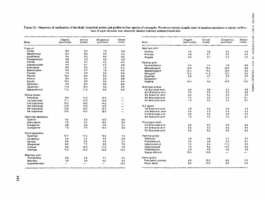
TABLE 12.-Sequence of ossification of the skull, branchial arches and girdles in four species of carangids. Numbers indicate length (mm) of smallest specimen in which ossification of each element was observed; dashes indicate undetermined size.
Elagatis Serio/a DecapterusBone bipinnulata zonata punctatus
Cranium:Vomer 6.0 6.5 76Mesethmoid 8.4 8.5 90Ectethmoid 9.4 8.5 9.0Parasphenoid 3.8 3.5 4.2Frontal 4.6 4.7 4.2Supraoccipital 4.6 4.7 4.2Exoccipital 6.0 6.5 76Basioccipital 6.0 6.5 7.6Parietal 7.0 5.5 5.2Plerotic 10.4 9.5 9.0Prootic 8.4 8.5 9.0Epiotic 10.4 9.5 9.0Sphenotic 9.5 8.5 9.0Opisthotic tt.5 to.3 9.0Basisphenoid 1t.5 1t.2 9.0
Orbital bones:Preorbital 8.4 11.2 10.01st suborbital 17.1 17.0 15.02nd suborbital 152 16.0 15.03rd suborbital 12.0 13.0 14.04th suborbital 13.0 14.0 1605th suborbital 13.0 150 16.0
Opercular apparatus:Opercle 5.4 55 10.0Interopercle 7.0 5.5 12.0Preopercle 3.8 3.6 3.5Subopercle 7.0 5.5 14.0
Hyoid apparatus:Hypohyal 13.1 11.0 10.0Ceratohyal 5.4 5.5 5.3Epihyal 6.0 65 5.5Glossohyal 8.0 7.5 9.0Urohyal 8.0 100 11.0Interhyal 17.0 15.0 22.0
Maxillary arch:Premaxillary 3.8 3.6 3.1Maxillary 3.8 3.6 4.2Supramaxillary
~~.....
Selenevomer
9.09.09.04.04.04.06.39.06.38.68.68.68.6909.0
9.09.0259.0
7.64.95.27.67.6
13.5
2.54.0
135
Elagat,s Seriofa Decapterus SeleneBone bipinnulata zonata punctatus vomer
Maxillary arch:
Dentary 4.6 3.6 4.2 4.0Articular 4.6 4.7 5.2 4.0Angular 46 4.7 5.2 5.2
Palatine arch:Hyomandibular 6.0 75 7.6 86Metapterygoid 12.0 10.4 t2.5 8.6Mesopterygoid 120 10.4 125 90Pterygoid 12.0 11.5 13.5 9.0Quadrate 4.6 4.7 5.3 4.0SymplecticPalatine 10.4 9.4 125 135
Branchial arches:t st Branchial arch 4.6 48 4.2 4.92nd Branchial arch 54 5.5 5.3 5.23rd Branchial arch 60 5.5 5.3 554th Branchial arch 7.0 5.5 5.3 6.15th Branchial arch 7.0 5.5 53 6.1
Gill rakers:1st Branchial arch 4.6 48 4.2 4.92nd Branchial arch 54 55 5.3 523rd Branchial arch 60 5.5 5.3 5.54th Branchial arch 7.0 5.5 5.3 6.1
Pharyngeal teeth:3rd Branchial arch 8.0 6.5 66 804th Branchial arch 8.0 6.5 6.6 805th Branchial arch 8.0 6.5 66 8.0
Pectoral girdle:Cleithrum 3.8 3.6 31 2.5Postcleithrum 5.4 48 52 6.1Hypercoracoid 72 84 11.0 9.0Hypocoracoid 7.2 84 11.0 90Posttemporal 5.4 4.8 4.2 4.9Supracleithrum 104 13.0
Pelvic girdle:Post pelvic process 8.0 100 8.0 5.0Pelvic keels 8.0 10.0 80 5.0

metamorphosis 2 to 3 irregular rows ofsharp teethare present.
ACKNOWLEDGMENTS
The author wishes to thank the Miami Laboratory, Southeast Fisheries Center of the NationalMarine Fisheries Service, which made availablethe working space, facilities, materials, and funds.She is deeply grateful to William J. Richards forsupervision and encouragement and to ThomasW. McKenney for the many helpful discussionsand suggestions on larval fish work. She wishes toexpress her appreciation to Elbert H. Ahlstrom forreviewing the manuscript and for his valuablecriticisms and suggestions, and also wishes toacknowledge the helpful comments from DonaldP. de Sylva, Hilary B. Moore, Charles E. Lane, andLowell P. Thomas. She is grateful to BarbaraPalko, Edward D. Houde, and George Miller forcarangid larvae given to her, and also wishes tothank Thomas Potthoff for information on staining fish larvae, Alexander Dragovich for translation of Russian literature, John Wise and JohnStimpson for assistance in computer work, andElizabeth Leonard for help in securing muchneeded literature.
This research project was completed while theauthor was on a University of the Philippines Faculty Fellowship and a scholarship grant from theInternational Women's Fishing Association ofPalm Beach, Fla.
LITERATURE CITED
ABOUSSOUAN. A.1968. Oeufs et larves de Teleosteen de 1'0uest Africain VI.
Larves de Chloroscombrus chrysurus (Linne) et deBlepharis crinitus (Mitchil\) (Carangidae). Bull. Inst. Fr.Afr. Noire. Ser. A. 30(3):226-237.
AHLSTROM, E. H.
1948. A record of pilchard eggs and larvae collected duringsurveys made in 1939 to 1941. U.S. Fish Wildl. Serv..Spec. Sci. Rep. 54:1-76.
AHUlTROM, E. H., AND O. P. BALL.
1954. Description of eggs and larvae of jack mackerel(Trachurus symmetricus) and distribution and abundanceof larvae in 1950 and 1951. U.S. Fish Wildl. Serv., Fish.Bull. 56:209-245.
APRIETO, V. L.1973. Early development of carangid fishes of the Gulf of
Mexico and the South Atlantic coast of the UnitedStates. Ph.D. Thesis, Univ. Miami, Coral Gables, 179 p.
BERRY, F. H.
1959. Young jack crevalles (Caranx species) off the southeastern Atlantic coast of the United States. U.S. Fish
442
FISHERY 1ll'1 IFTIN: VO!.. 7c. NO.
Wildl. Serv., Fish. Bull. 59:417-535.1fl68. A new species of carangid fish Wecapterus tab/) from
the Western Atlantic. Contrib. Mar. Sci. 13:145-167.1969. Elagatis bipinnulata (Pisces: Carangidae): Morphol
ogy of the fins and other characters. Copeia1969:454-463.
BOHLKE, J. E., AND C. C. G. CHAPLIN.
1968. Fishes of the Bahamas and adjacent tropicalwaters. Livingston Publishing Co., Wynnwood,Pa., 771p.
BRIGGS, J. C.
1960. Fishes of worldwide (circumtropicaD distribution. Copeia 1960:171-180.
CHACKO,P. I.1950. Marine plankton from waters around the Krusadai
Island. Proc. Indian Acad. Sci. 31, Sect. B:162-174.DEUlMAN, H. C.
1926. Fish eggs and larvae from the Java-Sea. 5. Caranxkurra, macrosoma and crumenophthalmus. 6. On a fewcarangid eggs and larvae. Treubia 8:199-218.
DEVANESAN, D. W., AND K. CHIDAMBARAM.
1941. On two kinds of fish eggs hatched out in the laboratory of the West Hill Biological Station, Calicut. CurroSci. (India) 10(5):259-261.
DOOLEY, J. K.1972. Fishes associated with the pelagic sargassum com
plex, with a discussion of the sargassumcommunity. Contrib. Mar. Sci. 16:1-32.
FARRIS, D. A.
1961. Abundance and distribution of eggs and larvae andsurvival of larvae of jack mackerel (Trachurussymmetricus). U.S. Fish Wildl. Serv., Fish. Bull.61:247-279.
FIELDS, H. A.
1962. Pompanos (Trachinotus spp.) of south Atlantic coastof the United States. U.S. Fish Wildl. Serv., Fish. Bull.62: 189-222.
FOWLER, H. W.1936. Marine Fishes of West Africa. Bull. Am. Mus. Nat.
Hist. 70(part 11):675-724.FUJII, R.
1969. Chromatophores and pigments. In W. S. Hoar and D.J. Randall (editors), Fish physiology, Vol. 3, p.307-353. Academic Press, N.Y.
GAETANI, D. DE.
1940. Stadi larvali e giovanili in Lichia glaucaRisso. Mem. R. Com. Talassogr., Ital. 270, 15 p.
GINSBURG, I.1952. Fishes of the family Carangidae of the northern Gulf
of Mexico and three related species. Publ.lnst. Mar. Sci.,Univ. Texas 2(2):43-117.
GOSLINE, W. A., AND V. E. BROCK.
1960. Handbook of Hawaiian fishes. University of HawaiiPress, Honolulu, 372 p.-
HERRE, A. W.1953. Checklist ofPhilippine fishes. U.S. Fish Wildl. Serv.,
Res. Rep. 20, 977 p.HILDEBRAND, S. F., AND L. E. CABLE.
1930. Development and life history of fourteen teleosteanfishes at Beaufort, N.C. Bull. U.S. Bur. Fish. 46:384-488.
HOUDE, E. D., AND B. J. PALKO.
1970. Laboratory rearing of the clupeid fish Harengulapensacolae from fertilized eggs. Mar. BioI. (Ber\.)5:354-358.
APRIETO: EARLY DEVELOPMENT OF FIVE CARANGlD FISHES
HUBBS, C. L.
1943. Terminology of early stage of fishes. Copeia1943:260.
JORDAN, D. S., AND B. W. EVERMANN.
1896. The fishes of North and Middle America: A descriptive catalogue of the species offish-like vertebrates foundin the waters of North America, north of the Isthmus ofPanama. Bull. U.S. Natl. Mus. 47, Part 1:895-945.
LAGLER, K. F., J. E. BARDACH, AND R. R. MILLER.
1962. Ichthyology. John Wiley & Sons, Inc., N.Y., 545 p.LUTKEN, C. F.
1880. Spolia Atlantica. Bidrag til kundaskab onforaforandringer nos Fiske under deres Vnext og Udvikling, saerlight nos nogle af Atlanterhavetshllhllsfiske. Kgl. Dan. Vidensk. Selsk. BioI. Skr. 5,12:049-613.
MANSUETI, A. J., AND J. D. HARDY, JR.
1967. Development offishes of the Chesapeake Bay region.An atlas of egg, larval, and juvenile stages, Part1. Natural Resources Institute, Univ. Maryland, 202 p.
MARSHALL, T. C.1965. Fishes of the Great Barrier Reefand coastal waters of
Queensland. Livingston Publ. Co., Narbeth, Pa., 566 p.MATHER, F. J. III
1952. Three species offishes, genusSeriola, in the waters ofCape Cod and vicinity. Copeia 1952:209-210.
1958. A preliminary review of the amberjacks, GenusSeriola, of the western Atlantic. In Proc. Int. Game FishConf., p. 1-13.
McKENNEY, T. W., E. C. ALEXANDER, AND G. L. Voss.
1958. Early development and larval distribution of thecarangid fish, Caranx crysos (Mitchim. ull. Mar. Sci.8:167-200.
MILLER, G. L., AND S. C. JORGENSON
1973. Meristic characters ofsome marine fishes ofthe western Atlantic Ocean. Fish Bull., U.S. 71:301-312.
MITANI, F.1960. Fishery biology of the yellowtail, Seriola quin
queradiata T.& S., inhabiting the waters aroundJapan. Mem. Fac. Agric. Kinki Univ. 1:81-300.
MITo, S.1961. Pelagic fish eggs from Japanese waters - II Lam
prida, Zeida, Mugilina, Scombrina, Carangina andStromateina. [In Jap., Engl. summ.] Sci. Bull. Fac.Agric. Kyushu Univ. lR(4):451-466.
MOSER, H. G., AND E. H. AHLSTROM.
1970. Development ofLanternfishes (Family Myctophidae)in the California Current. Part I. Species with narroweyed larvae. Bull. Los Ang. Cty. Mus. Nat. Hist. Sci.7,145 p.
NICHOLS, J. T.
1946. On young western Atlantic Seriolas. Copeia1946:259-260.
OKADA, Y.1966. Fishes of Japan. Illustrations and descriptions of
fishes of Japan. Uno Shoten Co., Ltd., Tokyo, 458 p.OKIYAMA, M.
1970. Studies on the early life history of the rainbow runner, Elagatis bipinnulata (Quoy and Gaimard) in theIndo-Pacific Oceans. Far East Fish. Res. Lab. (Shimizu)Bull. (3):167-186.
POSGAY, J. A., R. R. MARAK, AND R. C. HENNEMUTH.
1968. International Commission for the Northwest Atlantic Fisheries. Res. Doc. 68/85, 5 p.
SANZO, L.1931. Uovo, stadi embrionali e post-embrionali di Nauc
rates ductor L. Mem. R. Corn. Talassogr. Ital. 185, 14 p.1933. Uovo larvae e stadi giovanili de Seriola durrwrili
Risso. Mem. R. Corn. Talassogr. Ital. 205, 12 p.SCHNAKENBECK, W.
1931. Carangidae. Report on the Danish oceanographicalexpeditions 1908-1910 to the Mediterranean and Adjacent seas, No. 10, 2(BioJ.) A. 14: 1-20.
SHOJIMA, Y.1962. On the postlarvae and juveniles of Carangid fishes
collected together with the jelly-fishes. [In Jap., Engl.summ.] Bull. Seikai Reg. Fish. Res. Lab. 27:49-58.
STARKS, K.1911. The osteology and relationship of fishes belonging to
the family Carangidae. Stanford Univ. Publ. Univ. Ser.5:27-49.
SUBRAHMANYAM, C. B.
1964. Eggs and early development of a carangid fromMadras. J. Mar. BioI. Assoc. India 6(1):142-146.
SUZUKI, K.1962. Anatomical and taxonomical ~tudies on the carangid
fishes of Japan. Rep. Fac. Fish. Prefect. Univ. Mie4(2):43-232.
UCHIDA, K., Y. DOTU, S. MITO, AND K. NAKAHARA.
1958. The spawning and early life history of "buri"Japanese yellow tail, Seriola quinqueradiata Temmincket Schlegel. [In Jap., Engl. summ.] Sci. Bull. Fac. Agric.Kyushu Univ. 16:329-342.
UCHIDA, K., S. IMAI, S. MITO, S. FUJITA, M. UENO, Y. SHOJIMA, T.
SENTA, M. TAHUKU, AND Y. DOTU.
1958. Studies on the eggs, larvae andjuveniles ofJapanesefishes. Kyushu Univ., Fac. Agric., Fish. Dep., SecondLab. Fish. BioI., Ser. 1, 89 p., 86 pI.
VIJAYARAGHAVAN, P.1958. Studies on fish eggs and larvae of Madras
Coast. Univ. Madras, G. S. Press, 79 p.
WEITZMAN, S. H.1962. The osteology of BrycoTl meeki, a generalized
characid fish, with an osteological definition of the family.Stanford lchthyol. Bull. 8:1-77.
443